Pyrimidine nucleoside conformational analysis. Nuclear Overhauser effect and circular dichroism...
8
753 Pyrimidine Nucleoside Conformational Analysis. Nuclear Overhauser Effect and Circular Dichroism Correlations Phillip A. Hart* and Jeffrey P. Davis Contribution from the School of Pharmacy, University of Wisconsin, Madison, Wisconsin 53706. Received June 3, 1970 Abstract: In a continuing effort to unify the interpretation of various experimental approaches to nucleoside glycosidic conformation in solution we have measured and collated nuclear Overhauser effects, circular dichroism, and ultraviolet absorption spectra on cytidine, 2 ’,3 ’-isopropylidenecytidine, uridine, and 2 ‘,3 ’-isopropylidene- uridine in water and organic solvents. 2’,3 ‘-Isopropylideneuridine and 2 ’,3 ’-isopropylidenecytidine were found to form nonideal solutions in chlorocarbon solvents. Upon dilution, the typically negative BzU molecular ellipticities of these systems tend toward the positive values characteristicin water and dimethyl sulfoxide. Plausible origins of the concentration dependence of the complex curves resulting in these cases are considered. The NOE results in water and dimethyl sulfoxide indicate that all four nucleosides possess a considerable amount of the syn rotamer in hydrogen bond accepting solvents, with the isopropylidene derivatives favoring this conformation to a higher degree than the underivatized nucleosides. The possible consequences of these nmr observations coupled with new and published CD data are outlined for a previously published monograph of pyrimidine nucleoside BPu molecular ellipticity us. torsion angle. e have recently reported datal derived from nu- W clear Overhauser effect (NOE) measurements on pyrimidine nucleosides that were viewed as evidence for the existence of substantial amounts of the syn confor- mation in the conformer equilibrium of some of them. Although the intuitive interpretation of the relative enhancements is that they represent conformer popula- tions, the theory had not been developed that would permit quantitative conformational deductions to be made. Thus the conclusions had to be qualified. In addition, the syn conformation has been assumed to be a relatively unimportant contributor to pyrimidine nucleoside conformational equilibria, an assumption bolstered by model building, computational, and experi- mental ~ o r k . ~ - ~ The idea that the anti conformation for purine and pyrimidine nucleosides need not be ex- clusive has, however, been extant since Donohue and Trueblood expressed it, lo and a recent X-ray analysis of 4-thiouridine, performed by Sanger and Scheit”” and quoted in a broader context by Tavale and Sobell,llb lends credence to this idea for pyrimidine nucleosides. Circular dichroism (CD) observations are inherently unable to distinguish single conformers from appropri- ately weighted mixtures of conformers in the absence of unequivocal conformational models. However, CD * Address correspondence to this author, (1) P. A. Hart and J. P. Davis, Biochem. Biophys. Res. Commun., 34, (2) A. E. V. Haschemeyer and A. Rich, J. Mol. Biol., 27, 369 (1967). (3) F. Jordon and B. Pullman, Theor. Chim. Acta, 9, 242 (1968). (4) I. Tinoco, R. C. Davis, and S. R. Jaskunas in “Molecular Associ- ations in Biology,” B. Pullman, Ed., Academic Press, New York, N. Y., 1968, p 77. (5) V. Sassisekharan, A. V. Lakshminarayanan, and G. N. Rama- chandran in “Conformation of Biopolymers,” Vol. 2, G. N. Rama- chandran, Ed., Academic Press, New York, N. Y., 1967, p 641. (6) R. J. Cushley, K. A. Watanabe, and J. J. Fox, J. Amer. Chem. Soc., 89, 394 (1967). (7) R. J. Cushley, I. Wempen, and J. J. Fox, ibid., 90, 709 (1968). (8) D. W. Miles, M. J. Robins, R. K. Robins, M. W. Winkley, and H. Eyring, ibid., 91, 824 (1969). (9) (a) D. W. Miles, M. J. Robins, R. K. Robins, M. W. Winkley, and H. Eyring, ibid., 91, 831 (1969); (b) T. R. Emerson, R. J. Swan, and T. L. V. Ulbricht, Biochemistry, 6, 843 (1967). (10) J. Donohue and K. N. Trueblood, J.Md Bid, 2, 363 (1960). (11) (a) W. Sanger and K. H. Scheit, Angew. Chem., Int. Ed. Engl., 8, 139 (1969); (b) S. S. Tavale and H. M. Sobell, J. Mol. Bid, 48, 109 ( 1970). 733 (1969). studies of a variety of pyrimidine nucleosides led Miles, et uZ.,’~ to modify somewhat the previous conclusions of Ulbri~ht,~~ to establish approximately how molecular ellipticity of the B,, band (the long-wavelength transi- tion of pyrimidine nucleosides) should vary as a func- tion of torsion angle,’, and to embody the expected trend in a diagram reproduced in Figure 1. The dia- gram shows that extreme anti conformations of p pyrimidine nucleosides should be characterized by a large positive ellipticity associated with the B,, transi- tion, and that the extreme syn conformation should have a moderately large negative value. The values for the diagram were not all obtained from the same chromo- phoric system. The high positive values were derived from the experimental values of cytidine, 2’,3 ’-isopro- pylidenecytidine, and 1 -fl-D-arabinofuranosylcytosine on the assumption that these substances adopted the anti conformation in water. The negative values came from ellipticity measurements done on isopropylidene- cytidine in dichloroethane, in which case intramolecular 2-keto, 5 ’-hydroxyl hydrogen bonding was thought to force the adoption of a syn c~nformation.’~ In addi- tion, measurements on 6-methylcytidine (6-CH3-C) and 6-methyl-2’-deoxycytidine (6-CH3-2’-dC) in water, di- oxane, and acetonitrile provided the remaining negative values since in those cases the syn conformer is favored because of severe steric interactions between the 6-methyl group and the sugar C-2’, C-3’, and C-5’ hydrogens. Even though a quantitative relation of relative intra- molecular Overhauser enhancements to conformer populations has been de~eloped’~“,~ the experimental technique has not been sufficiently refined to provide data of the accuracy required to employ it rigorously. (12) The torsion angle is the parameter that defines the conformation about the glycosidic bond and has been defined by Donohue and True- blood10 and M. Sundaralingam and L. H. Jensen, J. Mol. Biol., 13, 930 (1966). (13) Infrared studies of OH frequency in chloroform have been interpreted in terms of syn conformations in the purine nucleoside series: J. Pitha, S. Chladek, and J. Smrt, Collect. Czech. Chem. Com- mun., 28, 1622 (1963). (14) (a) Roger E. Schirmer, Ph.D. Thesis, University of Wisconsin, 1970; (b) R. E. Schirmer, 3. H. Noggle, J. P. Davis, and P. A. Hart, J. Amer. Chem. Soc., 92, 3266 (1970). Hart, Davis 1 Pyrimidine Nucleoside Conformational Analysis
-
Upload
jeffrey-paul -
Category
Documents
-
view
213 -
download
0
Transcript of Pyrimidine nucleoside conformational analysis. Nuclear Overhauser effect and circular dichroism...

753
Pyrimidine Nucleoside Conformational Analysis. Nuclear Overhauser Effect and Circular Dichroism Correlations
Phillip A. Hart* and Jeffrey P. Davis
Contribution from the School of Pharmacy, University of Wisconsin, Madison, Wisconsin 53706. Received June 3, 1970
Abstract: In a continuing effort to unify the interpretation of various experimental approaches to nucleoside glycosidic conformation in solution we have measured and collated nuclear Overhauser effects, circular dichroism, and ultraviolet absorption spectra on cytidine, 2 ’,3 ’-isopropylidenecytidine, uridine, and 2 ‘,3 ’-isopropylidene- uridine in water and organic solvents. 2’,3 ‘-Isopropylideneuridine and 2 ’,3 ’-isopropylidenecytidine were found to form nonideal solutions in chlorocarbon solvents. Upon dilution, the typically negative BzU molecular ellipticities of these systems tend toward the positive values characteristic in water and dimethyl sulfoxide. Plausible origins of the concentration dependence of the complex curves resulting in these cases are considered. The NOE results in water and dimethyl sulfoxide indicate that all four nucleosides possess a considerable amount of the syn rotamer in hydrogen bond accepting solvents, with the isopropylidene derivatives favoring this conformation to a higher degree than the underivatized nucleosides. The possible consequences of these nmr observations coupled with new and published CD data are outlined for a previously published monograph of pyrimidine nucleoside BPu molecular ellipticity us. torsion angle.
e have recently reported datal derived from nu- W clear Overhauser effect (NOE) measurements on pyrimidine nucleosides that were viewed as evidence for the existence of substantial amounts of the syn confor- mation in the conformer equilibrium of some of them. Although the intuitive interpretation of the relative enhancements is that they represent conformer popula- tions, the theory had not been developed that would permit quantitative conformational deductions to be made. Thus the conclusions had to be qualified.
In addition, the syn conformation has been assumed to be a relatively unimportant contributor to pyrimidine nucleoside conformational equilibria, an assumption bolstered by model building, computational, and experi- mental ~ o r k . ~ - ~ The idea that the anti conformation for purine and pyrimidine nucleosides need not be ex- clusive has, however, been extant since Donohue and Trueblood expressed it, lo and a recent X-ray analysis of 4-thiouridine, performed by Sanger and Scheit”” and quoted in a broader context by Tavale and Sobell,llb lends credence to this idea for pyrimidine nucleosides.
Circular dichroism (CD) observations are inherently unable to distinguish single conformers from appropri- ately weighted mixtures of conformers in the absence of unequivocal conformational models. However, CD
* Address correspondence to this author, (1) P. A. Hart and J. P. Davis, Biochem. Biophys. Res. Commun., 34,
(2) A. E. V. Haschemeyer and A. Rich, J . Mol. Biol., 27, 369 (1967). (3) F. Jordon and B. Pullman, Theor. Chim. Acta, 9, 242 (1968). (4) I. Tinoco, R. C. Davis, and S. R. Jaskunas in “Molecular Associ-
ations in Biology,” B. Pullman, Ed., Academic Press, New York, N. Y., 1968, p 77.
( 5 ) V. Sassisekharan, A. V. Lakshminarayanan, and G . N. Rama- chandran in “Conformation of Biopolymers,” Vol. 2, G. N. Rama- chandran, Ed., Academic Press, New York, N. Y., 1967, p 641.
(6) R. J. Cushley, K. A. Watanabe, and J. J. Fox, J . Amer. Chem. Soc., 89, 394 (1967).
(7) R. J. Cushley, I. Wempen, and J. J. Fox, ibid., 90, 709 (1968). (8) D. W. Miles, M. J. Robins, R. K. Robins, M. W. Winkley, and
H. Eyring, ibid., 91, 824 (1969). (9) (a) D. W. Miles, M. J. Robins, R. K. Robins, M. W. Winkley, and
H. Eyring, ibid., 91, 831 (1969); (b) T. R. Emerson, R. J. Swan, and T. L. V. Ulbricht, Biochemistry, 6 , 843 (1967).
(10) J. Donohue and K. N. Trueblood, J . M d B i d , 2, 363 (1960). (11) (a) W. Sanger and K. H. Scheit, Angew. Chem., Int. Ed. Engl.,
8 , 139 (1969); (b) S. S. Tavale and H. M. Sobell, J . Mol. B i d , 48, 109 ( 1970).
733 (1969).
studies of a variety of pyrimidine nucleosides led Miles, et u Z . , ’ ~ to modify somewhat the previous conclusions of U l b r i ~ h t , ~ ~ to establish approximately how molecular ellipticity of the B,, band (the long-wavelength transi- tion of pyrimidine nucleosides) should vary as a func- tion of torsion angle,’, and to embody the expected trend in a diagram reproduced in Figure 1. The dia- gram shows that extreme anti conformations of p pyrimidine nucleosides should be characterized by a large positive ellipticity associated with the B,, transi- tion, and that the extreme syn conformation should have a moderately large negative value. The values for the diagram were not all obtained from the same chromo- phoric system. The high positive values were derived from the experimental values of cytidine, 2’,3 ’-isopro- pylidenecytidine, and 1 -fl-D-arabinofuranosylcytosine on the assumption that these substances adopted the anti conformation in water. The negative values came from ellipticity measurements done on isopropylidene- cytidine in dichloroethane, in which case intramolecular 2-keto, 5 ’-hydroxyl hydrogen bonding was thought to force the adoption of a syn c~nformat ion . ’~ In addi- tion, measurements on 6-methylcytidine (6-CH3-C) and 6-methyl-2’-deoxycytidine (6-CH3-2’-dC) in water, di- oxane, and acetonitrile provided the remaining negative values since in those cases the syn conformer is favored because of severe steric interactions between the 6-methyl group and the sugar C-2’, C-3’, and C-5’ hydrogens.
Even though a quantitative relation of relative intra- molecular Overhauser enhancements to conformer populations has been de~e loped’~“ ,~ the experimental technique has not been sufficiently refined to provide data of the accuracy required to employ it rigorously.
(12) The torsion angle is the parameter that defines the conformation about the glycosidic bond and has been defined by Donohue and True- blood10 and M. Sundaralingam and L. H. Jensen, J . Mol. Biol., 13, 930 (1966).
(13) Infrared studies of OH frequency in chloroform have been interpreted in terms of syn conformations in the purine nucleoside series: J. Pitha, S . Chladek, and J. Smrt, Collect. Czech. Chem. Com- mun., 28, 1622 (1963).
(14) (a) Roger E. Schirmer, Ph.D. Thesis, University of Wisconsin, 1970; (b) R. E. Schirmer, 3. H. Noggle, J. P. Davis, and P. A. Hart, J . Amer. Chem. Soc., 92, 3266 (1970).
Hart, Davis 1 Pyrimidine Nucleoside Conformational Analysis
754
30 r I - - - - B -anomer
-20. -3 0
-l80'-16d -120' -80' -40' 0' 40' SO' 120' l60'l80'
Figure 1. Nomograph of the expected relation of /3 pyrimidine nucleoside molecular ellipticity to the torsion angle +CN. Taken from ref 9a by permission.
We decided, therefore, to determine NOE enhancements for selected members of the set of nucleosides used by Miles to see if the conclusions reached by the nmr ex- periment agreed with those reached by the CD measure- ments. The models chosen were cytidine (C), uridine (U), 2',3 '-isopropylidenecytidine (i-C), and 2',3 '- isopropylideneuridine (i-U), all in water or polar sol- vents, and i-C in dichloroethane. In view of Miles' work this selection appeared to allow the examination of a series from anti-like to syn-like.
We found, however, that the chlorocarbon solutions of i-C were nonideal and that the NOE experiments could not be done on them. In addition, although i-U was not sufficiently soluble in the chlorocarbon solvents to even try the nmr experiments, it was sufficiently soluble in chloroform to do the CD experiments for completeness and it too was found to form nonideal solutions. With this information and further C D mea- surements the role of chiroptical results from "non- associating" solvents in a correlation diagram can be put into perspective.
Experimental Section The nucleosides used in this study were Sigma grade Sigma
Chemical Co. products. Isopropylidenecytidine was purchased either as the free base or was obtained following neutralization of the hydrochloride by passage through a Dowex-2 OH- column. The reagent grade solvents were treated as follows: methanol, distilled from Linde 3A molecular sieves; ethanol, distilled from Linde 4A molecular sieves; water, distilled and further deionized; acetonitrile, distilled from PZO5 and redistilled; chloroform, washed four times with water, dried over sodium sulfate, and distilled from molecular sieves; 1 ,Zdichloroethane, dried over sodium sulfate and distilled from molecular sieves; methylene chloride, distilled from calcium hydride; dimethyl sulfoxide, distilled from calcium hydride under vacuum and redistilled under vacuum.
CD measurements were performed on a Cary 60 recording spec- tropolarimeter fitted with a Model 6002 CD attachment with the slit programmed for a half-band width of 15 A. Measurements were made at path lengths ranging from 0.1 to 5 cm and for concen- trations ranging from 0.912 X 10⁻⁶ to 4.55 X M except where indicated. The C D is recorded as molecular ellipticity, [e], in units of deg cm2 dmol-1, and absorbances never exceeded 2.0. The solu- tions were not buffered so that ionic strength factors would not enter the NOE-CD comparisons. The resultant pH uncertainties are probably not severe. The instrument was calibrated using (+)-camphorsulfonic acid (Aldrich).
Ultraviolet absorption spectra were taken on a Beckman DK-2 recording spectrophotometer with cells of 1.0-cm path length and the concentration studies in chloroform and water were done on a
Cary 14 recording spectrophotometer in cells of 0.1-5.0-cm path- length. All uv and C D spectra in DMSO were run in a 0.1-cm cell.
Mean degrees of association in solution were measured on a Hewlett-Packard Mechrolab Division Model 301A vapor pressure osmometer using a 37' probe. Recrystallized biphenyl was used as an ideal solute standard in all solvents but in water, in which so- dium chloride was used. Standard runs were made before and after each nucleoside run in every solvent and reproducibility of A R readings was consistently within 3-5 %. Since precise molal osmotic coefficients for biphenyl in the various solvents employed are not available we have presented the osmometry data so that they simply indicate the deviation of i-C from ideality in chlorocarbon solvents taking the osmotic coefficient for biphenyl as very nearly one in all cases. The VPO data were identical within experimental error whether the reagent grade solvents were treated or untreated.
The conventional nmr spectra were taken on a Varian A60-A spectrometer. Perdeuteriomethylene chloride, ethanol, methanol, and acetonitrile were Stohler Isotope Corporation products. Pyridine-&, benzene-&, dimethyl-d, sulfoxide, and chloroform-dl were from Merck Sharpe and Dohme, and D20 was purchased from Diaprep, Inc. The NOE experiments were done in coaxial tubes on a Varian HA-100 nmr spectrometer as previously de- scribed.I.16 It was shown that peak height changes correspond well with peak area changes. The symbol fnm used to report the NOE data denotes here the fractional enhancement of the resonance m upon irradiation of resonance n. Nmr solvent mixtures are re- ported as per cent by volume before mixing.
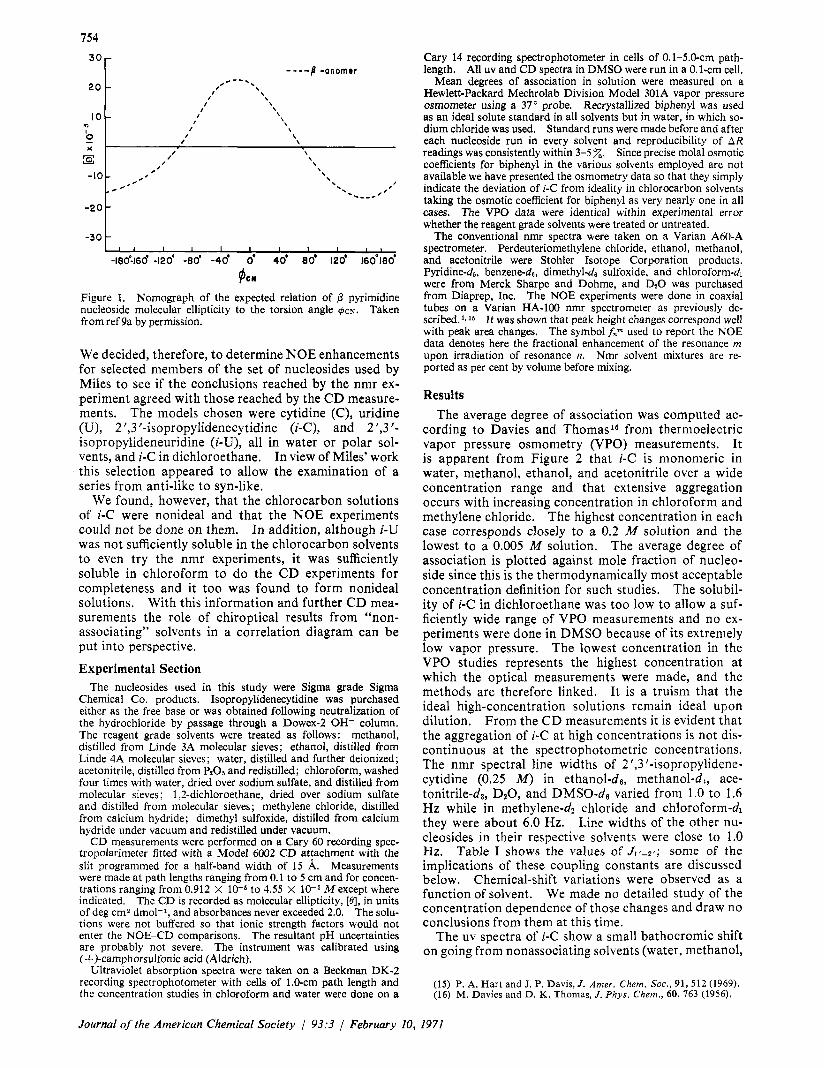
Results The average degree of association was computed ac-
cording to Davies and Thomas ' 6 from thermoelectric vapor pressure osmometry (VPO) measurements. It is apparent from Figure 2 that i-C is monomeric in water, methanol, ethanol, and acetonitrile over a wide concentration range and that extensive aggregation occurs with increasing concentration in chloroform and methylene chloride. The highest concentration in each case corresponds closely to a 0.2 M solution and the lowest to a 0.005 M solution. The average degree of association is plotted against mole fraction of nucleo- side since this is the thermodynamically most acceptable concentration definition for such studies. The solubil- ity of i-C in dichloroethane was too low to allow a suf- ficiently wide range of VPO measurements and no ex- periments were done in DMSO because of its extremely low vapor pressure. The lowest concentration in the VPO studies represents the highest concentration at which the optical measurements were made, and the methods are therefore linked. It is a truism that the ideal high-concentration solutions remain ideal upon dilution. From the C D measurements it is evident that the aggregation of i-C at high concentrations is not dis- continuous at the spectrophotometric concentrations. The nmr spectral line widths of 2',3'-isopropylidene- cytidine (0.25 M ) in ethanol-d6, methanol-d,, ace- tonitrile-da, D20, and DMSO-d6 varied from 1.0 to 1.6 Hz while in methylene-d2 chloride and chloroformd they were about 6.0 Hz. Line widths of the other nu- cleosides in their respective solvents were close to 1.0 Hz. Table I shows the values of J 1 ~ - ~ ~ ; some of the implications of these coupling constants are discussed below. Chemical-shift variations were observed as a function of solvent. We made no detailed study of the concentration dependence of those changes and draw no conclusions from them at this time.
The uv spectra of i-C show a small bathocromic shift on going from nonassociating solvents (water, methanol,
(15) P. A. Hart and J. P . Davis,J. Amer. Chem. Soc., 91, 5 1 2 (1969). (16) M. Davies and D. I<. Thomas, J . Phys. Chem., 60, 763 (1956).
Journal of the American Chemicat Society / 93:3 1 February 10, 1971
755
r ,
I I I I I I I .25 .SO .75 1.00 1.25 1.50
MOLE FRACTION ISOPROPYLIDENE CYTIDINE XIO’
Figure 2. 0 ; CHaCN, A; EtOH, A; HzO, 0 ; CHZClz, a. Calculated from vapor pressure osmometry data.
Average degree of self-association of 2‘,3’-isopropylidenecytidine as a function of concentration in: CHC13, 0 ; MeOH,
ethanol, and acetonitrile) to associating solvents (chloro- form, methylene chloride, and dichloroethane; Table 11). The absorbance maxima decrease by approximately
Table I. Coupling Constants
Nucleoside Solvent for 0.25 M soln Jif,zi, Hz ~
C DzO 62 benzene-de
in DMSO-d6 i-C DzO U 25 % pyr-db
in DaO DMSO-ds
i- U 75% DMSO-% in DzO
DMSO-de
3.5 2.4
2.8 3.2
4.6 2.4
2.4
Table 11. Uv Data for 2’,3/-Isopropylideneytidine
Nucleoside molar concn Amax, Emax,
Solvent x 104 nm x 10-4 Hz0 0.87 269 0.765 MeOH 0.42 270 0.780 EtOH 0.40 270 0.717 CHsCN 1.02 272 0.555 CHC13 0.82 213 0.550 CHtClz 0.82 274 0.513 CHtClCHzCl 0.88 274 0.502
30% on going from hydroxylic solvents to the non- hydrogen-bonding solvents. The ultraviolet spectra of the polar solutions show no concentration depen- dence, whereas the nonpolar solutions show small ab- sorbance and/or wavelength changes with concentration (Table 111), paralleling the concentration dependence shown in the CD experiments (see below).
Table 111. Uv Data for 2‘,3’-Isopropylidenecytidine
Nucleoside ----H20-- --CHCla- molar concn A,.,, Emax, Amax, emax,
x 104 nm x 10-4 nm X
0.0912 269 0.850 213 0.493 0.455 269 0.850 213 0.503 1.82 269 0.846 213 0.500 9.10 269 0.845 214 0.512 45.5 274 0.525
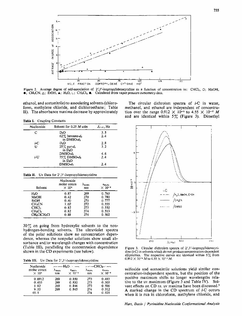
The circular dichroism spectra of i-C in water, methanol, and ethanol are independent of concentra- tion over the range 0.912 X to 4.55 X M and are identical within 5 % (Figure 3). Dimethyl
I c I I t
t ’ I I I I
3 50 h ( m r 1 300 2 5 0
Figure 3. Circular dichroism spectra of 2’,3’-isopropylidenecyti- dine (i-C) in solvents which do not produce concentration-dependent ellipticities. The respective curves are identical within 5 from 0.912 X Mto4.55 X 10-3M.
sulfoxide and acetonitrile solutions yield similar con- centration-independent spectra, but the position of the positive maximum shifts to longer wavelengths rela- tive to the uv maximum (Figure 3 and Table IV). Sol- vent effects on CD us. uv maxima have been discussed.8 A marked change in the CD spectrum of i-C occurs when it is run in chloroform, methylene chloride, and
Hart, Davis / Pyrimidine Nucleoside Conformational Analysis
756
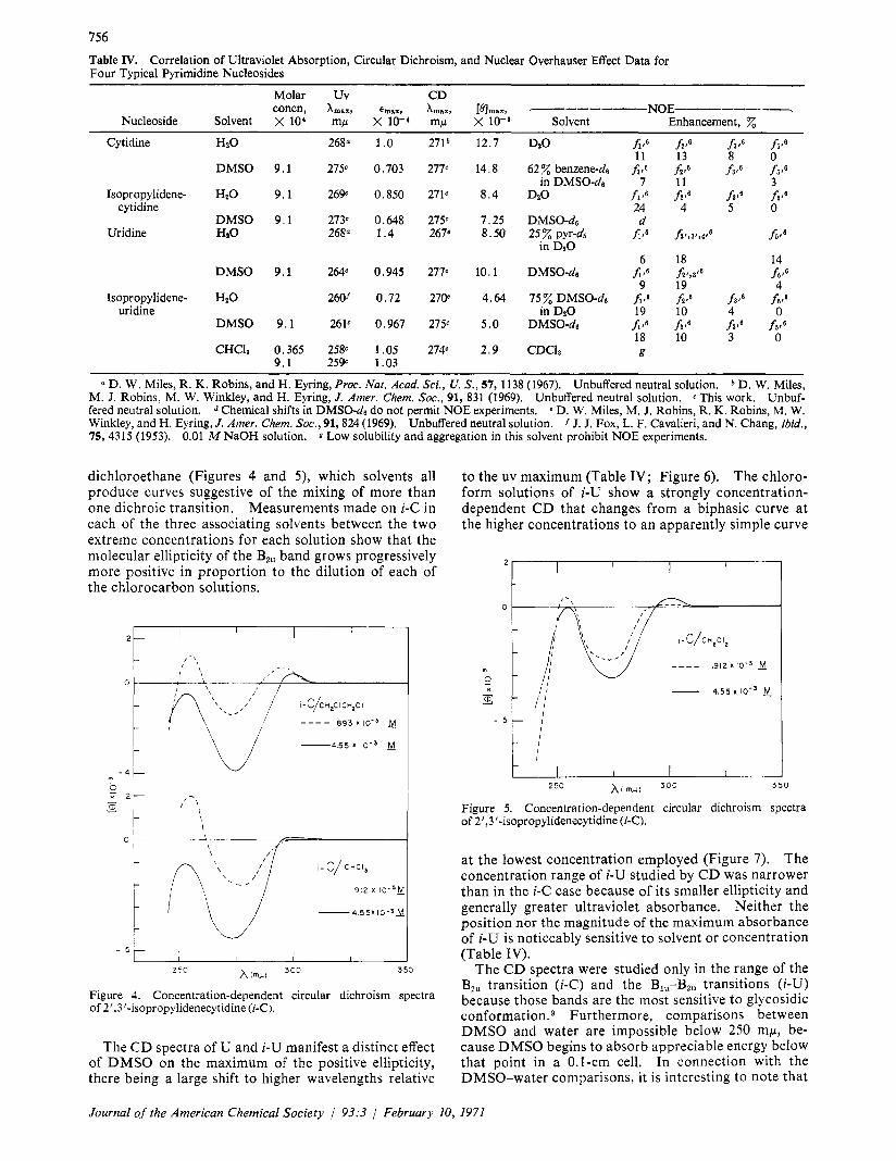
Table IV. Correlation of Ultraviolet Absorption, Circular Dichroism, and Nuclear Overhauser Effect Data for Four Typical Pyrimidine Nucleosides
Molar Uv CD concn, Amax, emax, Amax, [elm,,, --------- NOE------- 7
Nucleoside Solvent X 104 mu X 10-4 mu x 10-8 Solvent Enhancement. 'Z
Cytidine Hz0 268n 1.0 271b 12.7 Dz0 J i l 6 f i t 6 f 3 f 6 1 6 ' 6 11 13 8 0
in DMSO-d6 7 11 3 Isopropylidene- H 2 0 9.1 269 0.850 271C 8.4 D20 f i t 6 f i t 6 f 3 1 6 f d
cytidine 24 4 5 0
Uridine Ha0 26ga 1.4 267' 8.50 25 pyr-ds fi1,3',4'8 16'6
DMSO 9.1 27Y 0.703 277c 14.8 62% benzene-d6 f i , 6 fiJ f 3 , 6 f5 ,6
DMSO 9.1 273' 0.648 2750 7.25 DMSO-de d
in D 9 0 6 18
9 19 DMSO 9.1 264' 0.945 277' IO. 1 DMSO-de f i t 6 f i r , t ' 6
14 h 16
4 Isopropylidene- HzO 260f 0.72 27Oe 4.64 75z DMSO-d6 5.6 fi,' f 3 t 6 f5,'
uridine in D 2 0 19 10 4 0 DMSO 9.1 261' 0.967 275C 5.0 DMSO-de f i t 6 f i o 6 f 3 1 6 f v 6
18 10 3 0 CHCls 0.365 258C 1.05 274' 2.9 CDC13 g
9.1 259 1.03
a D. W . Miles, R. K. Robins, and H. Eyring, Proc. Nat. Acad. Sci., U. S., 57, 1138 (1967). Unbuffered neutral solution. D. W. Miles, M. J. Robins, M. W. Winkley, and H. Eyring, J. Amer. Chem. SOC., 91, 831 (1969). Unbuffered neutral solution. c This work. Unbuf- fered neutral solution. Chemical shifts in DMSO-d6 do not permit NOE experiments. e D. W. Miles, M. J. Robins, R. K. Robins, M. W. Winkley, and H. Eyring, J . Amer. Chem. SOC., 91,824 (1969). Unbuffered neutral solution. J J. J. Fox, L. F. Cavalieri, and N. Chang, ibid., 75, 4315 (1953). 0.01 M NaOH solution. Low solubility and aggregation in this solvent prohibit NOE experiments.
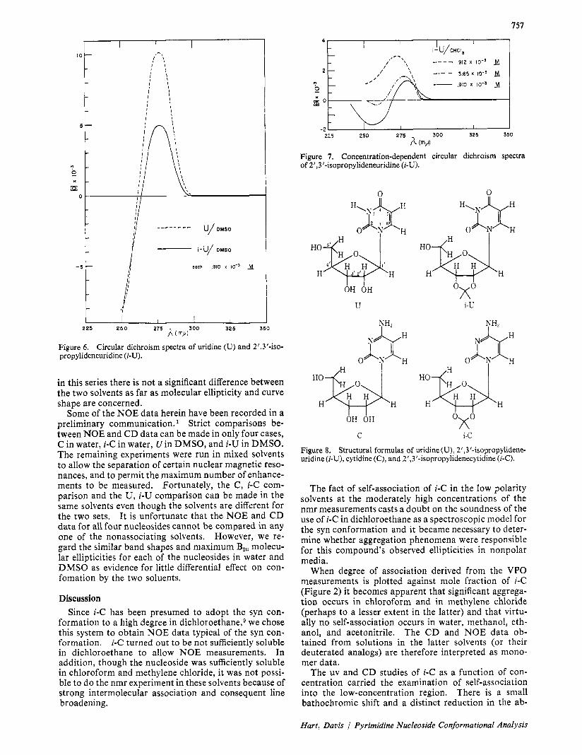
dichloroethane (Figures 4 and 5 ) , which solvents all produce curves suggestive of the mixing of more than one dichroic transition. Measurements made on i-C in each of the three associating solvents between the two extreme concentrations for each solution show that the molecular ellipticity of the B2,, band grows progressively more positive in proportion to the dilution of each of the chlorocarbon solutions.
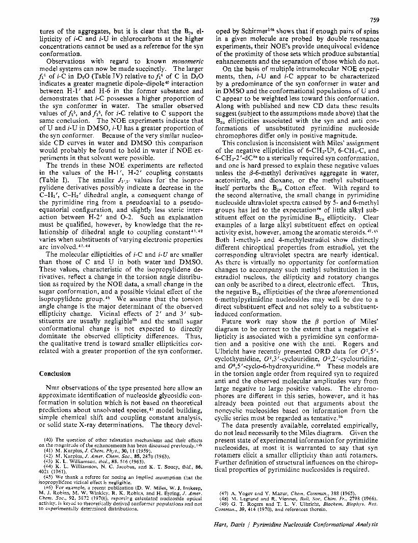
to the uv maximum (Table IV; Figure 6 ) . The chloro- form solutions of i-U show a strongly concentration- dependent C D that changes from a biphasic curve at the higher concentrations to an apparently simple curve
0 \ - \ \ I'
3 50 h c m i l i 300 2 50
Figure 4. Concentration-dependent circular dichroism spectra of 2',3'-isopropylidenecytidine (92).
The CD spectra of U and i-U manifest a distinct effect of DMSO on the maximum of the positive ellipticity, there being a large shift to higher wavelengths relative
at the lowest concentration employed (Figure 7). The concentration range of i-U studied by CD was narrower than in the i-C case because of its smaller ellipticity and generally greater ultraviolet absorbance. Neither the position nor the magnitude of the maximum absorbance of i-U is noticeably sensitive to solvent or concentration (Table IV).
The CD spectra were studied only in the range of the BZu transition (i-C) and the BI,-B2, transitions ( i -U) because those bands are the most sensitive to glycosidic conf~rmat ion .~ Furthermore, comparisons between DMSO and water are impossible below 250 mp, be- cause DMSO begins to absorb appreciable energy below that point in a 0.1-cm cell. In connection with the DMSO-water comparisons, it is interesting to note that
Journal of the American Chemical Society 93:3 / February 10, 1971
757
I
i-U/ DMSO
- 5 each ,910 x IO” M
2 25 2 5 0 275 325 350 A (
Figure 6. propylideneuridine (i-U).
Circular dichroism spectra of uridine (U) and 2’,3’-iso-
in this series there is not a significant difference between the two solvents as far as molecular ellipticity and curve shape are concerned.
Some of the NOE data herein have been recorded in a preliminary communication.’ Strict comparisons be- tween NOE and C D data can be made in only four cases, C in water, i-C in water, U in DMSO, and i-U in DMSO. The remaining experiments were run in mixed solvents to allow the separation of certain nuclear magnetic reso- nances, and to permit the maximum number of enhance- ments to be measured, Fortunately, the C , i-C com- parison and the U , i-U comparison can be made in the same solvents even though the solvents are different for the two sets. It is unfortunate that the NOE and C D data for all four nucleosides cannot be compared in any one of the nonassociating solvents. However, we re- gard the similar band shapes and maximum BPu molecu- lar ellipticities for each of the nucleosides in water and DMSO as evidence for little differential effect on con- fomation by the two soluents.
Discussion Since i-C has been presumed to adopt the syn con-
formation to a high degree in dichlor~ethane,~ we chose this system to obtain NOE data typical of the syn con- formation. i-C turned out to be not sufficiently soluble in dichloroethane to allow NOE measurements. In addition, though the nucleoside was sufficiently soluble in chloroform and methylene chloride, it was not possi- ble to do the nmr experiment in these solvents because of strong intermolecular association and consequent line broadening.
225 250 275 300 325 350
A “1
Figure 7. Concentration-dependent circular dichroism spectra of 2’,3’-isopropylideneuridine (i-U).
n 0
U i-U
HowH H H H H&$ H H H
OX0 OH OH
C i-C
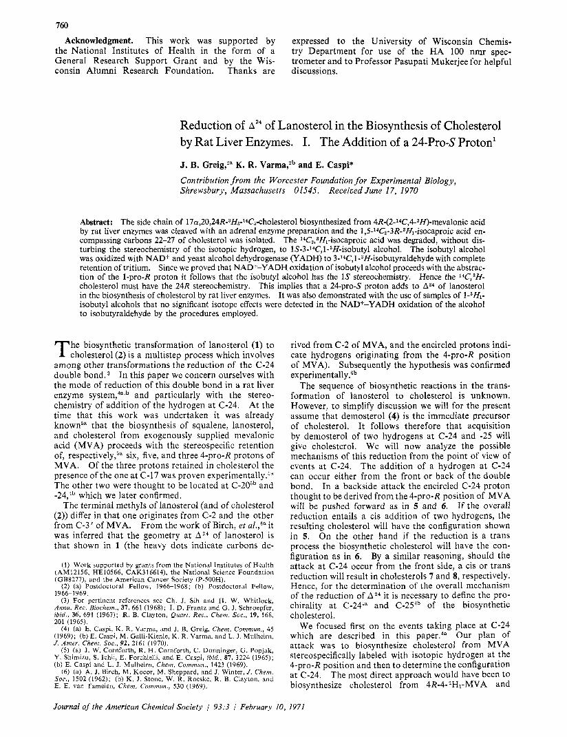
Figure 8. uridine (i-U), cytidine (C), and 2’,3’-isopropylidenecytidine (i-C).
Structural formulas of uridine (U), 2’,3’-isopropylidene-
The fact of self-association of i-C in the low polarity solvents at the moderately high concentrations of the nmr measurements casts a doubt on the soundness of the use of i-C in dichloroethane as a spectroscopic model for the syn conformation and it became necessary to deter- mine whether aggregation phenomena were responsible for this compound’s observed ellipticities in nonpolar media.
When degree of association derived from the VPO measurements is plotted against mole fraction of i-C (Figure 2) it becomes apparent that significant aggrega- tion occurs in chloroform and in methylene chloride (perhaps to a lesser extent in the latter) and that virtu- ally no self-association occurs in water, methanol, eth- anol, and acetonitrile. The C D and NOE data ob- tained from solutions in the latter solvents (or their deuterated analogs) are therefore interpreted as mono- mer data.
The uv and C D studies of i-C as a function of con- centration carried the examination of self-association into the low-concentration region. There is a small bathochromic shift and a distinct reduction in the ab-
Hart, Davis / Pyrimidine Nucleoside Conformational Analysis

758
sorbance correlated with decreasing solvent polarity (Table 11). Further, the absorbance and wavelength changes serve to differentiate the associating and non- associating solvents. Acetonitrile is anomalous in this study as it falls into the group of associating solvents on the basis of uv data, but into the group of nonasso- ciating solvents on the basis of the VPO measurements. The anomaly is not serious, however, for acetonitrile has been shown to cause hypochromicity relative to water in 9-pyranosyladenine without leading to ag- gregation. l7 Hypochromicity can, of course, be asso- ciated with aggregation18a,b and in that case the re- maining solvents seem to be appropriately grouped.
The complex concentration-dependent CD spectra of i-C and i-U (Figures 4, 5 , and 7) in chlorocarbon sol- vents are difficult to interpret rigorously. In general, the observed curve shapes suggest the appearance of a positive curve at the expense of a negative curve on going to lower concentrations. The complexity of the i-C curves is not completely removed even at the lower concentrations, while the lower concentrations of i-U in chloroform are characterized by distinctly simplified spectra and the lowest concentration curve shows very little of the negative contributor. The i-C curves in chloroform differ markedly from their water solution counterpartg in the 300-240-mp range. On the other hand, the i-U curves at higher concentrations in chloro- form are not fundamentally different from their polar solvent counterparts, there being a positive and nega- tive portion in both solvent types between 300 and 240 mp. The lower wavelength Cotton effect in this range has been associated with a B1, transitions in the uridine series and possible reasons for not seeing a correspond- ing Cotton effect in some cytidine derivativesg are either that the transition does not exist in those de- rivatives or that the transition gives a positive Cotton effect. In the latter case, the single positive curve that is seen in the cytidine derivatives might actually be an unresolved summation of the B,, and Bqu transitions. Magnetic circular dichroism studies verify this conclu- sion.lg The similarity between the i-U CD spectra in polar and nonpolar solvents may be coincidental, how- ever, since the biphasic nature of the chloroform curve disappears as the concentration is reduced. The pro- nounced, DMSO-induced shift of the Bzu ellipticity max- imum to higher wavelength relative to the uv absorbance maximum-a shift also caused to a lesser extent by water, dioxane, and acetonitrile-is probably due to a selective solvent effect on the B,, and B1, transitions. Such effects have been discussed.s
The appearance of new or complex C D bands has been treated in several contexts and attributed to sev- eral specific phenomena. The enhanced ellipticity of helical polynucleotides relative to the ellipticity of in- dependent monomer components has been related to intra- and intermolecular base-base interactions. In that case, the interacting chromophores become nearly the sole determinant of ellipticity.20-22 If these inter-
(17) E. Charney and M. Gellert, Biopolym. Symp., 1, 469 (1964). (18) (a) R. K. Nesbet, ibid., 129 (1964); (b) H. Devoe, ibid., 251
(19) W. Voelter, R. Records, E. Bunnenberg, and C. Djerassi, J .
(20) D. F. Bradley, 1. Tinoco, Jr., and R. W. Woody, Biopolymers, 1,
(21) C. A. Bush and J. Brahms, J . Chem. Phys., 46, 79 (1967). (22) C. A. Bush and I. Tinoco, Jr., J . Mol. Biol., 23, 601 (1967).
( 1964).
Amer. Chem. Soc., 90, 6163 (1968).
239 (1963).
actions are between identical chromophores, exciton splitting can result and one observes closely spaced CD curves of equal magnitude and opposite sign. Exciton coupling has been given considerable theoret- ical treatment. 20--25 The appearance of exciton split- ting has been noted in the stacking of nucleic acid and polynucleotide bases associated with helix forma- tion,26-28 in the interaction of dyes bound to dissym- metric macromolecules, 2 9 and in the self-association of several chlorophyll and protochlorophyll pigments. 30
If the interaction is between different chromophores with similar uv absorption maxima, the resultant inter- action has been called by Urry31s32 a reciprocal relation to describe the mutual effect of one chromophore on the other. Reciprocal relations are seen, for example, in the intramolecular association of the purine and pyr- idine moieties of NAD or NADH and the purine and isoalloxazine moieties of FAD. Both exciton splitting and reciprocal relations are special cases of electronic dipole-dipole coupling. Complex circular dichroism spectra have been observed in simpler systems as well and have been variously ascribed to vibronic coupling, 3 3
the presence of more than one conformation type,34-36 and the existence of several solvate types. 3 7 , 3 8 In these cases it is usual to observe unsymmetrical biphasic C D curves.
The curve shapes seen in the present experiments are too complex to allow one to factor out exciton contribu- tions and since the chromophores are identical one need not consider reciprocal relations. If the trends in curve shape with decreasing concentration can be in- terpreted in terms of the diluting-out of self-association, then the dissociation of aggregates is more extensive in the case of i-U than of i-C and the negative ellipticity observed for both is likely intrinsic only to the aggre- gates. The gradual enhancement of the positive con- tributor at the expense of the negative is a good indica- tion of the emergence of some new form (probably monomer) that exists only at a low, limiting, concentra- t i ~ n . ~ ~ Whatever species this is, it is apparently more prevalent in the case of i-U at the low concentration limit. These C D experiments do not reveal the struc-
(23) 1. Tinoco, Jr., Rudiut. Res., 20, 133 (1963). (24) E. A. Dratz, A. J. Schultz, and K. Sauer, Brookhuven Symp.
( 2 5 ) J. A. Schellman, Accounfs Chem. Res., 1, 144 (1968). (26) J. Brahms, A. M. Michelson, and K. E. Van Holde, J . Mol.
(27) J. N. Vournakis, H. A. Sheraga, G. W. Rushizky, and H. A.
(28) D. Poland, J. N. Vournakis, and H. A. Sheraga, ibid., 4, 223
(29) A. Blake and A. R. Peacocke, ibid., 5, 871 (1967). (30) C. Houssier and I<. Sauer, J . Amer. Chem. Soc., 92, 779 (1970). (31) D. W. Miles and D. W. Urry, Biochemistry, 7, 2791 (1968). (32) D. W. Miles and D. W. Urry, J . Biol. Chem., 243, 4181 (1968). (33) D. J. Severn and E. M. Kosower, J . Amer. Chem. Soc . , 91, 1710
(34) A. Moscowitz, K. M. Wellman, and C. Djerassi, Proc. Nut.
(35) K. M. Wellman, P. H. A. Laur, W. S. Briggs, A. Moscowitz, and
(36) G. Barth, W. Voelter, E. Bunnenberg, and C. Djerassi, Chem.
(37) C. Coulombeau and A. Rassat, Bull. Soc. Chim. Fr., 2673 (1963). (38) C. Coulombeau and A. Rassat, ibid., 3752 (1966). (39) The concentration below which no further ellipticity changes are
seen. I t was not actually reached in either case by our experiments. The unmasking of the conformation sensitive Bzu Cotton effect is presumably complete only below this point, It is difficult to reach in the present cases since one dilutes the solutions at the expense of meas- ured ellipticity and 10-5 M taxes the sensitivity of the instrument, given the ellipticity of the particular solutes.
Biol., 19, 303 (1966).
Biol., 15, 467 (1966).
Sober, Biopolymers, 4, 33 (1966).
( 19 66).
(1969).
Acud. Sci. U. S. , 50, 799 (1963).
C. Djerassi, J . Amer. Chem. Soc., 87, 66 (1965).
Commun., 355 (1969).
Journal of the American Chemical Society / 93:3 / February 10, 1971
759
oped by Schirmer14* shows that if enough pairs of spins in a given molecule are probed by double resonance experiments, their NOE's provide unequivocal evidence of the proximity of those sets which produce substantial enhancements and the separation of those which do not.
On the basis of multiple intramolecular NOE experi- ments, then, i-U and i-C appear to be characterized by a predominance of the syn conformer in water and in DMSO and the conformational populations of U and C appear to be weighted less toward this conformation. Along with published and new C D data these results suggest (subject to the assumptions made above) that the BBu ellipticities associated with the syn and anti con- formations of unsubstituted pyrimidine nucleoside chromophores differ only in positive magnitude.
This conclusion is inconsistent with Miles' assignment of the negative ellipticities of 6-CH3-US, 6-CH3-C, and 6-CH3-2'-dCga to a sterically required syn conformation, and one is hard pressed to explain these negative values unless the P-6-methyl derivatives aggregate in water, acetonitrile, and dioxane, or the methyl substituent itself perturbs the Bz,, Cotton effect. With regard to the second alternative, the small change in pyrimidine nucleoside ultraviolet spectra caused by 5 - and 6-methyl groups has led to the expectationga of little alkyl sub- stituent effect on the pyrimidine B2u ellipticity. Clear examples of a large alkyl substituent effect on optical activity exist, however, among the aromatic steroids. 4 7 , 4 8
Both I-methyl- and 4-methylestradiol show distinctly different chiroptical properties from estradiol, yet the corresponding ultraviolet spectra are nearly identical. As there is virtually no opportunity for conformation changes to accompany such methyl substitution in the estradiol nucleus, the ellipticity and rotatory changes can only be ascribed to a direct, electronic effect. Thus, the negative B,, ellipticities of the three aforementioned 6-methylpyrimidine nucleosides may well be due to a direct substituent effect and not solely to a substituent- induced conformation.
Future work may show the p portion of Miles' diagram to be correct to the extent that a negative el- lipticity is associated with a pyrimidine syn conforma- tion and a positive one with the anti. Rogers and Ulbricht have recently presented ORD data for 0 2 , 5 ' - cyclothymidine, 02,3 '-cyclouridine, 02,2'-cyclouridine, and 06,5 '-cyclo-6-hydroxyuridine. 49 These models are in the torsion angle order from required syn to required anti and the observed molecular amplitudes vary from large negative to large positive values. The chromo- phores are different in this series, however, and it has already been pointed out that arguments about the noncyclic nucleosides based on information from the cyclic series must be regarded as tentative.$"
The data presently available, correlated empirically, do not lead necessarily to the Miles diagram. Given the present state of experimental information for pyrimidine nucleosides, at most it is warranted to say that syn rotamers elicit a smaller ellipticity than anti rotamers. Further definition of structural influences on the chirop- tical properties of pyrimidine nucleosides is required.
tures of the aggregates, but it is clear that the B,, el- lipticity of i-C and i-U in chlorocarbons at the higher concentrations cannot be used as a reference for the syn conformation.
Observations with regard to known monomeric model systems can now be made succinctly. The larger
f16 of i-C in D,O (Table IV) relative tofi6 of C in D 2 0 indicates a greater magnetic d ip~ le -d ipo le~~ interaction between H-I ' and H-6 in the former substance and demonstrates that i-C possesses a higher proportion of the syn conformer in water. The smaller observed values offi6, andf36, for i-C relative to C support the same conclusion. The NOE experiments indicate that of U and i-U in DMSO, i-U has a greater proportion of the syn conformer. Because of the very similar nucleo- side C D curves in water and DMSO this comparison would probably be found to hold in water if NOE ex- periments in that solvent were possible.
The trends in these NOE experiments are reflected in the values of the H-l', H-2' coupling constants (Table I). The smaller values for' the isopro- pylidene derivatives possibly indicate a decrease in the C-HI', C-H,' dihedral angle, a consequent change of the pyrimidine ring from a pseudoaxial to a pseudo- equatorial configuration, and slightly less steric inter- action between H-2' and 0-2. Such an explanation must be qualified, however, by knowledge that the re- lationship of dihedral angle to coupling c o n ~ t a n t ~ ~ t ~ ~ varies when substituents of varying electronic properties are involved.48,44
The molecular ellipticities of i-C and i-U are smaller than those of C and U in both water .and DMSO. These values, characteristic of the isopropylidene de- rivatives, reflect a change in the torsion angle distribu- tion as required by the NOE data, a small change in the sugar conformation, and a possible vicinal effect of the isopropylidene g r o ~ p . ~ 5 We assume that the torsion angle change is the major determinant of the observed ellipticity change. Vicinal effects of 2' and 3' sub- stituents are usually negligiblegb and the small sugar conformational change is not expected to directly dominate the observed ellipticity differences. Thus, the qualitative trend is toward smaller ellipticities cor- related with a greater proportion of the syn conformer.
Conclusion
Nmr observations of the type presented here allow an approximate identification of nucleoside glycosidic con- formation in solution which is not based on theoretical predictions about unsolvated species, 46 model building, simple chemical shift and coupling constant analysis, or solid state X-ray determinations. The theory devel-
(40) The question of other relaxation mechanisms and their effects on the magnitude of the enhancements has been discussed previously.14b
(41) M. Karplus, J . Chem. Phys., 30, 11 (1959). (42) M. Karplus, J . Amer. Chem. Soc., 85, 2870 (1963). (43) I(. L. Williamson, ibid., 85, 516 (1963). (44) K. L. Williamson, N. C. Jacobus, and K. T. Souey, ibid., 86,
4021 (1961). (45) We thank a referee for noting an implied assumption that the
isopropylidene vicinal effect is negligible. (46) For example, a recent publication (D. W. Miles, W. J. Inskeep,
M. J. Robins, M. W. Winkley, R. K. Robins, and H. Eyring, J . Amer. Chem. Soc., 92, 3872 (1970)), reporting calculated nucleoside optical activity, is keyed to theoretically derived conformer populations and not to experimentally determined distributions.
(47) A. Yogev and Y . Mazur, Chem. Commun., 388 (1965). (48) M. Legrand and R. Viennet, Bull. SOC. Chim. Fr., 2798 (1966). (49) G. T. Rogers and T. L. V. Ulbricht, Biochem. Biophys. Res .
Commun., 39, 414 (1970), and references therein.
Hart, Davis Pyrimidine Nucleoside Conformational Analysis
760
the National Institutes of Health in the form of a General Research Support Grant and by the Wis- consin Alumni Research Foundation. Thanks are discussions.
Acknowledgment. This work was supported by expressed to the University of Wisconsin Chemis- try Department for use of the HA 100 nmr spec- trometer and to Professor Pasupati Mukerjee for helpful
Reduction of A24 of Lanosterol in the Biosynthesis of Cholesterol by Rat Liver Enzymes. 1. The Addition of a 24-Pro-$ Proton'
J. B. Greig,'" K. R. Varma,2b and E. Caspi*
Contribution from thc Worcester Foundation for Experimental Biology, Shrewsbury, Massachusetts 01545. Received June 17, 1970
Abstract: The side chain of 17~i,20,24R-~H~-*~C~-cholesterol biosynthesized from 4R-(2-14C,4-3H)-mevalonic acid by rat liver enzymes was cleaved with an adrenal enzyme preparation and the 1,5-14C~-3R-3Hl-isocaproic acid en- compassing carbons 22-27 of cholesterol was isolated. The 14C2,3Hl-iso~aproi~ acid was degraded, without dis- turbing the stereochemistry of the isotopic hydrogen, to 1S-3-14C,l-3H-isobutyl alcohol. The isobutyl alcohol was oxidized with NAD+ and yeast alcohol dehydrogenase (YADH) to 3-14C,1 -3H-isobutyraldehyde with complete retention of tritium, Since we proved that NAD+-YADH oxidation of isobutyl alcohol proceeds with the abstrac- tion of the I-pro-R proton it follows that the isobutyl alcohol has the 1s stereochemistry. Hence the 14C,3H- cholesterol must have the 24R stereochemistry. This implies that a 24-pro-S proton adds to AZ4 of lanosterol in the biosynthesis of cholesterol by rat liver enzymes. It was also demonstrated with the use of samples of l-3H1- isobutyl alcohols that no significant isotope effects were detected in the NAD+-YADH oxidation of the alcohol to isobutyraldehyde by the procedures employed.
he biosynthetic transformation of lanosterol (1) to T cholesterol (2) is a multistep process which involves among other transformations the reduction of the (2-24 double bond. In this paper we concern ourselves with the mode of reduction of this double bond in a rat liver enzyme system,4aab and particularly with the stereo- chemistry of addition of the hydrogen at (2-24. At the time that this work was undertaken it was already known5" that the biosynthesis of squalene, lanosterol, and cholesterol from exogenously supplied mevalonic acid (MVA) proceeds with the stereospecific retention of, respectively,'" six, five, and three 4-pro-R protons of MVA. Of the three protons retained in cholesterol the presence of the one at C-17 was proven e~perimentally.~" The other two were thought to be located at C-20jb and -24,4b which we later confirmed.
The terminal methyls of lanosterol (and of cholesterol (2)) differ in that one originates from C-2 and the other from C-3' of MVA. From the work of Birch, et al.,'ja it was inferred that the geometry at A Z 4 of lanosterol is that shown in 1 (the heavy dots indicate carbons de-
(1) Work supported by grants from the National Institutes of Health (AM12156, HE10566, CAK316614), the National Science Foundation (GB8277), and the American Cancer Society (P-500H).
(2) (a) Postdoctoral Fellow, 1966-1968; (b) Postdoctoral Fellow,
(3) For pertinent references see Ch. J . Sih and H. W. Whitlock, Annu. Rec. Biochem., 37,661 (1968); I. D. Frantz and G. J . Schroepfer, ibid., 36, 691 (1967); R . B. Clayton, Quart. Rea., Chem. Soc., 19, 168, 201 (1965).
(4) (a) E. Caspi, I<. R. Varma, and J . B. Greig, Chem. Commun., 45 (1969); (b) E. Caspi, M. Galli-Kienle, K . R. Varma, and L. J . Mulheirn, J. Amer. Chem. Soc., 92, 2161 (1970).
( 5 ) (a) J. W. Cornforth, R. H. Cornforth, C. Donninger, G. Popjak, Y. Shimizu, S. Ichii, E. Forchielli, and E. Caspi, ibid., 87, 3224 (1965); (b) E. Caspi and L. J. Mulheirn, Chem. Commun., 1423 (1969).
(6) (a) A. J . Birch, M. Kocor, M. Sheppard, and J. Winter, J . Chem. Soc., 1502 (1962); (b) K. J . Stone, W. R. Roeske, R. B. Clayton, and E. E. van Tamelen, Chem. Commun., 530 (1969).
1966-1969.
rived from C-2 of MVA, and the encircled protons indi- cate hydrogens originating from the 4-pro-R position of MVA). Subsequently the hypothesis was confirmed experimentally.'jb
The sequence of biosynthetic reactions in the trans- formation of lanosterol to cholesterol is unknown. However, to simplify discussion we will for the present assume that demosterol (4) is the immediate precursor of cholesterol. It follows therefore that acquisition by demosterol of two hydrogens at C-24 and -25 will give cholesterol. We will now analyze the possible mechanisms of this reduction from the point of view of events at C-24. The addition of a hydrogen at C-24 can occur either from the front or back of the double bond. In a backside attack the encircled (2-24 proton thought to be derived from the 4-pro-R position of MVA will be pushed forward as in 5 and 6. If the overall reduction entails a cis addition of two hydrogens, the resulting cholesterol will have the configuration shown in 5. On the other hand if the reduction is a trans process the biosynthetic cholesterol will have the con- figuration as in 6. By a similar reasoning, should the attack at (2-24 occur from the front side, a cis or trans reduction will result in cholesterols 7 and 8, respectively. Hence, for the determination of the overall mechanism of the reduction of A Z 4 it is necessary to define the pro- chirality at C-244a and C-254b of the biosynthetic cholesterol.
We focused first on the events taking place at C-24 which are described in this paper.4a Our plan of attack was to biosynthesize cholesterol from MVA stereospecifically labeled with isotopic hydrogen at the 4-pro-R position and then to determine the configuration at C-24. The most direct approach would have been to biosynthesize cholesterol from 4R-4-2Hl-MVA and
Journal of the American Chemical Society 93:3 February 10, 1971