Pulp Fibroblasts Contribution to the Local Control of Pulp ...
17
HAL Id: hal-03191414 https://hal.archives-ouvertes.fr/hal-03191414 Submitted on 7 Apr 2021 HAL is a multi-disciplinary open access archive for the deposit and dissemination of sci- entific research documents, whether they are pub- lished or not. The documents may come from teaching and research institutions in France or abroad, or from public or private research centers. L’archive ouverte pluridisciplinaire HAL, est destinée au dépôt et à la diffusion de documents scientifiques de niveau recherche, publiés ou non, émanant des établissements d’enseignement et de recherche français ou étrangers, des laboratoires publics ou privés. Pulp Fibroblasts Contribution to the Local Control of Pulp Inflammation via Complement Activation Chloé Le Fournis, Charlotte Jeanneau, Sandra Roumani, Thomas Giraud, Imad About To cite this version: Chloé Le Fournis, Charlotte Jeanneau, Sandra Roumani, Thomas Giraud, Imad About. Pulp Fibrob- lasts Contribution to the Local Control of Pulp Inflammation via Complement Activation. Journal of Endodontics, Elsevier, 2020, 46 (9), pp.S26-S32. 10.1016/j.joen.2020.06.029. hal-03191414
Transcript of Pulp Fibroblasts Contribution to the Local Control of Pulp ...
HAL Id: hal-03191414https://hal.archives-ouvertes.fr/hal-03191414
Submitted on 7 Apr 2021
HAL is a multi-disciplinary open accessarchive for the deposit and dissemination of sci-entific research documents, whether they are pub-lished or not. The documents may come fromteaching and research institutions in France orabroad, or from public or private research centers.
L’archive ouverte pluridisciplinaire HAL, estdestinée au dépôt et à la diffusion de documentsscientifiques de niveau recherche, publiés ou non,émanant des établissements d’enseignement et derecherche français ou étrangers, des laboratoirespublics ou privés.
Pulp Fibroblasts Contribution to the Local Control ofPulp Inflammation via Complement Activation
Chloé Le Fournis, Charlotte Jeanneau, Sandra Roumani, Thomas Giraud,Imad About
To cite this version:Chloé Le Fournis, Charlotte Jeanneau, Sandra Roumani, Thomas Giraud, Imad About. Pulp Fibrob-lasts Contribution to the Local Control of Pulp Inflammation via Complement Activation. Journal ofEndodontics, Elsevier, 2020, 46 (9), pp.S26-S32. �10.1016/j.joen.2020.06.029�. �hal-03191414�
1
Pulp Fibroblasts Contribution to the Local Control of Pulp
Inflammation via Complement Activation
Chloé Le Fournis1, Charlotte Jeanneau1, Sandra Roumani1, Thomas Giraud1,2, Imad About1*
1. Aix Marseille Univ, CNRS, ISM, Inst Movement Sci, Marseille, France.
2. APHM, Hôpital Timone, Service d'Odontologie, Marseille, France.
*Corresponding author :
Prof. Imad About
Institut des Sciences du Mouvement (ISM)
UMR 7287 CNRS & Université d'Aix-Marseille
Faculté d'Odontologie,
27 BD Jean Moulin
13385 MARSEILLE cedex 5
France
Tel: 04 86 13 68 59
Fax: 04 86 13 68 40
email: [email protected]
Acknowledgements
The authors declare no potential conflicts of interest regarding the authorship and publication of
this article.
Clinical significance:
Dental pulp inflammation control is a pre-requisite for a successful vital pulp therapy. Thus,
understanding the cellular and molecular mechanisms of pulp inflammation and its resolution via
local complement system activation would provide excellent therapeutic opportunities for
targeting pulp inflammation.
2
Abstract
Upon traumatic injuries or carious lesions, the elimination of bacteria infiltrating the pulp is
recognized as a prerequisite for initiating the regeneration process.
The Complement is a major system involved in initiating the inflammation and the subsequent
bacteria elimination. This plasma system of above 35 proteins is synthesized by the liver and some
immune cells. It is activated by three pathways: the classical, alternative and lectin pathways that
can be triggered by physical injuries, infection and biomaterials. It is an efficient system for
initiating the inflammatory reaction and for eliminating invading bacteria.
Recent data have shown that the pulp fibroblast represents a unique non-immune cell type able to
synthesize the Complement proteins. Indeed, after physical injuries/bacteria stimulation, the
pulp fibroblast has been shown to synthesize and to activate the complement system leading to the
production of biologically active molecules such as C5a, C3b and Membrane Attack Complex
(MAC). This local secretion represents a rapid and efficient mechanism for eliminating bacteria
invading the pulp, thus supporting the Complement activation from the plasma. Complement
proteins secreted by pulp fibroblasts allow cariogenic bacteria direct lysis via MAC formation on
their surface, phagocytic cell recruitment by producing C5a and opsonizing cariogenic bacteria
after C3b fixation on their surface, stimulating cariogenic bacteria phagocytosis.
Overall, this review highlights that, in addition to initiating the inflammatory reaction, pulp
fibroblasts also provide a powerful control of this inflammation via local Complement activation.
The pathogen elimination capacity by fibroblast-produced Complement demonstrates that this
system is a strong local actor in arresting bacterial progression into the dental pulp.
Keywords: Pulp biology, Complement system activation, Inflammation control, Pulp fibroblast

3
Introduction
Dental caries results from an oral microbiome imbalance where a high dietary carbohydrate
intake is metabolized into lactic, acetic, formic and propionic acids, suppressing acid-sensitive
bacteria (1-3). Subsequently, the bacterial community is reorganized and enriched with highly
acidogenic and acid-tolerant bacteria. Recent meta-transcriptomic analysis provided information
about the complex microbiota involved in carious process depending of the disease stage. Several
acidogenic cariogenic bacteria have been associated with caries, such as Streptococcus mutans,
Streptococcus sobrinus and Lactobacillus species (4). The production of these organic acids
demineralizes enamel and dentin allowing bacteria to progress towards the pulp (5).
Odontoblasts act as sensors of bacteria invading dentin tubules through their recognition of
factors released by dissolved dentin (such as TGFβ-1), bacteria toxins and bacterial patterns (such
as Lipopolysaccharides or flagellin) (6, 7). The activated odontoblasts produce antibacterial
molecules, such as β-defensins, that diffuse through the dentin to decrease the bacterial progression
(8). An important immunologic system named the complement system is involved in bacteria
destruction and in initiating the inflammation.
The complement system is known for three major roles: initiation of inflammation,
opsonization and lysis of microbes. The complement system circulating proteins are produced by
the liver and reach all body tissues through the bloodstream. Some tissues have a local production
of some Complement proteins in order to improve their defense mechanisms. For example,
astrocytes synthesize C1 to C8 proteins and represent the principal source of Complement
components in the brain (9,10). Rat retinal cells are able to synthetize C1q and C3 Complement
proteins in case of glaucoma (11). Tubular epithelial cells of the kidney are the predominant sources
of Complement proteins in this organ (12). These organs share terminal bloodstream localization,
requiring an additional local Complement production to prevent potential infection in case of a
decreased blood supply.
Fibroblasts from several tissues, such as skin, lung and synovial, have been shown to secrete
several Complement proteins (13,14). Because the dental pulp has a terminal circulation position
and is subject to caries and traumatic injuries, it was hypothesized that pulp fibroblasts might have
this local Complement protein secretion potential. Indeed, in 2014, it has been demonstrated that
dental pulp fibroblast is the first non-immune cell able to synthesize all the Complement
components under bacterial stimulation in order to initiate the inflammatory reaction (15).
However, to our knowledge, no studies have compared this secretion level to that of hepatocytes,
immune cells or other fibroblast cell types.
Additionally some studies have demonstrated that pulp fibroblasts play a central role in
inflammation by secreting pro-inflammatory cytokines in response to injuries or carious lesions.
Indeed, pulp fibroblasts have been shown to secrete IL-6, IL-8 (16), IL-1, and TNF-α (17). This
4
review will highlight the antibacterial role of pulp fibroblasts through the production of
Complement components C5a, C3b and C5b-C9 (membrane attack complex proteins).
Pulp fibroblasts synthetize all Complement components
The complement system is a powerful innate immune response. This system is composed
of more than 35 proteins, which are found in the serum in an inactive form (18,19). In the presence
of pathogens, Complement is activated. Inactive proteins convert to an active form after
interactions with each other in a highly specific enzymatic cascade, generating proteolytic
fragments that mediate numerous biological events. The Complement cascade can be activated by
the Classical, the Alternative or the Lectin pathways. In the Classical pathway, the C1 fragment is
activated by binding to an antigen/antibody complex. The Alternative pathway is constitutively
activated by spontaneous hydrolysis but may be triggered directly by foreign surfaces like
microorganisms or biomaterials. The Lectin pathway is activated by specific carbohydrate
expressed on microbial surfaces. Finally, these three pathways converge to C3 convertase enzyme
activation.
C3 convertase cleaves C3 protein into C3a and C3b fragments. C3a is known to be an
anaphylatoxin and is involved in the initiation of inflammation. Indeed, anaphylatoxins induce
smooth muscle contractions, increase blood vessel permeability, mast cell and basophil
degranulation and immune cell chemotaxis. C3b is an opsonin, it binds to the microorganism
surfaces (opsonization) facilitating their phagocytosis. Activation of the C3 convertase leads to the
C5 convertase formation which cleaves C5 into C5a and C5b fragments. C5a is also an
anaphylatoxin. On the other hand, C5b protein reacts with other Complement components
including C6, C7, C8, and C9, leading to the formation of the Membrane Attack Complex (MAC).
This structure creates pores onto the pathogen cell membrane and leads to cell lysis. Liver is the
primary site for circulating Complement proteins synthesis, but it has been shown that many cells
produce some Complement molecules either constitutively or in response to stimulation.
While fibroblasts have been traditionally recognized as quiescent cells responsible for
extracellular matrix production, they are more and more investigated as active players of the
immune system. Pulp fibroblasts isolated from primary human pulp cell cultures by magnetic
sorting and stimulated by a Gram-positive bacterial motif, the lipoteichoic acid (LTA), express all
the genes coding proteins required for efficient Complement activation (15). Subsequently, the
secretion by pulp fibroblasts and the role of each Complement proteins were studied, especially in
the context of a carious lesion. The next parts of this review will focus on the antibacterial effect
of C5a, C3b and MAC produced by pulp fibroblasts.
Fibroblast-produced C5a induces macrophage recruitment towards the lesion site
Monocytes are able to migrate from the bloodstream into the tissues under physiological
and inflammatory conditions. Under physiological conditions, monocyte recruitment allows
resident macrophage cell renewal. These resident macrophages act as immune system sentinel. In
5
case of infection, they release pro-inflammatory cytokines such as IL-1, TNF-α, and C5a (20).
These cytokines, as well as growth factors and bacterial products, allow the recruitment of
circulating monocytes and their differentiation into macrophages. The recruitment occurs through
the following steps: cell rolling, adhesion, extravasation and migration. First, blood vessel
endothelial cells are stimulated and secrete cytokines such as IL-4, IL-13, and TNF-α to attract
immune cells. Endothelial cells then express adhesion molecules, such as Selectins.
Oligosaccharides present on monocyte surface, named Sialyl-Lewis X, recognize Selectin motif
allowing the rolling step and permitting monocytes integrins surface activation. Integrins recognize
ICAM-1 and VCAM-1, constitutively expressed on endothelial cell surface, arresting the cell
rolling step by a strong interaction between monocytes and endothelial cells. Monocyte
cytoskeleton reorganizes forming pseudopodia allowing an extravasation through the gaps between
endothelial cells. This passage of cells throughout the blood vessels is called diapedesis. Once in
the interstitial fluid, monocytes are activated into macrophages. They migrate along a chemotactic
gradient toward the site of injury or infection. C5a protein is an anaphylatoxin providing a gradient
allowing macrophage recruitment to the injured/infected site. Indeed, C5a receptor is expressed on
endothelial cells surface. C5a fixation on endothelial cells increases vascular permeability allowing
fluids containing C5a to enter into the inflammatory site. C5a receptors are also expressed by
neutrophils, monocytes and macrophage where C5a acts as a chemotactic factor for their
recruitment to the infection site.
Under carious or traumatic injuries, an inflammatory reaction occurs in the pulp. Immune
cells, such as monocytes and macrophages, migrate to the lesion site (21). Several studies have
shown a predominance of macrophage cells in the inflamed pulp tissue (22,23) and their number
increases along with the cariogenic bacteria progression (24). Macrophages limit the bacterial
progression by bacteria phagocytosis.
The effect of carious and traumatic injuries have been studied on pulp fibroblast
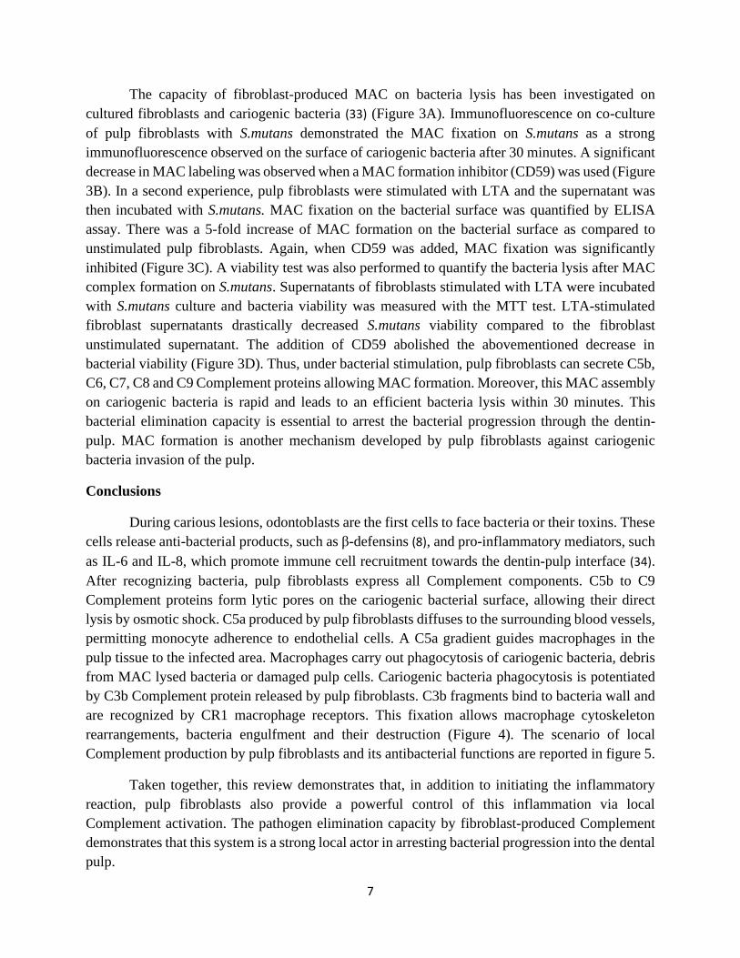
Complement C5a production and on chemotactic potential on macrophage-like cells (25,26) (Figure
1A). Pulp fibroblasts from primary pulp cell cultures were isolated, sorted and characterized. After
stimulation with LTA and/or physical injuries with scalpels pulp fibroblast C5a released was
quantified by ELISA. Pulp fibroblasts produce C5a without any stimulation. However, after their
stimulation with LTA or injuries, C5a production significantly increased (Figure 1B). Immune cell
migration was investigated with Boyden chambers using a human monocytic cell line model (THP-
1) activated to macrophage-like cells with Phorbol Myristate Acetate (PMA). After LTA
stimulation and physical injuries, macrophage-like cell migration significantly increased up to 3
times towards stimulated fibroblasts as compared to unstimulated cells (Figure 1C). These cells
express the C5aR (Figure 1D). The constitutive C5a secretion and macrophage-like cell
recruitment may have a physiological role in renewing the resident macrophage pool of sound
tissues. Indeed, resident macrophages act as sentinels (27). Under infection, C5a, binding to its
receptor (C5aR), increases CR1 receptor expression on macrophages, enhancing their phagocytic
capacity (28).
6
Fibroblast-produced C3b opsonizes cariogenic bacteria and enhances their phagocytosis
Phagocytosis is the capacity of specific cells, called phagocytes, to engulf and digest
micrometer-sized particles. Phagocytosis contributes to homeostasis, development, cell renewal
and immune response during the inflammatory acute phase. It is the first line of defense during
infection. Phagocytic cells include neutrophils, monocytes that differentiate into macrophages in
tissues, and dendritic cells. Pathogen phagocytosis occurs following 3 steps: adhesion, ingestion,
and digestion. During the adhesion, the phagocytic cell membrane binds to the particle to be
ingested. This stage involves the foreign pathogen lectins and the phagocyte membrane
glycoproteins. Phagocytic cells modify their plasma membrane to form pseudopods to swallow up
pathogens. A new vesicle is formed in the phagocytic cell cytoplasm called phagosome, trapping
the pathogen. The digestion step consists of the degradation of the pathogen by fusing the
phagosome with lysosomes to a phagolysosome. Lysosomes have an acidic content (pH is 4.5 to
5) due to the presence of proton pumps and ion channels on their membrane. Moreover, lysosomes
contain around 40 enzymes, such as lipases or proteases. Lysosomes discharge their content
composed of enzymes and acidic pH to the phagosome, allowing the degradation of the
phagocytosed particles. The digestion products are exocytosed (29). Opsonins are known to
enhance pathogen engulfment by phagocytes. There are two distinct types of opsonins: antibodies
and C3b Complement protein. Opsonins coat pathogens, acting as a flag for destruction, and
increase binding sites on the microbial surface to phagocytes. Opsonins binding to pathogens are
recognized by specific phagocytic cell receptors. C3b Complement protein is recognized by the
CR1 phagocyte receptor (30).
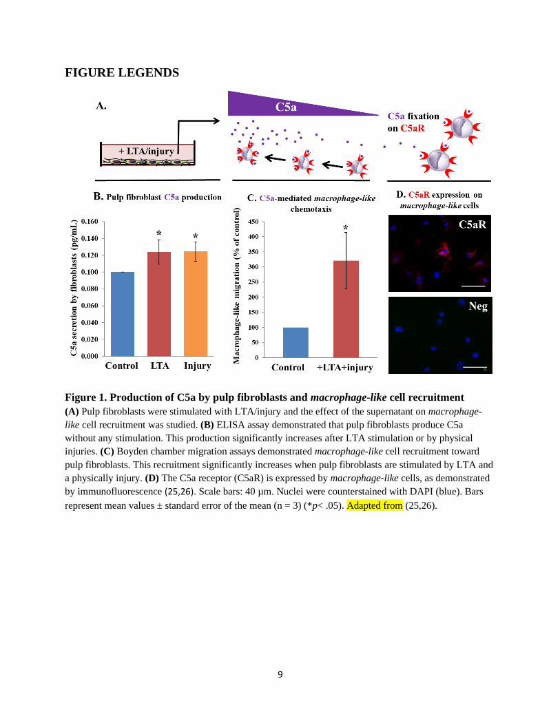
The ability of pulp fibroblasts to produce C3b opsonin and its role on cariogenic bacteria
have been recently investigated in vitro (31) (Figure 2A). Cultured pulp fibroblasts were found to
constitutively produce C3b. The presence of C3b protein was also confirmed in vivo in non-carious
pulp tooth sections. The C3b secretion is unmodified by an LTA fibroblast stimulation simulating
a carious lesion. When C3b protein produced by unstimulated pulp fibroblasts was incubated with
cariogenic bacteria, it bounds to their surface identifying them as a pathogenic target (Figure 2B).
This active fragment bound on bacteria membranes was recognized by CR1 macrophage-like
receptors and enhanced cariogenic bacteria phagocytosis (Figure 2C). A gentamycin protection
assay was performed to investigate the effect of fibroblast-produced C3b on cariogenic bacteria
phagocytosis by macrophage-like cells. There was a two-fold increase of phagocytosis in the
presence of fibroblast-produced C3b. When Cytochalasin D, an inhibitor of phagocytosis, was
used, cariogenic bacteria phagocytosis by macrophage-like cells was arrested demonstrating that
the engulfment process is phagocytosis and not only endocytosis or an intracellular bacteria (32)
(Figure 2D). Overall, these results suggest that pulp fibroblasts enhance cariogenic bacteria
elimination via C3b production and so contribute in arresting the carious bacteria progression.
Direct cariogenic bacteria lysis by fibroblast-produced Membrane Attack Complex (MAC)
7
The capacity of fibroblast-produced MAC on bacteria lysis has been investigated on
cultured fibroblasts and cariogenic bacteria (33) (Figure 3A). Immunofluorescence on co-culture
of pulp fibroblasts with S.mutans demonstrated the MAC fixation on S.mutans as a strong
immunofluorescence observed on the surface of cariogenic bacteria after 30 minutes. A significant
decrease in MAC labeling was observed when a MAC formation inhibitor (CD59) was used (Figure
3B). In a second experience, pulp fibroblasts were stimulated with LTA and the supernatant was
then incubated with S.mutans. MAC fixation on the bacterial surface was quantified by ELISA
assay. There was a 5-fold increase of MAC formation on the bacterial surface as compared to
unstimulated pulp fibroblasts. Again, when CD59 was added, MAC fixation was significantly
inhibited (Figure 3C). A viability test was also performed to quantify the bacteria lysis after MAC
complex formation on S.mutans. Supernatants of fibroblasts stimulated with LTA were incubated
with S.mutans culture and bacteria viability was measured with the MTT test. LTA-stimulated
fibroblast supernatants drastically decreased S.mutans viability compared to the fibroblast
unstimulated supernatant. The addition of CD59 abolished the abovementioned decrease in
bacterial viability (Figure 3D). Thus, under bacterial stimulation, pulp fibroblasts can secrete C5b,
C6, C7, C8 and C9 Complement proteins allowing MAC formation. Moreover, this MAC assembly
on cariogenic bacteria is rapid and leads to an efficient bacteria lysis within 30 minutes. This
bacterial elimination capacity is essential to arrest the bacterial progression through the dentin-
pulp. MAC formation is another mechanism developed by pulp fibroblasts against cariogenic
bacteria invasion of the pulp.
Conclusions
During carious lesions, odontoblasts are the first cells to face bacteria or their toxins. These
cells release anti-bacterial products, such as β-defensins (8), and pro-inflammatory mediators, such
as IL-6 and IL-8, which promote immune cell recruitment towards the dentin-pulp interface (34).
After recognizing bacteria, pulp fibroblasts express all Complement components. C5b to C9
Complement proteins form lytic pores on the cariogenic bacterial surface, allowing their direct
lysis by osmotic shock. C5a produced by pulp fibroblasts diffuses to the surrounding blood vessels,
permitting monocyte adherence to endothelial cells. A C5a gradient guides macrophages in the
pulp tissue to the infected area. Macrophages carry out phagocytosis of cariogenic bacteria, debris
from MAC lysed bacteria or damaged pulp cells. Cariogenic bacteria phagocytosis is potentiated
by C3b Complement protein released by pulp fibroblasts. C3b fragments bind to bacteria wall and
are recognized by CR1 macrophage receptors. This fixation allows macrophage cytoskeleton
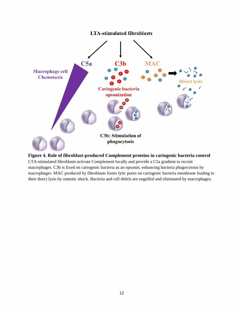
rearrangements, bacteria engulfment and their destruction (Figure 4). The scenario of local
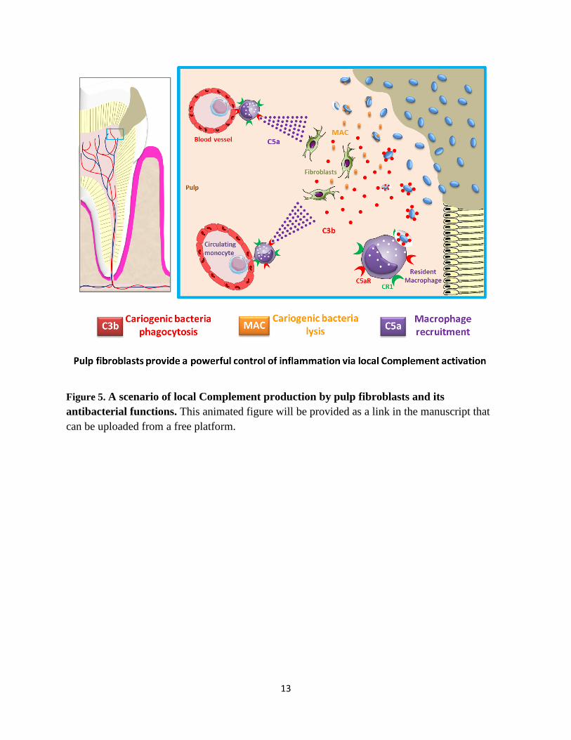
Complement production by pulp fibroblasts and its antibacterial functions are reported in figure 5.
Taken together, this review demonstrates that, in addition to initiating the inflammatory
reaction, pulp fibroblasts also provide a powerful control of this inflammation via local
Complement activation. The pathogen elimination capacity by fibroblast-produced Complement
demonstrates that this system is a strong local actor in arresting bacterial progression into the dental
pulp.

8
9
FIGURE LEGENDS
Figure 1. Production of C5a by pulp fibroblasts and macrophage-like cell recruitment
(A) Pulp fibroblasts were stimulated with LTA/injury and the effect of the supernatant on macrophage-
like cell recruitment was studied. (B) ELISA assay demonstrated that pulp fibroblasts produce C5a
without any stimulation. This production significantly increases after LTA stimulation or by physical
injuries. (C) Boyden chamber migration assays demonstrated macrophage-like cell recruitment toward
pulp fibroblasts. This recruitment significantly increases when pulp fibroblasts are stimulated by LTA and
a physically injury. (D) The C5a receptor (C5aR) is expressed by macrophage-like cells, as demonstrated
by immunofluorescence (25,26). Scale bars: 40 µm. Nuclei were counterstained with DAPI (blue). Bars
represent mean values ± standard error of the mean (n = 3) (*p< .05). Adapted from (25,26).
10
Figure 2. Opsonization and phagocytosis of S.mutans mediated by fibroblast-produced C3b
(A) Pulp fibroblasts were stimulated with LTA. (B) The fibroblast supernatant was incubated with
S.mutans. Immunofluorescence assay demonstrates the fibroblast produced C3b fixation on the bacterial
surface. (C) Pulp fibroblast supernatant was also incubated with macrophage-like cells. The C3b binding
to macrophage CR1 receptor was visualized. Immunofluorescence shows co-expression of C3b and CR1
on the merged images (D) The Gentamycin protection assay demonstrates a significant increase of
S.mutans phagocytosis by macrophage-like cells in the presence of stimulated or unstimulated fibroblast
supernatants. The addition of an inhibitor of phagocytosis, the Cytochalasin D, drastically decreases the
bacteria engulfment (31). Scale bars: 20 µm. Nuclei were counterstained with DAPI (blue). Bars represent
mean values ± standard error of the mean (n = 3) (*p< .05). Adapted from (31).

11
Figure 3. Membrane Attack Complex (MAC) formation by pulp fibroblasts and its fixation
on S.mutans and the subsequent bacteria lysis
(A) Pulp fibroblasts were stimulated by S.mutans and the MAC fixation on S.mutans and their lysis were
studied. (B) Co-culture of fibroblasts and S.mutans demonstrated that MAC is rapidly produced by pulp
fibroblasts. Within 30 min, its fixation on S.mutans is visible by immunofluorescence (MAC in red, FSP
in green). (C) MAC fixation increases after fibroblast stimulation with LTA and (D) significantly reduces
the bacteria viability. Incubation of cells/bacteria with CD59 demonstrates the implication of MAC (33).
Scale bars: 40 µm. Nuclei were counterstained with DAPI (blue). Bars represent mean values ± standard
error of the mean (n = 3) (*p< .05). Adapted from (33).
12
Figure 4. Role of fibroblast-produced Complement proteins in cariogenic bacteria control
LTA-stimulated fibroblasts activate Complement locally and provide a C5a gradient to recruit
macrophages. C3b is fixed on cariogenic bacteria as an opsonin, enhancing bacteria phagocytosis by
macrophages. MAC produced by fibroblasts forms lytic pores on cariogenic bacteria membrane leading to
their direct lysis by osmotic shock. Bacteria and cell debris are engulfed and eliminated by macrophages.
13
Figure 5. A scenario of local Complement production by pulp fibroblasts and its
antibacterial functions. This animated figure will be provided as a link in the manuscript that
can be uploaded from a free platform.
14
REFERENCES
1. Stephan RM. Intra-Oral Hydrogen-Ion Concentrations Associated With Dental Caries
Activity. J Dent Res. 1944;23:257–266.
2. Koenigs JW. Hydrogen peroxide and iron: a microbial cellulolytic system? Biotechnol
Bioeng Symp. 1975;5:151–159.
3. Featherstone JD. The science and practice of caries prevention. J Am Dent Assoc.
2000;131:887–899.
4. Kressirer CA, Chen T, Lake Harriman K, Frias-Lopez J, Dewhirst FE, Tavares MA, et al.
Functional profiles of coronal and dentin caries in children. J Oral Microbiol.
2018;10:1495976.
5. Krasse B. Biological factors as indicators of future caries. Int Dent J. 1988;38:219–225.
6. Magloire H, Romeas A, Melin M, Couble M-L, Bleicher F, Farges J-C. Molecular
Regulation of Odontoblast Activity under Dentin Injury. Adv Dent Res. 2001;15:46–50.
7. Farges J-C, Alliot-Licht B, Renard E, Ducret M, Gaudin A, Smith AJ, et al. Dental Pulp
Defence and Repair Mechanisms in Dental Caries. Mediat Inflamm. 2015;2015:1–16.
8. Dommisch H, Winter J, Açil Y, Dunsche A, Tiemann M, Jepsen S. Human beta-defensin
(hBD-1, -2) expression in dental pulp. Oral Microbiol Immunol. 2005;20:163–166.
9. Gasque P, Fontaine M, Morgan BP. Complement expression in human brain. Biosynthesis
of terminal pathway components and regulators in human glial cells and cell lines. J
Immunol. 1995;154:4726–4733.
10. Gasque P, Ischenko A, Legoedec J, Mauger C, Schouft MT, Fontaine M. Expression of the
complement classical pathway by human glioma in culture. A model for complement
expression by nerve cells. J Biol Chem. 1993;268:25068–25074.
11. Kuehn MH, Kim CY, Ostojic J, Bellin M, Alward WLM, Stone EM, et al. Retinal synthesis
and deposition of complement components induced by ocular hypertension. Exp Eye Res.
2006;83:620–628.
12. Brooimans RA, Stegmann AP, van Dorp WT, van der Ark AA, van der Woude FJ, van Es
LA, et al. Interleukin 2 mediates stimulation of complement C3 biosynthesis in human
proximal tubular epithelial cells. J Clin Invest. 1991;88:379–384.
13. Katz Y, Strunk RC. Synovial fibroblast-like cells synthesize seven proteins of the
complement system. Arthritis Rheum. 1988;31:1365–1370.
14. Morris KM, Colten HR, Bing DH. The first component of complement. A quantitative
comparison of its biosynthesis in culture by human epithelial and mesenchymal cells. J Exp
Med. 1978;148:1007–1019.
15
15. Chmilewsky F, Jeanneau C, Laurent P, About I. Pulp fibroblasts synthesize functional
complement proteins involved in initiating dentin-pulp regeneration. Am J Pathol.
2014;184:1991–2000.
16. Xiong H, Wei L, Peng B. IL-17 stimulates the production of the inflammatory chemokines
IL-6 and IL-8 in human dental pulp fibroblasts. Int Endod J. 2015;48:505–511.
17. Coil J, Tam E, Waterfield J. Proinflammatory Cytokine Profiles in Pulp Fibroblasts
Stimulated with Lipopolysaccharide and Methyl Mercaptan. J Endod. 2004;30:88–91.
18. Ricklin D, Hajishengallis G, Yang K, Lambris JD. Complement: a key system for immune
surveillance and homeostasis. Nat Immunol. 2010;11:785–797.
19. Jeanneau C, Lundy FT, El Karim IA, About I. Potential Therapeutic Strategy of Targeting
Pulp Fibroblasts in Dentin-Pulp Regeneration. J Endod. 2017;43:17–24.
20. Monk PN, Scola A-M, Madala P, Fairlie DP. Function, structure and therapeutic potential of
complement C5a receptors. Bri J Pharmacol. 2007;152:429–448.
21. Bergenholtz G. Pathogenic mechanisms in pulpal disease. J Endod. 1990;16:98–101.
22. Bruno KF, Silva JA, Silva TA, Batista AC, Alencar AHG, Estrela C. Characterization of
inflammatory cell infiltrate in human dental pulpitis: Immunological aspects of dental
pulpitis. Int Endod J. 2010;43:1013–1021.
23. Izumi T, Kobayashi I, Okamura K, Sakai H. Immunohistochemical study on the
immunocompetent cells of the pulp in human non-carious and carious teeth. Arch Oral Biol.
1995;40:609–614.
24. Hahn C-L, Liewehr FR. Innate Immune Responses of the Dental Pulp to Caries. J Endod.
2007;33:643–651.
25. Giraud T, Rufas P, Chmilewsky F, Rombouts C, Dejou J, Jeanneau C, et al. Complement
Activation by Pulp Capping Materials Plays a Significant Role in Both Inflammatory and
Pulp Stem Cells’ Recruitment. J Endod. 2017;43:1104–1110.
26. Giraud T, Jeanneau C, Bergmann M, Laurent P, About I. Tricalcium Silicate Capping
Materials Modulate Pulp Healing and Inflammatory Activity In Vitro. J Endod.
2018;44:1686–1691.
27. Davies LC, Taylor PR. Tissue-resident macrophages: then and now. Immunology.
2015;144:541–548.
28. Yancey KB, O’Shea J, Chused T, Brown E, Takahashi T, Frank MM, et al. Human C5a
modulates monocyte Fc and C3 receptor expression. J Immunol. 1985;135:465–470.
29. Flannagan RS, Jaumouillé V, Grinstein S. The Cell Biology of Phagocytosis. Annu Rev
Pathol. 2012;7:61–98.
16
30. Mosser DM, Zhang X. Measuring Opsonic Phagocytosis via Fcγ Receptors and
Complement Receptors on Macrophages. In: Coligan JE, Bierer BE, Margulies DH,
Shevach EM, Strober W, eds. Curr Protoc Immunol. 2011;Chapter 14:Unit 14.27.
31. Le Fournis C, Hadjichristou C, Jeanneau C, About I. Human Pulp Fibroblast Implication in
Phagocytosis via Complement Activation. J Endod. 2019;45:584–590.
32. Conner SD, Schmid SL. Regulated portals of entry into the cell. Nature. 2003;422:37–44.
33. Jeanneau C, Rufas P, Rombouts C, Giraud T, Dejou J, About I. Can Pulp Fibroblasts Kill
Cariogenic Bacteria? Role of Complement Activation. J Dent Res. 2015;94:1765–1772.
34. Farges J-C, Alliot-Licht B, Baudouin C, Msika P, Bleicher F, Carrouel F. Odontoblast
control of dental pulp inflammation triggered by cariogenic bacteria. Front Physiol.
2013;4:326.