PULP CAPPING - Swami Vivekanand Subharti … CAPPING- Dr...•Pulp capping is a technique used in...
32
PULP CAPPING Dr. Padmanabh Jha , Subharti Dental College, SVSU
Transcript of PULP CAPPING - Swami Vivekanand Subharti … CAPPING- Dr...•Pulp capping is a technique used in...
PULP CAPPING
Dr. Padmanabh Jha , Subharti Dental College, SVSU
Treatment of pulpally inflamed teeth presents
a unique challenge to dental clinician.
Capitalizes on the healing potential
of the noninflamed remaining portions
of the pulp. Dr. Padmanabh Jha , Subharti Dental College, SVSU
• Pulp capping is a technique used in dental restorations to prevent the dental pulp from necrosis, after being exposed, or nearly exposed during a cavity preparation.
• It is of 2 types:
Indirect pulp capping
Direct pulp capping.
Dr. Padmanabh Jha , Subharti Dental College, SVSU

Indirect Pulp Capping
This results in the deposition of tertiary dentin, which increases the distance between the affected dentin and the pulp, and in the deposition
of peritubular (sclerotic) dentin, which decrease dentin permeability.
Indirect pulp treatment is recommended for teeth that have deep carious lesions approximating the pulp but no signs or symptoms of pulp
degeneration.
It is defined as a procedure by which the deepest layer of the remaining carious dentin is covered with a biocompatible material to prevent pulp
exposure and additional trauma to the tooth.
Dr. Padmanabh Jha , Subharti Dental College, SVSU
The ultimate objective is to:
Arrest the carious process by promoting dentinal sclerosis
Stimulating promotion of reactionary dentin Remineralization of the carious dentin
Preserving pulpal vitality.
The procedure allows the tooth to use the natural protective mechanisms of the pulp against caries.
Based on the theory that a zone of affected, demineralized dentin exists between the outer infected layer of dentin and the pulp.
Dr. Padmanabh Jha , Subharti Dental College, SVSU
• When the infected dentin is removed, the affected dentin can remineralize and the odontoblasts form reactionary dentin, thus avoiding pulp exposure.
• To prevent microleakage, it is important to remove the carious denitn completely from the dentinoenamel junction (DEJ) and from the lateral walls of the cavity in order to achieve optimal interfacial seal between the tooth and the restorative material.
Dr. Padmanabh Jha , Subharti Dental College, SVSU

The dilemma that clinicians face lies in the assessment of how much caries to leave at
the pulpal or axial floor.
The carious tissue that should remain at the end of the cavity preparation is the
quantity that if removed, would result in overt exposure. It is difficult to determine
whether an area is an infected carious lesion or a bacteria-free demineralized
zone.
Dr. Padmanabh Jha , Subharti Dental College, SVSU
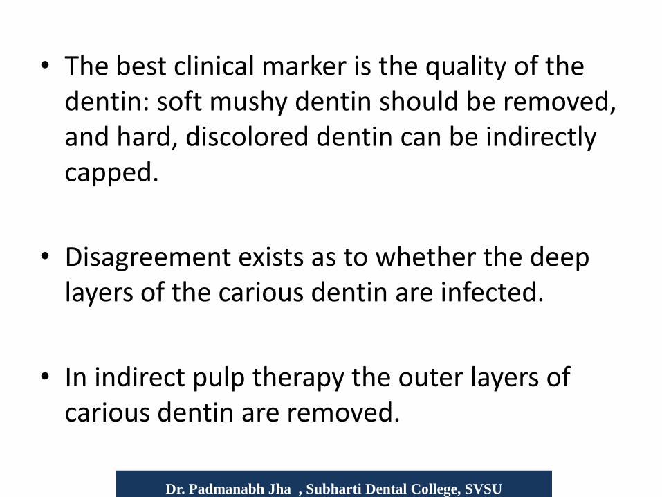
• The best clinical marker is the quality of the dentin: soft mushy dentin should be removed, and hard, discolored dentin can be indirectly capped.
• Disagreement exists as to whether the deep layers of the carious dentin are infected.
• In indirect pulp therapy the outer layers of carious dentin are removed.
Dr. Padmanabh Jha , Subharti Dental College, SVSU
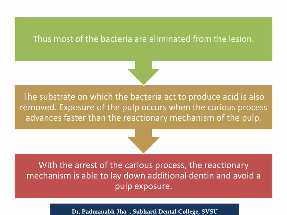
With the arrest of the carious process, the reactionary mechanism is able to lay down additional dentin and avoid a
pulp exposure.
The substrate on which the bacteria act to produce acid is also removed. Exposure of the pulp occurs when the carious process
advances faster than the reactionary mechanism of the pulp.
Thus most of the bacteria are eliminated from the lesion.
Dr. Padmanabh Jha , Subharti Dental College, SVSU

INDICATIONS History : absence of spontaneous pain
Mild discomfort from chemical & thermal stimuli.
Clinical examination: Large carious lesion
Absence of lymphadenopathy.
Normal adjacent gingiva.
Normal color of tooth.
Radiographic examination:
Large carious lesion in close proximity to pulp.
Normal PDL space.
No interradicular or periapical radiolucency.
Dr. Padmanabh Jha , Subharti Dental College, SVSU

CONTRAINDICATIONS History: Spontaneous pain.
Sharp, penetrating pain that persists after withdrawing stimulus.
Clinical examination: Excessive tooth mobility.
Tooth discoloration.
Nonresponsive to pulp testing techniques.
Large carious lesion with apparent pulp exposure.
Interrupted lamina dura.
Widened PDL space.
Radiolucency at the root apices or furcation areas.
Dr. Padmanabh Jha , Subharti Dental College, SVSU Dr. Padmanabh Jha , Subharti Dental College, SVSU

• Although carious dentin left in the tooth probably contains some bacteria, the number of organisms can be greatly diminished when this layer is covered with zinc oxide – eugenol (ZOE) or calcium hydroxide.
• Glass ionomer cements have been successfully used in indirect pulp therapy procedures in the last 2 decades, mainly because of their antimicrobial and remineralization effect on caries.
• Although calcium hydroxide and ZOE have traditionally been the materials placed over the remaining affected dentin, numerous reports of the use of acid etched and bonded composites have shown equally favourable results.
Dr. Padmanabh Jha , Subharti Dental College, SVSU

Two appointment technique
First appointment:
LA & isolation.
Establish cavity outline with high speed handpiece.
Remove soft, necrotic, infected dentin with large round bur in a slow speed handpiece.
Remove peripheral carious dentin with sharp spoon excavator.
Dr. Padmanabh Jha , Subharti Dental College, SVSU

Cover the remaining affected dentin with hard setting calcium hydroxide.
Fill the remainder of cavity with reinforced ZOE cement or GIC to achieve good seal.
Do not disturb this sealed cavityfor 6 - weeks. It may be necessary to use amalgam, composite or temporary crown as final restoration to maintain this seal.
Dr. Padmanabh Jha , Subharti Dental College, SVSU

Second appointment:
Bitewing radiograph assessed for the presence of reparative dentin.
LA & isolation.
Remove all temporary filling material, especially calcium hydroxide.
Remaining affected dentin should appear dehydrated and flaky and should be easily removed.
Dr. Padmanabh Jha , Subharti Dental College, SVSU

Dr. Padmanabh Jha , Subharti Dental College, SVSU
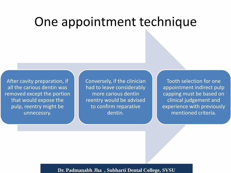
One appointment technique
After cavity preparation, if all the carious dentin was
removed except the portion that would expose the pulp, reentry might be
unnecessry.
Conversely, if the clinician had to leave considerably
more carious dentin reentry would be advised
to confirm reparative dentin.
Tooth selection for one appointment indirect pulp capping must be based on
clinical judgement and experience with previously
mentioned criteria.
Dr. Padmanabh Jha , Subharti Dental College, SVSU
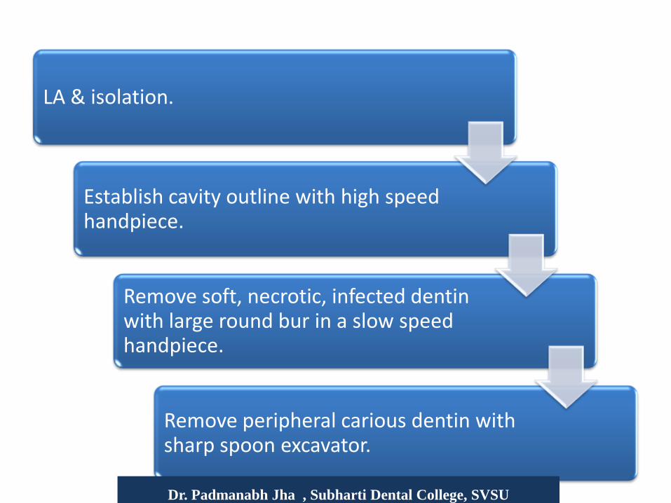
LA & isolation.
Establish cavity outline with high speed handpiece.
Remove soft, necrotic, infected dentin with large round bur in a slow speed handpiece.
Remove peripheral carious dentin with sharp spoon excavator.
Dr. Padmanabh Jha , Subharti Dental College, SVSU

Dr. Padmanabh Jha , Subharti Dental College, SVSU

DIRECT PULP CAPPING
Direct pulp capping involves the placement of a biocompatible agent on healthy pulp tissue that has been inadvertently exposed from caries excavation or traumatic
injury.
The treatment objective is to seal the pulp against bacterial leakage, encourage the pulp to wall off the exposure site by
initiating a dentin bridge, and maintain the vitality of the underlying pulp tissue regions.
Dr. Padmanabh Jha , Subharti Dental College, SVSU
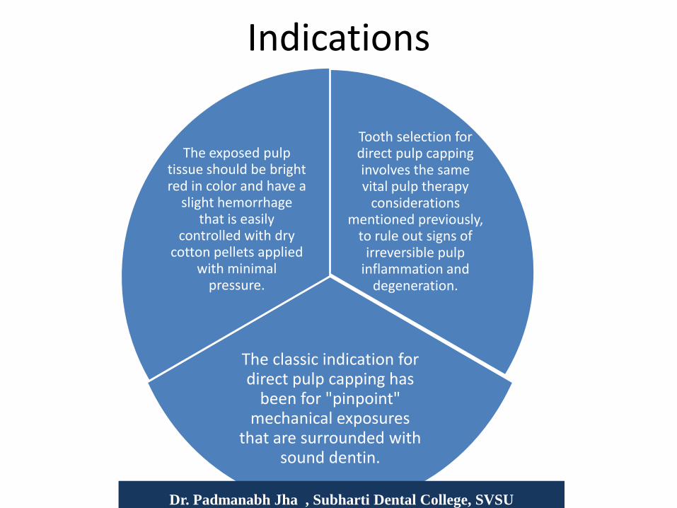
Indications
Tooth selection for direct pulp capping involves the same vital pulp therapy
considerations mentioned previously,
to rule out signs of irreversible pulp
inflammation and degeneration.
The classic indication for direct pulp capping has
been for "pinpoint" mechanical exposures
that are surrounded with sound dentin.
The exposed pulp tissue should be bright red in color and have a
slight hemorrhage that is easily
controlled with dry cotton pellets applied
with minimal pressure.
Dr. Padmanabh Jha , Subharti Dental College, SVSU

Contraindications
(1) spontaneous and nocturnal toothaches,
(2) excessive tooth mobility,
(3) thickening of the periodontal ligament,
(4) radiographic evidence of furcal or periradicular degeneration,
(5) uncontrollable hemorrhage at the time of exposure,
(6) purulent or serous exudate from the exposure.
Dr. Padmanabh Jha , Subharti Dental College, SVSU
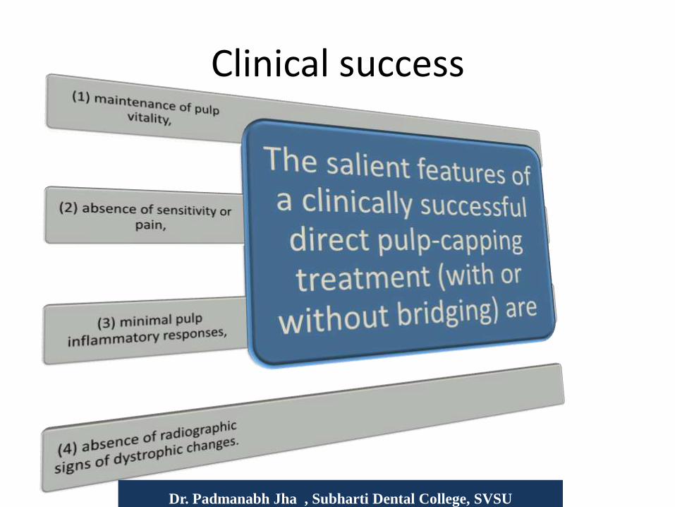
Clinical success
Dr. Padmanabh Jha , Subharti Dental College, SVSU
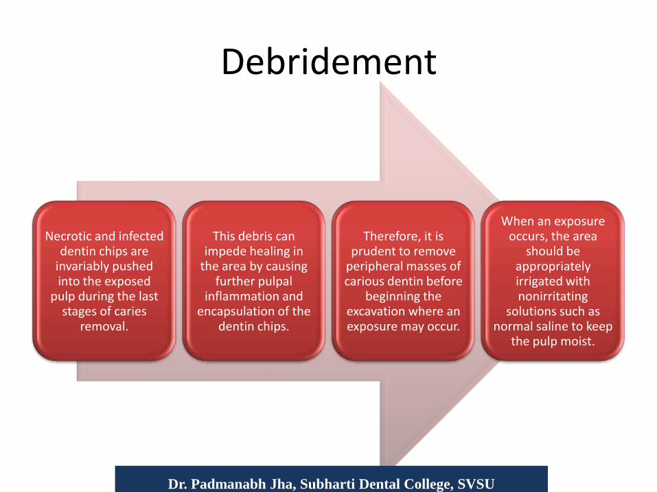
Debridement
Necrotic and infected dentin chips are
invariably pushed into the exposed
pulp during the last stages of caries
removal.
This debris can impede healing in
the area by causing further pulpal
inflammation and encapsulation of the
dentin chips.
Therefore, it is prudent to remove
peripheral masses of carious dentin before
beginning the excavation where an exposure may occur.
When an exposure occurs, the area
should be appropriately irrigated with nonirritating
solutions such as normal saline to keep
the pulp moist.
Dr. Padmanabh Jha, Subharti Dental College, SVSU

Hemorrhage and Clotting
Hemorrhage at the exposure site can be controlled with cotton pellet pressure.
Blood clot must not be allowed to form after the cessation of hemorrhage from the exposure site as it will impede pulpal healing.
The capping material must directly contact pulp tissue to exert a reparative dentin bridge response.
Hemolysis of erythrocytes results in an excess of hemosiderin and inflammatory cellular infiltrate, which prolongs pulpal healing.
Dr. Padmanabh Jha , Subharti Dental College, SVSU
Bacterial Contamination
• Bacterial microleakage under various restorations causes pulpal damage; in deep lesions, not the toxic properties of the cavity liners and/or restorative materials.
• The success of pulp-capping procedures is dependent on prevention of microleakage by an adequate seal.
Dr. Padmanabh Jha , Subharti Dental College, SVSU
Medications and Materials
• Many medicaments and materials have been suggested to cover pulp exposures and initiate tissue healing and/or hard structure repair.
• Calcium hydroxide, in one form or another, has been singled out as the medicament of choice for pulp exposures.
• Antibiotics, calcitonin, collagen, corticosteroids, cyanoacrylate, formocresol, and resorbable tricalcium phosphate cement have also been investigated, with varying degrees of success.
Dr. Padmanabh Jha , Subharti Dental College, SVSU

Calcium Hydroxide
• It produces coagulation necrosis at the contact surface of the pulp.
• The underlying tissue then differentiates into odontoblasts, which elaborate a matrix in about 4 weeks this results in the formation of a reparative dentin bridge, caused by the irritating quality of the highly alkaline calcium hydroxide, which has a pH of 11 to l2.
Dr. Padmanabh Jha , Subharti Dental College, SVSU

Calcium hydroxide has significant
antibacterial action, which has been identified as an
additional benefit in capping procedures.
There is some controversy as to
the source of calcium ions necessary for
dentinal bridge repair at the
exposure site.
Sciaky and Attalla demonstrated that calcium ions from
the capping material were not involved in
the bridge formation.
Stark and his colleagues,
however, believe that calcium ions from the capping medicament do
enter into bridge formation.
Dr. Padmanabh Jha , Subharti Dental College, SVSU

Dr. Padmanabh Jha , Subharti Dental College, SVSU

Alternative Agents to Calcium Hydroxide
Dr. Padmanabh Jha , Subharti Dental College, SVSU

https://www.youtube.com/watch?v=NIh9F4V1LQE
Dr. Padmanabh Jha , Subharti Dental College, SVSU