PulmonarySmoothMuscleinVertebrates:AComparativeReviewof ... - 2019... · often heterogeneous...
19
INVITED REVIEW Pulmonary Smooth Muscle in Vertebrates: A Comparative Review of Structure and Function Robert L. Cieri School of Biological Sciences, The University of Utah, 247 South 1400 East, 201 South Biology, Salt Lake City, UT 84112, USA 1 E-mail: [email protected] Synopsis Although the airways of vertebrates are diverse in shape, complexity, and function, they all contain visceral smooth muscle. The morphology, function, and innervation of this tissue in airways is reviewed in actinopterygians, lungfish, amphibians, non-avian reptiles, birds, and mammals. Smooth muscle was likely involved in tension regulation ancestrally, and may serve to assist lung emptying in fishes and aquatic amphibians, as well as maintain internal lung structure. In certain non-avian reptiles and anurans antagonistic smooth muscle fibers may contribute to intrapulmonary gas mixing. In mammals and birds, smooth muscle regulates airway caliber, and may be important in controlling the distribution of ventilation at rest and exercise, or during thermoregulatory and vocal hyperventilation. Airway smooth muscle is controlled by the autonomic nervous system: cranial cholinergic innervation generally causes excitation, cranial non-adrenergic, non-cholinergic innervation causes inhibition, and spinal adrenergic (SA) input causes species-specific, often heterogeneous contractions and relaxations. Introduction The airways and parenchymal tissues of vertebrate lungs are, nearly without exception, lined with smooth muscle controlled by the autonomic nervous system. The alteration and assignment of new func- tions for airway smooth muscle may have co- occurred with events in the evolution of the verte- brate lung, thus review and synthesis of what is known of this tissue should improve our knowledge of vertebrate pulmonary evolution (Fig. 1), perhaps shedding light on changes occurring when verte- brates left the water or developed new inspiratory and expiratory mechanisms. A comparative perspec- tive on airway smooth muscle may also benefit researchers working on the pathophysiology of this tissue in humans. Although there are several recent reviews of airways smooth muscle in humans (Stephens 2001; Canning 2006; Doeing and Solway 2013), most descriptions of pulmonary smooth mus- cle in other taxa are presented as parts of focused physiological investigations. A comprehensive review of the structure, function, and innervation patterns of airway smooth muscle in vertebrates has never been conducted and is presented here. This review briefly outlines and then covers in detail what is known of the morphology, function, and control mechanisms of smooth muscle tissues lining the air- ways in vertebrates. After a brief overview, this paper will review knowledge of pulmonary smooth muscle in different groups of extant vertebrates, summarize innervation and control patterns, and end with a brief conclusion. Brief overview of airway smooth muscle across vertebrates The ancestral vertebrate gas-containing organ likely originated as an outgrowth of the ventral pharynx (Liem 1988) which may have evolved to provide an additional oxygenation mechanism in hypoxic water conditions (Randall et al. 1981), to provide for buoyancy (Liem 1988) or to help oxygenate the heart (Farmer, 1997). Gas-containing derivatives of the gut tube are called lungs if they are paired and of ventral origin, and swim bladders if they are un- paired and of dorsal origin (Graham 1997). The evo- lution of lungs and swim bladders is summarized in Fig. 1. Among actinopterygians, the buoyancy role ß The Author(s) 2019. Published by Oxford University Press on behalf of the Society for Integrative and Comparative Biology. All rights reserved. For permissions please email: [email protected]. Integrative and Comparative Biology Integrative and Comparative Biology, pp. 1–19 doi:10.1093/icb/icz002 Society for Integrative and Comparative Biology Downloaded from https://academic.oup.com/icb/advance-article-abstract/doi/10.1093/icb/icz002/5298734 by SICB Member Access user on 08 May 2019
Transcript of PulmonarySmoothMuscleinVertebrates:AComparativeReviewof ... - 2019... · often heterogeneous...

INVITED REVIEW
Pulmonary Smooth Muscle in Vertebrates: A Comparative Review ofStructure and FunctionRobert L. Cieri
School of Biological Sciences, The University of Utah, 247 South 1400 East, 201 South Biology, Salt Lake City, UT 84112,
USA
1E-mail: [email protected]
Synopsis Although the airways of vertebrates are diverse in shape, complexity, and function, they all contain visceral
smooth muscle. The morphology, function, and innervation of this tissue in airways is reviewed in actinopterygians,
lungfish, amphibians, non-avian reptiles, birds, and mammals. Smooth muscle was likely involved in tension regulation
ancestrally, and may serve to assist lung emptying in fishes and aquatic amphibians, as well as maintain internal lung
structure. In certain non-avian reptiles and anurans antagonistic smooth muscle fibers may contribute to intrapulmonary
gas mixing. In mammals and birds, smooth muscle regulates airway caliber, and may be important in controlling the
distribution of ventilation at rest and exercise, or during thermoregulatory and vocal hyperventilation. Airway smooth
muscle is controlled by the autonomic nervous system: cranial cholinergic innervation generally causes excitation, cranial
non-adrenergic, non-cholinergic innervation causes inhibition, and spinal adrenergic (SA) input causes species-specific,
often heterogeneous contractions and relaxations.
Introduction
The airways and parenchymal tissues of vertebrate
lungs are, nearly without exception, lined with
smooth muscle controlled by the autonomic nervous
system. The alteration and assignment of new func-
tions for airway smooth muscle may have co-
occurred with events in the evolution of the verte-
brate lung, thus review and synthesis of what is
known of this tissue should improve our knowledge
of vertebrate pulmonary evolution (Fig. 1), perhaps
shedding light on changes occurring when verte-
brates left the water or developed new inspiratory
and expiratory mechanisms. A comparative perspec-
tive on airway smooth muscle may also benefit
researchers working on the pathophysiology of this
tissue in humans. Although there are several recent
reviews of airways smooth muscle in humans
(Stephens 2001; Canning 2006; Doeing and Solway
2013), most descriptions of pulmonary smooth mus-
cle in other taxa are presented as parts of focused
physiological investigations. A comprehensive review
of the structure, function, and innervation patterns
of airway smooth muscle in vertebrates has never
been conducted and is presented here. This review
briefly outlines and then covers in detail what is
known of the morphology, function, and control
mechanisms of smooth muscle tissues lining the air-
ways in vertebrates. After a brief overview, this paper
will review knowledge of pulmonary smooth muscle
in different groups of extant vertebrates, summarize
innervation and control patterns, and end with a
brief conclusion.
Brief overview of airway smooth muscleacross vertebrates
The ancestral vertebrate gas-containing organ likely
originated as an outgrowth of the ventral pharynx
(Liem 1988) which may have evolved to provide
an additional oxygenation mechanism in hypoxic
water conditions (Randall et al. 1981), to provide
for buoyancy (Liem 1988) or to help oxygenate the
heart (Farmer, 1997). Gas-containing derivatives of
the gut tube are called lungs if they are paired and of
ventral origin, and swim bladders if they are un-
paired and of dorsal origin (Graham 1997). The evo-
lution of lungs and swim bladders is summarized in
Fig. 1. Among actinopterygians, the buoyancy role
� The Author(s) 2019. Published by Oxford University Press on behalf of the Society for Integrative and Comparative Biology.
All rights reserved. For permissions please email: [email protected].
Integrative and Comparative BiologyIntegrative and Comparative Biology, pp. 1–19
doi:10.1093/icb/icz002 Society for Integrative and Comparative Biology
Dow
nloaded from https://academ
ic.oup.com/icb/advance-article-abstract/doi/10.1093/icb/icz002/5298734 by SIC
B Mem
ber Access user on 08 May 2019

dominates, although bichirs (Polypteriformes) have
lungs and the swim bladders of bowfins (Amia)
and gars (Lepisosteiformes) retain respiratory epithe-
lium (Graham 1997). Among teleosts, physoclists
have lost the connection between pharynx and
swim bladder while physostomes retain it.
All sarcopterygians have lungs, but only amniotes
have a trachea and intrapulmonary bronchi (Liem
1988; Duncker 2004). Tetrapod lungs are character-
ized by internal elaboration that increases surface
area. Lissamphibians and non-avian reptiles have
gas-exchanging pockets termed ediculi when they
form only one layer and faveoli when they form
multiple layers, and the pockets are rimmed by
thickened margins called trabeculae (Perry 1998).
The gas exchanging airways of birds are organized
into parallel tubes called parabronchi, and mammal
airways end in blind sacs called alveoli (Maina 2002).
Non-avian reptile lungs show varying amounts of
heterogeneity in the distribution of gas-exchanging
tissue, and birds have a system of compliant air
sacs that ventilate an isovolumetric gas-exchanging
region (Perry 1998).
Because the anatomy and function of the auto-
nomic nervous system differs across different verte-
brate groups, sympathetic and parasympathetic
divisions will be abandoned, and innervation will
be organized into cranial cholinergic (CC), cranial,
non-adrenergic non-cholinergic (NANC), and spinal
adrenergic (SA) nerve patterns. Investigations of au-
tonomic innervation and their findings are summa-
rized in Table 1. The function of autonomic
innervation on airway smooth muscle and variation
in autonomic nerve structure among vertebrates is
discussed in the following sections after a brief
outline.
Smooth muscle fibers regulate tone in blood vessels,
the gut tube, and urinary tubes (Tortora and Nielsen
2014), were likely the major regulatory component of
the earliest vertebrate gas organs. Because the lung is
an outgrowth of the pharynx (Hislop 2002), smooth
muscle almost certainly lined and regulated tension in
the ancestral vertebrate lung. Further evidence for an
ancestral smooth muscle layer in the lungs is provided
by the universal prevalence and pattern of SA innerva-
tion of smooth muscle in the lungs and swim bladders of
fishes. Smooth muscle plays a role in for expelling gases
or controlling the resorption or release of gases into
actinopterygian swim bladders.
Smooth muscle may be involved in deflation
among lungfishes, and aquatic amphibians. In lissam-
phibians and non-avian reptiles, smooth muscle prob-
ably functions largely to maintain internal lung
structures (Smith and Campbell 1976; Perry et al.
1989a). In non-avian reptiles, coordinated control of
antagonistic muscle fibers may generate important
intrapulmonary gas mixing through the bellowing ac-
tion of lung faveoli (Perry et al. 1989a; Daniels et al.
1994). Smooth muscle could also play a role in de-
flation among lungfishes, and aquatic amphibians.
Smooth muscle in birds and mammals is positioned
to regulate airway caliber but may also play important
roles in lung micromechanics. Parabronchial rigidity,
which enables birds to have a very thin blood–gas
barrier, may depend on the tension generated by
smooth muscle fibers guarding the respiratory atria
(Maina 2007), and sphincters guarding secondary
and parabronchi may allow air to bypass gas exchange
regions during vocal or thermoregulatory hyperventi-
lation (King and Cowie 1969), preventing excessive
water loss or respiratory alkalosis. In mammals,
changes in airway smooth muscle tone may directs
Fig. 1 Lung and swim bladder evolution in vertebrates. A dendrogram of major vertebrate groups with representative lung and swim
bladders superimposed. Drawings adapted from Duncker (1978), F€ange (1983), and Romer and Parsons (1977). The gas-exchange
tissue of non-avian reptiles and amphibians is organized into ediculi (single layer of chambers) or faveoli (multiple layers), which it forms
blind-end alveoli and tubular parabronchi in birds. See text for details. Cladogram topology from Kardong (2014).
2 R. L. Cieri
Dow
nloaded from https://academ
ic.oup.com/icb/advance-article-abstract/doi/10.1093/icb/icz002/5298734 by SIC
B Mem
ber Access user on 08 May 2019
air to reduce ventilation/perfusion inequality, protect
the lungs from inhalant particles, and regulate alveolar
volume at the expense of alveolar duct volume in
terminal airways (Greaves et al. 1986). Smooth muscle
development is extreme in marine mammals and may
be related to the biomechanics or physiology of diving
(Piscitelli et al. 2013).
Actinopterygii
Although the Actinopterygian swim bladder and
lungs of other vertebrates likely evolved from a
common ancestor with a primitive lung (Liem
1988), the diversification of the swim bladder in
fishes is independent of lung evolution in tetrapods.
Actinopterygian, physostomes maintain a connection
called the pneumatic duct between the swim bladder
and the foregut, whereas the swim bladder is ana-
tomically isolated in physoclists (Fig. 1; Brainerd
2015). Among most physostomes, smooth muscle
is present in part or all of the swim bladder wall
(Alexander 1966; Smith and Croll 2011) or pneu-
matic duct. Active swim bladder deflation, called
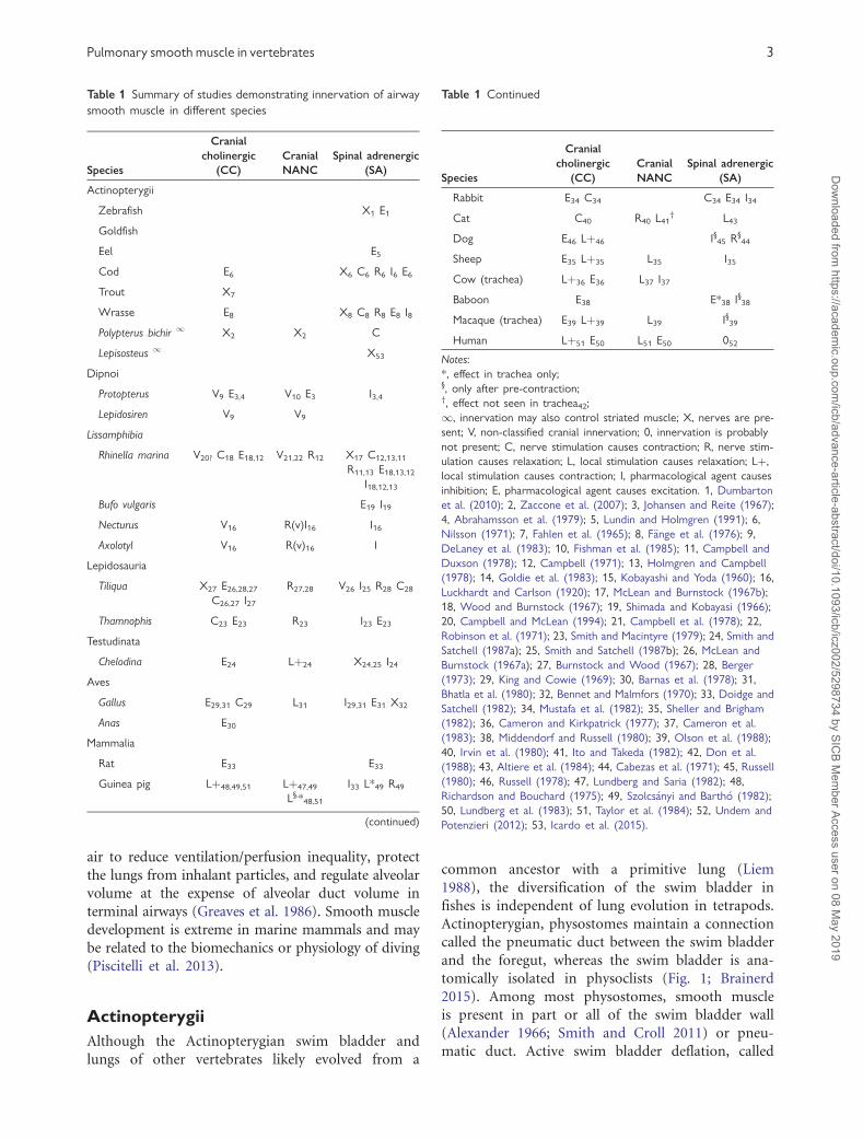
Table 1 Summary of studies demonstrating innervation of airway
smooth muscle in different species
Species
Cranial
cholinergic
(CC)
Cranial
NANC
Spinal adrenergic
(SA)
Actinopterygii
Zebrafish X1 E1
Goldfish
Eel E5
Cod E6 X6 C6 R6 I6 E6
Trout X7
Wrasse E8 X8 C8 R8 E8 I8
Polypterus bichir 1 X2 X2 C
Lepisosteus 1 X53
Dipnoi
Protopterus V9 E3,4 V10 E3 I3,4
Lepidosiren V9 V9
Lissamphibia
Rhinella marina V20? C18 E18,12 V21,22 R12 X17 C12,13,11
R11,13 E18,13,12
I18,12,13
Bufo vulgaris E19 I19
Necturus V16 R(v)I16 I16
Axolotyl V16 R(v)16 I
Lepidosauria
Tiliqua X27 E26,28,27
C26,27 I27
R27,28 V26 I25 R28 C28
Thamnophis C23 E23 R23 I23 E23
Testudinata
Chelodina E24 Lþ24 X24,25 I24
Aves
Gallus E29,31 C29 L31 I29,31 E31 X32
Anas E30
Mammalia
Rat E33 E33
Guinea pig Lþ48,49,51 Lþ47,49
L§,*48,51
I33 L*49 R49
(continued)
Table 1 Continued
Species
Cranial
cholinergic
(CC)
Cranial
NANC
Spinal adrenergic
(SA)
Rabbit E34 C34 C34 E34 I34
Cat C40 R40 L41† L43
Dog E46 Lþ46 I§45 R§44
Sheep E35 Lþ35 L35 I35
Cow (trachea) Lþ36 E36 L37 I37
Baboon E38 E*38 I§38
Macaque (trachea) E39 Lþ39 L39 I§39
Human Lþ51 E50 L51 E50 052
Notes:
*, effect in trachea only;§, only after pre-contraction;†, effect not seen in trachea42;
1, innervation may also control striated muscle; X, nerves are pre-
sent; V, non-classified cranial innervation; 0, innervation is probably
not present; C, nerve stimulation causes contraction; R, nerve stim-
ulation causes relaxation; L, local stimulation causes relaxation; Lþ,
local stimulation causes contraction; I, pharmacological agent causes
inhibition; E, pharmacological agent causes excitation. 1, Dumbarton
et al. (2010); 2, Zaccone et al. (2007); 3, Johansen and Reite (1967);
4, Abrahamsson et al. (1979); 5, Lundin and Holmgren (1991); 6,
Nilsson (1971); 7, Fahlen et al. (1965); 8, F€ange et al. (1976); 9,
DeLaney et al. (1983); 10, Fishman et al. (1985); 11, Campbell and
Duxson (1978); 12, Campbell (1971); 13, Holmgren and Campbell
(1978); 14, Goldie et al. (1983); 15, Kobayashi and Yoda (1960); 16,
Luckhardt and Carlson (1920); 17, McLean and Burnstock (1967b);
18, Wood and Burnstock (1967); 19, Shimada and Kobayasi (1966);
20, Campbell and McLean (1994); 21, Campbell et al. (1978); 22,
Robinson et al. (1971); 23, Smith and Macintyre (1979); 24, Smith and
Satchell (1987a); 25, Smith and Satchell (1987b); 26, McLean and
Burnstock (1967a); 27, Burnstock and Wood (1967); 28, Berger
(1973); 29, King and Cowie (1969); 30, Barnas et al. (1978); 31,
Bhatla et al. (1980); 32, Bennet and Malmfors (1970); 33, Doidge and
Satchell (1982); 34, Mustafa et al. (1982); 35, Sheller and Brigham
(1982); 36, Cameron and Kirkpatrick (1977); 37, Cameron et al.
(1983); 38, Middendorf and Russell (1980); 39, Olson et al. (1988);
40, Irvin et al. (1980); 41, Ito and Takeda (1982); 42, Don et al.
(1988); 43, Altiere et al. (1984); 44, Cabezas et al. (1971); 45, Russell
(1980); 46, Russell (1978); 47, Lundberg and Saria (1982); 48,
Richardson and Bouchard (1975); 49, Szolcs�anyi and Barth�o (1982);
50, Lundberg et al. (1983); 51, Taylor et al. (1984); 52, Undem and
Potenzieri (2012); 53, Icardo et al. (2015).
Pulmonary smooth muscle in vertebrates 3
Dow
nloaded from https://academ
ic.oup.com/icb/advance-article-abstract/doi/10.1093/icb/icz002/5298734 by SIC
B Mem
ber Access user on 08 May 2019

the “Gasspuckreflex” or gas-spitting reflex, often
involves contraction and relaxation of different
smooth muscle tissues (Smith and Croll 2011;
Zaccone et al. 2012a) and usually occurs before
deep sounding, although some authors think this
term should be reserved for gas loss due to decom-
pression (Harvey et al. 1968). Some physostomes can
also regulate swim bladder volume through secretion
and reabsorption of gases and this is the sole mech-
anism of swim bladder inflation or deflation in
physoclists (Smith and Croll 2011).
Smooth muscle is reported in tarpon (Megalops),
especially in the non-respiratory portion of the blad-
der (Seymour et al. 2008). The lungs of some trahi-
ras (Hoplerythrinus), obligate air-breathing
Amazonian physostomes, have two well-defined
layers of smooth muscle in the tunica media: a
thin longitudinal internal layer and a thicker circular
external layer, and a bundle of longitudinal smooth
muscle fibers running the length of the swim bladder
laterally (Cruz-Hofling et al. 1981). The lungs of
arowanas (Osteoglossum), closely-related facultative
air-breathers, are similar but lack the external longi-
tudinal bundle (Cruz-Hofling et al. 1981).
In general, deflation of the swim bladder is medi-
ated by output from the spinal autonomic nervous
system (Nilsson 1972, 2009; F€ange 1983), although
cranial autonomic innervation is also present
(Zaccone et al. 2012b). Smooth muscle anatomy and
innervation is comparatively well studied in the two-
chambered swim bladder of the zebrafish (Danio
rerio) (Fig. 1), which uses gas expulsion as the main
mechanism of deflation. Smooth muscle in the zebra-
fish swim bladder consists of a sphincter between the
bladder and esophagus and thick bands along the ven-
tral surface of the anterior and along the sides of the
posterior swim bladder chambers (which connects to
the pneumatic duct) (Finney et al. 2006), such that
relaxation of some muscle and contraction of others is
necessary to raise the swim bladder pressure about
8 mm Hg higher than ambient an cause deflation
(Robertson et al. 2008). The anterior chamber pos-
sesses a series of connective tissue folds which amplify
the effect of smooth muscle contraction enabling 85%
reductions in swim bladder volume in in situ studies
(Dumbarton et al. 2010). In vivo pressure recordings
and observation of in vivo and excised muscle con-
traction from noradrenaline stimulation (Dumbarton
et al. 2010) show that the circumferential fibers in the
anterior chamber contract after stimulation of b-adre-
nergic receptors to expel gas from the pneumatic duct
(Dumbarton et al. 2010). Differences in adrenergic
receptor type may make fine spinal autonomic control
possible: Finney et al. (2006) detected choline
acetyltransferase-immunoreactive somata only in the
muscles of the esophagus, but tyrosine hydroxylase
(HT) immunoreactive fibers were detected through-
out the swim bladder itself. Furthermore, in the gold-
fish (Carassius auratus), HT and substance P
immunoreactive fibers have been isolated by immu-
nohistochemical methods in the craniodorsal part of
the swim bladder, whereas the gas gland and pneu-
matic cells are reactive to vasointestinal peptide (VIP)
(Zaccone et al. 2012b).
Even though direct expulsion of gas is not possi-
ble in physoclists, smooth muscle contraction and
relaxation still regulates the amount of air in their
swim bladders. Often, the absorptive epithelium is
isolated from the main gas chamber by an oval con-
striction, commonly called a diaphragm. In toadfish,
cod, and perch (F€ange 1983), the caliber of this oval
constriction is determined by antagonistic circum-
ferential and radial smooth muscle fibers. A func-
tionally homologous structure exists in the wrasse
(Ctenolabrus) (Smith and Croll 2011). Autonomic
regulation of these structures is adrenergic, with a-
receptors regulating the opening radial muscles and
b-receptors regulating the closure circumferential
muscles (Nilsson 2009). Swim bladder smooth mus-
cle may have other functions other than expelling
gases: early reports on sea horse physiology reported
changes in swim bladder geometry with body posi-
tion (Peters 1951) which may be mediated by
smooth muscle.
Many actinopterygian fishes have striated muscle
in their lungs or swim bladders. Striated muscle is
found in the lungs of bichirs (Brainerd 1994a), and
the swim bladders of Lepisosteus, Amia (Notopterus),
and trahiras (Erythrinus) (Company and Rahn 1971;
Crawford 1971; Liem 1989). There is no functional
evidence that muscle helps to deflate the swim blad-
der Lepisosteus, but the striated muscle is positioned
to collapse the bladder and is innervated by SA
nerves. It is also surrounded by neuroepithelial bod-
ies containing VIP that are positive to serotonin
(Zaccone et al. 2012a; Icardo et al. 2015), consistent
with SA control. The original addition of striated
muscles in fish gas organs may have served to in-
crease the speed or force of exhalation, provide ac-
tive control of the gas organ for hydrostatic
purposes, or to decouple ventilation from hormonal
control. Other expiratory mechanisms are noted in
physostomes: striated muscle is implicated in exha-
lation in Notopterus, Lepisosteus, and Polypterus, and
Arapaima probably powers exhalation through buc-
cal expansion (Greenwood and Liem 1984).
4 R. L. Cieri
Dow
nloaded from https://academ
ic.oup.com/icb/advance-article-abstract/doi/10.1093/icb/icz002/5298734 by SIC
B Mem
ber Access user on 08 May 2019

Lungfish (Dipnoi)
Smooth muscle has been reported in all three fami-
lies of lungfish. In the Australian lungfish
(Neoceratodus), Grigg (1965) reported smooth mus-
cle in the transverse (primary) and reticulated (sec-
ondary) septa of the unpaired lung. In both septa, a
layer of smooth muscle is sandwiched between layers
of connective tissue (Grigg 1965). This smooth mus-
cle takes the form of thickened pillars in the dorso-
ventrally oriented transverse septa, and forms
thickened bands at the edges of the secondary septa.
Smooth muscle structure similar to that of
Neoceratodus has also been described in the African
lungfish (Protopterus), the African lungfish (Klika
1967; Maina and Maloiy 1985), and sections of the
paired lungs of the south American lungfish
(Lepidosiren) stain positive for smooth muscle
(Bishop and Foxon 1968).
Sarcopterygian fishes primarily use a two-stroke
buccal pump, with inspiration powered by buccal
floor muscles (Brainerd 1994b). Based on its ana-
tomical position, Grigg (1965) concluded that
smooth muscle, along with elastic tissues and hydro-
static pressure, was responsible for exhalation in
Neoceratodus. Lung smooth muscle contraction likely
maintains intrapulmonary pressure above atmo-
spheric through the lung emptying phase (Johansen
et al. 1967). Based on Grigg’s work and radiological
recordings, Bishop and Foxon (1968) hypothesized a
similar role for smooth muscle in Lepidosiren.
Studies of ventilation in Protopterus hypothesized
that lung emptying was passive (McMahon 1969)
however the smooth muscle has been shown to con-
tract dramatically with acetylcholine (Johansen and
Reite 1967), leading Johansen and Reite (1967) to
hypothesize an expiratory function for smooth mus-
cle in this taxon. Brainerd et al. (1993) found no
increase in pleuroperitoneal pressure during exhala-
tion in Protopterus suggesting that hypaxial muscle
contraction has no role in lung emptying, but pul-
monary smooth muscle lies inside the lung could
assist lung emptying while increasing only intrapul-
monary and not pleuroperitoneal pressure. Lung
emptying is correlated with electromyographic
recordings from the abdominal muscles, however,
in aestivating Protopterus aethiopicus on land where
lung pressure increases before exhalation (DeLaney
and Fishman 1977). DeLaney and Fishman (1977)
hypothesize that the abdominal contractions serve
to replace hydrostatic pressure which normally assists
lung emptying.
Lissamphibia
Smooth muscle is present in the middle layer of
lissamphibian lungs, between the inner and outer
epithelia. It is usually thin, consisting of only occa-
sional cells in European tree frogs (Hyla arborea) but
is very thick in Amphiuma, where it can be 50–
180mm in the lung wall and 1000mm in the septa
(Goniakowska-Witalifiska 1995). The lungs of cau-
dates usually contain a great deal of smooth muscle
(Czopek 1962) and have variable amounts of paren-
chymal elaboration. Cryptobranchids have large,
poorly vascularized lungs with very little smooth
muscle and do most gas exchange across the skin
(Guimond and Hutchison 1976). Hydrostatic pres-
sure and active contraction of the transversus
abdominus musculature contribute to exhalation in
this group (Brainerd 1999). In contrast, the aquatic
salamanders of Sirenoidea rely on the lungs for gas
exchange which have large amounts of smooth mus-
cle (Czopek 1962). In Amphiuma tridactylum,
smooth muscle is organized in the walls of primary
septa, and in thick rings at the openings to second-
ary septa in the anterior lung, and in the wall of the
lung in the middle and especially posterior sections
(Stark-Vancs et al. 1984). Contraction of these
muscles contracts the primary and secondary septa,
pulling the lung walls together reducing the sizes of
the septal openings. Treatment with acetylcholine
causes lung collapse, especially in the posterior
lung (Stark-Vancs et al. 1984; Fig. 2A).
In both genera of aquatic salamanders, Siren and
Amphiuma, radiographic studies (Guimond and
Hutchison 1974; Martin and Hutchison 1979) reveal
that the lung fully collapses during deflation.
Deflation and re-inflation occur evenly with the pos-
terior section collapsing first and re-inflating last, sug-
gesting that the smooth muscle in the posterior lung
wall is active in exhalation (Martin and Hutchison
1979). The transversus abdominus is active during
the second half of deflation, contributing to expira-
tion (Brainerd and Dumka 1995; Brainerd and
Monroy 1998; Brainerd 1999). Hydrostatic pressure
may also contribute to deflation (Guimond and
Hutchison 1973; Martin and Hutchison 1979). In
contrast, Simons et al. (2000) argue that smooth mus-
cle probably does not play a major role in exhalation
in adult tiger salamanders (Ambystoma tigrinum) be-
cause exhalation is much faster than in Amphiuma.
Caecilians have smooth muscle in the external lung
wall and in the septal walls (Maina and Maloiy 1988;
Kuehne and Junqueira 2000). Smooth muscle may be
important for exhalation in the two central American
Pulmonary smooth muscle in vertebrates 5
Dow
nloaded from https://academ
ic.oup.com/icb/advance-article-abstract/doi/10.1093/icb/icz002/5298734 by SIC
B Mem
ber Access user on 08 May 2019

species: Dermophis mexicanus (Carrier and Wake
1995; Bennett et al. 1999) and Typhlonectes natans,
where pressure in the pseudo-trachea exceeds ambient
pressure during exhalation (Prabha et al. 2000).
In frogs (Anurans), the degree of lung septation
varies. Primary, secondary, and tertiary septa
invested with smooth muscle divide the lungs of
the cane toad (Rhinella marina) into nested parti-
tions along the lung wall (Smith and Campbell
1976). Smooth muscle forms thick bundles at the
open edge of each order of partition, smaller strips
that run around the partitions at regular intervals,
occasional longitudinal bundles that run perpendic-
ular to the other strips and is scattered throughout
the lung wall (Smith and Campbell 1976). Smith and
Campbell (1976) hypothesize that smooth muscle
contraction in the lung of R. marina functions to
maintain the internal lung structure by closing the
trabeculae (Fig. 3A), pulling the septa internally away
from the lung wall, which is rigid due to intrapul-
monary pressure. In contrast, Lawry (1999) hypoth-
esized that contraction of septal smooth muscles
would serve to collapse the septa, which are held
rigid by hydrostatic pressure in blood vessels.
Under this model, active pumping of pulmonary
air could be accomplished by varying pulmonary
blood pressure and visceral smooth muscle tone in
the pulmonary septa (Lawry 1999).
Control of smooth muscle is best studied in R.
marina, where cranial and sacral pathways join to-
gether into a vagosympathetic trunk that innervates
the lung (Campbell et al. 1978). CC cause contrac-
tion of visceral lung muscle, and cranial NANC
fibers cause smooth lung relaxation, using either
NO, VIP, or ATP as a transmitter (Campbell and
McLean 1994). SA postganglionic nerves, also run-
ning through the vagosympathetic trunk, cause con-
traction of septal edge musculature and relaxation of
smooth muscles in the lung wall (Holmgren and
Campbell 1978). An immunohistochemical study
of nerve fibers before and after vagotomy in the
same species reported 50% NANC, 25% cholinergic,
and 25% adrenergic fibers (Campbell et al. 1978)
with only the NANC fibers persisting after dener-
vation, indicating that their cell bodies are intra-
pulmonary. The vagosympathetic trunk reaches
muscle bundles through small myelinated and
non-myelinated fibers at the septal margins.
Distally, each muscle cell is innervated by two pairs
of axons, one cholinergic and another adrenergic
(Campbell et al. 1978).
Early experiments on mudpuppies (Necturus)
found a marked inhibition of lung tone from the
infusion of adrenaline and pituitrin (5-HT and
vasopressin) (Luckhardt and Carlson 1920), consis-
tent with SA and NANC inhibitory innervation.
Further experiments further point to cranial inhibi-
tory innervation: vagotomy also causes contraction
of the lung in Necturus, which can be temporarily
reversed by stimulation of the severed vagal nerve
(Luckhardt and Carlson 1920). Vagotomy also causes
lung contraction in the axolotl (Ambystoma mexica-
num) and is inhibited by adrenaline (Luckhardt and
Carlson 1920).
Although hypaxial exhalation is probably a synap-
omorphy of living tetrapods, lissamphibians display
a diversity of pulmonary smooth muscle functions
including exhalation in some Caudata and
Gymnophiona (caecilians), and maintenance of in-
ternal lung structure in Anura. It is possible that
contraction of the transversus abdominus muscle
first acted in concert with smooth muscle contrac-
tion to effect lung emptying, eventually becoming
the dominant exhalation mechanism in amniotes.
In this case, perhaps smooth muscle and hypaxial
musculature contribute differentially to exhalation
depending on habitat and lung morphology in
amphibians. The lengthy lungs of Siren may require
smooth muscle contraction to empty completely,
and perhaps smooth muscle contraction is more im-
portant for effective lung emptying in animals with-
out well-developed ribs.
Non-avian reptiles
In general, the gas-exchanging parenchyma of reptile
lungs consists of distal chambers, termed ediculae or
faveoli, separated by first, second, and third order
septa and rimmed by thick openings called trabecu-
lae (Perry 1998) (Fig. 3). Thick smooth muscle bun-
dles or myoelastic bundles are always found in the
trabeculae (Fig. 3A, C). Modulation of smooth mus-
cle contraction has been hypothesized to maintain
internal lung structure at various degrees of inflation
(Perry and Duncker 1978), or even contribute to
intrapulmonary gas mixing (Daniels et al. 1994).
Smooth muscle has been found in the trabecular
rings of red-eared slider turtles (Trachemys scripta)
(Perry 1978), loggerhead turtles (Caretta caretta)
(Perry et al. 1989b), Australian snake-necked tor-
toises (Chelodina longicollis) (Smith and Satchell
1987a), tegus (Tupinambis teguixin) (Perry et al.
1989a), skinks (Tiliqua rugosa) (Burnstock and
Wood 1967; McLean and Burnstock 1967a), Nile
crocodiles (Crocodylus niloticus) (Perry 1988), geckos
(Gekko gecko) (Perry and Duncker 1978). In C. long-
icollis, fewer smooth muscle fibers were observed in
the thin-walled posterior lung (Smith and Satchell
6 R. L. Cieri
Dow
nloaded from https://academ
ic.oup.com/icb/advance-article-abstract/doi/10.1093/icb/icz002/5298734 by SIC
B Mem
ber Access user on 08 May 2019

1987a), compared with the anterior lung, where the
parenchyma is thicker.
In addition, many non-avian reptiles display an-
tagonistic smooth muscle groups in the walls of
ediculae or faveoli (Fig. 3D, G), with the trabecular
rim muscles positioned to hold the internal lung
structure rigid and open, and perpendicular muscles
in the walls of the trabecular septa positioned to
collapse the septa (Perry 1988). Such muscular ar-
rangement has been documented in T. teguixin
(Perry et al. 1989a), C. niloticus (Perry 1988), the
bull snake (Pituophis sayi) (Wallach 1998), and gar-
ter snake (Thamnophis sirtalis) (Pohunkov�a and
Hughes 1985). In the tegu, septal wall fibers run
parallel (Fig. 3D) to the septal edge, but perpendic-
ular (Fig. 3G) in the crocodile. Septal smooth mus-
culature is observed in the black mamba snake
(Dendroaspis polylepis) (Maina 1989), which proba-
bly also has trabecular rim smooth muscle. In con-
trast, no smooth muscle is found in the septal walls
of the unicameral lungs of T. rugosa (Burnstock and
Wood 1967). In species with antagonistic muscle
groups, the ratio of trabecular to septal smooth mus-
cle seems to increase with body size (Perry 1998),
perhaps indicating that relatively greater force is re-
quired to distort larger trabeculae.
Fig. 2 Smooth muscle and lung contraction in selected species. A) The effect of acetylcholine-induced contraction on the lung of
Amphiuma tridactylum. Sections of an inflated lung before (top) and after application of acetylcholine (below) show contraction of the
lung surface and septum. From (Stark-Vancs et al. 1984). B) Lung inflation (left) and C) deflation (right) are accompanied by collapse of
the interedicular septa (E) in central netted dragons (Ctenophorus nuchalis) from Daniels et al. (1994). D) Parabronchial cross-sectional
area before and after (E) spontaneous smooth muscle contraction in goose (Anser anser) lungs. From Barnas et al. (1978).
Pulmonary smooth muscle in vertebrates 7
Dow
nloaded from https://academ
ic.oup.com/icb/advance-article-abstract/doi/10.1093/icb/icz002/5298734 by SIC
B Mem
ber Access user on 08 May 2019
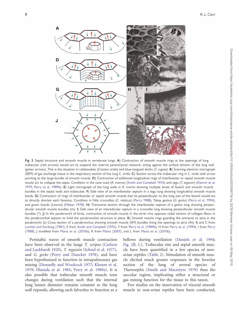
Peristaltic waves of smooth muscle contraction
have been observed in the lungs T. scripta (Carlson
and Luckhardt 1920), T. teguixin (Scheid et al. 1977),
and G. gecko (Perry and Duncker 1978), and have
been hypothesized to function in intrapulmonary gas
mixing (Donnelly and Woolcock 1977; Klemm et al.
1979; Hlastala et al. 1985; Perry et al. 1989a). It is
also possible that trabecular smooth muscle tone
changes during ventilation such that the internal
lung lumen diameter remains constant as the lung
wall expands, allowing each falveolus to function as a
bellows during ventilation (Daniels et al. 1994;
Fig. 2B, C). Trabecular rim and septal smooth mus-
cle have been quantified in a few species of non-
avian reptiles (Table 2). Stimulation of smooth mus-
cle elicited much greater responses in the faveolar
section of the lung of several species of
Thamnophis (Smith and Macintyre 1979) than the
saccular region, implicating either a structural or
gas mixing function for the tissue in this taxon.
Few studies on the innervation of visceral smooth
muscle in non-avian reptiles have been conducted,
Fig. 3 Septal structure and smooth muscle in vertebrate lungs. A) Contraction of smooth muscle rings at the openings of lung
trabecular (red arrows) would act to suspend the internal parenchymal network, acting against the surface tension of the lung wall
(green arrows). This is the situation in rattlesnakes (Crotalus viridis) and blue-tongued skinks (T. rugosa). B) Scanning electron micrograph
(SEM) of gas exchange tissue in the respiratory section of the lung C. viridis. C) Section across the trabecular ring in C. viridis with arrow
pointing to the large bundle of smooth muscle. D) Contraction of additional longitudinal rings of interfaveolar or septal smooth muscle
would act to collapse the septa. Condition in the cane toad (R. marina) (Smith and Campbell 1976) and tegu (T. teguixin) (Klemm et al.
1979; Perry et al. 1989b). E) Light micrograph of the lung walls in R. marina showing multiple levels of faveoli and smooth muscle
bundles in the septal walls and trabeculae. F) Side view of an interfaveolar septum in a tegu lung showing longitudinal smooth muscle
bands. G) Contraction of rings of interfaveolar or septal smooth muscle that lie perpendicular to the long axis of the faveoli would act
to directly shorten each faveolus. Condition in Nile crocodiles (C. niloticus) (Perry 1988), Tokay geckos (G. gecko) (Perry et al. 1994),
and green lizards (Lacerta) (Meban 1978). H) Transverse section through the interfaveolar septum of a gecko lung showing perpen-
dicular smooth muscle bundles (m). I) Side view of an interedicular septum in a crocodile lung showing perpendicular smooth muscle
bundles (*). J) In the parabronchi of birds, contraction of smooth muscle in the atrial rims opposes radial tension of collagen fibers in
the parabronchial septum to hold the parabronchial structure in place. K) Smooth muscle rings guarding the entrance to atria in the
parabronchi. L) Cross section of a parabronchus showing smooth muscle (SM) bundles lining the openings to atria (At). B and C from
Luchtel and Kardong (1981), E from Smith and Campbell (1976), F from Perry et al. (1989b), H from Perry et al. (1994), I from Perry
(1988), J modified from Maina et al. (2010b), K from Maina (2007), and L from Maina et al. (2010b).
8 R. L. Cieri
Dow
nloaded from https://academ
ic.oup.com/icb/advance-article-abstract/doi/10.1093/icb/icz002/5298734 by SIC
B Mem
ber Access user on 08 May 2019

but results are consistent. In general, CC innervation
is excitatory, cranial NANC innervation is inhibitory,
while adrenergic a-receptors mediate contraction and
b-receptors mediate inhibition (Berger 1973; Berger
and Burnstock 1979). Typical responses are seen in a
study of isolated lung strips from C. longicollis, where
transmural stimulation or direct application of ace-
tylcholine caused a rapid contraction of lung tissue,
which was abolished by hyoscine (a muscarinic re-
ceptor antagonist), demonstrating cholinergic excit-
atory input mediated through muscarinic receptors
(Smith and Satchell 1987b). Contraction caused by
transmural stimulation persisted despite the presence
of hyosine, implicating excitatory NANC or SA fibers
in addition to the CC pathway (Smith and Satchell
1987a), while noradrenaline caused a b-receptor-me-
diated relaxation blocked by propranolol (Smith and
Satchell 1987b). In whole-lung preparations of
Thamnophis, vagal nerve stimulation at high fre-
quency (20 Hz) caused contraction followed relaxa-
tions, while low frequency stimulation (2 Hz) caused
relaxation only (Smith and Macintyre 1979).
Atropine or hyoscine abolished the contraction, but
not the relaxation, pointing to CC-mediated excita-
tion and cranial NANC-mediated inhibition of air-
way smooth muscle in this group. Furthermore, ACh
administration caused lung contraction that was pre-
vented by atropine or hyoscine (Smith and
Macintyre 1979). Although stimulation of the spinal
efferent did not cause lung contraction, direct appli-
cation of noradrenaline causes contraction followed
by relaxation. The contraction and relaxation are
blocked by a- and b-blockers and agonists, respec-
tively (Smith and Macintyre 1979).
The rate of periodic smooth muscle contractions
increases with asphyxia in turtles (Carlson and
Luckhardt 1920) and increased CO2 concentration
while contractions are abolished in the absence of
CO2 (Scheid et al. 1977), supporting a gas mixing
role for the tissue in non-avian reptiles.
Intrapulmonary CO2 receptors may influence smooth
muscle: they have been documented throughout the
lungs of T. teguixin, can respond at 1.3 Hz to CO2,
and the relationship between CO2 and contraction is
maintained after vagotomy (Scheid et al. 1977).
Smooth muscle contraction also generates lung mech-
anoreceptor output (Scheid et al. 1977). Integration
and control may occur in intrapulmonary ganglia,
identified in the septal walls in C. longicollis (Smith
and Satchell 1987a), the base of the lung of pond
sliders (Chrysemys picta), T. rugosa, Thamnophis
(Jones 1912; Burnstock and Wood 1967; McLean
and Burnstock 1967b).
In the simple lung of T. rugosa, which has only one
layer of smooth muscle, the response to vagal stimu-
lation depends on smooth muscle tone: at high tone
vagal stimulation generates a small contraction and
large inhibition, whereas a large contraction and
smaller inhibition are generated at low tone
(Burnstock and Wood 1967). In this species, SA effer-
ents join the vagus nerve, similar to the condition in
R. marina. During sustained stimulation, the muscles
started a slow relaxation after about 30 s (Burnstock
and Wood 1967), indicating that some level of intrin-
sic control is present. a-receptors mediate excitatory
SA innervation and b-receptors mediate inhibitory
innervation (Berger 1973). Compared with R. marina,
immunohistochemical staining techniques indicate
that SA fibers innervate less fibers more densely, al-
though large spatial variation is seen in both species
(McLean and Burnstock 1967a, 1967b).
Aves
Smooth muscle is present throughout the avian lung
and forms a uniform sheet lining the primary bron-
chus (King and Cowie 1969; Cook and King 1970).
A network of spiral fiber bundles also encase the
tertiary bronchi (parabronchi), and secondary bron-
chi, where they are thicker (King and Cowie 1969).
Massive muscular sphincters surround the openings
to the parabronchi and atria, which are in turn lined
with more irregular smooth muscle bundles (King
and Cowie 1969; Maina et al. 2010a). Muscle fibers
in the primary bronchus run parallel to each other
Table 2 Quantitative data on smooth muscle in non-avian reptiles
Species
Lung tissue
volumem L/kg
body mass % of
total lung volume
Trabecular smooth
musclem L/kg
body mass % of
lung tissue
Septal smooth
musclem L/kg
body mass % of
lung tissue
Trabecular/
septal SM
Varanus exanthematicus1 9.12 (9%) 0.71 (8%) 0.09 (1%) 7.9 (89%)
Tupinambus teguixin (nigropunctatus) 3.39 (14.6%) 0.25 (7.3%) 0.25 (7.3%) 1.0 (50%)
Gekko gekko2 2.3 (19%) 0.4 (17%) 0.05 (2%) 8.0 (89%)
Crocodylus niloticus3 4.14 (11%) 0.3 (7%) 0.18 (4%) 1.7 (63%)
Source: Data from 1, Perry (1998); 2, Perry (1983); 3, Perry et al. (1994).
Pulmonary smooth muscle in vertebrates 9
Dow
nloaded from https://academ
ic.oup.com/icb/advance-article-abstract/doi/10.1093/icb/icz002/5298734 by SIC
B Mem
ber Access user on 08 May 2019
and the long axis of the bronchus and muscle fibers
and nerves are more densely packed than in the
parabronchi (Cook and King 1970).
Although the innervation of airway smooth mus-
cle in birds remains poorly studied, a few investiga-
tions yield general trends. King and Cowie (1969)
demonstrated CC contractions from vagal stimula-
tion that can be blocked with atropine and cause
reductions in atrial calibers, and often complete
atrial collapse. Application of catecholamines and
adrenaline cause local airway dilation and constric-
tion, respectively (King and Cowie 1969), although
simple application of atropine is not enough to cause
bronchodilation. The resting tone in bird bronchi
seems therefore to be intrinsic and not dependent
on CC input. Immunohistochemical investigations
found few SA nerves in the trachea, many in the
bronchi (most densely in the primary bronchus),
and only occasional nerves in the air sacs (Bennet
and Malmfors 1970; Bennett 1971). Local electrical
stimulation causes relaxation in chickens (Gallus gal-
lus), or contraction after the tissue is already relaxed
with adrenergic drugs (Bhatla et al. 1980). The in-
hibitory response to electrical stimulation is unaf-
fected by b-blockers (Bhatla et al. 1980), suggesting
a cranial NANC inhibitory innervation.
King and Cowie (1969) hypothesized that control
of smooth muscle in birds could cause intrapulmo-
nary gas mixing or prevent a respiratory alkalosis
during thermoregulatory hyperventilation by shunt-
ing lung gases away from the parabronchi, but
Barnas et al. (1978) found no consistent response
of smooth muscle tone or bronchial caliber from
changes in CO2 or O2 concentration. Smooth muscle
responses may have been blocked in this study, how-
ever, by the use of sodium pentobarbital for anes-
thetic, a known smooth muscle depressant (Altura
and Altura 1975). ACh causes strong rhythmic con-
tractions in the bronchi of G. gallus (King and Cowie
1969) that could facilitate pulmonary gas flow if they
occur naturally.
In geese anesthetized with sodium pentobarbitol,
Barnas et al. (1978) observed spontaneous increases
in airway resistance due to contraction of the
smooth muscle at the openings of parabronchi
(Fig. 2D, E). Similar contractions could be elicited
from mechanical or vagal stimulation (Barnas et al.
1978). Although the contractions caused by vagal
stimulation were abolished by atropine,
spontaneously-occurring contractions or contrac-
tions caused by mechanical stimulation could only
be blocked with a drug cocktail (Barnas et al. 1978),
suggesting that local control of airway smooth mus-
cle probably contributes to spontaneous contraction.
Smooth muscle contraction may be critical for
structural strength and maintenance of rigidity in
the avian lung (Fig. 3J). In the tension-integrity (ten-
segrity) model, the inward tensing force provided by
the atrial smooth muscle delimiting the lumen of the
parabronchi acts against outward tension forces pro-
vided by elastic and collagen fibers rendering the
entire parabronchial structure adaptively rigid
(Maina 2007; Maina et al. 2010b). Contraction of
parabronchial smooth muscle has also been hypoth-
esized to collapse parabronchi during diving in pen-
guins, preventing barotrauma (Ponganis et al. 2015).
Finally, bronchial smooth muscle may also con-
tribute to pulmonary fluid mechanics in the bird
lung. Inspiratory aerodynamic valving in the primary
bronchus, thought to be a major determinant of uni-
directional airflow (Banzett et al. 1987), is contingent
on airway caliber in the segmentum accelerans and
may therefore be regulated by contraction of smooth
muscle (Wang et al. 1988) in this region. The seg-
mentum accelerans dilates with elevated CO2 levels
(Wang et al. 1992), suggesting active control by
smooth muscle.
Mammalia
In mammals, bronchial smooth muscle develops
from the wrapping of mesenchyme around the grow-
ing ectoderm airways and is present in all of the
adult conducting airways. It is not present in the
gas-exchanging alveoli, but small smooth muscle
fibers may penetrate as far as the free edge of alve-
olar walls toward the alveolar duct (Weibel 1984). In
healthy human lungs, much more smooth muscle
relative to airway diameter is present in the terminal
airways [smooth muscle layer: bronchi diameter ra-
tio is 0.0049 in segmental bronchi, 0.0084 in bron-
chobronchiolar border, 0.0118 in the membranous
bronchioles, and 0.0215 in the terminal bronchioles
(Ebina et al. 1990)], implying a much greater capac-
ity to generate internal pressure in the terminal air-
ways because luminal airway pressure is proportional
to wall tension over airway radius. This allometry
may indicate the importance of distal bronchial
constriction.
One function of terminal bronchial smooth mus-
cle may be to regulate alveolar expansion. An in-
crease in smooth muscle tension at the level of the
alveolar duct will constrict the alveolar entrance
rings, increasing septal surface area and alveolar vol-
ume at the expense of alveolar duct volume (Greaves
et al. 1986). Increased surface tension will counteract
the effects of smooth muscle such that contraction of
smooth muscle in the alveolar ducts and alveolar
10 R. L. Cieri
Dow
nloaded from https://academ
ic.oup.com/icb/advance-article-abstract/doi/10.1093/icb/icz002/5298734 by SIC
B Mem
ber Access user on 08 May 2019

surface tension may thus interact to regulate alveolar
volume and surface area (Greaves et al. 1986).
Analysis of feline airways marked with tantalum
beads and frozen lungs show that alveolar and alve-
olar duct volume are proportional at high and me-
dium volumes (Storey and Staub 1962), but alveolar
volume decreases less than alveolar duct volume at
the lowest lung volumes (Klingele and Staub 1970).
At extremely low lung volumes, collapse of airways
occurs before total collapse of alveoli (atelectasis),
which is advantageous because it is substantially
more difficult to re-inflate a lung after atelectasis
than airway collapse (Greaves et al. 1986). Critical
opening pressures are approximately 4 cm H2O for
airways, but at least 15–25 cm H2O for alveoli
(Greaves et al. 1986). The smooth muscle of the
terminal bronchi might be important for keeping
the alveoli inflated at very low lung volumes, reduc-
ing the work in re-inflation.
Smooth muscle may also regulate the distribution
of ventilation by modulating airway caliber. Because
the force of gravity pulls down on the lungs and
pulmonary blood, the average alveolar size is greater
in the apex than the base of the lung in humans at
functional residual capacity (Glazier et al. 1966;
Milic-Emili et al. 1966), although the difference is
usually abolished at total lung capacity (Glenny
and Robertson 2011). The base of the lung should
therefore receive higher ventilatory flows than the
apex because volume of the alveoli in the base of
the lung will change in most during ventilation
(Glenny and Robertson 2011). Early studies con-
firmed this hypothesis but found little evidence of
gravitational gradients in the prone posture (Amis
et al. 1984; Orphanidou et al. 1986). Later work on
humans and animals using higher resolution imaging
techniques found limited gravitational gradients, but
largely only examined prone and supine postures
(Hoffman and Chon 2005; Glenny and Robertson
2011). Because gravity acts to pool blood at the bot-
tom of the lung, ventilation–perfusion matching may
be driven largely passively by gravity or by similarity
in the design of airway and blood vessel branching
patterns (Glenny and Robertson 2011). Smooth
muscle could attenuate the effect of gravity-induced
ventilation heterogeneity by constricting airways in
the base of the lung, such that more air would
flow to the apex than otherwise. Constriction of
the apical bronchi, conversely, may be advantageous
in upright humans if the gravitationally-induced per-
fusion heterogeneity is greater than the ventilation
heterogeneity. During exercise, the effect of gravity
on ventilation distribution is reduced, but ventila-
tion/perfusion inequality remains roughly constant
(Melsom et al. 1999). Local control of pulmonary
vascular resistance probably maintains ventilation–
perfusion matching (Glenny and Robertson 2011),
but modulation of local airway resistance from the
action of airway smooth muscle may also be
responsible.
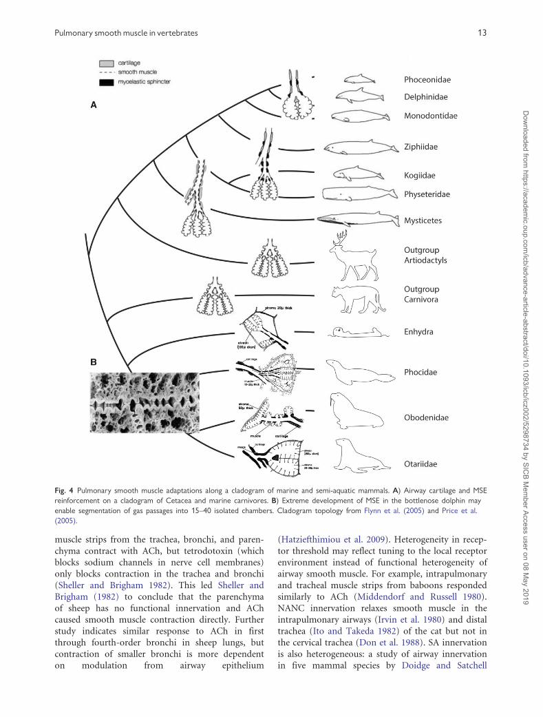
The lungs of diving mammals are often reinforced
with cartilage and smooth muscle (Kooyman 1973)
(Fig. 4). Pinnipeds generally have less reinforcement
than cetaceans (Denison and Kooyman 1973), and
phocids have more smooth muscle in the smaller
airways than otariids (Gray et al. 2006), although
the final few millimeters of pinniped terminal air-
ways are without cartilaginous reinforcement, and
the alveoli are organized in lobules protected by
thin stroma (Denison and Kooyman 1973). Smooth
muscle reinforcement may be related to diving
depth, as the deeper-diving Weddell seal
(Leptonychotes weddellii) has greater amounts of
smooth muscle than the crabeater seal (Lobodon car-
cinophaga) (Welsch and Drescher 1982). In the eared
seals (Otariidae), cartilage lines the terminal airways
all the way to the alveolar sacs, which are sur-
rounded by thick stroma (20–30mm), whereas termi-
nal bronchi of sea otters and walruses have thick
stroma and alveoli with and without cartilaginous
ducts (Denison and Kooyman 1973). Compared
with dog lungs, sea lion lungs collapse more
completely at pressure (Denison et al. 1971) because
cartilage maintains airway patency even at low pres-
sures, allowing all of the air to escape the alveoli
before conducting airways collapse. Most earless seals
(phocids) exhale before deep dives (Piscitelli et al.
2013) and contraction of smooth muscle in terminal
airways devoid of cartilage may enhance lung emp-
tying (Welsch and Drescher 1982). Contraction of
smooth muscle in phocid terminal airways may
also help to re-inflate collapsed alveoli after a dive,
a function provided by the thicker elastic stroma in
other pinnipeds.
In cetaceans, terminal bronchiole anatomy is
highly derived (Fig. 4). Delphinids replace longitudi-
nal smooth muscle with a layer of elastic tissue
(Wislocki 1948). In airways about 2 mm or smaller
in diameter, smooth muscle organizes into discrete
myoelastic sphincters (MES) between adjacent links
of hyaline cartilage both of which run between two
layers of antagonistic elastic tissue (Piscitelli et al.
2013). Contraction of the MES system in delphinids
can separate the terminal bronchi into 25–40 inde-
pendent chambers that may allow precise control of
air movement (Wislocki 1942; Fanning and Harrison
1974; Piscitelli et al. 2013; Fig. 4B). Smooth muscle
does not form MESs in baleen whales (Mysticetes)
Pulmonary smooth muscle in vertebrates 11
Dow
nloaded from https://academ
ic.oup.com/icb/advance-article-abstract/doi/10.1093/icb/icz002/5298734 by SIC
B Mem
ber Access user on 08 May 2019
and instead runs in a longitudinal layer through
highly elastic terminal airways devoid of cartilagi-
nous reinforcement (Piscitelli et al. 2013). Toothed
whales (Odontocetes) have cartilage rings and MESs
in the penultimate bronchi, and MSEs only in the
terminal bronchi and alveolar ducts (Wislocki 1929;
Fanning and Harrison 1974; Piscitelli et al. 2013).
Functional hypotheses for MESs include: air trapping
in the alveoli to continue gas exchange at depth,
enhancement of rapid exhalation, protection of alve-
oli from barotrauma, prevention of eversion or pro-
lapse of alveoli into the terminal bronchi, and the
protection of intercartilaginous tissue from baro-
trauma (Piscitelli et al. 2013).
Innervation and control of pulmonary smooth
muscle tissue is comparatively well studied in mam-
mals. The right and left vagus nerves carry choliner-
gic and NANC input from nerves in the rostral pole
of the nucleus ambiguus (the external formation) or
the rostral portion of the dorsal motor nucleus
(Undem and Potenzieri 2012). Preganglionic nerves
leading to airways receive mainly glutamate-driven
excitatory input from the nucleus tractus solitarius,
pons, hypothalamus, and amygdala (Haxhiu et al.
2005). Integration, relay, and reflex mediation occurs
in cranial autonomic ganglia along extra and intra-
pulmonary airways, which occur between nerve
trunk branches of the vagus in the European ferret
(Mustela putorius) and cat (Felis catus) (Dey et al.
1981; Undem and Potenzieri 2012). Although mor-
phology and distribution of ganglia is species-
specific, they are usually largest in the trachea and
main-stem bronchi but are also dispersed into the
intrapulmonary nerve plexuses (Undem and
Potenzieri 2012).
Airway smooth muscle receives both excitatory
and inhibitory cranial innervation; normal rhythmi-
cal contraction of the muscle is abolished after va-
gotomy (Jammes and Mei 1979; Undem and
Potenzieri 2012). The majority of effects of CC stim-
ulation are mediated by muscarinic cholinergic
receptors: the M3 subtype mediates contraction
(Eglen et al. 1996) while the role of M2 subtypes is
unclear, but may tune the response of smooth mus-
cle to the presence of additional transmitters and
peptides and function in negative feedback (Undem
and Potenzieri 2012). Inhibitory cranial innervation
is largely of the NANC variety in mammals and
wholly NANC in humans (Undem and Potenzieri
2012). NANC and CC innervation are both carried
by the vagus nerve but use distinct pathways and
ganglia and arise from distinct preganglionic nerves
(Undem and Potenzieri 2012). In guinea pigs, gan-
glia for cholinergic contraction are located in the
airway, whereas the NANC inhibitory ganglia are
in the esophageal plexus (Canning and Undem
1993). NANC innervation of the bovine trachea is
mimicked by VIP in tracheal strips (Cameron et al.
1983).
Smooth airway innervation of spinal origin is not
as well studied and varies by species in mammals:
stimulation of intrinsic bronchial adrenergic nerves
causes smooth muscle relaxation or contraction in
guinea pigs (Cavia porcellus), cats, and dogs (Canis
familiaris), but not in humans, monkeys, rats
(Doidge and Satchell 1982), or rabbits (Mustafa
et al. 1982; Undem and Potenzieri 2012). Mammals
with functional SA control express b1-adrenorecep-
tors and b2-adrenoreceptors while species without
functional control largely express b2-adrenorecep-
tors. An SA inhibitory pathway may be important
in mammals that frequently use panting for thermo-
regulation. Hyperventilation causes bronchoconstric-
tion in many mammals (Koyama et al. 1992; Nogami
et al. 1998; Suzuki and Freed 2000), and mammals
that pant may need to attenuate this response to
maintain a high lung compliance to reduce the
work of panting, or to maintain adequate levels of
gas exchange during hyperventilation. Sheep (Ovis
aries) and dogs (Rahardja et al. 2011), but not
guinea pigs (Richards 1970), rely extensively on
panting for thermoregulation. The SA inhibitory re-
sponse is confined to the trachea in guinea pigs
(Doidge and Satchell 1982), however, and may
have a different function in this species.
Spinal origin nerves dominate autonomic control
system of the airway vasculature and can also regu-
late smooth muscle in both systems through epi-
nephrine release from the adrenal medulla (Barnes
1986). GABA-mediated spinal preganglionic neurons
(in the lateral horn of the thoracic spinal cord) re-
ceive input from various parts of the central nervous
system and project to paravertebral ganglia and the
stellate ganglion (Undem and Potenzieri 2012). In
baboons, although a-adrenergic receptors could not
be located in intrapulmonary airways, b-receptor
adrenergic inhibition of contraction caused by hista-
mines is found throughout the lung (Middendorf
and Russell 1980). SA inhibition is demonstrated in
the Macaque trachea only after contraction with
phentolamine (an a-receptor agonist) (Olson et al.
1988).
There is evidence for heterogeneity in the inner-
vation of mammal lungs—some of the lung are more
often more affected by certain pathways than others.
In sheep lungs, greater doses of catecholamines are
needed to elicit relaxation in bronchi than the tra-
chea (Sheller and Brigham 1982). In addition,
12 R. L. Cieri
Dow
nloaded from https://academ
ic.oup.com/icb/advance-article-abstract/doi/10.1093/icb/icz002/5298734 by SIC
B Mem
ber Access user on 08 May 2019
muscle strips from the trachea, bronchi, and paren-
chyma contract with ACh, but tetrodotoxin (which
blocks sodium channels in nerve cell membranes)
only blocks contraction in the trachea and bronchi
(Sheller and Brigham 1982). This led Sheller and
Brigham (1982) to conclude that the parenchyma
of sheep has no functional innervation and ACh
caused smooth muscle contraction directly. Further
study indicates similar response to ACh in first
through fourth-order bronchi in sheep lungs, but
contraction of smaller bronchi is more dependent
on modulation from airway epithelium
(Hatziefthimiou et al. 2009). Heterogeneity in recep-
tor threshold may reflect tuning to the local receptor
environment instead of functional heterogeneity of
airway smooth muscle. For example, intrapulmonary
and tracheal muscle strips from baboons responded
similarly to ACh (Middendorf and Russell 1980).
NANC innervation relaxes smooth muscle in the
intrapulmonary airways (Irvin et al. 1980) and distal
trachea (Ito and Takeda 1982) of the cat but not in
the cervical trachea (Don et al. 1988). SA innervation
is also heterogeneous: a study of airway innervation
in five mammal species by Doidge and Satchell
Fig. 4 Pulmonary smooth muscle adaptations along a cladogram of marine and semi-aquatic mammals. A) Airway cartilage and MSE
reinforcement on a cladogram of Cetacea and marine carnivores. B) Extreme development of MSE in the bottlenose dolphin may
enable segmentation of gas passages into 15–40 isolated chambers. Cladogram topology from Flynn et al. (2005) and Price et al.
(2005).
Pulmonary smooth muscle in vertebrates 13
Dow
nloaded from https://academ
ic.oup.com/icb/advance-article-abstract/doi/10.1093/icb/icz002/5298734 by SIC
B Mem
ber Access user on 08 May 2019

(1982) demonstrated tracheal SA inhibition in the
guinea pig only, but bronchial inhibition in all spe-
cies studied excluding rats (guinea pigs, rabbits,
monkeys, humans).
Smooth muscle innervation
The innervation of airway smooth muscles also pro-
vides information about its evolution (Table 1). CC
output is excitatory in all species studied, with ACh
causing tissue contraction. This pattern is likely basal
to vertebrates and controlled the ancestral air-filled
organ. In the simplest scenario, CC input was the
only external autonomic control, and relaxation of
smooth muscle occurred intrinsically or at the ces-
sation of cholinergic stimulation. This scenario is
supported by the lack of cranial NANC innervation
in actinopterygians, where SA innervation often
stimulates contraction and relaxation of opposing
muscle groups.
The main condition in tetrapods is for NANC
nerves to mediate inhibition. Known exceptions are
Chelodina turtles, with a purportedly excitatory
NANC innervation (Smith and Satchell 1987a),
guinea pigs, where cranial NANC pathways can
cause tracheal contraction (Richardson and
Bouchard 1975; Taylor et al. 1984), and dogs, where
cranial NANC inhibitory pathways have not been
demonstrated. Airway smooth muscle in dogs and
sheep responds comparatively uniformly with relax-
ation to SA stimulation (Cabezas et al. 1971; Russell
1980; Sheller and Brigham 1982)—SA stimulation
could serve to antagonize reflex bronchoconstriction
resulting from hyperventilation in thermoregulatory
panting.
SA pathways seem to represent a parallel control
system for airway smooth muscle. While cranial
nerves provide the main inhibition and excitation
of smooth muscle throughout the lung or swim
bladder, SA fibers seem to target specific regions of
visceral smooth muscle. Heterogeneity of SA stimu-
lation is demonstrated in the mucosa of swim blad-
ders and R. marina, where SA nerves cause
contraction of septal rim musculature and relaxation
of lung wall musculature (Campbell and McLean
1994). Under this paradigm, studies of SA innerva-
tion of reptile lungs are needed to determine if an-
tagonistic smooth muscle groups are differentially
activated by SA innervation. In addition, study of
the innervation of smooth muscle in marine mam-
mals is needed. MESs are probably controlled by SA
innervation, while cranial autonomic innervation
controls smooth muscle tone throughout the lung.
Concluding remarks
Lung story short, airway smooth muscle has adapted
from an ancestral role regulating hollow-organ ten-
sion (Fig. 5A) to take on new roles reflecting the
morphological and physiological diversity of verte-
brate respiratory structures. In actinopterygians,
smooth muscle is involved in deflation and regulat-
ing gas absorption and secretion in the swim bladder
(Fig. 5B). Smooth muscle contraction suspends the
intrapulmonary structure (Fig. 5C) as well as poten-
tially contributing to intrapulmonary mixing in
many amphibians and non-avian reptiles (Fig. 5E)
and is the primary regulator of airway caliber and
ventilation distribution in birds and mammal
(Fig. 5F) lungs. CC innervation is excitatory, cranial
NANC innervation is largely inhibitory, and SA in-
nervation causes species-specific heterogeneous
responses. Further study of smooth muscle function
may shed further light on vertebrate lung evolution
Fig. 5 Hypothesized events in pulmonary smooth muscle evolution superimposed on a vertebrate cladogram. A) Ancestral smooth
muscle function of regulating tension and perhaps aiding exhalation and body orientation in the water column. B) Smooth muscle also
redistributes air in the swim bladder and regulates the opening of the pneumatic duct in physostomes. C) Smooth muscle contraction
maintains internal lung structure (see Fig. 2). D) Hypaxial muscle contraction supplements lung emptying. E) Antagonistic smooth
muscle groups facilitate intrapulmonary gas mixing (see Figs. 2, 3B). F) Smooth muscle regulates airway caliber. G) Smooth muscle
contributes to heavy airway reinforcement. Cladogram topology from Kardong (2014).
14 R. L. Cieri
Dow
nloaded from https://academ
ic.oup.com/icb/advance-article-abstract/doi/10.1093/icb/icz002/5298734 by SIC
B Mem
ber Access user on 08 May 2019

and the importance of lung micromechanics on pul-
monary function.
Acknowledgments
The author is indebted to EL Brainerd, DR Carrier,
CG Farmer, F Goller, and three anonymous
reviewers for comments that greatly improved the
manuscript.
Funding
This work was supported by the National Science
Foundation Graduate Research Fellowship Program
under Grant No. 1256065. Any opinions, findings,
and conclusions or recommendations expressed in
this material are those of the authors and do not
necessarily reflect the views of the National Science
Foundation.
References
Abrahamsson T, Holmgren S, Nilsson S, Pettersson K. 1979.
Adrenergic and cholinergic effects on the heart, the lung
and the spleen of the African lungfish, Protopterus aethio-
picus. Acta Physiol Scand 107:141–7.
Alexander RM. 1966. Physical aspects of swimbladder func-
tion. Biol Rev Camb Philos Soc 41:141–76.
Altiere RJ, Szarek JL, Diamond L. 1984. Neural control of
relaxation in cat airways smooth muscle. J Appl Physiol
57:1536–44.
Altura BT, Altura BM. 1975. Pentobarbital and contraction of
vascular smooth muscle. Am J Physiol 229:1635–40.
Amis TC, Jones HA, Hughes JMB. 1984. Effect of posture on
inter-regional distribution of pulmonary ventilation in
man. Respir Physiol 56:145–67.
Banzett RB, Butler JP, Nations CS, Barnas GM, Lehr JL, Jones
JH. 1987. Inspiratory aerodynamic valving in goose lungs
depends on gas density and velocity. Respir Physiol
70:287–300.
Barnas GM, Mather FB, Fedde MR. 1978. Response of avian
intrapulmonary smooth muscle to changes in carbon
dioxide concentration. Poult Sci 57:1400–7.
Barnes PJ. 1986. Neural control of human airways in health
and disease. Am Rev Respir Dis 134:1289–314.
Bennet T, Malmfors T. 1970. The adrenergic nervous system
of the domestic fowl (Gallus domesticus (L.)). Z Zellforsch
106:22–50.
Bennett T. 1971. The adrenergic innervation of the pulmo-
nary vasculature, the lung and the thoracic aorta, and on
the presence of aortic bodies in the domestic fowl (Gallus
gallus domesticus L.). Z Zellforsch 114:117–34.
Bennett WO, Summers AP, Brainerd EL. 1999. Confirmation
of the passive exhalation hypothesis for a terrestrial caeci-
lian, Dermophis mexicanus. Copeia 1999:206–9.
Berger PJ. 1973. Autonomic innervation of the visceral and
vascular smooth muscle of the lizard lung. Comp Gen
Pharmac 4:1–10.
Berger PJ, Burnstock G. 1979. Autonomic nervous systems.
In: Gans C, Northcutt RG, Ulinksi P, editors. Biology of
the Reptilia. London: Academic Press. p. 1–58.
Bhatla R, Ferguson CC, Richardson JB. 1980. The innervation
of smooth muscle in the primary bronchus of the chicken.
Can J Physiol Pharmacol 58:310–5.
Bishop IR, Foxon GEH. 1968. The mechanism of breathing in
the South American lungfish, Lepidosiren paradoxa; a ra-
diological study. J Zool Lond 154:263–71.
Brainerd EL. 1994a. Mechanical design of polypterid fish in-
tegument for energy storage during recoil aspiration. J Zool
232:7–19.
Brainerd EL. 1994b. The evolution of lung–gill bimodal
breathing and the homology of vertebrate respiratory
pumps. Am Zool 34:289–99.
Brainerd EL. 1999. New perspectives on the evolution of lung
ventilation mechanisms in vertebrates. Exp Biol Online
4:1–28.
Brainerd EL. 2015. Major transformations in vertebrate
breathing mechanisms. In: Dial KP, Shubin NH, Brainerd
EL, editors. Great transformations in vertebrate evolution.
Chicago (IL):University of Chicago Press. p. 47–61.
Brainerd EL, Ditelberg JS, Bramble DM. 1993. Lung ventila-
tion in salamanders and the evolution of vertebrate air-
breathing mechanisms. Biol J Linn Soc 49:163–83.
Brainerd EL, Dumka AM. 1995. Mechanics of ventilation in
an aquatic salamander, Amphiuma tridactylum. American
Society of Zoologists Annual Meeting, St. Louis, p. 144A.
Brainerd EL, Monroy J. 1998. Mechanics of lung ventilation
in a large aquatic salamander, Siren lacertina. J Exp Biol
201:673–82.
Burnstock G, Wood M. 1967. Innervation of the lungs of the
sleepy lizard (Trachysaurus rugosus)—II. Physiology and
pharmacology. Comp Biochem Physiol 22:815–31.
Cabezas GA, Graf PD, Nadel JA. 1971. Sympathetic versus
parasympathetic nervous regulation of airways in dogs. J
Appl Physiol 31:651–5.
Cameron AR, Johnston CF, Kirkpatrick CT, Kirkpatrick
MC. 1983. The quest for the inhibitory neurotransmitter
in bovine tracheal smooth muscle. Q J Exp Physiol
68:413–26.
Cameron AR, Kirkpatrick CT. 1977. A study of excitatory
neuromuscular transmission in the bovine trachea. J
Physiol 270:733–45.
Campbell GC. 1971. Autonomic innervation of the lung mus-
culature of a toad (Bufo marinus). Comp Gen Pharmacol
2:281–6.
Campbell G, Duxson M. 1978. The sympathetic innervation
of lung muscle in the toad Bufo marinus: a revision and an
explanation. Comp Biochem Physiol C 60:65–73.
Campbell GC, McLean JR. 1994. Lungs and swimbladders. In:
Nilsson S, Holmgren S, editors. Comparative physiology
and evolution of the autonomic nervous system. Chur,
Switzerland: Harwood Academic Publishers. p. 257–309.
Campbell GC, Haller CJ, Rogers DC. 1978. Fine structural
and cytochemical study of the innervation of smooth mus-
cle in an amphibian (Bufo marinus) lung before and after
denervation. Cell Tissue Res 432:419–32.
Canning BJ. 2006. Reflex regulation of airway smooth muscle
tone. J Appl Physiol 101:971–85.
Pulmonary smooth muscle in vertebrates 15
Dow
nloaded from https://academ
ic.oup.com/icb/advance-article-abstract/doi/10.1093/icb/icz002/5298734 by SIC
B Mem
ber Access user on 08 May 2019

Canning BJ, Undem BJ. 1993. Evidence that distinct neural
pathways mediate parasympathetic contractions and relax-
ations of guinea–pig trachealis. J Physiol 471:25–40.
Carlson AJ, Luckhardt AB. 1920. Studies on the visceral ner-
vous system. III. Lung automatism and lung reflexes in
reptilia (turtles: Chrysemys elegans and Malacoclemmys
lesueurii. Snake: Eutenia elegans). Am J Physiol 54:261–306.
Carrier DR, Wake MH. 1995. Mechanism of lung ventilation
in the caecilian Dermophis mexicanus. J Morphol
226:289–95.
Company NP, Rahn K. 1971. Air breathing of the garfish.
Respir Physiol 11:285–307.
Cook RD, King A. 1970. Observations on the ultrastructure of
the smooth muscle and its innervation in the avian lung. J
Anat 106:273–83.
Crawford R. 1971. Aquatic and aerial respiration in the bow-
fin, longnose gar and Alaska blackfish [PhD Thesis].
Toronto, Canada: University of Toronto.
Cruz-Hofling MA, Cruz-Landim C, Patelli AS. 1981.
Morphological and histochemical observations on swim-
bladders of an obligate water-breathing and a facultative
air-breathing fish from the Amazon. Zool Jb Anat
105:1–12.
Czopek J. 1962. Smooth muscles in the lungs of some uro-
deles. Nature 193:798.
Daniels CB, Mcgregor LK, Nicholas TE. 1994. The dragon’s
breath: a model for the dynamics of breathing and faveolar
ventilation in agamid lizards. Herepetologica 50:251–61.
DeLaney RG, Fishman A. 1977. Analysis of lung ventilation in
the aestivating lungfish Protopterus aethiopicus. Am J
Physiol 233:R181–7.
DeLaney RG, Laurent P, Galante R, Pack AI, Fishman A.
1983. Pulmonary mechanoreceptors in the dipnoi lungfish
Protopterus and Lepidosiren. Am J Physiol 244:R418–28.
Denison DM, Kooyman GL. 1973. The structure and function
of the small airways in pinniped and sea otter lungs. Respir
Physiol 17:1–10.
Denison DM, Warrell DA, West JB. 1971. Airway structure
and alveolar emptying in the lungs of sea lions and dogs.
Respir Physiol 13:253–60.
Dey RD, Shannon WA, Said SI. 1981. Localization of VIP-
immunoreactive nerves in airways and pulmonary vessels
of dogs, cat, and human subjects. Cell Tissue Res
220:231–8.
Doeing DC, Solway J. 2013. Airway smooth muscle in the
pathophysiology and treatment of asthma. J Appl Physiol
114:834–43.
Doidge JM, Satchell DG. 1982. Adrenergic and non-
adrenergic inhibitory nerves in mammalian airways. J
Auton Nerv Syst 5:83–99.
Don H, Baker DG, Richardson CA. 1988. Absence of non-
adrenergic noncholinergic relaxation in the cat cervical tra-
chea. J Appl Physiol 65:2524–30.
Donnelly P, Woolcock AJ. 1977. Ventilation and gas exchange
in the carpet python, Morelia spilotes variegata. J Comp
Physiol 418:403–18.
Dumbarton TC, Stoyek M, Croll RP, Smith FM. 2010.
Adrenergic control of swimbladder deflation in the zebra-
fish (Danio rerio). J Exp Biol 213:2536–46.
Duncker H-R. 1978. Coelom-gliederung der wirbeltiere-funk-
tionelle aspekte. Verh Anat Ges 72:91–112.
Duncker H-R. 2004. Vertebrate lungs: structure, topography
and mechanics. A comparative perspective of the progres-
sive integration of respiratory system, locomotor apparatus
and ontogenetic development. Respir Physiol Neurobiol
144:111–24.
Ebina M, Yaegashi H, Takahashi T, Motomiya M, Tanemura
M. 1990. Distribution of smooth muscles along the bron-
chial tree. A morphometric study of ordinary autopsy
lungs. Am Rev Respir Dis 141:1322–6.
Eglen RM, Hegde SS, Watson N. 1996. Muscarinic receptor
subtypes and smooth muscle function. Pharmacol Rev
48:531–65.
Fahlen G, Falck B, Rosengren E. 1965. Monoamines in the
Swimbladder of Gadus Callarias and Salmo Irideus. Acta
Physiol Scand 64:119–26.
F€ange R. 1983. Gas exchange in fish swim bladder. Rev
Physiol Biochem Pharmacol 97:111–58.
F€ange R, Holmgren S, Nilsson S. 1976. Autonomic nerve
control of the swimbladder of the goldsinny wrasse,
Ctenolabrus rupestris. Acta Physiol Scand 97:292–303.
Fanning J, Harrison R. 1974. The structure of the trachea and
lungs of the South Australian bottle-nosed dolphin. In:
Harrison RJ, editor. Functional anatomy of marine mam-
mals. Vol. 2. London: Academic Press. p. 22.
Farmer CG. 1997. Did lungs and the intracardiac shunt evolve
to oxygenate the heart in vertebrates? Paleobiology
23:358–72.
Finney JL, Robertson GN, McGee CAS, Smith FM, Croll RP.
2006. Structure and autonomic innervation of the swim
bladder in the zebrafish (Danio rerio). J Comp Neurol
495:587–606.
Fishman A, DeLaney RG, Laurent P. 1985. Circulatory adap-
tation to bimodal respiration in the dipnoan lungfish. J
Appl Physiol 59:285–94.
Flynn JJ, Finarelli JA, Zehr S, Hsu J, Nedbal MA. 2005.
Molecular phylogeny of the carnivora (mammalia): assess-
ing the impact of increased sampling on resolving enig-
matic relationships. Syst Biol 54:317–37.
Glazier JB, Hughes JM, Maloney JE, Pain MC, West JB. 1966.
Vertical gradient of alveolar size in dog lungs frozen in situ.
J Physiol 186:114P–5P.
Glenny RW, Robertson HT. 2011. Spatial distribution of ven-
tilation and perfusion: mechanisms and regulation. Compr
Physiol 1:373–95.
Goldie RG, Bertram JF, Warton A, Papadimitriou JM,
Paterson JW. 1983. A pharmacological and ultrastructural
study of alveolar contractile tissue in toad (Bufo marinus)
lung. Comp Biochem Physiol C 75:343–9.
Goniakowska-Witalifiska L. 1995. The histology and ultra-
structure of the amphibian lung. In: Pastor L, editor.
Histology, ultrastructure and immunoreactivity of the re-
spiratory organs. Servicio de Publicaciones de la
Universidad de Murica. p. 73–112.
Graham JB. 1997. Air breathing fishes: evolution, diversity,
and adaptation. London: Academic Press.
Gray R, Canfield P, Rogers T. 2006. Histology of selected
tissues of the leopard seal and implications for functional
adaptations to an aquatic lifestyle. J Anat 209:179–99.
Greaves IA, Hildebrandt J, Hoppin FG. 1986. Micromechanics
of the Lung. In: Macklem PT, Mead J, editors. Handbook
of physiology. American Physiological Society. p. 217–31.
16 R. L. Cieri
Dow
nloaded from https://academ
ic.oup.com/icb/advance-article-abstract/doi/10.1093/icb/icz002/5298734 by SIC
B Mem
ber Access user on 08 May 2019

Greenwood PH, Liem KF. 1984. Aspiratory respiration in
Arapaima gigas (Teleostei, Osteoglossomorpha): a reap-
praisal. J Zool 203:411–25.
Grigg GC. 1965. Studies on the Queensland lungfish,
Neoceratodus forsteri (Krefft). 3. Aerial respiration in rela-
tion to habits. Aust J Zool 13:413.
Guimond RW, Hutchison VH. 1973. Trimodal gas exchange
in the large aquatic salamander, Siren lacertina (Linnaeus).
Comp Biochem Physiol A Comp Physiol 46:249–68.
Guimond RW, Hutchison VH. 1974. Aerial and aquatic res-
piration in the Congo eel Amphiuma means means
(Garden). Respir Physiol 20:147–59.
Guimond RW, Hutchison VH. 1976. Gas exchange of the
giant salamanders of North America. In: Hughes G, editor.
Respiration of amphibious vertebrates. Academic Press. p.
313–38.
Harvey HH, Hoar WS, Bothern CR. 1968. Sounding response
of the kokanee and sockeye salmon. J Fish Res Board Can
25:1115–31.
Hatziefthimiou A, Kiritsi M, Kiropoulou C, Vasilaki A,
Sakellaridis N, Molyvdas PA. 2009. Regional differences
in the modulatory role of the epithelium in sheep airway.
Clin Exp Pharmacol Physiol 36:668–74.
Haxhiu MA, Prabha K, Moore CT, Acquah SS, Wilson CG,
Zaidi SI, Massari VJ, Ferguson DG. 2005. Brain stem
excitatory and inhibitory signaling pathways regulating
bronchoconstrictive responses. J Appl Physiol
98:1961–82.
Hislop AA. 2002. Airway and blood vessel interaction during
lung development. J Anat 201:325–34.
Hlastala MP, Standaert TA, Pierson DJ, Luchtel DL. 1985.
The matching of ventilation and perfusion in the lung of
the Tegu lizard, Tupinambis nigropunctatus. Respir Physiol
60:277–94.
Hoffman EA, Chon D. 2005. Computed tomography studies
of lung ventilation and perfusion. Proc Am Thorac Soc
2:492–8.
Holmgren S, Campbell GC. 1978. Adrenoceptors in the lung
of the toad Bufo marinus: regional differences in responses
to amines and to sympathetic nerve stimulation. Comp
Biochem Physiol C 60C:11–8.
Icardo JM, Colvee E, Lauriano ER, Capillo G, Guerrera
MC, Zaccone G. 2015. The structure of the gas bladder
of the spotted gar, Lepisosteus oculatus. J Morphol
276:90–101.
Irvin CG, Boileau R, Tremblay J, Martin RR, Macklem PT.
1980. Bronchodilatation: noncholinergic, nonadrenergic
mediation demonstrated in vivo in the cat. Science
207:791–2.
Ito Y, Takeda K. 1982. Non-adrenergic inhibitory nerves and
putative transmitters in the smooth muscle of cat trachea. J
Physiol 330:497–511.
Jammes Y, Mei N. 1979. Assessment of the pulmonary origin
of bronchoconstrictor vagal tone. J Physiol 291:305–16.
Johansen K, Reite OB. 1967. Effects of acetylcholine and bio-
genic amines on pulmonary smooth muscle in the African
lungfish, Protopterus aethiopicus. Acta Physiol Scand
71:248–52.
Johansen K, Lenfant C, Grigg GC. 1967. Respiratory control
in the lungfish, Neoceratodus forsteri (krefft). Comp
Biochem Physiol 20:835–54.
Jones AC. 1912. Innervation and nerve terminals of the rep-
tilian lung. J Comp Neurol 40:371–88.
Kardong KV. 2014. Vertebrates: comparative anatomy, func-
tion, evolution. 7th ed. New York (NY): McGraw-Hill.
King A, Cowie AF. 1969. The functional anatomy of the
bronchial muscle of the bird. J Anat 105:323–36.
Klemm RD, Gatz RN, Westfall JA, Fedde MR. 1979.
Microanatomy of the lung parenchyma of a tegu lizard
Tupinambis nigropunctatus. J Morphol 161:257–79.
Klika E. 1967. A contribution to the study of the lungs of the
Protopterus annectens and Polypterus senegalensis. Folia
Morphol (Praha) 15:168–75.
Klingele TG, Staub NC. 1970. Alveolar shape changes with
volume in isolated, air-filled lobes of cat lung. J Appl
Physiol 28:28–31.
Kobayashi S, Yoda C. 1960. Effects of adrenaline and acetyl-
choline upon the smooth muscle of isolated lungs of
Japanese toads. Acta Med Biol Niigata 8:241–50.
Kooyman GL. 1973. Respiratoy adaptations in marine mam-
mals. Am Zool 13:457–68.
Koyama S, Ohtsuka A, Horie T. 1992. Eucapnic
hyperventilation-induced bronchoconstriction in rabbits.
Tohoku J Exp Med 168:611–9.
Kuehne B, Junqueira LC. 2000. Histology of the trachea and
lung of Siphonops annulatus (Amphibia, Gymnophiona).
Rev Bras Biol 60:167–72.
Lawry J. 1999. A hydrostatic Michell framework supports frog
lungs. Bull Math Biol 61:683–700.
Liem KF. 1988. Forms and function of lungs: the evolution of
air breathing mechanisms. Am Zool 28:739–59.
Liem KF. 1989. Respiratory gas bladders in teleosts: functional
conservatism and morphological diversity. Integr Comp
Biol 29:333–52.
Luchtel DL, Kardong KV. 1981. Ultrastructure of the lung of
the rattlesnake, Crotalus viridis oreganus. J Morphol
169:29–47.
Luckhardt AB, Carlson AJ. 1920. Studies on the visceral sen-
sory nervous system. II. Lung automatism and lung reflexes
in the salamanders (Necturus, Axolotl). Am J Physiol
54:122–37.
Lundberg JM, Martling CR, Saria A. 1983. Substance P and
capsaicin-induced contraction of human bronchi. Acta
Physiol Scand 119:49–53.
Lundberg JM, Saria A. 1982. Bronchial smooth muscle con-
traction induced by stimulation of capsaicin-sensitive sen-
sory neurons. Acta Physiol Scand 116:473–6.
Lundin K, Holmgren S. 1991. An X-Ray study of the influ-
ence of vasoactive intestinal peptide and substance-P on
the secretion of gas into the swimbladder of a teleost
Gadus morhua. J Exp Biol 157:287–98.
Maina JN. 1989. The morphology of the lung of the black
mamba Dendroaspis polylepis (Reptilia: Ophidia: Elapidae).
A scanning and transmission electron microscopic study. J
Anat 167:31–46.
Maina JN. 2002. Structure, function and evolution of the gas
exchangers: comparative perspectives. J Anat 201:281–304.
Maina JN. 2007. Minutialization at its extreme best! The
underpinnings of the remarkable strengths of the air
and the blood capillaries of the avian lung: a conun-
drum. Respir Physiol Neurobiol 159:141–5 (author re-
ply 146).
Pulmonary smooth muscle in vertebrates 17
Dow
nloaded from https://academ
ic.oup.com/icb/advance-article-abstract/doi/10.1093/icb/icz002/5298734 by SIC
B Mem
ber Access user on 08 May 2019
Maina JN, Maloiy GMO. 1985. The morphometry of the lung
of the African lungfish (Protopterus aethiopicus): its
structural–functional correlations. Proc R Soc B Biol Sci
224:399–420.
Maina JN, Maloiy GMO. 1988. A scanning and transmission
electron microscopic study of the lung of a caecilian
Boulengerula taitanus. J Zool 215:739–52.
Maina JN, West JB, Orgeig S, Foot NJ, Daniels CB, Kiama
SG, Gehr P, Muhlfeld C, Blank F, Muller L. 2010a. Recent
advances into understanding some aspects of the structure
and function of mammalian and avian lungs. Physiol
Biochem Zool 83:792–807.
Maina JN, Jimoh SA, Hosie M. 2010b. Implicit mechanistic
role of the collagen, smooth muscle, and elastic tissue com-
ponents in strengthening the air and blood capillaries of
the avian lung. J Anat 217:597–608.
Martin KM, Hutchison VH. 1979. Ventilatory activity in
Amphiuma tridactylum and Siren lacertina (Amphibia,
Caudata). J Herpetol 13:427–34.
McLean JR, Burnstock G. 1967a. Innervation of the lungs of
the sleepy lizard (Trachysaurus rugosus)—I. Fluorescent his-
tochemistry of catecholamines. Comp Biochem Physiol
22:809–13.
McLean JR, Burnstock G. 1967b. Innervation of the lungs
of the toad (Bufo marinus)—II. Fluorescent histochem-
istry of catecholamines. Comp Biochem Physiol
22:767–73.
McMahon BR. 1969. A functional analysis of the aquatic and
aerial respiratory movements of an African lungfish,
Protopterus aethiopicus, with reference to the evolution of
the lung-ventilation mechanism in vertebrates. J Exp Biol
51:407–30.
Meban C. 1978. Functional anatomy of the lungs of the green
lizard, Lacerta viridis. J Anat 125:421–31.
Melsom MN, Flatebo T, Sjaastad OV, Aulie A, Nicolaysen G.
1999. Minor redistribution of ventilation and perfusion
within the lung during exercise in sheep. Acta Physiol
Scand 165:283–92.
Middendorf WF, Russell JA. 1980. Innervation of airway
smooth muscle in the baboon: evidence for a nonadrener-
gic inhibitory system. J Appl Physiol 48:947–56.
Milic-Emili J, Henderson JA, Dolovich MB, Trop D, Kaneko
K. 1966. Regional distribution of inspired gas in the lung. J
Appl Physiol 21:749–59.
Mustafa KY, Elkhawad AO, Bicik V, Mardlni IA, Thulesius O.
1982. Adrenergic and cholinergic induced contractions of
tracheal smooth muscle in the rabbit as demonstrated by a
new in vivo method. Acta Physiol Scand 114:129–34.
Nilsson S. 1971. Adrenergic innervation and drug responses
of the oval sphincter in the swimbladder of the cod (Gadus
morhua). Acta Physiol Scand 83:446–53.
Nilsson S. 1972. Autonomic vasomotor innervation in the gas
gland of the swimbladder of a teleost (Gadus morhua).
Comp Gen Pharmacol 3:371–5.
Nilsson S. 2009. Nervous control of fish swimbladders. Acta
Histochem 111:176–84.
Nogami H, Umeno E, Kano S, Hirose T, Nishima S. 1998.
Effect of nitric oxide synthase inhibitor on allergen- and
hyperventilation-induced bronchoconstriction in guinea-
pigs. Eur Respir J 12:1318–21.
Olson LE, Mason DE, Muir WW. 1988. Innervation of crab-
eating monkey trachealis muscle determined in vitro.
Respir Physiol 71:375–86.
Orphanidou D, Hughes JMB, Myers MJ, Al-Suhali AR,
Henderson B. 1986. Tomography of regional ventilation
and perfusion using krypton 81m in normal subjects and
asthmatic patients. Thorax 41:542–51.
Perry SF. 1978. Quantitative anatomy of the lungs of the red-
eared turtle, Pseudemys scripta elegans. Respir Physiol
35:245–62.
Perry SF. 1983. Reptilian lungs: functional anatomy and evo-
lution. New York: Springer-Verlag.
Perry SF. 1988. Functional morphology of the lungs of the
Nile crocodile, Crocodylus niloticus: non-respiratory param-
eters. J Exp Biol 117:99–117.
Perry SF. 1998. Lungs: comparative anatomy, functional mor-
phology and evolution. In: Gans C, Gaunt AS, editors.
Biology of the Reptilia. Ithaca (NY): Society for the
Study of Amphibians and Reptiles. p. 1–92.
Perry SF, Duncker H-R. 1978. Lung architecture, volume and
static mechanics in five species of lizards. Respir Physiol
34:61–81.
Perry SF, Aumann U, Maloney JE. 1989a. Intrinsic lung mus-
culature and associated ganglion cells in a teiid lizard,
Tupinambis nigropunctatus Spix. Herpetologica 45:217–27.
Perry SF, Darian-Smith C, Alston J, Limpus CJ, Maloney JE.
1989b. Histological structure of the lungs of the loggerhead
turtle, Caretta caretta, before and after hatching. Copeia
1989:1000–10.
Perry SF, Hein J, van Dieken E. 1994. Gas exchange mor-
phometry of the lungs of the tokay, Gekko gecko L.
(Reptilia: Squamata: Gekkonidae). J Comp Physiol B
164:206–214.
Peters HM. 1951. Beitrage zur okologischen physiologie des
seepferdes (Hippocampus brevirostris). Z Vgl Physiol
33:207–65.
Piscitelli MA, Raverty SA, Lillie MA, Shadwick RE. 2013. A
review of cetacean lung morphology and mechanics. J
Morphol 274:1425–40.
Pohunkov�a H, Hughes GM. 1985. Ultrastructure of the lungs
of the garter snake. Folia Morphol (Warsz) 33:254–8.
Ponganis PJ, Leger JS, Scadeng M. 2015. Penguin lungs and
air sacs: implications for baroprotection, oxygen stores and
buoyancy. J Exp Biol 218:720–30.
Prabha KC, Bernard DG, Gardner M, Smatresk NJ. 2000.
Ventilatory mechanics and the effects of water depth on
breathing pattern in the aquatic caecilian Typhlonectes
natans. J Exp Biol 203:263–72.
Price SA, Bininda-Emonds ORP, Gittleman JL. 2005. A com-
plete phylogeny of the whales, dolphins and even-toed
hoofed mammals (Cetartiodactyla). Biol Rev 80:445–73.
Rahardja DP, Toleng AL, Lestari VS. 2011. Thermoregulation
and water balance in fat-tailed sheep and Kacang goat un-
der sunlight exposure and water restriction in a hot and
dry area. Animal 5:1587–93.
Randall D, Burggren W, Farrell A, Haswell M. 1981. The
evolution of air breathing in vertebrates. Cambridge:
Press Syndicate of the University of Cambridge.
Richards S. 1970. The biology and comparative physiology of
thermal panting. Biol Rev 45:223–64.
18 R. L. Cieri
Dow
nloaded from https://academ
ic.oup.com/icb/advance-article-abstract/doi/10.1093/icb/icz002/5298734 by SIC
B Mem
ber Access user on 08 May 2019
Richardson JB, Bouchard T. 1975. Demonstration of a non-
adrenergic inhibitory nervous system in the trachea of the
guinea pig. J Allergy Clin Immunol 56:473–80.
Robertson GN, Lindsey BW, Dumbarton TC, Croll RP, Smith
FM. 2008. The contribution of the swimbladder to buoy-
ancy in the adult zebrafish (Danio rerio): a morphometric
analysis. J Morphol 269:666–73.
Robinson PM, McLean JR, Burnstock G. 1971. Ultrastructural
identification of non-adrenergic inhibitory nerve fibers. J
Pharmacol Exp Ther 179:149–60.
Romer A, Parsons T. 1977. The Vertebrate Body. 5th ed.
Philadelphia (PA): WB Sanders Co.
Russell JA. 1978. Responses of isolated canine airways to elec-
tric stimulation and acetylcholine. J Appl Physiol 45:690–
98.
Russell JA. 1980. Noradrenergic inhibitory innervation of ca-
nine airways. J Appl Physiol 48:16–22.
Scheid P, Kuhlmann WD, Fedde MR. 1977. Intrapulmonary
receptors in the Tegu lizard: II. Functional characteristics
and localization. Respir Physiol 29:49–62.
Seymour RS, Wegner NC, Graham JB. 2008. Body size and
the air-breathing organ of the Atlantic tarpon Megalops
atlanticus. Comp Biochem Physiol A Mol Integr Physiol
150:282–7.
Sheller JR, Brigham KL. 1982. Bronchomotor responses of
isolated sheep airways to electrical field stimulation. J
Appl Physiol 53:1088–93.
Shimada K, Kobayasi Y. 1966. Neural control of pulmonary
smooth muscle in the toad. Acta med biol Niigata
13:297–303.
Simons RS, Bennett WO, Brainerd EL. 2000. Mechanics of
lung ventilation in a post-metamorphic salamander,
Ambystoma tigrinum. J Exp Biol 203:1081–92.
Smith DG, Campbell GC. 1976. The anatomy of the pulmo-
nary vascular bed in the toad Bufo marinus. Cell Tissue Res
213:199–213.
Smith FM, Croll RP. 2011. Autonomic control of the swim-
bladder. Auton Neurosci Basic Clin 165:140–8.
Smith DG, Macintyre DH. 1979. Autonomic innervation of
the visceral and vascular smooth muscle of a snake lung
(Ophidia: Colubridae). Comp Biochem Physiol Part C
Comp Pharmacol 62:187–91.
Smith RV, Satchell DG. 1987. Innervation of the lung of the
Australian snake-necked tortoise, Chelodina longicollis.
Comp Biochem Physiol C 87:439–44.
Smith RV, Satchell DG. 1987. Histochemistry of the lung of
the Australian snake-necked tortoise Chelodina longicollis. J
Morphol 192:257–68.
Stark-Vancs VI Jr, Bell PB, Hutchison VH. 1984. Morphological
and pharmacological basis for pulmonary ventilation in
Amphiuma tridactylum. Cell Tissue Res 238:1–12.
Stephens NL. 2001. Airway smooth muscle. Lung 179:333–73.
Storey WF, Staub NC. 1962. Ventilation of terminal air units.
J Appl Physiol 17:391–7.
Suzuki R, Freed AN. 2000. Heparin inhibits eicosanoid me-
tabolism and hyperventilation-induced bronchoconstriction
in dogs. Am J Respir Crit Care Med 161:1850–4.
Szolcs�anyi J, Barth�o L. 1982. Capsaicin-sensitive non-cholin-
ergic excitatory innervation of the guinea-pig tracheobron-
chial smooth muscle. Neurosci Lett 34:247–51.
Taylor SM, Par�e PD, Schellenberg RR. 1984. Cholinergic and
nonadrenergic mechanisms in human and guinea pig air-
ways. J Appl Physiol 56:958–65.
Tortora G, Nielsen M. 2014. Principles of human anatomy.
13th ed. Hoboken (NJ): Wiley.
Undem BJ, Potenzieri C. 2012. Autonomic neural control of
intrathoracic airways. Comp Physiol 2:1241–67.
Wallach V. 1998. The lungs of snakes. In: Gans C, Gaunt AS,
editors. The biology of the reptilia. Society for the Study of
Amphibians and Reptiles. p. 93–296.
Wang N, Banzett RB, Butler JP, Fredberg JJ. 1988. Bird lung
models show that convective inertia effects inspiratory
aerodynamic valving. Respir Physiol 73:111–24.
Wang N, Banzett RB, Nations CS, Jenkins FA. 1992. An aero-
dynamic valve in the avian primary bronchus. J Exp Biol
262:441–5.
Weibel ER. 1984. The pathway for oxygen. Cambridge:
Harvard University Press.
Welsch U, Drescher HE. 1982. Light- and electron microscop-
ical observations on the terminal airways and the alveolar
parenchyma of the Antarctic seals Lobodon carcinophagus
and Leptonychotes weddelli. Polar Biol 1:105–14.
Wislocki GB. 1929. On the structure of the lungs of the
porpose (Tursiops truncatus). Am J Anat 44:47–77.
Wislocki GB. 1942. The lungs of the cetacea, with special
reference to the harbor porpoise (Phocoena phocoena
Linnaeus). Anat Rec 84:117–23.
Zaccone D, Dabrowski K, Lauriano ER, De Pasquale A, Macr�ıD, Satora L, Lanteri G. 2012a. The simultaneous presence
of neuroepithelial cells and neuroepithelial bodies in the
respiratory gas bladder of the longnose gar, Lepisosteus
osseus, and the spotted gar, L. oculatus. Acta Histochem
114:370–8.
Zaccone D, Sengar M, Lauriano ER, Pergolizzi S, Macri’ F,
Salpietro L, Favaloro A, Satora L, Dabrowski K, Zaccone
G. 2012b. Morphology and innervation of the teleost
physostome swim bladders and their functional evolu-
tion in non-teleostean lineages. Acta Histochem
114:763–72.
Zaccone G, Mauceri A, Maisano M, Giannetto A, Parrino V,
Fasulo S. 2007. Innervation and neurotransmitter localiza-
tion in the lung of the nile bichir Polypterus bichir bichir.
Anat Rec 290:1166–77.
Pulmonary smooth muscle in vertebrates 19
Dow
nloaded from https://academ
ic.oup.com/icb/advance-article-abstract/doi/10.1093/icb/icz002/5298734 by SIC
B Mem
ber Access user on 08 May 2019