The Regulation of Pulmonary Vascular Tone by Neuropeptides ...

PVDOMICS Protocol v1.3April 16, 2021 1
Pulmonary Vascular Disease
Phenomics Program -
PVDOMICS
Study Protocol
Version 1.3
April 16, 2021

PVDOMICS Protocol v1.3April 16, 2021 2
Table of Contents
1. Introduction ........................................................................................................................................... 4
2. Objectives ............................................................................................................................................. 4
2.1 Specific Aims: ............................................................................................................................... 5
2.2 Hypotheses .................................................................................................................................... 5
2.2.1 General Hypotheses .................................................................................................................. 5
2.2.2 Specific Hypotheses .................................................................................................................. 6
2.3 Study Design ................................................................................................................................. 7
2.3.1 Organizational Structure of the Study ....................................................................................... 7
2.3.2 Interactions with Other NHLBI Awards and the Pulmonary Hypertension Association .......... 8
3. Study Participants ................................................................................................................................. 9
3.1 Patient Recruitment ....................................................................................................................... 9
3.2 Inclusion/Exclusion Criteria ....................................................................................................... 10
3.2.1 Control Group ......................................................................................................................... 10
3.2.2 Comparator Groups ................................................................................................................. 11
3.2.3 PH Groups for Enrollment ...................................................................................................... 12
3.2.4 Phenotypic Characterization of PH Across WHO Groups...................................................... 13
3.3 Patient Timeline .......................................................................................................................... 15
3.4 Consenting Patients ..................................................................................................................... 15
3.5 Meeting Recruitment Goals ........................................................................................................ 15
4. Data Collection ................................................................................................................................... 16
4.1 Study Visits ................................................................................................................................. 16
4.2 Baseline Evaluation and Data Collection .................................................................................... 19
4.2.1 Demographics and History ...................................................................................................... 19
4.2.2 Quality of Life Questionnaires ................................................................................................ 19
4.2.3 Comorbid Conditions .............................................................................................................. 19
4.2.4 Medications ............................................................................................................................. 20
4.2.5 Physical Measurements ........................................................................................................... 20
4.2.6 Lung Function Measurements ................................................................................................. 21
4.2.7 Six Minute Walk Test ............................................................................................................. 21
4.2.8 Overnight Sleep Monitoring ................................................................................................... 21
4.2.9 Electrocardiogram (3).............................................................................................................. 22

PVDOMICS Protocol v1.3April 16, 2021 3
4.2.10 Imaging Studies....................................................................................................................... 22
4.2.10.1 Echocardiography ............................................................................................................... 22
4.2.10.2 Cardiac MRI ........................................................................................................................ 23
4.2.10.3 High Resolution Chest CT .................................................................................................. 25
4.2.10.4 Ventilation Perfusion Lung Scan ........................................................................................ 25
4.2.11 Right Heart Catheterization .................................................................................................... 25
4.2.12 Cardiopulmonary Exercise Testing ......................................................................................... 27
4.2.13 Laboratory Measurements ....................................................................................................... 28
4.3 Biospecimen Collection .............................................................................................................. 29
4.4 Longitudinal Follow-up .............................................................................................................. 30
5. Statistical Considerations .................................................................................................................... 30
5.1 Statistical Power for Specific Hypotheses .................................................................................. 32
5.2 General Methods ......................................................................................................................... 36
5.3 Methods by Aims ........................................................................................................................ 36
5.4 Missing Data ............................................................................................................................... 38
5.5 Software ...................................................................................................................................... 38
6. Regulatory Considerations .................................................................................................................. 38
6.1 Institutional Review Boards (IRBs) ............................................................................................ 38
6.2 Confidentiality and Ownership of Data and Biospecimens ........................................................ 38
6.3 Subject Information and Consent ................................................................................................ 39
6.4 Adverse Events ........................................................................................................................... 39
6.5 Incidental Findings ...................................................................................................................... 40
6.6 Observational Safety and Monitoring Board (OSMB) ............................................................... 41
7. Ancillary Studies ................................................................................................................................. 42
8. References ........................................................................................................................................... 43
APPENDIX 1: Definitions of Parenchymal and Non-Parenchymal Lung Diseases ................................. 58
APPENDIX 2: PVDOMICS Ancillary Studies Policy .............................................................................. 61
APPENDIX 3: Glossary of Terms ............................................................................................................. 66

PVDOMICS Protocol v1.3April 16, 2021 4
1. Introduction (1-12)
This NHLBI PVDOMICS protocol represents the core working protocol jointly created by the
six centers awarded the NHLBI U collaborative grant under RFA-HL-027 (Columbia/Cornell,
Johns Hopkins, Brigham and Women’s Hospital, Mayo Clinic, University of Arizona Tucson,
and Vanderbilt University) together with the project Data Coordinating Center (DCC)
(Cleveland Clinic) and the NHLBI. The need for such a project grew out of recognition of the
prognostic impact of pulmonary hypertension (PH) and right ventricular (RV) failure across a
wide spectrum of disease states that are currently classified under the Nice modification of the
World Health Organization (WHO) classification system.
It is recognized that patients with various forms of heart and lung disease exhibit varying degrees
of pulmonary vascular disease, leading to pulmonary vascular remodeling, pulmonary
hypertension, and right ventricular dysfunction. The genetic, molecular, and cellular processes
driving these phenomena are not well understood. Rapid advances in high throughput omic
methodology, combined with powerful bioinformatics and network biology capability, have
created the opportunity to conduct studies that broadly search for homologies and differences
across the spectrum of disease states associated with pulmonary hypertension, and determinants
of the spectrum of pulmonary vascular disease and RV compensation that accompanies these
conditions.
The 2010 NHLBI Pulmonary Vascular Strategic Plan identified the development of a
comprehensive cohort to define phenotypes integrating Omics technologies and systems
approaches as a top priority(2). The current WHO PH classification, based solely on
clinical/hemodynamic subsets, limits our ability to customize treatment for an individual patient,
or to assign meaningful clinical phenotype designations, e.g., Long-term survivor or Maladaptive
RV Hypertrophy (1). The overall goal of the PVDOMICS network is to perform
comprehensive phenotyping (demographic, physiologic, clinical chemistries, and imaging)
and endophenotyping (genomic, proteomic, metabolomic, coagulomic, cell and/or tissue
based) across the World Health Organization (WHO) classified PH clinical groups 1
through 5 as well as intermediate phenotypes (including those without overt PH) in order
to deconstruct the traditional classification and define new meaningful subclassifications of
patients with PVD. The long-term goal is utilization of endophenotypes/biomarkers for early
diagnosis, at-risk screening, and personalized approaches for interventions and/or preventions of
PVD.
The PVDOMIC protocol is designed to lead to this comprehensive understanding of patients
with pulmonary vascular disease based on phenotypes and endophenotypes.
2. Objectives (1-12)

PVDOMICS Protocol v1.3April 16, 2021 5
2.1 Specific Aims:
The first aim will be a natural product of the protocol, to identify the molecular basis of
pulmonary vascular disease regardless of WHO clinical classification, by comparison of
current subsets of PH patients with healthy subjects and with non-PH diseased comparators.
The second aim is to discover biological measures of disease and therapeutic responses that
may be useful not only in diagnosis but also as outcome measures in treatment and possibly
prevention trials.
1. Create an advanced description of structural and functional abnormalities of the heart
and pulmonary circulation in patients with PVD to define novel phenotypic clusters of
PVD.
a. Compare and contrast imaging assessment with Echo, CT and MRI, clinical and
hemodynamic and gas exchange data
b. Compare and contrast invasive exercise assessment with imaging, volume loading
and baseline catheterization data and etiology.
2. Create a detailed molecular endotype of all PVD patients, including genomic,
transcriptomic, proteomic, metabolomic, cell biomic and coagulomic metrics.
a. Test for known PH mutations, new genetic variants and genomic correlations with all
PVD and PH Group designations, including acute vasodilator responders, and
appropriate controls.
b. Compare and contrast transcriptomic, proteomic, metabolomic, cell biomic and
coagulomic data in all categories of demographic features, known etiology (such as
genetic) exercise physiology, pharmaceutical management and outcomes (where
feasible).
c. Compare all omics data without regard to PH Group designation to generate a new,
more accurate classification of pulmonary vascular disease leading to PH.
3. Cross-validate variants of PH between PVDOMIC genetic data with that of the Nichols
R24 and other available databases.
2.2 Hypotheses
2.2.1 General Hypotheses

PVDOMICS Protocol v1.3April 16, 2021 6
1. Epidemiological, biological, metabolomic and hemodynamic features will allow
differentiation of phenotypic similarities and differences among current World
Symposium PH Group categories of PH. These insights will lead to newer classification
of PH based on shared biological features.
2. The molecular basis of pulmonary vascular disease of all etiologies will be discovered
by integration of biological markers with careful phenotyping of all patients with PH
and comparing this data with healthy subjects and with non PH patients as diseased
comparators, such as emphysema and interstitial lung disease.
3. Racial and gender-related (or ancestry) genetic variation in phenotype, natural history
and responses to therapy will be discovered and lead to more precise diagnostic and
therapeutic approaches.
4. The response, adaptation and dysfunction of the right ventricle (RV) will be elucidated
by careful phenotyping, including specialized imaging and –omic correlations.
5. Exercise pathophysiology will lead to improved early diagnosis of PVD, elucidation of
RV- pulmonary vascular interactions, RV functional reserve, failure, and response to
therapies.
6. The biological and genetic features of patients with combined pulmonary venous
hypertension and PAH will lead to better differentiation of these two etiologies of PH,
and of shared biological mechanisms.
7. Epigenetic and RNA variants will influence the development, severity and type of PVD
and reveal therapeutic responses and form a basis for new therapies.
2.2.2 Specific Hypotheses
Examples of specific hypotheses that can be tested in the study are below.
1. Racial and ethnic (or ancestry) differences in transcriptomics, epigenetics and
mitochondrial haplotypes will inform PH pathogenesis by similarities and differences
among similar phenotypes with PH.
2. Patients with left heart dysfunction and PH will have patterns of omic measures that predict
the presence of combined PH. Patients with combined PH in WHO Group 2 will have
patterns of genomic vulnerability similar to patients with Group 1 PH, different from
Group 2 patients without severe PH.

PVDOMICS Protocol v1.3April 16, 2021 7
3. Patients with parenchymal lung disease and moderate to severe PH will have omic
signatures that reflect not only underlying pathogenesis (emphysema or fibrosis) but will
have patterns of genomic vulnerability similar to patients with Group 1 PH.
4. Patients with exercise-induced PH will have omic features that are similar to those with
Group 1 PH, different from those with normal exercise hemodynamics.
5. Connective tissue disease patients with PH (largely systemic sclerosis) will have
endothelial dysfunction measured as abnormalities in nitric oxide production,
arginine/ornithine/citrulline metabolism and vasoactive mediators that are similar to
patients with Group 1 PH and different from scleroderma patients without PH.
6. Genomic, transcriptomic and metabolomic patterns will distinguish the degree of right
ventricular compensation for a similar degree of right ventricular afterload in patients with
pulmonary hypertension, regardless of the underlying WHO category.
2.3 Study Design (13, 14)
2.3.1 Organizational Structure of the Study
The study will be governed by a Steering Committee comprised of the Principal Investigators
(PIs) of the PVDOMICS clinical centers and the DCC, the NHLBI Project Scientists
participating in the PVDOMICS, and the Steering Committee Chair of the PVDOMICS. The
Steering Committee has the primary responsibility for the study protocol, monitoring study
conduct, and reviewing data prior to reporting study results. It is also responsible for
determining policies in such areas as access to participant data, ancillary studies, publications
and presentations. Day-to-day decision-making is vested by the Steering Committee in an
Executive Committee consisting of the Steering Committee Chair, NIH Project Scientists, DCC
PI, and two rotating clinical center PIs. Study oversight is also provided by an Observational
Safety and Monitoring Board (OSMB) appointed by the NHLBI (See Section 6.6). Proposals for
ancillary studies of high scientific merit are encouraged to further enhance the scientific value of
the main study and to optimize the yield from collected data, images and biospecimens (see
Section 7). Upon conclusion of the study, data will be archived and shared according to NIH
policies.
There will be many central core facilities for PVDOMICS. These include ones for imaging,
lung physiology, clinical chemistry, right heart catheterization and cardiac pulmonary exercise
tests. These cores will provide central direction, personnel training, and supervision to the study,
while supporting ascertainment of high-quality standardized data. Study coordination,
centralized data management, biospecimen management and repository, and statistical
collaboration will be provided by the PVDOMICS DCC at the Cleveland Clinic. Also, the

PVDOMICS Protocol v1.3April 16, 2021 8
DCC’s Biorepository Core will have several omics cores to aid in carrying out the omics
analyses and interpretation. These cores include those for genomics and transcriptomics,
proteomics, metabolomics, cell biomics and coagulomics
2.3.2 Interactions with Other NHLBI Awards and the Pulmonary Hypertension
Association
The overall goal of the PVDOMICS network is to perform comprehensive phenotyping
(demographic, physiologic, clinical chemistries, and imaging) and endophenotyping (8, 15, 16)
(genomic, proteomic, metabolomic, cell and/or tissue based) across the World Health
Organization (WHO) classified PH clinical groups 1 through 5 in order to define new
subclassifications of patients based on characteristics that are associated with mechanisms of
pathogenesis (17). The network plans to leverage the synergistic scientific and operational
strengths of two NHLBI-awarded investigator-Initiated Resource-Related Research Project
Application (R24) grants on PH to support, facilitate and accelerate PVDOMICS goals: (1)
Pulmonary Hypertension Breakthrough Initiative (PHBI) (18, 19) and (2) the National Biological
Sample and Data Repository for PAH (20). The latter is led by Dr. Nichols [PI] at Cincinnati
Children’s Hospital Medical Center, and represents collaboration between academic PH centers
across the United States to collect a cohort of PAH patients to identify novel pathways or genetic
factors contributing to the disorder. They will collect and maintain biological material and
generate genetic data from 3000 WHO Group 1 PAH patients. Dr. Nichols will be invited to
participate in PVDOMICS Steering Committee meetings in order to optimize interactions and
leveraging of ideas and data. The PHBI aims to accrue PH lung and heart with detailed clinical
annotation of specimen lung and heart tissues and human primary endothelial and smooth muscle
cells derived from explanted PH and control lungs, which will be available for translational
approaches to endophenotype/biomarker discovery in PVDOMICS. Researchers in the PHBI
overlap with membership in PVDOMICS, including Dr. Geraci [PI] and Drs. Comhair, Aldred
and Erzurum. The PHBI R24 protocols, data, samples and cells will be leveraged for optimal
productivity of PVDOMICS. Specifically, Drs. Aldred and Geraci run the PHBI Genomics and
Mutation Analysis Cores, the goals of which are to develop a genomic catalog of PAH
specimens by mutational and genomic analysis of DNA and RNA from pulmonary and cardiac
tissues. Explant lung tissues are analyzed for mutations in known PAH genes, genome-wide
single nucleotide variant genotyping and expression analysis of mRNA and microRNA profiles.
Similar analyses will be performed in cardiac tissues as they are accrued. Novel markers
identified in the lung and heart tissues of PHBI subjects will be validated in peripheral
biospecimens obtained in PVDOMICS, providing a powerful link between the primary disease
sites and less invasive peripheral biomarkers. In addition, the network has entered into
agreement with the Pulmonary Hypertension Association (PHA) for advancing the goals of
PVDOMICS. PHA has committed support for expansion of the network to include 6 sites. PHA
is in the process of accrediting PH Care Centers (PHCCs) and registry data through the PHCCs’
participation in the PHA Registry (PHAR) (21). The PVDOMICS DCC will plan to harmonize

PVDOMICS Protocol v1.3April 16, 2021 9
data with PHAR, so that data may potentially be evaluated collectively for similar data. PHA
representatives will be invited to all PVDOMICS Steering Committee meetings to optimize
interactions and patient voice to the network.
3. Study Participants
3.1 Patient Recruitment
Patients, at risk comparators and true controls will be recruited amongst the centers. We will
recruit patients who present for evaluation of PH, heart failure, lung disease, dyspnea and/or
exercise intolerance from the various PH, heart failure, advanced lung disease clinics. Following
the catheterization, patients will be assigned to “buckets” to assure an appropriate enrollment
distribution across the traditional WHO PH Groups 1-5 or WHO 1-4 comparator groups at risk
for PVD associated with similar underlying diseases.
Each of the six centers will recruit patients across the spectrum of WHO PH
Groups (22)
Group 1 – Pulmonary arterial hypertension (PAH)
Group 2 – PH associated with left heart disease
Group 3 – PH associated with lung diseases and/or hypoxemia
Group 4 – PH attributed to chronic thromboembolic disease (CTEPH)
Group 5 – Miscellaneous
Ideally, each center will recruit patients with the following targets, but there may be center
variation based upon programmatic strengths:
Group 1 PH = 50; Group 2 PH = 50; Group 3 PH = 50; Group 4 PH = 8-9; Group 5 PH = 8-9.
WHO 1 comparators = 20: inclusive of more mild PH, exercise induced PH (ePAH), relatives of
patients with heritable PAH, CTD patients with mild or no PH.
WHO 2 comparators = 20-21 with 50% moderate PVD and 50% mild to no PVD.
WHO 3 comparators = 20-21 with 50% moderate PVD risk and 50% mild to no PVD.
WHO 4 comparators = 5 with chronic PE’s without associated PH.
Totals across all centers are:
Group 1 PH = 300; Group 2 PH = 300; Group 3 PH = 300; Group 4 PH = 50; Group 5 PH = 50.
For comparators: WHO 1 = 120; WHO 2 = 125; WHO 3 = 125; WHO 4 = 30; and 100
“healthy” controls.
We anticipate at least 25% incident disease enrollment.

PVDOMICS Protocol v1.3April 16, 2021 10
Partners or spouses or accompanying friends of patients participating in the study will be
recruited to serve as true healthy controls. An effort will be made to maintain parity with regard
to race, ethnicity, age and BMI with the patient population being recruited. Obesity will not be
an exclusion criterion.
Recruitment via advertisement may be employed to complement the recruitment of
accompanying subjects and to ensure appropriately matching cohorts.
3.2 Inclusion/Exclusion Criteria
PVD Cohort (patients and comparators) Inclusion Criteria:
- Patients ages >18 years of age referred for right heart catheterization for further
evaluation of known PVD or to be at risk for PVD due to established cardiac disease or
pulmonary disease
- Able to perform complete diagnostic testing listed subsequently (catheterization, echo,
exercise test, PFT’s, chest CT, QOL, ventilation/perfusion scan and ideally cardiac MRI)
- Subject signs informed consent to perform required testing for the protocol
Exclusion Criteria:
Dialysis dependent renal function
In the clinician’s opinion, too ill to perform the protocol testing
Pregnant or nursing
3.2.1 Control Group
“Healthy” Volunteers without end organ disease
N = 100 (approximately 16-17 per center)
Inclusion criteria:
Age 18 or above
Informed consent obtained
Normal cardiopulmonary screening by history, exam
Subcategories permitted:
a) Obesity
b) Diabetes without end organ disease
c) Hypertension without end organ disease
d) Hyperlipidemia

PVDOMICS Protocol v1.3April 16, 2021 11
e) Sleep apnea if being treated
Exclusion criteria:
Note will be made of all prescription and over the counter medications, vitamins,
supplements, contraception
Active malignancy other than localized non-melanoma skin cancer
Pregnant or nursing
3.2.2 Comparator Groups
I. WHO 1 comparators
a. Borderline PH mPAP 21 to < 25 (when presenting in symptomatic or at risk
patient population including SSc, Heritable PH)
b. Exercise induced (ePAH): Ex mPAP > 30, flow < 10 L/min and mPAP-Q slope >
3 (mmHg*min/L)
II. WHO 2 comparators (systolic heart failure (HFrEF), heart failure with preserved ejection
fraction (HFpEF), restrictive cardiomyopathy (RCM), hypertrophic cardiomyopathy
(HCM), valvular heart disease (VHD)). Exclusion: Takotsubo cardiomyopathy, apical
ballooning, acute myocarditis, must be > 6 months post-surgical or catheter based
valvular intervention.
a. Mild PVD risk associated with LHD
Left Heart Disease (LHD) with mPAP < 25
b. Moderate PVD risk associated with LHD
1. Isolated post-capillary pulmonary hypertension (Ipc-PH) mPAP 25,
PVR < 3, diastolic pressure gradient (DPG) < 7
2. Provocable mPCW > 18 or mPCW > 15 with large v waves following
challenge
III. WHO 3 comparators (parenchymal or non-parenchymal lung disease without resting PH)
a. Chronic Obstructive Pulmonary Disease (COPD) as defined by ATS criteria (23)
(see Appendix 1)
b. Idiopathic Pulmonary Fibrosis (IPF) as defined by ATS criteria (see Appendix 1)
c. Other Interstitial Lung Diseases (ILD), including combined pulmonary fibrosis
emphysema (CPFE) and scleroderma-related ILD (26) (see Appendix 1)
d. Obstructive Sleep Apnea (OSA) as defined by AASM criteria (see Appendix 1)
e. Obesity Hypoventilation Syndrome (OHS) as defined by AASM criteria (28) (see
Appendix 1)
f. Sarcoidosis as defined by ATS criteria (29) (see Appendix 1)

PVDOMICS Protocol v1.3April 16, 2021 12
g. For all categories
1. Mild to no associated PVD risk: mPAP < 21
2. Moderate associated PVD mPAP 21 to < 25
IV. WHO 4 comparators (chronic thromboembolic (CTE) disease with mPAP < 25)
3.2.3 PH Groups for Enrollment
A more inclusive PVD classification will include current WHO Groups 1-5 definitions and
utilization of a primary and secondary group classification where appropriate. Those with more
mild disease and/or considered to be at risk for development of PVD are outlined in the previous
comparator Section 3.2.2.
For the purposes of meeting the RFA enrollment criteria, PVD Groups will be classified
with a primary and where appropriate, secondary diagnosis according to the 5th World
Symposium on Pulmonary Hypertension (WSPH) (22) and to include the extended PVD risk
cohort comparisons. At regular intervals Adjudication Committee will review enrollment to be
sure that we are meeting the recruitment goals with a distribution across traditional and expanded
PVD criteria including severity of disease and ensuring that we meet the targets for traditional
WHO Group classification distribution. The committee will notify centers if we have to target
specific deficient enrollments. The committee will also review the mixed pathologies, in
particular the combined WHO 2, 3 and WHO 1, 3 with hypoxemia to be sure that similar criteria
are being used across centers. How well our present clinical phenotyping of the traditional
groups correlate with what we hope will be a new physiological definition coupled with a
pathobiological characterization will ultimately be addressed (Section 3.2.4).
PH Groups for the purposes of enrollment
I. WHO Group 1: rest mPAP 25, PVR > 3.0
a. IPAH, Heritable PAH, HIV, Portal hypertension, drug induced, CHD,
Schistosomiasis, PVOD, PCH;
b. WHO 1 - CTD (SSc, SLE, MCTD, Sjogren, RA,)
II. WHO Group 2 PVD: (HFrEF, HFpEF, VHD, RCM, HCM): mPAP 25, PCW >
15(systolic heart failure (HFrEF), heart failure with preserved ejection fraction (HFpEF),
restrictive cardiomyopathy (RCM), hypertrophic cardiomyopathy (HCM), valvular heart
disease (VHD) .
a. Combined precapillary and postcapillary (Cpc-PH) mPAP 25

PVDOMICS Protocol v1.3April 16, 2021 13
i. PVR > 3.0 if CO > 4;
ii. If CO < 4, then DPG > 7 or TPG > 12
III. WHO Group 3:
a. COPD/IPF/CPFE defined as in Appendix 1 with hemodynamically proven PH
(mPAP ≥ 25 mmHg) will be further categorized as follows as per the 5th World
Symposium recommendations (7):
i. Moderate PH-COPD, PH-IPF and PH-CPFE (35> mPAP > 25)
ii. Severe PH-COPD, severe PH-IPF, severe PH-CPFE defined as
mPAP > 35 or ≥ 25 with CI < 2.0 0 L/min/m2
b. Other ILD, non-parenchymal restrictive lung disease (RLD; neuromuscular disease or
thoracic cage abnormalities), OSA, OHS defined as in Appendix 1 with
hemodynamically proven PH (mPAP ≥ 25 mmHg) will be further categorized as in
III.a, though specific recommendations for classification of PH severity do not exist
for these disease states
IV. WHO Group 4:
a. High probability V/Q or low/intermediate V/Q and (+) CTA or pulmonary angiogram
consistent with chronic thromboembolic (CTE) disease
b. supportive, OR positive pulmonary angiogram
c. > 3 months therapeutic anticoagulation
d. mPAP ≥ 25, mPCW ≤ 15 mmHg
V. WHO Group 5:
a. Sarcoidosis as defined in Appendix 1
b. Myeloproliferative disease
c. Hemoglobinopathy
i. Sickle cell
ii. Thalassemia
3.2.4 Phenotypic Characterization of PH Across WHO Groups
To better characterize patients who present for PVD evaluation in an unbiased approach we
will categorize patients hemodynamically as pre vs post capillary and as mild, moderate and
severe PVD across the categories. Similarly, we will score associated medical conditions
across the full spectrum of patients. Specifically we will assess:

PVDOMICS Protocol v1.3April 16, 2021 14
I. Hemodynamics
a. Pre capillary
b. Post capillary
i. Pulmonary venous hypertension: iso-PH
ii. Combined pre and post capillary pathology: Cpc-PH
II. Parenchymal and Non-Parenchymal lung disease
a. COPD
b. IPF
c. CPFE
d. Other parenchymal lung diseases (scleroderma-related ILD, other collagen
vascular disease related ILD, hypersensitivity pneumonitis, drug-induced ILD,
etc.)
e. non-parenchymal RLD (neuromuscular disease and thoracic cage abnormalities),
f. OSA, OHS including:
i. Oxygenation
1. Resting
2. Nocturnal
3. Exercise
III. Associated medical diseases, conditions
a. Diabetes
b. Metabolic, e.g., thyroid disorders, metabolic syndrome, adrenal disorders
c. Atrial fibrillation
d. Autoimmune dx (SLE, Sjogren, RA, MCTD, antisynthetase syndrome, ACL,
LAC, autoimmune thyroid ITP, PBC)
i. Active Rx
e. Hepatic
i. Venous hypertension i.e., Non cirrhotic fibrotic liver disease
ii. Portal hypertension
iii. Hepatitis (treated or not)
iv. Nodular regenerative hyperplasia
f. Renal
i. CKD
g. Hypercoaguable state
h. Myeloproliferative Disease
i. Abnormal SPEP/IPEP
i. myeloma, amyloid
ii. MGUS
iii. POEMS

PVDOMICS Protocol v1.3April 16, 2021 15
j. Chronic hemolysis
k. Splenectomy
l. HIV
m. Sarcoidosis
3.3 Patient Timeline
Patients will be enrolled during years 2-4 of the study. Follow-up data to be collected or
measured are given in Section 4.4.
3.4 Consenting Patients
The informed consent process will afford the opportunity for the patients and their family
members to ask questions and express any concerns about participating in the study. Potential
study participants will also be given the opportunity to consider the requirements of participation
and the option of not participating in the study. Efforts will be made at all times to confirm that
patients understand the information and are making an informed voluntary decision to participate
in the study. Patients will also be encouraged to have family members and other trusted
individuals assist with their decision to participate in the study and address any questions or
concerns about participation in the study.
A consent form, approved by the IRB of the clinical center at which a patient is enrolled, will be
completed before any patient undergoes any study activity. Formal consent for the study will be
obtained by an investigator or study coordinator. Patients will have the option of including
family members or trusted advisors in their decision process. Patients will be given adequate
time to review the consent. Consent forms in languages other than English can be used
according to the policies of the clinical center’s IRB. Patients who cannot read will be read the
entire consent form and will sign the form in the presence of a witness.
A separate consent will be completed for healthy control subjects.
The original signed consent forms will be kept at each participating institution and maintained
according to the policies of the clinical center’s IRB.
3.5 Meeting Recruitment Goals
For the purposes of enrollment classification across the WHO Groups 1-5 designations, we will
have a cumulative running total and it will be reviewed by the Adjudication/Enrollment
Committee periodically for the 6 centers together to meet target enrollment goals based on the
primary diagnosis and to address if centers and cohorts are lagging in enrollment. It is feasible
that one center will have more primary lung disease patients, another center more scleroderma,
CTEPH or cardiac related PH patients, and slight differences in center specific enrollment that
PVDOMICS Protocol v1.3April 16, 2021 16
are compensated across the 6 centers is acceptable. We will utilize the primary PH diagnosis
although it is clear and part of the aim of the study to discover overlapping biological features
across traditional WHO Groups. Prior stated traditional definitions of WHO Groups 1-5 will be
used, a clinical designation of primary and secondary (if it exists) diagnosis will be included and
each center will use their current clinical diagnostic algorithm for categorization.
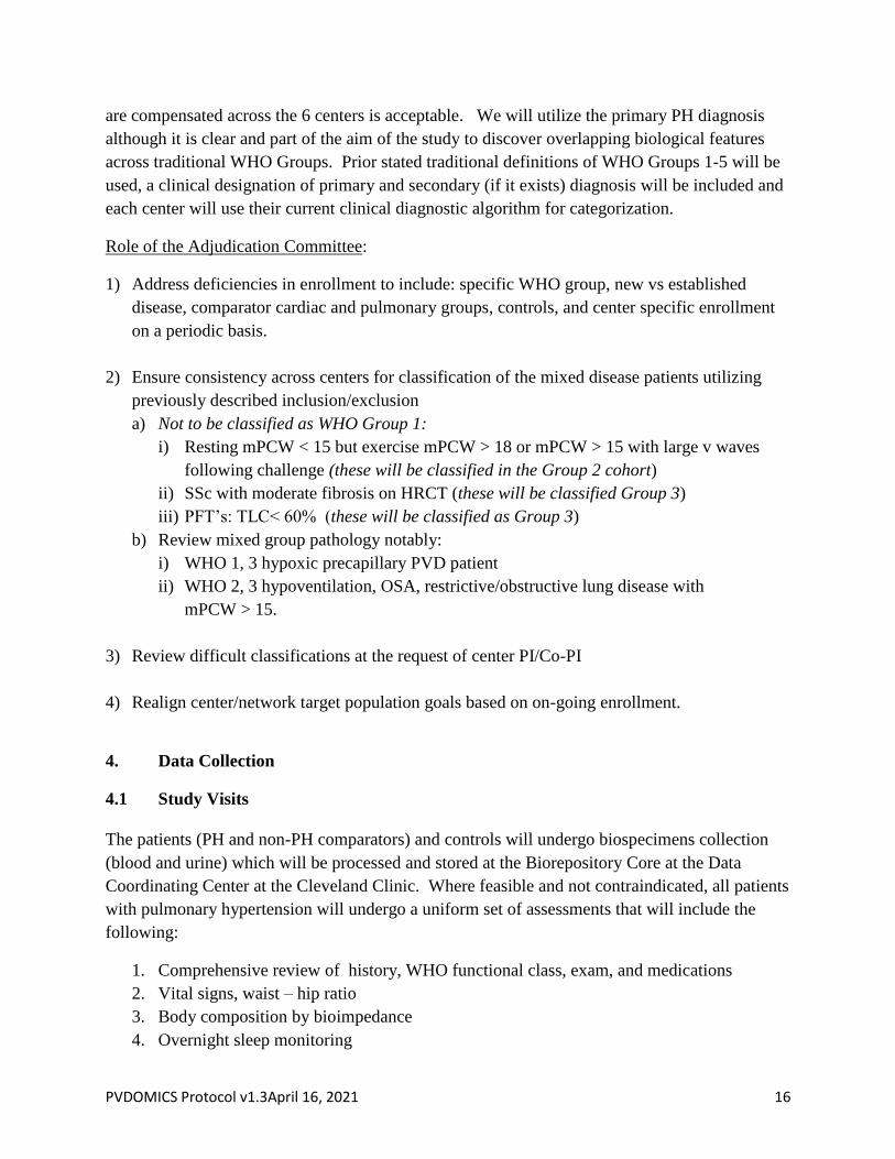
Role of the Adjudication Committee:
1) Address deficiencies in enrollment to include: specific WHO group, new vs established
disease, comparator cardiac and pulmonary groups, controls, and center specific enrollment
on a periodic basis.
2) Ensure consistency across centers for classification of the mixed disease patients utilizing
previously described inclusion/exclusion
a) Not to be classified as WHO Group 1:
i) Resting mPCW < 15 but exercise mPCW > 18 or mPCW > 15 with large v waves
following challenge (these will be classified in the Group 2 cohort)
ii) SSc with moderate fibrosis on HRCT (these will be classified Group 3)
iii) PFT’s: TLC< 60% (these will be classified as Group 3)
b) Review mixed group pathology notably:
i) WHO 1, 3 hypoxic precapillary PVD patient
ii) WHO 2, 3 hypoventilation, OSA, restrictive/obstructive lung disease with
mPCW > 15.
3) Review difficult classifications at the request of center PI/Co-PI
4) Realign center/network target population goals based on on-going enrollment.
4. Data Collection
4.1 Study Visits
The patients (PH and non-PH comparators) and controls will undergo biospecimens collection
(blood and urine) which will be processed and stored at the Biorepository Core at the Data
Coordinating Center at the Cleveland Clinic. Where feasible and not contraindicated, all patients
with pulmonary hypertension will undergo a uniform set of assessments that will include the
following:
1. Comprehensive review of history, WHO functional class, exam, and medications
2. Vital signs, waist – hip ratio
3. Body composition by bioimpedance
4. Overnight sleep monitoring

PVDOMICS Protocol v1.3April 16, 2021 17
5. Complete PFTs: spirometry, lung volumes and DLCO
6. Assessments of exercise tolerance (6 minute walk, cardiopulmonary exercise testing)
7. Complete HRQOL surveys (SF-36 v2®, MLHF, emPHasis-10)
8. Echocardiography
9. Cardiac MRI
10. Lung CT images
11. Ventilation perfusion scan
12. Cardiopulmonary exercise test
13. Right heart catheterization with inhaled nitric oxide vasodilator volume loading with 500
cc saline if not contraindicated, sampling of pulmonary artery, pulmonary capillary
wedge, and systemic arterial blood (if arterial line clinically indicated). Exercise will be
performed in selected centers and patients.
14. Phlebotomy for omics measurements
Table 4.1 lists all the measurements and tests that are to be collected. However, studies may be
scheduled over the course of 6 weeks. Suggested order of items and visit components are given
in the Manual of Operations. Test requirements are:
Right heart catheterization (with invasive CPET at centers performing this),
echocardiogram, MRI and omics blood draws should be performed within two weeks, but
data can be used if it is within one month allowing for individual needs.
The MRI can be performed either before or after the right heart catheterization. If they
are scheduled for the same day, the MRI ideally should be performed either before the
catheterization or usually a minimum of one hour after the right heart catheterization.

PVDOMICS Protocol v1.3April 16, 2021 18
Table 4.1
PVDOMICS Measurements and Tests
Measurements and Tests
Being Performed
Allotted Time and Subject
Accommodations
Review and sign consent
Medical history, review medications, NYHA functional class, allergies
Vital signs
Physical exam
Waist to hip circumference, Body composition bioimpedance
HRQOL surveys
Blood tests (CBC, CMP, NT BNP, HIV, Hepatitis, Rheum panel)
ECG
Echocardiogram
Spirometry, DLCO, Body Box* then 6 MWT
Clinically appropriate pre-catheterization discussion will have occurred
Overnight sleep monitoring
Cardiac Catheterization with lab work before, during and after
CPET
Recovery
MRI
High res CT*
Ventilation-perfusion lung scan*
Parking and lunch.
Wear comfortable clothes and shoes to walk in
Visit may require a hotel stay to get all events completed.
1-2 hours for consent, medication, medical history and exam
1 hour for blood tests waist to hip circumference, bioimpedance
1 hour for echo
2 hours for PFTs
30 min for HRQOL surveys
4-6 hour fast prior to some tests
* Acceptable if adequate test was performed within 1 year (3 months if have lung
disease) prior to enrollment

PVDOMICS Protocol v1.3April 16, 2021 19
4.2 Baseline Evaluation and Data Collection
Controls will undergo the same tests as the PH Groups (see Sections below). Healthy controls
will also have all the tests listed below except for a right heart catheterization (RHC), ventilation
perfusion lung scan and invasive CPET.
4.2.1 Demographics and History
Birth date, gender, race, ethnicity, zip code of most frequent domicile will be recorded.
Family history of pulmonary arterial hypertension
Occupational exposures (30-32) (for example mining, industrial solvents): duration
Infectious disease history (33-35) (HIV, Herpes virus, tuberculosis, tick-borne diseases, parasitic
diseases)
4.2.2 Quality of Life Questionnaires
Quality of life questionnaires will be completed by each patient. This includes the 36-item
health survey SF-36v2® (36), the Minnesota Living with Heart Failure (MLHF) Questionnaire
(37) and the EmPHasis10 survey (38).
4.2.3 Comorbid Conditions
Tobacco use (None ever, prior, current, total pack years)
Alcohol use (Current, number of drinks per week), Alcohol abuse history (none, prior, current)
Diabetes Type I, Type II, year of diagnosis, therapy (diet, oral agent (type), insulin)
Sleep disordered breathing (year of diagnosis, duration of treatment, current type of treatment)
Systemic hypertension (year of diagnosis, medications)
Cardiac diagnoses (arrhythmias, valve disease, rheumatic disease, CAD, etc.)
Pulmonary diagnoses (if biopsy proven, provide pathologic subtype)
Renal insufficiency (dialysis, history of renal transplant, fistula, meds)
Liver disease
Obesity

PVDOMICS Protocol v1.3April 16, 2021 20
Prior diet or methamphetamine drug exposure (name of medication and years of exposure)
Prior malignancy aside from localized non-melanoma skin cancer
Prior exposure to chemotherapeutic drugs (e.g., cyclophosphamide (39), multi-tyrosine kinase
inhibitors (40, 41))
Prior chest radiation
Prior chemotherapy
High altitude exposure (duration)
Estrogen containing medication exposure (duration)
History of DVT or PE or hypercoagulability
Relevant surgical history:
Cardiac surgery (type, date)
Previous atrial septostomy (date)
Pulmonary surgery (type, date)
Gastric bypass or banding (type, date)
Pacemaker
Defibrillator
4.2.4 Medications
A complete list of medications including dose, and over the counter or homeopathic products
should be compiled within 1 week of study entry. For intravenous or subcutaneous prostanoids
provide dose range, duration and peak dose (ng/kg/min). Oxygen use should be described,
including description of dose and when used (night, day, exertion, and approximate number of
hours per 24 hours). Historical PH therapy should be noted. For medications of special interest
(diuretics, any medications for heart or lung disease including pulmonary hypertension, use of
oxygen, and anticoagulants, the approximate duration of therapy should be categorized (less than
2 weeks, between 2 weeks and 3 months, 3 months to 12 months, or greater than 1 year).
4.2.5 Physical Measurements
Vital Signs

PVDOMICS Protocol v1.3April 16, 2021 21
Height, weight, Heart rate, rhythm, seated blood pressure, oxygen saturation (room air, or
specify oxygen amount). For patients with CHD (PDA) provide pre and post ductal saturation
(Right Arm and leg).
Waist – Hip Ratio
The waist circumference will be measured at the top of the iliac crest as illustrated in the Manual
of Operations. (42)
Body Composition by Bioelectrical Impedance Analysis (43, 44)
Bioelectrical impedance analysis (BIA) will be performed utilizing the Tanita 240S single
frequency device (Tanita Corporation of America, Arlington Heights, Illinois), except at
Vanderbilt, which may utilize the Tanita BC418, which is capable of the same measurement
technique. BIA will not be performed in patients with implantable pacemakers, defibrillators, or
electrical pumps.
4.2.6 Lung Function Measurements (45-49)
Spirometry, lung volumes and DLCO will be performed in all subjects unless test data from
within the past year is available. Subjects with evidence of parenchymal lung disease will be
required to have PFTs performed within 3 months of enrollment. PFTs are only acceptable if
done at a PVDOMICS-approved site using standardized, validated and agreed upon protocols.
Total lung capacity will be done by body plethysmography. Repeat PFTs closer to time of
enrollment will depend on clinical evaluation of possible change in functional status as assessed
by the investigator.
4.2.7 Six Minute Walk Test (50)
Six minute walk test will be performed in accordance with American Thoracic Society
guidelines. Continuous pulse oximetry should be used throughout the test. Resting O2 sat and
heart rate will be obtained and again at the end of the walk test. A Borg Dyspnea score will also
be acquired.
4.2.8 Overnight Sleep Monitoring (51)
Overnight sleep monitoring will be performed routinely in all enrollees to acquire standardized
baseline information on sleep disordered breathing and to quantify overnight oxygen saturation
patterns. Patients already receiving oxygen supplementation and/or positive airway pressure
therapy on a regular, nightly basis will be studied during use of these. A portable home
monitoring system (Nox T3, Carefusion) will be used, with recordings of nasal airflow, chest
wall and abdominal motion as well as pulse oximetry, heart rate, snoring and position. These
measures will permit scoring of central and obstructive apneas and hypopneas as well as to
quantify level of oxygen desaturation. Each center will purchase the equipment necessary to

PVDOMICS Protocol v1.3April 16, 2021 22
perform these studies, and study personnel will be trained in the proper application of the
devices. The results of the portable sleep monitoring will be provided to physicians caring for the
patients. The results of prior sleep studies as well as any additional testing or interventions
obtained as a consequence of the portable monitoring will be recorded. Sleep studies will be
scored using standardized methods for home sleep apnea testing and will conform to American
Academy of Sleep Medicine guidelines. The summary data will be electronically transmitted to
the DCC for integration into the database and for statistical analysis If this information is
available on a clinical basis within 1 year prior to study entry and with no change in nighttime
oxygen or sleep disorder therapy, and in the opinion of the investigator, no change in patient
condition that would impact results, then the clinically available data may be considered
adequate for inclusion in the case report form – if available, a copy of that study be acquired for
central review, quality assessment and scoring.
4.2.9 Electrocardiogram (3)
All participants should have an electrocardiographic test (12 lead ECG) done after enrollment.
4.2.10 Imaging Studies
4.2.10.1 Echocardiography
Inclusion criteria:
All subjects considered for participation in the PVDOMICS study as either PH or non-PH
comparators or controls will undergo a standardized transthoracic Echo exam (complete Echo
protocol is available for review in the MOP).
Exclusion criteria:
Clinical or hemodynamic instability requiring immediate therapy
Inability to communicate with the sonographer/follow commands for any reason and/or
provide consent (psychosis, agitation, etc.)
The PVDOMICS Echo protocol consists of the mandatory clinical protocol (based on the
standard ASE views), and an optional research protocol with 3D full volume LV and RV dataset.
Study subjects without known congenital systemic to pulmonary shunts, who did not have an
agitated saline test on their prior Echo, will need to undergo a peripheral IV line insertion and
injection of the agitated saline (“bubble study”) in order to rule out intracardiac shunt.
The Echo for the study will be performed only by the sonographers certified and trained in the
common and optional protocols based on the sonographer’s PVDOMICS Echo manual (see the
Manual of Operations).

PVDOMICS Protocol v1.3April 16, 2021 23
The sites’ Echo reports and images of the study subjects will be readily available for clinical
decision making by the local clinical teams and study investigators.
All of the Echo studies will be collected as DICOM files and uploaded by the local study
coordinators into the Echo Core Lab server for further assessment. The Echo variables for the
PVDOMICS dataset inclusion will be derived solely from the Core Lab data.
Justification
The Transthoracic Echocardiography will objectively reveal: (52-66)
Anatomy and morphology of the cardiac chambers
Presence or absence of structural heart disease
Hemodynamic status, volume status and functional performance (systolic and diastolic)
of the RV and the LV
Clinical features predominantly consistent with the WHO Group 2 PHT.
Gross anatomy and morphology of great vessels (aorta; main pulmonary artery)
Findings suggestive of intracardiac shunt
Presence or absence of a pericardial effusion
4.2.10.2 Cardiac MRI
Inclusion criteria:
All subjects considered for participation in the PVDOMICS study as either PH or non-PH
comparators or controls will undergo a standardized Cardiac MRI exam (complete MRI protocol
is available for review in the MOP).
Exclusion criteria:
Standard MRI contraindications; these may vary among the participating sites and may
depend on local policies/experience (example – scanning patients with permanent
pacemakers/ICDs)
Inability to obtain reliable gating data during the scan (significant arrhythmias with
highly irregular RR intervals (atrial fibrillation); significant tachycardia in NSR (HR
>120 bpm)
Clinical or hemodynamic instability requiring immediate therapy; severe dyspnea with
inability to lay flat/breath hold.
Inability to communicate with the MRI technician/follow commands for any reason
(psychosis, agitation, etc.)

PVDOMICS Protocol v1.3April 16, 2021 24
The PVDOMICS Cardiac MRI protocol consists of the mandatory common part and optional
part (please review the MOP).
The common MRI protocol will include the following:
Assessment of cardiac structure and function (SSFP sequences)
Hemodynamic assessment of valvular flow (phase-contrast imaging), including Qp/Qs
Tissue characterization, evaluation of inflammation, assessment of possible cardiac
masses/thrombi (T1 and T2 imaging)
Assessment of myocardial scar/fibrosis (delayed Gadolinium hyperenhancement
imaging)
- Non-contrast cMRI should still be performed in subjects selected with ESRD with
GFR<30, or gadolinium allergy.
Assessment of the pulmonary artery anatomy
The optional MRI protocol will include the following:
Assessment of extracellular volume fractionation (using pre-contrast T1 mapping)
Pulmonary artery compliance and vessel wall remodeling (high resolution sequences)
The sites’ MRI reports and images of the study subjects will be available for clinical decision
making and research considerations for the local clinical teams and study investigators.
All PVDOMICS MRI studies will be collected as DICOM files and uploaded by the local study
coordinators into the MRI Core Lab server for further assessment. The MRI variables for the
PVDOMICS dataset inclusion will be derived from the Core Lab data.
Justification
Cardiac MRI will objectively reveal: (60-66)
Fine details of the anatomy and morphology of cardiac chambers
Functional performance (systolic and diastolic function) of the RV and the LV;
reproducible volumetric data
Clinical features consistent with the WHO Groups 1, 2, 4 and 5.
Anatomy and morphology of great vessels (aorta; main pulmonary artery)
Findings suggestive of intracardiac shunt (anatomy; flow; Qp/Qs)
Presence of scar/fibrosis of the LV and/or RV

PVDOMICS Protocol v1.3April 16, 2021 25
4.2.10.3 High Resolution Chest CT (7, 67)
Non-contrast high resolution chest CT should be performed in all patients, unless the patient
refuses. This can be done within one year prior to enrollment if the images are available to the
DCC.
4.2.10.4 Ventilation Perfusion Lung Scan (3, 7, 67)
All patients are required to have a ventilation-perfusion lung scan for detection of possible
pulmonary emboli. V/Q scans performed within four years prior to enrollment can be used
provided acceptable results (no images required) are made available to the DCC. Also, a V/Q
scan is not required if the participant has lung parenchymal disease and has had a CT angiogram
(CTA) in the past year from which the results (images) are provided to the DCC Lung Imaging
Core.
4.2.11 Right Heart Catheterization
Right heart catheterization (RHC) is performed to confirm the diagnosis of PH, establish the
severity of disease and response to therapies, and assess prognosis (3, 68-72). All WHO Group
1-5 patients will undergo right heart catheterization as well as individuals in the comparator
groups. The study is expected to provide an assessment of right heart and pulmonary artery
filling pressures, pulmonary vascular resistance, cardiac output, and compartment-specific
oxygen saturations. All RHCs will be reviewed by the study’s central RHC Core.
During the right heart catheterization, pulmonary vasodilator testing will be done by acute
challenge with 100% oxygen, inhaled nitric oxide, and fluid loading if no contraindications.
(Fluid loading will not be performed if right heart catheterization is coupled with invasive
cardiopulmonary exercise testing). Blood samples will be collected for –omics testing.
A. General:
1. RHC to be performed using a minimum size 6F balloon occlusion catheter – this will prevent
shearing and activation of blood cells during sample acquisition.
2. Administration of heparin should be avoided; if patient is on heparin or low molecular weight
heparin, the drug should be stopped at least 30 minutes or 8 hours, respectively, prior to
catheterization unless medically necessary.
B. Measurements:
The following measurements or calculations detailed below will be collected as appropriate.
Each hemodynamic measurement should be performed at each stage and should be made both
during spontaneous breathing and during a brief expiratory pause, taking care to avoid Valsalva:
1. Vital signs a. Systemic blood pressure – systolic, diastolic, mean

PVDOMICS Protocol v1.3April 16, 2021 26
recorded as close as possible to time of PA pressure recording
b. Heart rate and rhythm – recorded at time of PA blood sample
c. Hemoglobin
d. BSA.
2. Pressure measurements:
a. Mean right atrial pressure (mid-A wave), peak A wave, peak V wave
pressures
b. Right ventricular systolic, minimal, and end-diastolic pressures
c. Pulmonary artery systolic, diastolic and mean pressure
d. Mean pulmonary capillary wedge pressure – mid a wave, peak a wave,
peak V wave (73)
e. Diastolic pressure gradient pull back (wedge to PA during quiet, held
end-expiration).
3. Oxygen saturations obtained during baseline pressure measurements:
a. Mixed venous oxygen saturation – should be obtained from the
pulmonary artery
b. Superior vena cava and pulmonary artery as screen to rule out shunt
c. Arterial if line in place; if not, finger oximetry saturation.
4. Blood samples obtained for -OMICs: volumes/types of tubes determined from OMICs
protocol.
a. Peripheral venous (26.5 ml)
b. Pulmonary artery (6.5 ml)
c. Pulmonary capillary wedge (6.5 ml)
d. Arterial – only if line already in place (6.5 ml).
5. Thermodilution/Fick cardiac output:
a. All sites will obtain thermodilution cardiac output (TDCO). A direct
Fick cardiac output will also be obtained if the study site is able to do
so.
b. Record VO2 (either directly measured or assumed)
c. The following will be calculated:
Cardiac index
Calculate stroke volume: SV = CO (TD and/or Fick)/HR
Calculate SV/PP (stroke volume/pulse pressure)
Pulmonary vascular resistance (PVR)
Systemic vascular resistance (SVR)
Right ventricular stroke work index (RVSWI)
Diastolic pulmonary vascular gradient (DPG)
Pulmonary to systemic flow ratio (Qp/Qs).
C. Challenges (74-76)
1. 100% Oxygen (77)
a. Performed in all patients except with known CO2 retention or those with
mPAP <20 mmHg

PVDOMICS Protocol v1.3April 16, 2021 27
pH < 7.32 AND pCO2 > 50 mmHg – contraindicated
pH > 7.32 AND pCO2 > 50 mmHg – at the discretion of the physician
b. Inhale 100% O2 for 5 minutes – through a face mask
c. Record measurements from B1, B2, B3, and B5
d. No washout period required.
2. Inhaled NO (78-83)
a. Performed in all patients except with PCWP ≥ 25 mmHg or those with
mPAP < 20 mmHg
b. Inhale iNO 40 ppm (added on to 100% oxygen) for 5 minutes
c. Record measurements from B1, B2, B3, and B5
d. Stop iNO and 100% oxygen.
3. Fluid challenge (84-86)
a. Performed in all patients except with baseline RA ≥ 15 or
PCWP ≥ 18 mmHg or having an invasive CPET (selection likely based
on center availability of invasive CPET)
b. Infuse 500 ml of 37°C 0.9% saline intravenously over 10 min
c. Record measurements from B1, B2, B3, and B5
d. Complete study.
Vasoresponders
Recognizing that acute vasoreactivity testing at the time of right heart catheterization has been
primarily defined by relatively arbitrary criteria to individuals who have non-Group 2 PH, we
will assess the phenotype of 'responder' to acute vasoreactivity testing. The response to
vasoreactivity testing at the time of right heart catheterization will be collected. A priori, there
will be a classification assigned as 'responder' or 'nonresponder' based on the change in the
pulmonary artery pressure from select agents such as inhaled nitric oxide. In PVDOMICS, we
will use current guidelines to classify the participant as a responder to the challenges if there is a
fall in the mean pulmonary artery pressure (mPAP) from baseline of 10 mmHg or greater to a
mean pulmonary artery pressure below 40 mmHg, without a fall in cardiac output. As a goal of
PVDOMICS, we will collect data for evidence-based assignment to a responder phenotype class
of PVD.
4.2.12 Cardiopulmonary Exercise Testing (CPET) (87-94)
Patients and age and gender matched sedentary controls absent the exclusion criteria below will
undergo a maximum incremental non-invasive CPET or an invasive CPET. To ensure consistent
stress and measurements, all studies should be performed in an upright cycle ergometry test.
Exclusion criteria:
Current exercise-induced angina
Inability to cycle (e.g., arthritis)

PVDOMICS Protocol v1.3April 16, 2021 28
Exertional syncope
Known potentially lethal exercise induced arrhythmia (e.g., ventricular tachycardia)
Severe symptomatic aortic stenosis
Resting systolic blood pressure ≥ 200 mmHg
The non-invasive CPET will consist of:
Maximum incremental cycling to exhaustion with:
o Breath by breath metabolic cart measurements of ventilation and pulmonary gas
exchange
o Pulse oximetry
o 12 lead ECG every minute
At some of the centers the CPET will be invasive (90), adding:
Radial artery line (optional)
IJ or brachial vein pulmonary artery line
Hemodynamics (RAP, PAP, PCW and AO, if available) obtained at each workload
increment. These measurements will be captured during spontaneous breathing and
during a brief expiratory pause if possible.
Cardiac output will be obtained by direct Fick when possible.
Blood collection from the pulmonary artery and the arterial line should be drawn at rest
(during the initial right heart catheterization), peak exercise, and 15 minutes post-
exercise. At each time point, 6.5 ml of blood should be drawn from the pulmonary artery
and, if one is already in place, the arterial line.
Justification:
The non-invasive CPET will objectively determine the degree of exertional impairment
(VO2peak, AT, OUES) and rule out a pulmonary mechanical limit (e.g., Group 3). It will
also estimate disease severity (ventilatory efficiency) (92) and suggest Group 1 vs. 2
during exercise (PETCO2 change) (93).
The invasive CPET will may further define the exercise phenotype (Group 1 vs. 2) and
determine the role of limb skeletal muscle dysfunction (by systemic O2 extraction), and
in mixed disease (i.e., combined Group 1 and 3) determine the functional limitation by
each contributor. Additionally, CPET can indirectly quantify gas exchange abnormalities
pertinent to pulmonary vascular disease such as dead space and A-a gradient.
4.2.13 Laboratory Measurements
Laboratory measurements and OMICS data will be integratively analyzed to understand the
molecular phenotype of pulmonary hypertension. Laboratory measurements will be performed
as necessary and may include (but not limited to): a complete blood count panel, metabolic panel

PVDOMICS Protocol v1.3April 16, 2021 29
(Glucose, Calcium, Albumin, Total Protein, Sodium, Potassium,CO2, Chloride, BUN,
Creatinine, ALP, ALT, AST, Bilirubin, Insulin), Cholesterol and Lipid panel (Total cholesterol,
HDL, LDL, Triglycerides), Cardiac injury panel (NT-ProBNP, Cardiac Troponin), inflammation
panel (MPO, C-reactive protein), iron metabolism panel (Iron, Ferritin, Transferrin) and
rheumatoid factor. Historical tests may be provided for HIV and hepatitis.
4.3 Biospecimen Collection
Blood (cells, serum, plasma) and urine samples will be collected for the molecular omics
analysis. Molecular network analysis incorporating genetics, genomics, epigenomics,
proteomics, metabolomics and coagulomics may be done in an unbiased manner but may also
take into account:
endothelial and smooth muscle cell biology with BMPR2 (18, 95, 96), KCKN3 (and
other ion channels) (97-101), endothelin (102-106), angiotensin (107, 108) , and
serotonin (109-114) mediator networks and circulating progenitor cells (115, 116) that
mark injury and repair processes
hypoxia signaling pathways (117-122)
coagulation: initiation of coagulation (procoagulant activity), coagulation cascade,
fibrinolysis (123-132)
markers of heart health(133-136) (BNP / ANP peptides; troponin), braveheart RNA (137)
function of hormone receptor signaling (e.g. estrogen receptor, aldosterone receptor)
(138-143)
cancer-like processes (144-147)
arachidonic acid signaling network (148-150)
nitric oxide synthases (NOS1-3) vs. arginase balance and phosphodiesterase 5 -
determinants of nitric oxide and guanylyl cyclase signaling (151-157)
metabolic shift in lung, vascular, heart and immune cells with mitochondrial remodeling;
metabolic syndrome, insulin resistance and type II diabetes (158-167)
matrix remodeling of the lungs, the pulmonary blood vessels and the heart (168-171)
(proteases to anti-protease imbalance; fibrosis )
oxidative stress in the cell environment and intracellular imbalance of the redox system
(172-175)
responses by the network hubs of Interleukin (IL)– 1 (176) and Tumor necrosis factor –
TNF (177-179) super-families
(131, 180-183) immune response mediators (network hubs of IL-13 / IL-4 including
resistin like molecule and the IL-33 receptor ST2 (135, 184-190); IL-17A (187, 191); IL-
6 (176, 177))
Interferon response (189, 192-197) (network hubs of type-I Interferons (IFN) and IFNγ)
(198-202)
Details of amounts and types of specimens are given in the Manual of Operations.

PVDOMICS Protocol v1.3April 16, 2021 30
4.4 Longitudinal Follow-up
All patients, or their designated contacts, will be contacted by telephone and/or letter at least
annually up to 10 years after enrollment if not seen at the enrolling center for follow-up. Vital
status and occurrence/date of any lung, heart or heart-lung transplantation will be determined
from this contact or by using center medical records. Cause of death will be ascertained by the
site investigators.
5. Statistical Considerations
We propose the following plan for discovering PVD types and subtypes using phenotype
and molecular measurements:
(1) When 750 subjects have been recruited, the first half of the anticipated study cohort, and
their phenotypic variables have been measured, including demographic and clinical variables, we
will perform model-based unsupervised clustering on this phenotypic data to cluster subjects into
novel phenotype-derived groups. Akaike Information Criteria (AIC) (203) and Bayesian
Information Criteria (BIC) (204) will be used to select the number of clusters. Based on a
preliminary analysis of 1900 subjects at the Cleveland Clinic, which have a subset of the planned
PVDOMICS phenotypic variables, we expect to derive four to six phenotype groups, with the
minimum group percentage being greater than 10%.
(2) 50 subjects will be chosen from each phenotype group in such a way as to maximize
variability across phenotype variables within each group. A full battery of omic measures, say M
variables, will be taken on biological samples from this subset, the omic discovery subject subset
(ODSS), with specific choices of biological layers (e.g., RNA, DNA, metabolome) and platforms
(e.g., sequencing, microarrays) to be determined.
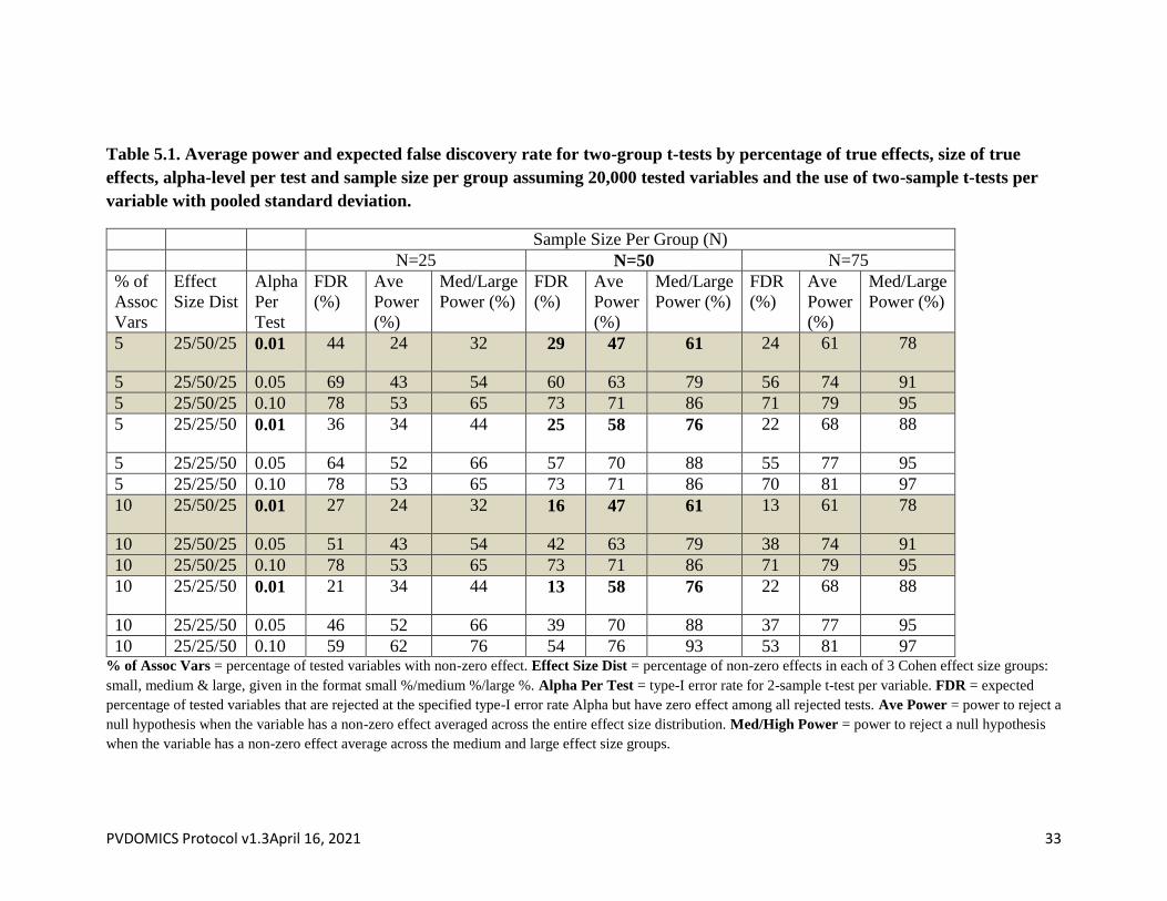
(3) Each omic variable will be tested for differences between each pair of phenotype groups
using the data from the ODSS. Power and expected false discovery rates for these comparisons
assuming M=20,000 omic measures are given in Table 5.1. Omic variables will be ranked by
their standardized effect sizes of differences between phenotype groups and their variability
within phenotype groups. Ranks will be combined using rank aggregation methods to generate a
single ranked list. The rank list will be pruned to account for correlations among omic variables.
Using the filtered rank list and considerations of cost and feasibility, P1 of the M omic variables
will be selected for measurement in all first-half subjects not in the ODSS, which we label
NODSS1. We anticipate NODSS1 will consist of between 300 and 500 subjects and expect P1 to
be much smaller than M.

PVDOMICS Protocol v1.3April 16, 2021 31
(4) After measuring the selected P1 omic variables in the NODSS1, clustering of the P1 omic
variables within each phenotype group, i.e., a form of supervised clustering, will be done to find
omic-based subtypes using the combined ODSS+NODSS1 subject sets. In addition, step (3)
above will be repeated using the combined subject sets and P1 omic variables on both the
phenotype groups alone and the omic-based subtypes to validate and filter the P1 omic variables.
A smaller number of omic variables, P2, may be chosen for measurement in the remaining half
of the study, labeled NODSS2, ~750 subjects.
(5) Finally, after measuring the phenotype variables and P2 omic variables on the NODSS2
subject set, step (4) will be repeated on the combined ODSS+NODSS1+NODSS2 set and on the
NODSS2 set alone. Adjusted Rand Index will be used to measure cluster stability of
ODSS+NODSS1 derived types and subtypes in the NODSS2 set.
Biological validity of the derived types and subtypes will be assessed by associating cluster
memberships with clinical outcomes, including survival times and event rates. We decided to
wait to form phenotype-based clusters until half of the proposed study size (1500) has been
enrolled to have a high probability (estimated from our preliminary data) of getting at least 50
subjects in each cluster. Our power calculations in Table 5.1 show that power and FDR for
discriminating omic variables between clusters fall below tolerable levels when there were less
than 50 subjects in a group. In those calculations, we assumed 20,000 omics variables were
measured in Step (2). While global DNA and mRNA measurements might be considered at this
step, leading to many more than 20,000 measures, we expect due to cost and information
considerations to measure a focused subset of these variables potentially combined with miRNA
and other lower dimensional omics platforms (e.g., protein arrays, metabolomics, coagulomics)
(205, 206). For example, if 10% of the 20,000 omic variables differ between 2 groups, with 50%
of these variables having a large effect, when performing testing at the 0.01 alpha level we have
76% power to detect a medium to large effect and overall FDR of 13% with 50 subjects per
group compared to just 44% power and overall FDR of 21% with 25 subjects per group.
Measuring all M omic variables on more than 50 subjects per group can substantially increase
power as shown in Table 5.1, but there may be insufficient funding to do complete measurement
in more than 50 samples per cluster assuming we find 4-6 phenotype clusters. If that situation
changes, either due to increased funding or decreased costs, we will consider increasing the omic
sample size per group. We propose to do multiple filtering steps on the omic variables to help
maximize the information content while minimizing costs. The extent of the filtering will depend
again on funding and costs per measure.
Our primary strategy is to first cluster/group by phenotypes in a larger subject set, determine the
most informative omic variables in a subset, and then determine molecular subtypes in the larger
set using the informative markers. This strategy fits well with the study’s primary Aims 1 and 2,
as Aim 1 is focused on understanding phenotype measures while Aim 2 is focused on exploiting
molecular measures. We will also perform cluster analyses where we cluster first with the omic
variables (in Steps (2), (4) & (5)), then either associate phenotypes with these molecular clusters

PVDOMICS Protocol v1.3April 16, 2021 32
or attempt to derive phenotypic subtypes within them. We will compare the phenotype-then-
omic to the omic-then-phenotype approaches based on biological validity, interpretability and
cluster overlap.
5.1 Statistical Power for Specific Hypotheses
Table 5.1 gives power and expected false discovery rates (FDR) for two-group comparisons for a
mix of effect sizes, group sizes, and alpha levels assuming 20,000 variables are measured. The R
package pwr was used to make the calculations. The nominal per-test alpha levels do not account
for multiple testing among cluster groups but since we are primarily interested in filtering omic
features and wish to include features that may only separate two of the groups, we are less
concerned about Type 1 errors. This table is useful for justifying our choice of 50 subjects to
measure complete omics in Step (2) as discussed above.
Table 5.2 gives minimum detectable fold changes (MDFC) between two independent groups for
various number of features tested, levels of variability (as measured by a common coefficient of
variation), feature intensity (as measured by average number of sequencing reads per feature)
and sample sizes. A desired power level of 80% and a conservative multiple test level per feature
of 0.10/# of assumed features were used in the calculations. The R package RNASeqPower,
which assumed that features followed a conditional negative binomial distribution and accounted
for sequencing based measurement error, was used to estimate the power of the designs. This
table can be used to assess our ability to detect meaningful differences in phenotype variables at
Steps (1)-(5) across phenotype clusters, assuming 50-500 phenotype variables. For example, if a
set of 50 phenotype variables are tested between phenotype-derived clusters (or across a priori
defined groups like WHO categories), we have 80% power to detect a fold change as small as
1.32 between groups assuming 100 subjects per group (cluster), phenotype variables are
measured with very little error (Feature Intensity=High) and variability across subjects is
medium (cv=0.5). This table can also be used to assess the MDFCs for omic variables measured
in Steps (2)-(5). For example, if after filtering in Step (3), we select P1=5000 omic variables to
measure in Step (4), we have a MDFC of 1.43 between two groups for say a gene expression
measurement assuming 40 subjects per group (subtype), gene expression is medium and
variability across subjects is small. Most of the MDFCs in Table 5.2 were below 2 for low to
medium variability scenarios, indicating that our designs in these scenarios are reasonably well
powered to find realistic fold changes from human samples.
PVDOMICS Protocol v1.3April 16, 2021 33
Table 5.1. Average power and expected false discovery rate for two-group t-tests by percentage of true effects, size of true
effects, alpha-level per test and sample size per group assuming 20,000 tested variables and the use of two-sample t-tests per
variable with pooled standard deviation.
Sample Size Per Group (N)
N=25 N=50 N=75
% of
Assoc
Vars
Effect
Size Dist
Alpha
Per
Test
FDR
(%)
Ave
Power
(%)
Med/Large
Power (%)
FDR
(%)
Ave
Power
(%)
Med/Large
Power (%)
FDR
(%)
Ave
Power
(%)
Med/Large
Power (%)
5 25/50/25 0.01 44 24 32 29 47 61 24 61 78
5 25/50/25 0.05 69 43 54 60 63 79 56 74 91
5 25/50/25 0.10 78 53 65 73 71 86 71 79 95
5 25/25/50 0.01 36 34 44 25 58 76 22 68 88
5 25/25/50 0.05 64 52 66 57 70 88 55 77 95
5 25/25/50 0.10 78 53 65 73 71 86 70 81 97
10 25/50/25 0.01 27 24 32 16 47 61 13 61 78
10 25/50/25 0.05 51 43 54 42 63 79 38 74 91
10 25/50/25 0.10 78 53 65 73 71 86 71 79 95
10 25/25/50 0.01 21 34 44 13 58 76 22 68 88
10 25/25/50 0.05 46 52 66 39 70 88 37 77 95
10 25/25/50 0.10 59 62 76 54 76 93 53 81 97 % of Assoc Vars = percentage of tested variables with non-zero effect. Effect Size Dist = percentage of non-zero effects in each of 3 Cohen effect size groups:
small, medium & large, given in the format small %/medium %/large %. Alpha Per Test = type-I error rate for 2-sample t-test per variable. FDR = expected
percentage of tested variables that are rejected at the specified type-I error rate Alpha but have zero effect among all rejected tests. Ave Power = power to reject a
null hypothesis when the variable has a non-zero effect averaged across the entire effect size distribution. Med/High Power = power to reject a null hypothesis
when the variable has a non-zero effect average across the medium and large effect size groups.

PVDOMICS Protocol v1.3April 16, 2021 34
Table 5.2. Minimum detectable fold change by subgroup size, feature intensity, feature variability and number of features
assuming desired 80% power, alpha level per test of 0.10/# of features.
Large Subgroups: 100 vs 100 Medium Subgroups: 40 vs 40 Small Subgroups: 10 vs
10
Feature Intensity Low Medium High Low Medium High Low Medium High
# Features Variability
20000 Low: cv=0.3 1.40 1.27 1.26 1.69 1.47 1.44 2.87 2.15 2.07
20000 Medium:
cv=0.5 1.57 1.48 1.47 2.04 1.85 1.83 4.18 3.43 3.36
20000 High: cv=1 2.23 2.16 2.15 3.55 3.37 3.35 12.63 11.36 11.23
5000 Low: cv=0.3 1.37 1.26 1.24 1.64 1.43 1.41 2.71 2.06 1.99
5000 Medium:
cv=0.5 1.53 1.45 1.44 1.97 1.79 1.77 3.86 3.20 3.14
5000 High: cv=1 2.13 2.07 2.06 3.31 3.15 3.13 10.97 9.93 9.82
500 Low: cv=0.3 1.32 1.23 1.21 1.56 1.38 1.36 2.43 1.91 1.85
500 Medium:
cv=0.5 1.46 1.39 1.38 1.83 1.68 1.67 3.34 2.83 2.78
500 High: cv=1 1.97 1.91 1.91 2.91 2.79 2.77 8.49 7.77 7.70
50 Low: cv=0.3 1.27 1.19 1.18 1.47 1.32 1.30 2.15 1.74 1.70
50 Medium:
cv=0.5 1.39 1.33 1.32 1.68 1.57 1.55 2.83 2.45 2.41
50 High: cv=1 1.79 1.75 1.74 2.51 2.42 2.41 6.32 5.85 5.81
Feature Intensity
Low=10 reads per feature Variability cv=coefficient of variation
Medium=100 reads per feature
High=1000 reads per feature

PVDOMICS Protocol v1.3April 16, 2021 35

PVDOMICS Protocol v1.3April 16, 2021 36
5.2 General Methods
In general, all quantitative variables will be summarized with standard descriptive statistics and
represented graphically with displays such as box plots, histograms and density function plots.
5.3 Methods by Aims
Aims 1 and 2a analyses will focus on the comparison of quantitative variables between current
WHO classifications, including comparisons to non-PH comparators and controls. The first part
will be comparing demographical, lab and other clinical variables. ANOVA will be used to
compare continuous variables and the chi-squared test will be applied to categorical variables.
Highly skewed variables may be transformed appropriately. In addition to comparisons across
existing classifications, we will also build prediction models for WHO classes plus non-PH
comparators using sparse regression methods (such as Elastic Net), random forests and multiple
fractional polynomials (207) in an effort to gain understanding of the predictability and drivers of
the existing systems. The second part will mainly be comparing various omic data, including
differential analysis, test for known PH mutations and new genetic variants, etc. Large amounts
of omic data will be generated by the Omic Cores. Much of the data will be high dimensional, on
multiple biological layers, e.g., transcriptome and metabolome, measured on tissues, e.g., blood,
from the same set of subjects. For example, we may receive mRNA-seq and Nanostring
nCounter miRNA measures of human blood transcriptome or mass spectrometry based measures
of the proteome and metabolome. Each layer presents challenges, sometimes unique, to
obtaining meaningful and reproducible inferences from relevant measures of that layer.
However, there is a common set of four broad steps we will apply to data from each layer to
address these challenges: (1) data acquisition, (2) quality checking and preprocessing, (3)
summarization and annotation and (4) statistical analysis. For example, with mRNA-seq data, the
four steps may involve (1) acquisition of fastq files generated by the sequencer, (2) trimming of
poor reads and alignment to the human reference genome or transcriptome to obtain bam files,
(3) counting aligned reads by Ensembl annotated genes or exons and (4) differential expression
analysis of gene or exon counts. We will attempt to incorporate the omic data into the prediction
models built using the clinical data.
Aim 2b analyses will aim to develop a new and more accurate classification of PH. To discover
molecular-based subtypes of PH, we will apply appropriate clustering or biclustering methods to
each layer, including methods that give estimates of the optimal number of subtypes such as
Bayesian mixture models, followed by feature reduction/selection to aid in interpretability and
reproducibility. Development of molecular-based subtypes and identification of features driving
the subtypes will aid in the development of a controlled vocabulary and new PH ontologies.
When feasible, we will attempt to build layer-specific network representations (208), e.g., co-
expression or mutual information networks from transcriptome data, to aid in understanding the
layer complexity and degree. Results from each layer will be compared and contrasted both
analytically and visually (209), e.g., by overlaying networks using Cytoscape. In addition, data

PVDOMICS Protocol v1.3April 16, 2021 37
from each layer will be associated with clinical outcomes and endophenotypes using sparse
regression methods to gain insight into the predictability of important downstream measures.
Because inferences from high-dimensional data are particularly prone to false discoveries and
non-reproducibility, and because we expect to get data from a large number of subjects over
multiple years (with some data not being produced for all subjects due to cost or tissue
availability), we will partition the work for each layer into two phases: discovery and validation.
In the discovery phase we will analyze data from subjects obtained in the early part of
enrollment, with the goal of finding subtypes, biomarkers and endophenotypes for later
validation, while in the validation phase we will analyze data from later subjects and measures
obtained at follow up(s). For example, we may build PH subtypes from mRNA-seq expression
profiles of for example, 300, early-recruited subjects, and then later validate the consistency and
relevance to clinical follow-up outcomes, such as death, of these profiles in the remaining
subjects with lower dimensional/less expensive measures.
While per-layer analysis will provide insight into PH/PVD pathobiology, the full power of the
proposed study comes from integrative analysis of the multiple biological layers, including the
clinical data layer. For example, while important changes in activity of some genes may be
reflected in their mRNA levels, other genes may show changes in protein levels or states but no
changes in mRNA level, perhaps through protein degradation/instability rather than protein
production. Integrative analysis is often more challenging than analysis of individual layers.
Recent methodological developments, mainly arising from similar desires of cancer and
cardiovascular researchers, have shown the feasibility and power of an integrative approach. We
will use the latest methods including Bayesian machine learning methods, multivariate
partitioning methods, sparse integrated clustering methods, and graphical models (210) to
maximize our ability to find meaningful PH subtypes, biomarkers and endophenotypes that use
the full spectrum of information available from multiple layers across samples. We will closely
monitor this fast moving area to ensure the best methods are chosen. For example, a recent
method that seems well suited to discovery of disease subtypes using multiple omic datasets
involves sparse integrative clustering.(211)
Aim 3 will examine the associations between various molecular biomarkers and longitudinal
outcomes. The mixed-effects models will be applied to repeated-measurement type of outcomes,
which include random effects to account for correlations of measurements within the same
subject. According to the variable type of the outcome, linear or generalized mixed models will
be used appropriate. The Cox models will be applied to time-to-event outcomes, e.g., death,
responses to therapy, etc. The proportional hazard assumption will be tested be checking the
Schoenfeld residuals.
Aim 4 to compare PVDOMIC genetic data with that of the Nichols R24 and other available
databases. These analyses will be mainly descriptive by appropriately summarizing and

PVDOMICS Protocol v1.3April 16, 2021 38
annotating the genetic variants identified from the PVODOMIC database and the Nichols R24
and other available database.
5.4 Missing Data
Maximum efforts will be made to minimize missing data. If necessary, the chained-equation
approach (212) will be used to impute missing values in all variables included in the analyses.
5.5 Software
All analyses will be performed with the open source R statistical and bioinformatic software. The
R environment includes cutting edge packages, many from the Bioconductor project, to perform
the bioinformatics analysis for every combination of biological layer, data type and analytical
goal we expect to see. In addition, R has strong interconnections to other important bioinformatic
and statistical tools and data sources, including Cytoscape for visualization, SAS for statistical
analysis, Oracle for database management and Ensemble for biological annotation.
6. Regulatory Considerations
6.1 Institutional Review Boards (IRBs)
The protocol will be submitted to the IRB of each clinical center for review and
approval. Centers may not recruit patients into the study until they have been documented as
being ready to enroll and their protocol has been approved by their IRB. Protocol amendments
and changes will be submitted to the IRB and approval must be received before
implementation. All patients enrolled in the study must sign and date an IRB-approved consent
form and medical records release form before any study related procedures are
undertaken. Study personnel will explain the study and answer all of the patient’s questions
before asking the patient to sign and date the consent form.
The Investigator will provide the Institutional Review Board (IRB) with all requisite material,
including a copy of the protocol, the informed consent and assent documents. The study will not
be initiated until the IRB provides written approval of the protocol, the informed consent form,
and the subject assent form, and until approved documents have been obtained by the
Investigator and copies received by the Sponsor. Appropriate reports on the progress of this
study by the Investigator will be made to the Institutional Review Board (IRB) and the Sponsor
in accordance with the applicable government regulations and in agreement with policy
established by the Sponsor.
6.2 Confidentiality and Ownership of Data and Biospecimens
All data and biospecimens will be maintained in secure locations. Data and biospecimens
collected from study evaluations will be identified by study identification codes. Identifying
PVDOMICS Protocol v1.3April 16, 2021 39
features including names and addresses will be known to the clinical center, but will not be
provided to the Data Coordinating Center (DCC) at the Cleveland Clinic.
The study will be conducted in compliance with applicable ICH guidelines, the ICH E6 GCP
guideline, and regulations, guidelines, and applicable laws of the local site where the study is
conducted. The study will be conducted with the approval of a duly constituted IRB/EC in
accordance with the requirement of US regulation Title 21 CFR Part 56-Institutional Reviews
Boards. The nature and risks of the study will be fully explained to each subject and written
consent obtained in accordance with the requirements of 21 CFR 50-Protectionof Human
Subjects. Subjects will be informed of their rights, including the right to withdraw from the
study at any time.
Use of study data and biospecimens, and resulting findings, during the PVDOMICS study are
subject to approval of the PVDOMICS Steering Committee and follow guidelines of the
Publications and Ancillary Study Committee. The data and biospecimens from subjects at a
clinical center are the property of the PVDOMICS study and are under the custody of that center.
The data and biospecimens are transmitted to the PVDOMICS DCC, but cannot be moved to
another institution unless approved by the PVDOMICS Steering Committee; for example, if the
Principal Investigator of the center moves to another place. At the conclusion of the
PVDOMICS study, the data and remaining biospecimens will be transferred to a PVDOMICS
specified biobank and/or to the National Heart, Lung and Blood Institute (NHLBI) Biological
Specimen and Data Repository Information Coordinating Center (BioLINCC) biobank and
become the property of one of these organizations. Future researchers may request data or
biospecimens from a biobank with approval following the guidelines of the biobank.
6.3 Subject Information and Consent
A properly executed, written informed consent in compliance with national and local regulation
and Good Clinical Practice (GCP) guidelines will be obtained from each prior to entering the
study or performing any study-related procedures that are not part of the subject’s standard care.
The Investigator will submit a copy of the informed consent document to the Institutional
Review Board (IRB) for review and approval before research subjects are enrolled. The
Investigator will provide a copy of the signed informed consent form to the subject and the
originals will be maintained in the subject’s study binder.
6.4 Adverse Events
A. Definition of an Adverse Event
Adverse event. An adverse event (AE) shall be considered any detrimental change in the
patient’s condition.
Anticipated adverse event. Anticipated adverse events are defined for each of the protocol
procedures in the sections specifically written for those procedures.

PVDOMICS Protocol v1.3April 16, 2021 40
Unanticipated adverse event. Any adverse event that results in risk or harm to the subject or
others that differs from the known, predicted possible effects of the research protocol. An
unanticipated adverse event is one that varies in nature, intensity, or frequency from information
in the informed consent document.
Serious adverse event. Any event that results in death, inpatient hospitalization or prolongation
of existing hospitalization, a persistent or significant disability or incapacity, or a congenital
anomaly or birth defect. Important medical events that may not result in death, be life-
threatening, or require hospitalization may be considered a serious adverse event when, based
upon appropriate medical judgment, they may jeopardize the patient or subject and may require
medical or surgical intervention to prevent one of the outcomes listed above.
B. Monitoring of adverse events related to the study
The occurrence of an AE may come to the attention of study personnel during study visits or
telephone interviews or by a patient presenting for medical care. The AEs that are expected from
participation in the study are given in the consent form.
6.5 Incidental Findings
In this study, research subjects, at risk cohort comparators, true controls and all five WHO
groups of pulmonary hypertension patients will be recruited. Subjects will undergo many tests
as part of the study protocol, including but not limited to, overnight sleep monitoring, complete
PFTs, echocardiography, cardiac MRI, chest CT, blood work and ventilation perfusion
scan. Each subject will identify a primary care provider and their contact information upon
entrance into the study. This will be kept in the research file at each site by the research
coordinator. Although these research tests are part of a research protocol, they may reveal
incidental findings (abnormalities) in study subjects that do not pertain to pulmonary
hypertension.
If there is a problem or abnormality found during the medical tests done at their site, the subject
will be notified about it by the research investigator there. If the subject would like, their
primary care provider can also be notified. If an abnormality is found by a PVDOMICS core
lab during research analyses, this finding will be reported by the DCC to the Principal
Investigator and the lead coordinator at the subject’s site. Results from genetic testing will only
be reported if the subject agrees at the start of the study to have these findings reported. Only
genetic results related to pulmonary hypertension risk or other clinically actionable genes (as
defined by the American College of Medical Genetics) will be reported. The central core
laboratory that is doing genetic testing is a research facility and does not have the ability to
provide genetic test results to the subjects. Therefore, the information will be conveyed to the
site PI in general terms only. Site investigators will refer the subject to a genetic counselor to
discuss options for clinical testing in a CLIA-approved laboratory. The cost of genetic
counseling and clinical genetic testing is not covered under the research protocol.

PVDOMICS Protocol v1.3April 16, 2021 41
The study site will provide acknowledgement to the DCC of their receipt of an incidental
finding. The study site once notified by the DCC is responsible for the subject notification. We
cannot forecast what incidental findings may be found on these tests and their clinical
significance. That is why this information will be provided to the clinical center Principal
Investigator that knows the subject at their site. Incidental findings will be given to the subject
and the health care provider by the study site personnel as further delineated in the Manual of
Procedures.
Many of the studies performed locally at a site will likely be placed into the subject’s medical
record, which means they would be available to the subject and their healthcare provider. If an
abnormality is found by a PVDOMICS core lab during research analyses, the results will not be
placed into the subject’s medical record.
6.6 Observational Safety and Monitoring Board (OSMB)
The OSMB was established by the NHLBI in accordance with NIH policies and is responsible
for monitoring of patient safety and review of study performance. The OSMB consists of a
chair, clinicians with expertise in pulmonary hypertension, bioethics, and biostatistics. An
NHLBI scientist other than the NHLBI’s Project Scientist serves as the Executive Secretary to
the OSMB. The OSMB meets once per year in person and by teleconference at other times as
necessary.
The purpose of monitoring is to verify that the rights and well-being of human subjects are
protected. The reported study data are accurate, complete, and verifiable from source documents.
The conduct of the study is in compliance with the currently approved protocol/amendment, with
GCP, and with applicable regulatory requirement(s). The OSMB will also provide oversight and
monitoring of ancillary studies (see Section 7).
Monitoring of Centers
Clinical Centers will be monitored primarily by on-going reports based on data collected in the
study database and by conference call discussions. Reports on recruitment, retention, adherence,
missing visits, missing procedures, missing data items, and procedural quality control will be
generated regularly (e.g., weekly) and posted on the study website or sent via email to
investigators and the NHLBI Project Officer, as appropriate.
Clinical centers will be site visited as part of the quality review of the performance of each site.
These visits may be performed at each center every other year unless needed more frequently.
Site visit reports should be provided that include the significant findings/facts, deviations and
deficiencies, conclusions, actions taken or to be taken, and/or actions recommended to secure
compliance.

PVDOMICS Protocol v1.3April 16, 2021 42
7. Ancillary Studies
To enhance the value of the PVDOMICS Study, the Steering Committee encourages ancillary
studies be done in collaboration with the PVDOMICS Study investigators. An ancillary study is one
based on information, images or biospecimens from the PVDOMICS Study participants in an
investigation that is relevant to, yet not part of the main goals of the PVDOMICS parent study, and
derives support from non-PVDOMICS Study funds. It is anticipated that a typical ancillary study
will propose the collection of additional data not collected or analyzed as part of the PVDOMICS
Study parent data set.
Participation in, and approval of, an ancillary study is subject to review by the PVDOMICS Steering
Committee. Also, the PVDOMICS Study Observational Study Monitoring Board (OSMB) will be
notified of all ancillary studies and will provide oversight. Namely, the OSMB will determine if
there are any safety concerns and if the study impacts the parent PVDOMICS study. The OSMB
will monitor ancillary studies in a similar manner as the parent study.
A detailed description of ancillary study principles, approval and operational procedures are
provided in Appendix 2 (which will also be in the PVDOMICS Manual of Operations).

PVDOMICS Protocol v1.1, June 16, 2016 43
8. References
1. Dweik, R. A., S. Rounds, S. C. Erzurum, S. Archer, K. Fagan, P. M. Hassoun, N. S. Hill, M. Humbert, S. M. Kawut, M. Krowka, E. Michelakis, N. W. Morrell, K. Stenmark, R. M. Tuder, J. Newman, and A. T. S. C. o. P. H. Phenotypes. 2014. An official American Thoracic Society Statement: pulmonary hypertension phenotypes. Am J Respir Crit Care Med 189: 345-355.
2. Erzurum, S., S. I. Rounds, T. Stevens, M. Aldred, J. Aliotta, S. L. Archer, K. Asosingh, R. Balaban, N. Bauer, J. Bhattacharya, H. Bogaard, G. Choudhary, G. W. Dorn, 2nd, R. Dweik, K. Fagan, M. Fallon, T. Finkel, M. Geraci, M. T. Gladwin, P. M. Hassoun, M. Humbert, N. Kaminski, S. M. Kawut, J. Loscalzo, D. McDonald, I. F. McMurtry, J. Newman, M. Nicolls, M. Rabinovitch, J. Shizuru, M. Oka, P. Polgar, D. Rodman, P. Schumacker, K. Stenmark, R. Tuder, N. Voelkel, E. Sullivan, R. Weinshilboum, M. C. Yoder, Y. Zhao, D. Gail, and T. M. Moore. 2010. Strategic plan for lung vascular research: An NHLBI-ORDR Workshop Report. Am J Respir Crit Care Med 182: 1554-1562.
3. Hoeper, M. M., H. J. Bogaard, R. Condliffe, R. Frantz, D. Khanna, M. Kurzyna, D. Langleben, A. Manes, T. Satoh, F. Torres, M. R. Wilkins, and D. B. Badesch. 2013. Definitions and diagnosis of pulmonary hypertension. J Am Coll Cardiol 62: D42-50.
4. Kim, N. H., M. Delcroix, D. P. Jenkins, R. Channick, P. Dartevelle, P. Jansa, I. Lang, M. M. Madani, H. Ogino, V. Pengo, and E. Mayer. 2013. Chronic thromboembolic pulmonary hypertension. J Am Coll Cardiol 62: D92-99.
5. McLaughlin, V. V., S. L. Archer, D. B. Badesch, R. J. Barst, H. W. Farber, J. R. Lindner, M. A. Mathier, M. D. McGoon, M. H. Park, R. S. Rosenson, L. J. Rubin, V. F. Tapson, J. Varga, D. American College of Cardiology Foundation Task Force on Expert Consensus, A. American Heart, P. American College of Chest, I. American Thoracic Society, and A. Pulmonary Hypertension. 2009. ACCF/AHA 2009 expert consensus document on pulmonary hypertension a report of the American College of Cardiology Foundation Task Force on Expert Consensus Documents and the American Heart Association developed in collaboration with the American College of Chest Physicians; American Thoracic Society, Inc.; and the Pulmonary Hypertension Association. J Am Coll Cardiol 53: 1573-1619.
6. Robbins, I. M., T. M. Moore, C. J. Blaisdell, and S. H. Abman. 2012. National Heart, Lung, and Blood Institute Workshop: improving outcomes for pulmonary vascular disease. Circulation 125: 2165-2170.
7. Seeger, W., Y. Adir, J. A. Barbera, H. Champion, J. G. Coghlan, V. Cottin, T. De Marco, N. Galie, S. Ghio, S. Gibbs, F. J. Martinez, M. J. Semigran, G. Simonneau, A. U. Wells, and J. L. Vachiery. 2013. Pulmonary hypertension in chronic lung diseases. J Am Coll Cardiol 62: D109-116.
8. Soubrier, F., W. K. Chung, R. Machado, E. Grunig, M. Aldred, M. Geraci, J. E. Loyd, C. G. Elliott, R. C. Trembath, J. H. Newman, and M. Humbert. 2013. Genetics and genomics of pulmonary arterial hypertension. J Am Coll Cardiol 62: D13-21.
9. Tuder, R. M., S. L. Archer, P. Dorfmuller, S. C. Erzurum, C. Guignabert, E. Michelakis, M. Rabinovitch, R. Schermuly, K. R. Stenmark, and N. W. Morrell. 2013. Relevant issues in the pathology and pathobiology of pulmonary hypertension. J Am Coll Cardiol 62: D4-12.
10. Vachiery, J. L., Y. Adir, J. A. Barbera, H. Champion, J. G. Coghlan, V. Cottin, T. De Marco, N. Galie, S. Ghio, J. S. Gibbs, F. Martinez, M. Semigran, G. Simonneau, A. Wells, and W. Seeger. 2013. Pulmonary hypertension due to left heart diseases. J Am Coll Cardiol 62: D100-108.
11. Voelkel, N. F., R. A. Quaife, L. A. Leinwand, R. J. Barst, M. D. McGoon, D. R. Meldrum, J. Dupuis, C. S. Long, L. J. Rubin, F. W. Smart, Y. J. Suzuki, M. Gladwin, E. M. Denholm, D. B. Gail, L. National Heart, C. Blood Institute Working Group on, and F. Molecular Mechanisms of Right Heart. 2006. Right ventricular function and failure: report of a National Heart, Lung, and Blood Institute

PVDOMICS Protocol v1.1, June 16, 2016 44
working group on cellular and molecular mechanisms of right heart failure. Circulation 114: 1883-1891.
12. Vonk-Noordegraaf, A., F. Haddad, K. M. Chin, P. R. Forfia, S. M. Kawut, J. Lumens, R. Naeije, J. Newman, R. J. Oudiz, S. Provencher, A. Torbicki, N. F. Voelkel, and P. M. Hassoun. 2013. Right heart adaptation to pulmonary arterial hypertension: physiology and pathobiology. J Am Coll Cardiol 62: D22-33.
13. Omenn, G. S. 2013. On best practices: the Institute of Medicine scheme for developing, validating, and demonstrating clinical utility of omics-based diagnostic and predictive tests. Proteomics Clin Appl 7: 748-755.
14. 2012. In Evolution of Translational Omics: Lessons Learned and the Path Forward. C. M. Micheel, S. J. Nass, and G. S. Omenn, eds, Washington (DC).
15. Said, S. I., and S. A. Hamidi. 2011. Pharmacogenomics in pulmonary arterial hypertension: Toward a mechanistic, target-based approach to therapy. Pulm Circ 1: 383-388.
16. Sutendra, G., and E. D. Michelakis. 2013. Pulmonary arterial hypertension: challenges in translational research and a vision for change. Sci Transl Med 5: 208sr205.
17. McDermott, J. E., J. Wang, H. Mitchell, B. J. Webb-Robertson, R. Hafen, J. Ramey, and K. D. Rodland. 2013. Challenges in Biomarker Discovery: Combining Expert Insights with Statistical Analysis of Complex Omics Data. Expert Opin Med Diagn 7: 37-51.
18. Drake, K. M., D. Zygmunt, L. Mavrakis, P. Harbor, L. Wang, S. A. Comhair, S. C. Erzurum, and M. A. Aldred. 2011. Altered MicroRNA processing in heritable pulmonary arterial hypertension: an important role for Smad-8. Am J Respir Crit Care Med 184: 1400-1408.
19. Tuder, R. M., S. H. Abman, T. Braun, F. Capron, T. Stevens, P. A. Thistlethwaite, and S. G. Haworth. 2009. Development and pathology of pulmonary hypertension. J Am Coll Cardiol 54: S3-9.
20. Pauciulo, M., K. Lutz, A. Reponen, C. Winslow, A. Walsworth, M. Barve, J. Harley, M. Barnes, K. Marsolo, L. Martin, and W. C. Nichols. 2014. National Biological Sample And Data Repository For Pulmonary Arterial Hypertension. Am J Respir Crit Care Med 189: A4746.
21. Chakinala, M., and M. McGoon. 2014. Pulmonary hypertension care centers. Advances in Pulmonary Hypertension 12: 175-178.
22. Simonneau, G., M. A. Gatzoulis, I. Adatia, D. Celermajer, C. Denton, A. Ghofrani, M. A. Gomez Sanchez, R. Krishna Kumar, M. Landzberg, R. F. Machado, H. Olschewski, I. M. Robbins, and R. Souza. 2013. Updated clinical classification of pulmonary hypertension. J Am Coll Cardiol 62: D34-41.
23. Celli, B. R., W. MacNee, and A. E. T. Force. 2004. Standards for the diagnosis and treatment of patients with COPD: a summary of the ATS/ERS position paper. Eur Respir J 23: 932-946.
24. Raghu, G., H. R. Collard, J. J. Egan, F. J. Martinez, J. Behr, K. K. Brown, T. V. Colby, J. F. Cordier, K. R. Flaherty, J. A. Lasky, D. A. Lynch, J. H. Ryu, J. J. Swigris, A. U. Wells, J. Ancochea, D. Bouros, C. Carvalho, U. Costabel, M. Ebina, D. M. Hansell, T. Johkoh, D. S. Kim, T. E. King, Jr., Y. Kondoh, J. Myers, N. L. Muller, A. G. Nicholson, L. Richeldi, M. Selman, R. F. Dudden, B. S. Griss, S. L. Protzko, H. J. Schunemann, and A. E. J. A. C. o. I. P. Fibrosis. 2011. An official ATS/ERS/JRS/ALAT statement: idiopathic pulmonary fibrosis: evidence-based guidelines for diagnosis and management. Am J Respir Crit Care Med 183: 788-824.
25. van den Hoogen, F., D. Khanna, J. Fransen, S. R. Johnson, M. Baron, A. Tyndall, M. Matucci-Cerinic, R. P. Naden, T. A. Medsger, Jr., P. E. Carreira, G. Riemekasten, P. J. Clements, C. P. Denton, O. Distler, Y. Allanore, D. E. Furst, A. Gabrielli, M. D. Mayes, J. M. van Laar, J. R. Seibold, L. Czirjak, V. D. Steen, M. Inanc, O. Kowal-Bielecka, U. Muller-Ladner, G. Valentini, D. J. Veale, M. C. Vonk, U. A. Walker, L. Chung, D. H. Collier, M. Ellen Csuka, B. J. Fessler, S. Guiducci, A. Herrick, V. M. Hsu, S. Jimenez, B. Kahaleh, P. A. Merkel, S. Sierakowski, R. M. Silver, R. W. Simms, J.

PVDOMICS Protocol v1.1, June 16, 2016 45
Varga, and J. E. Pope. 2013. 2013 classification criteria for systemic sclerosis: an American college of rheumatology/European league against rheumatism collaborative initiative. Ann Rheum Dis 72: 1747-1755.
26. Cottin, V., H. Nunes, P. Y. Brillet, P. Delaval, G. Devouassoux, I. Tillie-Leblond, D. Israel-Biet, I. Court-Fortune, D. Valeyre, J. F. Cordier, and P. Groupe d'Etude et de Recherche sur les Maladies Orphelines. 2005. Combined pulmonary fibrosis and emphysema: a distinct underrecognised entity. Eur Respir J 26: 586-593.
27. Epstein, L. J., D. Kristo, P. J. Strollo, Jr., N. Friedman, A. Malhotra, S. P. Patil, K. Ramar, R. Rogers, R. J. Schwab, E. M. Weaver, M. D. Weinstein, and M. Adult Obstructive Sleep Apnea Task Force of the American Academy of Sleep. 2009. Clinical guideline for the evaluation, management and long-term care of obstructive sleep apnea in adults. J Clin Sleep Med 5: 263-276.
28. Al Dabal, L., and A. S. Bahammam. 2009. Obesity hypoventilation syndrome. Ann Thorac Med 4: 41-49.
29. 1999. Statement on sarcoidosis. Joint Statement of the American Thoracic Society (ATS), the European Respiratory Society (ERS) and the World Association of Sarcoidosis and Other Granulomatous Disorders (WASOG) adopted by the ATS Board of Directors and by the ERS Executive Committee, February 1999. Am J Respir Crit Care Med 160: 736-755.
30. Montani, D., A. Descatha, E. Lau, J. Le Pavec, L. Savale, X. Jaïs, B. Girerd, I. Zendah, F. Perros, P. Dorfmuller, F. Soubrier, O. Sitbon, G. Simonneau, and M. Humbert. 2015. Occupational Exposure: A Risk Factor For Pulmonary Veno-Occlusive Disease. Am J Respir Crit Care Med 191: A5517.
31. Marchiori, E., S. Lourenco, T. D. Gasparetto, G. Zanetti, C. M. Mano, and L. F. Nobre. 2010. Pulmonary talcosis: imaging findings. Lung 188: 165-171.
32. Vearrier, D., and M. I. Greenberg. 2011. Occupational health of miners at altitude: adverse health effects, toxic exposures, pre-placement screening, acclimatization, and worker surveillance. Clin Toxicol (Phila) 49: 629-640.
33. Flores, S. C., and S. Almodovar. 2013. Human immunodeficiency virus, herpes virus infections, and pulmonary vascular disease. Pulm Circ 3: 165-170.
34. Thienemann, F., A. Dzudie, A. O. Mocumbi, L. Blauwet, M. U. Sani, K. M. Karaye, O. S. Ogah, I. Mbanze, A. Mbakwem, P. Udo, K. Tibazarwa, A. S. Ibrahim, R. Burton, A. Damasceno, S. Stewart, and K. Sliwa. 2014. Rationale and design of the Pan African Pulmonary hypertension Cohort (PAPUCO) study: implementing a contemporary registry on pulmonary hypertension in Africa. BMJ Open 4: e005950.
35. de Koning, J., J. A. Hoogkamp-Korstanje, M. R. van der Linde, and H. J. Crijns. 1989. Demonstration of spirochetes in cardiac biopsies of patients with Lyme disease. J Infect Dis 160: 150-153.
36. Maruish ME. (Ed.). User’s manual for the SF-36v2 Health Survey (3rd ed.). Lincoln, RI: QualityMetric Incorporated, 2011.
37. Rector TS, Kubo SH,., Cohn JN. 1987. Patients' Self-Assessment of Their Congestive Heart Failure: Content, Reliability and Validity of a New Measure, the Minnesota Living With Heart Failure Questionnaire. Heart Failure 3: 198-209.
38. Yorke, J., P. Corris, S. Gaine, J. S. Gibbs, D. G. Kiely, C. Harries, V. Pollock, and I. Armstrong. 2014. emPHasis-10: development of a health-related quality of life measure in pulmonary hypertension. Eur Respir J 43: 1106-1113.
39. Ranchoux, B., S. Gunther, R. Quarck, M. C. Chaumais, P. Dorfmuller, F. Antigny, S. J. Dumas, N. Raymond, E. Lau, L. Savale, X. Jais, O. Sitbon, G. Simonneau, K. Stenmark, S. Cohen-Kaminsky, M. Humbert, D. Montani, and F. Perros. 2015. Chemotherapy-induced pulmonary hypertension: role of alkylating agents. Am J Pathol 185: 356-371.

PVDOMICS Protocol v1.1, June 16, 2016 46
40. Hennigs, J. K., G. Keller, H. J. Baumann, F. Honecker, S. Kluge, C. Bokemeyer, T. H. Brummendorf, and H. Klose. 2011. Multi tyrosine kinase inhibitor dasatinib as novel cause of severe pre-capillary pulmonary hypertension? BMC Pulm Med 11: 30.
41. Montani, D., E. Bergot, S. Gunther, L. Savale, A. Bergeron, A. Bourdin, H. Bouvaist, M. Canuet, C. Pison, M. Macro, P. Poubeau, B. Girerd, D. Natali, C. Guignabert, F. Perros, D. S. O'Callaghan, X. Jais, P. Tubert-Bitter, G. Zalcman, O. Sitbon, G. Simonneau, and M. Humbert. 2012. Pulmonary arterial hypertension in patients treated by dasatinib. Circulation 125: 2128-2137.
42. Nishida, C., G. T. Ko, and S. Kumanyika. 2010. Body fat distribution and noncommunicable diseases in populations: overview of the 2008 WHO Expert Consultation on Waist Circumference and Waist-Hip Ratio. Eur J Clin Nutr 64: 2-5.
43. Lee, S. Y., and D. Gallagher. 2008. Assessment methods in human body composition. Curr Opin Clin Nutr Metab Care 11: 566-572.
44. Mulasi, U., A. J. Kuchnia, A. J. Cole, and C. P. Earthman. 2015. Bioimpedance at the bedside: current applications, limitations, and opportunities. Nutr Clin Pract 30: 180-193.
45. Macintyre, N., R. O. Crapo, G. Viegi, D. C. Johnson, C. P. van der Grinten, V. Brusasco, F. Burgos, R. Casaburi, A. Coates, P. Enright, P. Gustafsson, J. Hankinson, R. Jensen, R. McKay, M. R. Miller, D. Navajas, O. F. Pedersen, R. Pellegrino, and J. Wanger. 2005. Standardisation of the single-breath determination of carbon monoxide uptake in the lung. Eur Respir J 26: 720-735.
46. Miller, M. R., J. Hankinson, V. Brusasco, F. Burgos, R. Casaburi, A. Coates, R. Crapo, P. Enright, C. P. van der Grinten, P. Gustafsson, R. Jensen, D. C. Johnson, N. MacIntyre, R. McKay, D. Navajas, O. F. Pedersen, R. Pellegrino, G. Viegi, J. Wanger, and A. E. T. Force. 2005. Standardisation of spirometry. Eur Respir J 26: 319-338.
47. Stoller, J. K., S. Basheda, D. Laskowski, M. Goormastic, and K. McCarthy. 1993. Trial of standard versus modified expiration to achieve end-of-test spirometry criteria. Am Rev Respir Dis 148: 275-280.
48. Stoller, J. K., A. S. Buist, B. Burrows, R. G. Crystal, R. J. Fallat, K. McCarthy, M. D. Schluchter, N. T. Soskel, and R. Zhang. 1997. Quality control of spirometry testing in the registry for patients with severe alpha1-antitrypsin deficiency. alpha1-Antitrypsin Deficiency Registry Study Group. Chest 111: 899-909.
49. Wanger, J., J. L. Clausen, A. Coates, O. F. Pedersen, V. Brusasco, F. Burgos, R. Casaburi, R. Crapo, P. Enright, C. P. van der Grinten, P. Gustafsson, J. Hankinson, R. Jensen, D. Johnson, N. Macintyre, R. McKay, M. R. Miller, D. Navajas, R. Pellegrino, and G. Viegi. 2005. Standardisation of the measurement of lung volumes. Eur Respir J 26: 511-522.
50. Laboratories, A. T. S. C. o. P. S. f. C. P. F. 2002. ATS statement: guidelines for the six-minute walk test. Am J Respir Crit Care Med 166: 111-117.
51. Ismail, K., K. Roberts, P. Manning, C. Manley, and N. S. Hill. 2015. OSA and pulmonary hypertension: time for a new look. Chest 147: 847-861.
52. Bossone, E., A. D'Andrea, M. D'Alto, R. Citro, P. Argiento, F. Ferrara, A. Cittadini, M. Rubenfire, and R. Naeije. 2013. Echocardiography in pulmonary arterial hypertension: from diagnosis to prognosis. J Am Soc Echocardiogr 26: 1-14.
53. Janda, S., N. Shahidi, K. Gin, and J. Swiston. 2011. Diagnostic accuracy of echocardiography for pulmonary hypertension: a systematic review and meta-analysis. Heart 97: 612-622.
54. D'Andrea, A., R. Naeije, E. Grunig, P. Caso, M. D'Alto, E. Di Palma, L. Nunziata, L. Riegler, R. Scarafile, R. Cocchia, O. Vriz, R. Citro, R. Calabro, M. G. Russo, and E. Bossone. 2014. Echocardiography of the pulmonary circulation and right ventricular function: exploring the physiologic spectrum in 1,480 normal subjects. Chest 145: 1071-1078.
55. Grunig, E., P. Henn, A. D'Andrea, M. Claussen, N. Ehlken, F. Maier, R. Naeije, C. Nagel, F. Prange, J. Weidenhammer, C. Fischer, and E. Bossone. 2013. Reference values for and determinants of

PVDOMICS Protocol v1.1, June 16, 2016 47
right atrial area in healthy adults by 2-dimensional echocardiography. Circ Cardiovasc Imaging 6: 117-124.
56. Kong, D., X. Shu, L. Dong, C. Pan, L. Cheng, H. Yao, and D. Zhou. 2013. Right ventricular regional systolic function and dyssynchrony in patients with pulmonary hypertension evaluated by three-dimensional echocardiography. J Am Soc Echocardiogr 26: 649-656.
57. Lancellotti, P., W. Budts, D. De Wolf, J. U. Voigt, A. Pasquet, J. De Sutter, A. Friart, S. Huez, B. Paelinck, J. L. Vachiery, and I. Working Group on Non-Invasive Cardiac. 2013. Practical recommendations on the use of echocardiography to assess pulmonary arterial hypertension--a Belgian expert consensus endorsed by the Working Group on Non-Invasive Cardiac Imaging. Acta Cardiol 68: 59-69.
58. Lang, R. M., L. P. Badano, V. Mor-Avi, J. Afilalo, A. Armstrong, L. Ernande, F. A. Flachskampf, E. Foster, S. A. Goldstein, T. Kuznetsova, P. Lancellotti, D. Muraru, M. H. Picard, E. R. Rietzschel, L. Rudski, K. T. Spencer, W. Tsang, and J. U. Voigt. 2015. Recommendations for cardiac chamber quantification by echocardiography in adults: an update from the American Society of Echocardiography and the European Association of Cardiovascular Imaging. J Am Soc Echocardiogr 28: 1-39 e14.
59. Rudski, L. G., W. W. Lai, J. Afilalo, L. Hua, M. D. Handschumacher, K. Chandrasekaran, S. D. Solomon, E. K. Louie, and N. B. Schiller. 2010. Guidelines for the echocardiographic assessment of the right heart in adults: a report from the American Society of Echocardiography endorsed by the European Association of Echocardiography, a registered branch of the European Society of Cardiology, and the Canadian Society of Echocardiography. J Am Soc Echocardiogr 23: 685-713; quiz 786-688.
60. Benza, R., R. Biederman, S. Murali, and H. Gupta. 2008. Role of cardiac magnetic resonance imaging in the management of patients with pulmonary arterial hypertension. J Am Coll Cardiol 52: 1683-1692.
61. Kang, K. W., H. J. Chang, Y. J. Kim, B. W. Choi, H. S. Lee, W. I. Yang, C. Y. Shim, J. Ha, and N. Chung. 2011. Cardiac magnetic resonance imaging-derived pulmonary artery distensibility index correlates with pulmonary artery stiffness and predicts functional capacity in patients with pulmonary arterial hypertension. Circ J 75: 2244-2251.
62. Klein, A. L., S. Abbara, D. A. Agler, C. P. Appleton, C. R. Asher, B. Hoit, J. Hung, M. J. Garcia, I. Kronzon, J. K. Oh, E. R. Rodriguez, H. V. Schaff, P. Schoenhagen, C. D. Tan, and R. D. White. 2013. American Society of Echocardiography clinical recommendations for multimodality cardiovascular imaging of patients with pericardial disease: endorsed by the Society for Cardiovascular Magnetic Resonance and Society of Cardiovascular Computed Tomography. J Am Soc Echocardiogr 26: 965-1012 e1015.
63. Kruger, S., P. Haage, R. Hoffmann, C. Breuer, A. Bucker, P. Hanrath, and R. W. Gunther. 2001. Diagnosis of pulmonary arterial hypertension and pulmonary embolism with magnetic resonance angiography. Chest 120: 1556-1561.
64. Peacock, A. J., S. Crawley, L. McLure, K. Blyth, C. D. Vizza, R. Poscia, M. Francone, I. Iacucci, H. Olschewski, G. Kovacs, A. Vonk Noordegraaf, J. T. Marcus, M. C. van de Veerdonk, and F. P. Oosterveer. 2014. Changes in right ventricular function measured by cardiac magnetic resonance imaging in patients receiving pulmonary arterial hypertension-targeted therapy: the EURO-MR study. Circ Cardiovasc Imaging 7: 107-114.
65. Rajaram, S., A. J. Swift, D. Capener, C. A. Elliot, R. Condliffe, C. Davies, C. Hill, J. Hurdman, R. Kidling, M. Akil, J. M. Wild, and D. G. Kiely. 2012. Comparison of the diagnostic utility of cardiac magnetic resonance imaging, computed tomography, and echocardiography in assessment of suspected pulmonary arterial hypertension in patients with connective tissue disease. J Rheumatol 39: 1265-1274.

PVDOMICS Protocol v1.1, June 16, 2016 48
66. Vonk-Noordegraaf, A., and R. Souza. 2012. Cardiac magnetic resonance imaging: what can it add to our knowledge of the right ventricle in pulmonary arterial hypertension? Am J Cardiol 110: 25S-31S.
67. Piazza, G., and S. Z. Goldhaber. 2011. Chronic thromboembolic pulmonary hypertension. N Engl J Med 364: 351-360.
68. Andersen, M. J., and B. A. Borlaug. 2014. Invasive hemodynamic characterization of heart failure with preserved ejection fraction. Heart Fail Clin 10: 435-444.
69. Maron, B. A. 2015. Hemodynamics should be the primary approach to diagnosing, following, and managing pulmonary arterial hypertension. Can J Cardiol 31: 515-520.
70. Rich, J. D., and S. Rich. 2014. Clinical diagnosis of pulmonary hypertension. Circulation 130: 1820-1830.
71. Schwartzenberg, S., M. M. Redfield, A. M. From, P. Sorajja, R. A. Nishimura, and B. A. Borlaug. 2012. Effects of vasodilation in heart failure with preserved or reduced ejection fraction implications of distinct pathophysiologies on response to therapy. J Am Coll Cardiol 59: 442-451.
72. Shah, R. V., and M. J. Semigran. 2008. Pulmonary hypertension secondary to left ventricular systolic dysfunction: contemporary diagnosis and management. Curr Heart Fail Rep 5: 226-232.
73. Ryan, J. J., J. D. Rich, T. Thiruvoipati, R. Swamy, G. H. Kim, and S. Rich. 2012. Current practice for determining pulmonary capillary wedge pressure predisposes to serious errors in the classification of patients with pulmonary hypertension. Am Heart J 163: 589-594.
74. Costa, E. L., C. Jardim, H. B. Bogossian, M. B. Amato, C. R. Carvalho, and R. Souza. 2005. Acute vasodilator test in pulmonary arterial hypertension: evaluation of two response criteria. Vascul Pharmacol 43: 143-147.
75. Skoro-Sajer, N., N. Hack, R. Sadushi-Kolici, D. Bonderman, J. Jakowitsch, W. Klepetko, M. A. Hoda, M. P. Kneussl, P. Fedullo, and I. M. Lang. 2009. Pulmonary vascular reactivity and prognosis in patients with chronic thromboembolic pulmonary hypertension: a pilot study. Circulation 119: 298-305.
76. Tonelli, A. R., H. Alnuaimat, and K. Mubarak. 2010. Pulmonary vasodilator testing and use of calcium channel blockers in pulmonary arterial hypertension. Respir Med 104: 481-496.
77. Leuchte, H. H., C. J. Baezner, R. A. Baumgartner, P. Mernitz, C. Neurohr, and J. Behr. 2013. Acute hemodynamic responses to supplemental oxygen and their prognostic implications in pulmonary hypertension. Respiration 85: 400-407.
78. Abman, S. H. 2013. Inhaled nitric oxide for the treatment of pulmonary arterial hypertension. Handb Exp Pharmacol 218: 257-276.
79. Cockrill, B. A., R. M. Kacmarek, M. A. Fifer, L. M. Bigatello, L. C. Ginns, W. M. Zapol, and M. J. Semigran. 2001. Comparison of the effects of nitric oxide, nitroprusside, and nifedipine on hemodynamics and right ventricular contractility in patients with chronic pulmonary hypertension. Chest 119: 128-136.
80. Hoeper, M. M., H. Olschewski, H. A. Ghofrani, H. Wilkens, J. Winkler, M. M. Borst, J. Niedermeyer, H. Fabel, and W. Seeger. 2000. A comparison of the acute hemodynamic effects of inhaled nitric oxide and aerosolized iloprost in primary pulmonary hypertension. German PPH study group. J Am Coll Cardiol 35: 176-182.
81. Ichinose, F., J. D. Roberts, Jr., and W. M. Zapol. 2004. Inhaled nitric oxide: a selective pulmonary vasodilator: current uses and therapeutic potential. Circulation 109: 3106-3111.
82. Jiang, B. H., J. Maruyama, A. Yokochi, H. Amano, Y. Mitani, and K. Maruyama. 2002. Correlation of inhaled nitric-oxide induced reduction of pulmonary artery pressure and vascular changes. Eur Respir J 20: 52-58.

PVDOMICS Protocol v1.1, June 16, 2016 49
83. Krasuski, R. A., J. J. Warner, A. Wang, J. K. Harrison, V. F. Tapson, and T. M. Bashore. 2000. Inhaled nitric oxide selectively dilates pulmonary vasculature in adult patients with pulmonary hypertension, irrespective of etiology. J Am Coll Cardiol 36: 2204-2211.
84. Andersen, M. J., S. J. Hwang, G. C. Kane, V. Melenovsky, T. P. Olson, K. Fetterly, and B. A. Borlaug. 2015. Enhanced pulmonary vasodilator reserve and abnormal right ventricular: pulmonary artery coupling in heart failure with preserved ejection fraction. Circ Heart Fail 8: 542-550.
85. Fujimoto, N., B. A. Borlaug, G. D. Lewis, J. L. Hastings, K. M. Shafer, P. S. Bhella, G. Carrick-Ranson, and B. D. Levine. 2013. Hemodynamic responses to rapid saline loading: the impact of age, sex, and heart failure. Circulation 127: 55-62.
86. Robbins, I. M., A. R. Hemnes, M. E. Pugh, E. L. Brittain, D. X. Zhao, R. N. Piana, P. P. Fong, and J. H. Newman. 2014. High prevalence of occult pulmonary venous hypertension revealed by fluid challenge in pulmonary hypertension. Circ Heart Fail 7: 116-122.
87. Kovacs, G., A. Berghold, S. Scheidl, and H. Olschewski. 2009. Pulmonary arterial pressure during rest and exercise in healthy subjects: a systematic review. Eur Respir J 34: 888-894.
88. Kovacs, G., A. Olschewski, A. Berghold, and H. Olschewski. 2012. Pulmonary vascular resistances during exercise in normal subjects: a systematic review. Eur Respir J 39: 319-328.
89. Lewis, G. D., E. Bossone, R. Naeije, E. Grunig, R. Saggar, P. Lancellotti, S. Ghio, J. Varga, S. Rajagopalan, R. Oudiz, and M. Rubenfire. 2013. Pulmonary vascular hemodynamic response to exercise in cardiopulmonary diseases. Circulation 128: 1470-1479.
90. Maron, B. A., B. A. Cockrill, A. B. Waxman, and D. M. Systrom. 2013. The invasive cardiopulmonary exercise test. Circulation 127: 1157-1164.
91. Tolle, J. J., A. B. Waxman, T. L. Van Horn, P. P. Pappagianopoulos, and D. M. Systrom. 2008. Exercise-induced pulmonary arterial hypertension. Circulation 118: 2183-2189.
92. Sun, X. G., J. E. Hansen, R. J. Oudiz, and K. Wasserman. 2001. Exercise pathophysiology in patients with primary pulmonary hypertension. Circulation 104: 429-435.
93. Hansen, J. E., G. Ulubay, B. F. Chow, X. G. Sun, and K. Wasserman. 2007. Mixed-expired and end-tidal CO2 distinguish between ventilation and perfusion defects during exercise testing in patients with lung and heart diseases. Chest 132: 977-983.
94. Kovacs, G., R. Maier, E. Aberer, M. Brodmann, S. Scheidl, N. Troster, C. Hesse, W. Salmhofer, W. Graninger, E. Gruenig, L. J. Rubin, and H. Olschewski. 2009. Borderline pulmonary arterial pressure is associated with decreased exercise capacity in scleroderma. Am J Respir Crit Care Med 180: 881-886.
95. Drake, K. M., B. J. Dunmore, L. N. McNelly, N. W. Morrell, and M. A. Aldred. 2013. Correction of nonsense BMPR2 and SMAD9 mutations by ataluren in pulmonary arterial hypertension. Am J Respir Cell Mol Biol 49: 403-409.
96. Newman, J. H., L. Wheeler, K. B. Lane, E. Loyd, R. Gaddipati, J. A. Phillips, 3rd, and J. E. Loyd. 2001. Mutation in the gene for bone morphogenetic protein receptor II as a cause of primary pulmonary hypertension in a large kindred. N Engl J Med 345: 319-324.
97. Ma, L., D. Roman-Campos, E. D. Austin, M. Eyries, K. S. Sampson, F. Soubrier, M. Germain, D. A. Tregouet, A. Borczuk, E. B. Rosenzweig, B. Girerd, D. Montani, M. Humbert, J. E. Loyd, R. S. Kass, and W. K. Chung. 2013. A novel channelopathy in pulmonary arterial hypertension. N Engl J Med 369: 351-361.
98. Olschewski, A. 2010. Targeting TASK-1 channels as a therapeutic approach. Adv Exp Med Biol 661: 459-473.
99. Fernandez, R. A., J. Wan, S. Song, K. A. Smith, Y. Gu, M. Tauseef, H. Tang, A. Makino, D. Mehta, and J. X. Yuan. 2015. Upregulated expression of STIM2, TRPC6, and Orai2 contributes to the
PVDOMICS Protocol v1.1, June 16, 2016 50
transition of pulmonary arterial smooth muscle cells from a contractile to proliferative phenotype. Am J Physiol Cell Physiol 308: C581-593.
100. Fernandez, R. A., P. Sundivakkam, K. A. Smith, A. S. Zeifman, A. R. Drennan, and J. X. Yuan. 2012. Pathogenic role of store-operated and receptor-operated ca(2+) channels in pulmonary arterial hypertension. J Signal Transduct 2012: 951497.
101. Chan, M. C., A. S. Weisman, H. Kang, P. H. Nguyen, T. Hickman, S. V. Mecker, N. S. Hill, G. Lagna, and A. Hata. 2011. The amiloride derivative phenamil attenuates pulmonary vascular remodeling by activating NFAT and the bone morphogenetic protein signaling pathway. Mol Cell Biol 31: 517-530.
102. Barst, R. J., D. Langleben, A. Frost, E. M. Horn, R. Oudiz, S. Shapiro, V. McLaughlin, N. Hill, V. F. Tapson, I. M. Robbins, D. Zwicke, B. Duncan, R. A. Dixon, L. R. Frumkin, and S.-S. Group. 2004. Sitaxsentan therapy for pulmonary arterial hypertension. Am J Respir Crit Care Med 169: 441-447.
103. Yu, J., L. Taylor, J. Wilson, S. Comhair, S. Erzurum, and P. Polgar. 2013. Altered expression and signal transduction of endothelin-1 receptors in heritable and idiopathic pulmonary arterial hypertension. J Cell Physiol 228: 322-329.
104. Galie, N., A. L. Hinderliter, A. Torbicki, T. Fourme, G. Simonneau, T. Pulido, N. Espinola-Zavaleta, G. Rocchi, A. Manes, R. Frantz, M. Kurzyna, S. F. Nagueh, R. Barst, R. Channick, K. Dujardin, A. Kronenberg, I. Leconte, M. Rainisio, and L. Rubin. 2003. Effects of the oral endothelin-receptor antagonist bosentan on echocardiographic and doppler measures in patients with pulmonary arterial hypertension. J Am Coll Cardiol 41: 1380-1386.
105. Maron, B. A., Y. Y. Zhang, K. White, S. Y. Chan, D. E. Handy, C. E. Mahoney, J. Loscalzo, and J. A. Leopold. 2012. Aldosterone inactivates the endothelin-B receptor via a cysteinyl thiol redox switch to decrease pulmonary endothelial nitric oxide levels and modulate pulmonary arterial hypertension. Circulation 126: 963-974.
106. Green, D. S., C. Rupasinghe, R. Warburton, J. L. Wilson, C. O. Sallum, L. Taylor, A. Yatawara, D. Mierke, P. Polgar, and N. Hill. 2013. A cell permeable peptide targeting the intracellular loop 2 of endothelin B receptor reduces pulmonary hypertension in a hypoxic rat model. PLoS One 8: e81309.
107. Chung, W. K., L. Deng, J. S. Carroll, N. Mallory, B. Diamond, E. B. Rosenzweig, R. J. Barst, and J. H. Morse. 2009. Polymorphism in the angiotensin II type 1 receptor (AGTR1) is associated with age at diagnosis in pulmonary arterial hypertension. J Heart Lung Transplant 28: 373-379.
108. Maron, B. A., and J. A. Leopold. 2014. The role of the renin-angiotensin-aldosterone system in the pathobiology of pulmonary arterial hypertension (2013 Grover Conference series). Pulm Circ 4: 200-210.
109. Roberts, K. E., M. B. Fallon, M. J. Krowka, R. L. Benza, J. A. Knowles, D. B. Badesch, R. S. Brown, Jr., D. B. Taichman, J. Trotter, S. Zacks, E. M. Horn, S. M. Kawut, and G. Pulmonary Vascular Complications of Liver Disease Study. 2009. Serotonin transporter polymorphisms in patients with portopulmonary hypertension. Chest 135: 1470-1475.
110. Olson, T. P., E. M. Snyder, R. P. Frantz, S. T. Turner, and B. D. Johnson. 2007. Repeat length polymorphism of the serotonin transporter gene influences pulmonary artery pressure in heart failure. Am Heart J 153: 426-432.
111. Penumatsa, K., S. Abualkhair, L. Wei, R. Warburton, I. Preston, N. S. Hill, S. W. Watts, B. L. Fanburg, and D. Toksoz. 2014. Tissue transglutaminase promotes serotonin-induced AKT signaling and mitogenesis in pulmonary vascular smooth muscle cells. Cell Signal 26: 2818-2825.
112. Sadoughi, A., K. E. Roberts, I. R. Preston, G. P. Lai, D. H. McCollister, H. W. Farber, and N. S. Hill. 2013. Use of selective serotonin reuptake inhibitors and outcomes in pulmonary arterial hypertension. Chest 144: 531-541.

PVDOMICS Protocol v1.1, June 16, 2016 51
113. Wei, L., R. R. Warburton, I. R. Preston, K. E. Roberts, S. A. Comhair, S. C. Erzurum, N. S. Hill, and B. L. Fanburg. 2012. Serotonylated fibronectin is elevated in pulmonary hypertension. Am J Physiol Lung Cell Mol Physiol 302: L1273-1279.
114. Liu, Y., M. Li, R. R. Warburton, N. S. Hill, and B. L. Fanburg. 2007. The 5-HT transporter transactivates the PDGFbeta receptor in pulmonary artery smooth muscle cells. FASEB J 21: 2725-2734.
115. Lanzola, E., S. Farha, S. C. Erzurum, and K. Asosingh. 2013. Bone marrow-derived vascular modulatory cells in pulmonary arterial hypertension. Pulm Circ 3: 781-791.
116. Asosingh, K., M. A. Aldred, A. Vasanji, J. Drazba, J. Sharp, C. Farver, S. A. Comhair, W. Xu, L. Licina, L. Huang, B. Anand-Apte, M. C. Yoder, R. M. Tuder, and S. C. Erzurum. 2008. Circulating angiogenic precursors in idiopathic pulmonary arterial hypertension. Am J Pathol 172: 615-627.
117. Taylor, B. J., C. R. Mojica, T. P. Olson, P. R. Woods, R. P. Frantz, and B. D. Johnson. 2013. A possible role for systemic hypoxia in the reactive component of pulmonary hypertension in heart failure. J Card Fail 19: 50-59.
118. Farha, S., K. Asosingh, W. Xu, J. Sharp, D. George, S. Comhair, M. Park, W. H. Tang, J. E. Loyd, K. Theil, R. Tubbs, E. Hsi, A. Lichtin, and S. C. Erzurum. 2011. Hypoxia-inducible factors in human pulmonary arterial hypertension: a link to the intrinsic myeloid abnormalities. Blood 117: 3485-3493.
119. Fijalkowska, I., W. Xu, S. A. Comhair, A. J. Janocha, L. A. Mavrakis, B. Krishnamachary, L. Zhen, T. Mao, A. Richter, S. C. Erzurum, and R. M. Tuder. 2010. Hypoxia inducible-factor1alpha regulates the metabolic shift of pulmonary hypertensive endothelial cells. Am J Pathol 176: 1130-1138.
120. Teng, X., D. Li, H. C. Champion, and R. A. Johns. 2003. FIZZ1/RELMalpha, a novel hypoxia-induced mitogenic factor in lung with vasoconstrictive and angiogenic properties. Circ Res 92: 1065-1067.
121. Smith, K. A., and J. X. Yuan. 2014. Hypoxia-inducible factor-1alpha in pulmonary arterial smooth muscle cells and hypoxia-induced pulmonary hypertension. Am J Respir Crit Care Med 189: 245-246.
122. Wan, J., A. Yamamura, A. M. Zimnicka, G. Voiriot, K. A. Smith, H. Tang, R. J. Ayon, M. S. Choudhury, E. A. Ko, J. Wang, C. Wang, A. Makino, and J. X. Yuan. 2013. Chronic hypoxia selectively enhances L- and T-type voltage-dependent Ca2+ channel activity in pulmonary artery by upregulating Cav1.2 and Cav3.2. Am J Physiol Lung Cell Mol Physiol 305: L154-164.
123. Olman, M. A., R. Z. Gan, R. T. Yen, I. Villespin, R. Maxwell, C. Pedersen, R. Konopka, J. Debes, and K. M. Moser. 1994. Effect of chronic thromboembolism on the pulmonary artery pressure-flow relationship in dogs. J Appl Physiol (1985) 76: 875-881.
124. Lang, I. M., J. J. Marsh, M. A. Olman, K. M. Moser, and R. R. Schleef. 1994. Parallel analysis of tissue-type plasminogen activator and type 1 plasminogen activator inhibitor in plasma and endothelial cells derived from patients with chronic pulmonary thromboemboli. Circulation 90: 706-712.
125. Lang, I. M., J. J. Marsh, M. A. Olman, K. M. Moser, D. J. Loskutoff, and R. R. Schleef. 1994. Expression of type 1 plasminogen activator inhibitor in chronic pulmonary thromboemboli. Circulation 89: 2715-2721.
126. Olman, M. A., J. J. Marsh, I. M. Lang, K. M. Moser, B. R. Binder, and R. R. Schleef. 1992. Endogenous fibrinolytic system in chronic large-vessel thromboembolic pulmonary hypertension. Circulation 86: 1241-1248.
127. Noth, I., K. J. Anstrom, S. B. Calvert, J. de Andrade, K. R. Flaherty, C. Glazer, R. J. Kaner, M. A. Olman, and N. Idiopathic Pulmonary Fibrosis Clinical Research. 2012. A placebo-controlled randomized trial of warfarin in idiopathic pulmonary fibrosis. Am J Respir Crit Care Med 186: 88-95.

PVDOMICS Protocol v1.1, June 16, 2016 52
128. Schleef, R. R., M. A. Olman, L. A. Miles, and J. L. Chuang. 2001. Modulating the fibrinolytic system of peripheral blood mononuclear cells with adenovirus. Hum Gene Ther 12: 439-445.
129. Olman, M. A., N. Mackman, C. L. Gladson, K. M. Moser, and D. J. Loskutoff. 1995. Changes in procoagulant and fibrinolytic gene expression during bleomycin-induced lung injury in the mouse. J Clin Invest 96: 1621-1630.
130. Bakouboula, B., O. Morel, A. Faure, F. Zobairi, L. Jesel, A. Trinh, M. Zupan, M. Canuet, L. Grunebaum, A. Brunette, D. Desprez, F. Chabot, E. Weitzenblum, J. M. Freyssinet, A. Chaouat, and F. Toti. 2008. Procoagulant membrane microparticles correlate with the severity of pulmonary arterial hypertension. Am J Respir Crit Care Med 177: 536-543.
131. Morse, J. H., R. J. Barst, M. Fotino, Y. Zhang, E. Flaster, A. E. Gharavi, M. J. Fritzler, M. Dominguez, and E. Angles-Cano. 1997. Primary pulmonary hypertension, tissue plasminogen activator antibodies, and HLA-DQ7. Am J Respir Crit Care Med 155: 274-278.
132. Ogawa, A., A. L. Firth, S. Ariyasu, I. Yamadori, H. Matsubara, S. Song, D. R. Fraidenburg, and J. X. Yuan. 2013. Thrombin-mediated activation of Akt signaling contributes to pulmonary vascular remodeling in pulmonary hypertension. Physiol Rep 1: e00190.
133. Frantz, R. P., S. McDevitt, and S. Walker. 2012. Baseline NT-proBNP correlates with change in 6-minute walk distance in patients with pulmonary arterial hypertension in the pivotal inhaled treprostinil study TRIUMPH-1. J Heart Lung Transplant 31: 811-816.
134. Baliga, R. S., L. Zhao, M. Madhani, B. Lopez-Torondel, C. Visintin, D. Selwood, M. R. Wilkins, R. J. MacAllister, and A. J. Hobbs. 2008. Synergy between natriuretic peptides and phosphodiesterase 5 inhibitors ameliorates pulmonary arterial hypertension. Am J Respir Crit Care Med 178: 861-869.
135. Chida, A., H. Sato, M. Shintani, T. Nakayama, Y. Kawamura, Y. Furutani, K. Inai, T. Saji, R. Matsuoka, S. Nonoyama, and T. Nakanishi. 2014. Soluble ST2 and N-terminal pro-brain natriuretic peptide combination. Useful biomarker for predicting outcome of childhoodpulmonary arterial hypertension. Circ J 78: 436-442.
136. Avouac, J., C. Meune, C. Chenevier-Gobeaux, D. Borderie, G. Lefevre, A. Kahan, and Y. Allanore. 2015. Cardiac Biomarkers in Systemic Sclerosis: Contribution of High-Sensitivity Cardiac Troponin in Addition to N-Terminal Pro-Brain Natriuretic Peptide. Arthritis Care Res (Hoboken) 67: 1022-1030.
137. Klattenhoff, C. A., J. C. Scheuermann, L. E. Surface, R. K. Bradley, P. A. Fields, M. L. Steinhauser, H. Ding, V. L. Butty, L. Torrey, S. Haas, R. Abo, M. Tabebordbar, R. T. Lee, C. B. Burge, and L. A. Boyer. 2013. Braveheart, a long noncoding RNA required for cardiovascular lineage commitment. Cell 152: 570-583.
138. Morse, J. H., E. M. Horn, and R. J. Barst. 1999. Hormone replacement therapy: a possible risk factor in carriers of familial primary pulmonary hypertension. Chest 116: 847.
139. Austin, E. D., R. Hamid, A. R. Hemnes, J. E. Loyd, T. Blackwell, C. Yu, J. A. Phillips Iii, R. Gaddipati, S. Gladson, E. Gu, J. West, and K. B. Lane. 2012. BMPR2 expression is suppressed by signaling through the estrogen receptor. Biol Sex Differ 3: 6.
140. Pugh, M. E., and A. R. Hemnes. 2010. Development of pulmonary arterial hypertension in women: interplay of sex hormones and pulmonary vascular disease. Womens Health (Lond Engl) 6: 285-296.
141. Maron, B. A., and J. A. Leopold. 2010. Aldosterone receptor antagonists: effective but often forgotten. Circulation 121: 934-939.
142. Maron, B. A., W. M. Oldham, S. Y. Chan, S. O. Vargas, E. Arons, Y. Y. Zhang, J. Loscalzo, and J. A. Leopold. 2014. Upregulation of steroidogenic acute regulatory protein by hypoxia stimulates aldosterone synthesis in pulmonary artery endothelial cells to promote pulmonary vascular fibrosis. Circulation 130: 168-179.

PVDOMICS Protocol v1.1, June 16, 2016 53
143. Preston, I. R., K. D. Sagliani, R. R. Warburton, N. S. Hill, B. L. Fanburg, and I. Z. Jaffe. 2013. Mineralocorticoid receptor antagonism attenuates experimental pulmonary hypertension. Am J Physiol Lung Cell Mol Physiol 304: L678-688.
144. Chen, J., H. Tang, J. R. Sysol, L. Moreno-Vinasco, K. M. Shioura, T. Chen, I. Gorshkova, L. Wang, L. S. Huang, P. V. Usatyuk, S. Sammani, G. Zhou, J. U. Raj, J. G. Garcia, E. Berdyshev, J. X. Yuan, V. Natarajan, and R. F. Machado. 2014. The sphingosine kinase 1/sphingosine-1-phosphate pathway in pulmonary arterial hypertension. Am J Respir Crit Care Med 190: 1032-1043.
145. Federici, C., K. M. Drake, C. M. Rigelsky, L. N. McNelly, S. L. Meade, S. A. Comhair, S. C. Erzurum, and M. A. Aldred. 2015. Increased Mutagen Sensitivity and DNA Damage in Pulmonary Arterial Hypertension. Am J Respir Crit Care Med.
146. Drake, K. M., S. A. Comhair, S. C. Erzurum, R. M. Tuder, and M. A. Aldred. 2015. Endothelial chromosome 13 deletion in congenital heart disease-associated pulmonary arterial hypertension dysregulates SMAD9 signaling. Am J Respir Crit Care Med 191: 850-854.
147. Aldred, M. A., S. A. Comhair, M. Varella-Garcia, K. Asosingh, W. Xu, G. P. Noon, P. A. Thistlethwaite, R. M. Tuder, S. C. Erzurum, M. W. Geraci, and C. D. Coldren. 2010. Somatic chromosome abnormalities in the lungs of patients with pulmonary arterial hypertension. Am J Respir Crit Care Med 182: 1153-1160.
148. Rosenzweig, E. B., D. Kerstein, and R. J. Barst. 1999. Long-term prostacyclin for pulmonary hypertension with associated congenital heart defects. Circulation 99: 1858-1865.
149. Stearman, R. S., A. R. Cornelius, X. Lu, D. S. Conklin, M. J. Del Rosario, A. M. Lowe, M. T. Elos, L. M. Fettig, R. E. Wong, N. Hara, J. D. Cogan, J. A. Phillips, 3rd, M. R. Taylor, B. B. Graham, R. M. Tuder, J. E. Loyd, and M. W. Geraci. 2014. Functional prostacyclin synthase promoter polymorphisms. Impact in pulmonary arterial hypertension. Am J Respir Crit Care Med 189: 1110-1120.
150. Nana-Sinkam, P., R. J. Oyer, R. S. Stearman, S. Sotto-Santiago, M. D. Moore, T. M. Bull, M. C. Grady, Q. Choudhery, R. A. Nemenoff, K. Lane, J. E. Loyd, and M. W. Geraci. 2005. Prostacyclin synthase promoter regulation and familial pulmonary arterial hypertension. Chest 128: 612S.
151. Kao, C. C., S. H. Wedes, J. W. Hsu, K. M. Bohren, S. A. Comhair, F. Jahoor, and S. C. Erzurum. 2015. Arginine metabolic endotypes in pulmonary arterial hypertension. Pulm Circ 5: 124-134.
152. Xu, W., F. T. Kaneko, S. Zheng, S. A. Comhair, A. J. Janocha, T. Goggans, F. B. Thunnissen, C. Farver, S. L. Hazen, C. Jennings, R. A. Dweik, A. C. Arroliga, and S. C. Erzurum. 2004. Increased arginase II and decreased NO synthesis in endothelial cells of patients with pulmonary arterial hypertension. FASEB J 18: 1746-1748.
153. Machado, R. F., M. V. Londhe Nerkar, R. A. Dweik, J. Hammel, A. Janocha, J. Pyle, D. Laskowski, C. Jennings, A. C. Arroliga, and S. C. Erzurum. 2004. Nitric oxide and pulmonary arterial pressures in pulmonary hypertension. Free Radic Biol Med 37: 1010-1017.
154. Raychaudhuri, B., R. Dweik, M. J. Connors, L. Buhrow, A. Malur, J. Drazba, A. C. Arroliga, S. C. Erzurum, M. S. Kavuru, and M. J. Thomassen. 1999. Nitric oxide blocks nuclear factor-kappaB activation in alveolar macrophages. Am J Respir Cell Mol Biol 21: 311-316.
155. Kaneko, F. T., A. C. Arroliga, R. A. Dweik, S. A. Comhair, D. Laskowski, R. Oppedisano, M. J. Thomassen, and S. C. Erzurum. 1998. Biochemical reaction products of nitric oxide as quantitative markers of primary pulmonary hypertension. Am J Respir Crit Care Med 158: 917-923.
156. Bhatia, S., R. P. Frantz, C. J. Severson, L. A. Durst, and M. D. McGoon. 2003. Immediate and long-term hemodynamic and clinical effects of sildenafil in patients with pulmonary arterial hypertension receiving vasodilator therapy. Mayo Clin Proc 78: 1207-1213.
157. Lewis, G. D., R. Shah, K. Shahzad, J. M. Camuso, P. P. Pappagianopoulos, J. Hung, A. Tawakol, R. E. Gerszten, D. M. Systrom, K. D. Bloch, and M. J. Semigran. 2007. Sildenafil improves exercise
PVDOMICS Protocol v1.1, June 16, 2016 54
capacity and quality of life in patients with systolic heart failure and secondary pulmonary hypertension. Circulation 116: 1555-1562.
158. Talati, M., and A. Hemnes. 2015. Fatty acid metabolism in pulmonary arterial hypertension: role in right ventricular dysfunction and hypertrophy. Pulm Circ 5: 269-278.
159. Assad, T. R., and A. R. Hemnes. 2015. Metabolic Dysfunction in Pulmonary Arterial Hypertension. Curr Hypertens Rep 17: 20.
160. Hemnes, A. R., E. L. Brittain, A. W. Trammell, J. P. Fessel, E. D. Austin, N. Penner, K. B. Maynard, L. Gleaves, M. Talati, T. Absi, T. Disalvo, and J. West. 2014. Evidence for right ventricular lipotoxicity in heritable pulmonary arterial hypertension. Am J Respir Crit Care Med 189: 325-334.
161. Pugh, M. E., I. M. Robbins, T. W. Rice, J. West, J. H. Newman, and A. R. Hemnes. 2011. Unrecognized glucose intolerance is common in pulmonary arterial hypertension. J Heart Lung Transplant 30: 904-911.
162. Haslip, M., I. Dostanic, Y. Huang, Y. Zhang, K. S. Russell, M. J. Jurczak, P. Mannam, F. Giordano, S. C. Erzurum, and P. J. Lee. 2015. Endothelial uncoupling protein 2 regulates mitophagy and pulmonary hypertension during intermittent hypoxia. Arterioscler Thromb Vasc Biol 35: 1166-1178.
163. Sharp, J., S. Farha, M. M. Park, S. A. Comhair, E. L. Lundgrin, W. H. Tang, R. D. Bongard, M. P. Merker, and S. C. Erzurum. 2014. Coenzyme Q supplementation in pulmonary arterial hypertension. Redox Biol 2: 884-891.
164. Lundgrin, E. L., M. M. Park, J. Sharp, W. H. Tang, J. D. Thomas, K. Asosingh, S. A. Comhair, F. P. DiFilippo, D. R. Neumann, L. Davis, B. B. Graham, R. M. Tuder, I. Dostanic, and S. C. Erzurum. 2013. Fasting 2-deoxy-2-[18F]fluoro-D-glucose positron emission tomography to detect metabolic changes in pulmonary arterial hypertension hearts over 1 year. Ann Am Thorac Soc 10: 1-9.
165. Leopold, J. A. 2015. Obesity-related cardiomyopathy is an adipocyte-mediated paracrine disease. Trends Cardiovasc Med 25: 127-128.
166. Zhang, Z., C. W. Liew, D. E. Handy, Y. Zhang, J. A. Leopold, J. Hu, L. Guo, R. N. Kulkarni, J. Loscalzo, and R. C. Stanton. 2010. High glucose inhibits glucose-6-phosphate dehydrogenase, leading to increased oxidative stress and beta-cell apoptosis. FASEB J 24: 1497-1505.
167. White, K., Y. Lu, S. Annis, A. E. Hale, B. N. Chau, J. E. Dahlman, C. Hemann, A. R. Opotowsky, S. O. Vargas, I. Rosas, M. A. Perrella, J. C. Osorio, K. J. Haley, B. B. Graham, R. Kumar, R. Saggar, R. Saggar, W. D. Wallace, D. J. Ross, O. F. Khan, A. Bader, B. R. Gochuico, M. Matar, K. Polach, N. M. Johannessen, H. M. Prosser, D. G. Anderson, R. Langer, J. L. Zweier, L. A. Bindoff, D. Systrom, A. B. Waxman, R. C. Jin, and S. Y. Chan. 2015. Genetic and hypoxic alterations of the microRNA-210-ISCU1/2 axis promote iron-sulfur deficiency and pulmonary hypertension. EMBO Mol Med 7: 695-713.
168. Olman, M. A. 2009. Beyond TGF-beta: a prostaglandin promotes fibrosis. Nat Med 15: 1360-1361.
169. Rahaman, S. O., L. M. Grove, S. Paruchuri, B. D. Southern, S. Abraham, K. A. Niese, R. G. Scheraga, S. Ghosh, C. K. Thodeti, D. X. Zhang, M. M. Moran, W. P. Schilling, D. J. Tschumperlin, and M. A. Olman. 2014. TRPV4 mediates myofibroblast differentiation and pulmonary fibrosis in mice. J Clin Invest 124: 5225-5238.
170. Kim, Y. M., L. Haghighat, E. Spiekerkoetter, H. Sawada, C. M. Alvira, L. Wang, S. Acharya, G. Rodriguez-Colon, A. Orton, M. Zhao, and M. Rabinovitch. 2011. Neutrophil elastase is produced by pulmonary artery smooth muscle cells and is linked to neointimal lesions. Am J Pathol 179: 1560-1572.

PVDOMICS Protocol v1.1, June 16, 2016 55
171. Nickel, N. P., E. Spiekerkoetter, M. Gu, C. G. Li, H. Li, M. Kaschwich, I. Diebold, J. K. Hennigs, K. Y. Kim, K. Miyagawa, L. Wang, A. Cao, S. Sa, X. Jiang, R. W. Stockstill, M. R. Nicolls, R. T. Zamanian, R. D. Bland, and M. Rabinovitch. 2015. Elafin Reverses Pulmonary Hypertension via Caveolin-1-Dependent Bone Morphogenetic Protein Signaling. Am J Respir Crit Care Med 191: 1273-1286.
172. Lane, K. L., M. Talati, E. Austin, A. R. Hemnes, J. A. Johnson, J. P. Fessel, T. Blackwell, R. L. Mernaugh, L. Robinson, C. Fike, L. J. Roberts, 2nd, and J. West. 2011. Oxidative injury is a common consequence of BMPR2 mutations. Pulm Circ 1: 72-83.
173. Bakhshi, F. R., M. Mao, A. N. Shajahan, T. Piegeler, Z. Chen, O. Chernaya, T. Sharma, W. M. Elliott, R. Szulcek, H. J. Bogaard, S. Comhair, S. Erzurum, G. P. van Nieuw Amerongen, M. G. Bonini, and R. D. Minshall. 2013. Nitrosation-dependent caveolin 1 phosphorylation, ubiquitination, and degradation and its association with idiopathic pulmonary arterial hypertension. Pulm Circ 3: 816-830.
174. Handy, D. E., J. Loscalzo, and J. A. Leopold. 2013. Systems analysis of oxidant stress in the vasculature. IUBMB Life 65: 911-920.
175. Wong, C. M., I. R. Preston, N. S. Hill, and Y. J. Suzuki. 2012. Iron chelation inhibits the development of pulmonary vascular remodeling. Free Radic Biol Med 53: 1738-1747.
176. Humbert, M., G. Monti, F. Brenot, O. Sitbon, A. Portier, L. Grangeot-Keros, P. Duroux, P. Galanaud, G. Simonneau, and D. Emilie. 1995. Increased interleukin-1 and interleukin-6 serum concentrations in severe primary pulmonary hypertension. Am J Respir Crit Care Med 151: 1628-1631.
177. Matura, L. A., C. E. Ventetuolo, H. I. Palevsky, D. J. Lederer, E. M. Horn, S. C. Mathai, D. Pinder, C. Archer-Chicko, E. Bagiella, K. E. Roberts, R. P. Tracy, P. M. Hassoun, R. E. Girgis, and S. M. Kawut. 2015. Interleukin-6 and tumor necrosis factor-alpha are associated with quality of life-related symptoms in pulmonary arterial hypertension. Ann Am Thorac Soc 12: 370-375.
178. Germain, M., M. Eyries, D. Montani, O. Poirier, B. Girerd, P. Dorfmuller, F. Coulet, S. Nadaud, S. Maugenre, C. Guignabert, W. Carpentier, A. Vonk-Noordegraaf, M. Levy, A. Chaouat, J. C. Lambert, M. Bertrand, A. M. Dupuy, L. Letenneur, M. Lathrop, P. Amouyel, T. J. de Ravel, M. Delcroix, E. D. Austin, I. M. Robbins, A. R. Hemnes, J. E. Loyd, E. Berman-Rosenzweig, R. J. Barst, W. K. Chung, G. Simonneau, D. A. Tregouet, M. Humbert, and F. Soubrier. 2013. Genome-wide association analysis identifies a susceptibility locus for pulmonary arterial hypertension. Nat Genet 45: 518-521.
179. Kao, D. D., S. R. Oldebeken, A. Rai, E. Lubos, J. A. Leopold, J. Loscalzo, and D. E. Handy. 2013. Tumor necrosis factor-alpha-mediated suppression of dual-specificity phosphatase 4: crosstalk between NFkappaB and MAPK regulates endothelial cell survival. Mol Cell Biochem 382: 153-162.
180. Becker, M. O., A. Kill, M. Kutsche, J. Guenther, A. Rose, C. Tabeling, M. Witzenrath, A. A. Kuhl, H. Heidecke, H. A. Ghofrani, H. Tiede, R. T. Schermuly, N. Nickel, M. M. Hoeper, I. Lukitsch, M. Gollasch, W. M. Kuebler, S. Bock, G. R. Burmester, D. Dragun, and G. Riemekasten. 2014. Vascular receptor autoantibodies in pulmonary arterial hypertension associated with systemic sclerosis. Am J Respir Crit Care Med 190: 808-817.
181. Bourji, K. I., R. M. Khair, R. L. Damico, T. M. Kolb, S. C. Mathai, and P. M. Hassoun. 2015. Vascular receptor autoantibodies in pulmonary arterial hypertension. Am J Respir Crit Care Med 191: 602.
182. Morse, J. H., S. Antohi, K. Kasturi, S. Saito, M. Fotino, M. Humbert, G. Simonneau, R. J. Basst, and C. A. Bona. 2000. Fine specificity of anti-fibrillin-1 autoantibodies in primary pulmonary hypertension syndrome. Scand J Immunol 51: 607-611.
183. Mirrakhimov, A. E., and N. S. Hill. 2014. Primary antiphospholipid syndrome and pulmonary hypertension. Curr Pharm Des 20: 545-551.

PVDOMICS Protocol v1.1, June 16, 2016 56
184. Yamaji-Kegan, K., E. Takimoto, A. Zhang, N. C. Weiner, L. W. Meuchel, A. E. Berger, C. Cheadle, and R. A. Johns. 2014. Hypoxia-induced mitogenic factor (FIZZ1/RELMalpha) induces endothelial cell apoptosis and subsequent interleukin-4-dependent pulmonary hypertension. Am J Physiol Lung Cell Mol Physiol 306: L1090-1103.
185. Graham, B. B., M. M. Mentink-Kane, H. El-Haddad, S. Purnell, L. Zhang, A. Zaiman, E. F. Redente, D. W. Riches, P. M. Hassoun, A. Bandeira, H. C. Champion, G. Butrous, T. A. Wynn, and R. M. Tuder. 2010. Schistosomiasis-induced experimental pulmonary hypertension: role of interleukin-13 signaling. Am J Pathol 177: 1549-1561.
186. Daley, E., C. Emson, C. Guignabert, R. de Waal Malefyt, J. Louten, V. P. Kurup, C. Hogaboam, L. Taraseviciene-Stewart, N. F. Voelkel, M. Rabinovitch, E. Grunig, and G. Grunig. 2008. Pulmonary arterial remodeling induced by a Th2 immune response. J Exp Med 205: 361-372.
187. Park, S. H., W. C. Chen, N. Esmaeil, B. Lucas, L. M. Marsh, J. Reibman, and G. Grunig. 2014. Interleukin 13- and interleukin 17A-induced pulmonary hypertension phenotype due to inhalation of antigen and fine particles from air pollution. Pulm Circ 4: 654-668.
188. Cho, W. K., C. M. Lee, M. J. Kang, Y. Huang, F. J. Giordano, P. J. Lee, T. K. Trow, R. J. Homer, W. C. Sessa, J. A. Elias, and C. G. Lee. 2013. IL-13 receptor alpha2-arginase 2 pathway mediates IL-13-induced pulmonary hypertension. Am J Physiol Lung Cell Mol Physiol 304: L112-124.
189. Christmann, R. B., E. Hayes, S. Pendergrass, C. Padilla, G. Farina, A. J. Affandi, M. L. Whitfield, H. W. Farber, and R. Lafyatis. 2011. Interferon and alternative activation of monocyte/macrophages in systemic sclerosis-associated pulmonary arterial hypertension. Arthritis Rheum 63: 1718-1728.
190. Angelini, D. J., Q. Su, K. Yamaji-Kegan, C. Fan, X. Teng, P. M. Hassoun, S. C. Yang, H. C. Champion, R. M. Tuder, and R. A. Johns. 2009. Resistin-like molecule-beta in scleroderma-associated pulmonary hypertension. Am J Respir Cell Mol Biol 41: 553-561.
191. Radstake, T. R., L. van Bon, J. Broen, A. Hussiani, R. Hesselstrand, D. M. Wuttge, Y. Deng, R. Simms, E. Lubberts, and R. Lafyatis. 2009. The pronounced Th17 profile in systemic sclerosis (SSc) together with intracellular expression of TGFbeta and IFNgamma distinguishes SSc phenotypes. PLoS One 4: e5903.
192. Gibbons, E., S. Promislow, R. A. Davies, G. Chandy, D. J. Stewart, C. D. Vladamir, C. Pugliese, R. Dunne, and L. M. Mielniczuk. 2015. Reversible pulmonary arterial hypertension associated with interferon-beta treatment for multiple sclerosis. Can Respir J.
193. Govern, E. M., E. P. Judge, E. Kavanagh, S. Gaine, and T. Lynch. 2015. Interferon beta related pulmonary arterial hypertension; an emerging worrying entity? Mult Scler Relat Disord 4: 284-286.
194. Savale, L., C. Sattler, S. Gunther, D. Montani, M. C. Chaumais, S. Perrin, X. Jais, A. Seferian, R. Jovan, S. Bulifon, F. Parent, G. Simonneau, M. Humbert, and O. Sitbon. 2014. Pulmonary arterial hypertension in patients treated with interferon. Eur Respir J 44: 1627-1634.
195. Eloranta, M. L., K. Franck-Larsson, T. Lovgren, S. Kalamajski, A. Ronnblom, K. Rubin, G. V. Alm, and L. Ronnblom. 2010. Type I interferon system activation and association with disease manifestations in systemic sclerosis. Ann Rheum Dis 69: 1396-1402.
196. George, P. M., E. Oliver, P. Dorfmuller, O. D. Dubois, D. M. Reed, N. S. Kirkby, N. A. Mohamed, F. Perros, F. Antigny, E. Fadel, B. E. Schreiber, A. M. Holmes, M. Southwood, G. Hagan, S. J. Wort, N. Bartlett, N. W. Morrell, J. G. Coghlan, M. Humbert, L. Zhao, and J. A. Mitchell. 2014. Evidence for the involvement of type I interferon in pulmonary arterial hypertension. Circ Res 114: 677-688.
197. Strassheim, D., S. R. Riddle, D. L. Burke, M. W. Geraci, and K. R. Stenmark. 2009. Prostacyclin inhibits IFN-gamma-stimulated cytokine expression by reduced recruitment of CBP/p300 to STAT1 in a SOCS-1-independent manner. J Immunol 183: 6981-6988.

PVDOMICS Protocol v1.1, June 16, 2016 57
198. Tamosiuniene, R., W. Tian, G. Dhillon, L. Wang, Y. K. Sung, L. Gera, A. J. Patterson, R. Agrawal, M. Rabinovitch, K. Ambler, C. S. Long, N. F. Voelkel, and M. R. Nicolls. 2011. Regulatory T cells limit vascular endothelial injury and prevent pulmonary hypertension. Circ Res 109: 867-879.
199. Fairfax, K. C., E. Amiel, I. L. King, T. C. Freitas, M. Mohrs, and E. J. Pearce. 2012. IL-10R blockade during chronic schistosomiasis mansoni results in the loss of B cells from the liver and the development of severe pulmonary disease. PLoS Pathog 8: e1002490.
200. Ito, T., T. Okada, H. Miyashita, T. Nomoto, M. Nonaka-Sarukawa, R. Uchibori, Y. Maeda, M. Urabe, H. Mizukami, A. Kume, M. Takahashi, U. Ikeda, K. Shimada, and K. Ozawa. 2007. Interleukin-10 expression mediated by an adeno-associated virus vector prevents monocrotaline-induced pulmonary arterial hypertension in rats. Circ Res 101: 734-741.
201. Chada, M., S. Nogel, A. M. Schmidt, A. Ruckel, S. Bosselmann, J. Walther, T. Papadopoulos, K. von der Hardt, J. Dotsch, W. Rascher, and M. A. Kandler. 2008. Anakinra (IL-1R antagonist) lowers pulmonary artery pressure in a neonatal surfactant depleted piglet model. Pediatr Pulmonol 43: 851-857.
202. Voelkel, N. F., R. M. Tuder, J. Bridges, and W. P. Arend. 1994. Interleukin-1 receptor antagonist treatment reduces pulmonary hypertension generated in rats by monocrotaline. Am J Respir Cell Mol Biol 11: 664-675.
203. Akaike, H. 1974. A new look at the statistical model identification. . IEEE Transactions on Automatic Control 19: 716-723.
204. Schwarz, G. E. 1978. Estimating the dimension of a model. Annals of Statistics 6: 461-464. 205. Lewis, G. D., D. Ngo, A.R. Hemnes, L. Farrell, C. Domos, P.P. Pappagianopoulos, B. P. Dhakal, A.
Souza, X. Shi, M. E. Pugh, A. Beloartsev, S. Sinha, C. B. Clish, and R. E. Gerszten. 2016. Metabolic profiling of right ventricular-pulmonary vascular function reveals circulating biomarkers of pulmonary hypertension. J Am Coll Cardiol 67: 174-189.
206. E., C., García-Álvarez A, and V. J. 2016. The quest for metabolic biomarkers of pulmonary hypertension. J Am Coll Cardiol 67: 190-192.
207. Royston, P., and D. G. Altman. 1994. Regression using fractional polynomials of continuous covariates: parsimonious parametric modelling. Applied Statistics: 429-467.
208. Zhang, B., Y. Tian, and Z. Zhang. 2014. Network biology in medicine and beyond. Circ Cardiovasc Genet 7: 536-547.
209. Gehlenborg, N., S. I. O'Donoghue, N. S. Baliga, A. Goesmann, M. A. Hibbs, H. Kitano, O. Kohlbacher, H. Neuweger, R. Schneider, D. Tenenbaum, and A. C. Gavin. 2010. Visualization of omics data for systems biology. Nat Methods 7: S56-68.
210. Kristensen, V. N., O. C. Lingjaerde, H. G. Russnes, H. K. Vollan, A. Frigessi, and A. L. Borresen-Dale. 2014. Principles and methods of integrative genomic analyses in cancer. Nat Rev Cancer 14: 299-313.
211. Shen, R., S. Wang, and Q. Mo. 2013. Sparse integrative clustering of multiple omics data sets. the Annals of Applied Statistics 7: 269.
212. White, I. R., P. Royston, and A. M. Wood. 2011. Multiple imputation using chained equations: Issues and guidance for practice. Statistics in Medicine 30: 377-399.

PVDOMICS Protocol v1.1, June 16, 2016 58
APPENDIX 1: Definitions of Parenchymal and Non-Parenchymal Lung Diseases
I. Diagnosis of COPD (23)
Severity
Postbronchodilator
FEV1/FVC FEV1 % predicted
At risk* > 0.7 ≥ 80
Mild COPD ≤ 0.7 ≥ 80
Moderate COPD ≤ 0.7 50-80
Severe COPD ≤ 0.7 30-50
Very severe COPD ≤ 0.7 < 30
*At risk: history of or current use of inhaled tobacco
II. Diagnosis of IPF (24)
(a) Exclusion of other known causes of ILD (e.g. domestic and occupational
environmental exposures, connective tissue disease, and drug toxicity)
(b) The presence of a UIP pattern on HRCT in patients not subjected to surgical lung
biopsy
(c) Specific combinations of HRCT and surgical lung biopsy pattern in patients
subjected to surgical lung biopsy (see Tables A1.1, A1.2, A1.3)
(d) Severity of IPF
(i) Mild FVC > 70% and DLCO > 60%
(ii) Moderate FVC 50-70% or DLCO 40 to 60% (if FVC and DLCO are discordant,
more severely reduced value determines overall severity)
(iii) Severe FVC <50% or DLCO < 40%
III. Diagnosis of CPFE (26)
a. Radiographic evidence of centrilobular and/or paraseptal emphysema in the upper lobes
and pulmonary fibrosis in the lower lobes
IV. Diagnosis of SSc-ILD (25)
a. Pulmonary fibrosis seen on high resolution CT or chest radiography, most pronounced in
the basilar portions of the lungs, or occurrence of “velcro” crackles on auscultation, not
due to another cause such as congestive heart failure.
V. Diagnosis of OSA (27)

PVDOMICS Protocol v1.1, June 16, 2016 59
a. Number of obstructive events (apneas, hypopneas + respiratory event related arousals) on
PSG is greater than 15 events/hr or greater than 5/hour in a patient who reports any of the
following:
Daytime sleepiness
Unrefreshing sleep
Fatigue
Insomnia
Waking up breath holding, gasping, or choking
Bed partner describing loud snoring, breath interruptions, or both
b. OSA severity :
i. Mild for RDI ≥ 5 and < 15
ii. Moderate for RDI ≥ 15 and < 30
iii. Severe for RDI > 30/hr
VI. Diagnosis of OHS (28)
a. Sleep disordered breathing (either OSA or central sleep apnea or both)
b. BMI > 30 kg/m2
c. Daytime hypercapnia (PCO2 > 45 mmHg) in the absence of other known causes of
alveolar hypoventilation
VII. Diagnosis of Sarcoidosis (29)
a. Histologic findings of noncaseating epitheliod cell granulomas on biopsy and exclusion
of other causes
Table A1.1: HRCT Criteria for UIP Pattern

PVDOMICS Protocol v1.1, June 16, 2016 60
Table A1.2: Histopathological Criteria for UIP Pattern
Table A1.3: Combination of HRCT and Lung Biopsy for the Diagnosis of IPF

PVDOMICS Protocol v1.1, June 16, 2016 61
APPENDIX 2: PVDOMICS Ancillary Studies Policy
A2.1 Purpose
To enhance the value of the PVDOMICS Study, the Steering Committee welcomes proposals from
individual investigators to carry out ancillary studies in collaboration with the PVDOMICS Study
investigators.
A2.2 Definition of Ancillary Study
An ancillary study is one based on information, images or biospecimens from the PVDOMICS
Study participants in an investigation that is relevant to, yet not part of the main goals of the
PVDOMICS parent study, and derives support from non-PVDOMICS Study funds. It is anticipated
that a typical ancillary study will propose the collection of additional data not collected or analyzed
as part of the PVDOMICS Study parent data set.
A2. 3 Ancillary Study Principles
1) Participation in, and approval of, an ancillary study is subject to review by the PVDOMICS
Steering Committee. Also, the PVDOMICS Study Observational Study Monitoring Board
(OSMB) will be notified of all ancillary studies and will provide oversight. Namely, the
OSMB will determine if there are any safety concerns and if the study impacts the parent
PVDOMICS study. The OSMB will monitor ancillary studies in a similar manner as the
parent study.
2) Approval by the Steering Committee will be defined as a majority of votes in favor of the
proposal. In the case of an ancillary study which includes subject participation at all clinical
centers, two-thirds approval of the Steering Committee membership will be required.
Furthermore, the centers participating must each approve.
3) An ancillary study must receive Steering Committee approval before a grant to support it is
submitted to a funding agency or to local institutional authorities (e.g., IRB), and before the
study is permitted to begin.
4) All PVDOMICS ancillary study proposals initiated by a non-PVDOMICS investigator as PI
must include as a Co-investigator at least one PVDOMICS Study PI or Co-investigator.
5) Ancillary studies require external (non-PVDOMICS Study) funding. Any ancillary study
must have sufficient funding to cover the costs incurred by the PVDOMICS Study Clinical
Centers and Cores (e.g., to process or ship samples), and the Data Coordinating Center
(DCC) (for tasks such as sample selection, data management, preparing and documenting
analysis files, participating in statistical analysis, and integrating the new ancillary data into
the combined PVDOMICS Study database). Also, studies using Repository biospecimens

PVDOMICS Protocol v1.1, June 16, 2016 62
must have adequate support for handling and using the specimens. Special consideration will
be given to requests for ancillary studies to be funded through training grants or career
development awards through the NIH or other peer-reviewed funding sources.
6) Considerations for approval of ancillary studies
The proposed study:
A) must meet requirements of the highest scientific merit.
B) must not, or minimally, interfere with the completion of the main objectives of
PVDOMICS Study.
C) must not, or minimally, adversely affect participant cooperation or compliance
with PVDOMICS Study.
D) must not create a serious diversion of PVDOMICS Study resources.
E) must put minimal demand on scarce PVDOMICS Study resources, such as
blood samples.
F) must require the unique characteristics of the PVDOMICS Study patient data to
accomplish its goals.
G) must have adequate resources to effectively complete the project.
H) must agree to provide the ancillary data to the PVDOMICS Study (also see 10
and 13).
I) must not jeopardize the public image of the PVDOMICS Study.
J) consider using the entire cohort for testing, rather than individual centers or
isolated subgroups when appropriate.
7) Once an ancillary study is approved, if a change occurs in the structure or concept of the
study (for example as a result of the NIH review process), including any change in data
elements to be collected or analyzed, or any change to study aims, such changes must be
disclosed to the PVDOMICS Study Steering Committee, for review and approval before the
proposal is (re-)submitted to a funding agency.
8) A written progress report on ancillary studies must be made periodically (e.g., at time of
Steering Committee meetings) to the Steering Committee.
9) All data collected under the auspices of an ancillary study is expected to adhere to the same
high standards of quality applied to data collected in the core PVDOMICS Study. A plan for
quality control must be submitted to the DCC for funded studies. In addition, once the
ancillary study is initiated, periodic quality control reports must be sent to the DCC.
10) Data from ancillary studies will be made available to the DCC either on a real time basis
using direct data entry into the DCC’s computer server or through periodic transfers of
ancillary data to the DCC.
11) Images, tracings and biosamples may usually be kept by the ancillary study investigators.
During the PVDOMICS Study operation, further use of the samples beyond the objectives
that have been approved by the PVDOMICS Steering Committee is prohibited without
additional consent from the Steering Committee. The restrictions on specimens imposed by

PVDOMICS Protocol v1.1, June 16, 2016 63
the Veterans Affairs for specimens collected at any VA sites must be observed. Also, the
NHLBI has the option to require that the images, tracings (or copies of) and remaining
biosamples be transferred to the NHLBI Biological Specimen and Data Repository
Information Coordinating Center (BioLINCC) Repository at the end of the PVDOMICS
Study.
12) Unless specifically arranged, all analyses will take place at the DCC and be conducted under
the supervision of its biostatistician-investigators in collaboration with the ancillary study
investigators. Under specifically approved circumstances, datasets will be released to
external investigators for local analysis.
13) Proposals for abstracts and manuscripts resulting from all ancillary studies shall be submitted
to the Steering Committee for review and approval before establishment of a writing
committee or a submission for publication or presentation. It is anticipated that principal
investigators of approved ancillary studies will lead at least one scientific paper emerging
from the ancillary study analyses.
14) An archival copy of the collected data and/or laboratory results not already held at the DCC
will be sent to the PVDOMICS Study Data Coordinating Center at the conclusion of the data
analysis and publication of the main ancillary study results. This transfer is the responsibility
of the ancillary study PVDOMICS Study collaborator(s). Once transferred back to the
PVDOMICS, these ancillary data will become part of the aggregate PVDOMICS Study data.
15) Information about proposed ancillary studies, and progress and results from approved
ancillary studies are considered to be confidential and are not to be shared with others outside
of the PVDOMICS Study except as provided for by the PVDOMICS Study Publications and
Ancillary Studies Policy. Ancillary study investigators can share information among their
co-investigators and with PVDOMICS Study investigators.
A2.4 Funding of Ancillary Studies
Ancillary studies will not be funded by the PVDOMICS Study, but will require an independent
source of funding.
A2.5 Approval Procedures
1. Proposals may be generated by a participating clinical center or by other interested
investigators providing at least one PVDOMICS Study PI or Co-investigator is included as a co-
investigator. These applications are submitted to the Data Coordinating Center for review by the
PVDOMICS Steering Committee.
2. There will be a two-step review by the Steering Committee. The first step is to have the
proposal reviewed for its concept and general acceptability. This will be done in 2-4 weeks after
submission. A short description of the study including the following information should be
submitted.

PVDOMICS Protocol v1.1, June 16, 2016 64
a. Hypotheses to be tested.
Specific outcome variables that will be assessed.
Need for data and specimens from the DCC or Repositories.
b. Significance of the proposed ancillary study.
c. How will performance of this ancillary study affect the PVDOMICS Study? Specifically:
i. Will there be any data/specimen/image collection beyond that specified in the
PVDOMICS Study protocol? If so, what additional information/samples will be
obtained? What, if any, impact will this additional information/sample have on the
main study?
ii. How much additional participant burden and time will be required to complete this
ancillary study?
iii. Will additional funds be requested for the study and what will their source be?
3. If this proposal is acceptable in concept to the Steering Committee, a more detailed proposal
should be written and submitted for review. This proposal should include detailed information
on:
a. Hypotheses to be tested.
b. Background and significance of the study.
c. Conduct and performance of the study including specifying the study population and the
data to be collected.
d. PVDOMICS Study staff and DCC burden. Costs for this work need to be included in the
project’s support.
e. Sample size justification.
f. Quality control of the data.
g. Data analysis methods.
4. The Steering Committee will review the proposal within 2-4 weeks. The decision can be for
approval, modifications with further review, or disapproval.
A2.6 Publication of Ancillary Study Results
The policies regarding publications and presentations of the result of ancillary studies are the
same as those governing the publications and presentations of results of the main study (see
Manual of Operations). These policies are designed to:
1. Assure timely publication of the results to the appropriate professional audiences.
2. Avoid premature publications of results that might compromise the performance of the main
study or that might compromise the ability to publish the results in high quality peer
reviewed journals.

PVDOMICS Protocol v1.1, June 16, 2016 65
3. Maintain high standards of the published material.
4. To guard against duplicate publication of results, unless in review articles after the results
have been published in a peer-reviewed article.
5. Assure equitable attribution of credit to all of the professionals participating in the ancillary
study and the PVDOMICS Study.

PVDOMICS Protocol v1.1, June 16, 2016 66
APPENDIX 3: Glossary of Terms
Abbreviation and Term Definition
ALP- Alkaline phosphatase is a hydrolase enzyme responsible for removing phosphate groups from many types of molecules, including nucleotides, proteins, and alkaloids.
ALT- Alanine Aminotransferase Blood test measures the amount of this enzyme in the blood. ALT is found mainly in the liver, but also in smaller amounts in the kidneys, heart, muscles, and pancreas
ANOVA-Analysis of Variance is a collection of statistical models used in order to analyze the differences of means between groups
ANP-Atrial Natriuretic Peptide is a powerful vasodilator, and a protein (polypeptide) hormone secreted by heart muscle cells
Antisynthetase Syndrome is a chronic autoimmune condition that affects the muscles and various other parts of the body. The signs and symptoms can vary but may include muscle inflammation (myositis), polyarthritis (inflammation of many joints), interstitial lung disease and Raynaud phenomenon
AO-Aortic pressure Or central aortic pressure is the blood pressure at the root of the aorta
ASE-American Society of Echocardiography
American Society of Echocardiography is an organization of professionals committed to excellence in cardiovascular ultrasound and its application to patient care through education, advocacy, research, innovation and service to our members and the public.
AST- Aspartate Aminotransferase It is an enzyme found in various cells, including liver cells, and elevated levels may indicate liver damage
AT-Anaerobic Threshold The point at which you begin working your muscles without oxygen, from an aerobic level, believed to be at about 87% of your Maximum Heart Rate.
BIA-Bioelectrical Impedance Analysis
is a commonly used method for estimating body composition. It actually determines the electrical impedance, or opposition to the flow of an electric current through body tissues which can then be used to calculate an estimate of total body water
BMI-Body Mass Index a measure of body fat based on height and weight that applies to adult men and women
BMPR2-Bone Morphogenetic Protein Receptor-II
A gene on chromosome 2q33-q34 that encodes a member of the bone morphogenetic protein (BMP)-receptor family of transmembrane serine/threonine kinases, which binds BMPs and plays a central role in endochondral bone formation and embryogenesis
BSA-Body Surface Area The total surface area of the human body

PVDOMICS Protocol v1.1, June 16, 2016 67
BUN-Blood Urea Nitrogen A blood test that measures the amount of nitrogen in your blood that comes from the waste product urea
CAD-Coronary Artery Disease is a narrowing of the blood vessels (coronary arteries) that supply oxygen and blood to the heart.
CBC-Complete Blood Count complete blood count measures the quantity of all the different types of cells in the blood
CFR-Code of Federal Regulations is the codification of the general and permanent rules and regulations (sometimes called administrative law) published in the Federal Register by the executive departments and agencies of the federal government of the United States
CHD-Congenital Heart Disease heart condition or defect that develops in the womb before a baby is born
CI-Cardiac Index is a hemodynamic parameter that relates the cardiac output from left ventricle in one minute to body surface area
CKD-Chronic Kidney Disease Kidney disease occurs when the kidneys are damaged and cannot function properly
CMP-Comprehensive Metabolic Panel is used as a broad screening tool to evaluate organ function and electrolytes.
CO-Cardiac Output describes the volume of blood being pumped by the heart, in particular by a left or right ventricle, per unit time
CO2-Carbon Dioxide colorless, odorless gas produced by burning carbon and organic compounds and by respiration.
COPD-Chronic Obstructive Pulmonary Disease
group of lung diseases that block airflow and make breathing difficult
CPC-PH Combined precapillary post capillary pulmonary hypertension
Pulmonary hypertension with both pre and post capillary component
CPET-Cardiopulmonary Exercise Testing
method used to assess the performance of the heart and lungs at rest and during exercise
CPFE-Combine Pulmonary Fibrosis and Emphysema
Individuals with combined pulmonary fibrosis and emphysemagroup of lung diseases that block airflow and make breathing difficult
CRF-Clinical Research Form Forms used in a research study to record data
CT/CTA-Computed tomography/Computed tomography angiography
is a type of medical exam that combines a computed tomography scan with an injection of a special dye called contrast material to produce pictures of blood vessels and tissues in a part of your body
CTD-Connective Tissue Disease
A connective tissue disease is any disease that has the connective tissues of the body as a primary target of pathology
CTE disease Chronic thromboembolic disease
CTEPH-Chronic Thromboembolic Pulmonary Hypertension
Chronic thromboembolic pulmonary hypertension is a form of pulmonary hypertension caused by blood clots in the lungs.

PVDOMICS Protocol v1.1, June 16, 2016 68
CXR-Chest X-ray
is a projection radiograph of the chest used to diagnose conditions affecting the chest, its contents, and nearby structures
DICOM-Digital Imaging and Communications in Medicine
is a standard for handling, storing, printing, and transmitting information in medical imaging
DLCO-Diffusion Capacity This test measures how well gases move through the lung and into the bloodstream
DNA- Deoxyribonucleic acid encodes the genetic instructions used in the development and functioning of all known living organisms and many viruses
DPG-Diastolic Pulmonary Vascular Pressure Gradient
the difference between invasive diastolic pulmonary artery pressure and mean pulmonary capillary wedge pressure
DVT-Deep Vein Thrombosis is a blood clot in a deep vein
ECG-Electrocardiogram is a diagnostic tool that measures and records the electrical activity of the heart
E-PAH-Exercise-Induced Pulmonary Hypertension
is actually high blood pressure in the lungs which is unmasked by exercise.
ESRD-End Stage Renal Disease Chronic renal disease or Stage 5 kidney disease
FEV1-Forced Expiratory Volume at one second
is defined as the amount of air which can be forcibly exhaled from the lungs in the first second of a forced exhalation
FFM-Fat Free Mass Fat free tissue
FM-Fat Mass Amount of fat in tissue
GFR-Glomerular Filtration Rate test to measure level of kidney function and determine the stage of kidney disease. It can be calculated from the results of blood creatinine test, age, body size and gender.
HCM-Hypertrophic Cardiomyopathy
is a disease of the muscle of the heart in which a portion of the myocardium is thickened, creating functional impairment of the cardiac muscle
HDL-High Density Lipoprotein Is a lipoprotein that removes fat molecules from cells which want to export fat molecules
HFpEF-Heart Failure with preserved ejection fraction
The amount of blood pumped from the left ventricle with each heartbeat is within normal limits but the heart is unable to pump sufficiently to maintain blood flow to meet the body’s needs
HfrEF-Heart Failure with reduced ejection
The amount of blood pumped from the left ventricle is reduced with each heart beat and the heart is unable to pump sufficiently to maintain blood flow to meet the body’s needs.
HRCT- High Resolution CT Is computed tomography (CT) with high resolution
HIV-Human Immunodeficiency Disease
It is the virus that can lead to acquired immunodeficiency syndrome
HR-Heart rate Number of times the heart beats per minute
HRQOL-Health-Related Quality of Life Questionnaire relating to how one’s health affects their quality of life

PVDOMICS Protocol v1.1, June 16, 2016 69
ICD- implantable cardioverter-defibrillator
is a device implantable inside the body, able to perform both cardioversion, defibrillation and pacing of the heart.It is the virus that can lead to acquired immunodeficiency syndrome
ILD-Interstitial Lung Disease
is a large group of lung disorders associated with inflammation and scarring of lung tissue
IPAH-Idiopathic Pulmonary Arterial Hypertension
An increase of blood pressure in the pulmonary artery, pulmonary vein, or pulmonary capillaries without a known cause
IPC-PH-Isolated Post Capillary Pulmonary Hypertension
Post capillary pulmonary hypertension
IPEP-Immunoglobin Protein Electrophoresis
Are tests looking for abnormal proteins in the bloodis a large group of lung disorders associated with inflammation and scarring of lung tissue
IPF-Idiopathic Pulmonary Fibrosis is a lung progressive disease. In IPF, lung tissue becomes scarred. The scarring typically starts at the edges of the lungs and progresses towards the center of the lungs.
IRB-Institutional Review Board is a committee that has been formally designated to approve, monitor, and review biomedical and behavioral research involving humans
ITP-Idiopathic Thrombocytopenic Purpura
is a bleeding disorder caused by an abnormally low level of platelets in the patient’s blood
LA-Left Atrium
is one of four chambers in the heart. It receives oxygenated blood from the pulmonary veins, and pumps it into the left ventricle, via the mitral valve
LAC-Lupus Anticoagulant is an immunoglobulin that binds to phospholipids and proteins associated with the cell membrane
LDL-Low Density Lipoprotein LDL is one of the five major groups of lipoproteins. LDL particles are sometimes referred to as bad cholesterol because they can transport their content of fat molecules into artery walls, attract macrophages, and thus drive atherosclerosis
LHD-Left Heart Disease Disease on the left side of the heart
LV-Left Ventricle is one of four chambers of the heart. It is located in the bottom left portion of the heart below the left atrium
LVEF-Left Ventricular Ejection Fraction
is the fraction of outbound blood pumped from the left ventricle with each heartbeat
LVEDP-Left Ventricular end diastolic pressure LV-Left Ventricle
is the volume of blood in the left ventricle at end load or filling is one of four chambers of the heart.
MCTD-Mixed Connective Tissue Disease
is a rare autoimmune disorder that is characterized by features commonly seen in three different connective tissue disorders: systemic lupus erythematous, scleroderma, and polymyositis
MGUS- Monoclonal gammopathy of undetermined significance
is a condition in which an abnormal protein (monoclonal protein, or M protein)

PVDOMICS Protocol v1.1, June 16, 2016 70
MLHF-Minnesota Living with Heart Failure Questionnaire
It is a questionnaire for patients to answer on how heart failure is affecting their lives.
MOP-Manual of Procedures It is a “how to” guide for a study or business so that procedures are done uniformly and with consistency.
mPAP-Mean Pulmonary Artery Pressure
is a measure of the blood pressure found in the pulmonary artery. This is measured by inserting a catheter into the pulmonary artery. The mean pressure is typically 9 – 18 mmHg
MRA-Magnetic Resonance Angiogram
is a type of MRI that looks specifically at the body’s blood vessels
MRI-Magnetic Resonance Imaging
Magnetic resonance imaging is a test that uses a magnetic field and pulses of radio wave energy to make pictures of organs and structures inside the body
MWT-Minute Walk Test A test that determines how far a person can walk during a specific period of time
NHLBI- National Heart Lung Blood Institute
The primary responsibility this division of the National Institute of Health is the scientific investigation of heart, blood vessel, lung, and blood diseases. NHLBI oversees resources and research, demonstration, prevention, education, control, and sleep disorders and training activities in these fields.
iNO-inhaled Nitric Oxide is a pulmonary vasodilator that plays a major role in regulating vascular muscle tone.
Non parenchymal RLD-Restrictive Lung Disease
a category of extrapulmonary, pleural, or parenchymal respiratory diseases that restrict lung expansion.
NT BNP-N-terminal pro b-type natriuretic peptide
A blood test that measures a certain enzyme that may be used to help detect, diagnose, and evaluate the severity of heart failure.
NYHA-New York Heart Association classification
provides a classification of the extent of heart failure. Patients are placed in one of four categories based on how much they are limited during physical activity; the limitations/symptoms are in regard to normal breathing and varying degrees in shortness of breath and/or angina pain
O2-Oxygen a colorless, odorless reactive gas, the chemical element of atomic number 8 and the life-supporting component of the air
OHS-Obesity Hypoventilation Syndrome
is a condition in some obese people in which poor breathing leads to lower oxygen and higher carbon dioxide levels in the blood.
OSA-Obstructive Sleep Apnea
is a common and serious disorder in which breathing repeatedly stops for 10 seconds or more during sleep
OTC-Over-the-Counter Medications or supplements that can be purchased without a prescription.
OUES-Oxygen Uptake Efficiency Slope has been suggested as a submaximal measurement of cardiorespiratory fitness that is independent of exercise intensity

PVDOMICS Protocol v1.1, June 16, 2016 71
PA-Pulmonary Artery the artery carrying blood from the right ventricle of the heart to the lungs for oxygenation.
PAH-Pulmonary Arterial Hypertension
Disease identified by high blood pressure in the arteries of the lungs.
PBC-Primary Biliary Cirrhosis is an autoimmune disease of the liver.disease identified by high blood pressure in the arteries of the lungs
PCH -– Pulmonary capillary emangiomatosis
A rare vascular proliferative condition characterized by tumorous capillary vessels in the lung that can lead to pulmonary hypertension
PCW-pulmonary capillary wedge pressure mPCW (mean pulmonary capillary wedge pressure
the pressure measured by wedging a pulmonary catheter with an inflated balloon into a small pulmonary arterial branch
PDA-Patent ductus arteriosus (PDA) is a heart defect that occurs when the ductus arteriosus (the temporary fetal blood vessel that connects the aorta and the pulmonary artery) does not close at birth.
PDG-Pulmonary end-diastolic gradient
is a Doppler measure that correlates with pulmonary artery diastolic pressures
PE-Pulmonary Embolism is a blockage of the main artery of the lung or one of its branches by a substance that has travelled from elsewhere in the body
PET-Positron emission tomography Uses positron emitting radionuclides, or tracers, to look at the function of tissues and organs in the body
PFTs-Pulmonary Function Tests
Pulmonary function tests are a group of tests that measure how well the lungs take in and release air and how well they move gases such as oxygen from the atmosphere into the body's circulation
PH or PHT-Pulmonary Hypertension An increase of blood pressure in the pulmonary artery, pulmonary vein, or pulmonary capillaries
POEMS-Polyneuropathy, Organomegaly, Endocrinopathy, Monoclonal Gammopathy,
A condition characterized by polyneuropathy, organomegaly, endocrinopathy, monoclonal gammopathy, and skin changes.
PP-Pulse Pressure The variation in blood pressure occurring in an artery during the cardiac cycle
PVD-Pulmonary Vascular Disease Disease involving the vessels of the lungs. An increase of blood pressure in the pulmonary artery, pulmonary vein, or pulmonary capillaries.
PVDOMICS- Pulmonary Vascular Disease Phenomics Program
A research protocol developed by six centers who were awarded the collaborative grant under RFA-HL-027
PVH-Pulmonary Venous Hypertension
Pulmonary Hypertension that involves the pulmonary circulation at the postcapillary level
PVOD-Pulmonary Veno-Occlusive Disease
is a rare form of pulmonary hypertension caused by progressive blockage of the small veins in the lungs
PVR-Pulmonary Vascular Resistance Refers to the resistance that must be overcome to push blood through the vasculature of the lungs
QOL-Quality of life questionnaire Questionnaire for patients related to how they perceive their quality of their life

PVDOMICS Protocol v1.1, June 16, 2016 72
Qp/Qs-Pulmonary-to-Systemic flow ratio
Pulmonary to systemic blood flow ratio requires calculation of pulmonary artery and aortic lumens and systolic velocity curves
RA-room air Oxygen comprises 21% of the air.
RA-Rheumatoid Arthritis is a long lasting autoimmune disorder that primarily affects joints. It typically results in warm, swollen, and painful joints. Pain and stiffness often worsen following rest. Most commonly the wrist and hands are involved with typically the same joints involved on both sides of the body. The disease may also affect other parts of the body
RAP-Right Atrial Pressure is the pressure of blood in the thoracic vena cava, near the right atrium of the heart.
RCM-Restrictive Cardiomyopathy The heart muscle becomes rigid and unable to relax and fill with blood
RHC-Right Heart Catheterization
is the passing of a catheter into the right side of the heart and the arteries leading to the lungs.
RLD-Restrictive Lung Disease Category of extrapulmonary, pleural, or parenchymal respiratory diseases that restrict lung expansion,
resulting in a decreased lung volume, an increased work of breathing, and inadequate ventilation and/or oxygenation.
RNA- Ribonucleic acid Biological roles in coding, decoding, regulation, and expression of genesis
RHC-Right Heart Catheterization
is the passing of a catheter into the right side of the heart and the arteries leading to the lungs. It is done to monitor the heart's function and blood flow.
RV-Right Ventricle The muscular chamber of the heart which accepts blood from the right atrium and pumps it through the pulmonary artery
RVSWI-Right Ventricular Stroke Work Index
A measure of the work done by the right ventricle with each contraction and equal to the stroke volume multiplied by the arterial pressure and divided by the body surface
SF-36- The Short Form (36) Health Survey
is a 36-item, patient-reported survey of patient health. The SF-36 is a measure of health status and an abbreviated variant of it, the SF-6D, is commonly used in health economics as a variable in the quality-adjusted life year calculation to determine the cost-effectiveness of a health treatment.
SLE- Systemic lupus erythematosus is an autoimmune disease in which the body's immune system mistakenly attacks healthy tissue
SPEP- Serum protein electrophoresis are tests looking for abnormal proteins in the blood
SSc-Systemic Scleroderma
is an autoimmune or connective tissue disease. It is characterized by thickening of the skin caused by accumulation of collagen, and by injuries to the smallest arteries

PVDOMICS Protocol v1.1, June 16, 2016 73
SV-Stroke Volume is the volume of blood pumped from the left ventricle of the heart per beat
SVR-Systemic Vascular Resistance The resistance offered by the peripheral circulation
TBW-Total Body Water is the water content of a body is contained in the tissues, the blood, the bones and elsewhere
TD-Thermodilution TDCO-Thermodilution Cardiac Output
relating to or being a method of determining cardiac output by measurement of the change in temperature in the bloodstream after injecting a measured amount of cool fluid (as saline)
TLC-Total Lung Capacity the volume of gas contained in the lung after a full inhalation is an autoimmune disease in which the body's immune system mistakenly attacks healthy tissue
TPG-Transpulmonary Gradient defined by the difference between mean pulmonary arterial pressure and left atrial pressure
UIP-Usual interstitial pneumonia is a form of lung disease characterized by progressive scarring of both lungs. The scarring (fibrosis) involves the supporting framework (interstitium) of the lung.
VC-Vital Capacity is the maximum amount of air a person can expel from the lungs after a maximum inhalation
VHD-Valvular Heart Disease is any disease process involving one or more of the four valves of the heart
VO2-Oxygen Consumption
is a measure of the volume of oxygen that is used by your body to convert the energy from the food you eat into the energy molecules
V/Q-Ventilation perfusion scan
is a type of imaging using scintigraphy and medical isotopes to evaluate the circulation of air and blood within a patient's lungs, in order to determine the ventilation/perfusion ratio
WHO-World Health Organization
A specialized agency of the United Nations (UN) that is concerned with international public health
WHO Groups Definitions of Pulmonary Hypertension created during the World Symposium of Pulmonary Hypertension
WSPH-World Symposium on Pulmonary Hypertension
An international symposium on pulmonary hypertension