Pulmonary interstitium
46
Topic chest: Interstitium Anatomy & Physiology Chaiyapongse-Thorsang R1 Aj. Wiwatana
-
Upload
fern-ferretie -
Category
Health & Medicine
-
view
165 -
download
0
Transcript of Pulmonary interstitium

Topic chest: Interstitium Anatomy & Physiology
Chaiyapongse-Thorsang R1
Aj. Wiwatana


Board exam(2008)
About interstitial edema, which is false?
A. Near drowning is pure hydrostatic edema.
B. Post-obstructive edema is pure hydrostatic.
C. At High altitude can find increased permeability with diffuse airspace damage.
D. Pressure > 25 mmHg.
E. Early drowning?
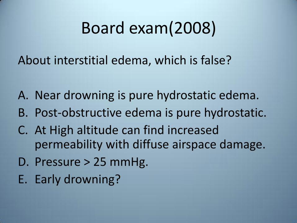
Interstitium
• Connective tissues within the lung
– Basement membrane of alveoli and capillaries
– Perivascular and perilymphatic tissues
• Functions
– Supporting lung
– Fluid balance
– Repair and remodelling

Interstitium
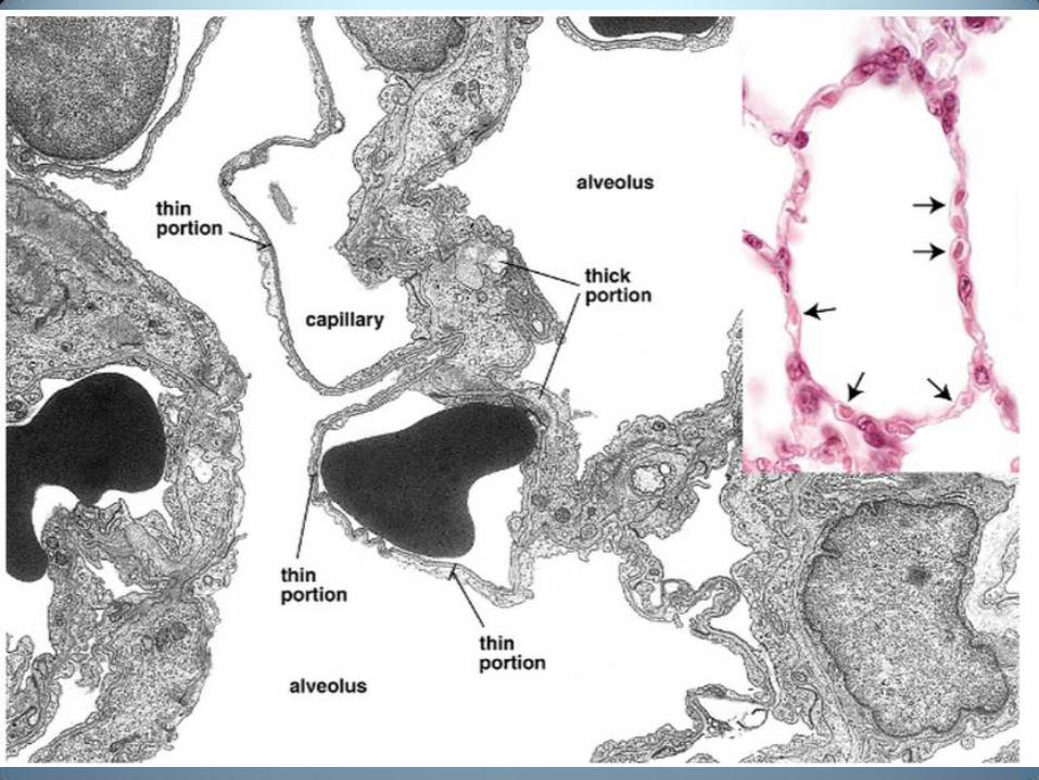
• Thin portions(tight junction) of interalveolar septa:
– Basal lamina + adjacent capillary endothelium
– Strict diffusion barrier between alveolar surface and
underlying tissues
• Thick portion:
– Tissue fluid can accumulate or cross into the alveoli
– Lymphatic vessels in the connective tissue of the terminal
bronchioles drain fluid that accumulates in the thick
portion of the septum
Interstitium: thick portion
• Component: extracellular matrix
– Collagen: tensile strength
– Elastin: flexibility
– Proteoglycans:
• Maintain hydration
• Adhesive activity
• Component of growth factors

Interstitium: thick portion
• Component: interstitial cell
– Macrophage: wander about on the epithelial
surfaces (septum and air space)
• Clear the respiratory spaces of inhaled particles
• Migrate to the bronchioles or the lymphatics/lymphoid
tissue (interstitium)
– Fibroblast

Interstitium
• 3 zones – Peripheral connective tissue(pleural)
– Axial connective tissue(central, bronchovascular)
– Parenchymal connective tissue(intralobular)

Axial connective tissue
• Originate at the hilum
• Surrounds the bronchovascular structures
• Extend peripherally
• Terminate at
centre of the acini

Peripheral connective tissue
• Subpleural space and interlobular lung septa

Parenchymal connective tissue
• Penetrate into secondary pulmonary lobule and lie along intralobular venule

Axial Peripheral
Parenchymal

Interstitial structures


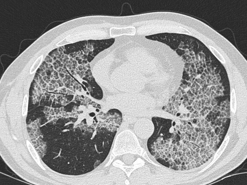


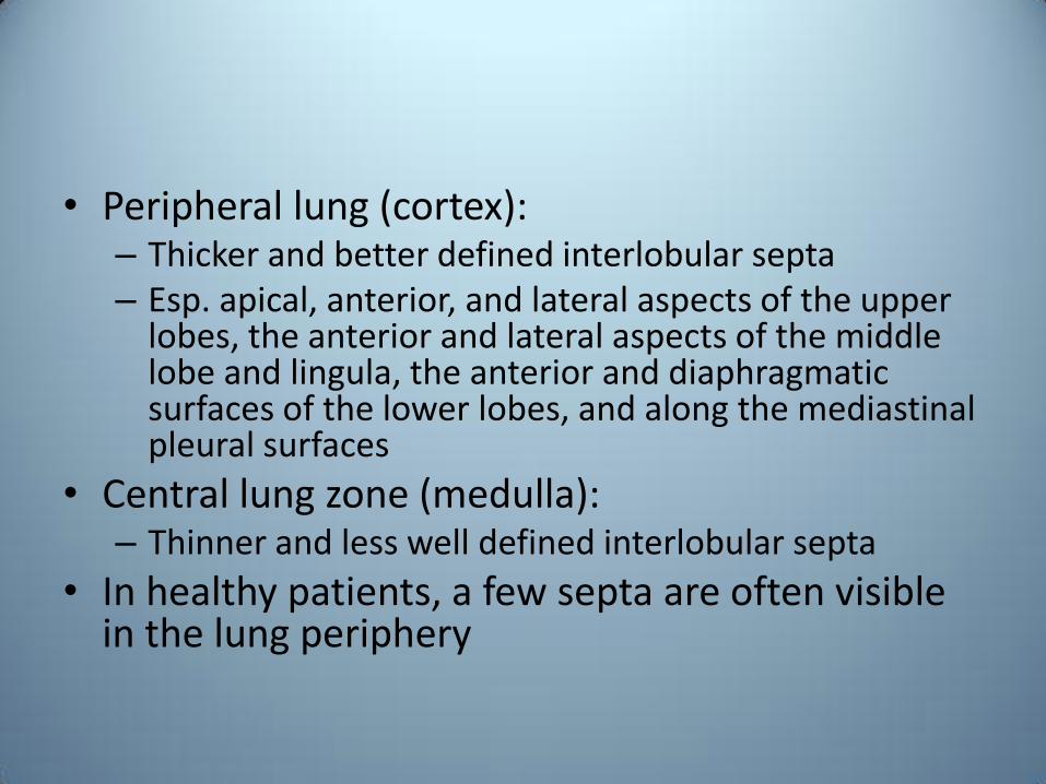
• Peripheral lung (cortex): – Thicker and better defined interlobular septa – Esp. apical, anterior, and lateral aspects of the upper
lobes, the anterior and lateral aspects of the middle lobe and lingula, the anterior and diaphragmatic surfaces of the lower lobes, and along the mediastinal pleural surfaces
• Central lung zone (medulla): – Thinner and less well defined interlobular septa
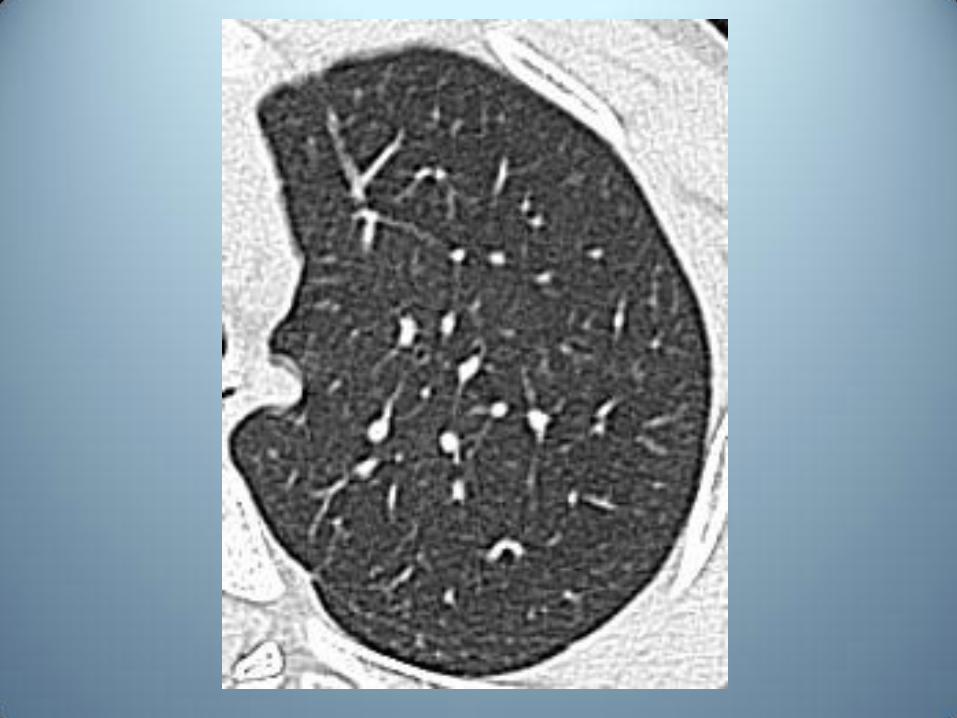
• In healthy patients, a few septa are often visible in the lung periphery

Cortex Medulla
Rich interlobular septum Poor interlobular septum
Low blood flow High blood flow
Low air flow High air flow
High lymphatic flow Low lymphatic flow


Physiology
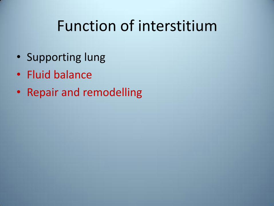
Function of interstitium
• Supporting lung
• Fluid balance
• Repair and remodelling

Fluid balance
• Starling force
• Net fluid movement between compartments ( Jv )
• Capillary hydrostatic pressure ( Pc )
• Interstitial hydrostatic pressure ( Pi )
• Capillary oncotic pressure ( πc ) • Interstitial oncotic pressure ( πi ) • Filtration coefficient ( Kf )
• Reflection coefficient ( σ )
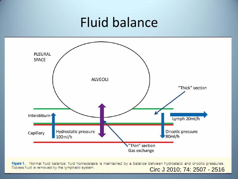
Fluid balance
Circ J 2010; 74: 2507 - 2516

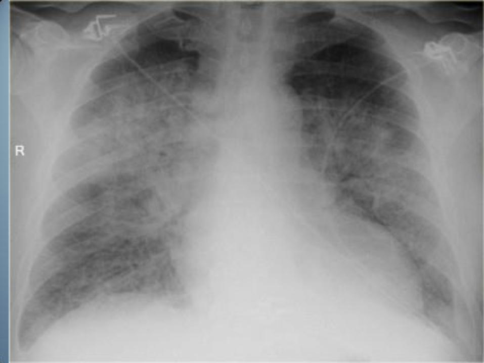
Fluid balance: leakage
Color Atlas of Pathophysiology © 2000 Thieme

Fluid balance: leakage
Circ J 2010; 74: 2507 - 2516

Fluid balance: leakage
Circ J 2010; 74: 2507 - 2516

Repair, remodelling and matrix turnover
• Critical element of lung biology
• Lung remodelling: chronic inflammation
• Matrix turnover: dynamic balance between accumulation and distribution of matrix component
– Synthesis
– Degradation

Am J Physiol Lung Cell Mol Physiol 298: L715–L731, 2010


Board exam(2008)
About interstitial edema, which is false?
A. Near drowning is pure hydrostatic edema.
B. Post-obstructive edema is pure hydrostatic.
C. At High altitude can find increased permeability with diffuse airspace damage.
D. Pressure > 25 mmHg
E. Early drowning?


Board exam
Which type of the pulmonary interstitium produces a peribronchial cuffing picture?
A. Axial
B. Peripheral
C. Parenchymal
D. Peripheral and parenchymal
E. Axial and peripheral

Board exam
Which type of the pulmonary interstitium is visible in HRCT of the normal lung?
A. Axial
B. Peripheral
C. Axial and parenchymal
D. Axial and peripheral
E. Peripheral and parenchymal

Board exam
Which interstitial component is damaged in emphysema?
A. Collagen
B. Proteoglycans
C. Elastin
D. Basement membrane
E. Fibroblast
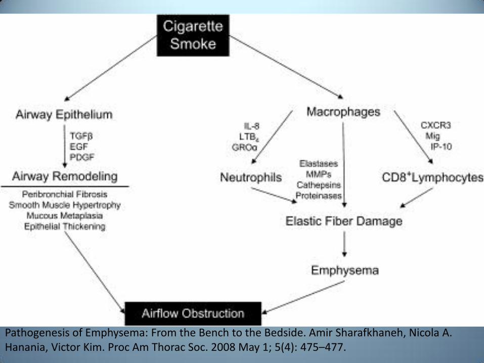
Pathogenesis of Emphysema: From the Bench to the Bedside. Amir Sharafkhaneh, Nicola A. Hanania, Victor Kim. Proc Am Thorac Soc. 2008 May 1; 5(4): 475–477.

THANK YOU