Pulmonary Function TestsPulmonary Function Tests · Three Types of Pulmonary Function TestsThree...
48
Pulmonary Function Tests Pulmonary Function Tests
Transcript of Pulmonary Function TestsPulmonary Function Tests · Three Types of Pulmonary Function TestsThree...
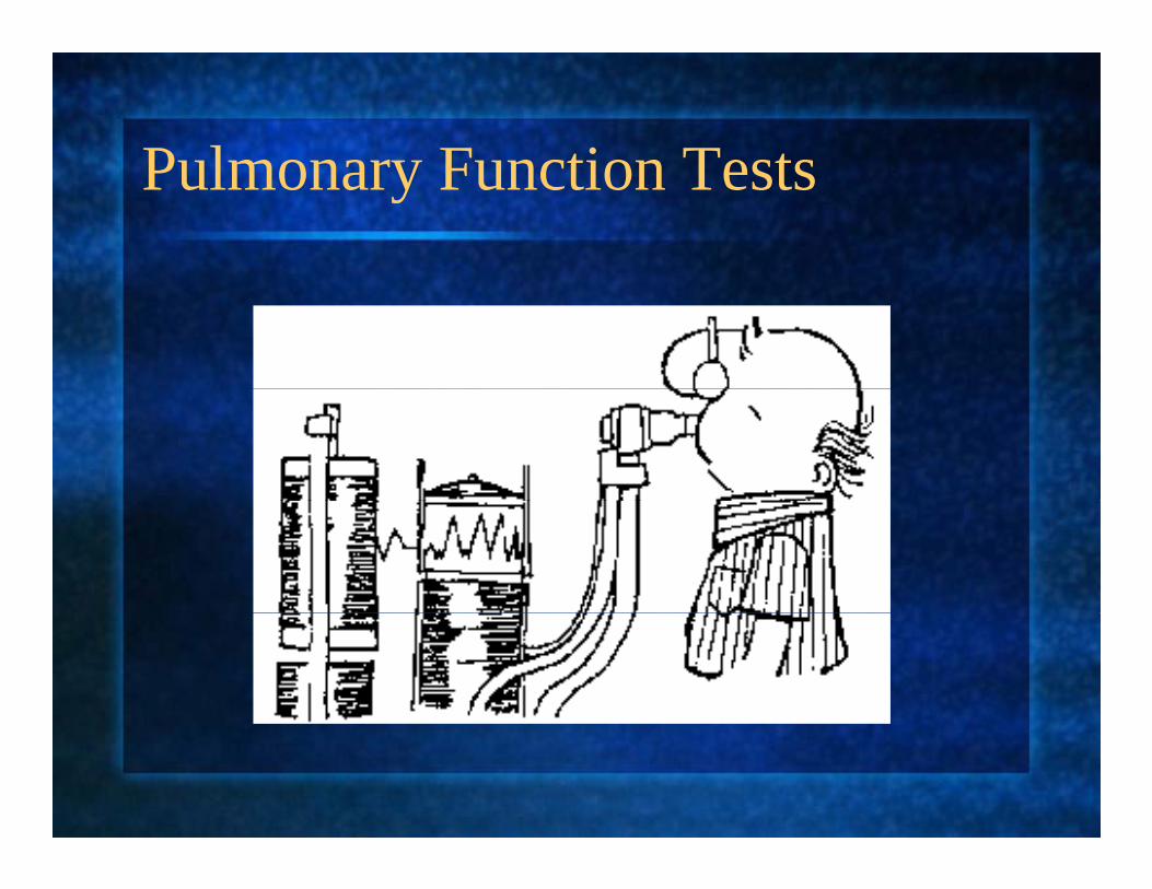
Pulmonary Function TestsPulmonary Function Tests
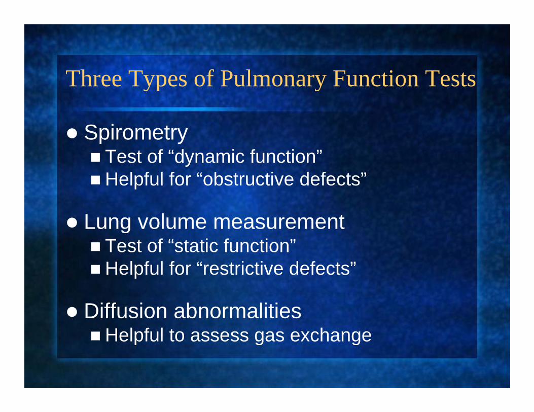
Three Types of Pulmonary Function TestsThree Types of Pulmonary Function Tests
SpirometrSpirometryTest of “dynamic function”Helpful for “obstructive defects”Helpful for obstructive defects
Lung volume measurementgTest of “static function”Helpful for “restrictive defects”
Diffusion abnormalities Helpful to assess gas exchangeHelpful to assess gas exchange

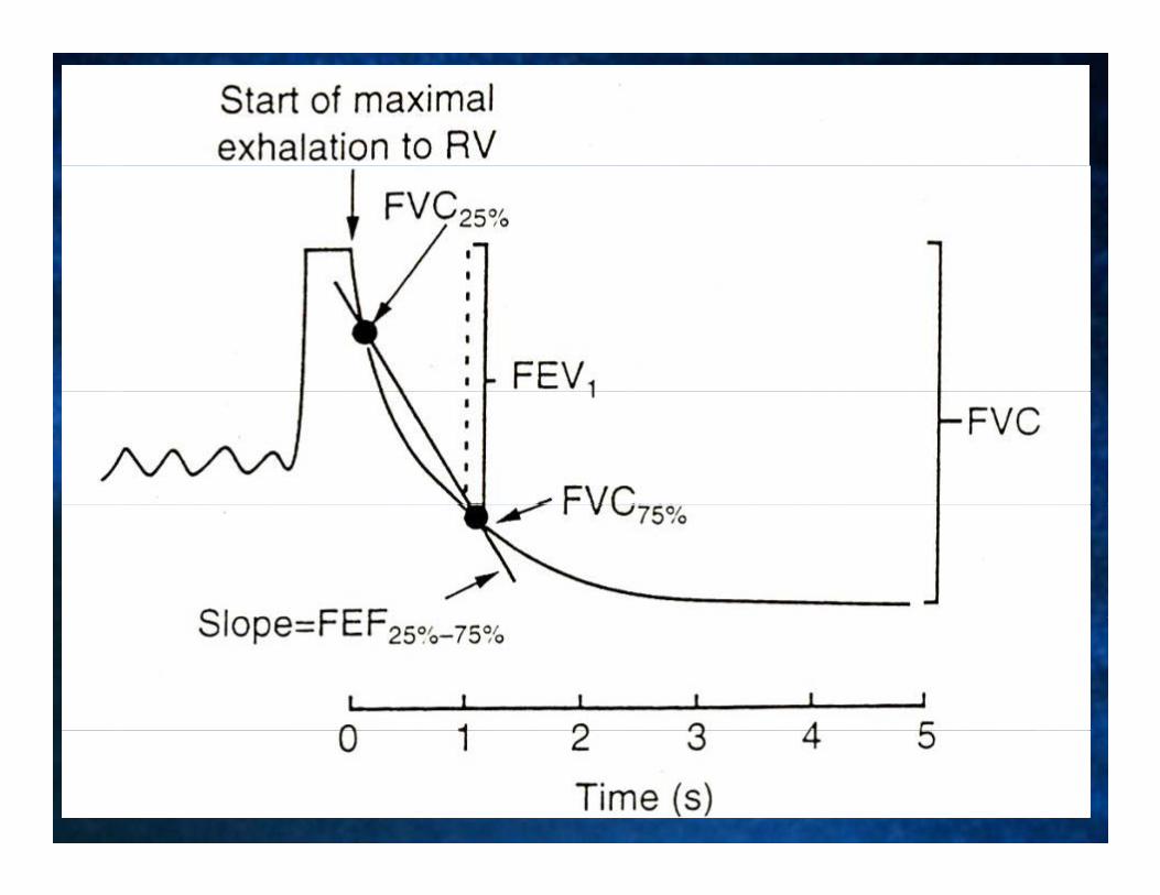
Spirometryp y
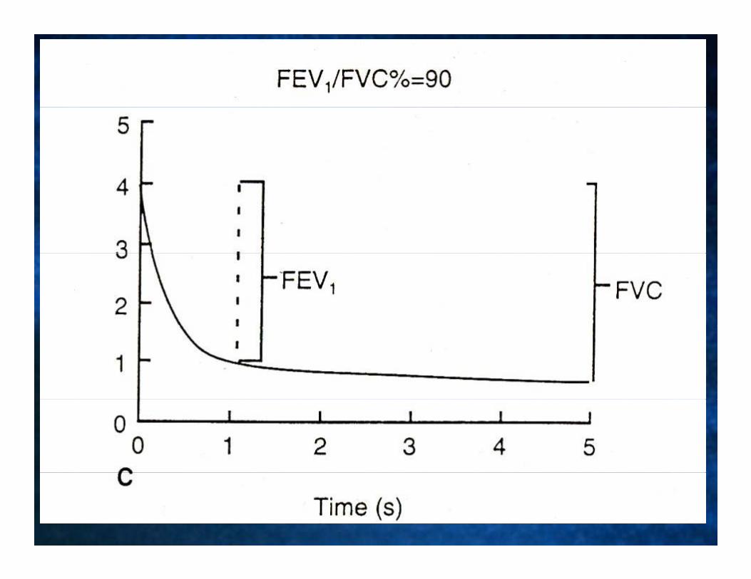

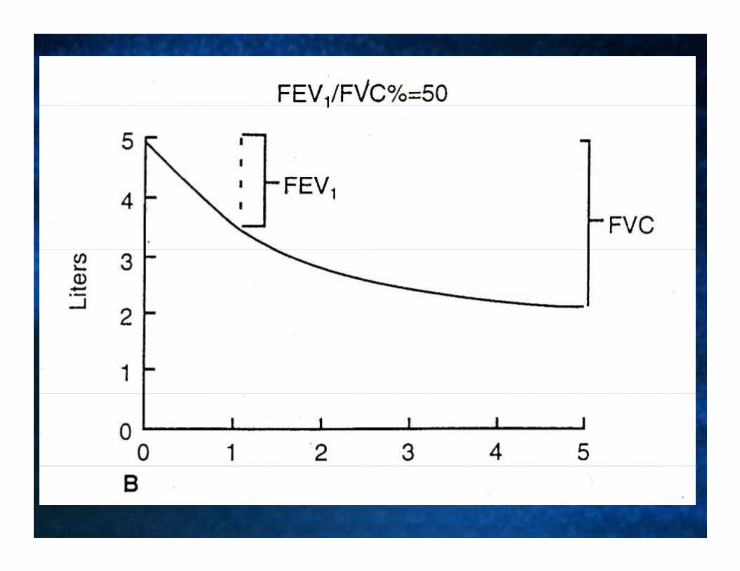

PFTs: airflow obstructionPFTs: airflow obstruction
D i i t i fl d/ lDecrease in expiratory airflow and/or volumeFEV1 decreasedFVC normal or decreasedFVC normal or decreasedFEV1/FVC decreased*FEF decreasedFEF25-75 decreased
*definition of obstructive defectdefinition of obstructive defect


Types of Airflow ObstructionTypes of Airflow Obstruction
I t l i l b t tiIntraluminal obstructionIntramural obstructionExtraluminal obstructionExtraluminal obstructionUpper airway obstruction“Mixed”Mixed

Types of Airflow ObstructionTypes of Airflow Obstruction
Intraluminal:e.g., Secretions
Intramural:e.g., Edema
Extraluminal:e.g., Loss of radial traction

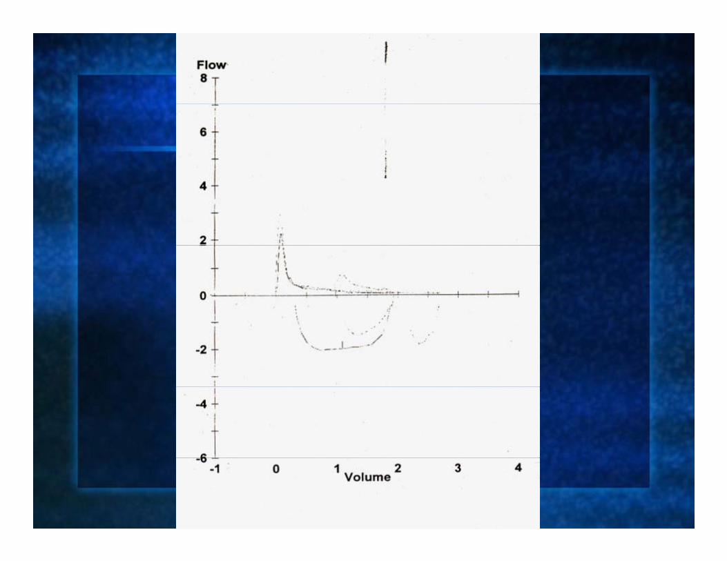
Dynamic Airway Compression during y y p gForced Expiration
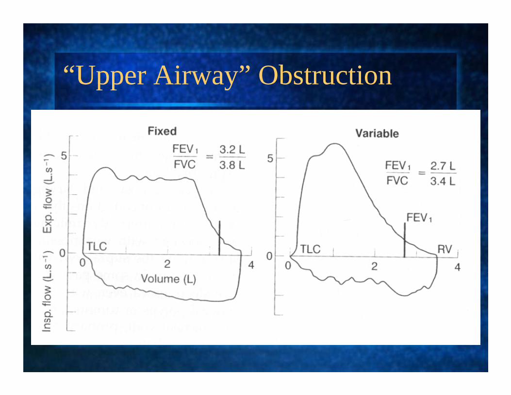
“Upper Airway” ObstructionUpper Airway Obstruction
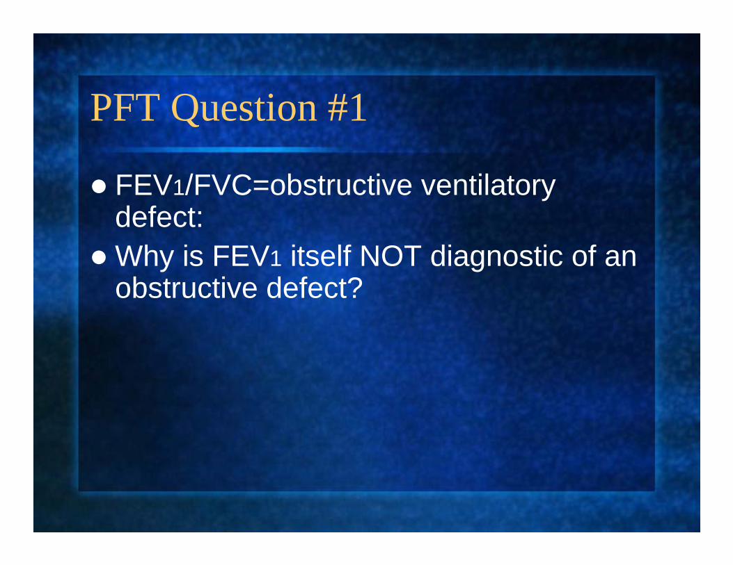
PFT Question #1PFT Question #1
FEV /FVC b t ti til tFEV1/FVC=obstructive ventilatory defect: Wh i FEV it lf NOT di ti fWhy is FEV1 itself NOT diagnostic of an obstructive defect?

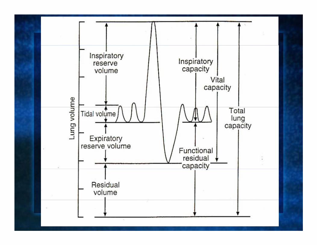
Lung VolumesLung Volumes
“St ti f ti ”“Static function”Gas Equilibration (“wash in” and “wash
t”)out”)Body plethysmography
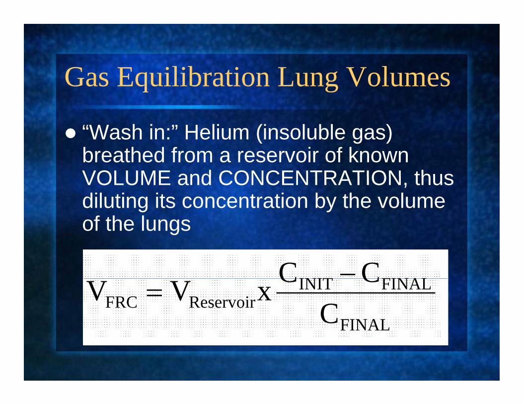
Gas Equilibration Lung VolumesGas Equilibration Lung Volumes
“W h i ” H li (i l bl )“Wash in:” Helium (insoluble gas) breathed from a reservoir of known VOLUME and CONCENTRATION thusVOLUME and CONCENTRATION, thus diluting its concentration by the volume of the lungsof the lungs
FINALINIT CC −
FINAL
FINALINITReservoirFRC C
CCxVV =FINAL
Gas Equilibration Lung VolumesGas Equilibration Lung Volumes
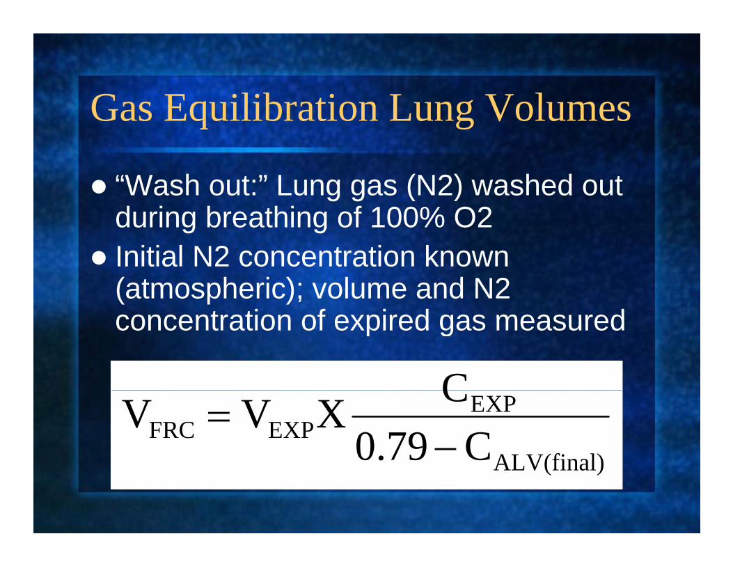
“W h t ” L (N2) h d t“Wash out:” Lung gas (N2) washed out during breathing of 100% O2 I iti l N2 t ti kInitial N2 concentration known (atmospheric); volume and N2 concentration of expired gas measuredconcentration of expired gas measured
C
ALV(final)
EXPEXPFRC C0.79
CXVV−
=ALV(final)

Plethysmographic Lung VolumesPlethysmographic Lung Volumes
Cl d t t t t t tClosed system at constant temperatureLungs and airway closed system when occludedPanting at FRCPanting at FRCInhalation ↓ intrathoracic pressure, ↑ volume

Boyle’s LawAt
Boyle s Law
2211 VPVP = 2211At constant temperature
( )( )ΔVVΔPPVP FRCFRCFRCFRC ++=

Plethysmographic Lung VolumesPlethysmographic Lung Volumes
( )( )ΔVVΔPPVP FRCFRCFRCFRC ++=
ΔPΔVΔVPΔPVVP FRCFRCFRCFRC +++=
ΔPΔVΔVPΔPV0 FRCFRC ++=
FRCFRC PΔVV =
Close to zero
FRCFRC PΔP
V
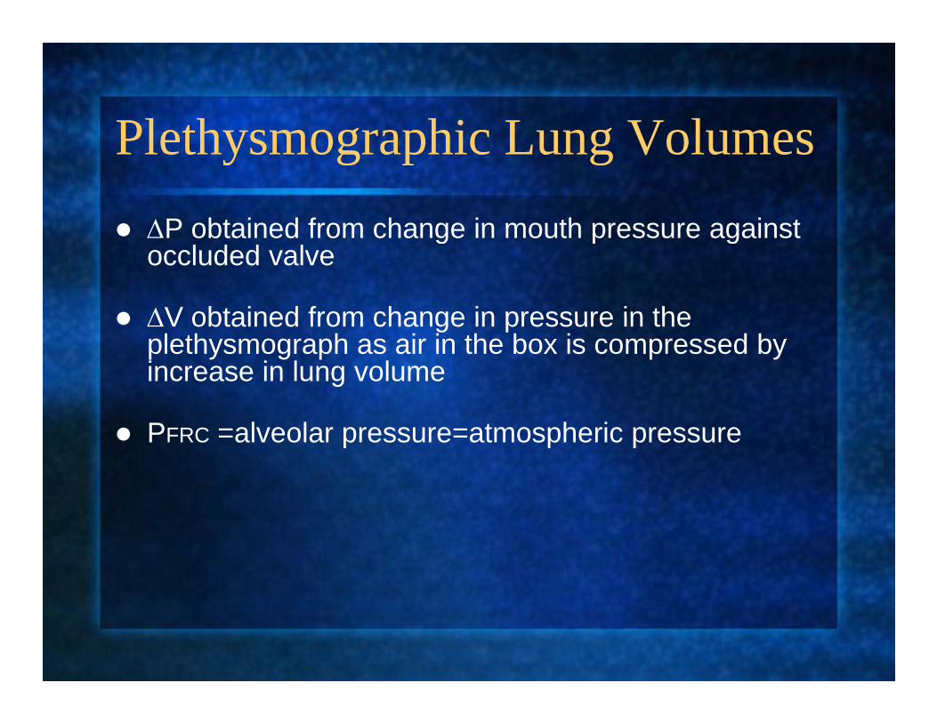
Plethysmographic Lung VolumesPlethysmographic Lung VolumesΔP obtained from change in mouth pressure againstΔP obtained from change in mouth pressure against occluded valve
ΔV obtained from change in pressure in theΔV obtained from change in pressure in the plethysmograph as air in the box is compressed by increase in lung volume
PFRC =alveolar pressure=atmospheric pressure

PFTs: Restrictive ventilatory defectPFTs: Restrictive ventilatory defect
A d i l iA decrease in lung expansionFEV1 decreasedFVC decreasedFEV1/FVC normal or increased1Total Lung Capacity (TLC) decreased*
* Definition of restrictive ventilatory defect

PFT Questions #3 and #4PFT Questions #3 and #4
Wh i FVC it lf NOT di ti fWhy is FVC itself NOT diagnostic of a restrictive ventilatory defect?
Wh i VC it lf t di ti fWhy is VC itself not diagnostic of a restrictive ventilatory defect?

Types of Restrictive DefectsTypes of Restrictive Defects
P h l l/d t tiParenchymal removal/destructionParenchymal infiltrationExtrapulmonary deformityReduced force generation

Restrictive patternsRestrictive patterns
P h l l diParenchymal lung diseasethoracic cage restrictionS t i d i TLC VC FRC RVSymmetric decrease in TLC, VC, FRC, RV
Neuromuscular weaknessIC mainly decreasedTLC and VC decreased FRC d RV dFRC and RV spared

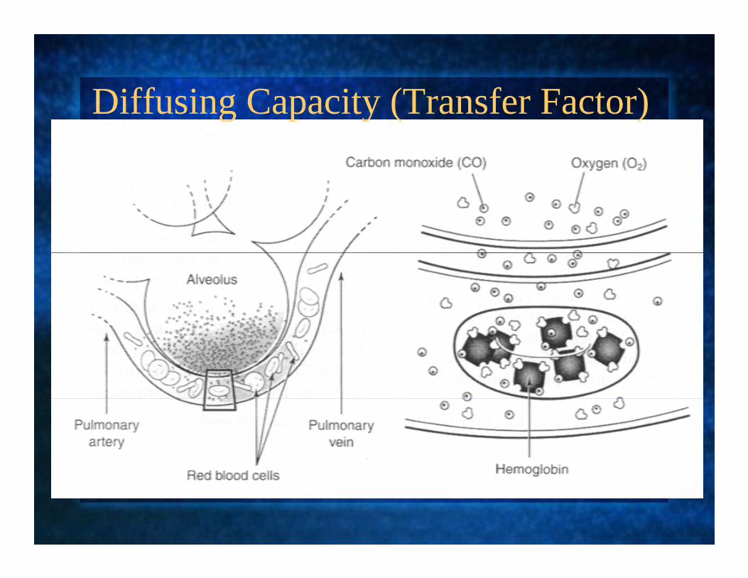
Diffusing Capacity (Transfer Factor)Diffusing Capacity (Transfer Factor)
Diffusing Capacity for CO (DL )Diffusing Capacity for CO (DLCO)
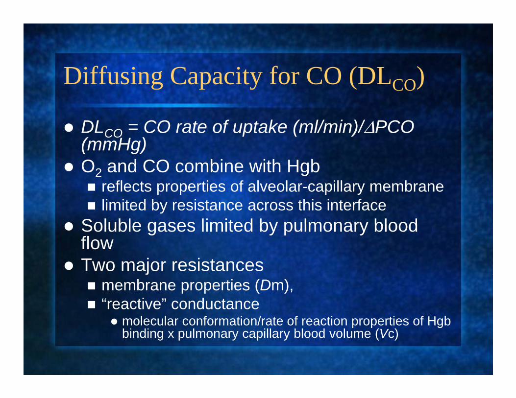
DL = CO rate of uptake (ml/min)/ΔPCODLCO = CO rate of uptake (ml/min)/ΔPCO (mmHg)O2 and CO combine with Hgb2 g
reflects properties of alveolar-capillary membranelimited by resistance across this interface
Soluble gases limited by pulmonary bloodSoluble gases limited by pulmonary blood flowTwo major resistances j
membrane properties (Dm),“reactive” conductance
molecular conformation/rate of reaction properties of Hgbmolecular conformation/rate of reaction properties of Hgb binding x pulmonary capillary blood volume (Vc)

Diffusing Capacity for CO (DL )Diffusing Capacity for CO (DLCO)
OHb
ACO PP
VkCODL ))((−
=
kCO = rate constant for CO uptake ( aka
OHb 2
kCO = rate constant for CO uptake ( aka “permeability factor” or Krogh coefficient)VA = alveolar volumeP P = effective gas pressure (barometricPb – PH20 = effective gas pressure (barometric pressure - partial pressure of water)Units = conductance (ml CO/min/mmHg)
Diffusing Capacity for CO (DL )Diffusing Capacity for CO (DLCO)
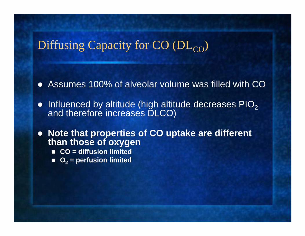
Assumes 100% of alveolar volume was filled with CO
Influenced by altitude (high altitude decreases PIOInfluenced by altitude (high altitude decreases PIO2and therefore increases DLCO)
Note that properties of CO uptake are differentNote that properties of CO uptake are different than those of oxygen
CO = diffusion limitedO2 = perfusion limited2 p

Determinants of DLDeterminants of DLCO
G di tGas gradientSolubilityHemoglobinRate of gas uptake by Hgb
Capillary blood volumeAlveolar-capillary membrane propertiesp y p p
ThicknessSurface area
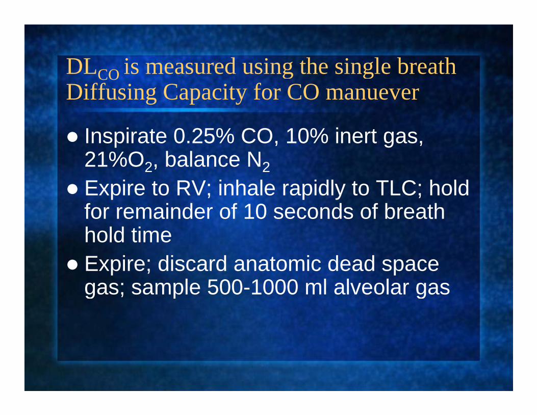
DLCO is measured using the single breath CO g gDiffusing Capacity for CO manuever
I i t 0 25% CO 10% i tInspirate 0.25% CO, 10% inert gas, 21%O2, balance N2E i t RV i h l idl t TLC h ldExpire to RV; inhale rapidly to TLC; hold for remainder of 10 seconds of breath hold timehold time Expire; discard anatomic dead space gas; sample 500 1000 ml alveolar gasgas; sample 500-1000 ml alveolar gas

Limitation of the single-breath gDiffusing Capacity manuever
SB UNDERESTIMATE t diff iSB may UNDERESTIMATE true diffusing capacity in emphysema if it underestimates gas dilution VA since DLCO =(kcOxVA)/(Pb-gas dilution VA since DLCO (kcOxVA)/(PbPH20)

Causes of Decreased “Measured” Diffusing Capacity
P h l L blParenchymal Lung problemLoss of alveoli
EmphysemaEmphysemaResection
Abnormal alveoli (rest of the course…)Pulmonary vascular problem
Pulmonary vascular diseasePulmonary embolismPulmonary embolism
AnemiaAbnormal hemoglobin
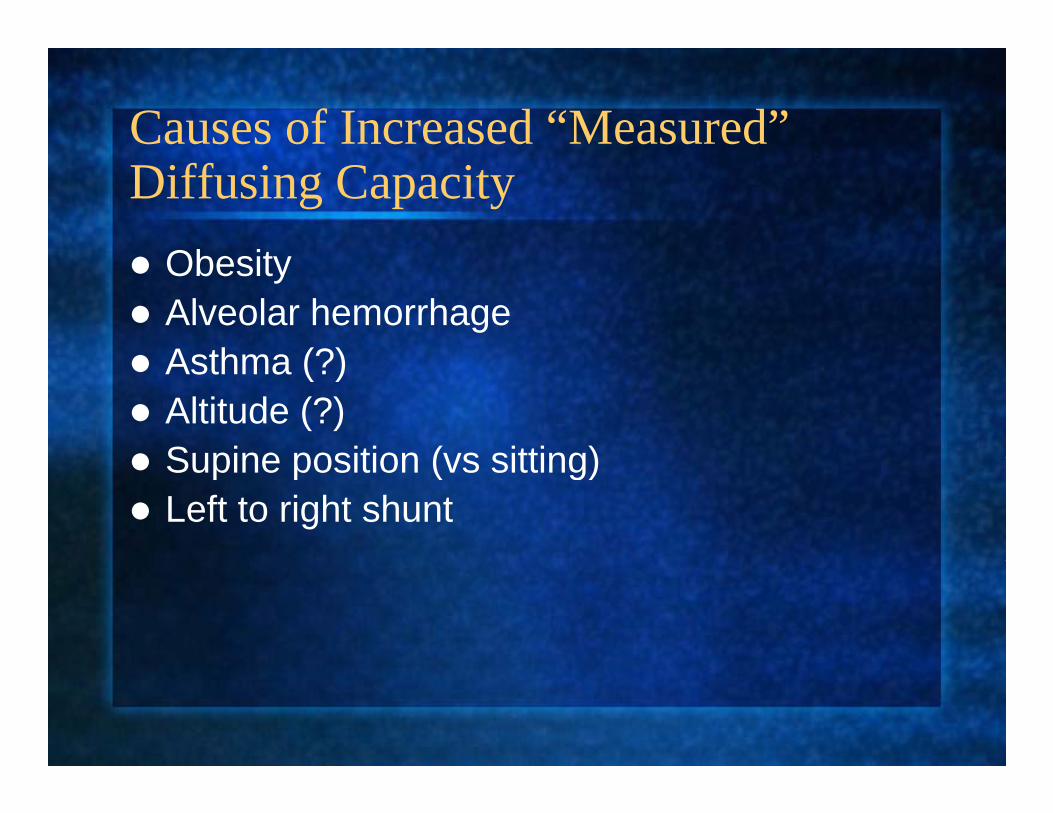
Causes of Increased “Measured” Diffusing Capacity
Ob itObesityAlveolar hemorrhageAsthma (?)Asthma (?)Altitude (?)Supine position (vs sitting)Supine position (vs sitting)Left to right shunt

“Diffusion Capacity” vs DiffusionDiffusion Capacity vs Diffusion
Decreased diffusing capacity can result fromDecreased diffusing capacity can result from numerous abnormalities unrelated to diffusion block itself Diffusion abnormality as a cause of hypoxemiaDiffusion abnormality as a cause of hypoxemia
Diffusion block or other inability to transfer gas completely (eg, low PIO2+ increased circulatory time) so that insufficient transfer of alveolar PO2 occur
Decreased diffusing capacity without diffusion blocklow alveolar volume,low Hgb


DLCO PearlDLCO Pearl
I l t d DLCO d ith lIsolated DLCO decrease with normal spirometry and volumes
P l l di dPulmonary vascular disorderInterstitial disorder not yet, or no longer, affecting parenchymal volumeaffecting parenchymal volumeAbnormality of Hgb
anemiacarboxyHgbmethHgb

Pre-operative Pulmonary p yAssessment: PFTs
C li ti hi h t f th i dComplications: highest for thoracic and upper abdominal (ie, near the diaphragm)diaphragm)All having lung resection, orthopoedic and lower abdominal with lung diseaseand lower abdominal with lung disease, or smokingAge>60 yearsAge>60 years

Postoperative Pulmonary RisksPostoperative Pulmonary Risks
S i t FEV FVC 70%Spirometry: FEV1 or FVC <70%, FEV1/FVC<65%P CO >45 mmHg DLCO<40% in COPDPaCO2>45 mmHg, DLCO<40% in COPDNone contraindicateLung resection: FEV1 best for pulmonaryLung resection: FEV1 best for pulmonary reserve and post op complications; post op FEV1 <30% predicted=increased long term
t lit d i di t t blmortality and immediate post op problems
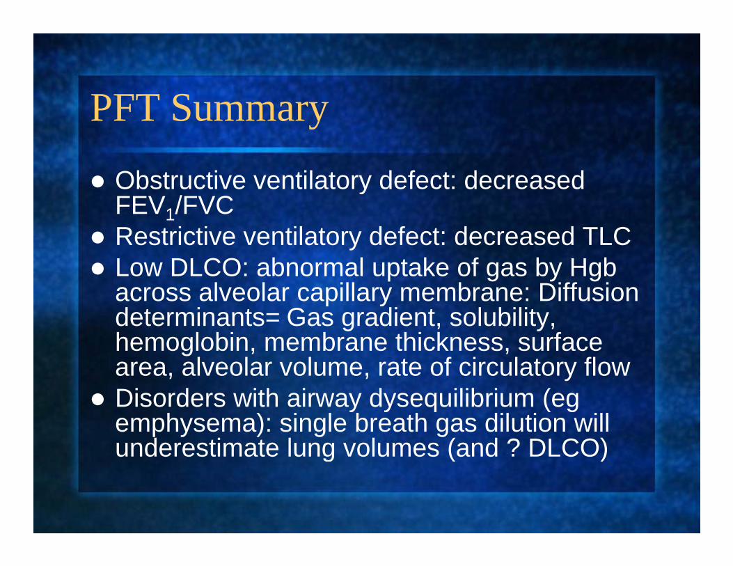
PFT SummaryPFT Summary
Obstructive ventilatory defect: decreasedObstructive ventilatory defect: decreased FEV1/FVCRestrictive ventilatory defect: decreased TLCyLow DLCO: abnormal uptake of gas by Hgb across alveolar capillary membrane: Diffusion determinants= Gas gradient solubilitydeterminants= Gas gradient, solubility, hemoglobin, membrane thickness, surface area, alveolar volume, rate of circulatory flowDi d ith i d ilib i (Disorders with airway dysequilibrium (eg emphysema): single breath gas dilution will underestimate lung volumes (and ? DLCO)g ( )

S iSeries ‘‘ATS/ERS TASK FORCE: STANDARDISATION OF LUNG FUNCTION TESTING’’ di d b C d G i iTESTING’’ Edited by V. Brusasco, R. Crapo and G. Viegi.
General considerations for lung function testing
Eur Respir J 2005; 26: 153–161


![Shrinking Lung Syndrome: A Pulmonary Manifestation of ... · scan]) and pulmonary function tests (PFTs). Pulmonary function tests were carried out in our pulmonary function laboratory,](https://static.fdocuments.us/doc/165x107/5f03189c7e708231d40783f1/shrinking-lung-syndrome-a-pulmonary-manifestation-of-scan-and-pulmonary-function.jpg)