Pulmonary Function Testing Part Two Handout Part... · 1r furpro\q iru krxuv 1r dqwlklvwdplqhv iru...
26
8/3/2017 1 Arthur Jones, EdD, RRT This Presentation is Approved for 2.0 CRCE Credit Hours Pulmonary Function Testing Part II Learning Objectives Describe the purposes, physiologic bases devices & methods for diffusing capacity testing Describe the purposes, physiologic bases devices & methods for specialized testing regimens Describe the purposes, physiologic bases devices & methods for cardiopulmonary exercise testing Describe the purposes, physiologic bases devices & methods for metabolic testing Interpret results from diffusing capacity tests, specialized tests, cardiopulmonary exercise tests & metabolic tests Specialized Testing Bronchodilator benefit Bronchial challenge testing Exhaled nitric oxide analysis Preoperative testing Testing for disability Diffusing Capacity Testing Diffusing Capacity Measures the rate of diffusion of gas across alveolar- capillary membrane Measured as mL (gas)/min/mm Hg (pressure gradient) See links below to view animation of diffusion Anatomic Diffusion Pathway Alveolar air Alveolar wall Surfactant layer Alveolar epithelium Alveolar basement membrane Interstitial space
Transcript of Pulmonary Function Testing Part Two Handout Part... · 1r furpro\q iru krxuv 1r dqwlklvwdplqhv iru...

8/3/2017
1
Arthur Jones, EdD, RRT
This Presentation is Approved for2.0 CRCE Credit Hours
Pulmonary FunctionTesting Part II
Learning Objectives
Describe the purposes, physiologic bases devices & methods for diffusing capacity testing
Describe the purposes, physiologic bases devices & methods for specialized testing regimens
Describe the purposes, physiologic bases devices & methods for cardiopulmonary exercise testing
Describe the purposes, physiologic bases devices & methods for metabolic testing
Interpret results from diffusing capacity tests, specialized tests, cardiopulmonary exercise tests & metabolic tests
Specialized Testing
Bronchodilator benefit
Bronchial challenge testing
Exhaled nitric oxide analysis
Preoperative testing
Testing for disability
Diffusing Capacity Testing
Diffusing Capacity
Measures the rate of diffusion of gas across alveolar-capillary membrane
Measured as mL (gas)/min/mm Hg (pressure gradient)
See links below to view animation of diffusion
Anatomic Diffusion Pathway
Alveolar air
Alveolar wall Surfactant layer Alveolar epithelium Alveolar basement membrane
Interstitial space
8/3/2017
2
Anatomic Diffusion Pathway
Capillary wall Capillary basement membrane Capillary endothelium
Plasma
RBC Erythrocyte membrane Intracellular erythrocyte fluid
Hemoglobin
See links below for illustration of diffusion pathway
Physical LawsGoverning Diffusion
Henry's law: the amount of gas dissolving in a liquid is proportional to the partial pressure of the gas derives the solubility coefficient (Ks) of the gas
Physical LawsGoverning Diffusion
Graham's law: the rate of diffusion through a liquid is Directly proportional to its Ks Inversely proportional to its GMW CO2 diffuses 20x
the rate of O2
High solubility of CO2 diffusion defects will not affect CO2 exchange
Physical LawsGoverning Diffusion
Fick's law: gas diffusion is Directly proportional to
• Alveolar surface area• Pressure gradient
Inversely proportional to• Alveolar thickness• Gram molecular weight of gas
Diffusion Limitations of Gases
O2 can be diffusion &/or perfusion limited
CO is diffusion limited, only ideal gas to measure diffusing capacity
Lung diffusing capacity is measured as DLCO - diffusion in lung of carbon monoxide
Diffusion Tests
DLCOsb (single breath): most common method Advantages
• Simple technique• Rapid analysis
Disadvantages• Sensitive to V/Q mismatching• Patient must be capable of breath holding for 10 sec

8/3/2017
3
Diffusion Tests
DLCOrb (rebreathing) Advantages
• Most accurate method• Least sensitive to V/Q mismatching• Can be used during exercise
Disadvantage• Requires rapid analyzers• Complex calculations
Diffusion Tests
DLCOib (intrabreath): analysis during a single exhalation Advantages
• Does not require breath hold• Can be used during exercise
Disadvantage• Sensitive to VQ mismatch• Complex calculations
DLCO Testing Indications
Evaluation & follow up of parenchymal lung diseases
Evaluation & follow-up of emphysema & cystic fibrosis
Evaluation of cardiovascular diseases
Evaluation of pulmonary involvement in systemic inflammatory & collagen vascular diseases
DLCO Testing Indications
Evaluation of the effects of chemotherapy agents or other drugs, e.g. amiodarone
Evaluation of pulmonary hemorrhage
Evaluation for pneumonectomy or lung reduction surgery
Evaluation for disability
Contraindicationsfor Diffusion Tests
CO toxicity
Severe hypoxemia (O2 removed during test)
Inability to cooperate, e.g. breath holding
Large meal or vigorous exercise immediately before the test
Smoking within 24 hours of test
DLCOsb Equipment
Spirometer
Automatic valve - for gas delivery, breath holding, & sampling
End-tidal sampler
Gas analyzers - CO & He
Gas mixture 0.3% CO 10% He 21% O2
Balance N2
8/3/2017
4
DLCOsb Procedure
Patient performs FVC maneuver
Inspires to TLC
Holds breath for 9 - 11 sec
Exhales
Alveolar sample collected between 750 - 1000 mL
FYI see links below for AARC CPG on DLCO testing
DLCOsb Procedure
Calculation VA - alveolar volume 60 - correction from sec to min PB - barometric pressure T - breath hold time (sec) Ln - natural logarithm FCOI - initial fraction of CO FCOF - final fraction of CO
DLCOsb = VA (STPD) x 60 x Ln FCOI(PB - PH2O) (T) FCOF
DLCOsb Acceptability Criteria
Test volume must be > 90% previously measured VC
End-inspiratory breath hold must be 9 - 11 sec
Expiration to RV ≤ 4 sec
VD must clear before alveolar sampling
Reproducibility criteria: two tests within 10% or 3.0 ml CO/min/mm Hg
Report: mean value of two tests
DLCOsb Predicted Value
Normal DLCO = 25 ml/min/mm Hg ± 20%
Predicted based on BSA Hb - 1 mg/dL 7% change in DLCO
Age - inverse relationship
Interpretation must consider lung volume
Factors Affecting DLCO
Alveolar surface area
V/Q abnormalities
Parenchymal thickening, e.g. fibrosis
Edema
Consolidation
Pulmonary capillary pressure
RBC, Hb quantities
Pulmonary capillary quantity
Conditions WithIncreased DLCO
Obesity
Asthma
Left-to-right shunt
CHF (without edema)
Early polycythemia
Large lung volume
Exercise
Supine position

8/3/2017
5
Conditions WithDecreased DLCO
Decreased surface area Emphysema Lung resection
Increased wall thickness Hypersensitivity pneumonitis Fibrosis Sarcoidosis
Decreased carrying capacity - anemia
Prognostic Value of DLCO
Determines when COPD develops into emphysema
Predicts complications after surgical resection of lung
Predicts mortality in pulmonary arterial hypertension
Bronchodilator Benefit &Bronchial Challenge
Testing
Bronchodilator Benefit Testing
Purpose: determine value of bronchodilators in patient management
Indications Clinical evidence of reactive airwaysWheezing Dyspnea FEV1 % < 70%
Bronchodilator Benefit Testing
Preconditions for testing No short-acting beta agonists or anticholinergics for 4 H No long-acting beta agonists for 12 H No long-acting anticholinergic for 24 H No cromlyn, nedocromil for 24 H No leukotriene modifiers for 24 HMaintain inhaled steroids
Bronchodilator Benefit Testing
Laboratory requirements Cooperative patient Skilled technologistMaintained & calibrated equipment ACLS capabilities Patient care capabilities in institution

8/3/2017
6
Bronchodilator Benefit Testing
Pretests - may include Spirometry, e.g. FEV1
sGaw measurement Lung volumes Diffusing capacity
Bronchodilator Benefit Testing
Medication administration Beta agonist: 1 pf Q30s x 4 Ipratropium: 1 pf Q30s x 4
Interval before post-testing Beta agonist: 10 - 15 min Ipratropium: 30 min
Bronchodilator Benefit Testing
Significant improvements> 12% & 200 mL increase in FEV1 or FVC> 30% increase in sGaw
Bronchodilator Benefit Testing
Insignificant improvementMay test after time using a medicationMay test with a different medication Check for symptomatic improvement
Decreased post-test parameters Paradoxical drug response Fatigue
Bronchodilator Benefit Testing
Calculating % change
%FEV1 change = Post FEV1 - Pre FEV1 x 100Pre FEV1
Example: Pre= 1.2L, Post = 1.7L
%FEV1 change = 1.7L - 1.2L x 100 = 42%1.2L
Bronchial Challenge Testing
Purposes Detect airway hyperreactivity Isolate cause of hyperreactivity Quantify severity of bronchospasm Assess changes in bronchoreactivity

8/3/2017
7
Bronchial Challenge Testing
Indications Exclude a diagnosis of airway hyperreactivity Evaluate occupational asthma Assess the severity of bronchospasm Determine the relative risk of developing asthma Assess response to therapeutic interventions
Bronchial Challenge Testing
Contraindications Symptoms, e.g. wheeze, cough Ventilatory impairment Recent cardiac event or stroke Cerebral aneurysm Uncontrolled hypertension Current use of anticholinesterase agent Pregnancy, lactation
Bronchial Challenge Testing
Provocative agentsMethacholine: parasympathetic stimulator
• Most common• Prepared by pharmacy
Histamine: mechanism of action uncertain
Exercise: exercise-induced bronchospasm (EIB)
FYI see links below for AARC CPG on methacholine challenge
Bronchial Challenge Testing
Side effectsMethacholine
• Headache• Itching• Signs & symptoms of severe allergic reaction
Histamine• Same as for methacholine• Flushing
FYI see links below for ATS standards on challenge testing
Bronchial Challenge Testing
Preconditions No bronchodilators, as for bronchodilator benefit test No systemic steroids for 12 hours No cromolyn for 48 hours No antihistamines for 48 hours No exercise, cold air for 2 hours No smoking for 6 hours No caffeine for 6 hours No beta-blocking agents
Methacholine Challenge Testing
Procedure 5 breath dosimeter
• Standardizes dose by volume• Most precise• Requires dosimeter
2 minute tidal breathing• Standardizes dose by time• Requires only small volume nebulizer
See links below to view dosimeter

8/3/2017
8
Methacholine Challenge Testing
Procedure Baseline mechanics - FVC, FEV1 , sGaw, etc. Inhaled NSS (control dose)Wait 3 minutes Repeat measure FEV1 ≤ 80% (of pretest) reactivity stop test FEV1 ≥ 80% (of pretest) non-reactivity proceed
Methacholine Challenge Testing
Procedure: 5 breath dosimeter 5 breaths methacholine - 0.0625 to 16 mg/mLWait 3 minutes Repeat, until
• FEV1 ≤ 80% control• Methacholine concentration = 16 mg/mL
Methacholine Challenge Testing
Procedure: 2 min tidal breathing Administer NSS control dose Post-test, as for dosimeter Administer methacholine in five quadrupled doses or ten
doubled doses from 0.0625 - 16 mg/mLWait 3 minutes between Repeat, until
• FEV1 ≤ 80% control• Methacholine concentration = 16 mg/mL
Methacholine Challenge Testing
Evaluation of results Provocative dose (PC20)
• Where FEV1 decreased by 20%• Calculated using last & next-to-last dosages
sGAW decrease of 35% positive response
Histamine Challenge Testing
Preconditions similar to methacholine challenge, with addition of abstention from antihistamines & H1 receptor antagonists (48 H)
Procedure similar to methacholine, with ascending double-dosing from 0.03 to 10 mg/mL
Exercise Challenge Testing
Purpose: to diagnose exercise-induced bronchospasm (EIB)
Indicated for patients with normal resting PFTs who report dyspnea on exertion

8/3/2017
9
Exercise Challenge Testing
PreconditionsWithhold activity & medications, as for methacholine
challenge Pretest ECG Pretest FEV1 ≥ 65% predicted Room temperature < 25 C º
Relative humidity ≤ 50%
Exercise Challenge Testing
Procedure Baseline mechanics Nose clips to remove nasal conditioning Continuous ECG & BP Exercise on treadmill or bicycle ergometer
Exercise Challenge Testing
Procedure Low level exercise for 1 - 2 min Vigorous exercise
• 85% HR max - maximum heart rate = (220 - age)• 6 - 8 minutes
Post-test mechanics
Exercise Challenge Testing
Evaluation of results Greatest response usually 5 - 10 min after exercise - may
be severe Key value = % decrease in mechanics produced by
exercise EIB signified by decrease >10% Normal response is for FEV1 & sGAW to increase
(improve)
Exhaled Nitric Oxide Analysis
Nitric Oxide (NO) Physiology
NO: multipurpose molecule that mediates many physiologicprocesses, including
Smooth muscle relaxation Platelet inhibition Neurotransmission Apoptosis (programmed cell death) Immune regulation
FYI see links below for article on eNO & asthma
8/3/2017
10
Nitric Oxide (NO) Physiology
NO synthesis catalyzed by NO synthases Endothelial Neural Induced - by inflammatory cytokines, e.g. as in asthma
Nitric Oxide (NO) Physiology
eNO is a noninvasive marker for airway inflammation, that Increases in patients with atopic (allergic) asthma Decreases in asthmatic subjects treated with inhaled
corticosteroids Correlates with the sputum eosinophil quantity
Diagnostic Utility of eNO
FeNO for lung transplant patient may detect infection, rejection, & bronchiolitis obliterans
FeNO reflects degree of asthma control by steroids
Asthma diagnosis based on FeNO is less expensive than standard methods
FYI see links below for article with asthmamanagement algorithm using FeNO
Diagnostic Utility of eNO
Smoking does NOT devalue FeNO in asthma control
FeNO analysis is NOT validated for acute exacerbations
FeNO reflects inflammation, NOT bronchospasm
FeNO Analysis
Chemiluminescent analyzer
FeNO reported in parts per billion (ppb) @ L/sec
Measurement techniques Off-line: sample collected in device for later analysis Online: sample collected at the mouth Nasal sampling
FYI see links below for informationon chemiluminescent analyzer
FeNO Analysis
Off-line sampling Patient inhales to TLC from NO scrubber or reservoir of
NO-free gas Exhales VC with 5 cm H2O resistance @ 0.35 L/sec Sample collected in mylar balloon Analysis within 12 H
8/3/2017
11
FeNO Analysis
Online sampling Patient inhales to TLC through scrubber Patient exhales VC into analyzer at controlled resistance
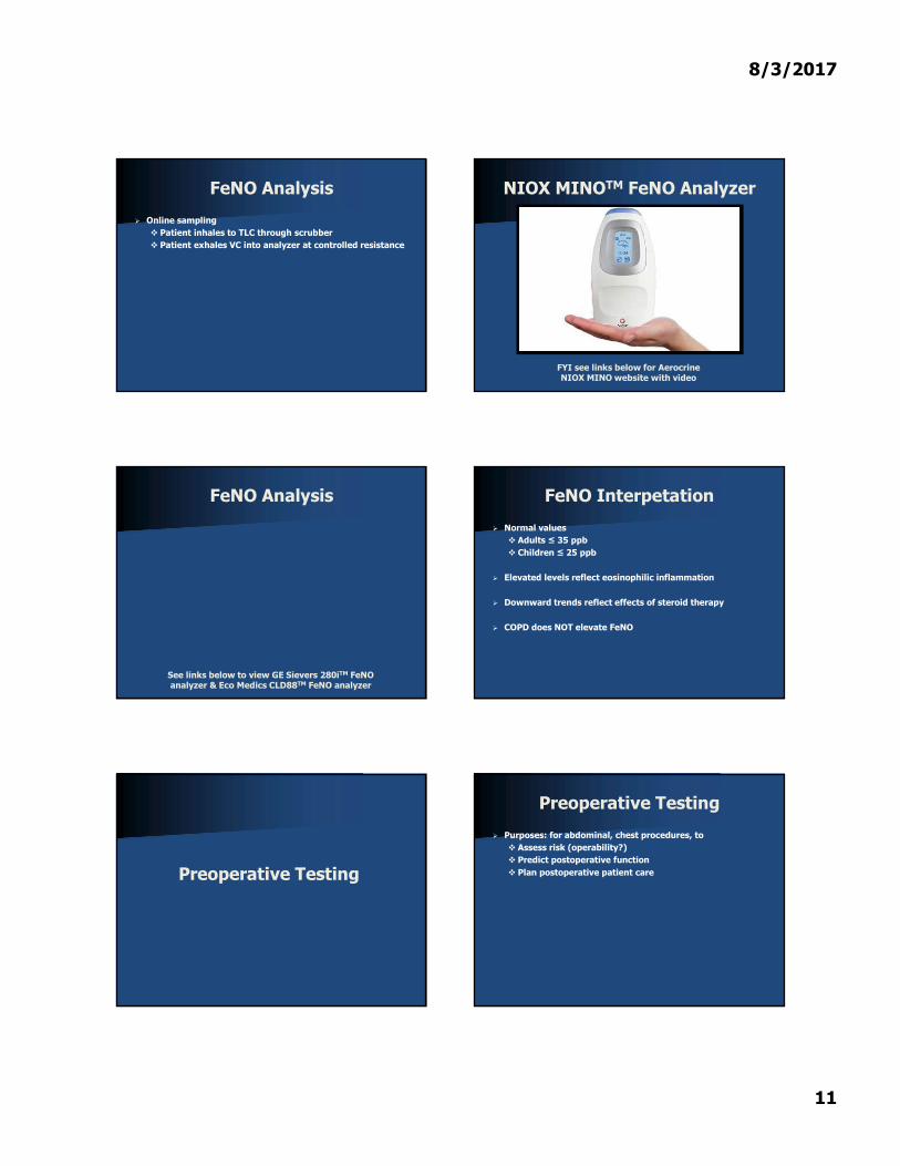
NIOX MINOTM FeNO Analyzer
FYI see links below for AerocrineNIOX MINO website with video
FeNO Analysis
See links below to view GE Sievers 280iTM FeNOanalyzer & Eco Medics CLD88TM FeNO analyzer
FeNO Interpetation
Normal values Adults ≤ 35 ppb Children ≤ 25 ppb
Elevated levels reflect eosinophilic inflammation
Downward trends reflect effects of steroid therapy
COPD does NOT elevate FeNO
Preoperative Testing
Preoperative Testing
Purposes: for abdominal, chest procedures, to Assess risk (operability?) Predict postoperative function Plan postoperative patient care
8/3/2017
12
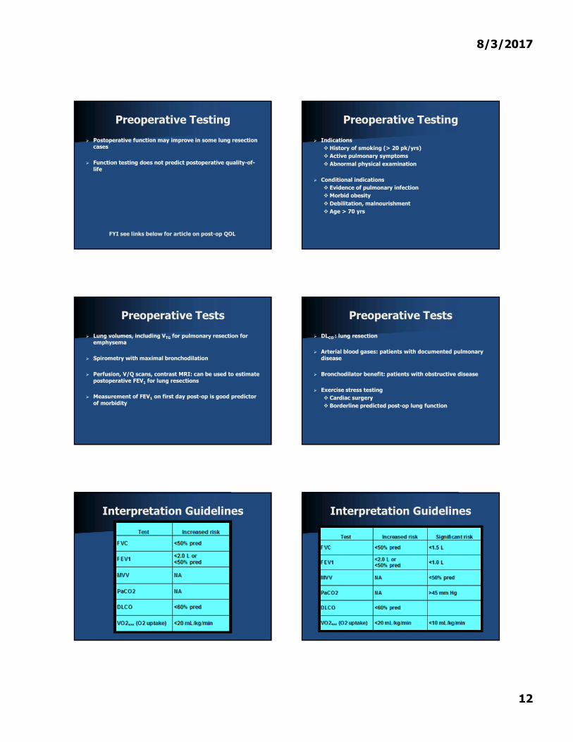
Preoperative Testing
Postoperative function may improve in some lung resection cases
Function testing does not predict postoperative quality-of-life
FYI see links below for article on post-op QOL
Preoperative Testing
Indications History of smoking (> 20 pk/yrs) Active pulmonary symptoms Abnormal physical examination
Conditional indications Evidence of pulmonary infectionMorbid obesity Debilitation, malnourishment Age > 70 yrs
Preoperative Tests
Lung volumes, including VTG for pulmonary resection for emphysema
Spirometry with maximal bronchodilation
Perfusion, V/Q scans, contrast MRI: can be used to estimate postoperative FEV1 for lung resections
Measurement of FEV1 on first day post-op is good predictor of morbidity
Preoperative Tests
DLCO : lung resection
Arterial blood gases: patients with documented pulmonary disease
Bronchodilator benefit: patients with obstructive disease
Exercise stress testing Cardiac surgery Borderline predicted post-op lung function
Interpretation Guidelines Interpretation Guidelines
8/3/2017
13
Testing for Disability
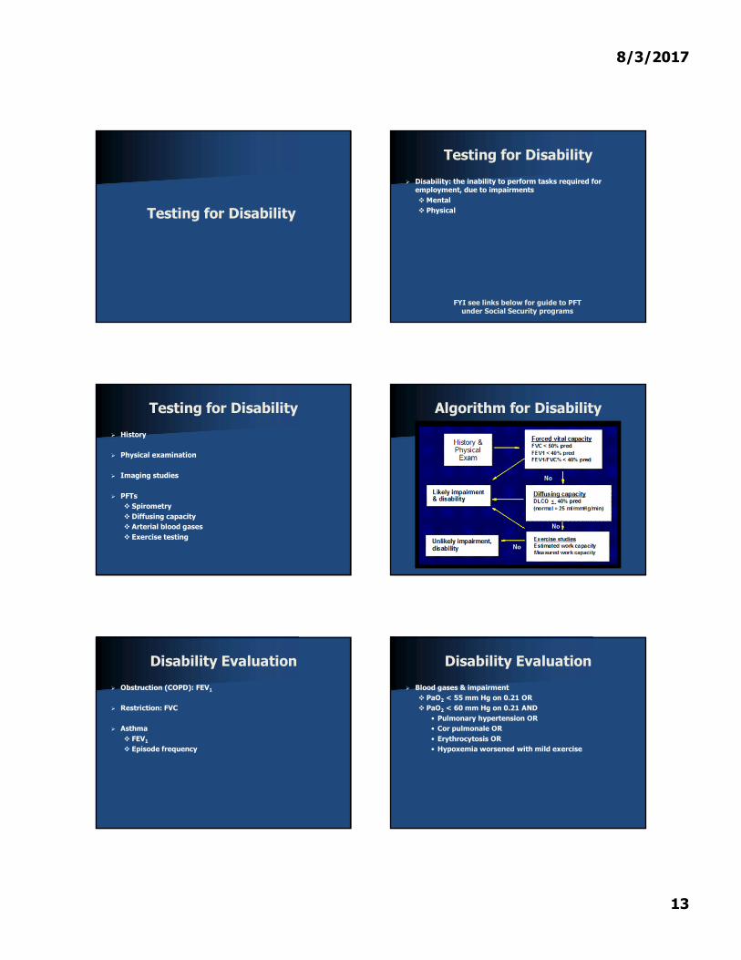
Testing for Disability
Disability: the inability to perform tasks required for employment, due to impairmentsMental Physical
FYI see links below for guide to PFTunder Social Security programs
Testing for Disability
History
Physical examination
Imaging studies
PFTs Spirometry Diffusing capacity Arterial blood gases Exercise testing
Algorithm for Disability
Disability Evaluation
Obstruction (COPD): FEV1
Restriction: FVC
Asthma FEV1
Episode frequency
Disability Evaluation
Blood gases & impairment PaO2 < 55 mm Hg on 0.21 OR PaO2 < 60 mm Hg on 0.21 AND
• Pulmonary hypertension OR• Cor pulmonale OR• Erythrocytosis OR• Hypoxemia worsened with mild exercise

8/3/2017
14
Disability Evaluation
Additional factors to consider Subject cooperation (malingering) Hx of emergency treatment for asthma Failure to receive appropriate care Deconditioning (couch potato) Coexisting disorders Impairment that is difficult to measure
Cardiopulmonary Exercise Testing (CPET)
Indications forExercise Testing
Diagnose cardiopulmonary disorders, often to distinguish between cardiac vs. pulmonary dx
Measure impairment (disability)
Evaluate therapy
Develop exercise prescriptions (rehabilitation)
Assess fitness for occupations, physical activities, etc.
Contraindications forExercise Testing
Limiting neurologic, neuromuscular, or orthopedic conditions
Pulmonary contraindications FEV1 < 30% Room air PaO2 < 40 mm Hg PaCO2 > 70 mm Hg Severe pulmonary hypertension
Contraindications forExercise Testing
Cardiovascular conditions Acute pericarditis CHF Recent MI Heart block: 2nd or 3rd degree Tachydysrhythmias Uncontrolled hypertension Unstable angina Recent systemic or pulmonary embolus Aortic stenosis
Pulmonary ChangesWith Exercise
TV increases early
Respiratory rate increases late
Vd/Vt decreases
V/Q equalizes
Capillary transit time decreases - increased velocity of blood
8/3/2017
15
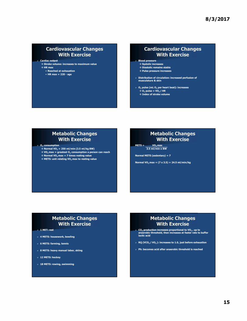
Cardiovascular ChangesWith Exercise
Cardiac output Stroke volume: increases to maximum value HR max
• Reached at exhaustion• HR max = 220 - age
Cardiovascular ChangesWith Exercise
Blood pressure Systolic increases Diastolic remains stable Pulse pressure increases
Distribution of circulation: increased perfusion of musculature & skin
O2 pulse (mL O2 per heart beat): increases O2 pulse = VO2 / HR Index of stroke volume
Metabolic ChangesWith Exercise
O2 consumption Normal VO2 = 250 ml/min (3.5 ml/kg BW) VO2 max = greatest O2 consumption a person can reach Normal VO2 max = 7 times resting valueMETS: unit relating VO2 max to resting value
Metabolic ChangesWith Exercise
METS = VO2 max3.5 ml/min x BW
Normal METS (sedentary) = 7
Normal VO2 max = (7 x 3.5) = 24.5 ml/min/kg
Metabolic ChangesWith Exercise
1 MET: rest
4 METS: housework, bowling
6 METS: farming, tennis
8 METS: heavy manual labor, skiing
12 METS: hockey
18 METS: rowing, swimming
Metabolic ChangesWith Exercise
CO2 production increases proportional to VO2 , up to anaerobic threshold, then increases at faster rate to buffer lactic acid
RQ (VCO2 / VO2 ): increases to 1.0, just before exhaustion
Ph: becomes acid after anaerobic threshold is reached
8/3/2017
16
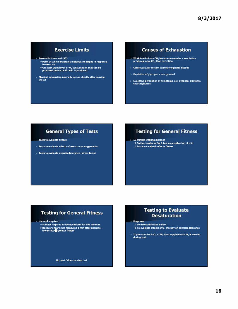
Exercise Limits
Anaerobic threshold (AT) Point at which anaerobic metabolism begins in response
to exercise Greatest work level, or O2 consumption that can be
produced before lactic acid is produced
Physical exhaustion normally occurs shortly after passing the AT
Causes of Exhaustion
Work to eliminate CO2 becomes excessive - ventilation produces more CO2 than excretion
Cardiovascular system cannot oxygenate tissues
Depletion of glycogen - energy need
Excessive perception of symptoms, e.g. dyspnea, dizziness, chest tightness
General Types of Tests
Tests to evaluate fitness
Tests to evaluate effects of exercise on oxygenation
Tests to evaluate exercise tolerance (stress tests)
Testing for General Fitness
12 minute walking distance Subject walks as far & fast as possible for 12 min Distance walked reflects fitness
Testing for General Fitness
Harvard step test Subject steps up & down platform for five minutes Recovery heart rate measured 1 min after exercise -
lower rate greater fitness
Up next: Video on step test
Testing to Evaluate Desaturation
Purposes To detect diffusion defect To evaluate effects of O2 therapy on exercise tolerance
If pre-exercise SaO2 < 90, then supplemental O2 is needed during test

8/3/2017
17
Testing to Evaluate Desaturation
Subject exercises on treadmill or ergometer for 6 min Parameters monitored
• Pre-exercise SaO2 , SpO2 : correlate values• Exercise SpO2
• ECG• Blood pressure
Testing to Evaluate Desaturation
Results Normally, SpO2 increases due to improved VQ matching SpO2 > 90% after 6 minutes no desaturation SpO2 decreased by 5% or drops to less than 85%, test
terminated & results are positive for desaturation & likely diffusion defect
Types of ExerciseTolerance Tests
Constant work
Incremental work: more common Staged increments - stepwise Ramp increments - constant
Exercise Testing Equipment
Treadmill Advantages
• Familiar exercise• Typical activity (ADL)
Disadvantages• Subject weight is a factor• Large, heavy, noisy• Expensive• Safety issues
FYI see links below for video on treadmill misadventures
Exercise Testing Equipment
Bicycle ergometer Advantages
• Workload unaffected by weight• Workload precisely measured• Small, portable• Inexpensive• Safer than treadmill
Exercise Testing Equipment
Bicycle ergometer Disadvantages
• Unfamiliar exercise• Not ADL
Yields results slightly different from treadmill
FYI see links below for article onexercise testing in clinical practice

8/3/2017
18
Exercise Testing Equipment
Gas volume measurement device
Gas collection, mixing devices
Gas analyzers: O2 , CO2
Pulse oximeter
ECG monitor: filtered for motion artifact
Blood pressure monitor
Crash cart
Exercise Tolerance Testing
Preparation of subject Comfortable clothes No meal within 2 H No smoking, coffee within 2 H Continue medications as prescribed Orient to equipment & procedures - include hand signals
Exercise Tolerance Testing
Preliminary assessment Hx & Px 12 lead ECG PFTs
• Spirometry• MVV• DLCO
Exercise Tolerance Testing
Obtain resting values Arterial blood gas Lactate (in some labs) SpO2 : correlate with SaO2
TV, f, VE PetCO2 , PetO2
HR, BP, ECG pattern
Exercise Tolerance Testing
Practice at minimal work level: check monitors & equipment
Exercise: increase workload Intervals Ramp
Monitor continuously or sample at each work level -depends on system
See links below for image of exercise testing
Exercise Tolerance Testing
Indicators to stop test Exhaustion: desired endpoint CNS symptoms: vertigo, etc. Nausea, vomiting Chest pain, SOB SpO2 drop > 5% Dysrhythmias: frequent PVCs, etc. PSYS > 250 mm Hg Equipment failure
8/3/2017
19
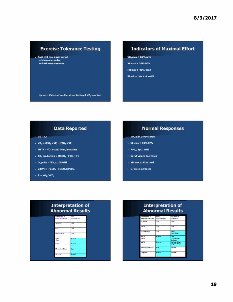
Exercise Tolerance Testing
Post-test cool down periodMinimal exercise Final measurements
Up next: Videos of cardiac stress testing & VO2 max test
Indicators of Maximal Effort
VO2 max ≥ 85% pred
VE max ≥ 70% MVV
HR max > 90% pred
Blood lactate ≥ 4 mM/L
Data Reported
VE, TV, f
VO2 = (FiO2 x VI) - (FEO2 x VE)
METS = VO2 max/3.5 ml/min x BW
CO2 production = (FECO2 - FiCO2) VE
O2 pulse = VO2 x 1000/HR
Vd/Vt = (PaCO2 - PetCO2)/PaCO2
R = VO2 /VCO2
Normal Responses
VO2 max ≥ 95% pred
VE max ≥ 70% MVV
PaO2 , SpO2 WNL
Vd/Vt values decreases
HR max ≥ 90% pred
O2 pulse increases
Interpretation ofAbnormal Results
Interpretation ofAbnormal Results

8/3/2017
20
Interpretation ofAbnormal Results
Metabolic Testing
Purposes of Metabolic Testing
Measure nutritional requirements
Measure relative metabolic contributions of Carbohydrates Lipids Protein
Rationale for Bedside Assessment
Critically ill patients have highly variable metabolic needs
Patients often are NPO & receive all nourishment via total parenteral nutrition (TPN)
Specific Indications for Testing
COPD
Multiple trauma
Acute pancreatitis
Organ transplant patients
Morbid obesity
Hyper or hypo-metabolism
Prolonged mechanical ventilation & NPO status (weaning)
Sources of Nutritional Depletion
Vomiting, NG suctioning
Diarrhea
Malabsorption
Elevated metabolism due to Fever Surgery Trauma

8/3/2017
21
Complications of Malnourishment
Impaired function of all organ systems
Immunocompromise
Delayed wound healing
Increased ventilatory load due to Increased oxygen demand Increased CO2 production
FYI see links below for article on calorimetry & weaning
Methods for Nutritiional Assessment
Anthropometric Skin fold thickness Arm circumference
Laboratory assessment - serum proteins
Calorimetry
Calorimetry Methods
Direct: complete enclosure of body & measurement of heat production
Indirect: uses VO2 , VCO2 , VE to calculate energy expenditure Closed circuit method Open circuit method
FYI see links below for AARC CPG on indirectcalorimetry during mechanical ventilation
Closed Circuit Method
Subject rebreathes in closed system
CO2 is absorbed
O2 measured volumetrically with spirometer VI - VE = VO2
Not compatible with current ventilators
Open Circuit Method
Hood or canopy for spontaneously breathing patients
Ventilator - attaches at airway
Measured parameters FiO2 & FEO2
FiCO2 & FECO2
VE
See links below to view open circuit calorimetry with hood
Open Circuit Method
Calorimeters
See links below to view

8/3/2017
22
Test Administration
Patient preparation Avoid stimulants prior to test Fast for 2 - 4 H, if PO Continuous feedings, if NPO No ventilator adjustments immediately before testing
(within 90 min)
Neutral thermal environment must be maintained - no thermal stress
Measurements made during steady state
Calculated Parameters
Resting energy expenditure (REE) VO2
VCO2
UN (urinary N2 ) - not critical to test
Caloric equivalents for Carbohydrates Lipids Protein
Respiratory quotient (RQ)
Significance of Results
RQ < 0.67 or RQ > 1.3 error
RQ < 0.7 starvation or ketosis
RQs for predominant substrate Carbohydrates = 1.0 Lipids = 0.71 Protein = 0.82
REE > caloric intake underfeeding
REE < caloric intake overfeeding
Summary & Review
Diffusing capacity Gas laws Pathophysiology DLCOsb - most common Normal DLCOsb = 25 mL/min/mm Hg Increased with obesity, asthma Decreased with emphysema, fibrosis
Summary & Review
Bronchodilator benefit: before & after bronchodilator tests Indications Preconditions Procedure Significant improvement
• 12% FEV1 increase & 200 mL FEV1 or FVC increase• 30% sGAW increase
Summary & Review
Bronchial challenge testing: detects & measures airway reactivity Provocative agents: methacholine, histamine, or exercise Preconditions, procedure Significant results
• PC20: dose where FEV1 decreased by 20%• Decrease in mechanics produced by exercise
8/3/2017
23
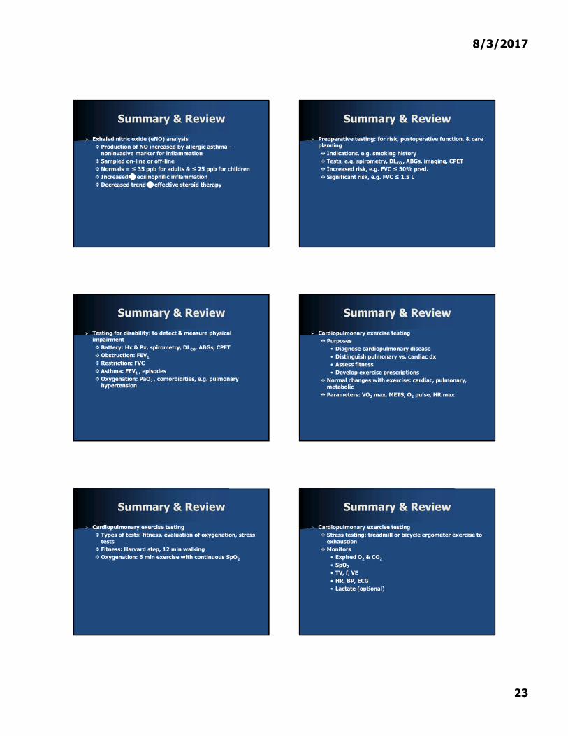
Summary & Review
Exhaled nitric oxide (eNO) analysis Production of NO increased by allergic asthma -
noninvasive marker for inflammation Sampled on-line or off-line Normals = ≤ 35 ppb for adults & ≤ 25 ppb for children Increased eosinophilic inflammation Decreased trend effective steroid therapy
Summary & Review
Preoperative testing: for risk, postoperative function, & care planning Indications, e.g. smoking history Tests, e.g. spirometry, DLCO , ABGs, imaging, CPET Increased risk, e.g. FVC ≤ 50% pred. Significant risk, e.g. FVC ≤ 1.5 L
Summary & Review
Testing for disability: to detect & measure physical impairment Battery: Hx & Px, spirometry, DLCO, ABGs, CPET Obstruction: FEV1
Restriction: FVC Asthma: FEV1 , episodes Oxygenation: PaO2 , comorbidities, e.g. pulmonary
hypertension
Summary & Review
Cardiopulmonary exercise testing Purposes
• Diagnose cardiopulmonary disease• Distinguish pulmonary vs. cardiac dx• Assess fitness• Develop exercise prescriptions
Normal changes with exercise: cardiac, pulmonary, metabolic
Parameters: VO2 max, METS, O2 pulse, HR max
Summary & Review
Cardiopulmonary exercise testing Types of tests: fitness, evaluation of oxygenation, stress
tests Fitness: Harvard step, 12 min walking Oxygenation: 6 min exercise with continuous SpO2
Summary & Review
Cardiopulmonary exercise testing Stress testing: treadmill or bicycle ergometer exercise to
exhaustionMonitors
• Expired O2 & CO2
• SpO2
• TV, f, VE• HR, BP, ECG• Lactate (optional)

8/3/2017
24
Summary & Review
Cardiopulmonary exercise testing Indicators of maximal effort, e.g. HR max > 90% pred. Normal responses: oxygenation stable or improved, O2
pulse increases
Summary & Review
Cardiopulmonary exercise testing Indicators of maximal effort, e.g. HR max > 90% pred. Normal responses: oxygenation stable or improved, O2
pulse increases Interpretation
• Poor conditioning: normal pulmonary parameters & O2 pulse
• Pulmonary disease: low pulmonary parameters• Cardiac disease: low O2 pulse
Summary & Review
Metabolic testing: to measure nutritional requirements & determine metabolic contributions Indications, e.g. prolonged mechanical ventilation Calorimetry methods
• Direct vs. indirect• Indirect: closed vs. open circuits
Summary & Review
Metabolic testing Calorimeter attached to airway & measures
• FiO2 , FEO2
• FiCO2 , FECO2
• VE Calculated values
• Resting energy expenditure (REE)• Caloric equivalents• Respiratory quotient (RQ)
Summary & Review
Results may detectMeasurement error Starvation or ketosis Contributions from carbohydrates, lipids, proteins Underfeeding Overfeeding
References
Ruppel GL. Manual of Pulmonary Function Testing, 9th Ed. Chaps. 5, 7, 9. 2009 Mosby-Elsevier; St. Louis.
Madama VC. Pulmonary function testing and cardiopulmonary stress testing 2nd ed. Chaps. 6, 8, 14, 15. 1998; Delmar; Albany.
Brunelli A, Refai MA, Salati M, Sabbatini A, MorganHughes NJ, Rocco G. Carbon monoxide lung diffusion capacity improves risk stratification in patients without airflow limitation: evidence for systematic measurement before lung resection. Eur J Cardiothorac Surg. 2006 Apr;29(4):567-70.

8/3/2017
25
References
Saydain G, Beck KC, Decker PA, Cowl CT, Scanlon PD. Clinical significance of elevated diffusing capacity.Chest. 2004 Feb;125(2):446-52.
Bobbio A, Chetta A, Carbognani P, Internullo E, Verduri A, Sansebastiano G, Rusca M, Olivieri D. Changes in pulmonary function test and cardiopulmonary exercise capacity in COPD patients after lobar pulmonary resection. Eur J Cardiothorac Surg. 2005 Nov;28(5):754-8.
Lamberto C, Nunes H, Le Toumelin P, Duperron F, Valeyre D, Clerici C. Membrane and capillary blood components of diffusion capacity of the lung for carbon monoxide in pulmonary sarcoidosis: relation to exercise gas exchange. Chest. 2004 Jun;125(6):2061-8.
References
Lee JS, Ra SW, Chae EJ, Seo JB, Lim SY, Kim TH, Lee SD, Oh YM. Validation of the Lower Limit of normal Diffusing Capacity for Detecting Emphysema. Respiration. 2010 Jan 29.
Wang JS. Relationship of carbon monoxide pulmonary diffusing capacity to postoperative cardiopulmonary complications in patients undergoing pneumonectomy. Kaohsiung J Med Sci. 2003 Sep;19(9):437-46.
Chandra S, Shah SJ, Thenappan T, Archer SL, Rich S, Gomberg-Maitland M. Carbon monoxide diffusing capacity and mortality in pulmonary arterial hypertension. J Heart Lung Transplant. 2010 Feb;29(2):181-7. Epub 2009 Sep 26.
References
Rodway GW, Choi J, Hoffman LA, Sethi JM. Exhaled nitric oxide in the diagnosis and management of asthma: clinical implications. Chron Respir Dis. 2009;6(1):19-29.
Price D, Berg J, Lindgren P. An economic evaluation of NIOX MINO airway inflammation monitor in the United Kingdom. Allergy. 2009 Mar;64(3):431-8. Epub 2009 Jan 29.
Kwok MY, Walsh-Kelly CM, Gorelick MH. The role of exhaled nitric oxide in evaluation of acute asthma in a pediatric emergency department. Acad Emerg Med. 2009 Jan;16(1):21-8. Epub 2008 Nov 27.
References
Hewitt RS, Modrich CM, Cowan JO, Herbison GP, Taylor DR. Outcomes using exhaled nitric oxide measurements as an adjunct to primary care asthma management. Prim Care Respir J. 2009;18(4):320-7.
Ferguson MK, Gaissert HA, Grab JD, Sheng S. Pulmonary complications after lung resection in the absence of chronic obstructive pulmonary disease: the predictive role of diffusing capacity. J Thorac Cardiovasc Surg. 2009 Dec;138(6):1297-302.
Sue DY, Wasserman K. Impact of integrative cardiopulmonary exercise testing on clinical decision making. Chest. 1991 Apr;99(4):981-92.
References
Chetta A, Tzani P, Marangio E, Carbognani P, Bobbio A, Olivieri D. Respiratory effects of surgery and pulmonary function testing in the preoperative evaluation. Acta Biomed. 2006 Aug;77(2):69-74.
Milani RV, Lavie CJ, Mehra MR, Ventura HO. Understanding the basics of cardiopulmonary exercise testing. Mayo Clin Proc. 2006;81(12):1603-11.
ERS Task Force, Palange P, Ward SA, Carlsen KH, Casaburi R, Gallagher CG, Gosselink R, O'Donnell DE, Puente-Maestu L, Schols AM, Singh S, Whipp BJ. Recommendations on the use of exercise testing in clinical practice. Eur Respir J. 2007;29(1):185-209.
References
Cote CG, Pinto-Plata V, Kasprzyk K, Dordelly LJ, Celli BR. The 6-min walk distance, peak oxygen uptake, and mortality in COPD. Chest. 2007;132(6):1778-85.
American Thoracic Society; American College of Chest Physicians. ATS/ACCP Statement on cardiopulmonary exercise testing. Am J Respir Crit Care Med. 2003 Jan 15;167(2):211-77. Review. Erratum in: Am J Respir Crit Care Med. 2003 May 15;1451-2.
Rasekaba T, Lee AL, Naughton MT, Williams TJ, Holland AE. The six-minute walk test: a useful metric for the cardiopulmonary patient. Intern Med J. 2009 Aug;39(8):495-501.

8/3/2017
26
References
Benzo RP, Sciurba FC. Oxygen Consumption, Shuttle Walking Test and the Evaluation of Lung Resection. Respiration. 2009 Aug 11.
Benzo RP, Paramesh S, Patel SA, Slivka WA, Sciurba FC. Optimal protocol selection for cardiopulmonary exercise testing in severe COPD. Chest. 2007 Nov;132(5):1500-5.
Varela, et al. Measured FEV1 in the first postoperative day, and not ppoFEV1, is the best predictor of cardiorespiratory morbidity after lung resection. Eur J Cardiothorac Surg 2007 31: 518-521
References
Ferrazza AM, Martolini D, Valli G, Palange P. Cardiopulmonary exercise testing in the functional and prognostic evaluation of patients with pulmonary diseases. Respiration 2009;77(1):3-17.
Pan AM, Stiell IG, Clement CM, Acheson J, Aaron SD. Feasibility of a structured 3-minute walk test as a clinical decision tool for patients presenting to the emergency department with acute dyspnoea. Emerg Med J. 2009 Apr;26(4):278-82.
Buschmann HC, Petermann W. Differential diagnosis of restrictive lung diseases: utility of cardiopulmonary exercise testing. Pneumologie. 2010 Jan;64(1):28-36.
![5HTXHVW IRU 4XDOLILFDWLRQV $UL]RQD 'HSDUWPHQW RI ... › sites › default › files › Sunrise Engineering Inc_1.pdf5htxhvw iru 4xdolilfdwlrqv 6rolflwdwlrq 1r ^wk í ó r ì ì ì](https://static.fdocuments.us/doc/165x107/60bb2dd689264910f6468737/5htxhvw-iru-4xdolilfdwlrqv-ulrqd-hsduwphqw-ri-a-sites-a-default-a-files.jpg)