Pulmonary Embryology - 1 File Download
520
Pulmonary Embryology Jason Ryan, MD, MPH
Transcript of Pulmonary Embryology - 1 File Download
Pulmonary EmbryologyJason Ryan, MD, MPH
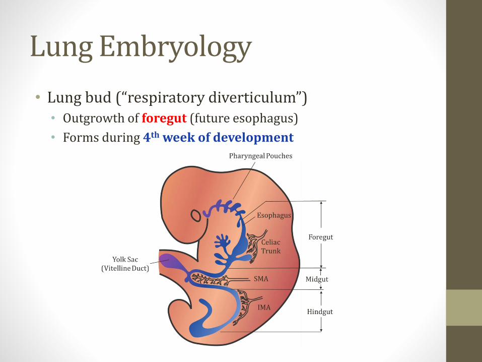
Lung Embryology
• Lung bud (“respiratory diverticulum”)• Outgrowth of foregut (future esophagus)
• Forms during 4th week of development
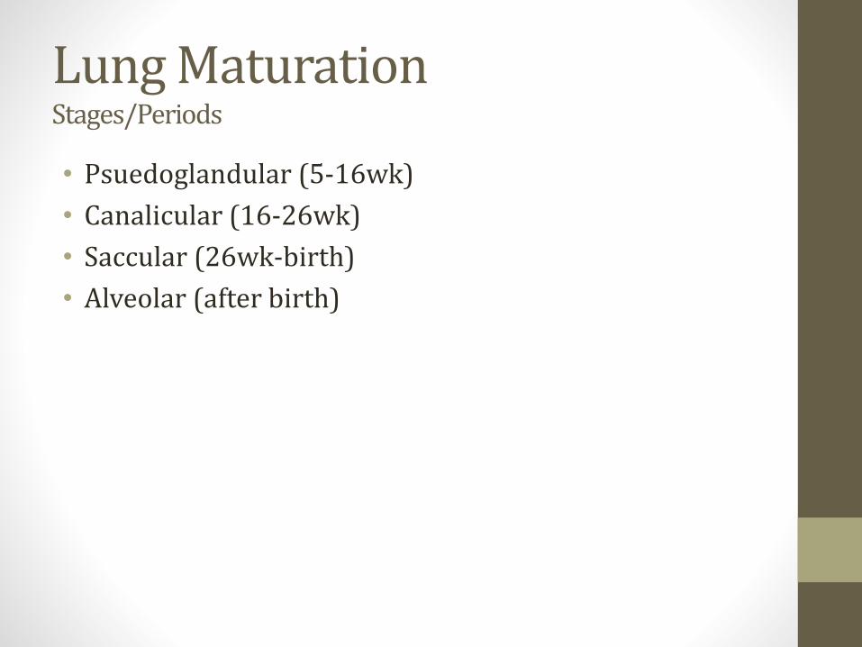
Lung Maturation Stages/Periods
• Psuedoglandular (5-16wk)
• Canalicular (16-26wk)
• Saccular (26wk-birth)
• Alveolar (after birth)
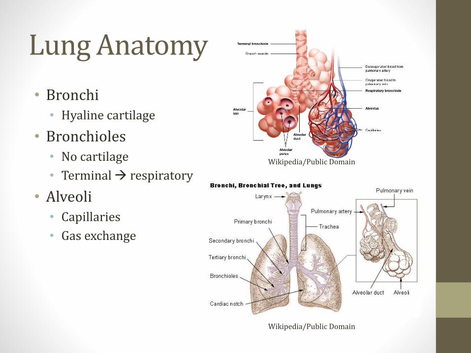
Lung Anatomy
• Bronchi• Hyaline cartilage
• Bronchioles• No cartilage
• Terminal → respiratory
• Alveoli• Capillaries
• Gas exchange
Wikipedia/Public Domain
Wikipedia/Public Domain
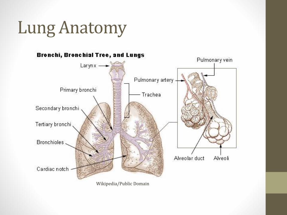
Lung Anatomy
Wikipedia/Public Domain

Lung Anatomy
Wikipedia/Public Domain
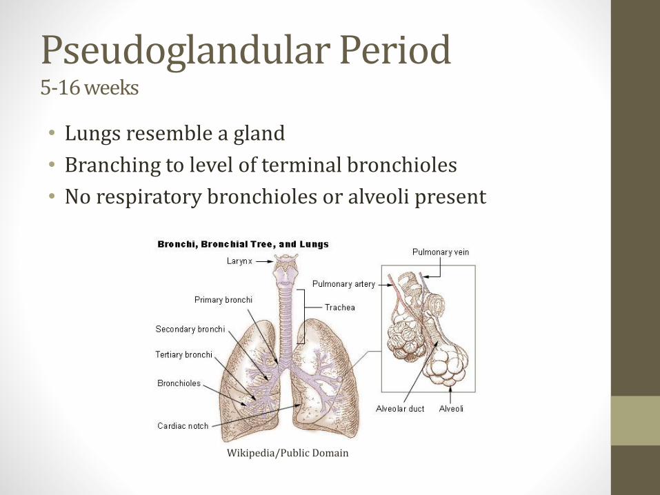
Pseudoglandular Period5-16 weeks
• Lungs resemble a gland
• Branching to level of terminal bronchioles
• No respiratory bronchioles or alveoli present
Wikipedia/Public Domain
Fetal Respiration
• Fetal breathing movements occur in utero
• Baby aspirates amniotic fluid
• Stimulates lung development
• Growth of respiratory muscles
• Important for growth during pseudoglandular phase

Fetal Respiration
• Oligohydramnios:• Pulmonary hypoplasia
• Part of Potter’s sequence
• Caused by fetal kidney abnormalities
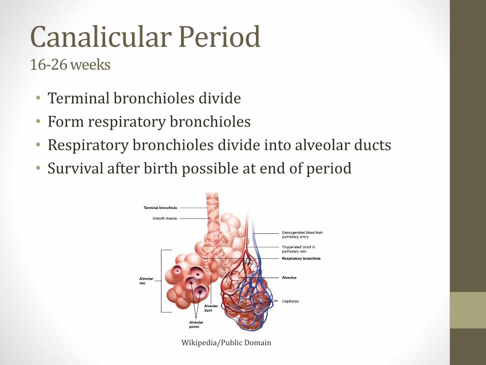
Canalicular Period16-26 weeks
• Terminal bronchioles divide
• Form respiratory bronchioles
• Respiratory bronchioles divide into alveolar ducts
• Survival after birth possible at end of period
Wikipedia/Public Domain

Canalicular Period16-26 weeks
• Airway lumens become larger
• Type II pneumocytes form• Produce surfactant
• Lowers surface tension
• Keeps alveoli open
Wikipedia/Public Domain
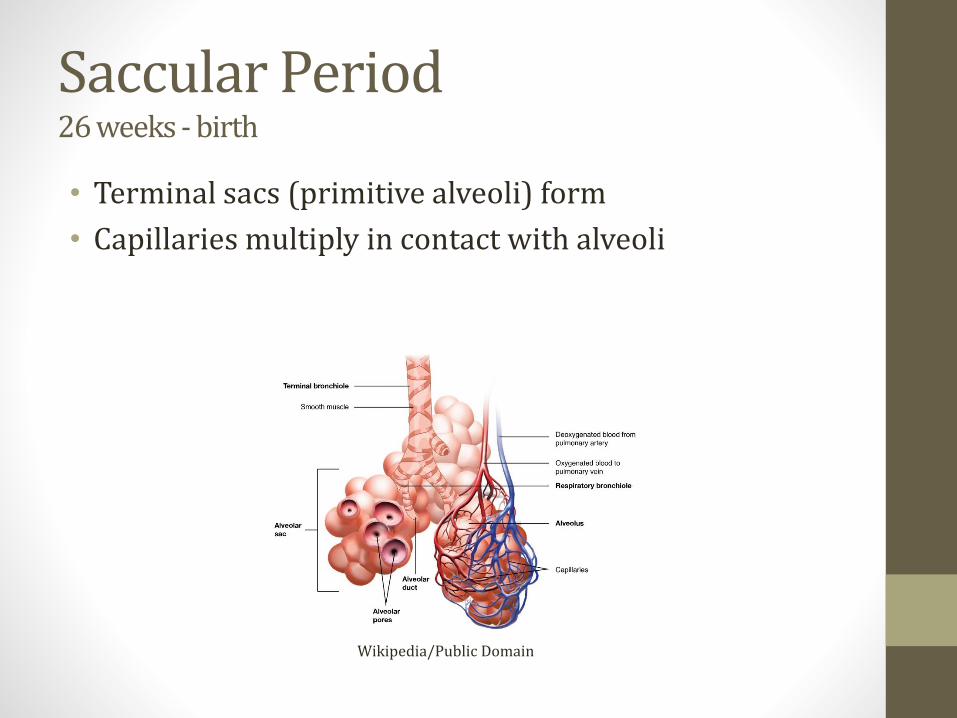
Saccular Period26 weeks - birth
• Terminal sacs (primitive alveoli) form
• Capillaries multiply in contact with alveoli
Wikipedia/Public Domain

Alveolar PeriodAfter birth
• At birth, only about 1/3 of alveoli present
• Following birth:• ↑ number of respiratory bronchioles and alveoli
• Continued lung development through age 10

Alveolarization
• Airspaces subdivided
• New walls formed (septa)
Johannes Schittny. Cell Tissue Res. 2017; 367 (3) 427

Bronchopulmonary Dysplasia
• Occurs in premature infants
• Treated in NICU
• Surfactant, oxygen, mechanical ventilation
• Oxygen toxicity and lung trauma
• Alveolarization does not progress normally
• Respiratory problems during infancy
• Often improves during childhood

Johannes Schittny. Cell Tissue Res. 2017; 367 (3) 427
Pulmonary Hypoplasia
• Oligohydramnios (Potter’s sequence)
• Congenital diaphragmatic hernia• Defective formation pleuroperitoneal membrane
• Hole in diaphragm
• Abdominal organs herniate into chest
• In utero herniation → pulmonary hypoplasia
• Often fatal

Bronchogenic Cysts
• Abnormal budding of foregut
• Usually found in mediastinum
• Contain clear fluid • Air seen when infected
Bronchogenic Cysts
• Do not communicate with lungs
• Lined by respiratory epithelium • Columnar, ciliated
• Walls contain cartilage (diagnostic criteria)
• Often asymptomatic
• May lead to pneumonia, compression of airway


The Radiology Assistant

Pulmonary Vascular Resistance
• In utero• PVR is high
• Canalicular stage: few/no pulmonary capillaries
• Later stages: hypoxemia → vasoconstriction
• Umbilical venous blood: PaO2 30mmHg; O2sat=80%
• Only about 10% of cardiac output to lungs
• At birth• PVR falls significantly
• 100% cardiac output through lungs
Pulmonary AnatomyJason Ryan, MD, MPH

Respiratory Tract
Zones
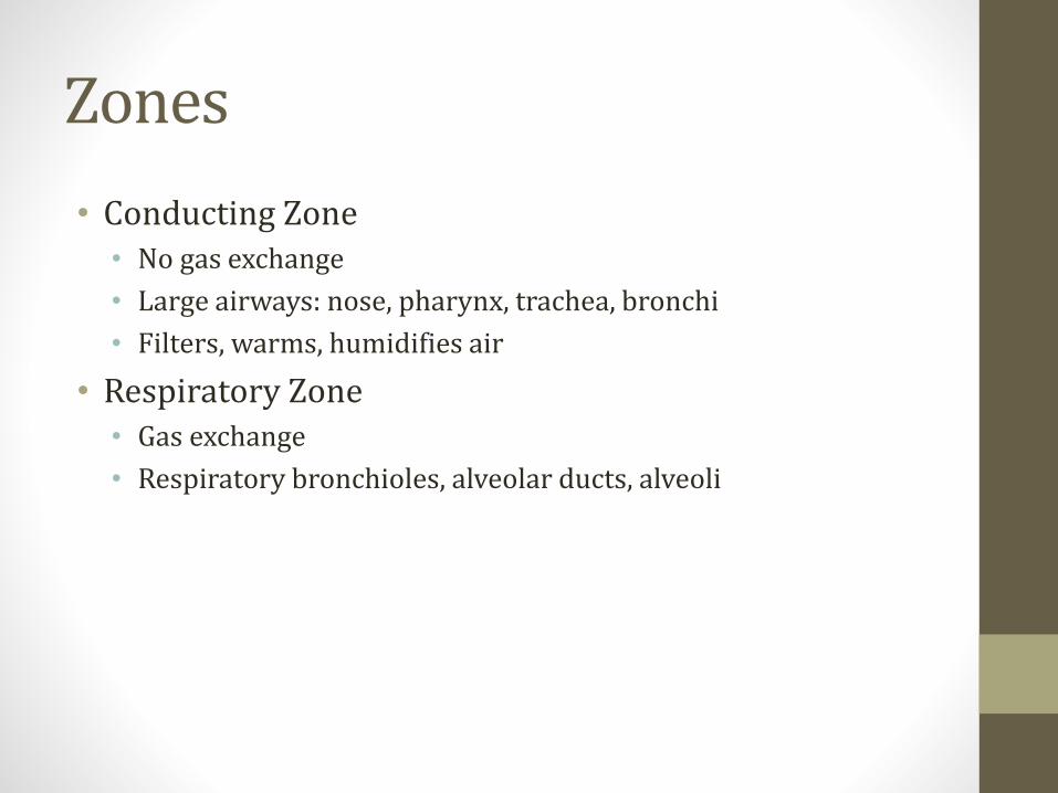
• Conducting Zone• No gas exchange
• Large airways: nose, pharynx, trachea, bronchi
• Filters, warms, humidifies air
• Respiratory Zone• Gas exchange
• Respiratory bronchioles, alveolar ducts, alveoli
Mucous
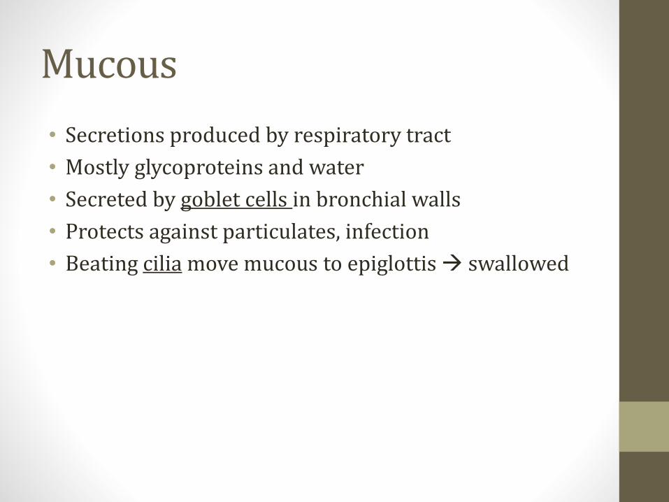
• Secretions produced by respiratory tract
• Mostly glycoproteins and water
• Secreted by goblet cells in bronchial walls
• Protects against particulates, infection
• Beating cilia move mucous to epiglottis → swallowed
Alveoli
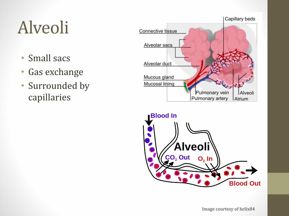
• Small sacs
• Gas exchange
• Surrounded by capillaries
Image courtesy of helix84

Alveolar Cells: Pneumocytes
• Type 1• Most common (97% of cells)
• Thin for gas exchange
• Type 2• Produce surfactant
• Can proliferate to other cells – key for regeneration after injury
• Club cells (bronchioles)• Surfactant
• Detoxification

Surfactant
• Exhale → alveoli shrink
• Could collapse → atelectasis
• ↓ efficiency gas exchange
• Surfactant allows alveoli to avoid collapse
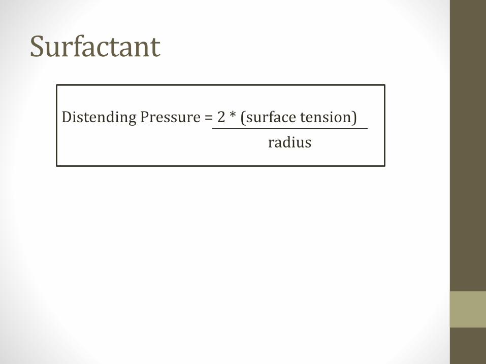
Surfactant
Distending Pressure = 2 * (surface tension)
radius
Surfactant
• Secreted by type 2 pneumocytes
• Mix of lecithins
• Especially dipalmitoylphosphatidylcholine
Fetal Lung Maturity
• Lungs are “mature” when enough surfactant present
• Occurs around 35 weeks
• Lecithin–sphingomyelin ratio (L/S ratio)
• Both produced equally until ~35 weeks
• Ratio >2.0 in amniotic fluid suggests lungs mature
• Preterm delivery: betamethasone used to stimulate surfactant production in lungs

Neonatal respiratory distress syndrome• Hyaline membrane disease
• Atelectasis
• Severe hypoxemia/↑pCO2 (poor ventilation)
• Poorly responsive to O2• Lungs collapsed (alveoli)
• Intrapulmonary shunting
Neonatal respiratory distress syndrome• Risk factors:
• Prematurity
• Maternal diabetes: high insulin levels decrease surfactant production
• Cesarean delivery: lack of vaginal compression stress leads to reduced fetal cortisol and reduction in surfactant
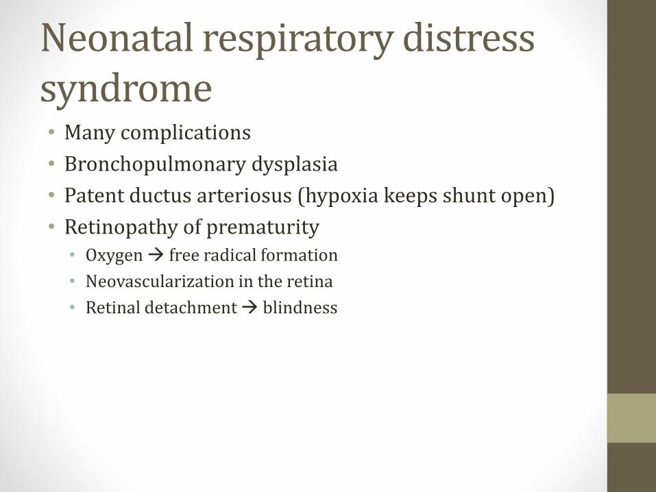
Neonatal respiratory distress syndrome• Many complications
• Bronchopulmonary dysplasia
• Patent ductus arteriosus (hypoxia keeps shunt open)
• Retinopathy of prematurity • Oxygen → free radical formation
• Neovascularization in the retina
• Retinal detachment → blindness
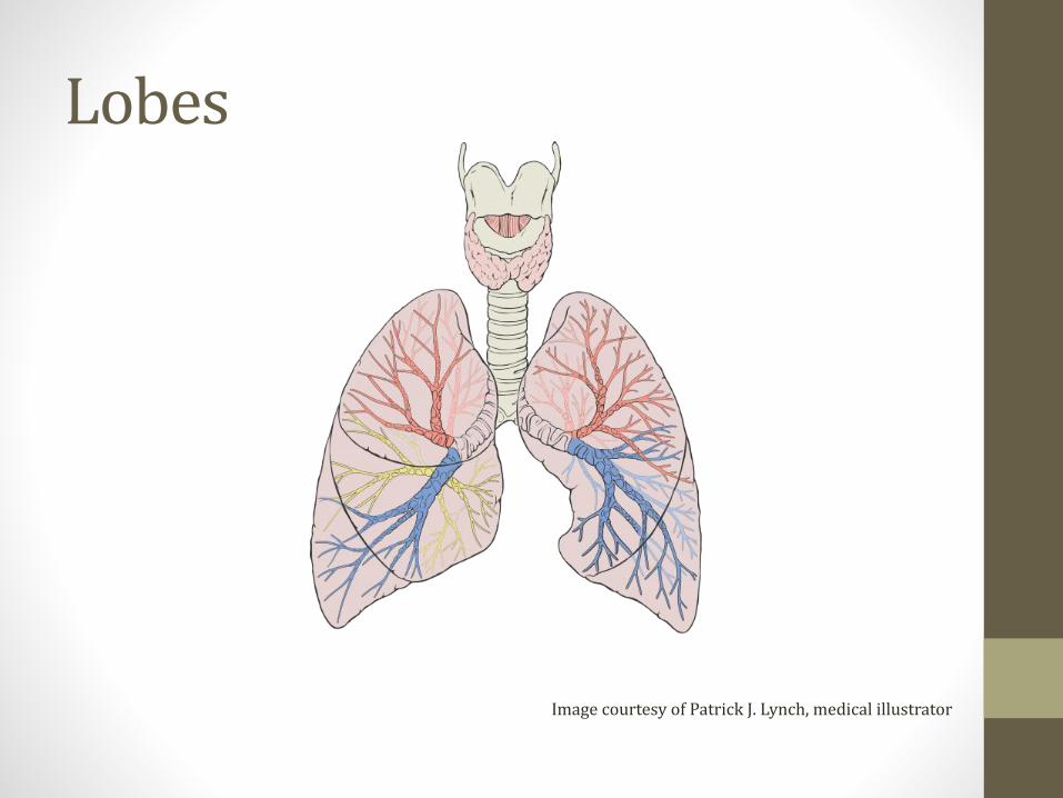
Lobes
Image courtesy of Patrick J. Lynch, medical illustrator
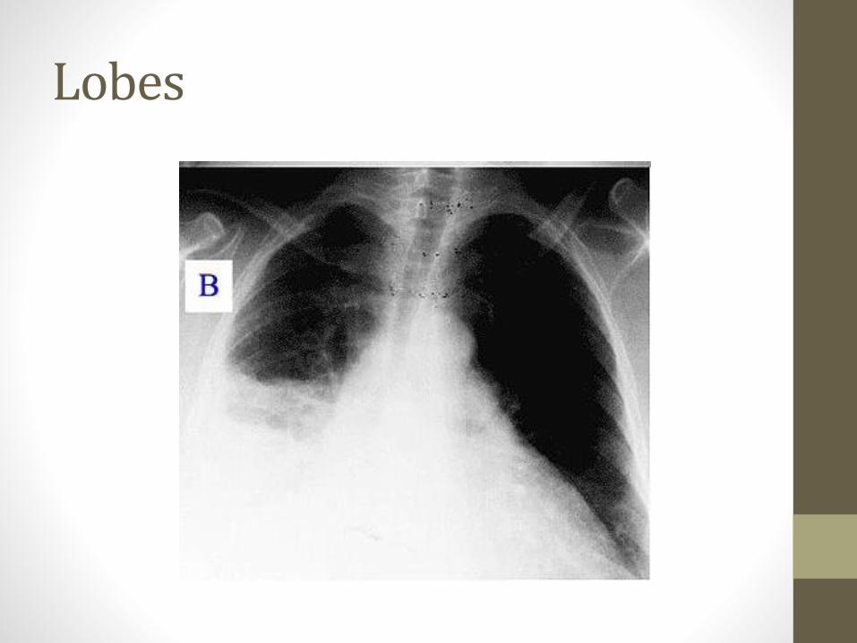
Lobes

Aspiration
• Right lung is more common site of aspiration• Right bronchus wider
• Less angle
• More vertical path to lung
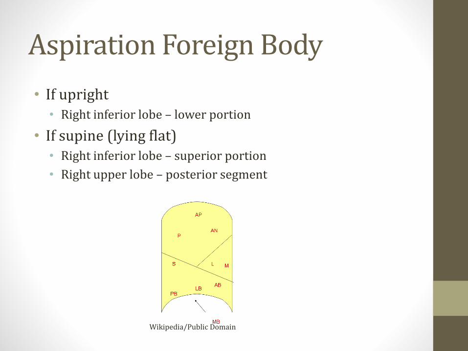
Aspiration Foreign Body
• If upright• Right inferior lobe – lower portion
• If supine (lying flat)• Right inferior lobe – superior portion
• Right upper lobe – posterior segment
Wikipedia/Public Domain
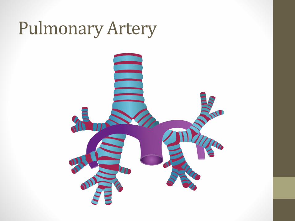
Pulmonary Artery
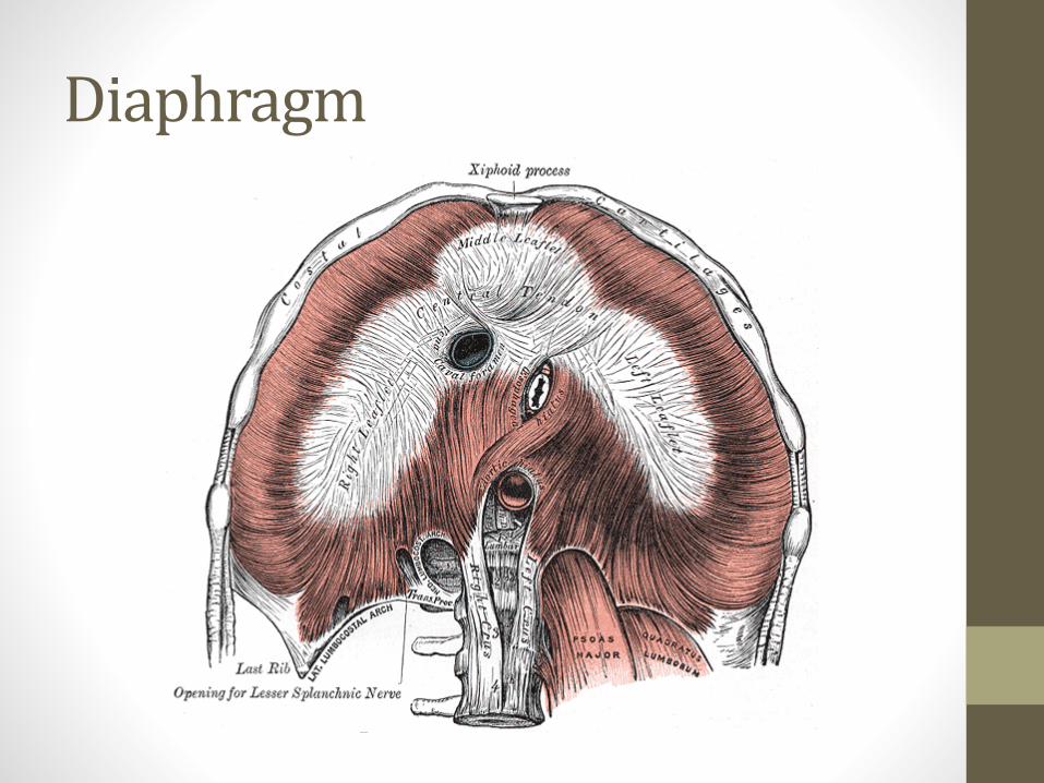
Diaphragm

Diaphragm
• Caval opening• T8
• IVC
• Esophageal hiatus• T10
• Esophagus, Vagus nerve
• Aortic hiatus• T12
• Aorta, thoracic duct, azygous vein

Diaphragm
• Innervated by C3, C4, C5 (Phrenic nerve)
• Diaphragm irritation → “referred” shoulder pain• Classic example is gallbladder disease
• Also lower lung masses
• Irritation can cause dyspnea and hiccups
• Cut nerve → diaphragm elevation, dyspnea• “Paradoxical movement” →Moves up with inspiration
• Can see on fluoroscopy (“sniff test”)
Muscles of Quiet Respiration
• Diaphragm → inspiration
• Exhalation is passive with normal (“quiet”) breathing
Exercise Breathing
• Inspiration (neck)• Scalenes – raise ribs
• Sternocleidomastoids – raise sternum
• Exhalation (abdomen)• Rectus muscle
• Internal/external obliques
• Transverse abdominis
• Internal intercostals
• Use of accessory muscles in respiratory distress
Pulmonary PhysiologyJason Ryan, MD, MPH

Physiology Concepts
1. Lung volumes/capacities
2. Ventilation and dead space
3. Lung and chest wall pressures
4. Lung compliance
5. Air flow resistance

Lung Volumes
Image courtesy of Michal Komorniczak, Medical Illustrations
Lung Volumes
• Tidal volume• In/out air with each quiet breath
• Expiratory reserve volume• Extra air pushed out with force beyond TV
• RV remains in lungs
• Inspiratory reserve volume• Extra air can be drawn in with force beyond TV
• Lungs filled to capacity
• Residual volume• Air that can’t be blown out no matter how hard you try

Lung CapacitiesCapacity = sum of two volumes
• Total lung capacity• Sum of all volumes
• RV + ERV+ IRV + TV
• Inspiratory capacity• Most air you can inspire
• TV + IRV
• Vital capacity• Most you can exhale
• TV + IRV + ERV

RV
ERV
IRV
TV
Lung CapacitiesCapacity = sum of two volumes
• Functional Residual Capacity• Residual volume after quiet expiration
• RV + ERV
• Volume when system is relaxed
• Chest wall pulling out = lungs pulling in
Ventilation
• Ventilation = volume x frequency (resp rate)• 500cc per breath x 20 breaths per minute
• 10,000cc/min
• Alveolar ventilation = useful for gas exchange
• Dead space ventilation = wasted ventilation• Nose, trachea, other areas with no gas exchange

Dead Space
• Space filled with air but no gas exchange
• #1: Anatomic dead space• Volume of conducting portions respiratory tract
• Nose, trachea
• #2: Physiologic dead space• Anatomic PLUS volume of alveoli that don’t exchange gas
• Insufficient perfusion
• Apex is largest contributor
• Physiologic dead space increases many diseases
Ventilation
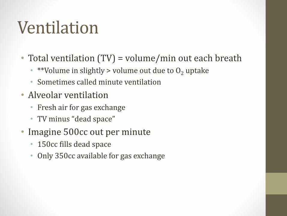
• Total ventilation (TV) = volume/min out each breath• **Volume in slightly > volume out due to O2 uptake
• Sometimes called minute ventilation
• Alveolar ventilation• Fresh air for gas exchange
• TV minus “dead space”
• Imagine 500cc out per minute• 150cc fills dead space
• Only 350cc available for gas exchange
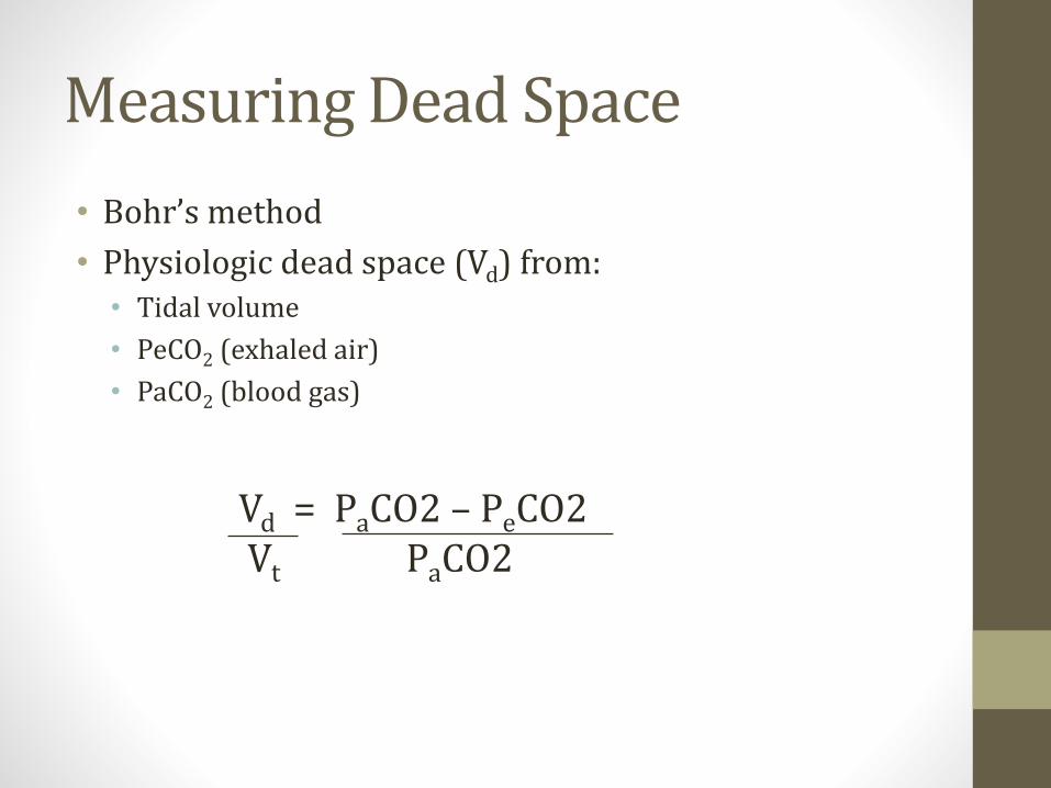
Measuring Dead Space
• Bohr’s method
• Physiologic dead space (Vd) from:• Tidal volume
• PeCO2 (exhaled air)
• PaCO2 (blood gas)
Vd = PaCO2 – PeCO2Vt PaCO2

Pressures
Alveoli
Patm = 760mmHg or 0mmHg
Patm = 760mmHg or 0mmHg
Δ = 0 No Flow

Pressures
Alveoli
Patm = 760mmHg or 0mmHg
Patm = 765mmHg or +5mmHg
Δ = +5 Flow Out
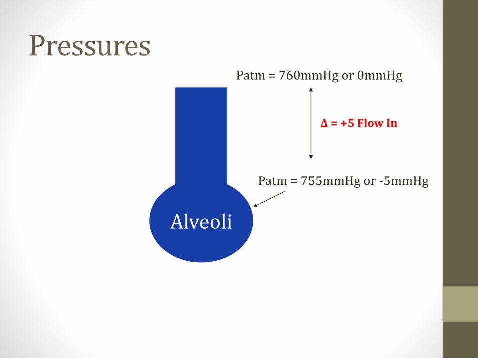
Pressures
Alveoli
Patm = 760mmHg or 0mmHg
Patm = 755mmHg or -5mmHg
Δ = +5 Flow In

Lung and Chest Wall
• Lungs tend to collapse • Pull inward/recoil
• Chest wall tends to expand • Spring outward

Lung Volumes and Pressures
0 20 40
Vo
lum
e
Pressure
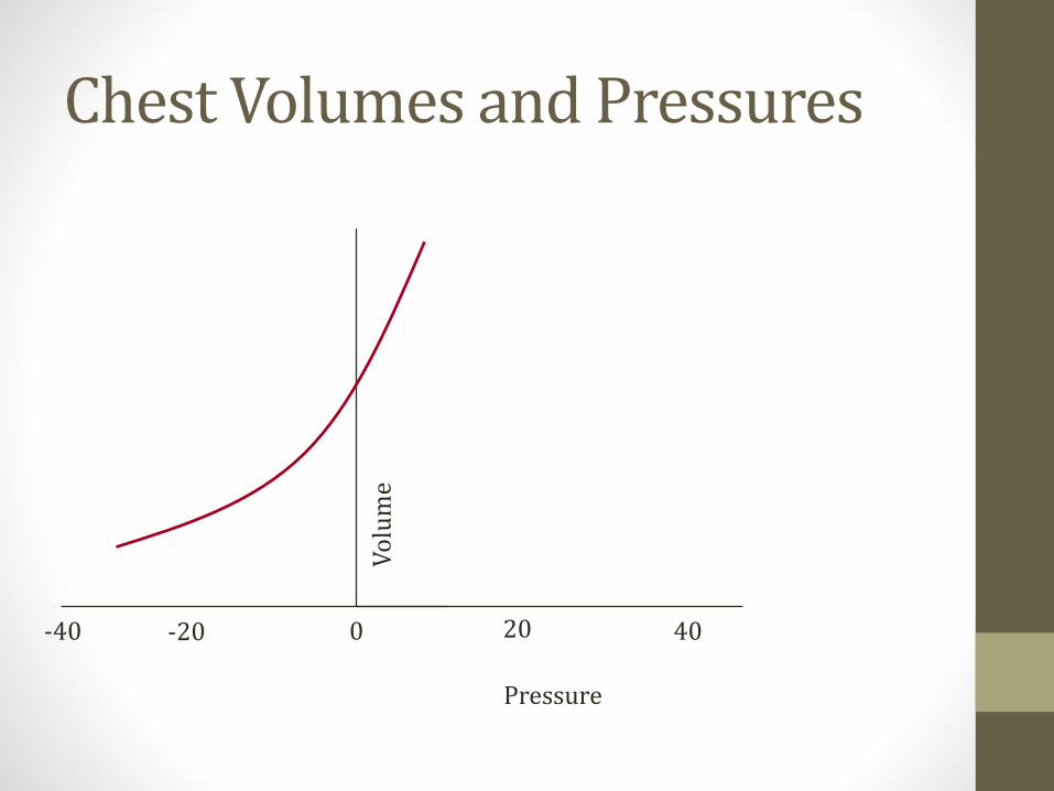
Chest Volumes and Pressures
Vo
lum
e
-40 -20 0 20 40
Pressure
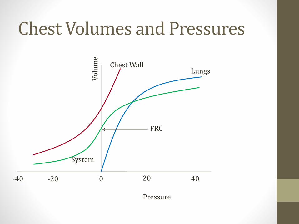
Chest Volumes and Pressures
Vo
lum
e
-40 -20 0 20 40
Chest WallLungs
System
FRC
Pressure

Functional residual capacity
• Lung in = chest out
• Volume where lungs rest after quiet exhalation
• Pressure inside system is zero• No ↑/↓ pressure from push/pull of lungs or chest wall
• Pressure = atmospheric pressure

Pressures
Alveoli
Chest Wall
Pleural Space
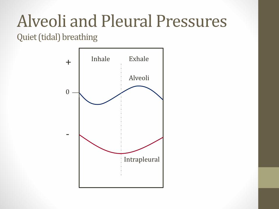
Alveoli and Pleural PressuresQuiet (tidal) breathing
0
+
-
Alveoli
Intrapleural
Inhale Exhale
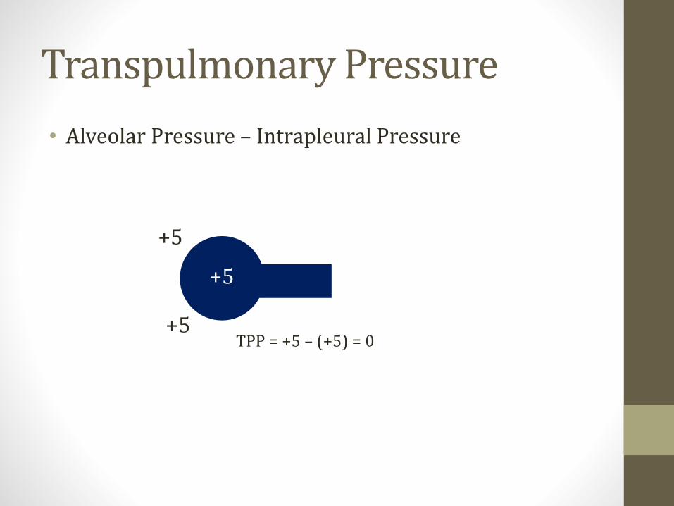
Transpulmonary Pressure
• Alveolar Pressure – Intrapleural Pressure
+5
+5
+5TPP = +5 – (+5) = 0

Transpulmonary Pressure
• Alveolar Pressure – Intrapleural Pressure
-30
0
-30TPP = 0 – (-30) = +30
(+) Pressure: Holds airway open(-) Pressure: Airway collapse
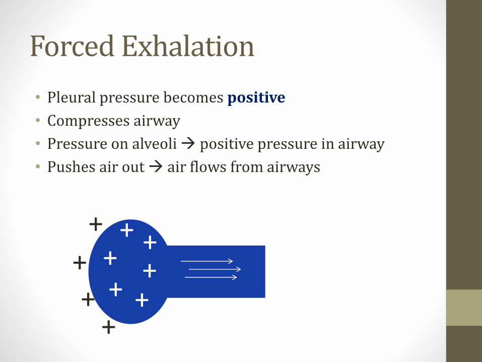
Forced Exhalation
• Pleural pressure becomes positive
• Compresses airway
• Pressure on alveoli → positive pressure in airway
• Pushes air out → air flows from airways
+
++
+
++ +
++
+

Equal Pressure Point
• Pleural pressure = airway pressure
• Beyond this point airway collapses
• In healthy lungs: EPP occurs in cartilaginous airways
• Prevents airway collapse
+
++
+
+ +
++++60
+90+75 +60
+60
+60
Pleural Pressure + Elastic Recoil
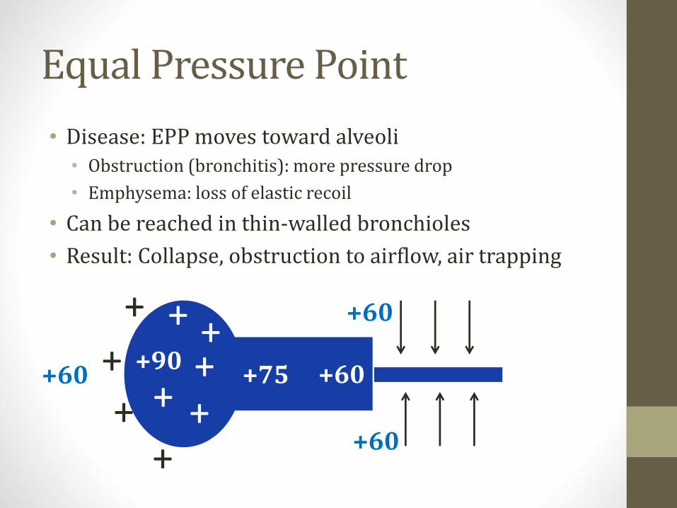
Equal Pressure Point
• Disease: EPP moves toward alveoli• Obstruction (bronchitis): more pressure drop
• Emphysema: loss of elastic recoil
• Can be reached in thin-walled bronchioles
• Result: Collapse, obstruction to airflow, air trapping
+
++
+
+ +
++++60
+90+75 +60
+60
+60
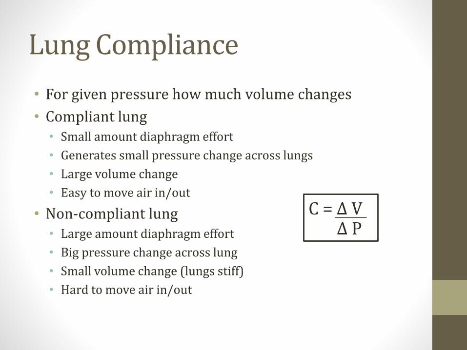
Lung Compliance
• For given pressure how much volume changes
• Compliant lung• Small amount diaphragm effort
• Generates small pressure change across lungs
• Large volume change
• Easy to move air in/out
• Non-compliant lung• Large amount diaphragm effort
• Big pressure change across lung
• Small volume change (lungs stiff)
• Hard to move air in/out
C = Δ VΔ P
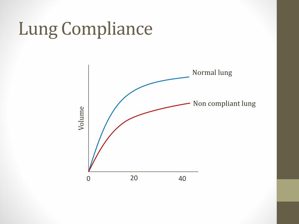
Lung Compliance
0 20 40
Vo
lum
eNormal lung
Non compliant lung

Lung Compliance
• Decreased• Pneumonia
• Pulmonary edema
• Pulmonary fibrosis
• Increased• Emphysema (floppy lungs)
• Aging
C = Δ VΔ P

Resistance to Air Flow
• Upper airways about 50% resistance • Nose, mouth, pharynx
• Lower airway resistance• Highest in medium bronchi (turbulent flow)
• Lowest in terminal bronchioles - slows laminar flow
Res
ista
nce
Medium Bronchi
Trachea
Terminalbronchioles
Air vessel size

HemoglobinJason Ryan, MD, MPH
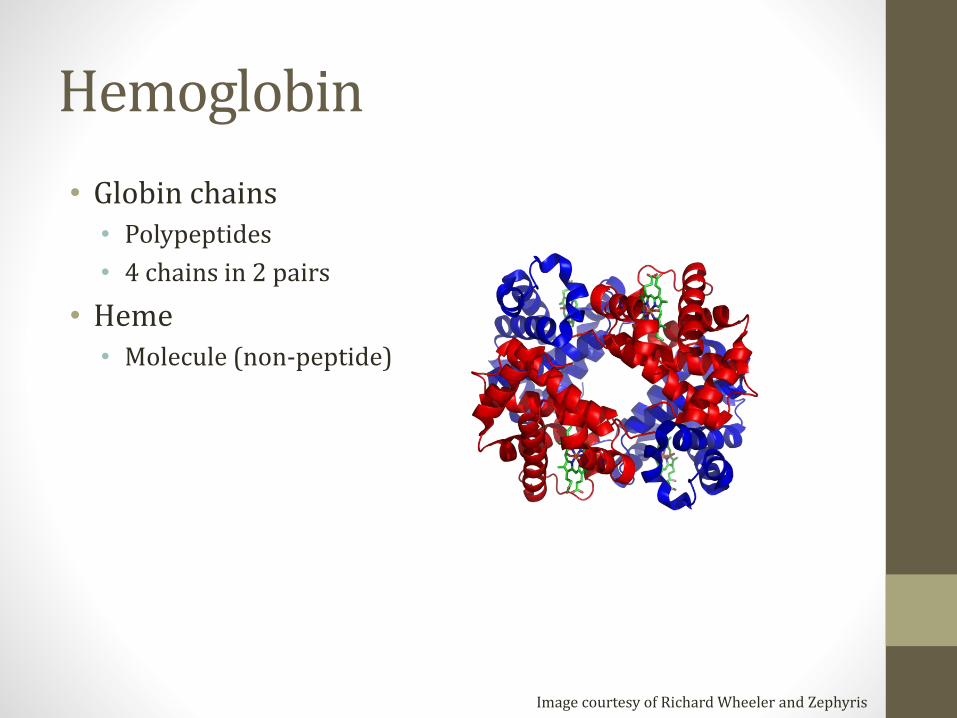
Hemoglobin
• Globin chains• Polypeptides
• 4 chains in 2 pairs
• Heme• Molecule (non-peptide)
Image courtesy of Richard Wheeler and Zephyris

Globin Protein Types
• Alpha (α)
• Beta (β)
• Gamma (γ)
• Delta (δ)
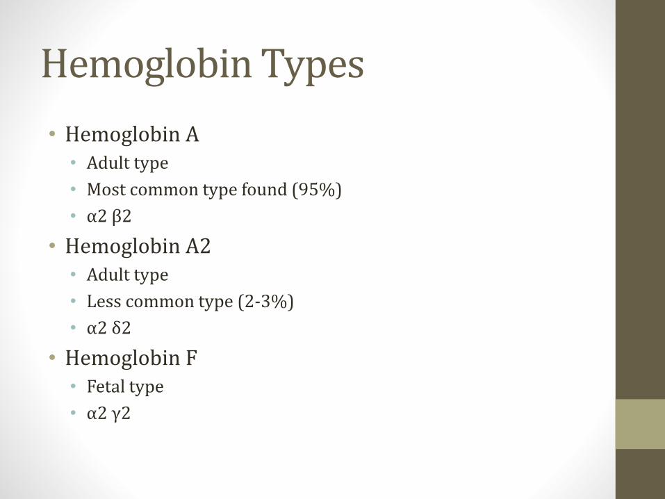
Hemoglobin Types
• Hemoglobin A• Adult type
• Most common type found (95%)
• α2 β2
• Hemoglobin A2• Adult type
• Less common type (2-3%)
• α2 δ2
• Hemoglobin F• Fetal type
• α2 γ2
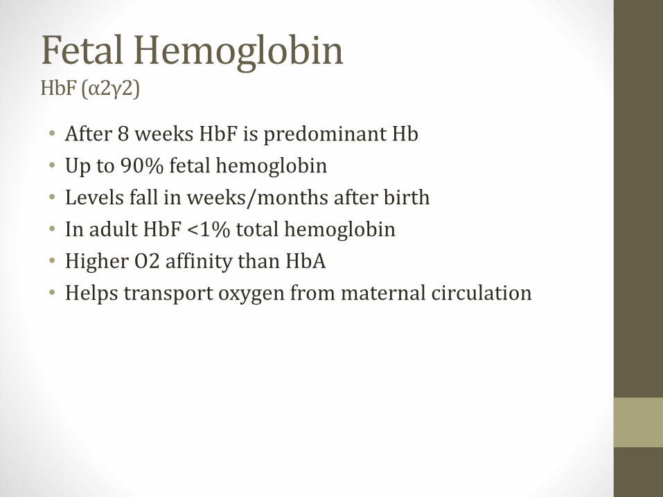
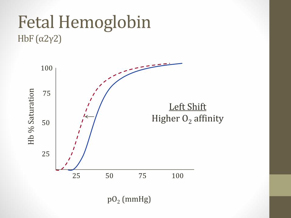
Fetal HemoglobinHbF (α2γ2)
• After 8 weeks HbF is predominant Hb
• Up to 90% fetal hemoglobin
• Levels fall in weeks/months after birth
• In adult HbF <1% total hemoglobin
• Higher O2 affinity than HbA
• Helps transport oxygen from maternal circulation

Globin Chain Diseases
• α-thalassemia • Underproduction alpha chain
• β-thalassemia minor/major• Underproduction beta chain
• Sickle Cell Anemia• HbS production
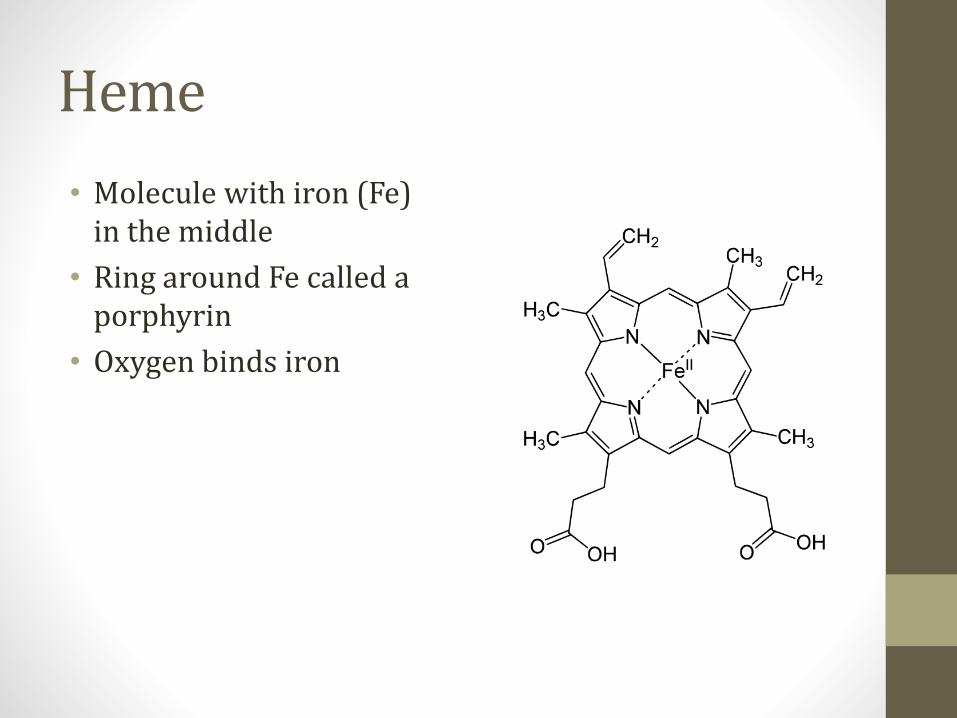
Heme
• Molecule with iron (Fe) in the middle
• Ring around Fe called a porphyrin
• Oxygen binds iron

2,3-Bisphosphoglycerate2,3 BPG
• Found in RBCs
• Promotes O2 release from hemoglobin
• Increasing levels:• Decrease oxygen affinity of hemoglobin
• Increase delivery oxygen to tissues
• More BPG at high altitude

2,3 Bisphosphoglycerate
• Created from diverted 1,3 BPG (glycolysis)
• Sacrifices ATP from glycolysis
1,3-bisphosphoglycerate
3-phosphoglycerate
2-phosphoglycerate
Phosphoenolpyruvate
Pyruvate
Glyceraldehyde-3-phosphate
2,3 BPG
BPGMutase
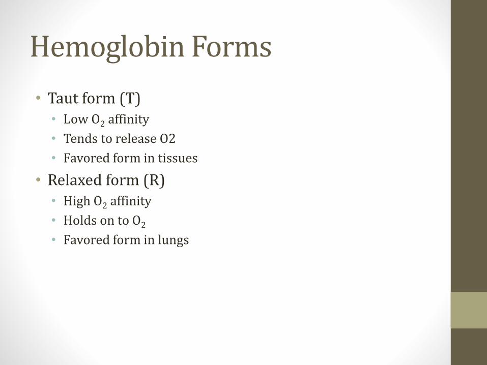
Hemoglobin Forms
• Taut form (T)• Low O2 affinity
• Tends to release O2
• Favored form in tissues
• Relaxed form (R)• High O2 affinity
• Holds on to O2
• Favored form in lungs
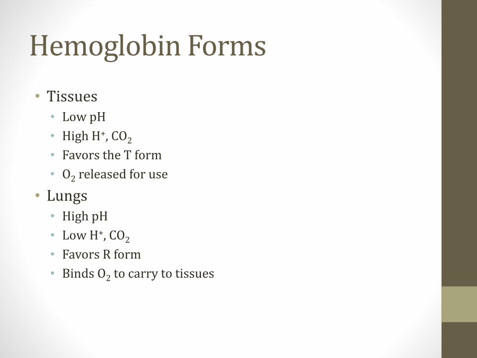
Hemoglobin Forms
• Tissues• Low pH
• High H+, CO2
• Favors the T form
• O2 released for use
• Lungs• High pH
• Low H+, CO2
• Favors R form
• Binds O2 to carry to tissues
Dissociation Curves
25 50 75 100
pO2 (mmHg)
25
50
75
100
Hb
% S
atu
rati
on
Cooperativity
• Four heme groups do not undergo simultaneous oxygenation
• First O2 molecule that binds INCREASES affinity of hemoglobin for 2nd molecule
• Makes curve S shaped

Allosteric Effects
• Allosteric proteins change affinity for binding when influenced by other (smaller) molecules
• Usually multi-subunit proteins
• Hemoglobin is an allosteric structure
• Cooperativity is a positive allosteric effect
Hgb Allosteric Effectors
• pH
• Temperature
• 2,3 BPG
• CO2

Right Curve ShiftsEasier to release O2
25 50 75 100
pO2 (mmHg)
25
50
75
100
Hb
% S
atu
rati
on
Right Shift↑Co2, BPG, Temp, H+

Left Curve ShiftsHarder to release O2
25 50 75 100
pO2 (mmHg)
25
50
75
100
Hb
% S
atu
rati
on
Left Shift↓Co2, BPG, Temp, H+
Fetal HemoglobinHbF (α2γ2)
25 50 75 100
pO2 (mmHg)
25
50
75
100
Hb
% S
atu
rati
on
Left ShiftHigher O2 affinity
Carbon Monoxide
• Binds to iron in heme
• 240x the affinity of oxygen
• Blocks O2 binding sites (less O2 can be absorbed)
• Other binding sites cannot offload O2
• Allosteric modification of hemoglobin
• Shifts dissociation curve left

Carbon Monoxide
25 50 75 100
pO2 (mmHg)
25
50
75
100
Hb
% S
atu
rati
on
Normal
50% CO Hb
Left shift
carrying capacity
Carbon Monoxide Poisoning
• Nonspecific symptoms
• Headache most common
• Malaise, nausea, dizziness
• Classic (but rare) sign: Cherry red lips• Carboxyhemoglobin is red
• Do not see blue lips (cyanosis)
Carbon Monoxide Poisoning
• Standard pulse oximetry normal• Cannot differentiate carboxyhemoglobin/oxyhemoglobin
• Diagnosis: carboxyhemoglobin level• Normal <3%
• Smokers 10-15%
• >15% suggest poisoning
• Treatment: Oxygen

Methemoglobinemia
• Iron in hemoglobin normally reduced (Fe2+)
• Certain drug oxidize iron to Fe3+
• When Fe3+ is present →methemoglobin
• Fe3+ cannot bind oxygen
• Remaining Fe2+ cannot release to tissues
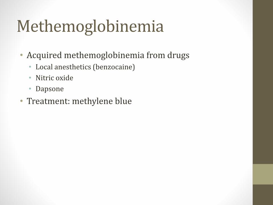
Methemoglobinemia
• Acquired methemoglobinemia from drugs• Local anesthetics (benzocaine)
• Nitric oxide
• Dapsone
• Treatment: methylene blue

Clinical Scenario
• Endoscopy patient
• Benzocaine spray used for throat analgesia
• Post procedure shortness of breath
• “Chocolate brown blood”
• O2 sat (pulse oximetry) = variable (80s-90s)
• PaO2 (blood gas) = normal
• Also premature babies given NO for pulmonary vasodilation

Cyanide Poisoning
• Cyanide binds to Fe3+ in cells
• Blocks electron transport chain in mitochondria
• Aerobic metabolism stops• Functional hypoxia
• Especially bad for brain and heart
• Anaerobic metabolism occurs
• Lactic acidosis occurs
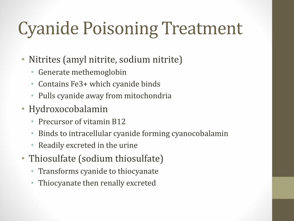
Cyanide Poisoning Treatment
• Nitrites (amyl nitrite, sodium nitrite)• Generate methemoglobin
• Contains Fe3+ which cyanide binds
• Pulls cyanide away from mitochondria
• Hydroxocobalamin• Precursor of vitamin B12
• Binds to intracellular cyanide forming cyanocobalamin
• Readily excreted in the urine
• Thiosulfate (sodium thiosulfate)• Transforms cyanide to thiocyanate
• Thiocyanate then renally excreted

Pulmonary CirculationJason Ryan, MD, MPH
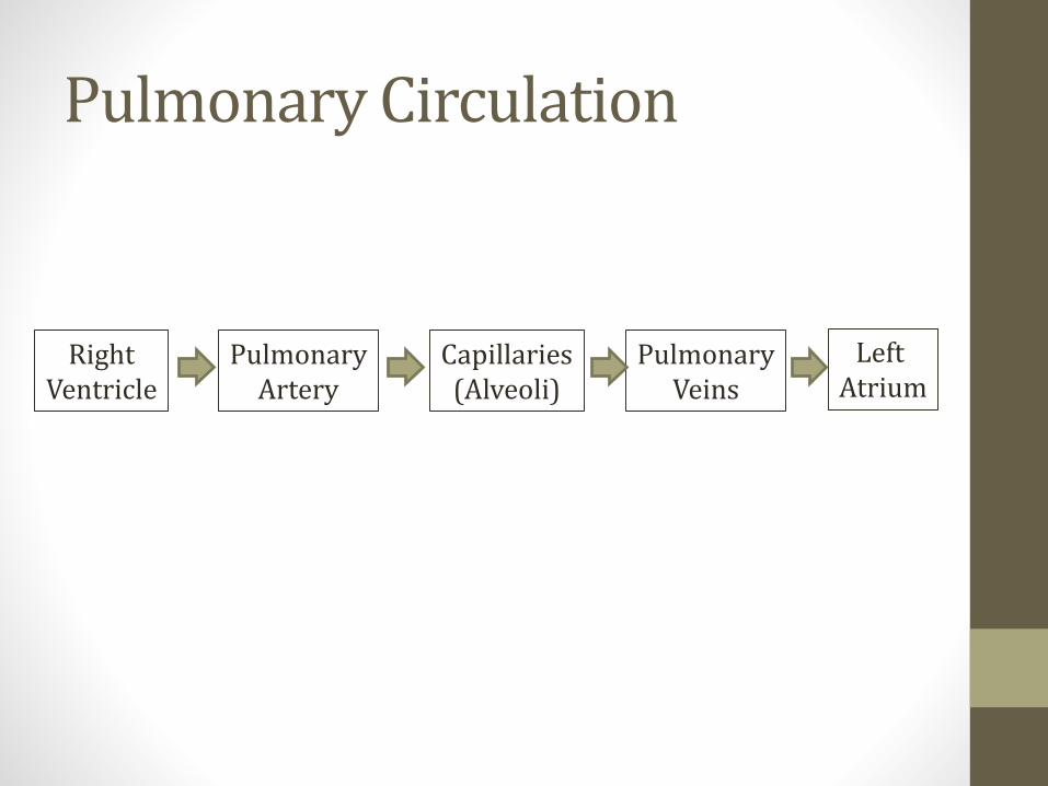
Pulmonary Circulation
RightVentricle
PulmonaryArtery
Capillaries(Alveoli)
PulmonaryVeins
Left Atrium

Pulmonary Circulation
• Low pressure system• Systemic: 120/80
• Pulmonary artery: 24/12
• Walls of pulmonary artery very thin• Little smooth muscle
• Low resistance to flow
• Very distensible (compliant)
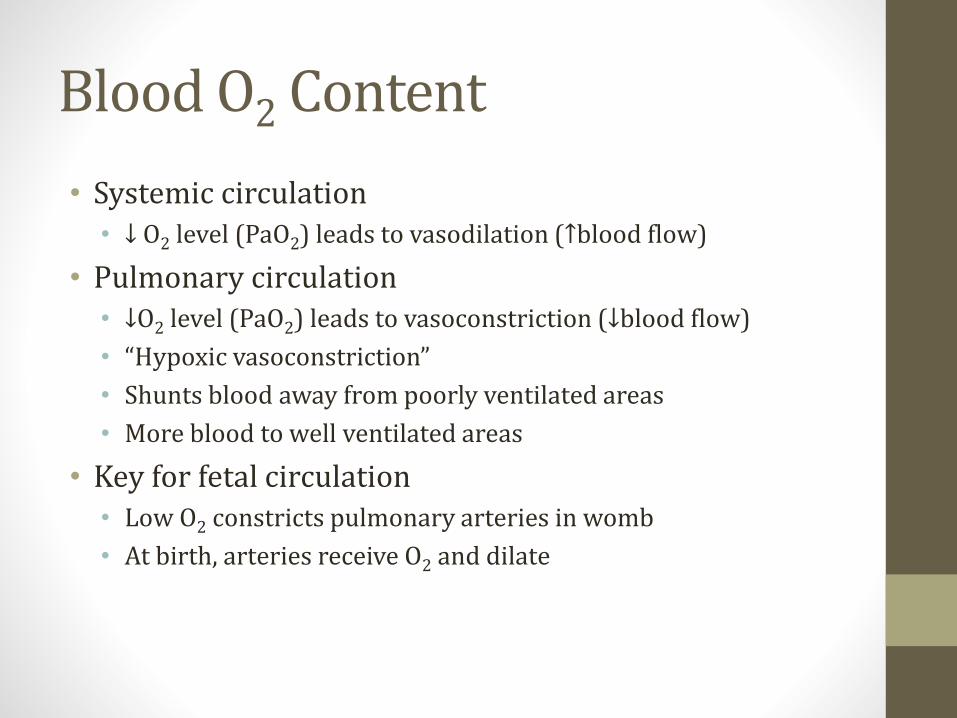
Blood O2 Content
• Systemic circulation• ↓ O2 level (PaO2) leads to vasodilation (↑blood flow)
• Pulmonary circulation• ↓O2 level (PaO2) leads to vasoconstriction (↓blood flow)
• “Hypoxic vasoconstriction”
• Shunts blood away from poorly ventilated areas
• More blood to well ventilated areas
• Key for fetal circulation• Low O2 constricts pulmonary arteries in womb
• At birth, arteries receive O2 and dilate

Blood O2 Content
PaO2
Blo
od
Flo
w
Systemic
Lungs
Diffusion
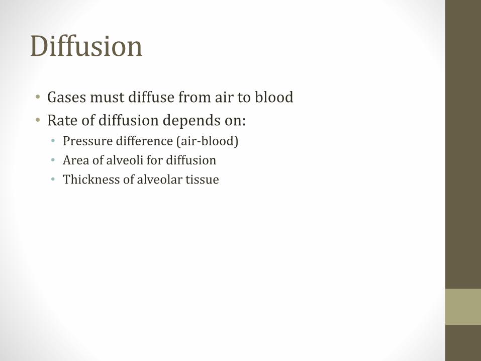
• Gases must diffuse from air to blood
• Rate of diffusion depends on:• Pressure difference (air-blood)
• Area of alveoli for diffusion
• Thickness of alveolar tissue
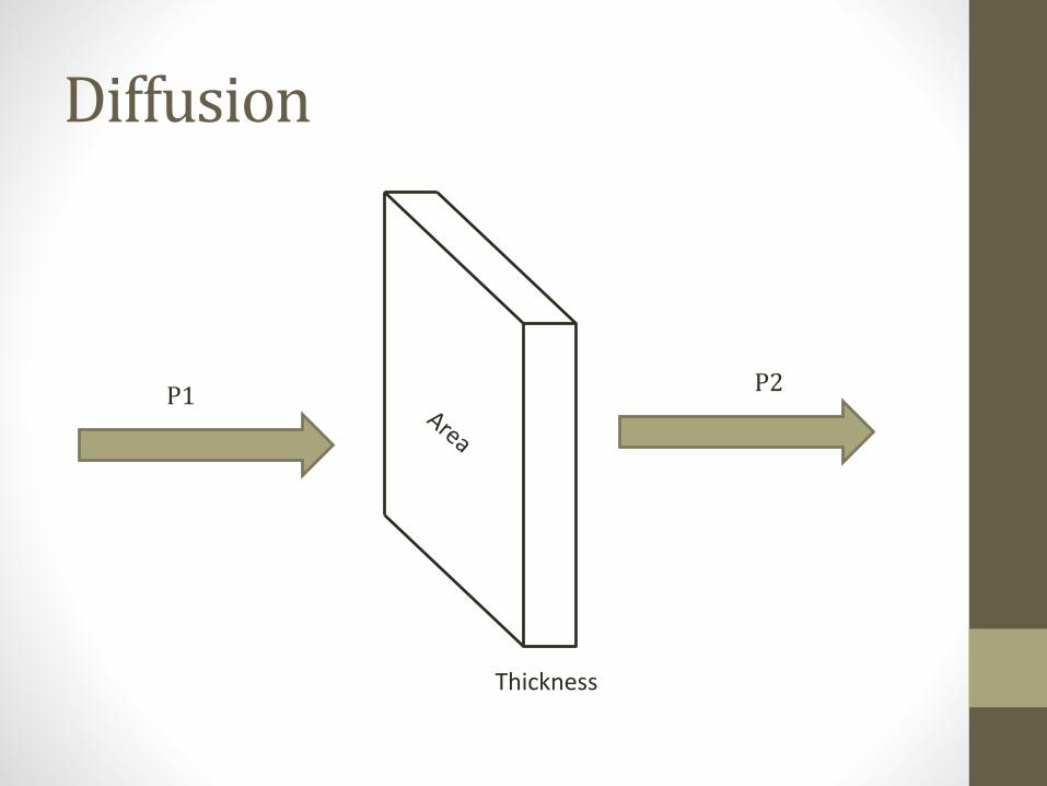
Diffusion
Thickness
P1P2
Diffusion
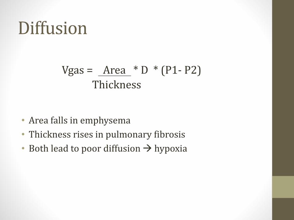
• Area falls in emphysema
• Thickness rises in pulmonary fibrosis
• Both lead to poor diffusion → hypoxia
Vgas = Area * D * (P1- P2)
Thickness
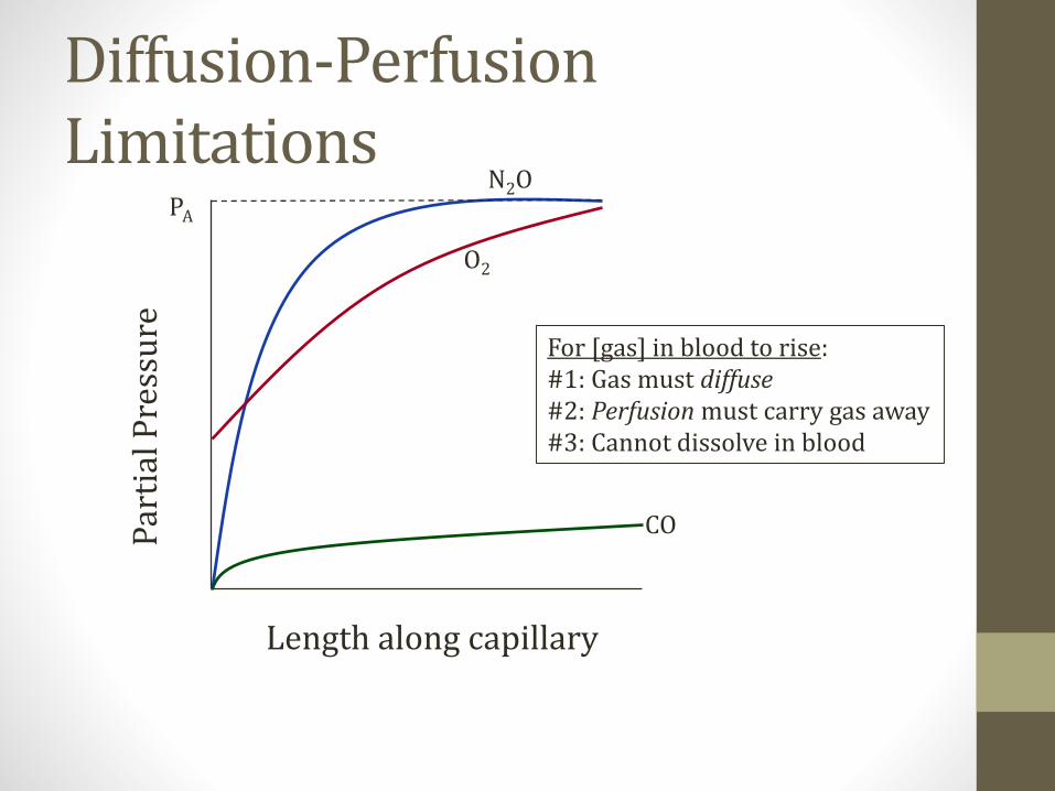
Diffusion-Perfusion Limitations
Length along capillary
Par
tial
Pre
ssu
re
O2
CO
N2OPA
For [gas] in blood to rise:#1: Gas must diffuse#2: Perfusion must carry gas away#3: Cannot dissolve in blood
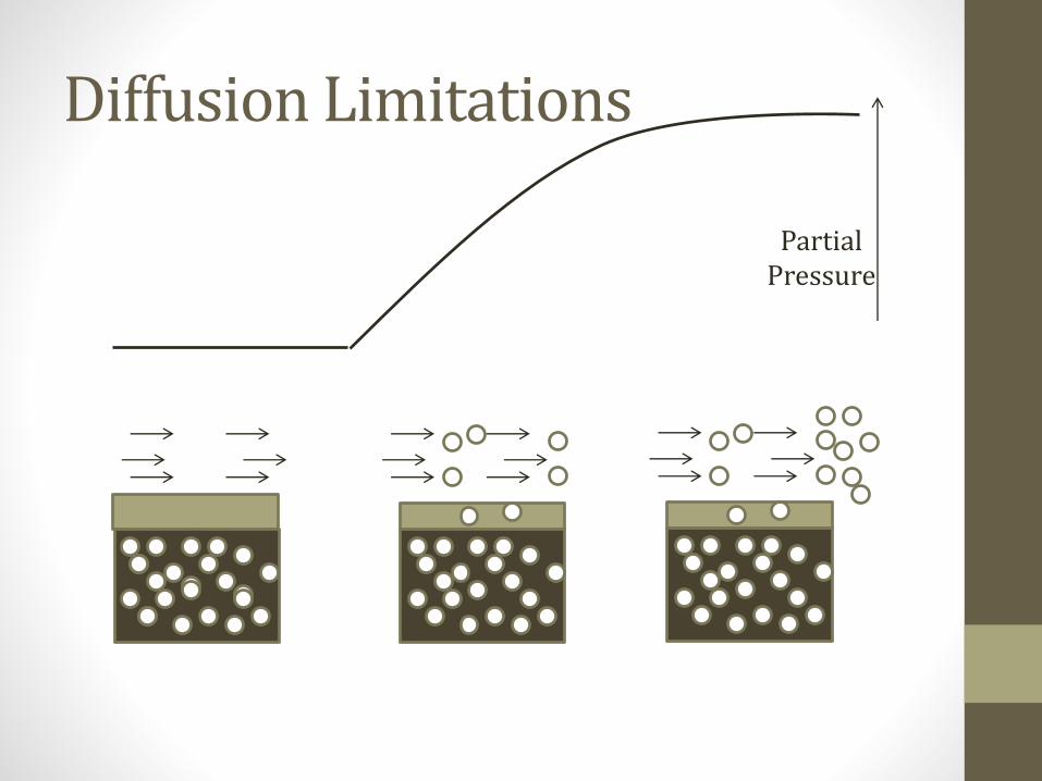
Diffusion Limitations
PartialPressure
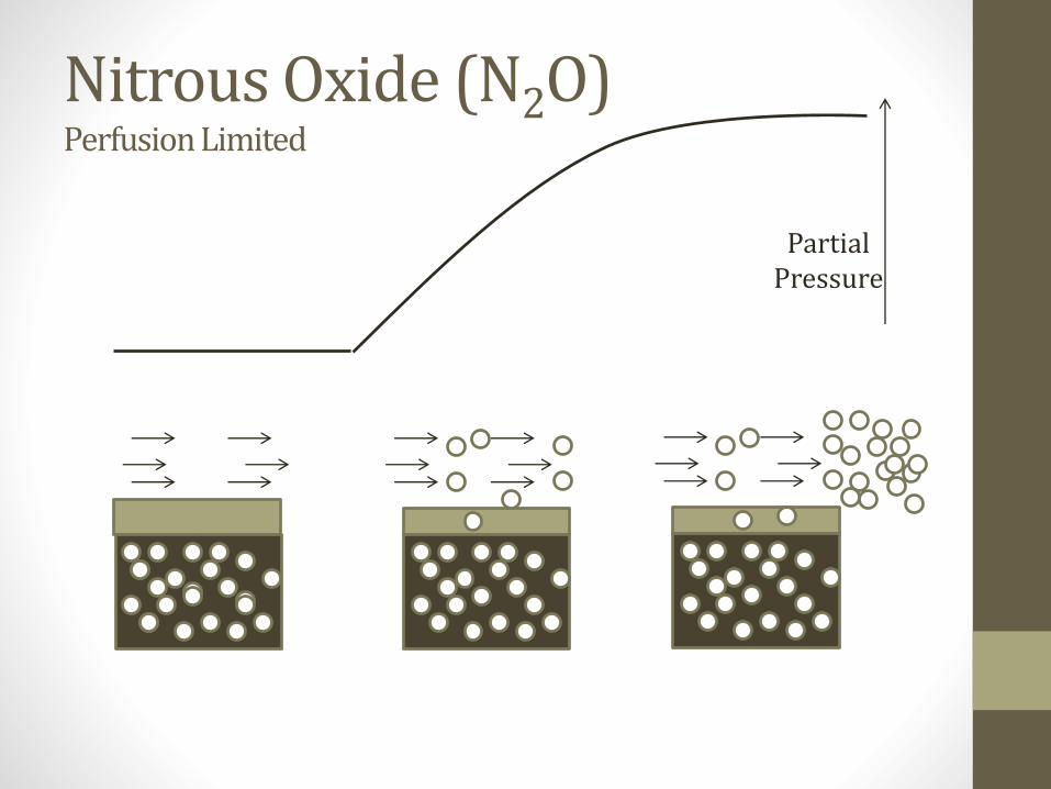
Nitrous Oxide (N2O)Perfusion Limited
PartialPressure
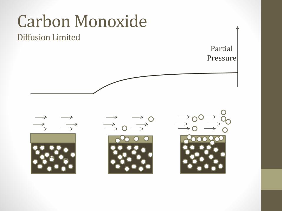
Carbon MonoxideDiffusion Limited
PartialPressure
Oxygen
• Normal healthy subjects: O2 uptake perfusion limited
• Disease (i.e. fibrosis, emphysema): diffusion limited

Diffusion-Perfusion Limitations
Length along capillary
Par
tial
Pre
ssu
re
O2
CO
N2OPA
For [gas] in blood to rise:#1: Gas must diffuse#2: Perfusion must carry gas away#3: Cannot dissolve in blood
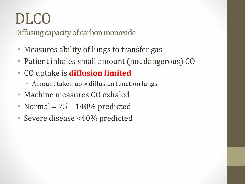
DLCODiffusing capacity of carbon monoxide
• Measures ability of lungs to transfer gas
• Patient inhales small amount (not dangerous) CO
• CO uptake is diffusion limited• Amount taken up ≈ diffusion function lungs
• Machine measures CO exhaled
• Normal = 75 – 140% predicted
• Severe disease <40% predicted

O2 Diffusion-Perfusion
Length along capillary
Par
tial
Pre
ssu
re
Healthy
Disease
PA
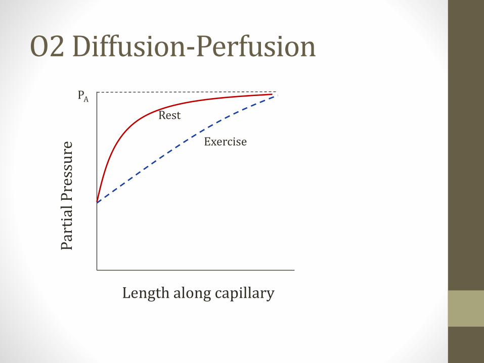
O2 Diffusion-Perfusion
Length along capillary
Par
tial
Pre
ssu
re
Rest
Exercise
PA

Pulmonary Vascular ResistanceResistance to blood flow
• Two vessels: alveolar and arteriolar
• Increased lung volumes stretch alveolar vessels• Makes them longer with smaller diameter
• Increases resistance
• Decreased lung volumes narrow arteriolar vessels• Increases resistance
PVR
Exhale Inhale
FRC

Pulmonary Hypertension
• Normal PA pressure• 24/12
• Mean 10-14mmHg
• Pulmonary hypertension• Mean pressure >25mmHg
• Loud P2 = pulmonary hypertension• Look for accentuated or loud second heart sound
• Left upper sternal border

Pulmonary Hypertension
• Main symptom is dyspnea
• Untreated can lead to “cor pulmonale”• Chronic high pressure in right ventricle
• Right ventricle hypertrophies
• Eventually dilates and fails
• Jugular venous distension
• Lower extremity edema
• Hepatomegaly
• Death from heart failure or arrhythmia
Pulmonary Hypertension
• Gold standard for diagnosis: right heart cath
• Non-invasive diagnosis by echocardiography• Estimate PA pressure
• Visualize right heart structures
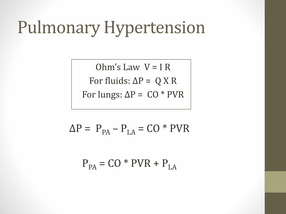
Ohm’s Law V = I R
For fluids: ΔP = Q X R
For lungs: ΔP = CO * PVR
Pulmonary Hypertension
ΔP = PPA – PLA = CO * PVR
PPA = CO * PVR + PLA
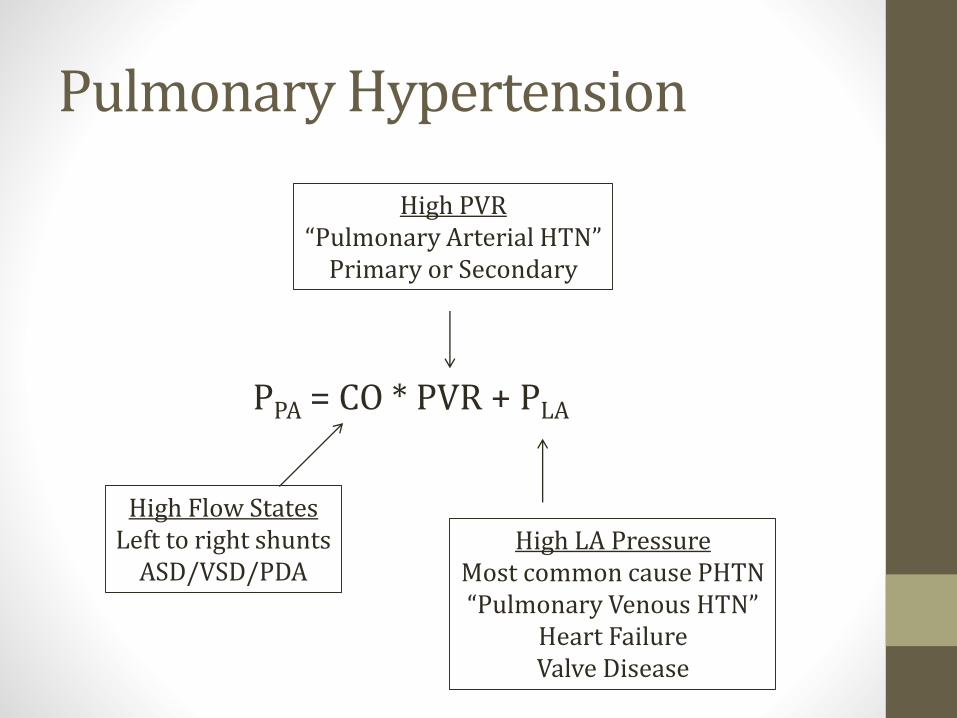
Pulmonary Hypertension
PPA = CO * PVR + PLA
High LA PressureMost common cause PHTN“Pulmonary Venous HTN”
Heart FailureValve Disease
High PVR“Pulmonary Arterial HTN”
Primary or Secondary
High Flow StatesLeft to right shunts
ASD/VSD/PDA
Pulmonary Hypertension
• Secondary causes high PVR• COPD
• Chronic pulmonary emboli
• Pulmonary fibrosis (scleroderma)
• Sleep apnea or high altitude (chronic hypoxia)
• HIV

1° Pulmonary Hypertension
• Rare disease
• Classically affects young women
• Increased endothelin (vasoconstrictor)
• Decreased NO
• Vasoconstriction, smooth muscle proliferation
• High pulmonary pressures
1° Pulmonary Hypertension
• Associated with BMPR2 gene mutations• Bone morphogenetic protein receptor type II
• Up to 25% of idiopathic cases
• Up to 80% familial cases
• Abnormal endothelial and vascular smooth muscle growth/proliferation

1° Pulmonary Hypertension
• Progressive dyspnea and right heart failure if untreated
• Treatments (all lower PVR):• Epoprostenol: Prostacyclin (IV)
• Bosentan: Antagonist endothelin-1 receptors (PO)
• Sildenafil: Inhibits PDE-5 in smooth muscle of lungs (PO)

HypoxiaJason Ryan, MD, MPH

Oxygen delivery to tissues
• Delivery = Cardiac Output * O2 Content of blood
• For proper O2 delivery need:• Normal cardiac output
• Normal O2 content
What determines O2 content?
• #1: O2 binding capacity • How much O2 blood can hold
• Determined by hemoglobin
• #2: % Saturation• % Hemoglobin molecules saturated
• #3: Dissolved O2
• O2 directly dissolved in blood

PaO2
• Partial pressure oxygen in blood
• Obtained from an arterial blood gas
• Reflects amount of O2 dissolved in blood
• Normal: >80mmHg
Bobjgalindo
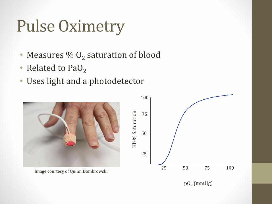
Pulse Oximetry
• Measures % O2 saturation of blood
• Related to PaO2
• Uses light and a photodetector
Image courtesy of Quinn Dombrowski

Oxygen Content
O2 Content = (O2 Binding Capacity ) * (% Sat) + (Dissolved O2)(ml O2/dl)
(1.39 * Hb) 0.003 PaO2
Hypoxemia, Hypoxia, Ischemia
• Hypoxemia: Low oxygen content of blood
• Hypoxia: Low O2 delivery to tissues
• Ischemia: Loss of blood flow
Pixabay/Public Domain
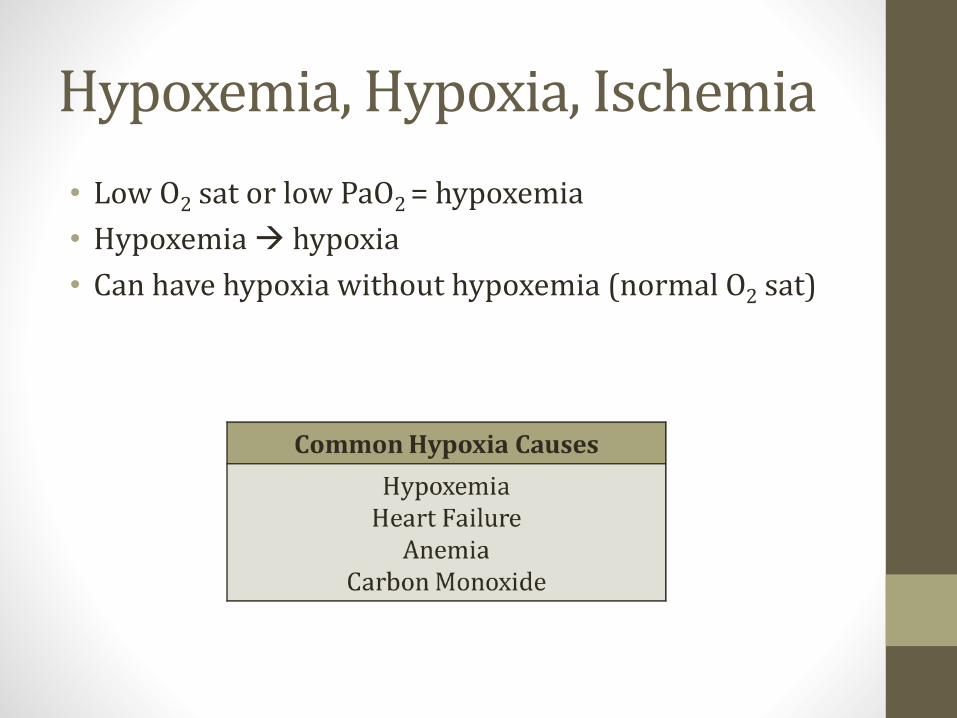
Hypoxemia, Hypoxia, Ischemia
• Low O2 sat or low PaO2 = hypoxemia
• Hypoxemia → hypoxia
• Can have hypoxia without hypoxemia (normal O2 sat)
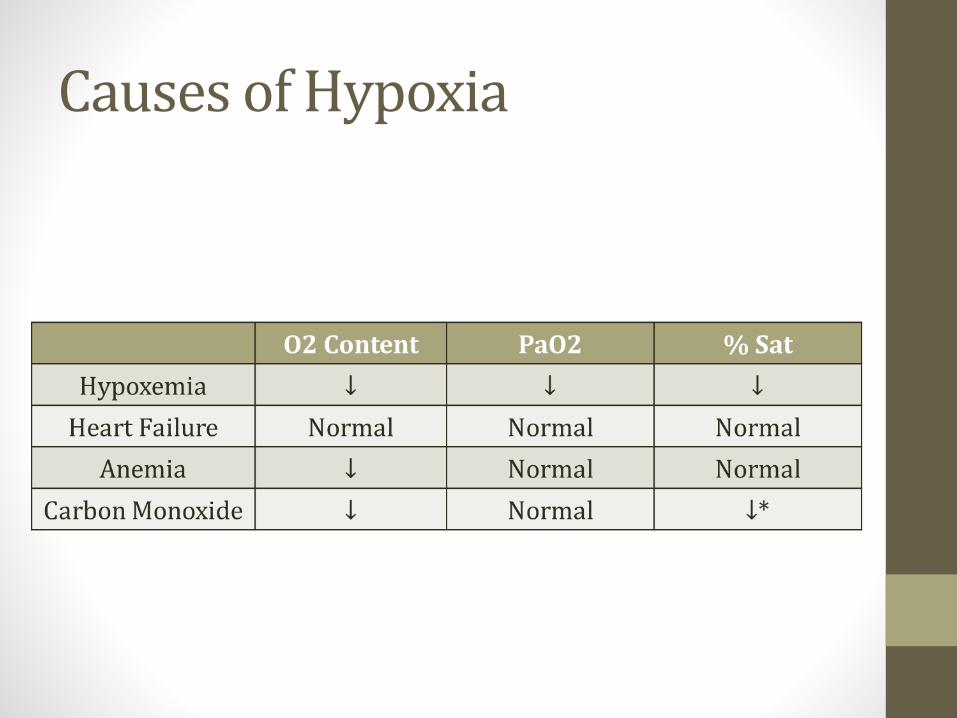
Heart Failure
• ↓ cardiac output
• ↓ blood flow to tissues → hypoxia
• O2 content of blood may be normal
• PaO2 and O2 sat may be normal
O2 Content = (O2 Binding Capacity ) * (% Sat) + (Dissolved O2)

Anemia
• Oxygenation of blood by lungs is normal
• Oxygen carrying capacity of blood reduced
• Low O2 content of blood
• PaO2 and O2 sat normal
Databese Center for Life Science (DBCLS)
O2 Content = (O2 Binding Capacity ) * (% Sat) + (Dissolved O2)
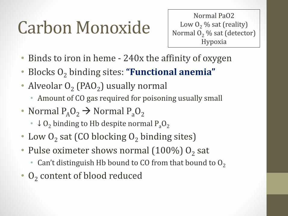
Carbon Monoxide
• Binds to iron in heme - 240x the affinity of oxygen
• Blocks O2 binding sites: “Functional anemia”
• Alveolar O2 (PAO2) usually normal• Amount of CO gas required for poisoning usually small
• Normal PAO2→ Normal PaO2
• ↓ O2 binding to Hb despite normal PaO2
• Low O2 sat (CO blocking O2 binding sites)
• Pulse oximeter shows normal (100%) O2 sat• Can’t distinguish Hb bound to CO from that bound to O2
• O2 content of blood reduced
Normal PaO2Low O2 % sat (reality)
Normal O2 % sat (detector)Hypoxia
Causes of Hypoxia
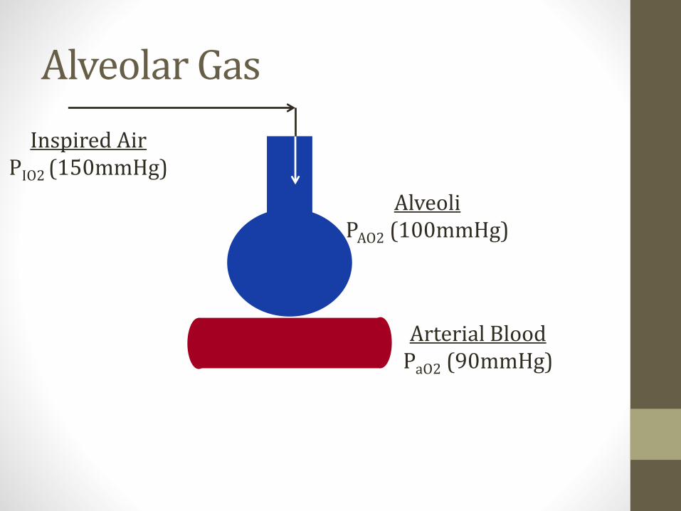
Alveolar Gas
AlveoliPAO2 (100mmHg)
Arterial BloodPaO2 (90mmHg)
Inspired AirPIO2 (150mmHg)
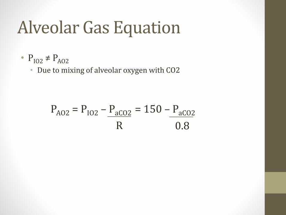
Alveolar Gas Equation
• PIO2 ≠ PAO2
• Due to mixing of alveolar oxygen with CO2
PAO2 = PIO2 – PaCO2 = 150 – PaCO2
R 0.8
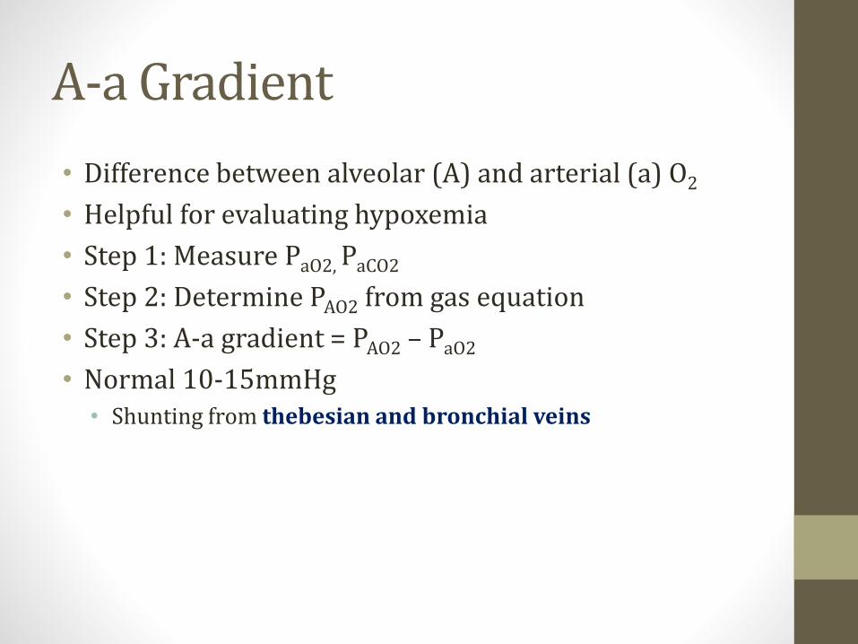
A-a Gradient
• Difference between alveolar (A) and arterial (a) O2
• Helpful for evaluating hypoxemia
• Step 1: Measure PaO2, PaCO2
• Step 2: Determine PAO2 from gas equation
• Step 3: A-a gradient = PAO2 – PaO2
• Normal 10-15mmHg• Shunting from thebesian and bronchial veins

A-a Gradient
• Hypoxemia with normal A-a gradient• Alveoli working
• Not inhaling enough O2
• Hypoventilation• Reduced respiratory rate
• Reduced tidal volume
• Narcotics, neuromuscular weakness, obesity
• High altitude
• Can treat with more oxygen
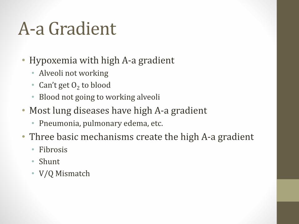
A-a Gradient
• Hypoxemia with high A-a gradient• Alveoli not working
• Can’t get O2 to blood
• Blood not going to working alveoli
• Most lung diseases have high A-a gradient• Pneumonia, pulmonary edema, etc.
• Three basic mechanisms create the high A-a gradient• Fibrosis
• Shunt
• V/Q Mismatch
Ventilation & PerfusionJason Ryan, MD, MPH
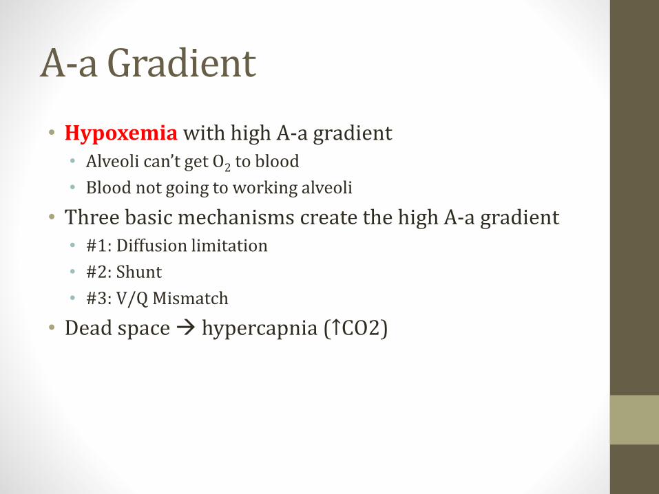
A-a Gradient
• Hypoxemia with high A-a gradient• Alveoli can’t get O2 to blood
• Blood not going to working alveoli
• Three basic mechanisms create the high A-a gradient• #1: Diffusion limitation
• #2: Shunt
• #3: V/Q Mismatch
• Dead space → hypercapnia (↑CO2)

Diffusion Limitation
• Increased A-a gradient
• Hypoxemia
• Less effect on CO2
• Seen in pulmonary fibrosis• Hypoxemia
• Hypercapnia
• Destruction alveolar capillaries → dead space
• Ventilation without perfusion
• Dead space may cause hypercapnia

Ventilation-Perfusion
• Ideally ventilation to lung is matched by perfusion• Ideal V/Q = 1
• Normal lungs:• 4L/min air into lungs
• 5L/min blood into lungs
• V/Q = 0.8
• If V/Q too high or too low, ventilation is inefficient
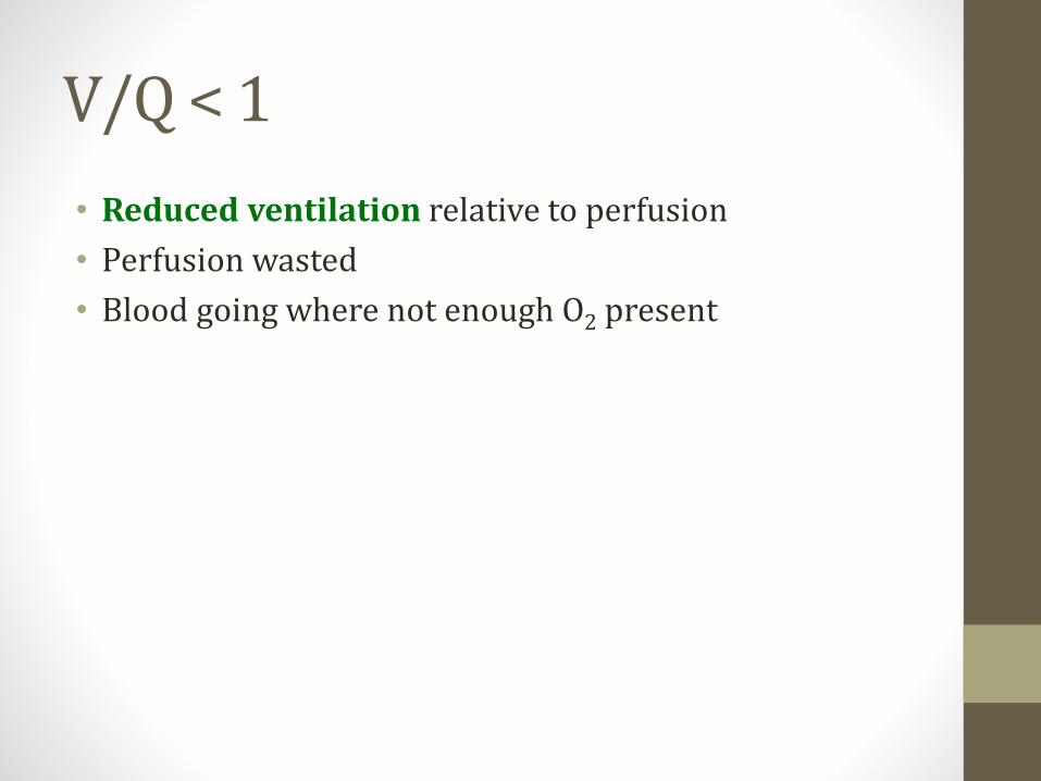
V/Q < 1
• Reduced ventilation relative to perfusion
• Perfusion wasted
• Blood going where not enough O2 present
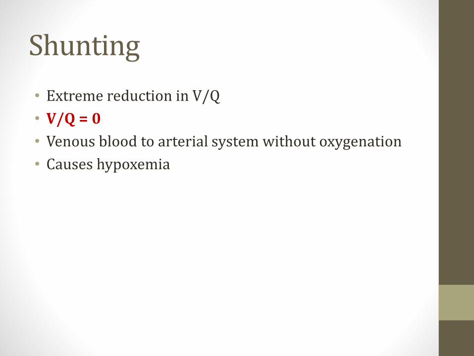
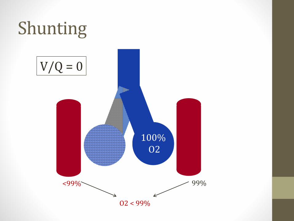
Shunting
• Extreme reduction in V/Q
• V/Q = 0
• Venous blood to arterial system without oxygenation
• Causes hypoxemia

Shunting
• Anatomic shunting• Blood bypasses lungs/alveoli completely
• Intra-cardiac, pulmonary AVMs
• Physiologic shunting• Blood goes to alveoli that don’t work
• Non-ventilated alveoli are perfused
• One example: Atelectasis (collapsed airway)
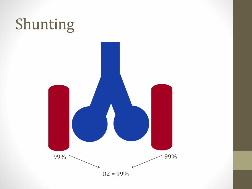
Shunting
O2 = 99%
99% 99%
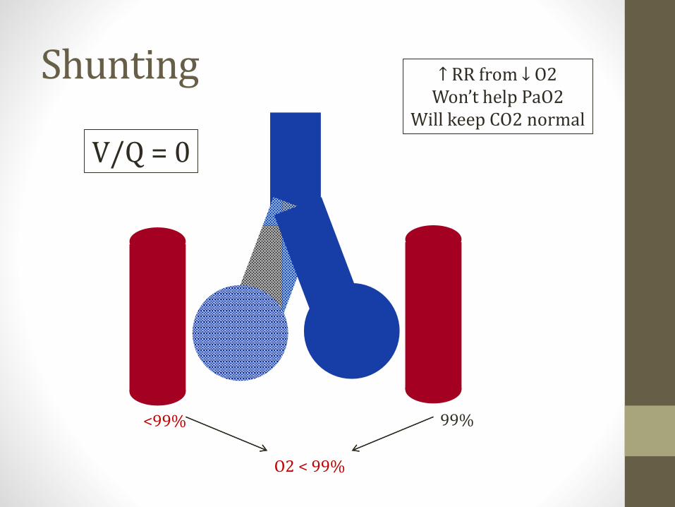
Shunting
O2 < 99%
<99% 99%
V/Q = 0
↑ RR from ↓ O2Won’t help PaO2
Will keep CO2 normal
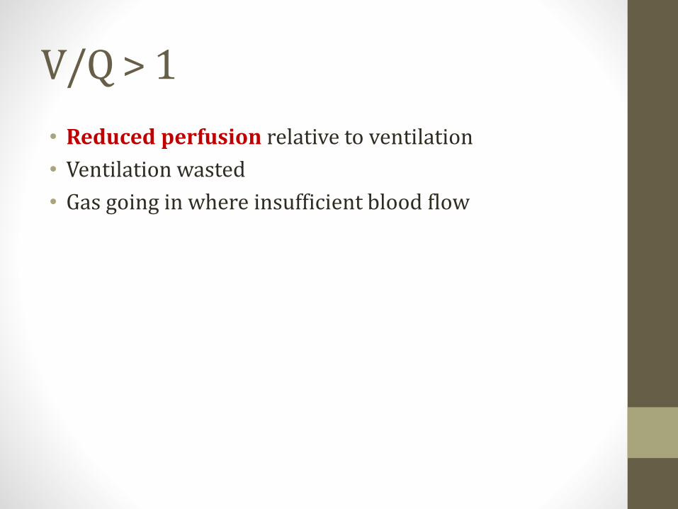
V/Q > 1
• Reduced perfusion relative to ventilation
• Ventilation wasted
• Gas going in where insufficient blood flow

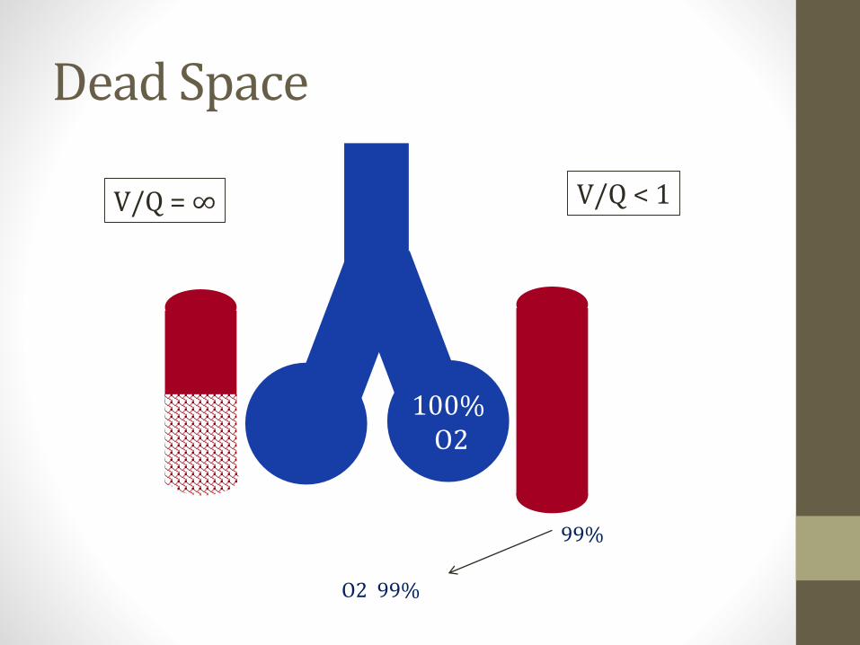
Dead Space
• Extreme increase in V/Q
• V/Q = ∞
• Anatomic dead space• Volume of conducting portions respiratory tract
• Nose, trachea
• Physiologic dead space• Anatomic PLUS volume of alveoli that don’t exchange gas
• Insufficient perfusion
• Apex is largest contributor
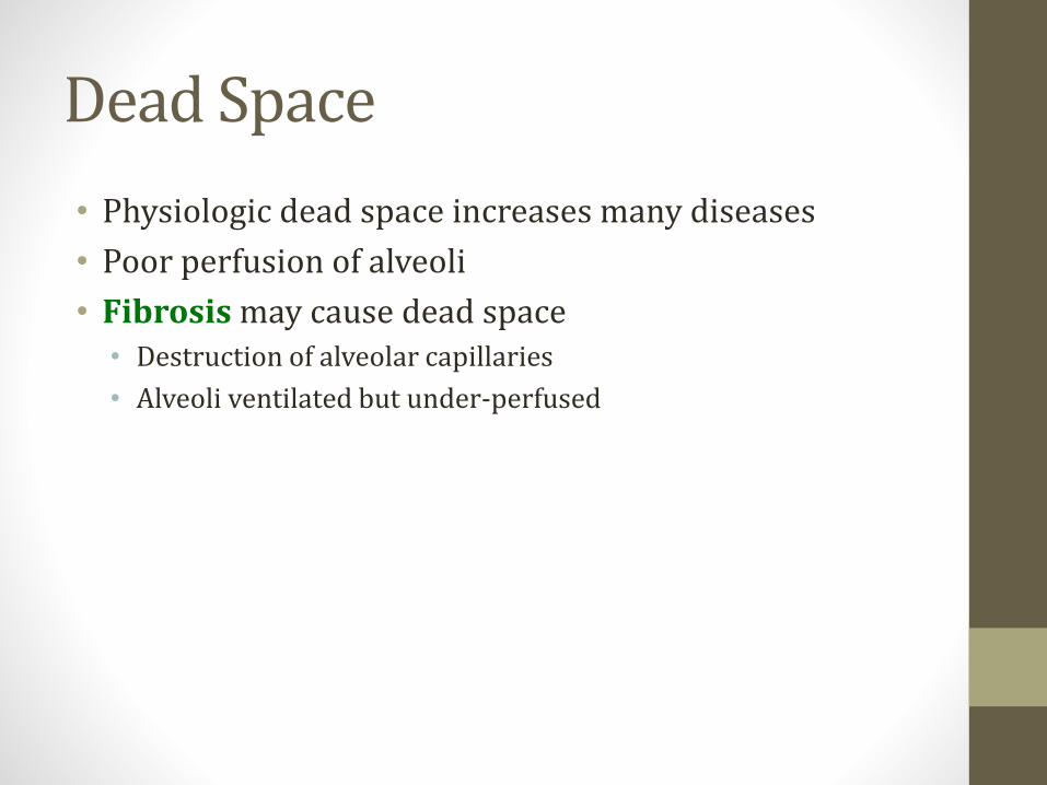
Dead Space
• Physiologic dead space increases many diseases
• Poor perfusion of alveoli
• Fibrosis may cause dead space• Destruction of alveolar capillaries
• Alveoli ventilated but under-perfused
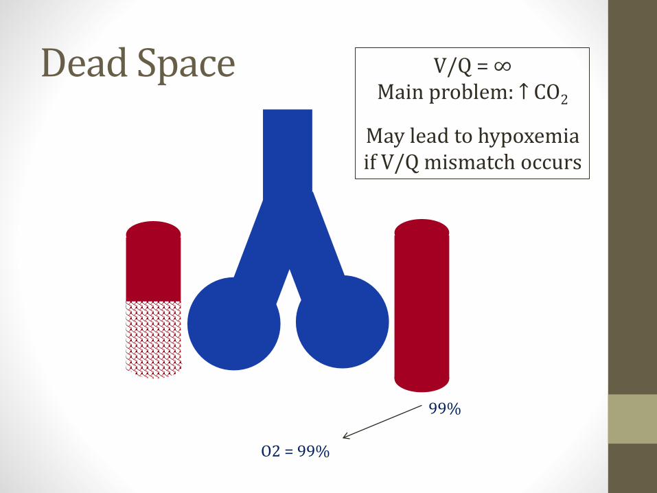
Dead Space
O2 = 99%
99%
V/Q = ∞Main problem: ↑ CO2
May lead to hypoxemiaif V/Q mismatch occurs

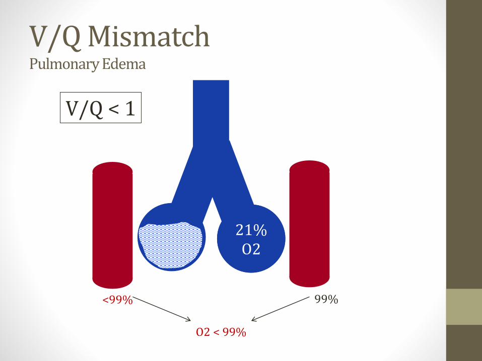
V/Q Mismatch
• Intermediate state• Some lung areas low V/Q
• Others high V/Q
• Inadequate ventilation
• Reduced oxygenation of blood
• ↑ RR → CO2 normal
V/Q = 0 V/Q = ∞
Shunt Dead Space
V/Q Mismatch
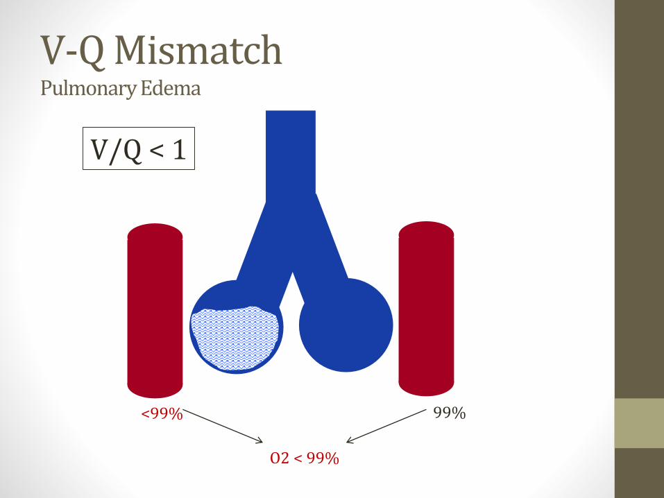
V-Q MismatchPulmonary Edema
O2 < 99%
<99% 99%
V/Q < 1
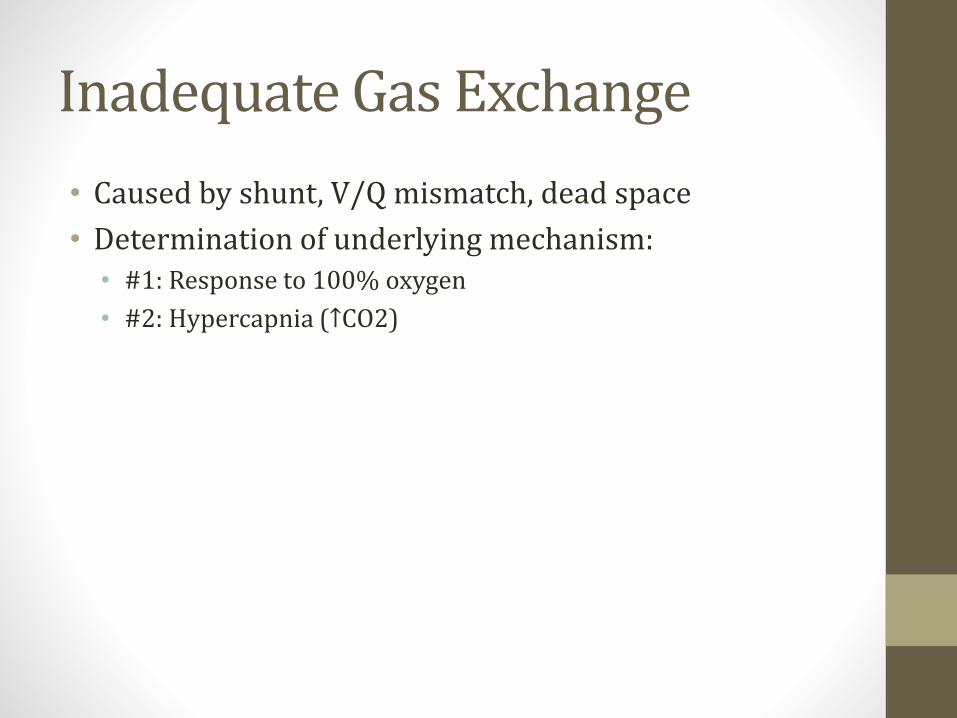
Inadequate Gas Exchange
• Caused by shunt, V/Q mismatch, dead space
• Determination of underlying mechanism:• #1: Response to 100% oxygen
• #2: Hypercapnia (↑CO2)

100% Oxygen
• Shunting (V/Q=0) won’t correct with 100% oxygen
• Functioning alveoli extracting maximum O2
• Increase alveolar O2 does not help
• Sick alveoli do not receive 100% O2 (↓V)

Shunting
O2 < 99%
<99% 99%
V/Q = 0
Shunting
O2 < 99%
<99% 99%
V/Q = 0
100%O2

100% Oxygen
• Dead space will correct with 100% oxygen
• V/Q mismatch will correct with 100% oxygen
• Increasing O2 content in alveoli → Increased PaO2

Dead Space
21% O2
O2 < 99%
<99%
V/Q = ∞ V/Q < 1
Dead Space
100%O2
O2 99%
99%
V/Q = ∞ V/Q < 1
V/Q MismatchPulmonary Edema
O2 < 99%
<99% 99%
V/Q < 1
21% O2
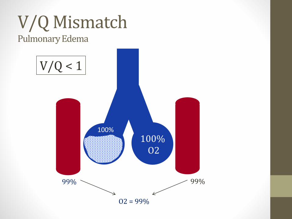
V/Q MismatchPulmonary Edema
O2 = 99%
99% 99%
V/Q < 1
100%O2
100%
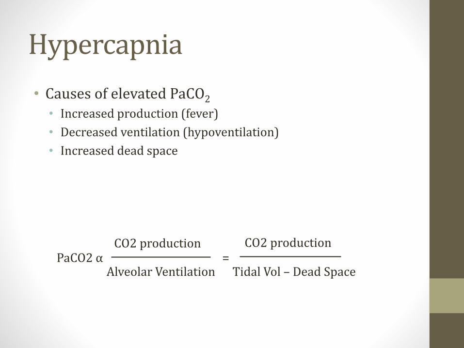
Hypercapnia
• Causes of elevated PaCO2
• Increased production (fever)
• Decreased ventilation (hypoventilation)
• Increased dead space
PaCO2 αCO2 production
Alveolar Ventilation
CO2 production
Tidal Vol – Dead Space=

Hypercapnia
• Dead space: classic cause of hypercapnia
• Perfusion problem
• Ventilation wasted
• Elevated PaCO2
PaCO2 αCO2 production
Alveolar Ventilation
CO2 production
Tidal Vol – Dead Space=
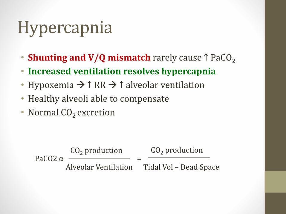
Hypercapnia
• Shunting and V/Q mismatch rarely cause ↑ PaCO2
• Increased ventilation resolves hypercapnia
• Hypoxemia → ↑ RR → ↑ alveolar ventilation
• Healthy alveoli able to compensate
• Normal CO2 excretion
PaCO2 αAlveolar Ventilation Tidal Vol – Dead Space
=CO2 production CO2 production
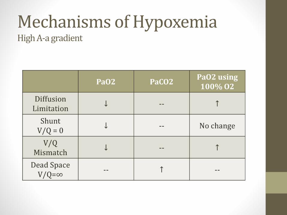
Mechanisms of HypoxemiaHigh A-a gradient

Ventilation-Perfusion
ApexLess Blood FlowLess Ventilation
BaseMost Blood FlowMost Ventilation
Both decrease bottom to topBlood flow decreases more
V/Q ratio changes

Ventilation-PerfusionApex
V/Q > 3Lots of Ventilation (relative)
High O2, Low CO2
BaseV/Q <0.6
Low Ventilation (relative)Low O2, High CO2
Overall V/Q = 0.8
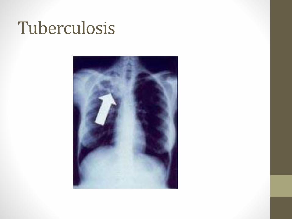
Tuberculosis

Mechanisms by Disease
• Most diseases (COPD, PNA, CHF) have hypoxemia from multiple mechanisms• PNA may cause V/Q mismatch or shunt
• COPD may cause alveoli and blood vessel destruction
• Some examples worth knowing• Intra-cardiac shunt: Pure shunt mechanism
• Inhale a peanut: V/Q = 0
• Pulmonary Embolism: V/Q = ∞
Ventilation-Perfusion
• With exercise overall V/Q approaches 1• More blood flow
• More ventilation
• ↑ ventilation > ↑blood flow
Lung Pressure Zones
• Because of gravity, more pressure in artery (Pa) and veins (Pv) at bottom compared to top of lungs• As you move up lungs, Pa and Pv fall
• Alveolar pressure (PA) same throughout lung
• In theory, PA at apex can become > Pa /Pv
• This compresses vessels
• Less blood flow/perfusion
• Very high V/Q
• More dead space
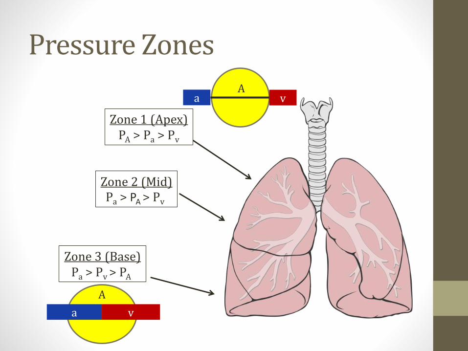
Pressure Zones
Zone 1 (Apex)PA > Pa > Pv
Zone 3 (Base)Pa > Pv > PA
a v
A
a vA
Zone 2 (Mid)Pa > PA > Pv
Carbon DioxideJason Ryan, MD, MPH
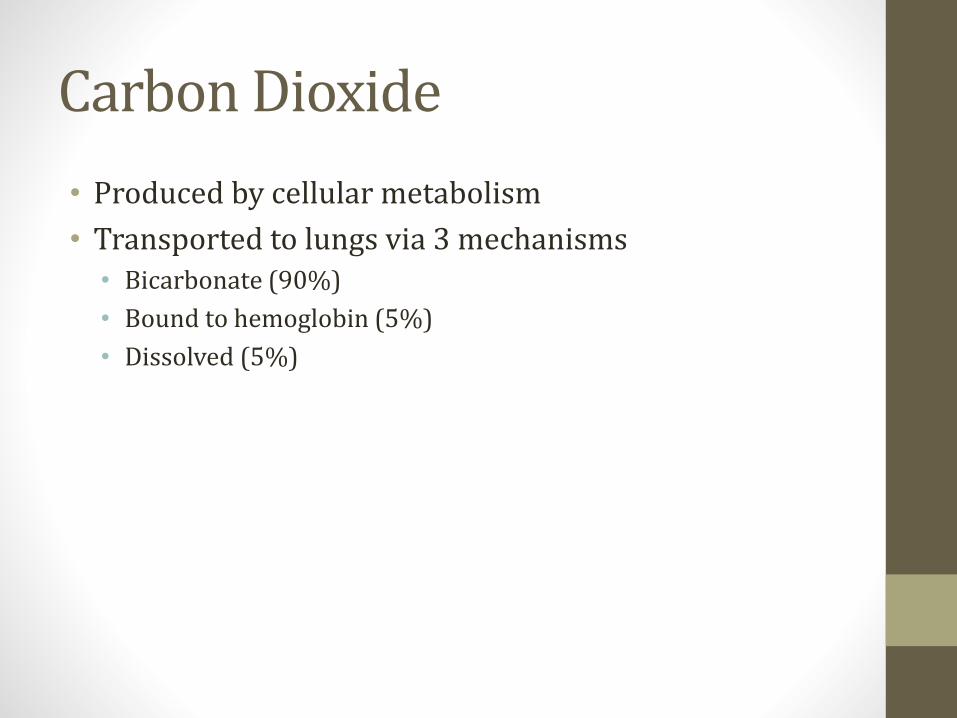
Carbon Dioxide
• Produced by cellular metabolism
• Transported to lungs via 3 mechanisms• Bicarbonate (90%)
• Bound to hemoglobin (5%)
• Dissolved (5%)
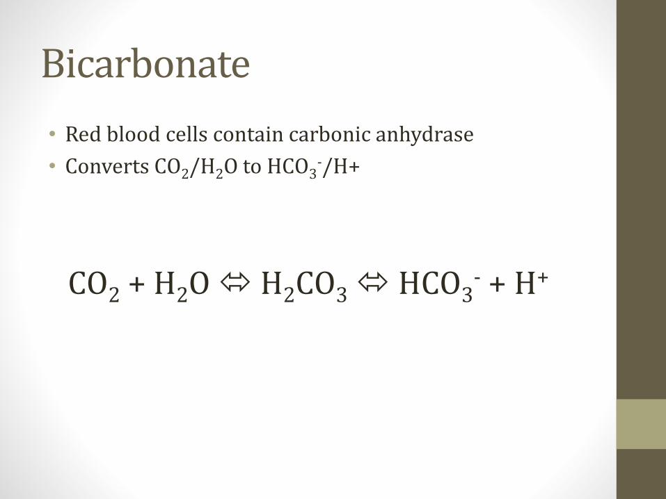
Bicarbonate
• Red blood cells contain carbonic anhydrase
• Converts CO2/H2O to HCO3-/H+
CO2 + H2O H2CO3 HCO3- + H+
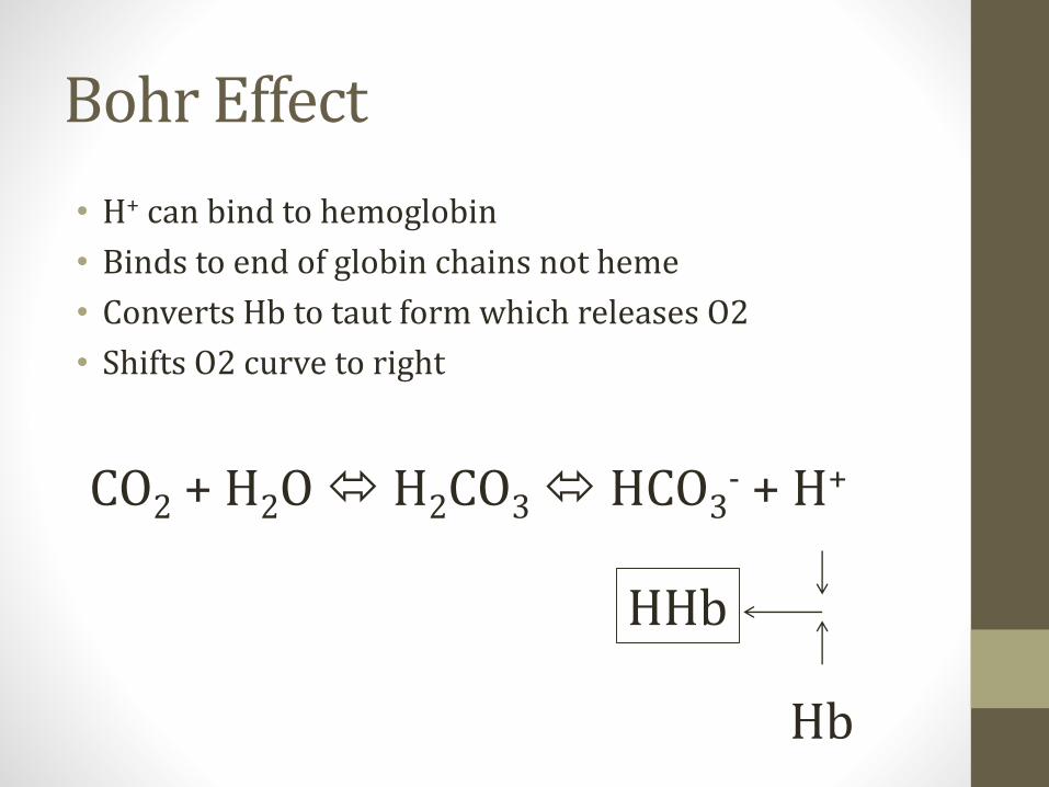
Bohr Effect
• H+ can bind to hemoglobin
• Binds to end of globin chains not heme
• Converts Hb to taut form which releases O2
• Shifts O2 curve to right
CO2 + H2O H2CO3 HCO3- + H+
Hb
HHb

Bohr EffectEasier to release O2
25 50 75 100
pO2 (mmHg)
25
50
75
100
Hb
% S
atu
rati
on
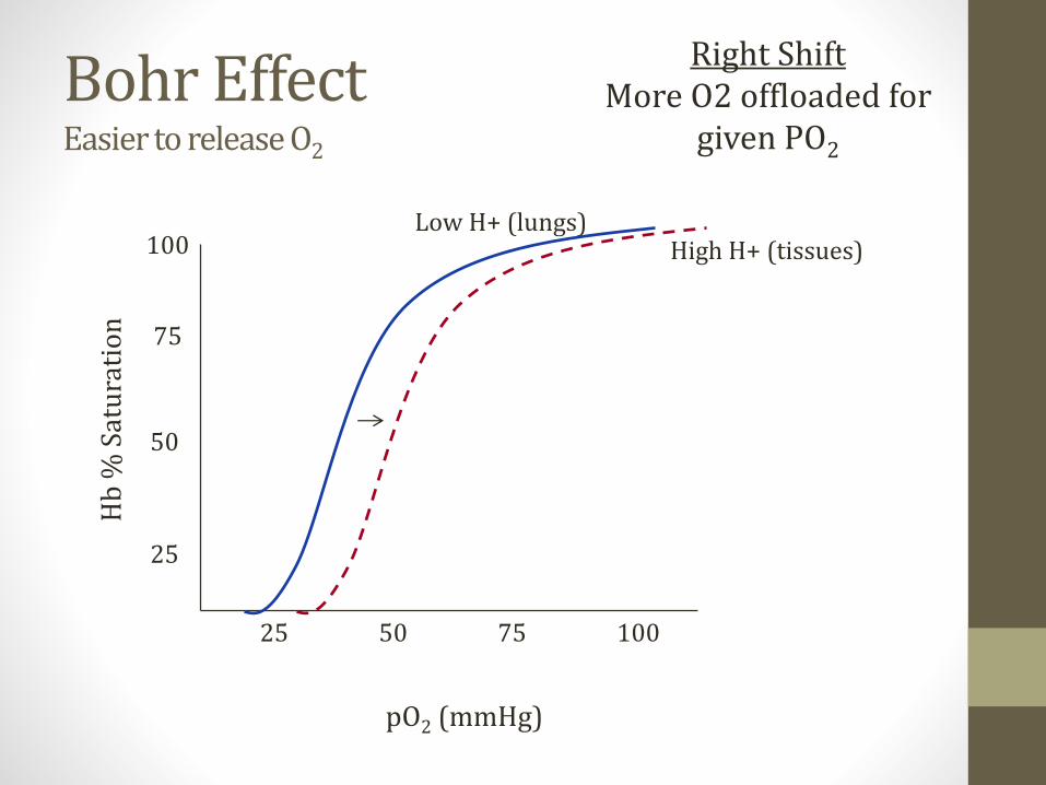
Bohr EffectEasier to release O2
25 50 75 100
pO2 (mmHg)
25
50
75
100
Hb
% S
atu
rati
on
Right ShiftMore O2 offloaded for
given PO2
Low H+ (lungs)High H+ (tissues)
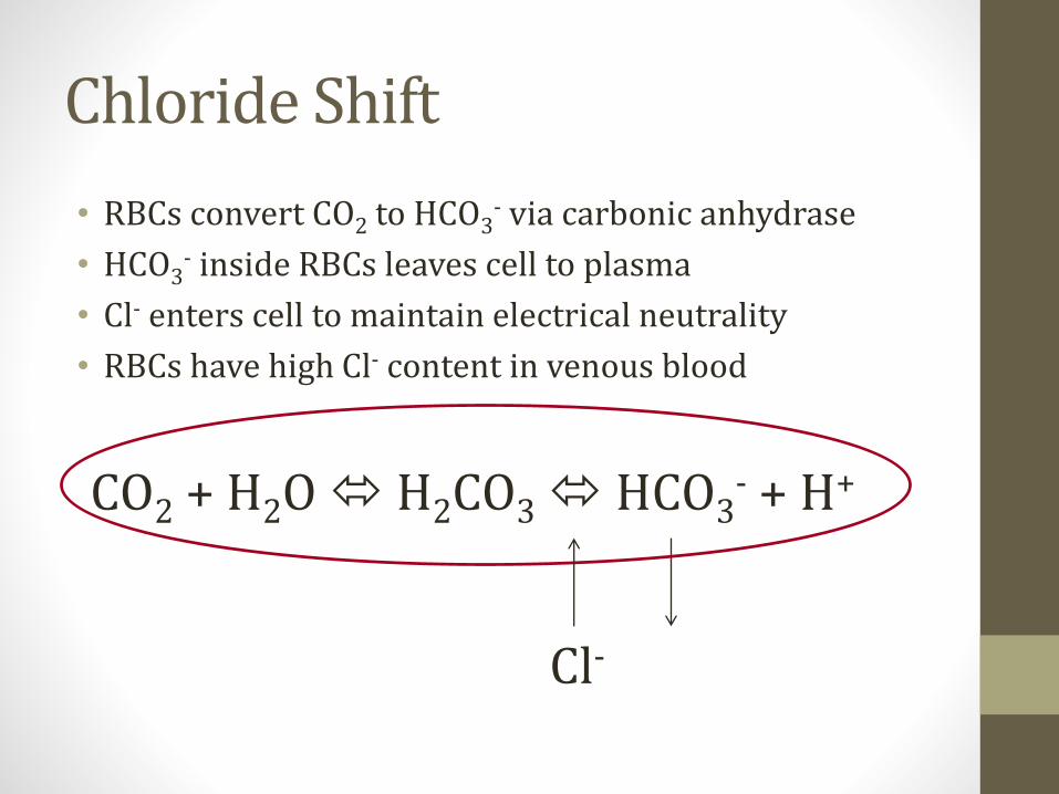
Chloride Shift
• RBCs convert CO2 to HCO3- via carbonic anhydrase
• HCO3- inside RBCs leaves cell to plasma
• Cl- enters cell to maintain electrical neutrality
• RBCs have high Cl- content in venous blood
CO2 + H2O H2CO3 HCO3- + H+
Cl-

Carbaminohemoglobin
• Hemoglobin bound to CO2
• In low O2 environment Hb more likely to bind CO2
• Haldane Effect

Haldane Effect
HbCO2
Content
PCO2
Low High
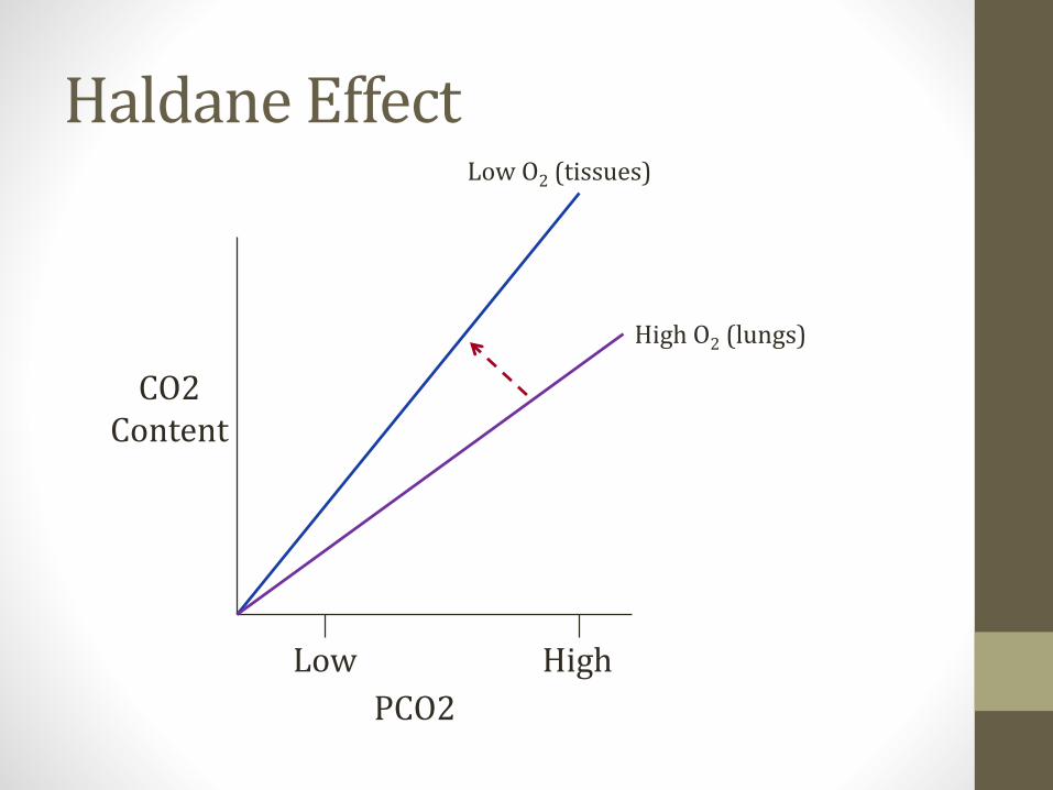
Haldane Effect
CO2Content
PCO2
Low High
Low O2 (tissues)
High O2 (lungs)
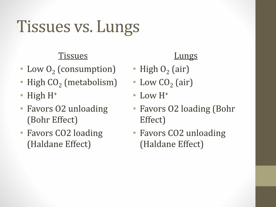
Tissues vs. Lungs
Tissues
• Low O2 (consumption)
• High CO2 (metabolism)
• High H+
• Favors O2 unloading (Bohr Effect)
• Favors CO2 loading (Haldane Effect)
Lungs
• High O2 (air)
• Low CO2 (air)
• Low H+
• Favors O2 loading (Bohr Effect)
• Favors CO2 unloading (Haldane Effect)
CO2 in Special Situations
• High Altitude
• Exercise
• Cerebral Blood Flow
• Control of Respiration

High Altitude
• Lower atmospheric pressure
• Lower pO2
• Hypoxia → hyperventilation
• ↓pCO2→ respiratory alkalosis (pH rises)
• After 24-48hrs, kidneys will excrete HCO3-
• pH will fall back toward normal

High Altitude
• ↑ erythropoietin• Will raise Hct and Hgb over 10-14 days
• Chronic hypoxic vasoconstriction• Pulmonary hypertension
• Can lead to RVH

High Altitude
• Changes to O2 dissociation curve
• Alkalosis causes leftward shift
• Also stimulates production of 2,3 BPG• Increased levels when RBCs need to release more O2
• This shifts curve back toward normal position

High Altitude
25 50 75 100
pO2 (mmHg)
25
50
75
100
Hb
% S
atu
rati
on
Alkalosis↑ 2,3 BPG
Cumulative Effect
Exercise
• ↑ O2 consumption
• ↑ CO2 production
• ↑ ventilation

Exercise
• More CO2 produced by muscles
• CO2 levels in venous blood rise
• More O2 consumed by muscles
• O2 levels in venous blood fall
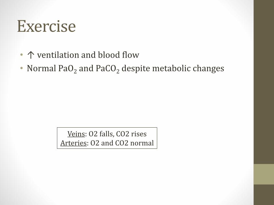
Exercise
• ↑ ventilation and blood flow
• Normal PaO2 and PaCO2 despite metabolic changes
Veins: O2 falls, CO2 risesArteries: O2 and CO2 normal

O2 Diffusion-Perfusion
Length along capillary
Par
tial
Pre
ssu
re
Rest
Exercise
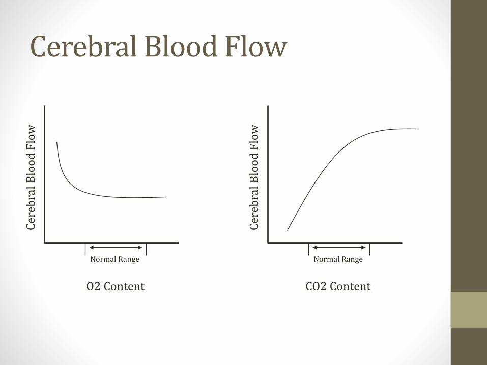
Cerebral Blood FlowC
ereb
ral
Blo
od
Flo
w
O2 Content
Normal RangeC
ereb
ral
Blo
od
Flo
w
CO2 Content
Normal Range
Panic Attacks
• Hyperventilation
• Low CO2
• Hypocapnia→ cerebral vasoconstriction
• CNS symptoms (dizziness, blurred vision)

CO2 and Breathing Control
• PaCO2 is the major stimulus for breathing
• Central chemoreceptors in medulla most important
• Peripheral chemoreceptors: carotid and aortic bodies• Can sense CO2 but more sensitive to O2
• High PaCO2 → increased respiratory rate
• Low PaCO2 → decreased respiratory rate

CO2 and Breathing Control
• COPD patients chronically retain CO2
• Worsens when excess oxygen therapy given
• Theory: response to CO2 blunted• Oxygen level becomes major stimulus
• Respiratory depression with high O2
• New data indicates more complex• Haldane effect

CO2 and Breathing Control
• Oxygen can mask hypoventilation
• High CO2 level useful to determine ventilation status
• Clinical scenario:• Patient with pneumonia
• O2 applied via nasal cannula
• O2 level 95%
• Blood gas: PaCO2 = 60mmHg (high)
CO2 and Breathing Control
• Clinical scenario• Patient with neuromuscular disease (ALS)
• O2 saturation on O2 95%
• Blood gas: PaCO2 = 60 (high)
• Respiratory muscles failing
• Symptoms of high CO2
• Lethargy
• Confusion
• Agitation
Lung Physical ExamJason Ryan, MD, MPH

Lung Exam
• Percussion• Finger against thorax → tap
• Auscultation• Stethoscope thorax
• Upper, mid, lower lung fields
• Special techniques• Fremitus
• Pectoriloquy

Percussion
• Normal sounds = resonant
• Abnormal: dull or hyperresonant
• Dull• Pleural effusion
• Consolidation (pneumonia)
• Hyperresonant→ air trapped• Pneumothorax
• Emphysema
Lung Auscultation
• Normal breath sounds are vesicular
• Most all pathologic lung processes result in decreased lung sounds over affected area
Adventitious Lung Sounds
• Rales
• Wheezes
• Rhonchi
• Bronchial breath sounds
• Stridor

Rales
• Also called crackles
• Small airways “pop” open after collapse
• Early inspiratory, late inspiratory or expiratory
• Classic causes• Pulmonary edema (bases)
• Pneumonia
• Interstitial fibrosis

Wheezes
• Air flows through narrowed bronchi
• Usually expiratory or inspiratory/expiratory
• Classic cause is asthma
• Other causes:• Heart failure (cardiac asthma)
• Chronic bronchitis
• Obstruction (tumor; localized wheeze)
Rhonchi
• Secretions in large airways
• Course breath sounds
• Classic cause is COPD

Bronchial Breath Sounds
• High pitched lung sounds
• Like flow through tube
• Longer expiratory phase than normal
• Seen in pneumonia with consolidation
Stridor
• Wheeze that is almost entirely inspiratory
• Usually loudest over neck
• Indicates partial obstruction of larynx or trachea
• Some classic causes• Laryngotracheitis (croup)
• Epiglottitis (Hib in children)
• Retropharyngeal abscess
• Diphtheria

Pectoriloquy
• Sounds over chest through stethoscope
• Bronchophony• Voice sounds are louder and clearer
• Whispered pectoriloquy• Whispered “99-99-99”
• Should be muffled
• Abnormal if clear
• Egophony: “Eeeeee” sounds like “Aaaay”
• All indicated fluid in lungs: Effusion, consolidation
Fremitus
• Place hands on patients back
• Patient says “ninety-nine”
• Vibrations travel through airways to back
• Varies with density of lung tissue
• Only common condition with increased fremitus is lobar pneumonia
• Decreased in most other processes• Pleural effusion
• Pneumothorax
• Atelectasis

Nail Clubbing
• Associated with many pulmonary diseases
• Bronchiectasis
• Cystic Fibrosis
• Lung tumors
• Pulmonary fibrosis
• Also cyanotic congenital heart disease
Image courtesy of James Heilman, MD
Image courtesy of Jfdwolff
Pulmonary Function TestsJason Ryan, MD, MPH
Dyspnea
• Many, many causes
• Deconditioning
• Cardiac causes
• Anemia
• Pulmonary causes

Pulmonary Dyspnea
• Obstruction• Can’t get air out of lungs
• Air trapped
• Poor oxygenation
• Restriction• Can’t get air into lungs
• Poor oxygenation
Pulmonary Function Testing
• Determining flows, volumes in lung
• Helps determine cause of dyspnea• Sometimes unclear from history, exam, x-ray, etc.
• Helps determine disease severity/progression• Many diseases monitored by PFTs
• COPD, Pulmonary Fibrosis
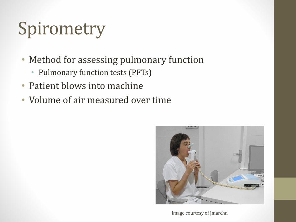
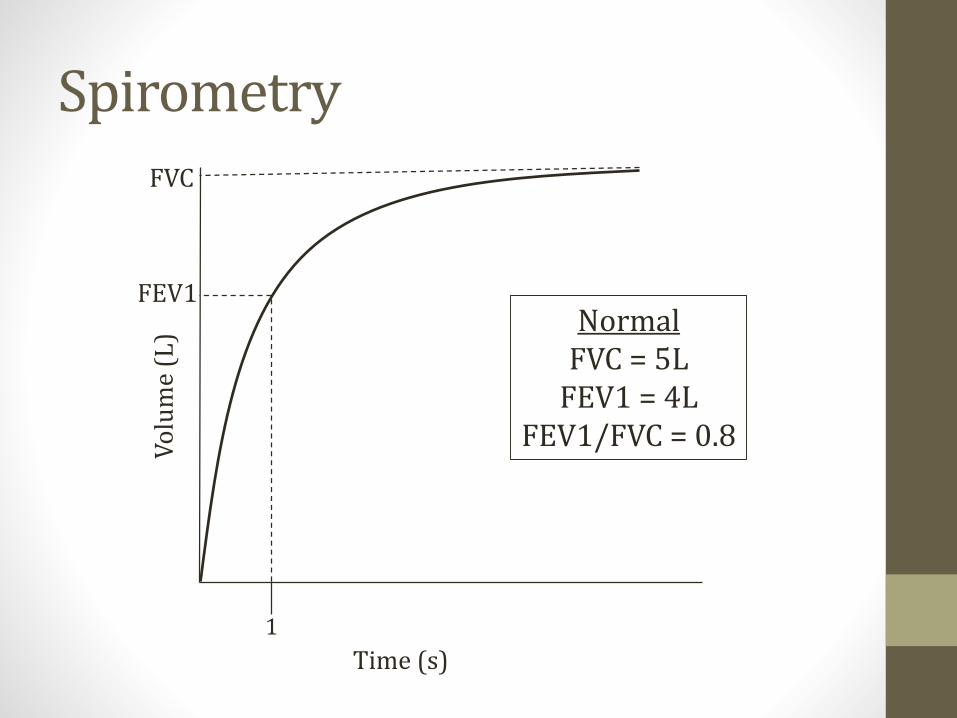
Spirometry
• Method for assessing pulmonary function• Pulmonary function tests (PFTs)
• Patient blows into machine
• Volume of air measured over time
Image courtesy of Jmarchn
SpirometryV
olu
me
(L)
Time (s)
1
FEV1
FVC
NormalFVC = 5L
FEV1 = 4LFEV1/FVC = 0.8
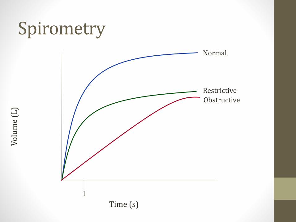
SpirometryV
olu
me
(L)
Time (s)
1
Normal
Restrictive
Obstructive

SpirometryV
olu
me
(L)
Time (s)
1
Normal
Restrictive
Obstructive
FEV1
FEV1
FEV1

SpirometryV
olu
me
(L)
Time (s)
1
Normal
Restrictive
Obstructive
FVC
FVCFVC

Summary
• FEV1 and FVC fall in both obstructive and restrictive diseases
• FEV1 falls MORE than FVC in obstructive
FEV1 FVC FEV1/FVC
Obstructive ↓↓ ↓ ↓
Restrictive ↓ ↓ >80%
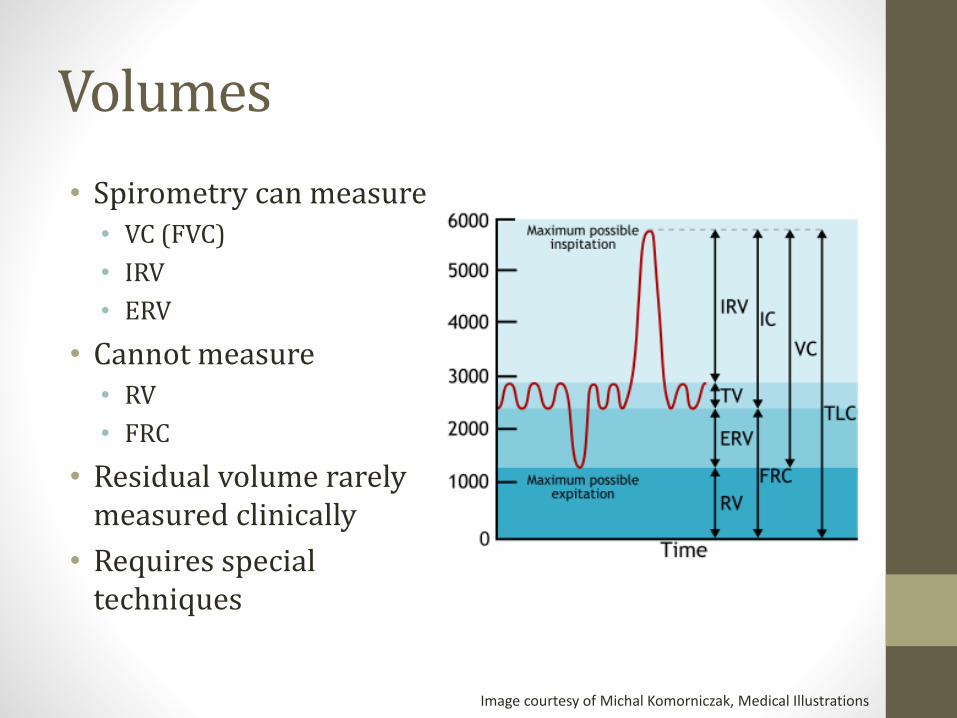
Volumes
• Spirometry can measure• VC (FVC)
• IRV
• ERV
• Cannot measure• RV
• FRC
• Residual volume rarely measured clinically
• Requires special techniques
Image courtesy of Michal Komorniczak, Medical Illustrations
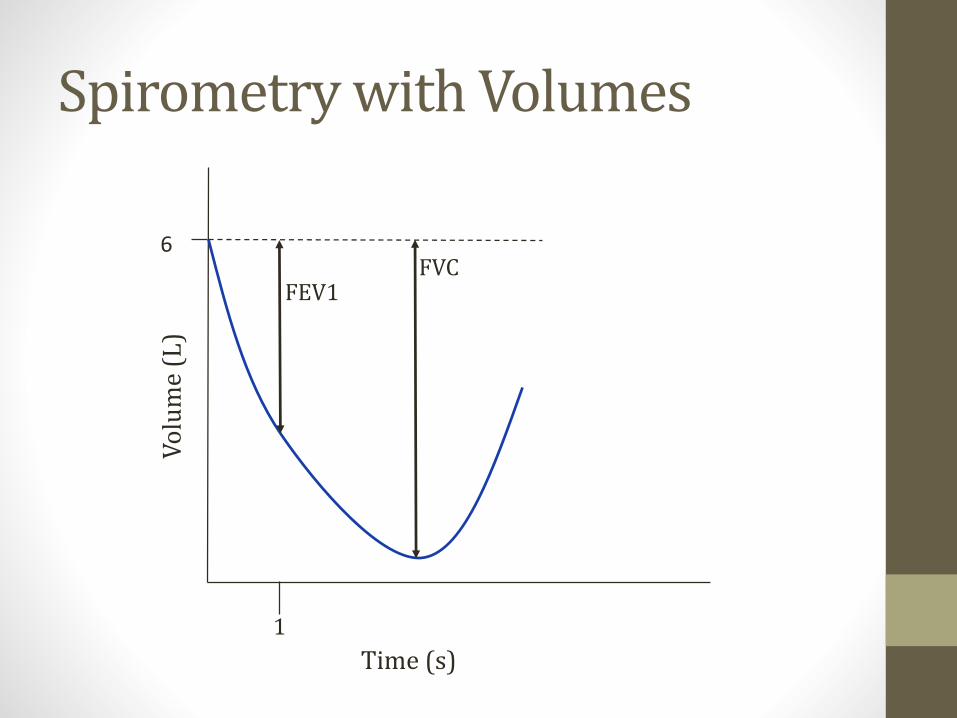
Spirometry with VolumesV
olu
me
(L)
1
6
Time (s)
FEV1FVC

Spirometry with VolumesV
olu
me
(L)
1
Time (s)
Obstructive
Restrictive
Normal
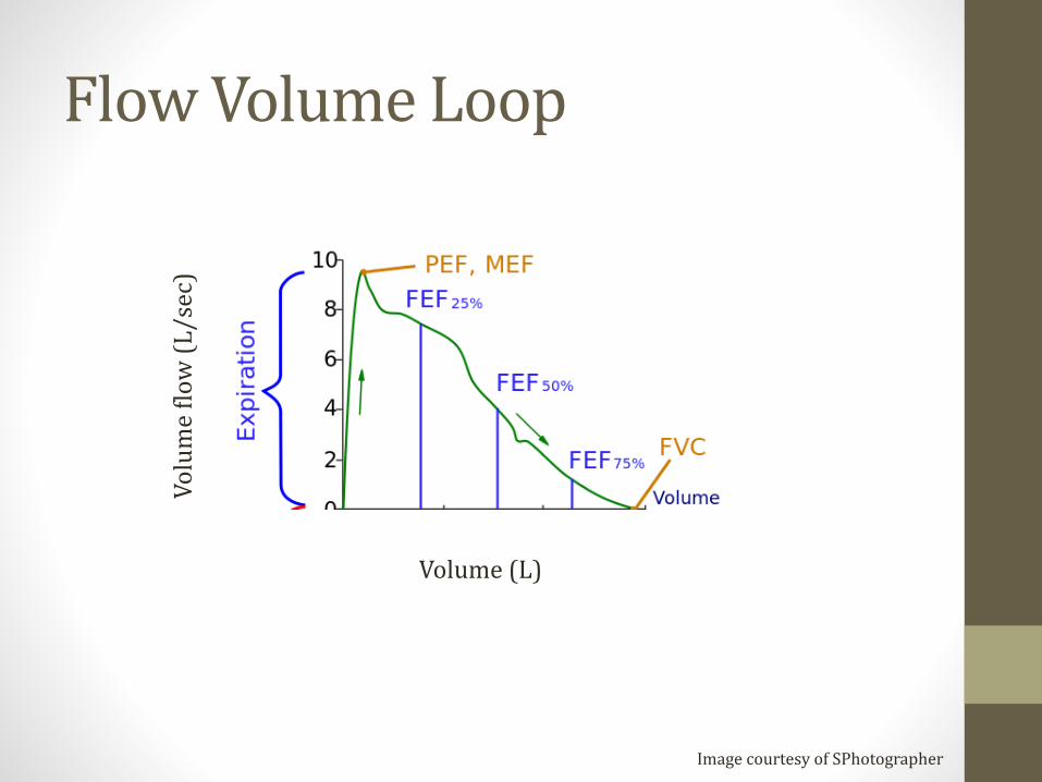
Flow Volume Loop
Image courtesy of SPhotographer
Volume (L)
Vo
lum
e fl
ow
(L
/sec
)
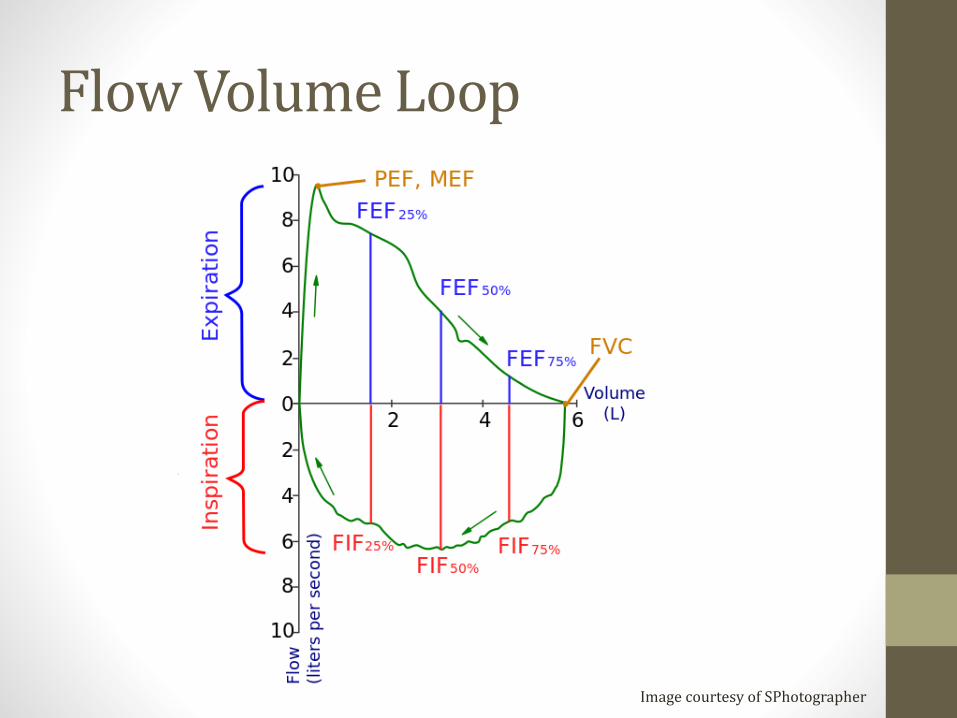
Flow Volume Loop
Image courtesy of SPhotographer

Flow Volume Loops
Image courtesy of Yaser Ammar,
Work of Breathing
• Work proportional to resistance
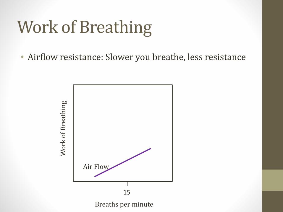
Work of Breathing
• Airflow resistance: Slower you breathe, less resistance
Breaths per minute
Wo
rk o
f B
reat
hin
g
15
Air Flow

Work of Breathing
• Elastic resistance: Faster you breathe, less resistance
Breaths per minute
Wo
rk o
f B
reat
hin
g
15
Elastic
Work of Breathing
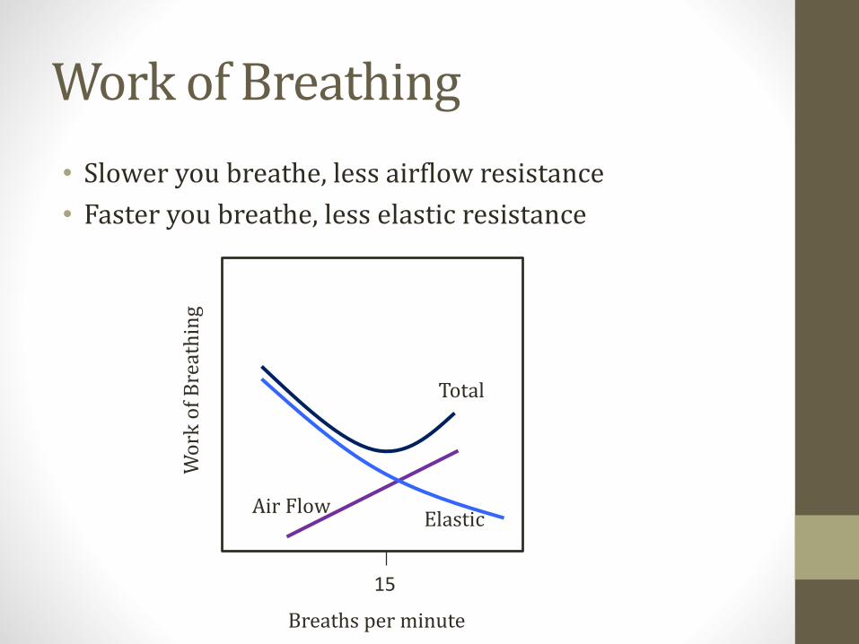
• Slower you breathe, less airflow resistance
• Faster you breathe, less elastic resistance
Breaths per minute
Wo
rk o
f B
reat
hin
g
15
Total
Air FlowElastic

Work of Breathing
• Increases in obstructive and restrictive disease
• Different patterns
Normal
Restrictive
Obstructive
Breaths per minute
Wo
rk o
f B
reat
hin
g
15

Obstructive Lung DiseaseJason Ryan, MD, MPH
Obstructive Lung Diseases
• Key points: Air trapping, slow flow out, less air out
• Reduced FEV1 (slow flow out)
• Reduced FVC (less air out)
• Reduced FEV1/FVC (hallmark)
Residual & Total Lung Volume
• Both go up in obstructive disease• From air trapping
• Both fall in restrictive disease• Less air fills the lungs due to restriction

Obstructive Lung Diseases
• Chronic bronchitis
• Emphysema
• Asthma
• Bronchiectasis
Chronic Bronchitis
• Chronic cough
• Productive of sputum
• At least 3 months over two years
• No other cause of cough present
• Strongly associated with smoking
Chronic Bronchitis
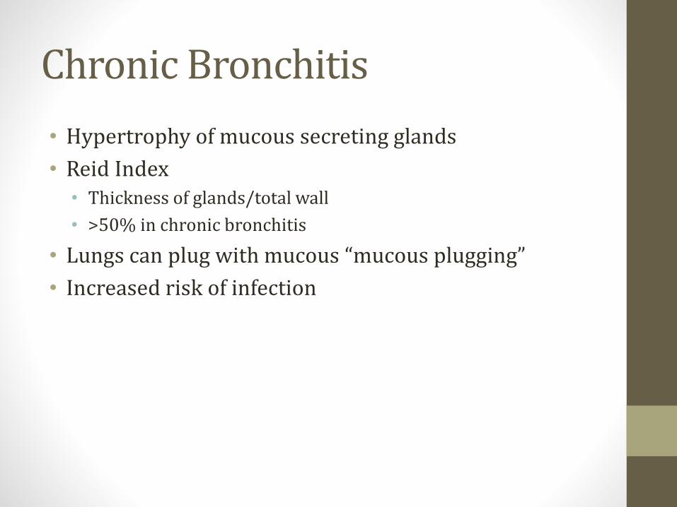
• Hypertrophy of mucous secreting glands
• Reid Index• Thickness of glands/total wall
• >50% in chronic bronchitis
• Lungs can plug with mucous “mucous plugging”
• Increased risk of infection

Chronic Bronchitis
• Poor ventilation of lungs
• Increased CO2
• Decreased O2
• Hypoxic vasoconstriction
• Pulmonary hypertension
• Right heart failure (cor pulmonale)
Chronic Bronchitis
• Cough
• Wheezing
• Crackles
• Dyspnea
• Cyanosis (shunting)

Shunting
O2 < 99%
<99% 99%
Emphysema
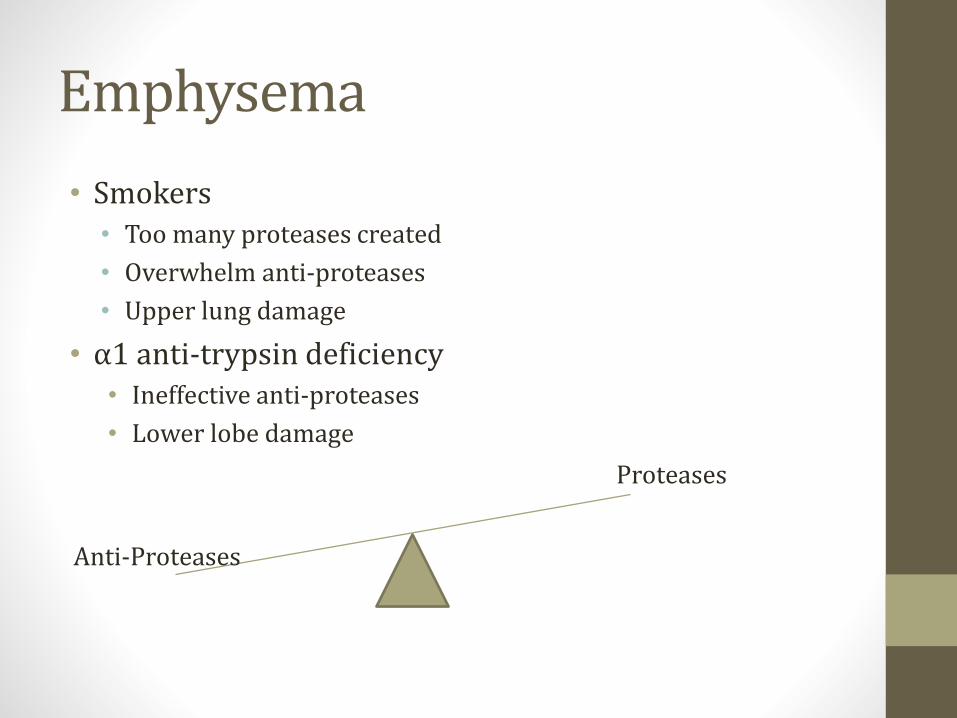
• Smokers• Too many proteases created
• Overwhelm anti-proteases
• Upper lung damage
• α1 anti-trypsin deficiency• Ineffective anti-proteases
• Lower lobe damage
Proteases
Anti-Proteases
Emphysema
• Destruction of alveoli• Smoke activates macrophages
• Recruitment of neutrophils
• Release of proteases
• Loss of elastic recoil
• Small airways collapse on exhalation
• Air “trapped” in lungs
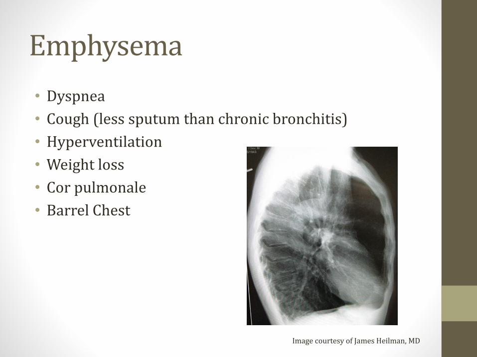
Emphysema
• Dyspnea
• Cough (less sputum than chronic bronchitis)
• Hyperventilation
• Weight loss
• Cor pulmonale
• Barrel Chest
Image courtesy of James Heilman, MD

Acinus
• Acinus = bronchiole + alveoli
• Smokers = centriacinar damage
• α1 anti-trypsin deficiency = panacinar

Image courtesy of Nephron
Image courtesy of James Heilman, MD

Chest Volumes and Pressures
Vo
lum
e
-40 -20 0 20 40
Chest WallLungs
System
FRC

Blue Bloater – Pink Puffer
• Chronic Bronchitis – Blue Bloater• Cyanosis from shunting (blue)
• Air trapping (bloated)
• Emphysema – Pink Puffer• Loss of alveoli
• Loss of surface area for O2 absorption (dead space)
• Hyperventilation to compensate (puffer)
• Initially this maintains O2 level (pink)
COPD
• Chronic Obstructive Pulmonary Disease
• Includes chronic bronchitis, emphysema, asthma
• Many similar symptoms (cough, dyspnea, wheezing)
• Many similar treatments
α1 Anti-trypsin Deficiency
• Inherited (autosomal co-dominant)
• Decreased or dysfunctional AAT
• AAT balances naturally occurring proteases
• Elastase found in neutrophils & alveolar macrophages
α1 Anti-trypsin Deficiency
• Lung• Panacinar emphysema
• Imbalance between neutrophil elastase (destroys elastin) and elastase inhibitor AAT (protects elastin)
• Lower lung damage
• Liver cirrhosis • Abnormal α1 builds up in liver
• Only occurs in phenotypes with pathologic polymerization of AAT in endoplasmic reticulum of hepatocytes
• Some patients have severe AAT deficiency but no intra-hepatocytic accumulation

α1 Anti-trypsin Deficiency
• Classic case• Typical COPD symptoms: cough, sputum, wheeze
• Younger patient (40s)
• Imaging: emphysematous changes most prominent at bases
• Obstructive PFTs
• Question often asks about panacinar involvement
• These patients should NEVER smoke• Stimulates neutrophil elastase production

Asthma
• Reversible bronchoconstriction
• Usually due to allergic stimulus• Type I hypersensitivity reaction
• Airways are HYPERresponsive
• Common in children
• Associated with other allergic (atopic) conditions• Rhinitis, eczema
• May have family history of allergic reactions
Asthma Triggers
• URI
• Allergens (animal dander, dust mites, mold, pollens)
• Stress
• Exercise
• Cold
• Aspirin
AERDAspirin Exacerbated Respiratory Disease
• Asthma, chronic rhinosinusitis, nasal polyposis• Chronic asthma/rhinosinusitis symptoms
• Acute exacerbations after ingestion aspirin or NSAIDs
• Dysregulation of arachidonic acid metabolism
• Overproduction leukotrienes
• Treatment: Leukotriene receptor antagonists • Montelukast, Zafirlukast
Asthma Symptoms
• Episodic symptoms
• Dyspnea, wheezing, cough
• Hypoxia during episodes
• Decreased I/E ratio
• Reduced peak flow
• Mucous plugging (airway obstruction/shunt)
• Death: Status asthmaticus
Asthma Diagnosis
• Usually classic history/physical exam
• Methacholine challenge• Muscarinic agonist
• Causes bronchoconstriction
• Administer increasing amounts of nebulized drug
• Spirometry after each dose
• Look for dose at which FEV1 falls significantly
• If dose is low → positive test
Asthma Pathology
• Recurrent episodes
• Smooth muscle hypertrophy
• Inflammation

Asthma Pathology
• Classic sputum findings• Curschmann’s spirals
• Charcot-Leyden crystals
Image courtesy of Jmh649
Image courtesy of Patho
Pulsus Paradoxus
• Most frequent non-cardiac causes are asthma/COPD

Bronchiectasis
• Result of chronic, recurrent airway inflammation
• Airways become permanently dilated
• Obstruction• Large airways dilated
• Small/medium airways thickened bronchial walls

Bronchiectasis
Image courtesy of Laura Fregonese, Jan StolkImage courtesy of Yale Rosen
Bronchiectasis Symptoms
• Recurrent infections
• Cough, excessive sputum (foul smelling)
• Hemoptysis
• Cor pulmonale
• Amyloidosis
Bronchiectasis Etiologies
• Obstruction (tumor)
• Smoking
• Cystic fibrosis
• Kartagener’s syndrome
• Allergic bronchopulmonary aspergillosis

Primary Ciliary DyskinesiaImmotile-cilia syndrome
• Cilia unable to beat, beat normally, or absent
• Inherited (autosomal recessive)
• Gene mutations dynein structure/formation
• Dynein = motor protein creates movement
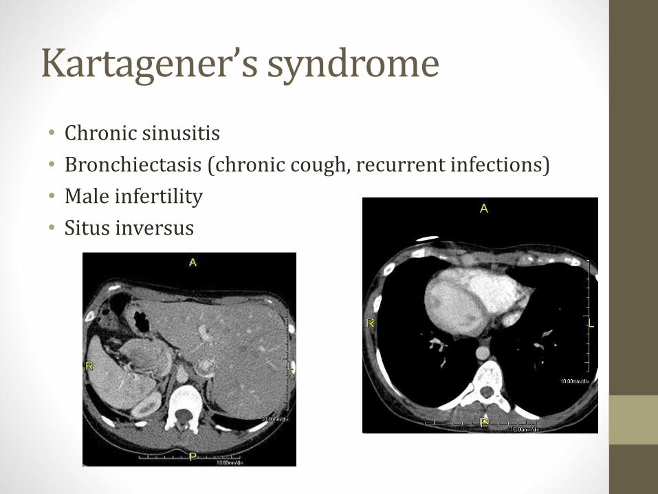
Kartagener’s syndrome
• Chronic sinusitis
• Bronchiectasis (chronic cough, recurrent infections)
• Male infertility
• Situs inversus

Kartagener’s syndrome
• Classic case:• Child
• Recurrent sinus/ear infections
• Chronic cough
• Bronchiectasis on chest CT
• Obstruction on PFTs
• Situs inversus
• Question often asks about dynein protein

ABPAAllergic bronchopulmonary aspergillosis
• Hypersensitivity (allergic) reaction to aspergillus
• Lungs become colonized with Aspergillus fumigatus• Low virulence fungus
• Only infects immunocompromised or debilitated lungs
• Occurs predominantly in asthma and CF patients
• ABPA patients:• Increases Th2 CD4+ cells
• Synthesis interleukins
• Eosinophilia
• IgE antibody production
ABPAAllergic bronchopulmonary aspergillosis
• Classic case• Asthma or CF patient
• Recurrent episodes cough, fever, malaise
• Brownish mucus plugs, hemoptysis
• Peripheral blood eosinophilia
• High IgE level
• Bronchiectasis on imaging
• PFTs with obstruction
• Diagnosis: Skin testing aspergillosis
• Treatment: Steroids
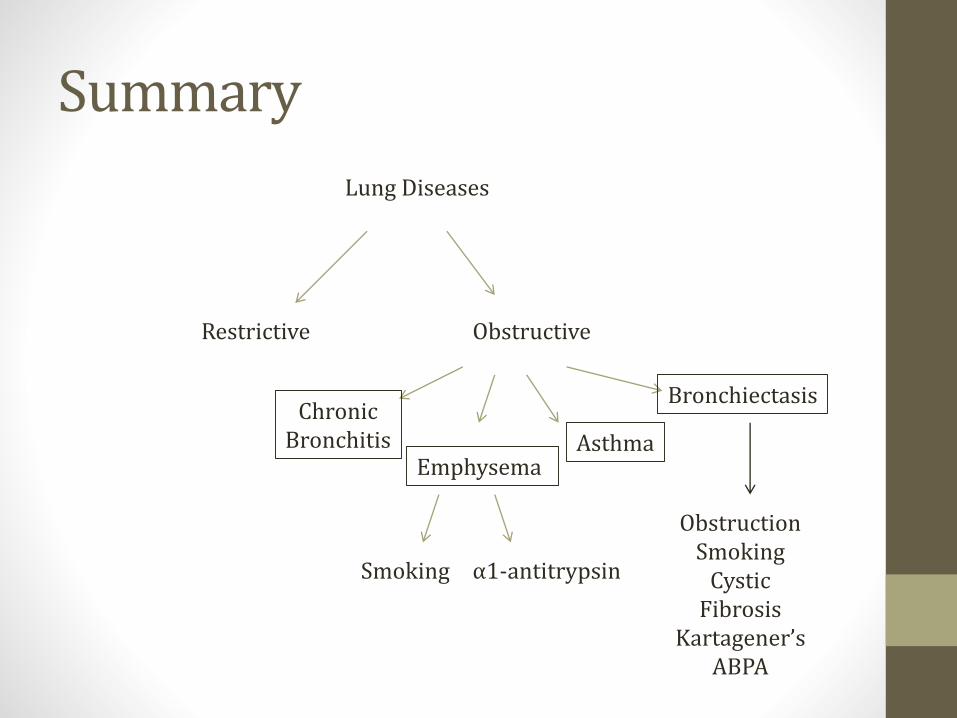
Summary
Lung Diseases
Restrictive Obstructive
ChronicBronchitis
EmphysemaAsthma
Bronchiectasis
ObstructionSmoking
Cystic Fibrosis
Kartagener’sABPA
Smoking α1-antitrypsin
Restrictive Lung DiseaseJason Ryan, MD, MPH

Restrictive Lung Diseases
• Key points: Can’t get air in → less air out
• Reduced FVC (less air in/out)
• Reduced FEV1 (less air in/out)
• Normal (>80%) FEV1/FVC (hallmark)
Causes
• #1: Poor breathing mechanics
• #2: Interstitial lung diseases
Poor Breathing Mechanics
• Not a primary pulmonary issue
• Under-ventilation of lungs
• Alveoli working: A-a gradient normal
• Neuromuscular • ALS, Polio, myasthenia gravis
• Structural• Scoliosis
• Morbid obesity
Interstitial Lung Disease
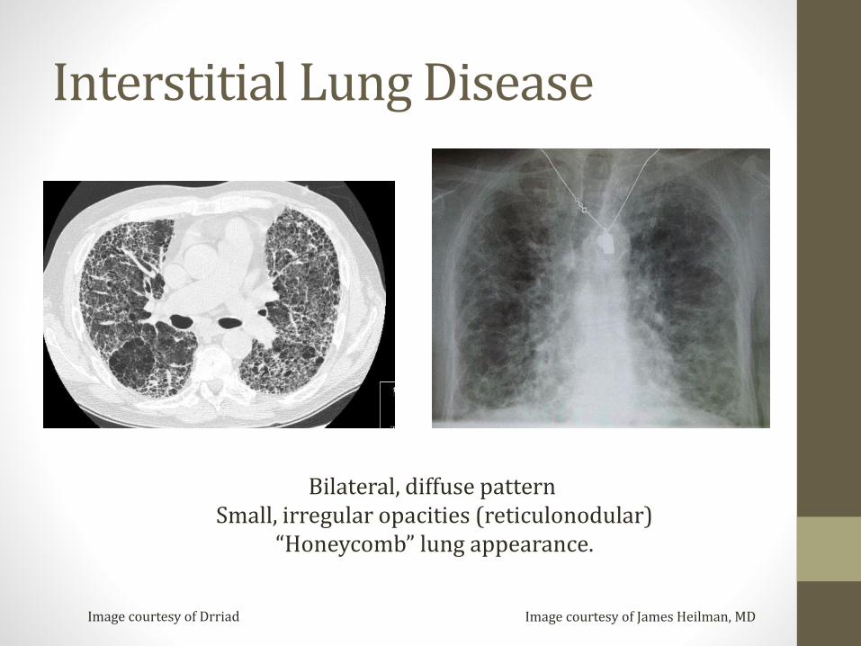
Image courtesy of James Heilman, MDImage courtesy of Drriad
Bilateral, diffuse pattern Small, irregular opacities (reticulonodular)
“Honeycomb” lung appearance.

DLCODiffusing capacity in lung of carbon monoxide
• DLCO separates cases restrictive disease
• Restriction with normal DLCO• Extra-pulmonary cause: obesity
• Restriction with low DLCO• Interstitial lung disease

DLCO
• DLCO = diffusing capacity of carbon monoxide
• Measures ability of lungs to transfer gas to RBCs
• Patient inhales small amount (not dangerous) CO
• CO uptake is diffusion limited• Amount taken up ≈ diffusion function lungs
• Machine measures CO exhaled
• Normal = 75 – 140% predicted
• Severe disease <40% predicted
Low DLCO Conditions
• Interstitial lung disease
• Emphysema
• Abnormal vasculature• Pulmonary hypertension
• Pulmonary embolism
• Prior lung resection
• Anemia• Corrects when adjusted for Hb level
Interstitial Diseases
• “Diffuse parenchymal lung diseases”
• Large group of disorders
• Similar clinical, radiographic, physiologic, or pathologic manifestations

Interstitial Diseases
• Idiopathic pulmonary fibrosis
• Systemic diseases with interstitial lung features• Scleroderma
• Rheumatoid arthritis
• Goodpasture’s
• Wegener’s
• Sarcoidosis
• Pneumoconiosis
• Drug toxicity (amiodarone, methotrexate)
• Hypersensitivity pneumonitis
Idiopathic pulmonary fibrosis
• Most common type: Idiopathic interstitial pneumonia
• Slow onset dyspnea
• Typically affects adults over the age of 40

PneumoconiosisOccupational lung diseases
• Coal miner’s lung
• Silicosis
• Asbestosis
Coal miner’s lung
• Inhalation of coal dust particles
• CXR or Chest CT: • Small, rounded, nodular opacities
• Preference for the upper lobes

Silicosis
• Inhalation of silica in quartz, granite, or sandstone
• Most widespread pneumoconiosis in US
• Foundries (metal production facilities)
• Sandblasting (abrasive blasting)
• Mines
Silicosis
• Macrophages react to silica
• Inflammation → fibroblasts → collagen
• High prevalence of TB• Impaired macrophage killing
• High prevalence of bronchogenic carcinoma
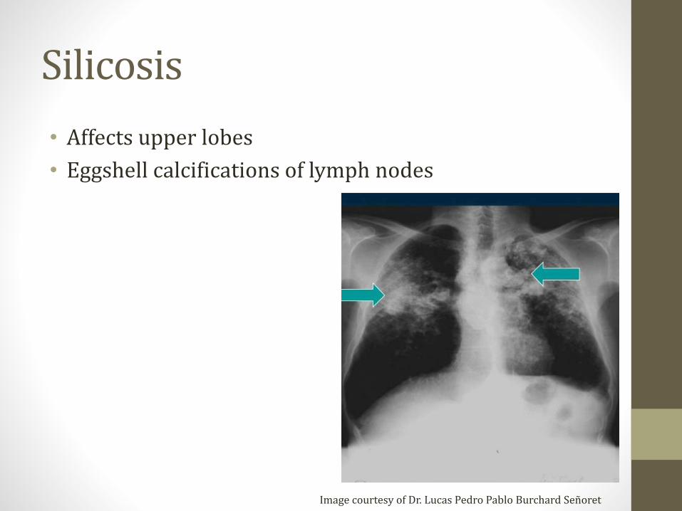
Silicosis
• Affects upper lobes
• Eggshell calcifications of lymph nodes
Image courtesy of Dr. Lucas Pedro Pablo Burchard Señoret
Asbestosis
• Inhalation of asbestos fibers
• Shipbuilding, roofing, plumbing
• Classically affects lower lobes
• Three clinical problems:• Interstitial lung disease (asbestosis)
• Pleural plaques
• Lung cancer

Asbestosis
• CXR: Calcified pleural plaques pathognomonic
• Path: Asbestos bodies (ferruginous body)• Asbestos fibers surrounded by a coating of iron and protein
Image courtesy of www.learningradiology.comImage courtesy of Nephron
Asbestosis
• Bronchogenic carcinoma
• Mesothelioma• Asbestos is the only known risk factor for mesothelioma
• Occurs decades after exposure
• Pleural thickening and pleural effusion
• Slow onset symptoms (dyspnea, cough, chest pain)
• Poor prognosis

Drug toxicity
• Bleomycin
• Busulfan
• Amiodarone
• Methotrexate
Hypersensitivity pneumonitis
• Hypersensitivity reaction to environmental antigen• Agricultural dusts
• Microorganisms (fungal, bacterial, or protozoa)
• Chemicals
• Mixed type III/IV hypersensitivity
• Classic case is a farmer’s lung• Moldy hay, grain exposure
• Also common in bird/poultry handlers• Waste from birds → dried, finely dispersed dust
Hypersensitivity pneumonitis
• Classic case• Farmer or bird handler
• Cough, dyspnea, chest tightness
• Diffuse crackles
• Diagnosis (challenging): • Bronchoalveolar lavage
• Inhalation challenge
• Lung biopsy
• Treatment:• Avoid exposure
• Steroids

Treatment of COPD & AsthmaJason Ryan, MD, MPH

COPD and Asthma Drugs
• Short-acting bronchodilators• Albuterol
• Ipratropium
• Long-acting bronchodilators• Salmeterol, Formoterol
• Tiotropium
• Steroids
β2 Agonists
• Activate adenylate cyclase → ↑cAMP
• Relax bronchiole smooth muscle
• Short acting: Albuterol• Nebulizer or inhaler
• Use during acute attacks (prn)
• Long acting: Salmeterol, Formoterol• Not used as monotherapy (always with ICS)
• Systemic side effects (rare)• Hypertension, arrhythmia
Muscarinic Antagonists
• Vagal nerve → Ach → Bronchoconstriction
• MA drugs block M receptors smooth muscle
• Prevents bronchoconstriction

Muscarinic Antagonists
• Short acting: Ipratropium
• Long acting: Tiotropium
Steroids
• Inhaled: Beclomethasone, Fluticasone, Budesonide
• Oral: Prednisone
• IV: Methylprednisolone (Solumedrol)

Steroids
• Inhibit synthesis of cytokines
• Bind to glucocorticoid receptor (GR)
• Many, many immunosuppressive effects
• ↓ expression many interleukins, IFN-γ, TNF-α, GM-CSF
• Inactivation NF-KB• Transcription factor
• Induces production of TNF-α

Steroids
• Common side effect is oral candidiasis (“thrush”)
• Patients instructed to rinse after inhalation
Image courtesy of James Heilman, MD
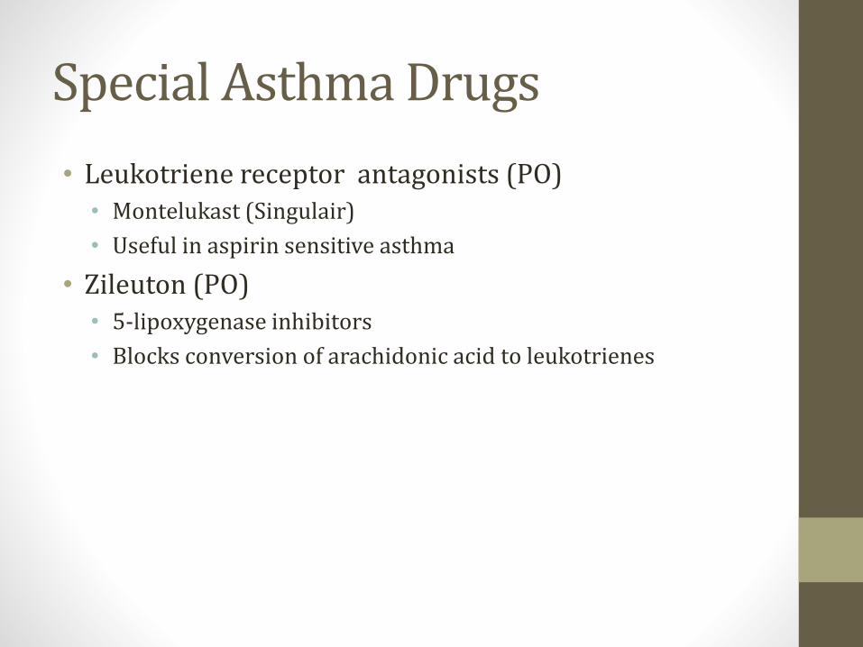
Special Asthma Drugs
• Leukotriene receptor antagonists (PO)• Montelukast (Singulair)
• Useful in aspirin sensitive asthma
• Zileuton (PO)• 5-lipoxygenase inhibitors
• Blocks conversion of arachidonic acid to leukotrienes
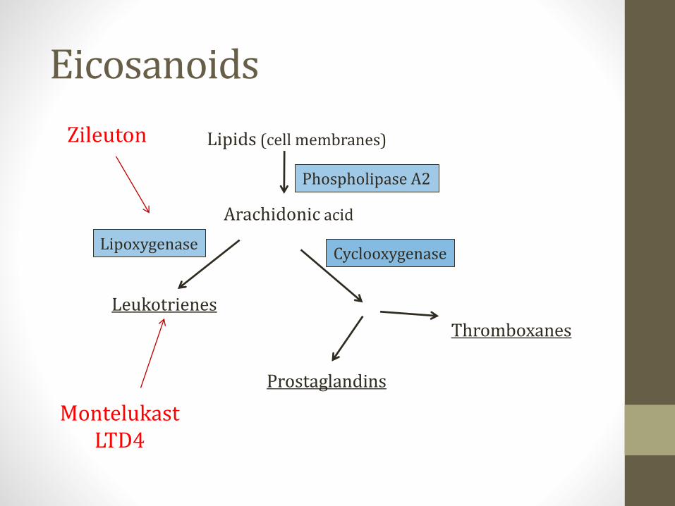
Eicosanoids
Lipids (cell membranes)
Arachidonic acid
Leukotrienes
Lipoxygenase
Thromboxanes
Prostaglandins
Cyclooxygenase
Phospholipase A2
MontelukastLTD4
Zileuton

Special Asthma Drugs
• Omalizumab (SQ injection)• IgG monoclonal antibody
• Inhibits IgE binding to IgE receptor on mast cells & basophils
• Cromolyn (inhaler/nebulizer)• Inhibits mast cell degranulation
• Blocks release of histamine, leukotrienes
Theophylline
• Methylxanthines
• Multiple, complex mechanisms
• Bronchodilation• Likely through inhibition PDE
• Less hydrolysis (breakdown) cAMP
• ↑cAMP
• Also down-regulates inflammatory cell functions
Theophylline
• Narrow therapeutic index
• Levels must be monitored
• Dose must be titrated
• Goal is a peak serum concentration 10 to20mg/L

Theophylline
• Metabolized by P450
• Many drug-drug interactions
• Common culprits:• Cimetidine
• Ciprofloxacin
• Erythromycin
• Clarithromycin
• Verapamil
• St. John’s Wart
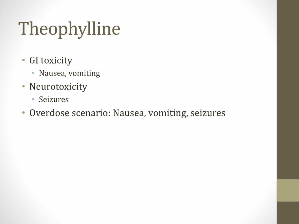
Theophylline
• GI toxicity• Nausea, vomiting
• Neurotoxicity• Seizures
• Overdose scenario: Nausea, vomiting, seizures

Theophylline
• Cardiotoxicity• Blocks adenosine receptors
• Increased heart rate
• Arrhythmias (atrial tachycardia, atrial flutter)
• Cause of death in overdose/poisoning
• Key clinical scenario• Patient on theophylline for asthma/COPD
• SVT
• Adenosine fails to slow heart rate
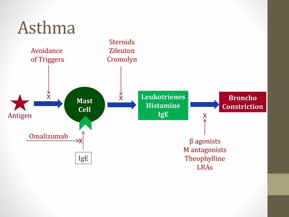
Asthma
Mast Cell
Antigen
LeukotrienesHistamine
IgE
Avoidanceof Triggers
IgE
OmalizumabX
X BronchoConstriction
SteroidsZileuton
Cromolyn
X
β agonistsM antagonistsTheophylline
LRAs
X
Special COPD Drugs
• Theophylline
• Roflumilast (PO)• Phosphodiesterase-4 (PDE-4) inhibitor
• Decreases inflammation
• May relax airway smooth muscle
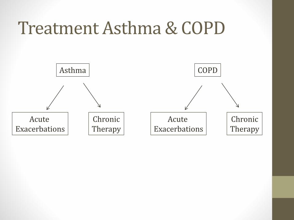
Treatment Asthma & COPD
Asthma
Acute Exacerbations
ChronicTherapy
COPD
Acute Exacerbations
ChronicTherapy

COPD: Acute Exacerbations
• Oxygen
• Nebulized albuterol +/- ipratropium (Combivent)
• IV or oral corticosteroids• Prednisone 60mg daily
• Methylprednisolone 80mg IV q8hrs
• Antibiotics (severe, hospitalized patients)• Fluoroquinolones
• Amoxicillin/clavulanate
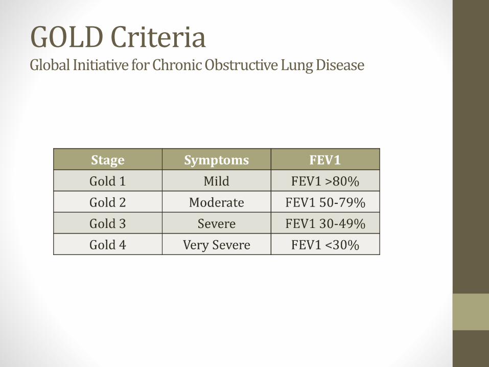
GOLD CriteriaGlobal Initiative for Chronic Obstructive Lung Disease
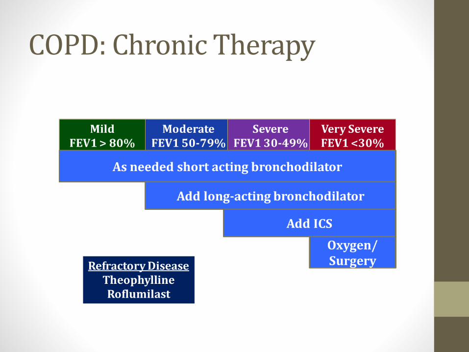
COPD: Chronic Therapy
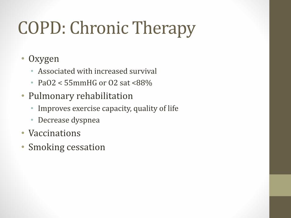
COPD: Chronic Therapy
• Oxygen• Associated with increased survival
• PaO2 < 55mmHG or O2 sat <88%
• Pulmonary rehabilitation• Improves exercise capacity, quality of life
• Decrease dyspnea
• Vaccinations
• Smoking cessation
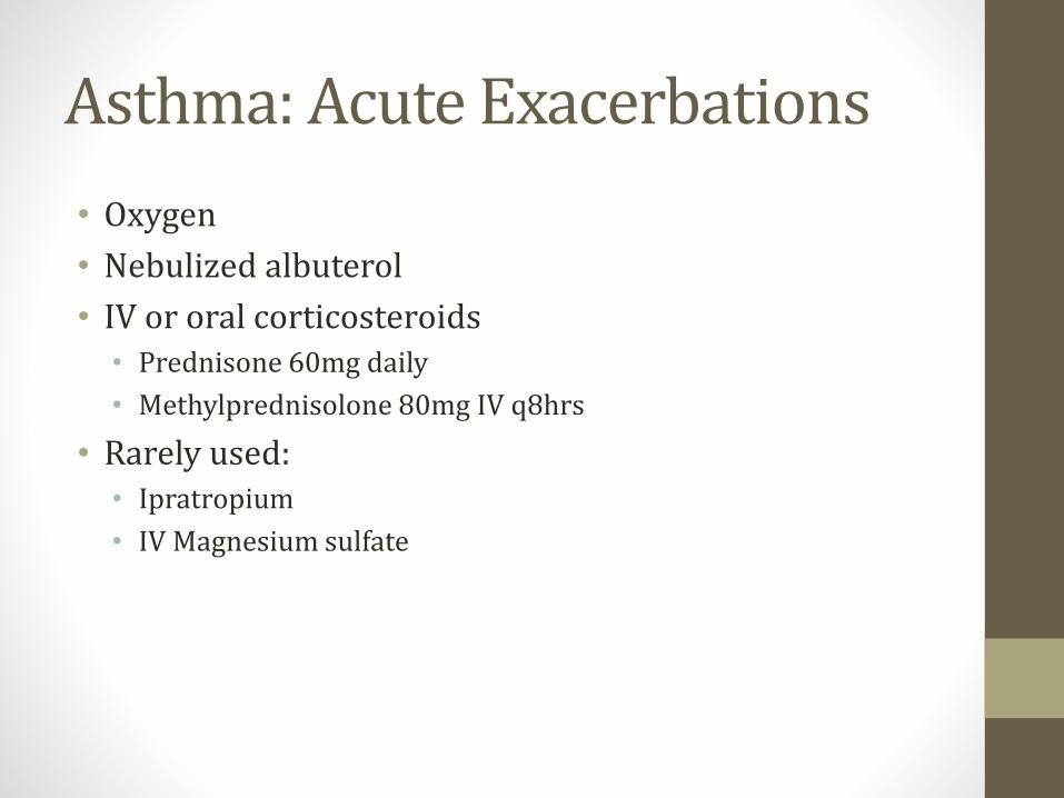
Asthma: Acute Exacerbations
• Oxygen
• Nebulized albuterol
• IV or oral corticosteroids• Prednisone 60mg daily
• Methylprednisolone 80mg IV q8hrs
• Rarely used:• Ipratropium
• IV Magnesium sulfate

Asthma: Chronic Therapy
Surgical Treatment
• For advanced “end-staged” COPD
• Lung volume reduction surgery/Bullectomy• Remove diseased lung tissue
• Allow healthy lung tissue more room to expand
• Lung transplant

PneumoniaJason Ryan, MD, MPH

Pneumonia
• Infection of the lungs
• Three patterns• Lobar
• Bronchopneumonia
• Interstitial (atypical)
Lobar Pneumonia
• Classic form of pneumonia (S. pneumoniae)
• Bacteria acquired in nasopharynx
• Aerosolized to alveolus
• Enter alveolar type II cells
• Pneumococci multiply in alveolus
• Invade alveolar epithelium
• Pass from one alveolus to next (pores of Cohn)
• Inflammation/consolidation of lobes
• Can involve entire lung
Lobar Pneumonia
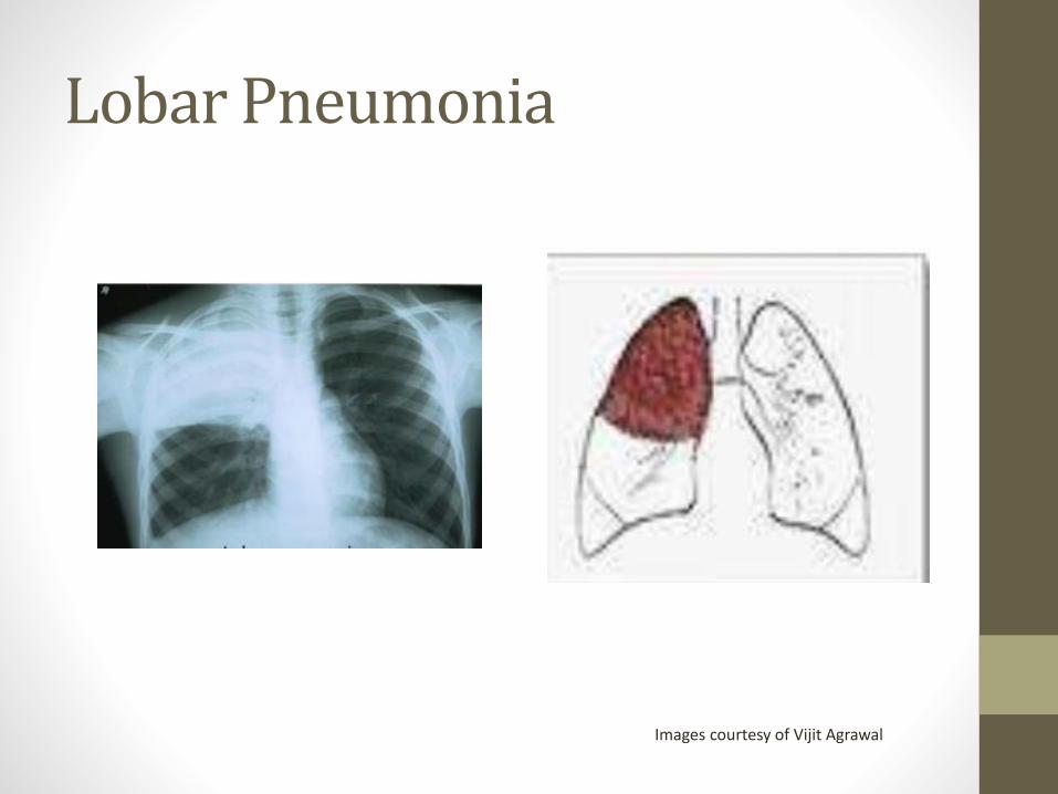
Images courtesy of Vijit Agrawal
Four Lobar Stages
• #1: Congestion (1st 24 hours)• Alveolar capillaries dilate
• Exudate of bacteria develops
• #2: Red hepatization (2-3days)• Exudate of RBCs, neutrophils, fibrin
• “Fresh" exudate: RBCs/WBCs intact
• Pneumococci alive
• Lobes look red
Four Lobar Stages
• #3: Gray hepatization (4-6days)• Gray, firm lobe
• Exudate with neutrophils/fibrin
• RBCs disintegrate
• Dying pneumococci
• #4: Resolution• Return to normal (little scarring)
• Enzymes digests exudate
• Type II pneumocyte key for regeneration
Bronchopneumonia
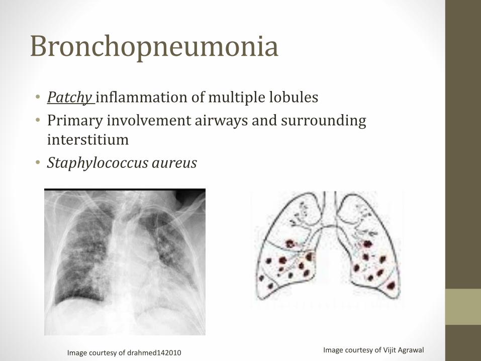
• Patchy inflammation of multiple lobules
• Primary involvement airways and surrounding interstitium
• Staphylococcus aureus
Image courtesy of Vijit AgrawalImage courtesy of drahmed142010
Interstitial Pneumonia
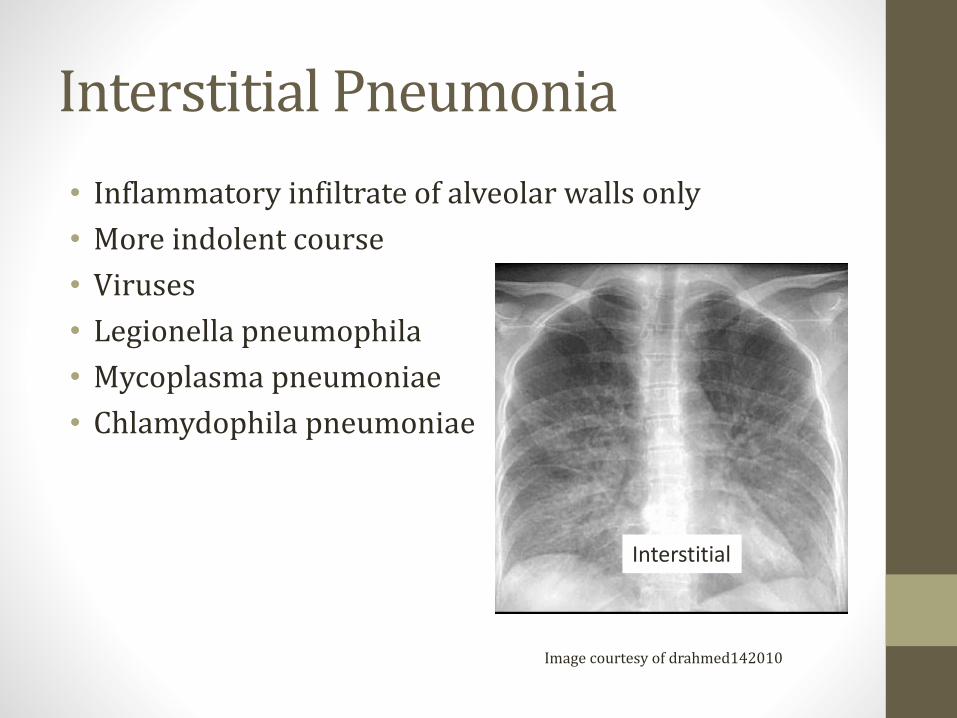
• Inflammatory infiltrate of alveolar walls only
• More indolent course
• Viruses
• Legionella pneumophila
• Mycoplasma pneumoniae
• Chlamydophila pneumoniae
Interstitial
Image courtesy of drahmed142010
Atypical Pneumonia
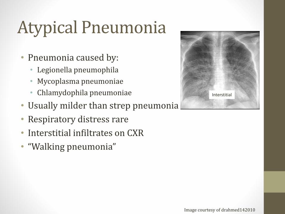
• Pneumonia caused by:• Legionella pneumophila
• Mycoplasma pneumoniae
• Chlamydophila pneumoniae
• Usually milder than strep pneumonia
• Respiratory distress rare
• Interstitial infiltrates on CXR
• “Walking pneumonia”
Interstitial
Image courtesy of drahmed142010
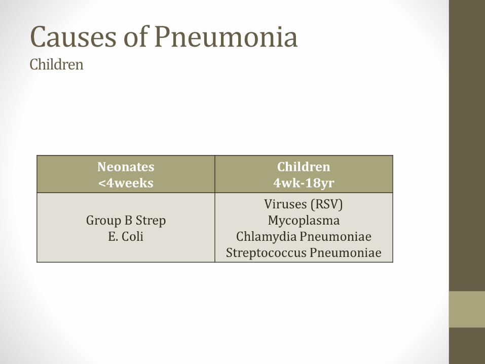
Causes of PneumoniaChildren

Causes of Pneumonia Adults
• S. pneumoniae – most common
• Haemophilus influenzae
• Mycoplasma pneumoniae
• C. pneumoniae
• Legionella
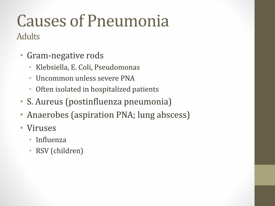
Causes of Pneumonia Adults
• Gram-negative rods• Klebsiella, E. Coli, Pseudomonas
• Uncommon unless severe PNA
• Often isolated in hospitalized patients
• S. Aureus (postinfluenza pneumonia)
• Anaerobes (aspiration PNA; lung abscess)
• Viruses• Influenza
• RSV (children)
Signs/Symptoms
• High Fever
• Cough
• Sputum production
• Elevated WBC
• Pleuritic chest pain

Diagnosis
• Usually:• History
• Physical exam
• X-ray (sometimes CT scan)
• Rarely• Sputum culture
• Bronchoalveolar lavage

Clinical Classes of Pneumonia
• Community acquired• Usually S. Pneumoniae, H. Influenza, S. Aureus
• Sometimes Mycoplasma, Chlamydia, Legionella (atypicals)
• Nosocomial• Bad bugs
• Often gram negatives (Pseudomonas, Klebsiella, E. Coli)
• Hospital Acquired
• Ventilator-associated pneumonia (VAP)
• Healthcare-associated pneumonia (HCAP; nursing homes)
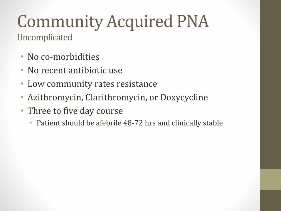
Community Acquired PNAUncomplicated
• No co-morbidities
• No recent antibiotic use
• Low community rates resistance
• Azithromycin, Clarithromycin, or Doxycycline
• Three to five day course• Patient should be afebrile 48-72 hrs and clinically stable
Community Acquired PNAComplicated
• COPD, CKD, Diabetes, CHF, Alcoholism
• Recent antibiotic use
• Fluoroquinolone (levofloxacin)
• Amoxicillin plus azithromycin
Nosocomial PNA
• Lots of resistance to antibiotics
• Gram negative rods• E. coli, Klebsiella, Enterobacter, Pseudomonas, Acinetobacter
• Staph Aureus including MRSA
• Often cover for pseudomonas, MRSA
• Sometimes multi-drug combinations
• Cefepime or Ceftazidime
• Imipenem or Meropenem
• Piperacillin-tazobactam (Zosyn)
Complications
• Sepsis
• Respiratory failure
• Lung abscesses
• Pleural effusion
• ARDS
ARDSAcute Respiratory Distress Syndrome
• Triggered by various lung injuries
• Injury → release of pro-inflammatory cytokines • TNF, interleukins
• Cytokines recruit neutrophils to lungs
• Neutrophils release toxic mediators • Reactive oxygen species, proteases
• Damage to capillary endothelium and alveolar epithelium
• Protein escapes from vascular space
• Fluid pours into the interstitium
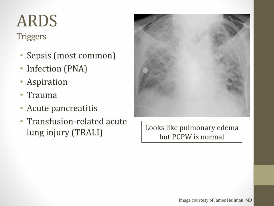
ARDSTriggers
• Sepsis (most common)
• Infection (PNA)
• Aspiration
• Trauma
• Acute pancreatitis
• Transfusion-related acute lung injury (TRALI)
Image courtesy of James Heilman, MD
Looks like pulmonary edemabut PCPW is normal
ARDSTreatment
• Mechanical ventilation
• Low tidal volume
• Supportive care (fluids, nutrition)
• VAP pneumonia is serious complication
Legionella
• First identified at American Legion convention
• Infection from inhalation of aerosolized bacteria• Not airborne
• Outbreaks at hotels with contaminated water
• Can cause nosocomial pneumonia in nursing homes

LegionellaSymptoms
• Initially mild pneumonia symptoms• Fever; mild, slightly productive cough
• Can progress to severe pneumonia
• GI symptoms • Watery diarrhea, nausea, vomiting, and abdominal pain
• Hyponatremia (Na<130 meq/L) common• Can occur in any PNA but more common Legionella

LegionellaDiagnosis
• Special culture requirements• Does not gram stain well
• Buffered charcoal yeast extract agar (BCYE)
• Iron and cysteine added for growth
• Supplemented with antibiotics and silver dyes• Antimicrobials prevent overgrowth by competing organisms
• Dyes give distinctive color to Legionella
• Urinary antigen test• Rapid test available in minutes
• Does not test for all Legionella types
LegionellaDiagnosis
• Classic Case• Mild cough
• Watery diarrhea
• Confusion (low Na)
• Negative bacteria on gram stain
• Diagnose with urinary antigen test
• Treatment: Fluoroquinolone or Azithromycin
Pontiac Fever
• Mild form of Legionella infection
• Fever, malaise, chills, fatigue, and headache
• No respiratory complaints
• Chest radiograph usually normal
Mycoplasma Pneumonia
• Atypical pneumonia
• Can’t see on gram stain (no cell wall)
• Classically causes outbreaks in young adults• College dorm residents
• Military recruits
• CXR looks worse than symptoms
• Can cause autoimmune hemolytic anemia• IgM antibody → RBC antigen
• “Cold” hemolytic anemia
• Stevens-Johnson syndrome
Influenza Virus
• Atypical pneumonia
• Influenza A or B viruses
• Fever, headache, myalgia, and malaise
• Nonproductive cough, sore throat, runny nose
• Major complication is secondary pneumonia• Strep pneumoniae, Staph aureus, H. influenzae
• Worsening symptoms after initial improvement
• Cause of death

CMV
• Pneumonia in transplant patients on immunosuppressive drugs
• “Owl eye” intranuclear inclusions
RSVRespiratory Syncytial Virus
• Viral respiratory infection in infants
• Often seasonal outbreaks (Nov – April)
• Most common cause lower respiratory tract illness in children• Bronchiolitis, pneumonia, acute respiratory failure
• Often starts as upper airway infection• Runny nose
• Few days later, lower tract symptoms• Wheezing often present
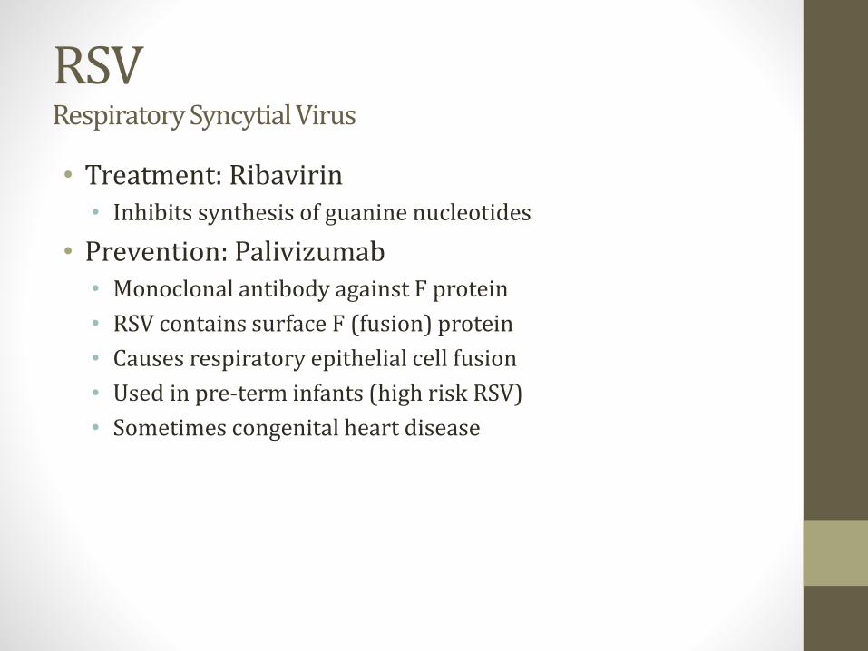
RSVRespiratory Syncytial Virus
• Treatment: Ribavirin• Inhibits synthesis of guanine nucleotides
• Prevention: Palivizumab• Monoclonal antibody against F protein
• RSV contains surface F (fusion) protein
• Causes respiratory epithelial cell fusion
• Used in pre-term infants (high risk RSV)
• Sometimes congenital heart disease

RSVRespiratory Syncytial Virus
• Classic case• Young child (often <2yo)
• Fever, runny nose
• Few days later, cough, wheezing
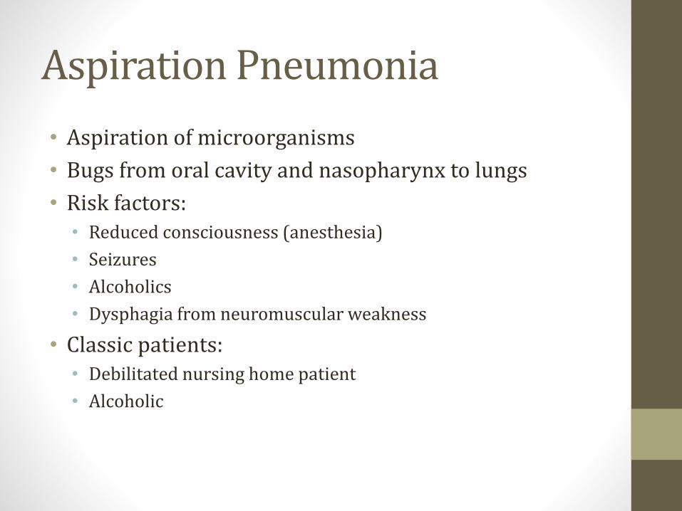
Aspiration Pneumonia
• Aspiration of microorganisms
• Bugs from oral cavity and nasopharynx to lungs
• Risk factors: • Reduced consciousness (anesthesia)
• Seizures
• Alcoholics
• Dysphagia from neuromuscular weakness
• Classic patients:• Debilitated nursing home patient
• Alcoholic

Aspiration Pneumonia
• Klebsiella
• Staph Aureus
• Anaerobic bacteria • Peptostreptococcus
• Fusobacterium
• Prevotella
• Bacteroides
• Clindamycin first-line therapy
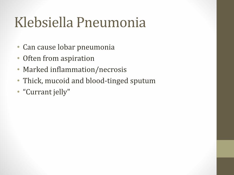
Klebsiella Pneumonia
• Can cause lobar pneumonia
• Often from aspiration
• Marked inflammation/necrosis
• Thick, mucoid and blood-tinged sputum
• “Currant jelly"
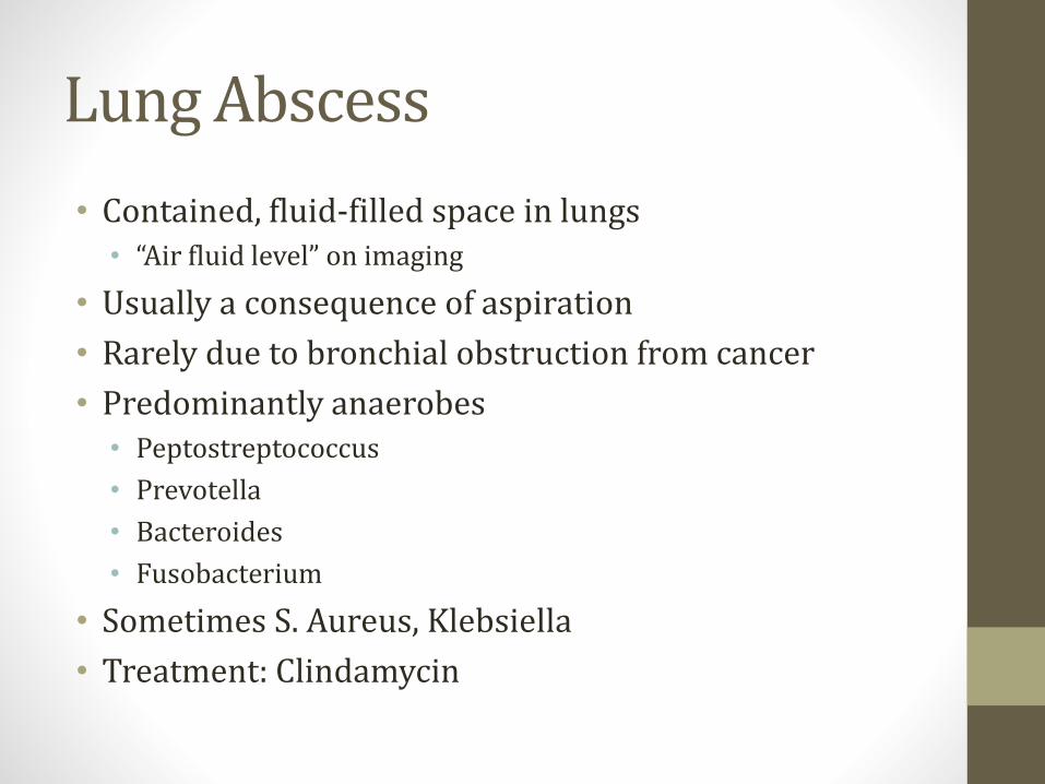
Lung Abscess
• Contained, fluid-filled space in lungs• “Air fluid level” on imaging
• Usually a consequence of aspiration
• Rarely due to bronchial obstruction from cancer
• Predominantly anaerobes• Peptostreptococcus
• Prevotella
• Bacteroides
• Fusobacterium
• Sometimes S. Aureus, Klebsiella
• Treatment: Clindamycin

PCPPneumocystis jirovecii
• Diffuse interstitial pneumonia
• Requires immunocompromise• Classically HIV
• AIDS-defining illness
• Yeast → inhaled• Usually no symptoms if immune system intact

PCPPneumocystis jirovecii
• Diagnosed by microscopy• Sputum sample or BAL
• Staining required → cannot be cultured
• Special stains used• Silver stains often used
Image courtesy of Yale Rosen

PCPPneumocystis jirovecii
• Treatments• TMP-SMX (first line)
• Dapsone
• Pentamidine
• Prophylaxis• TMP-SMX when CD4 <200cells/microL
• High dose steroid or other immunosuppressant
Pleural DiseaseJason Ryan, MD, MPH
What are the pleura?
• Two layers of tissue surrounding lungs• Visceral pleura – attached to lung
• Parietal pleura – attached to chest wall
• Pleural space/cavity – between layers
• Pleural lined by mesothelial cells
• Secrete small amount pleural fluid for lubrication

Pneumothorax
• Air in pleural space
• Two types to know about• Spontaneous
• Tension
Spontaneous PTX
• Primary• Rupture of subpleural bleb
• Common in tall, thin young males
• Secondary• Older patients with underlying pulmonary disease
• COPD

Spontaneous PTX
• Sudden onset dyspnea
• Sometimes pleuritic chest pain
• CXR for diagnosis
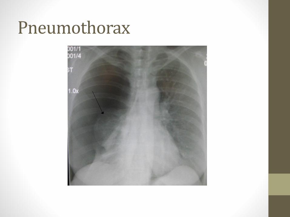
Pneumothorax
PneumothoraxTreatment
• 100% Oxygen• Displaces nitrogen from capillary blood
• ↑gradient for nitrogen reabsorption from pleural space
• Chest tube• Larger pneumothoraces (>15% lung volume)

Tension PTX
• Usually from trauma
• Air enters pleural space but cannot leave
• Medical emergency
• Emergent thoracentesis/chest tube placement
• Trachea deviates AWAY from affected side
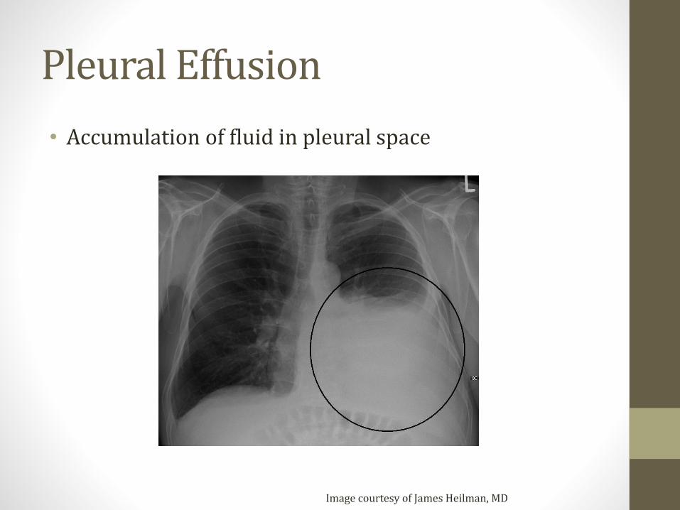
Pleural Effusion
• Accumulation of fluid in pleural space
Image courtesy of James Heilman, MD
Pleural Effusion
• Three general etiologies• Transudative
• Exudative
• Lymphatic

Transudative Effusion
• Something driving fluid into pleural space
• Most commonly due to CHF (High pressure)
• Other causes:• Nephrotic syndrome (low protein)
• Cirrhosis (low albumin)
• Mostly fluid in effusion
• Very little protein in effusion
• Usually treat for underlying cause (no drainage)

Exudative Effusion
• Fluid leaking into pleural space• High vascular permeability
• Many causes
• Malignancy
• Pneumonia
• More protein in pleural fluid vs. transudative
• Usually requires drainage
Transudate vs. Exudate
• Thoracentesis to obtain fluid sample
• Test for protein, LDH
• Light’s Criteria – Exudate if:• Pleural protein/serum protein greater than 0.5
• Pleural LDH/serum LDH greater than 0.6
• Pleural LDH greater than 2/3 upper limits normal LDH
Lymphatic Effusions“Chylothorax”
• Lymphatic fluid effusion
• From thoracic duct obstruction/injury
• Malignancy most common cause
• Trauma (usually surgical)
• Milky-appearing fluid
• Very high triglycerides• TG usually > 110 mg/dL
Other Effusions
• Hemothorax• High Hct in fluid
• Empyema• Infected pleural fluid
• Pus, putrid odor, positive culture
• Malignant effusion• Positive cytology

Mesothelioma
• Pleural tumor
• Asbestos is only known risk factor• Decades after exposure
• Imaging: Pleural thickening and pleural effusion
• Slow onset symptoms (dyspnea, cough, chest pain)
• Poor prognosis• Median survival 4 to 13 months untreated
• 6 to 18 months treated with chemo

Lung CancerJason Ryan, MD, MPH
Common Cancers
• Breast
• Prostate
• Lung (most deadly)
• Colorectal
Lung Cancer Risk Factors
• Cigarette smoking• Polycyclic Aromatic Hydrocarbons (PAHs)
• Radiation Therapy • Hodgkin's and breast cancer survivors
• Environmental toxins• Asbestos
• Radon
Symptoms
• Usually advanced at presentation
• Cough, dyspnea, rarely hemoptysis
• Usually leads to chest imaging
Diagnosis
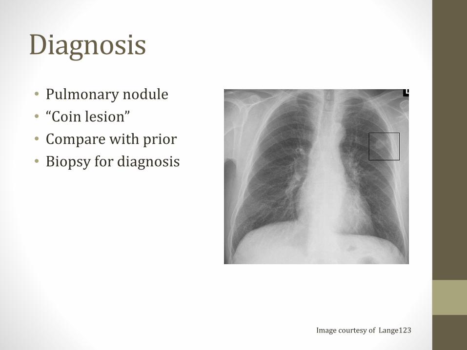
• Pulmonary nodule
• “Coin lesion”
• Compare with prior
• Biopsy for diagnosis
Image courtesy of Lange123

Benign Pulmonary Nodules
• Granulomas (80% benign nodules)
• Hamartomas• Lung tissue and cartilage (with scattered calcification)

Granulomas
• Fungi • Histoplasmosis (patient from Midwest, Miss/Ohio river valley)
• Coccidioidomycosis (southwest, California)
• Mycobacteria • Usually tuberculosis

Lung Cancers
• Small cell (15%)• Fast growing; Early mets
• Non amenable to surgical resection
• Smokers
• Treated with chemo
• Poor prognosis
• Non-small cell (Most Common: 85%)• Can sometimes be resected
• Better prognosis
• Smokers and non-smokers

Small Cell Cancer
• Poorly differentiated small cells
• Classic in male smokers
• Neuroendocrine tumor
• Central tumor
Image courtesy of Wikipedia
Small Cell CancerParaneoplastic Syndromes
• ACTH• Cushing syndrome
• Progressive obesity
• Hyperglycemia
• ADH• SIADH
• Hyponatremia (confusion)
• Antibodies • Antibodies against pre-synaptic Ca channels in neurons
• Block release of acetylcholine
• Lambert-Eaton syndrome
• Main symptom is weakness
Non-Small Cell Cancers
• Squamous Cell Carcinoma
• Adenocarcinoma
• Large cell carcinoma
• Bronchioloalveolar Carcinoma
• Carcinoid tumor
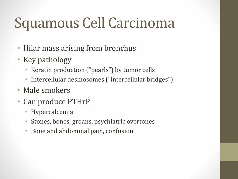
Squamous Cell Carcinoma
• Hilar mass arising from bronchus
• Key pathology• Keratin production (“pearls”) by tumor cells
• Intercellular desmosomes ("intercellular bridges")
• Male smokers
• Can produce PTHrP• Hypercalcemia
• Stones, bones, groans, psychiatric overtones
• Bone and abdominal pain, confusion
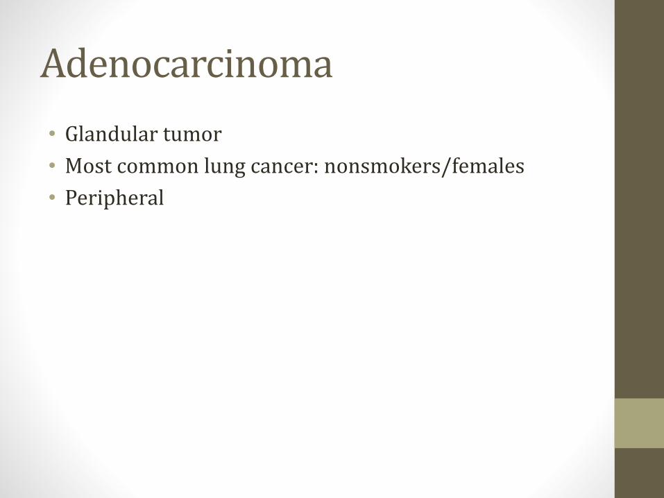
Adenocarcinoma
• Glandular tumor
• Most common lung cancer: nonsmokers/females
• Peripheral
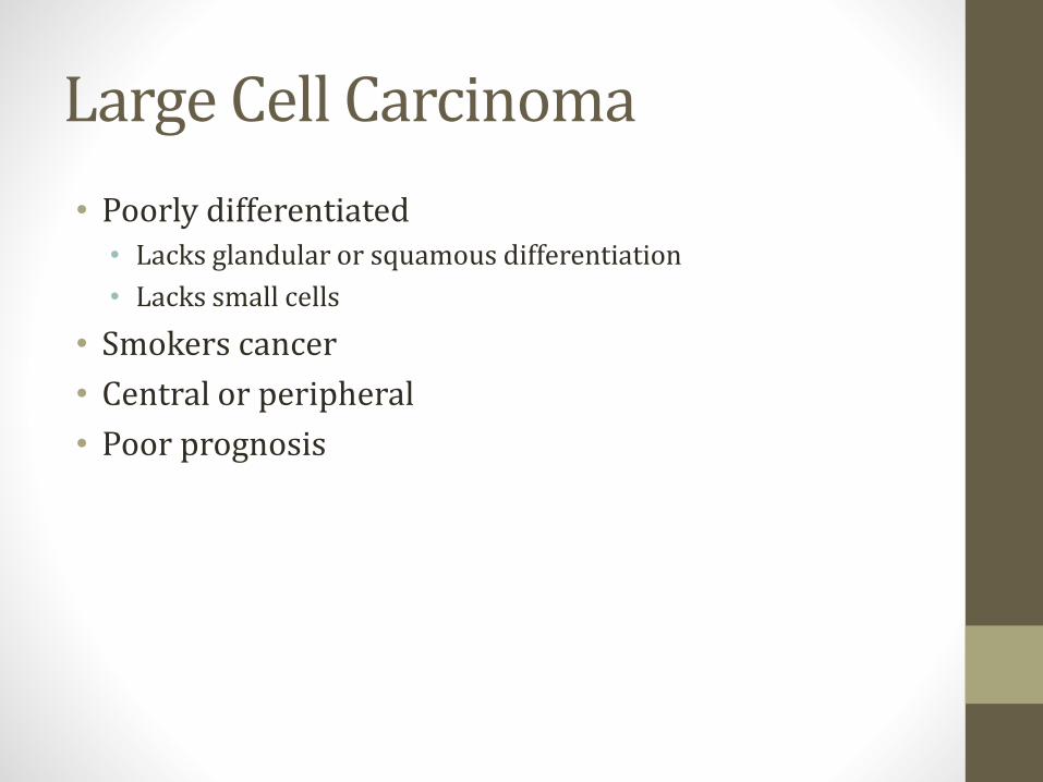
Large Cell Carcinoma
• Poorly differentiated• Lacks glandular or squamous differentiation
• Lacks small cells
• Smokers cancer
• Central or peripheral
• Poor prognosis
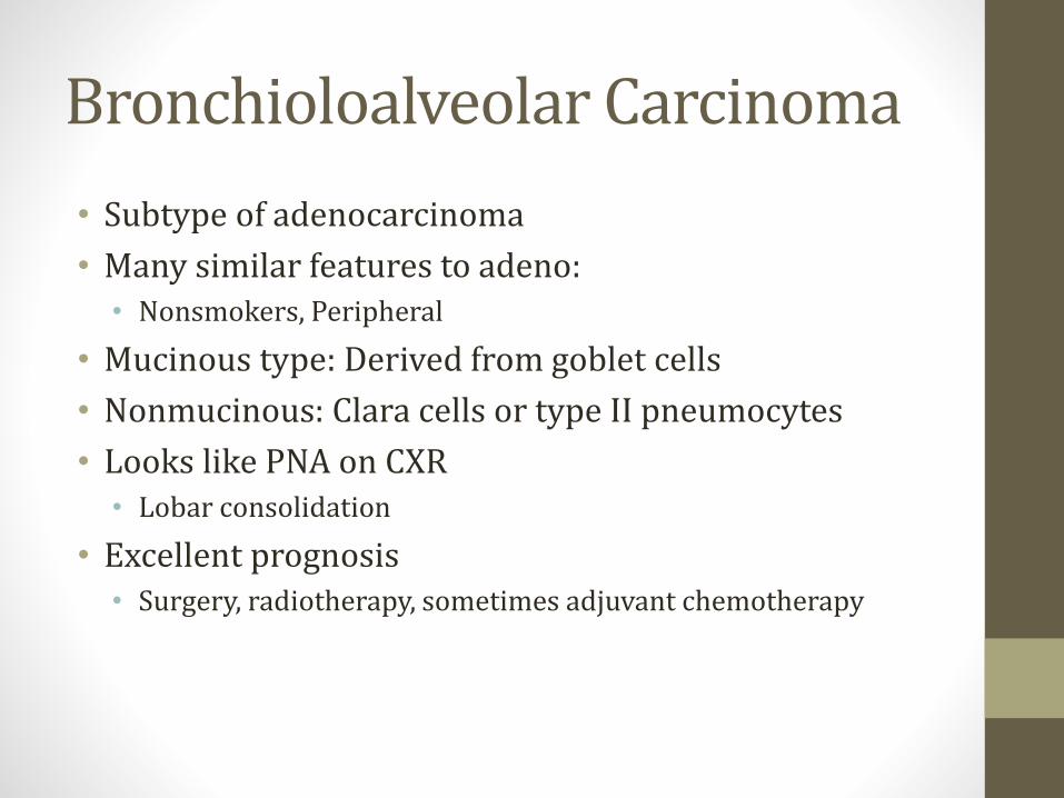
Bronchioloalveolar Carcinoma
• Subtype of adenocarcinoma
• Many similar features to adeno:• Nonsmokers, Peripheral
• Mucinous type: Derived from goblet cells
• Nonmucinous: Clara cells or type II pneumocytes
• Looks like PNA on CXR• Lobar consolidation
• Excellent prognosis• Surgery, radiotherapy, sometimes adjuvant chemotherapy

Carcinoid tumor
• Neuroendocrine
• Well-differentiated cells
• Chromogranin positive
• Non-smokers
• Rarely causes carcinoid syndrome• Secretion of serotonin
• Flushing, diarrhea

Complications
• Pleural effusions• Tap fluid, send for cytology
• Phrenic nerve compression• Diaphragm paralysis
• Dyspnea
• Hemidiaphragm elevated on CXR
• Sniff test
• Recurrent laryngeal nerve compression• Hoarseness
SVC Syndrome
• Obstruction of blood flow through SVC
• Can be caused by compression from tumor• Lung Masses: NSCLC, SCLC
• Mediastinal Masses: Lymphoma
• Other causes include thrombosis• Indwelling catheters, pacemaker wires
• Facial swelling or head fullness
• Arm swelling
• Can cause increased ICP• Headaches, confusion, coma
• Cranial artery rupture
SVC Syndrome
• Usually diagnosed CXR or CT Chest
• Various treatment options:• Anticoagulation for thrombus
• Steroids (lymphoma)
• Chemo/Radiation
• Endovascular stenting
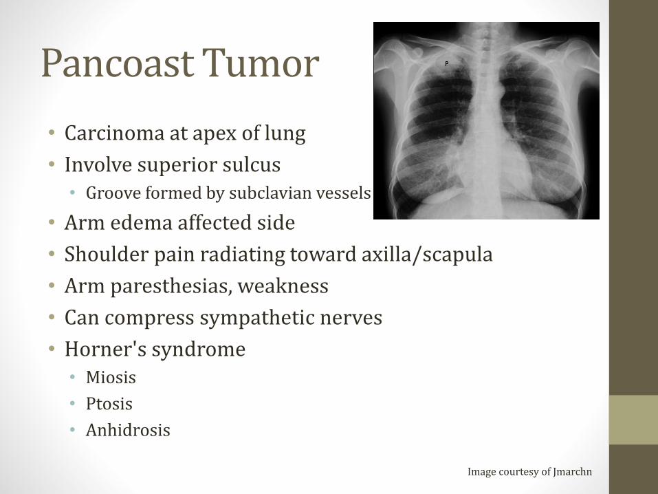
Pancoast Tumor
• Carcinoma at apex of lung
• Involve superior sulcus• Groove formed by subclavian vessels
• Arm edema affected side
• Shoulder pain radiating toward axilla/scapula
• Arm paresthesias, weakness
• Can compress sympathetic nerves
• Horner's syndrome• Miosis
• Ptosis
• Anhidrosis
Image courtesy of Jmarchn

Metastasis from Lung Cancer
• Adrenals• Usually found on imaging without symptoms
• Brain• Headache, neuro deficits, seizures
• Bone• Pathologic fractures
• Liver • Hepatomegaly, jaundice

Metastasis to Lung
• More common than primary lung tumors
• Most commonly from breast or colon cancer
• Usually multiple lesions on imaging

Sleep ApneaJason Ryan, MD, MPH
Sleep Apnea
• Apnea = cessation of breathing
• Sleep apnea = cessation of breathing during sleep
• Usually >10 seconds
• Multiple episodes per night are typical
Sleep Apnea Symptoms
• Unrestful sleep
• Daytime somnolence
• Loud snoring
Sleep Apnea Types
• Central sleep apnea• No effort to breathe
• Obstructive sleep apnea• Decreased air flow despite effort to breathe
Central Sleep Apnea
• Patients with marginal ventilation when awake• Hypoventilate when awake
• Fall asleep → apnea periods
• Central nervous system disease (encephalitis)
• Neuromuscular diseases (polio, ALS)
• Severe kyphoscoliosis
• Narcotics

Central Sleep Apnea
• Cheyne-Stokes breathing• Cyclic breathing
• Delayed detection/response to changes in PaCO2
• Common in heart failure and stroke patients
Normal Breathing Cheyne-Stokes
Images courtesy of Aleksa Lukic
Obstructive Sleep Apnea
• Recurrent soft tissue collapse in the pharynx
• Strongest risk factor is obesity
Sleep Apnea Complications
• HTN
• Pulmonary HTN
• Arrhythmias
• Sudden death
Erythropoiesis
• Chronic hypoxia
• EPO release

Sleep Apnea Diagnosis
• Polysomnography
• “Sleep study”
• Patient sleeps in monitored setting
• EEG, eye movements
• O2 level, HR, respiratory rate
• Number of apnea episodes recorded

Sleep Apnea Treatments
• Weight loss• Takes time; not best option for exhausted patients
• CPAP• First line for symptomatic patients
• Upper airway surgery• Severe disease
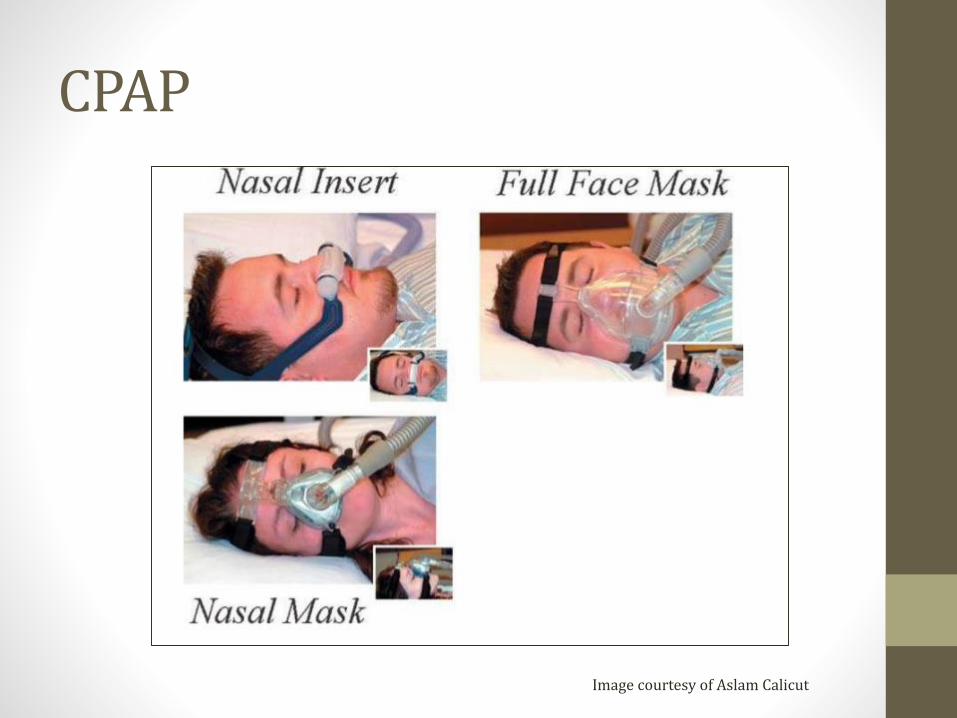
CPAP
Image courtesy of Aslam Calicut

Cystic FibrosisJason Ryan, MD, MPH
Cystic Fibrosis
• Inherited genetic disease• Autosomal recessive pattern
• Both parents must be carriers
• Results in thick, sticky mucus in lungs/GI tract
• Common cause chronic lung disease in children

CFTRCystic Fibrosis Transmembrane Regulator
• CFTR protein is abnormal in CF
• CFTR gene encodes for the abnormal protein
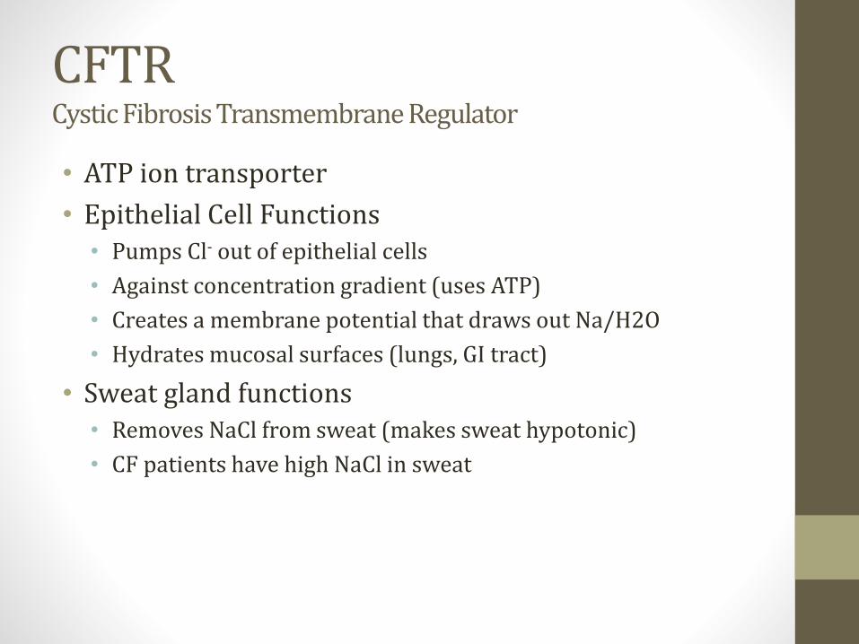
CFTRCystic Fibrosis Transmembrane Regulator
• ATP ion transporter
• Epithelial Cell Functions• Pumps Cl- out of epithelial cells
• Against concentration gradient (uses ATP)
• Creates a membrane potential that draws out Na/H2O
• Hydrates mucosal surfaces (lungs, GI tract)
• Sweat gland functions• Removes NaCl from sweat (makes sweat hypotonic)
• CF patients have high NaCl in sweat
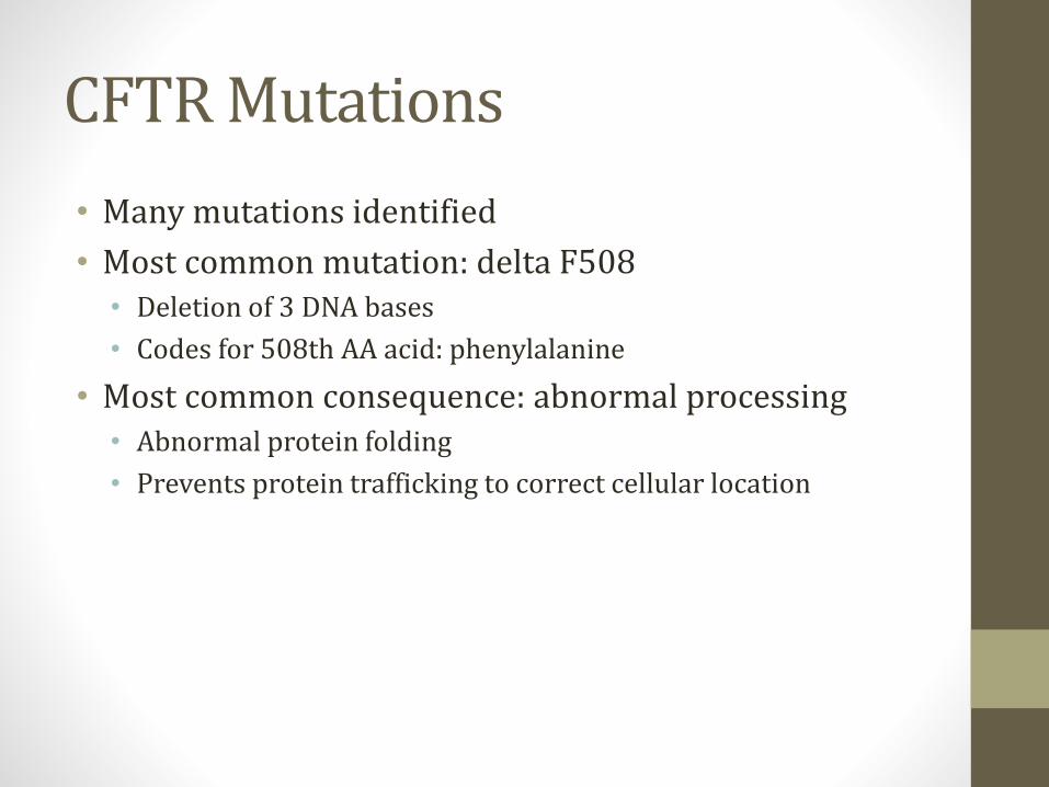
CFTR Mutations
• Many mutations identified
• Most common mutation: delta F508• Deletion of 3 DNA bases
• Codes for 508th AA acid: phenylalanine
• Most common consequence: abnormal processing• Abnormal protein folding
• Prevents protein trafficking to correct cellular location
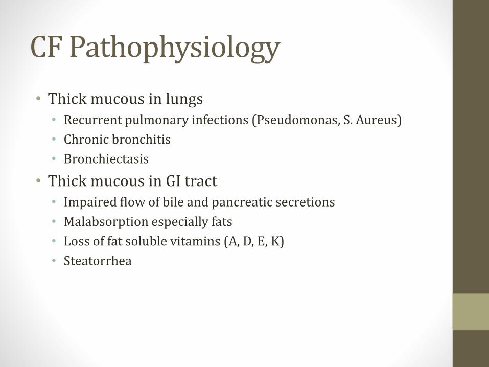
CF Pathophysiology
• Thick mucous in lungs• Recurrent pulmonary infections (Pseudomonas, S. Aureus)
• Chronic bronchitis
• Bronchiectasis
• Thick mucous in GI tract• Impaired flow of bile and pancreatic secretions
• Malabsorption especially fats
• Loss of fat soluble vitamins (A, D, E, K)
• Steatorrhea
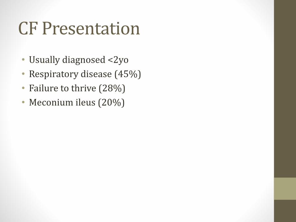
CF Presentation
• Usually diagnosed <2yo
• Respiratory disease (45%)
• Failure to thrive (28%)
• Meconium ileus (20%)

CF Lung Disease
• Productive cough
• Hyperinflation of lungs on CXR
• Obstructive pattern
• Later disease• Chronic bronchitis
• Bronchiectasis
• Acute exacerbations
• Pseudomonas aeruginosa: major pathogen in CF
Pancreatic insufficiency
• Chronic pancreatitis
• CF-related diabetes
• Fat malabsorption
• Steatorrhea: • Frequent stools
• Foul-smelling stools
• Oily or greasy
• Stools may float
Pancreatic insufficiency
• Deficiencies of fat-soluble vitamins: A, D, E, and K
• Vitamin K: coagulopathy
• Vitamin D: rickets
• Vitamin A: Night blindness
• Vitamin E: Ataxia, hemolysis
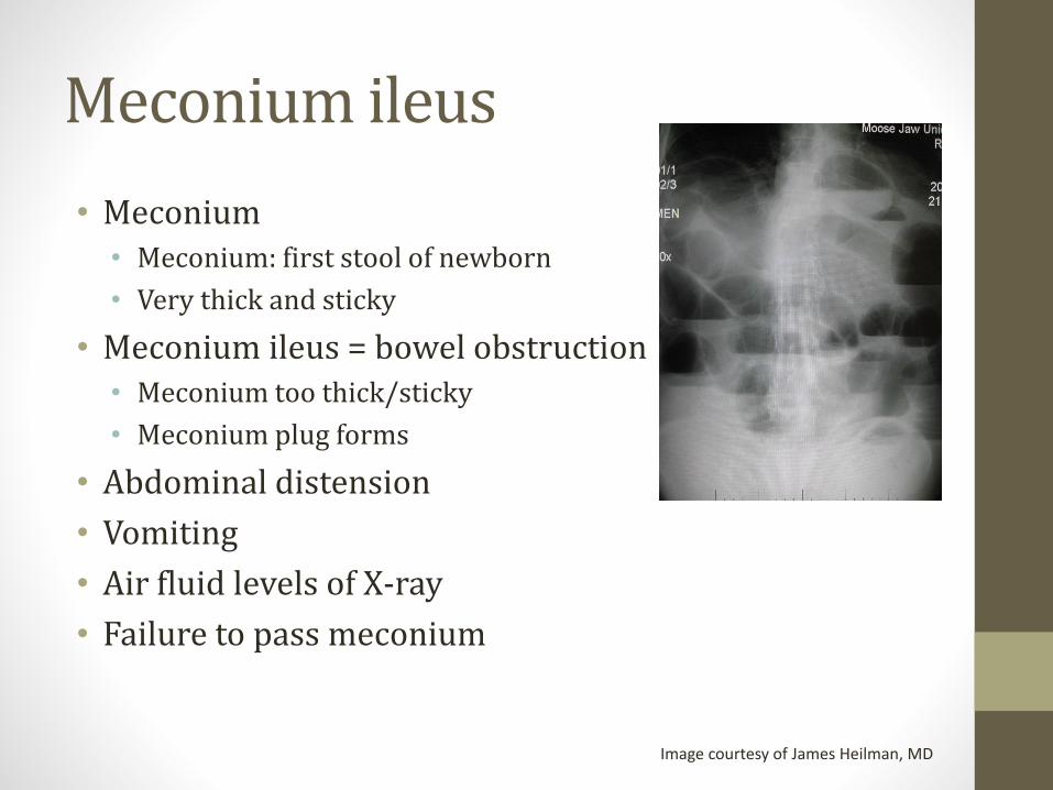
Meconium ileus
• Meconium• Meconium: first stool of newborn
• Very thick and sticky
• Meconium ileus = bowel obstruction • Meconium too thick/sticky
• Meconium plug forms
• Abdominal distension
• Vomiting
• Air fluid levels of X-ray
• Failure to pass meconium
Image courtesy of James Heilman, MD
Other symptoms
• Biliary disease• Bile duct obstruction
• Pale or clay colored stool
• Elevation of LFTs
• Hepatomegaly
• Cirrhosis
• Gallstones
Other symptoms
• Infertility• 95 percent males with CF are infertile
• Absent vas deferens
• Problem is sperm transport not spermatogenesis
• Can have children with assisted techniques
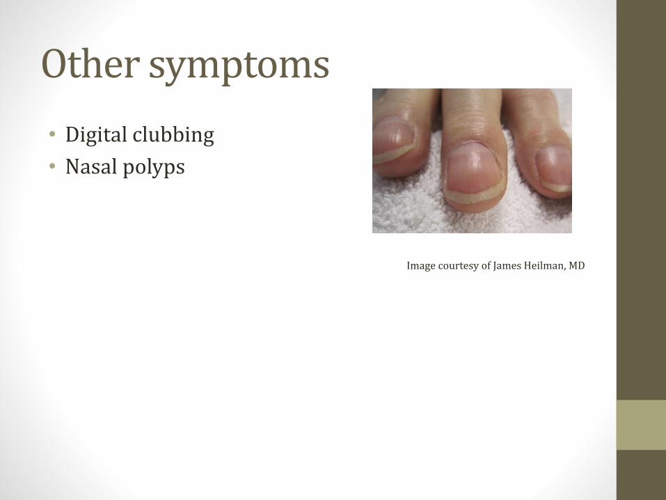
Other symptoms
• Digital clubbing
• Nasal polyps
Image courtesy of James Heilman, MD
Diagnosis
• Sweat chloride test
• Pilocarpine iontophoresis
• Pilocarpine gauze placed on skin
• Electrode placed over gauze
• Small electrical current drives pilocarpine into skin
• Sweating occurs
• Sweat collected on filter paper
• Chloride content analyzed
• High chloride level suggests CF
• DNA testing done if sweat test abnormal
Diagnosis
• Rare CF patients have negative sweat test• Usually have milder disease
• Often recurrent pulmonary and sinus infections
• If symptoms highly suggestive, can test nasal transepithelial potential difference• Measure nasal voltage
• CF patients: more negative voltage
• Due to abnormal sodium processing

Treatment
• Promote clearance of airway secretions• Inhaled DNase (dornase alfa)
• Inhaled saline
• N-acetylcysteine
• Ivacaftor (tablets)• Increased chloride ion flux
• Only for patients with G551D mutation
• Exacerbations are treated with antibiotics
• Lung transplantation
Other Treatments
• Pancreatic enzyme replacement
• Vitamins (A, D, E, K)
• Vaccinations
Prognosis
• Average life span ~ 37 years
• Death from lung complications
Screening
• Prenatal• Test for 23 most common CF mutations in US
• Often test mother first and stop if negative
• Newborn• ↑ blood levels immunoreactive trypsinogen (IRT)
• Blood test → if positive → sweat test
TuberculosisJason Ryan, MD, MPH

Tuberculosis
• Ancient disease: Found in mummies!
• Old name: Consumption
• Tubercle = round nodule
• Tuberculosis = multiple round nodules
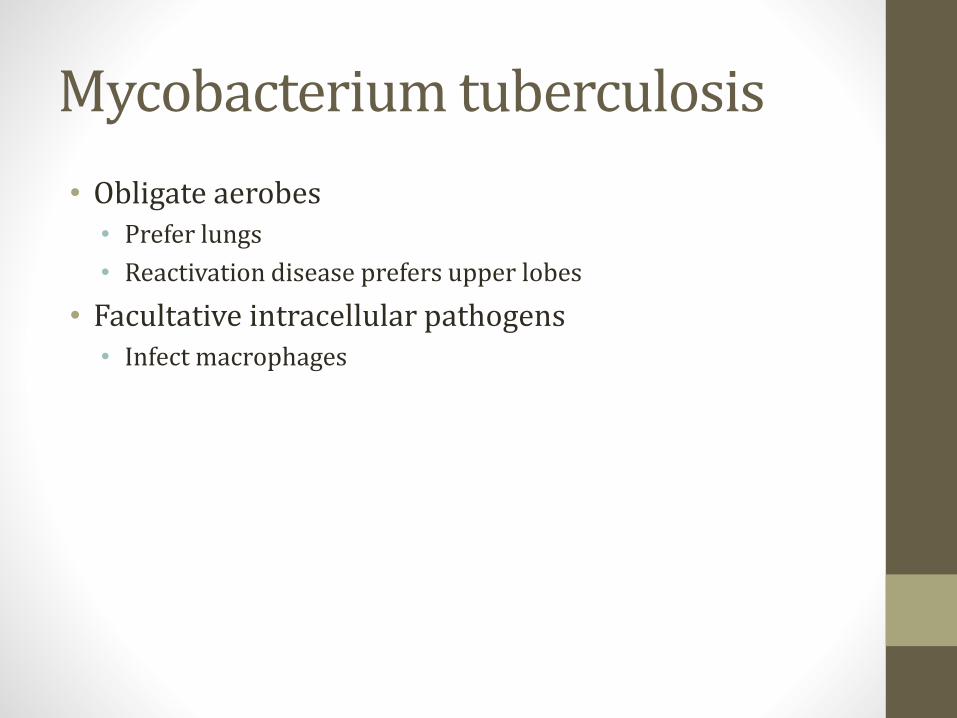
Mycobacterium tuberculosis
• Obligate aerobes• Prefer lungs
• Reactivation disease prefers upper lobes
• Facultative intracellular pathogens• Infect macrophages

Culture of TB
• Difficult to culture• Special media used
• Lowenstein-Jensen agar
• Slow growing
• Does not stain well with Gram stain
• This is due to mycolic acids in cell wall• Also fatty acids and complex lipids
Acid Fast
• Cell walls impermeable to many dyes
• Stain with very concentrated dyes plus heat• Lipid soluble; contain phenols
• Once stained, plate rinsed with acid decolorizer• “Acid fast stain”
• TB resists decolorization with acid solvents
• Some other bacteria (Nocardia) also do this

Virulence Factors
• Trehalose dimycolate ("cord factor")• Helps evade immune response
• Causes granuloma formation
• Triggers cytokine release
• Sulfatides• Glycolipids
• Inhibits fusion of phagosomes/lysosomes
• Catalase-peroxidase• Resists host cell oxidation

Spread of TB
• Spreads through the air
• Active TB patient’s cough, sneeze, etc.
• Inhaled by uninfected person
• Can spread rapidly in crowded areas

Exposure to TB
• Most patients will not develop active disease• Infection can clear or remain “latent”
• Small proportion patients develop active disease

Primary TBClinical Picture
• Mainly a disease of childhood or chemo patients• Ineffective immune response
• Gradual onset: weeks
• Fever
• Cough
• Pleuritic chest pain
• Fatigue, arthralgias
Primary TBPathophysiology
• First week• TB infects macrophages
• Phagocytosed
• Intracellular bacterial proliferation

Primary TBPathophysiology
• Two to four weeks• Cell-mediated immune system controls TB
• TH1 response
• Activation of CD4+ T cells
• Interferon-γ secreted
• Activated macrophages and cytotoxic T lymphocytes

Granulomas
• Granulomatous inflammation
• Caseating necrosis
• Macrophages transform to :• Epithelioid cells
• Langhans giant cells
• Fibroblasts activated → collagen
• T-cell mediated delayed type hypersensitivity reaction• Type IV hypersensitivity reaction
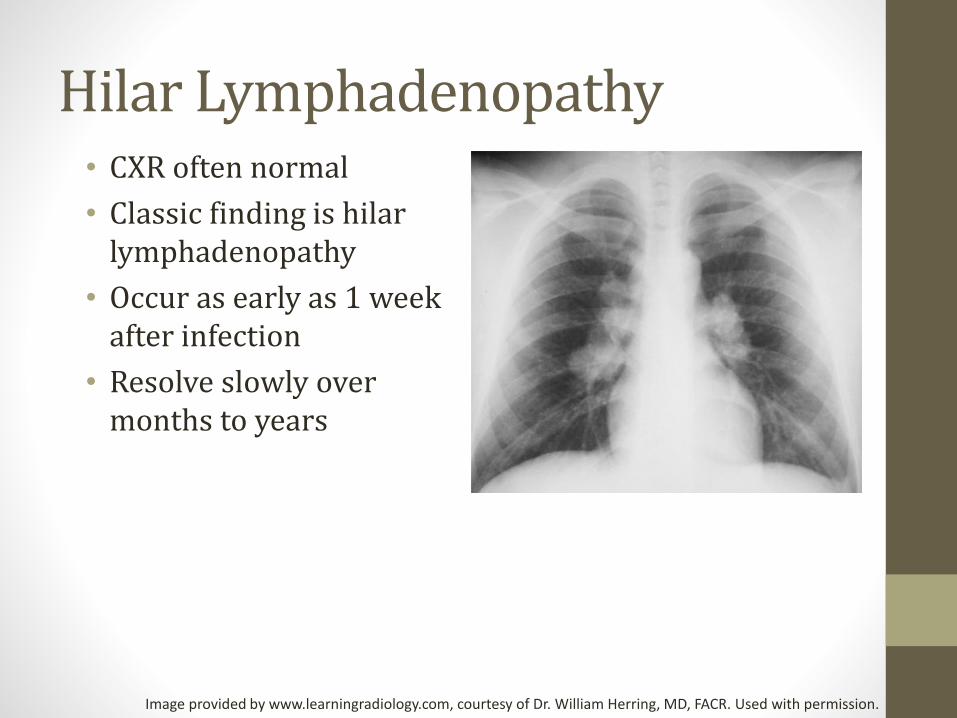
Hilar Lymphadenopathy• CXR often normal
• Classic finding is hilarlymphadenopathy
• Occur as early as 1 week after infection
• Resolve slowly over months to years
Image provided by www.learningradiology.com, courtesy of Dr. William Herring, MD, FACR. Used with permission.

Ghon Foci• Ghon foci form
• Granulomas
• Subpleural
• Mid to lower lungs
• Ghon foci plus lymph node is Ghon complex
• Calcified Ghon complex is a Ranke complex
Images courtesy of kaziomer
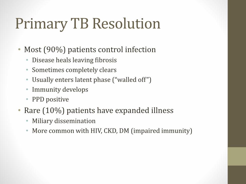
Primary TB Resolution
• Most (90%) patients control infection • Disease heals leaving fibrosis
• Sometimes completely clears
• Usually enters latent phase (“walled off”)
• Immunity develops
• PPD positive
• Rare (10%) patients have expanded illness• Miliary dissemination
• More common with HIV, CKD, DM (impaired immunity)
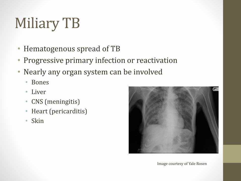
Miliary TB
• Hematogenous spread of TB
• Progressive primary infection or reactivation
• Nearly any organ system can be involved• Bones
• Liver
• CNS (meningitis)
• Heart (pericarditis)
• Skin
Image courtesy of Yale Rosen
Miliary TB
• Pott’s disease• Spine infection (osteomyelitis)
• Back pain, fever, night sweats, weight loss
• Constrictive pericarditis
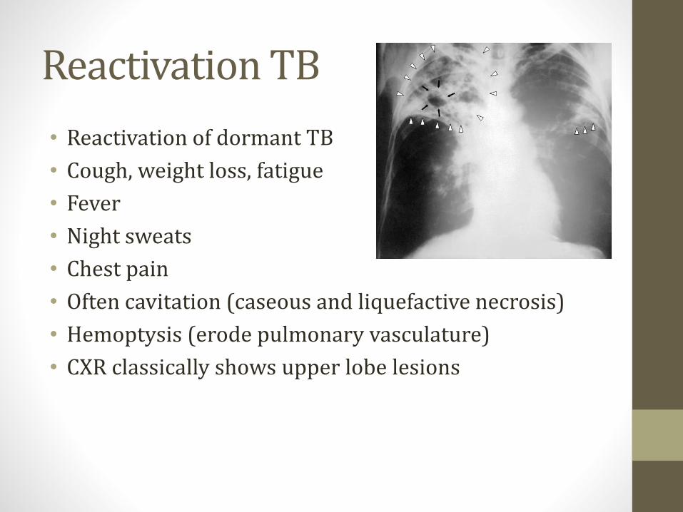
Reactivation TB
• Reactivation of dormant TB
• Cough, weight loss, fatigue
• Fever
• Night sweats
• Chest pain
• Often cavitation (caseous and liquefactive necrosis)
• Hemoptysis (erode pulmonary vasculature)
• CXR classically shows upper lobe lesions

Reactivation TB
• Can occur when immune compromise develops
• HIV infection
• TNF-α inhibitors• Used in autoimmune diseases
• Etanercept, Infliximab
• Diabetes
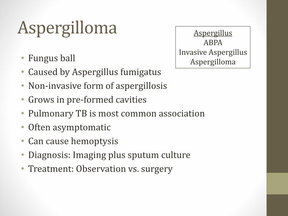
Aspergilloma
• Fungus ball
• Caused by Aspergillus fumigatus
• Non-invasive form of aspergillosis
• Grows in pre-formed cavities
• Pulmonary TB is most common association
• Often asymptomatic
• Can cause hemoptysis
• Diagnosis: Imaging plus sputum culture
• Treatment: Observation vs. surgery
AspergillusABPA
Invasive AspergillusAspergilloma
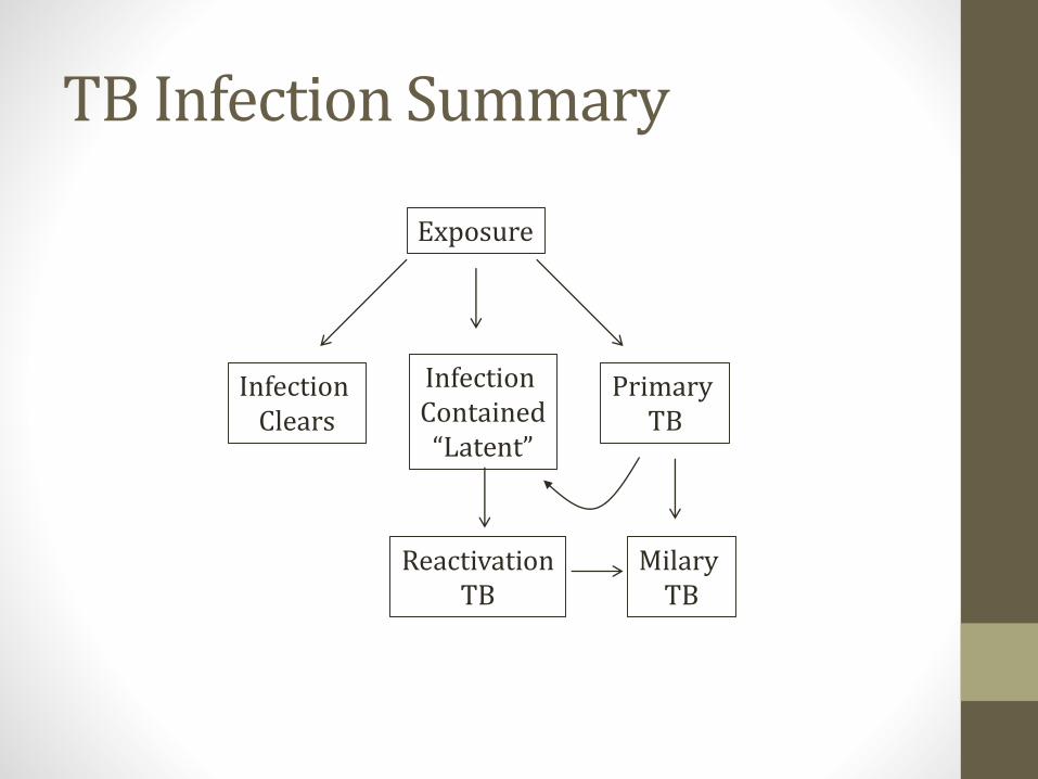
TB Infection Summary
Exposure
Infection Clears
Infection Contained“Latent”
Primary TB
MilaryTB
ReactivationTB

Diagnosis of Active TB
• Usual method: 3 sputum samples • Usually about 8hrs apart
• Spontaneous or induced
• Induced: Inhalation of aerosolized saline by nebulizer
• Acid-fast smear and culture

Diagnosis of Active TB
• Not necessary to hospitalize just for TB suspicion
• Outpatients: Remain at home, avoid visitors, mask
• Inpatients: Respiratory isolation• Private room
• Negative air pressure
• Persons entering must wear a respirator
• Tight seal over the nose and mouth
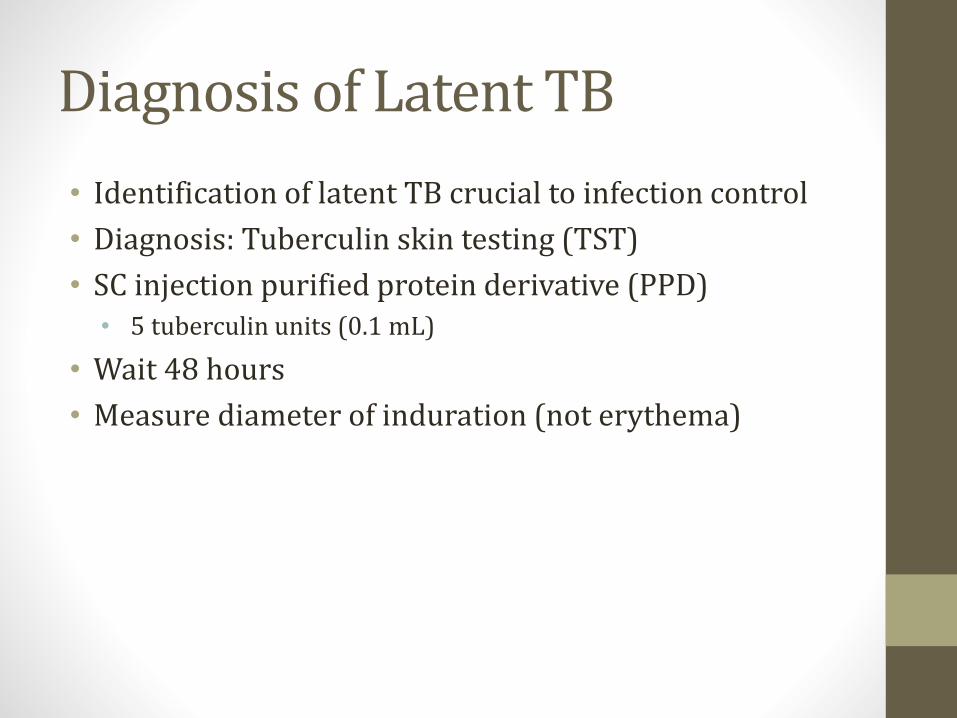
Diagnosis of Latent TB
• Identification of latent TB crucial to infection control
• Diagnosis: Tuberculin skin testing (TST)
• SC injection purified protein derivative (PPD)• 5 tuberculin units (0.1 mL)
• Wait 48 hours
• Measure diameter of induration (not erythema)

PPD Testing
* Silicosis, CKD, DM, IV drug users, homeless, prison employees, others

PPD Testing
• False negatives can occur
• Immunosuppressive drugs• Corticosteroids
• TNF-α inhibitors
• Immunocompromised• HIV
• CKD
• Malnutrition
• Diseased lymph system• Sarcoidosis
• Some lymphomas or leukemias
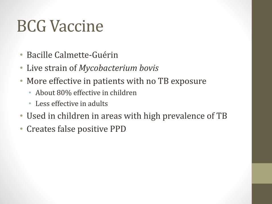
BCG Vaccine
• Bacille Calmette-Guérin
• Live strain of Mycobacterium bovis
• More effective in patients with no TB exposure• About 80% effective in children
• Less effective in adults
• Used in children in areas with high prevalence of TB
• Creates false positive PPD
Treatment of Positive PPD
• Most patients with latent TB will not develop disease
• Small proportion may reactivate
• Prophylaxis lowers risk
• Commonly isoniazid (INH) for 9 months
• Further PPD testing not indicated• Will remain positive for life

Treatment of Active TB
• Requires multi-drug regimens
• Typical regimen:• Isoniazid
• Rifampin
• Pyrazinamide
• Ethambutol
• Sometimes streptomycin
• Sometimes direct observation therapy (DOT)
• Risk of Multi-drug resistant (MDR) TB

Isoniazid
• Blocks synthesis of mycolic acids
• Bacteria lose their acid fastness
• katG-encoded catalase-peroxidase• Converts INH to active form
• Mutations lead to INH resistance
• Monotherapy produces resistance
Isoniazid
• Neurotoxic• Neuropathy, ataxia, and paresthesia
• Competes with B6 as co-factor neurotransmitter synthesis
• Pyridoxine (B6) co-administered
• Limits neurotoxicity
• Hepatotoxic (check LFTs)• Probably related to metabolites of INH
• Drug-induced lupus

Rifampin
• Inhibit bacterial DNA-dependent RNA polymerase
• Blocks RNA synthesis
• Main side effects are liver, GI• Increased LFTs
• GI upset: nausea, cramps, diarrhea
• Red/orange discoloration fluids (not dangerous)• Urine
• Saliva
• Sweat, tears
• CSF

RifampinOther uses
• Leprosy
• Meningococcal prophylaxis
• Chemoprophylaxis in contacts of children HiB
Pyrazinamide
• Mechanism unknown• Converted to pyrazinoic acid (PZA)
• May be more active in acidic environment inside macrophages
• Hepatotoxic• Can raise LFTs
• Competes with uric acid for excretion in kidneys• Can raise uric acid levels
• Hyperuricemia
• Gout exacerbations
Ethambutol
• Inhibits arabinosyl transferase• Polymerizes arabinose for mycobacteria cell walls
• Key side effect: optic neuropathy• Red-green color blindness
• Difficulty discriminating red and green hues
• Loss of visual acuity
• Reversible
Streptomycin
• Older, aminoglycoside drug
• Inhibits bacterial 30S ribosomal subunit• Prevents protein synthesis
• Lots of resistance• Mutations of genes for ribosomal proteins
Tuberculosis Key Points
• Mycolic acid cell walls → acid fast
• Infects macrophages (intracellular)
• Delayed type hypersensitivity reaction
• Hilar lymphadenopathy; Ghon complex
• Reactivation in upper lobes (immunosuppressed)
• Latent infection diagnosed with PPD
• Treat latent disease with INH
• Treat active disease with multidrug regimen

SarcoidosisJason Ryan, MD, MPH

Sarcoidosis
• Granulomatous disease• Granulomas form many places in the body
• Immune-mediated• Immune cells play major role
• Unknown cause
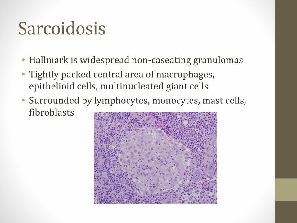
Sarcoidosis
• Hallmark is widespread non-caseating granulomas
• Tightly packed central area of macrophages, epithelioid cells, multinucleated giant cells
• Surrounded by lymphocytes, monocytes, mast cells, fibroblasts
Pathology
• Cell mediated immune process
• Accumulation of TH1 CD4+ helper T cells• High CD4:CD8 ratio
• Secrete IL-2 and interferon-γ
• IL-2 stimulates TH1 proliferation
• IFN-γ activates macrophages
• Ultimately leads to granuloma formation
• Key players: CD4 T cells, IL-2, IFN- γ

Organ Involvement
• Lungs (most common)
• Skin
• Eye
• Heart• Conduction disease (heart block)
• Cardiomyopathy
• Many other systems rarely involved• Renal: Renal failure
• CNS: Neurosarcoid, Bells Palsy, Motor loss
• Any system can be involved
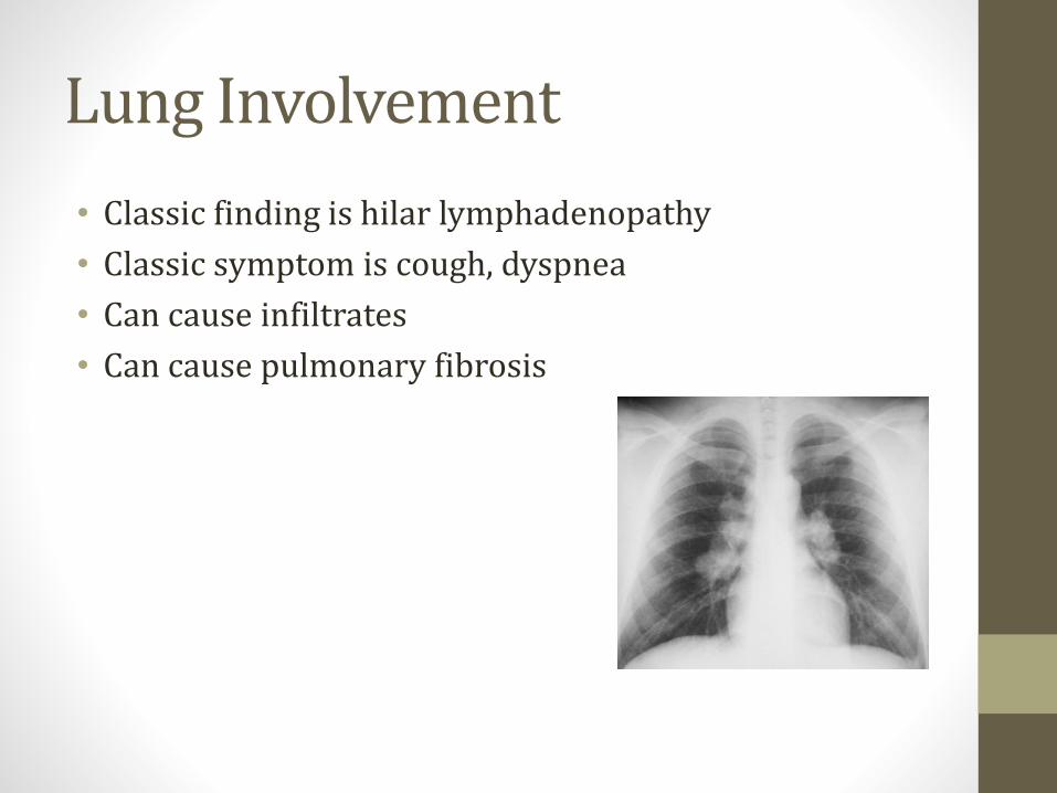
Lung Involvement
• Classic finding is hilar lymphadenopathy
• Classic symptom is cough, dyspnea
• Can cause infiltrates
• Can cause pulmonary fibrosis

Skin Involvement
• Many lesions possible• Plaques, maculopapules, subcutaneous nodules
• Classic lesion is erythema nodosum• Inflammation of fat cells under skin
• Tender red nodules
• Usually on both shins
Image courtesy of James Heilman, MD

Eye Involvement: Uveitis
• Can involve many parts of eye
• Classic is uveitis
• Uvea:• Iris, ciliary body, choroid
• Uveitis Types• Anterior (iris, ciliary)
• Posterior (choroid)
• Often mild symptoms• Dry eye, blurry vision
• Often detected on routine exam
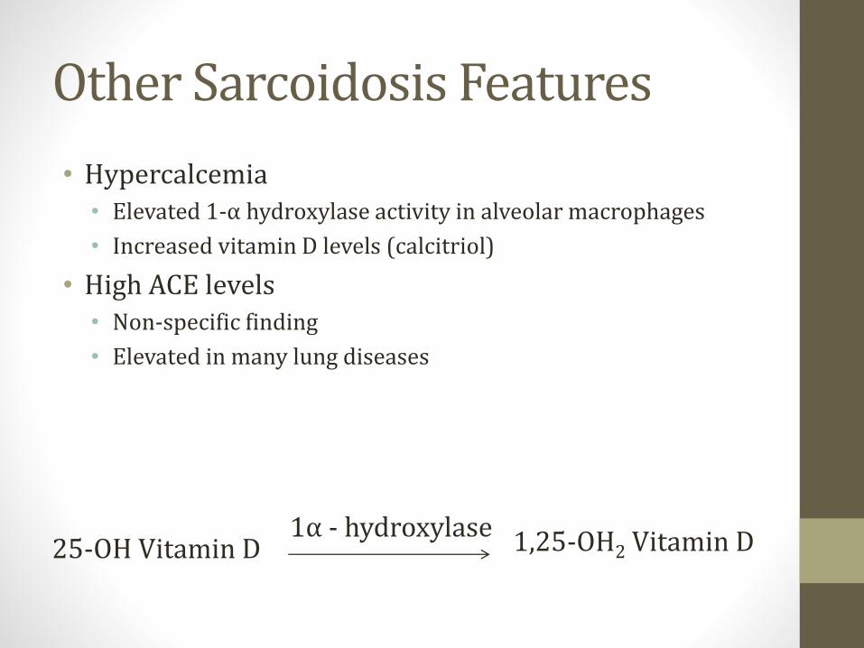
Other Sarcoidosis Features
• Hypercalcemia• Elevated 1-α hydroxylase activity in alveolar macrophages
• Increased vitamin D levels (calcitriol)
• High ACE levels• Non-specific finding
• Elevated in many lung diseases
25-OH Vitamin D 1,25-OH2 Vitamin D1α - hydroxylase

Classic Presentation
• African American female
• Hilar lymphadenopathy
• Cough, dyspnea
• Often asymptomatic, detected on routine chest x-ray
Treatment
• Steroids
• Other immunosuppressants• Methotrexate
• Azathioprine
• Mycophenolate


Pulmonary EmbolismJason Ryan, MD, MPH
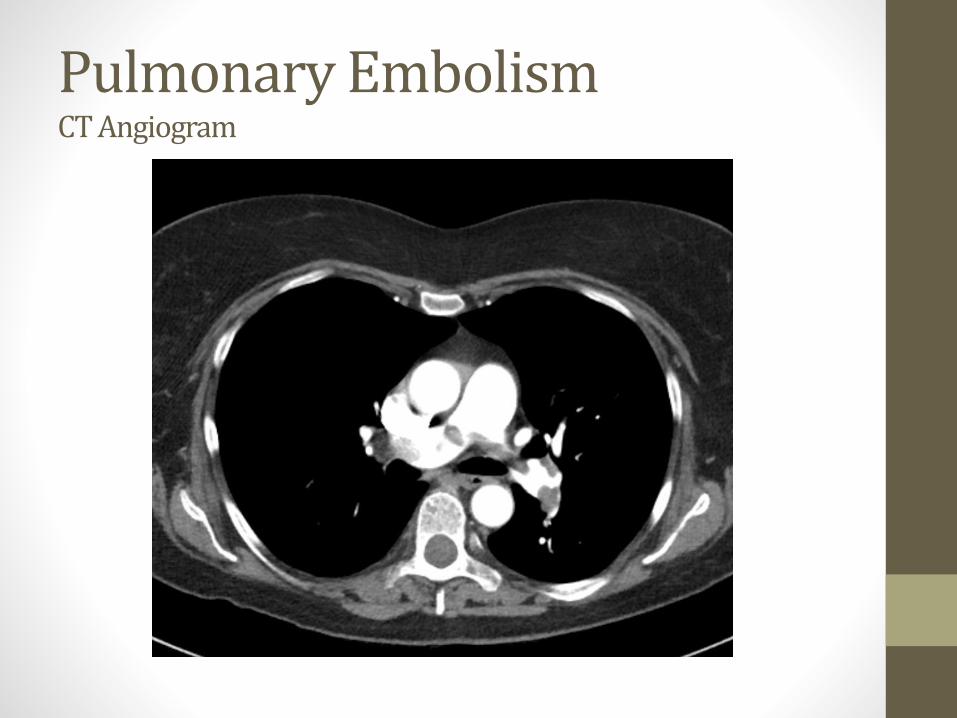
Pulmonary EmbolismCT Angiogram
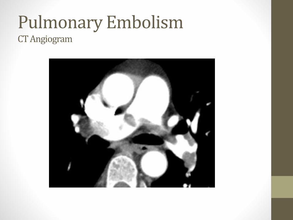
Pulmonary EmbolismCT Angiogram
Pulmonary Embolism
• Thrombus in pulmonary artery
• Rarely formed in heart or pulmonary vasculature
• Majority come from femoral vein or deep leg veins
• Travels to lung via IVC → RA → RV
Pulmonary Embolism
• Can be “unprovoked”
• Often secondary to a hypercoagulable state• Secondary: Malignancy, surgery, etc.
• Primary: Protein C/S deficiency, ATIII deficiency, etc.
Pulmonary Embolism
• Chest pain• Classic presentation is pleuritic
• Respiratory distress• Dyspnea
• Hypoxemia
• Tachypnea
• Massive PE can cause sudden death• Obstruction to flow through pulmonary arteries
• Small, chronic emboli: pulmonary hypertension
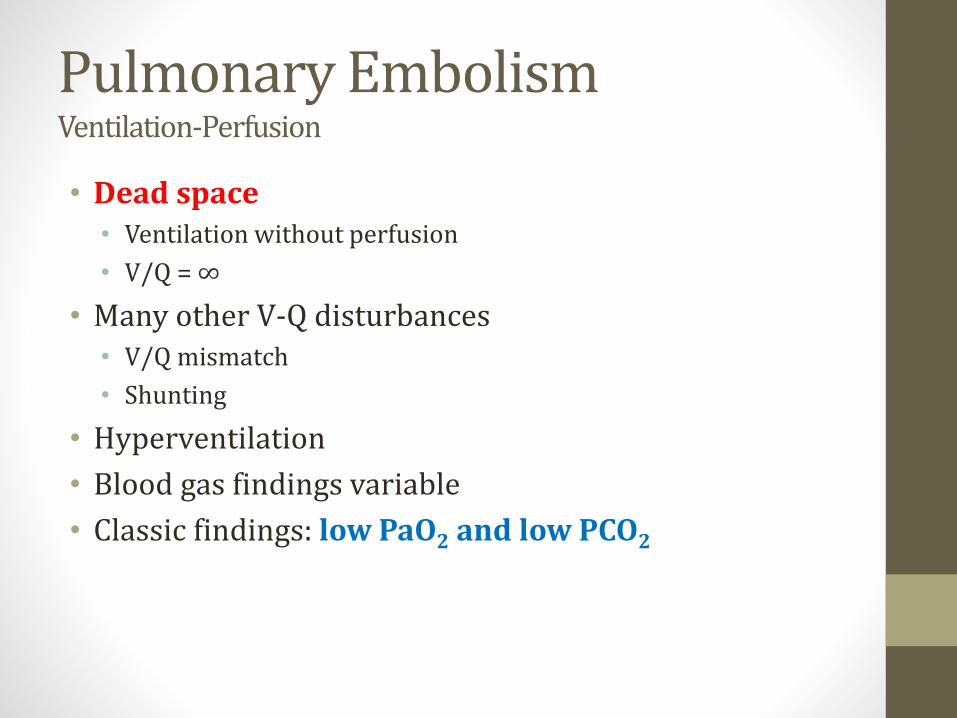
Pulmonary EmbolismVentilation-Perfusion
• Dead space • Ventilation without perfusion
• V/Q = ∞
• Many other V-Q disturbances• V/Q mismatch
• Shunting
• Hyperventilation
• Blood gas findings variable
• Classic findings: low PaO2 and low PCO2

Deep Vein Thrombosis
• Thrombus within a deep vein
• Usually occurs in calf or thigh
• Commonly femoral/popliteal veins
• Can extend or “grow”
• Precedes pulmonary embolism
• Often 2° hypercoagulable state
Bruce Blaus/Wikipedia
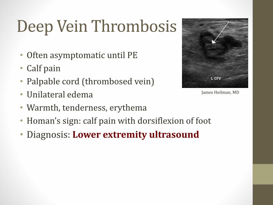
Deep Vein Thrombosis
• Often asymptomatic until PE
• Calf pain
• Palpable cord (thrombosed vein)
• Unilateral edema
• Warmth, tenderness, erythema
• Homan’s sign: calf pain with dorsiflexion of foot
• Diagnosis: Lower extremity ultrasound
James Heilman, MD

Deep Vein Thrombosis
• Similar treatment to PE
• “DVT/PE”
• “Venous thromboembolism” (VTE)
• Prevention important in hospitalized patients
• Hypercoagulable
• Immobility, stasis of blood, inflammation
• Prophylaxis: SQ heparin, LMWH
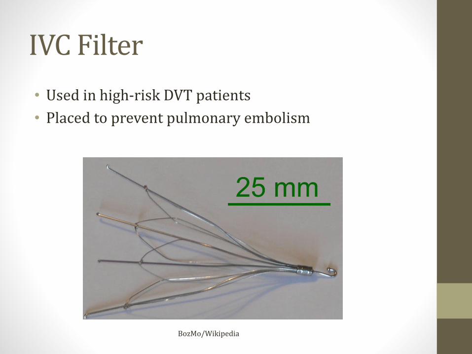
IVC Filter
• Used in high-risk DVT patients
• Placed to prevent pulmonary embolism
BozMo/Wikipedia

S1Q3T3
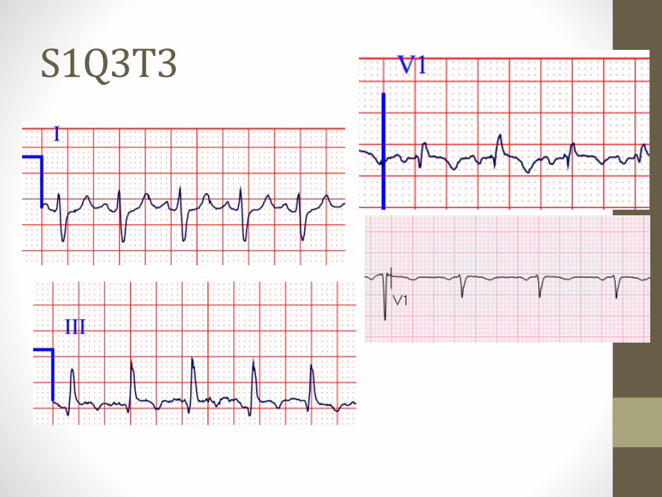
S1Q3T3
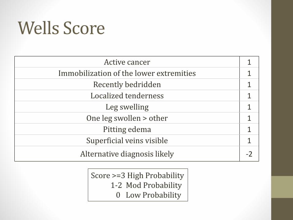
Active cancer 1
Immobilization of the lower extremities 1
Recently bedridden 1
Localized tenderness 1
Leg swelling 1
One leg swollen > other 1
Pitting edema 1
Superficial veins visible 1
Alternative diagnosis likely -2
Wells Score
Score >=3 High Probability1-2 Mod Probability
0 Low Probability

D-dimer
• Degradation product of fibrin
• Sensitive but not specific (unidirectional)• Levels elevated in DVT/PE
• Levels also elevated in many, many other conditions
• Useful when normal in setting of low-mod Wells score

Pulmonary EmbolismDiagnosis
• CT angiogram
• VQ Scan
Westgate EJ, FitzGerald GAPulmonary Embolism in a Woman Taking Oral Contraceptives and Valdecoxib.
PLoS Medicine Vol. 2, No. 7, e197. doi:10.1371/journal.pmed.0020197

Treatment DVT/PE
• Initial treatment with heparin or LMWH
• Transition to warfarin (oral)
• Massive PE: thrombolysis (tPA)
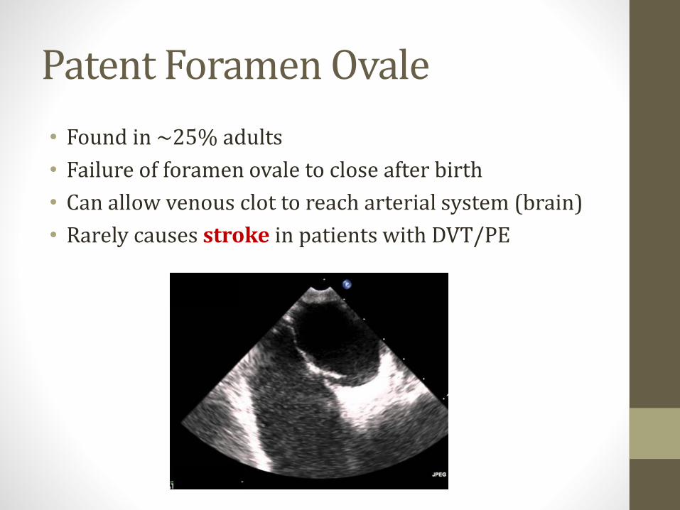
Patent Foramen Ovale
• Found in ~25% adults
• Failure of foramen ovale to close after birth
• Can allow venous clot to reach arterial system (brain)
• Rarely causes stroke in patients with DVT/PE
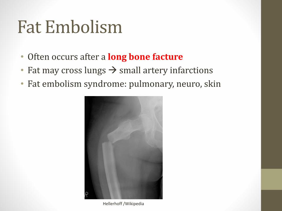
Fat Embolism
• Often occurs after a long bone facture
• Fat may cross lungs → small artery infarctions
• Fat embolism syndrome: pulmonary, neuro, skin
Hellerhoff /Wikipedia
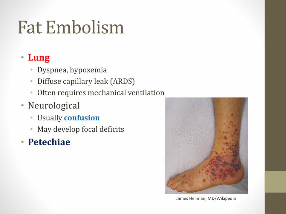
Fat Embolism
• Lung• Dyspnea, hypoxemia
• Diffuse capillary leak (ARDS)
• Often requires mechanical ventilation
• Neurological• Usually confusion
• May develop focal deficits
• Petechiae
James Heilman, MD/Wikipedia
Amniotic Fluid Embolism
• During labor or shortly after
• Amniotic fluid, fetal cells, fetal debris enter maternal circulation
• Inflammatory reaction
• Often fatal
Wikipedia/Public Domain
Amniotic Fluid Embolism
• Phase I• Pulmonary artery vasospasm → pulmonary hypertension
• Right heart failure
• Hypoxia
• Myocardial capillary damage → left heart failure
• Pulmonary capillary damage → ARDS
• Acute respiratory distress syndrome
• Key features: respiratory distress, ↓O2, hypotension

Amniotic Fluid Embolism
• Phase II (hemorrhagic phase)• Massive hemorrhage
• DIC
• Key feature: bleeding
• Seizures also often occur

Chest X-raysJason Ryan, MD, MPH
Chest X-ray
• Difficult to see different structures
• Many, many normal variants
• Many, many pathologic findings
• Reasonable goals:• Basic chest anatomy
• Classic examples of pathology

Chest Anatomy
RightAtrium
SVC
IVC
RightVentricle
LeftAtrium
LeftVentricle
PulmonaryArtery
AorticKnob
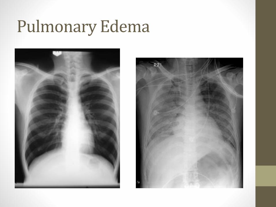
Pulmonary Edema
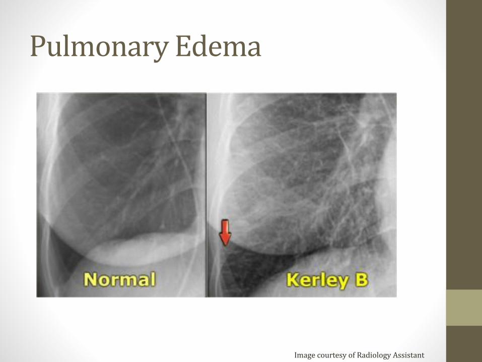
Pulmonary Edema
Image courtesy of Radiology Assistant

Pleural Effusion
Image courtesy of James Heilman, MD

Pneumothorax

Lobar Pneumonia
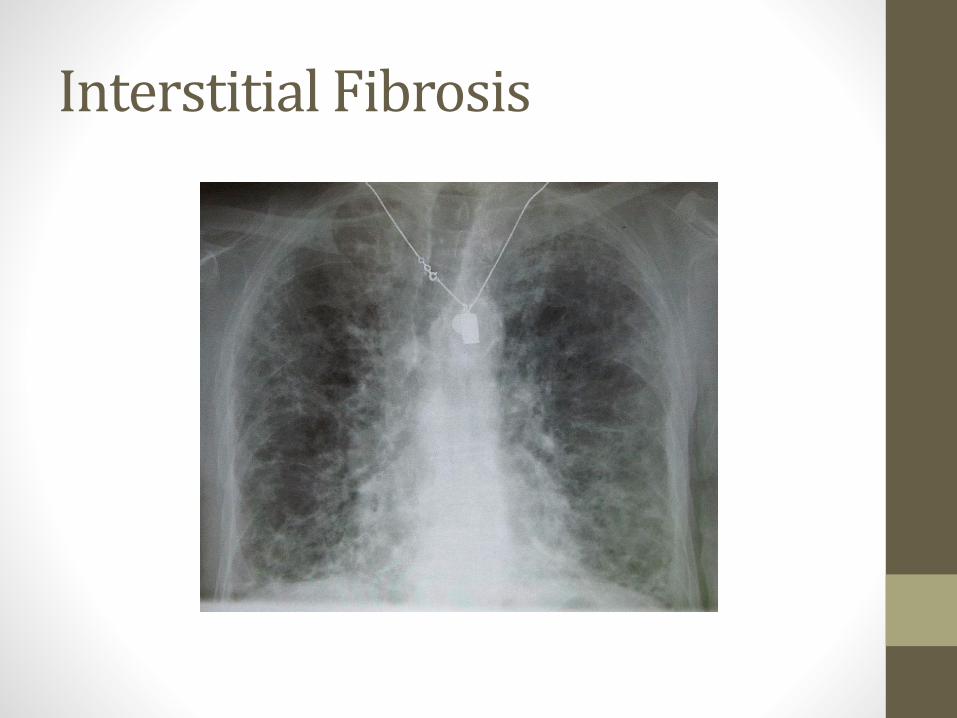
Interstitial Fibrosis
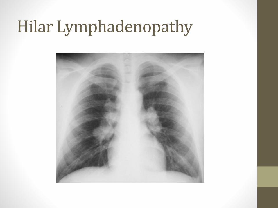
Hilar Lymphadenopathy
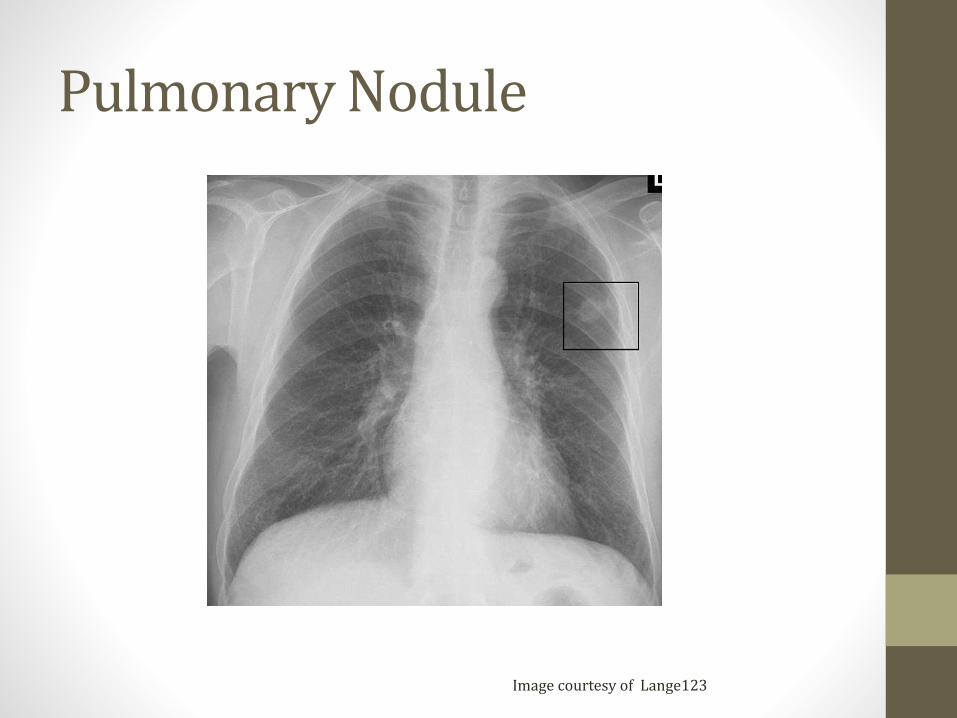
Pulmonary Nodule
Image courtesy of Lange123