PULMONARY ARTERIAL HYPERTENSION: A CORRELATION...
103
University of Padua School of Doctoral Research in Medical, Clinical and Experimental Sciences Course of Studies in Cardiovascular Sciences 20° cycle PULMONARY ARTERIAL HYPERTENSION: A CORRELATION BETWEEN SEVERITY OF DISEASE AND NEW BIOMARKERS OF ENDOTHELIAL DAMAGE AND REPAIR. Director: Professor Gaetano Thiene Supervisor: Professor Luciano Daliento Doctoral Candidate: Riccardo Proietti
Transcript of PULMONARY ARTERIAL HYPERTENSION: A CORRELATION...
University of Padua
School of Doctoral Research in Medical, Clinical and Experimental Sciences
Course of Studies in Cardiovascular Sciences
20° cycle
PULMONARY ARTERIAL HYPERTENSION: A
CORRELATION BETWEEN SEVERITY OF DISEASE
AND NEW BIOMARKERS OF ENDOTHELIAL DAMAGE
AND REPAIR.
Director: Professor Gaetano Thiene
Supervisor: Professor Luciano Daliento
Doctoral Candidate: Riccardo Proietti
2
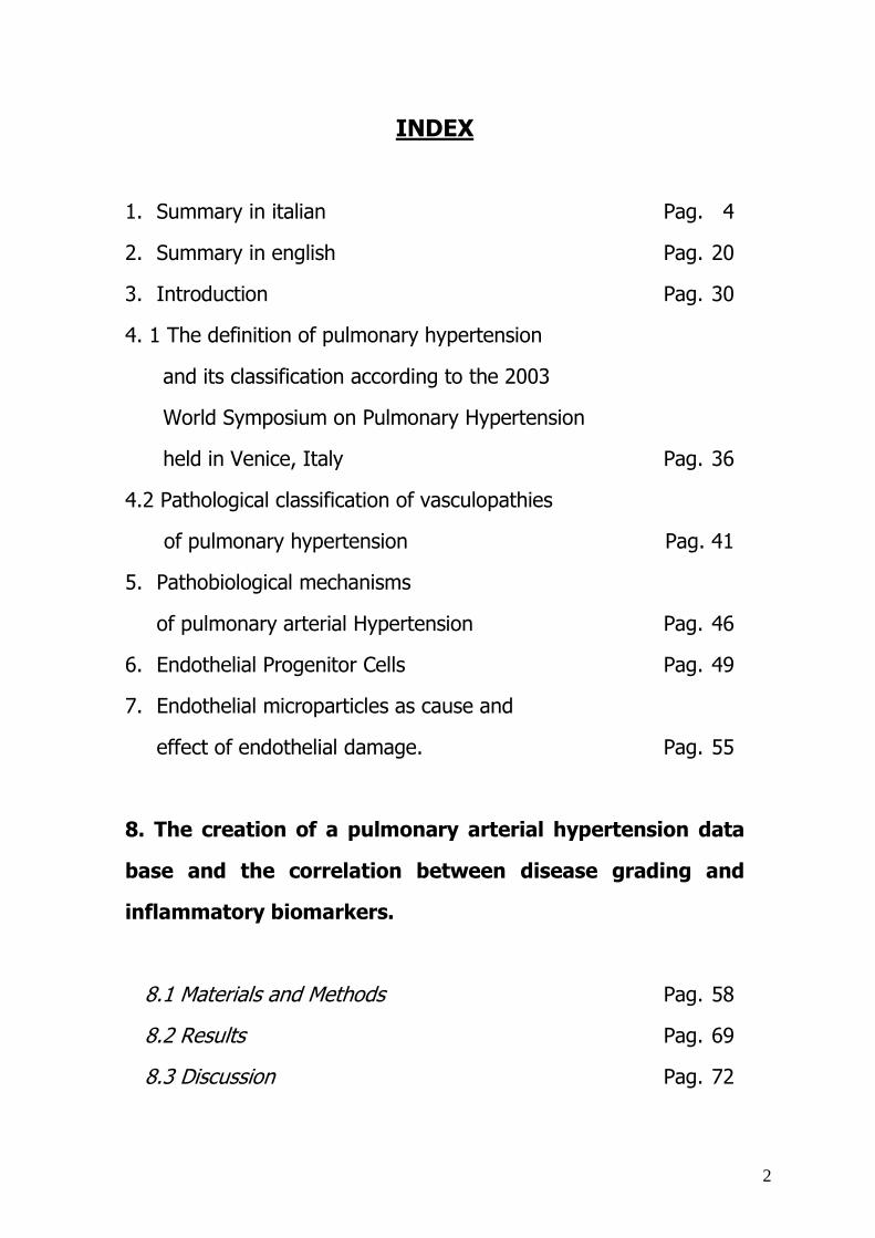
INDEX
1. Summary in italian Pag. 4
2. Summary in english Pag. 20
3. Introduction Pag. 30
4. 1 The definition of pulmonary hypertension
and its classification according to the 2003
World Symposium on Pulmonary Hypertension
held in Venice, Italy Pag. 36
4.2 Pathological classification of vasculopathies
of pulmonary hypertension Pag. 41
5. Pathobiological mechanisms
of pulmonary arterial Hypertension Pag. 46
6. Endothelial Progenitor Cells Pag. 49
7. Endothelial microparticles as cause and
effect of endothelial damage. Pag. 55
8. The creation of a pulmonary arterial hypertension data
base and the correlation between disease grading and
inflammatory biomarkers.
8.1 Materials and Methods Pag. 58
8.2 Results Pag. 69
8.3 Discussion Pag. 72
3
9. Correlation between pulmonary arterial hypertension
and endothelial damage and repair indexes
9.1 Study population Pag. 74
9.2 Materials and Methods Pag. 76
9.3 Results Pag. 79
9.4 Discussion Pag. 85
10. CONCLUSION Pag. 87
Bibliography Pag. 90

4
1 Summary in italian
L’ipertensione arteriosa polmonare è una condizione clinica che può
manifestarsi in forma idiopatica o associata ad una varietà di
patologie1. Anche se queste malattie presentano una differente
eziologia, l’ipertensione polmonare in esse riscontrata è
caratterizzata da tre predominanti meccanismi patobiologici che ne
determinano l’epifenomeno clinico-patologico: l’attivazione di uno
stato flogistico sistemico, una precoce disfunzione endoteliale ed
una aumentata angiogenesi2,3.
Un numero considerevole di evidenze scientifiche suggerisce il
possibile coinvolgimento della flogosi sistemica nella patogenesi
dell’ipertensione arteriosa polmonare. Elevati livelli circolanti di
citochine infiammatorie (IL-1 ed IL-6), l’aumentata espressione
polmonare di alcune chemochine (MIP-1α e MCP-1) e l’ infiltrazione
perivascolare di cellule infiammatorie (cellule T e macrofagi) è stata
evidenziata in pazienti con ipertensione arteriosa polmonare4-7.
Recentemente elevati livelli circolanti del CD40 ligando, una
proteina trans-membrana implicata nello sviluppo di molte malattie
infiammatorie come i disturbi autoimmuni, le reazioni da rigetto di
5
trapianto ed i processi aterosclerotici, sono stati riscontrati nei
pazienti con ipertensione arteriosa polmonare8.
Tuttavia ulteriori indagini sono necessarie per stabilire il ruolo dei
meccanismi infiammatori nello sviluppo di tale patologia2,3. Primo
scopo del nostro studio è stato quello di valutare lo stato
infiammatorio di tali soggetti così come appare dai principali indici
bioumorali di flogosi.
Sino ad oggi gli esami ematochimici non sono stati utilizzati
clinicamente nello screening o nel follow-up dei pazienti con
ipertensione polmonare; tuttavia diverse molecole (BNP,
endotelina-1, acido urico, serotonina, NO, D-dimero, troponina T)
sono elevate nel plasma di soggetti con tale patologia9.
Si è pensato quindi di valutare se i livelli di alcuni comuni indici
aspecifici di infiammazione, quali la conta dei globuli bianchi, la
formula leucocitaria, la VES, i livelli di PCR, fibrinogeno ed acido
urico fossero aumentati nei pazienti con ipertensione arteriosa
polmonare, rispetto a controlli sani; abbiamo inoltre voluto valutare
se esiste una correlazione tra l’aumento di questi indici di flogosi e
la severità della malattia ipertensiva polmonare, valutata sulla base
della classe NYHA e sulla distanza percorsa durante il 6MWT.
6
L’altro aspetto che il nostro studio ha considerato è stata la
presenza degli indici di danno e riparazione endoteliale nei soggetti
affetti da ipertensione arteriosa polmonare.
È consolidato in letteratura che nei pazienti con ipertensione
arteriosa polmonare il danno all’integrità e alla funzione endoteliale
(diminuita produzione di NO, di prostacicline e di sostanze
antitrombotiche; ed aumentata produzione di trobassano A2, di
endotelina-1 e di sostanze pro-trombotiche10) portano a molteplici
alterazioni nei normali processi omeostatici del sistema vascolare
polmonare e determinano un rimodellamento importante della
parete vasale attraverso l’instaurazione di un complesso circolo
vizioso mediato dall’ipossia che ne consegue11.
Il rimodellamento della parete arteriosa si caratterizza per un
ispessimento determinato dall’accumulo di proteine della matrice
extracellulare e dalla proliferazione e migrazione delle cellule
muscolari liscie, stimolata dallo stravaso nella matrice extracellulare
di mitogeni12. Recenti lavori su animali attribuiscono un ruolo
importante nella patogenesi e nel mantenimento dell’ispessimento
parietale alla proliferazione dei vasa vasorum avventiziali13. Le
lesioni plessiformi che sono caratteristiche ma non patognomoniche
7
di ipertensione arteriosa polmonare costituiscono la forma più
complessa di rimodellamento parietale12.
In tale contesto fisiopatologico è nato il nostro interesse nel
valutare nei soggetti affetti da questa malattia le concentrazioni
sieriche delle microparticelle endoteliali e dei progenitori delle
cellule endoteliali.
Infatti recentemente un considerevole interesse scientifico è stato
rivolto alle microparticelle prodotte dallo sfaldamento della
membrana citoplasmatica cellulare, come nuovo marker di danno
endoteliale14. Un elevato livello plasmatico di microparticelle
identificate principalmente dai cluster CD31+/CD42- sono state
riscontrate in soggetti con patologia del sistema cardiovascolare
quale sindrome coronarica acuta, arteriopatia periferica e diabete
mellito13. In tutte queste patologie le microparticelle endoteliali
vengono considerate un indice di danno vascolare14,15. Un recente
lavoro eseguito in pazienti con insufficienza renale severa ha
mostrato che le microparticelle sono in grado di inibire il rilascio di
ossido nitrico dalla cellule endoteliali suggerendo che esse non
costituiscono solamente il risultato di un danno ma hanno un
possibile ruolo nella genesi della disfunzione16.
8
La scoperta nel sangue circolante di progenitori di cellule
endoteliali17 capaci di differenziarsi in multipli fenotipi cellulari
(cellule muscolari lisce e cellule endoteliali) fa supporre che essi
siano coinvolti nei meccanismi di angiogenesi e vasculogenesi
dell’adulto nonché costituiscano un pool cellulare in grado di
rigenerare il tessuto endoteliale perso e sostituire le cellule
disfunzionanti18, creando quindi una differente prospettiva al
tradizionale concetto per cui si riteneva che fossero le cellule
adiacenti ad una lesione a proliferare e re-endotelizzare l’esistente
vasculatura18.
Tuttavia discordanti sono i risultati degli studi che investigano se i
progenitori partecipino solamente al mantenimento dell’omeostasi
vascolare o intervengono anche nella patogenesi delle differenti
patologie18. Nagaya et al19 hanno mostrato, in studi su animali, che
i progenitori delle cellule endoteliali somministrate per via
endovenosa sono incorporate nelle arteriole polmonari e che se
trasdotte con plasmidi contenenti adrenomedullina migliorano in
maniera significativa il grado di ipertensione polmonare.
Uno studio preliminare recentissimo su 33 pazienti ha mostrato che
l’infusione intravenosa di progenitori delle cellule endoteliali in
soggetti con ipertensione arteriosa polmonare è praticabile e sicura
9
e che può avere effetti benefici sulla capacità funzionale e sulla
emodinamica polmonare20.
In questo contesto scientifico il nostro studio ha l’intento di valutare
il rapporto tra le concentrazioni delle micropaticelle CD31+/CD42
ed i progenitori delle cellule endoteliali in una popolazione di
soggetti con ipertensione arteriosa polmonare idiopatica o associata
correlando i valori ottenuti con gli indici strumentali di malattia.
In questo studio abbiamo quindi valutato se esista una correlazione
tra severità della patologia e disfunzione endoteliale stimata tramite
dosaggio delle microparticelle CD31+/CD42- , ed abbiamo correlato
la severità della patologia con il numero di progenitori delle cellule
endoteliali circolanti.
CREAZIONE DATA BASE IPERTENSIONE POLMONARE E
CORRELAZIONE TRA GRADING DELLA MALATTIA E STATO
FLOGISTICO
La nostra ricerca ha avuto come primo intento quello di identificare
i pazienti affetti da ipertensione polmonare, ricoverati al Policlinico
di Padova, e rivalutare la loro diagnosi clinica sulla base della nuova
classificazione elaborata nel corso del Congresso Internazionale di
Venezia 2003.
10
Ciò ha permesso di constatare la casistica relativa a tale patologia
afferita presso questo centro di terzo livello, creare un data base di
pazienti aggiornato secondo la nuova nomenclatura, ed effettuare
delle analisi sulla base dei dati recuperati.
Con tale fine abbiamo richiesto presso gli Uffici amministrativi
dell’Azienda Ospedaliera di Padova l’elenco dei pazienti ricoverati al
Policlinico Universitario dal 2000 al 2006 che presentavano come
diagnosi di dimissione primaria o secondaria il codice DRG 4160
(ipertensione polmonare primitiva) od il codice 4168 (altre forme di
ipertensione polmonare) della classificazione delle malattie ICD-9-
CM79.
La ricerca tramite codice DRG ha selezionato 255 cartelle. 35 di
queste cartelle, un numero relativamente esiguo, pari al 14 %
sono state escluse per incongruenze dei valori di pressione
polmonare riportati all’esame ecocardiografico e\o invasivo e della
patologia riportata alla dimissione con la diagnosi di ipertensione
polmonare. L’analisi delle restanti 255 cartelle ha individuato 36
pazienti affetti da ipertensione arteriosa polmonare: nove pazienti
risultavano affetti da ipertensione polmonare idiopatica e 27 da
forme associate, di cui 11 a cardiopatia congenita, 5 ad una
malattia del collagene, 8 ad AIDS, 3 ad ipertensione portale (2

11
affetti da cirrosi epatica ed 1 da sindrome di Budd-Chiari). Nel
nostro data base il numero totale dei pazienti affetti da forme
secondarie di ipertensione polmonare risultava di 182 soggetti, e
sono stati esclusi.
Nella nostra analisi abbiamo considerato i pazienti con ipertensione
arteriosa polmonare idiopatica o associata, appartenenti al primo
gruppo della Classificazione Internazionale di Venezia del 2003,
poiché il nostro obiettivo è stato correlare l’esistenza e l’entità della
malattia ipertensiva polmonare con una attivazione umorale
infiammatoria, indipendentemente che sia o non sia presente una
patologia sistemica sottostante.
Dalla analisi dei dati rilevati dalle cartelle cliniche dei 36 pazienti
affetti da ipertensione arteriosa polmonare idiopatica o associata e
da quelli ottenuti in 36 soggetti sani, bilanciati per età e sesso con i
primi, si è potuto rilevare quanto segue:
1. I pazienti affetti da ipertensione arteriosa polmonare avevano
una conta di globuli bianchi (6700±113/mm3 vs 6360±156/mm3) e
livelli di VES (21±2,5 mm vs 16±2,7 mm) sovrapponibili ai soggetti
di controllo sani, tuttavia è emersa una differenza significativa
(p<0.05) nei livelli di PCR (4.7±0,09 mg/l vs 1,6±0,04 mg/l),
fibrinogeno (465±53 mg/dl vs 319±66 mg/dl), acido urico (5,1±0,8
12
mg/dl vs 3,2±0,7 mg/dl) tra i due gruppi; in particolare questi
ultimi indici sono risultati essere significativamente più alti nei
pazienti con ipertensione arteriosa polmonare. Al contrario i livelli di
albumina sono risultati essere inferiori nei pazienti con ipertensione
polmonare rispetto ai controlli sani (3.2±0,8 g/dl vs 4.1±1,1
g/dl,p=0,026).
2. Nei soggetti con ipertensione arteriosa polmonare, dal
confronto dei livelli dei principali indici di flogosi nel gruppo di
pazienti in classe NYHA III e IV vs quelli in classe NYHA I e II, sono
emersi valori più alti dei parametri infiammatori nei soggetti nel
primo gruppo: PCR (6.9±0,08 mg/l vs 2.8±0,05 mg/l), fibrinogeno
(513±69 mg/dl vs 408±88 mg/dl) ed acido urico (7.6±1,1 mg/dl vs
2,6±1,4 mg/dl). Non sono emerse differenze significative nei livelli
degli altri indici di flogosi presi in considerazione nel nostro studio
tra i pazienti raggruppati nelle diverse classi NYHA.
3. Nei pazienti con ipertensione arteriosa polmonare, dalle
analisi di correlazione tra indici di flogosi ed esito del 6MWT è
emerso che i soggetti con minore distanza percorsa al 6MWT
avevano livelli più alti di PCR (rho= -0.47, p=0.002), di fibrinogeno
(r=-0.33, p=0.009) e di acido urico (r=-0.36, p=0.008). La VES, la
conta dei globuli bianchi ed albumina non sono risultate essere

13
significativamente associate all’entità della limitazione funzionale dei
pazienti con ipertensione arteriosa polmonare.
In conclusione possiamo affermare che i pazienti con ipertensione
arteriosa polmonare presentano una attivazione sub-clinica della
cascata infiammatoria, la quale potrebbe essere in relazione con il
grado di compromissione della funzione vascolare polmonare.
Anche se l’incremento di comuni indicatori aspecifici di flogosi non
rappresenta uno strumento dirimente per la diagnosi di
ipertensione arteriosa polmonare, tuttavia, l’impiego di alcuni tra
questi indicatori (PCR, fibrinogeno ed acido urico) potrebbe essere
di una certa utilità nel migliorare la stratificazione del danno
vascolare polmonare del paziente con ipertensione arteriosa
polmonare oltre che consentire un più preciso inquadramento dello
stadio della malattia vascolare polmonare.
Inoltre una serie di rilevanti considerazioni emergono dalla analisi
dalla caratteristiche cliniche dei pazienti affetti da ipertensione
polmonare inclusi nel nostro data base.
La sintomatologia presentata pone molti problemi di diagnosi
differenziale con numerose altre patologie che presentano analoga
manifestazione, quali la cardiopatia ischemica, lo scompenso
cardiaco congestizio, le pneumopatie, la tromboembolia polmonare
14
cronica. La distinzione risulta estremamente difficile sulla base del
racconto anamnestico e soltanto la positività dell’anamnesi familiare
o patologica remota per le forme associate potrebbe suggerire il
sospetto di tale malattia. L’altro dato rilevante, é che tali soggetti
giungono alla diagnosi clinica tardivamente ed in fase avanzata
della malattia. Ciò potrebbe essere attribuito alla necessità di un
iter diagnostico che prevede numerose indagini strumentali come
approccio alla sintomatologia riferita. Inoltre nelle forme associate il
clinico sovente, tende ad interessarsi prevalentemente della
patologia principale trascurando l’evoluzione ed il trattamento della
malattia polmonare. Emerge quindi chiaramente che il sospetto
clinico e la successiva diagnosi di ipertensione polmonare è di
difficile elaborazione e costituisce essenzialmente una diagnosi di
esclusione.
CORRELAZIONE TRA IPERTENSIONE POLMONARE ED INDICI DI
DANNO E RIPARAZIONE ENDOTELIALE.
Il nostro studio è stato eseguito su di un gruppo di 15 pazienti
affetti da ipertensione arteriosa polmonare (4 con IAP idiopatica, 5
con forma associata a collagenopatia, 5 con forma associata a
cardiopatia congenita ed 1 caso di ipertensione polmonare
associata ad ipertensione portale) e seguiti presso l’ospedale

15
Martini di Torino, l’ospedale provinciale Sant’Andrea di Vercelli e
l’ospedale San Luigi Gonzaga di Orbassano. Tutti i pazienti hanno
firmato consenso informato per la partecipazione allo studio. I
soggetti in esame erano tutti in terapia con antagonisti recettoriali
dell’endotelina. Tutti i pazienti avevano un anamnesi negativa per
eventi cardiovascolari, dislipidemia, diabete, fumo ed ipertensione
arteriosa. In tutti i pazienti un ecocolorDoppler dei tronchi
sopraaortici ha mostrato valori normali di spessore medio-intimale
ed il test di vasoreattività dell’arteria brachiale è risultato nella
norma.
La popolazione in esame è stata confrontata con 15 soggeti sani
bilanciati per sesso ed età con la popolazione affetta da
ipertensione arteriosa polmonare.
Tutte le determinazioni sono state effettuate a digiuno, tra le 8:00
e le 10:00 del mattino. La misurazione del numero di
microparticelle endoteliali è stata effettuata come descritto da
Jimenez e coll.69. In breve, ogni paziente è stato sottoposto a
prelievo venoso ed il sangue ottenuto è stato sottoposto a
centrifugazioni sequenziali volte ad ottenere plasma povero in
piastrine (PPP). Ogni campione di PPP è stato quindi incubato per
30 minuti a 4°C con anticorpi fluorescenti: anti-CD31 coniugato con
16
il fluorocromo ficoeritrina (PE) e anti-CD42a coniugato con il
fluorocromo fluoresceina isotiocianato (FITC). Trascorso tale tempo
su ogni campione è stata eseguita un’analisi citofluorimetrica volta
all’individuazione della sottopopolazione delle MPs caratterizzata da:
presenza dell’antigene 31 sulla superficie della membrana, assenza
dell’antigene 42a e dimensione inferiore a 1,5 µm. Tale operazione
è stata condotta eseguendo un “gate” dimensionale mediante
l’utilizzo di sfere di calibrazione per citofluorimetro a diametro noto.
Per la misurazione del numero di progenitori endoteliali, ad ogni
partecipante allo studio è stato prelevato un campione di sangue in
provette con EDTA come anticoagulante. Ciascun campione di
sangue intero è stato sottoposto a centrifugazione in gradiente di
densità allo scopo di separare le cellule mononucleate (linfociti e
monociti). Dopo lavaggi sequenziali con PBS, volti ad eliminare
l’eventuale contaminazione piastrinica, le cellule ottenute sono state
incubate a 4°C per 30 minuti con anticorpi anti-CD34-FITC ed anti-
KDR-PE. Dopo tale tempo su ogni campione è stata eseguita
un’analisi citofluorimetrica volta all’individuazione dei progenitori
endoteliali caratterizzati dalla co-espressione degli antigeni CD34 e
KDR. Le analisi statistiche sono state effettuate con SPSS 15.0. La
significatività statistica è considerata per valori di p<0.05.
17
Dal nostro studio emerge che i pazienti con ipertensione arteriosa
polmonare hanno un maggior numero/µl di microparticelle
endoteliali (835.8±15.7 vs 710.9±31.7; p<0.01) ed un minor
numero/ml di progenitori endoteliali (423.9±7.4 vs 498.2±26.2;
p<0.01), rispetto ai controlli sani. L’analisi di correlazione tra il
numero di microparticelle endoteliali ed i livelli di PAPs determinata
con esame ecocardiografico ha mostrato che esiste una relazione
significativamente positiva tra queste due variabili (r=0.609;
p=0.004). Inoltre la correlazione tra numero di progenitori circolanti
delle cellule endoteliali e valori di PAPs hanno mostrato una
associazione inversa (r=-0.463; p=0.05).
Dall’analisi di correlazione tra numero di microparticelle e valori del
6 MWtest è emerso che esiste una associazione tra le suddette
variabili in quanto al diminuire della distanza percorsa si verifica un
incremento del numero di microparticelle (r=-0.573, p=0.001). Al
contrario il numero dei progenitori delle cellule endoteliali
diminuisce con il ridursi dei risultati del 6MW test (r=0.444;
p=0.014).
Conclusioni
L’ equilibrio tra danno endoteliale e riparazione dell’endotelio
danneggiato è essenziale per il mantenimento dell’omeostasi
18
vascolare. Dai risultati del nostro studio riteniamo che le
microparticelle endoteliali CD31+/CD42- possano rappresentare un
nuovo valido indicatore del danno prodotto a livello dell’albero
vascolare polmonare. D’altra parte, sembra emergere sempre con
maggiore evidenza che il danno endoteliale rappresenti una
condizione essenziale ma non sufficiente per deteriorare in modo
significativo la funzione vascolare. Affinché ciò possa realizzarsi, si
ritiene indispensabile che al danno vascolare si associ un’alterazione
dei meccanismi di riparazione del danno stesso.
Oggi, si pensa che i progenitori endoteliali possano assolvere alla
funzione di riparazione del danno endoteliale; pertanto, tutte le
condizioni capaci di influenzare il numero o la funzione dei
progenitori endoteliali possono contribuire in modo significativo alla
riparazione del danno endoteliale o alla disfunzione vascolare.
I risultati del nostro studio hanno dimostrato che l’ipertensione
arteriosa polmonare può essere associata in modo rilevante a
questi due importanti fenomeni, il cui corretto equilibrio garantisce
la normale funzione vascolare; in altre parole, l’ipertensione
arteriosa polmonare, associandosi ad una frammentazione delle
cellule endoteliali mature, incrementando la formazione di
microparticelle endoteliali e riducendo il numero e la vitalità dei

19
progenitori endoteliali circolanti, costituisce l’espressione funzionale
di una disfunzione vascolare in grado di determinare un
rimodellamento strutturale e biologico dell’albero vascolare
polmonare e di interressare complessi meccanismi omeostatici
sistemici.
20
2 Summary in English
Pulmonary arterial hypertension (PAH) is a clinical condition that
can present in either an idiopathic form or associated with a variety
of other conditions1. Pulmonary arterial hypertension is
characterized by three predominant pathobiological mechanisms
that influence the clinical-pathologic phenomenon: the activation
of a systemic inflammatory reaction, a premature endothelial
dysfunction and an increased angiogenesis2,3. However, further
investigations are needed, to establish the role of inflammatory
mechanisms in the development of PAH. The principal goal of this
study, was to evaluate the inflammatory state of such pathologies
specifically by examining the inflammatory profile in these subjects.
To date, blood test indexes have not been used clinically in either
screening or follow-up of PAH patients; stating from the fact that
several molecules (BNP, endotelin-1, uric acid, serotonin, NO, D-
dimer, troponin T) are elevated in these patients9. We proposed an
investigation looking at plasma levels of several inflammatory
indexes in PAH patients. We studied white blood cell count and
formula, ESR, CRP levels, fibrinogen and uric acid levels.
Furthermore we wanted to find out if there was a correlation
21
between an increase in these phlogistic indexes and the severity of
PAH based upon the NYHA classification and the 6MWT.
Another aim of this study was to establish the presence of
endothelial damage and repair indexes in PAH subjects.
CD31+/CD42- micro-particles and endothelial cell progenitors are
now considered new markers for endothelial dysfunction. The
second part of our study was to determe a possible relationship
between CD31+/CD42- micro-particles and endothelial progenitor
cells in a population of subjects with idiopathic or associated PAH.
To do this, we compared the plasma levels of CD31+/CD42- micro-
particles and endothelial progenitor cells with diagnostic indexes of
disease (echocardiographic PAPs, 6MWT). In conclusion, this study
investigated a possible correlation exist between the severity of
pathology and endothelial dysfunction estimated by CD31+/CD42—
microparticle and endothelial progenitor cells count.
THE CREATION OF A PULMONARY HYPERTENSION DATABASE
AND CORRELATION BETWEEN DISEASE GRADING AND
INFLAMMATORY BIOMARKERS
Aim of our research was to identify patients affected by
pulmonary hypertension (PH), admitted to the University Hospital of
Padua and re-evaluate their clinical diagnoses on the basis of the
22
new classifications adopted at The 2003 World Symposium on
Pulmonary Arterial Hypertension held in Venice, Italy.
According to this new classification we have created a new
database of patients and we have analyzed the collected data
accordingly.
We have obtained from University of Padua, the lists of patients
admitted between 2000 and 2006 and discharged with the code of
the disease classification ICD-9-CM79 4160 (primary pulmonary
hypertension) or 4168 (other forms of pulmonary hypertension).
We have selected 255 cases. Thirty-five (14%) were excluded for
incongruence between the values of pulmonary pressure that were
obtained from echocardiographic and/or invasive examination and
diagnosis reported at discharge. The analysis of the cases identified
36 patients affected by PAH: 9 with idiopathic PAH and 27 with
associated form of PAH (which 11 presented congenital
cardiopathy, 5 collagen disease, 8 AIDS, 3 portal hypertension). In
our database the total number of patients affected by other forms
of PH was 184 subjects.
Since we limited our research to forms of PAH, patients with other
forms were excluded.
23
In our analyses we considered patients with all the forms of
pulmonary hypertension, which were included in the first group of
the International classification held in Venice 2003. The aim of this
was to correlate the existence and the severity of the pulmonary
damage to inflammatory indexes, either in the absence or presence
of any underliyng systemic disease.
The results relative to the medical charts of the 36 patients with
idiopathic or associated PAH and the data obtained from the control
group of 36 healthy subjects, the following was revealed:
1) the patients affected by PAH compared to healthy control
subjects had white blood cell counts of 6700±113/mm3 vs
6360±156/mm3 and ESR levels of 21±2.5 mm vs 16±2.7 mm
respectively. However, a significant difference emerged (p<0.05)
in the CRP (4.7±0.09 mg/l vs 1,6±0.04 mg/l), fibrinogen (465±53
mg/dl vs 319±66 mg/dl), uric acid (5,1±0.8 mg/dl vs 3,2±0.7
mg/dl) levels between the two groups; all these indexes were
significantly higher in patients with pulmonary arterial hypertension.
On the other hand, the levels of albumin were inferior in patients
with pulmonary arterial hypertension compared to healthy controls
(3.1±0.8 g/dl vs 4.1±1.1 g/dl, p=0.026).
24
2) In subjects with pulmonary arterial hypertension comparing the
principal inflammatory indices levels between the combined group
of patients in the NYHA III and IV classes vs those subjects in the
NYHA classes I and II, emerged higher levels for all inflammatory
parameters in the first group: CRP (6.9±0.08 mg/l vs 2.8±0.05
mg/l), fibrinogen (513±69 mg/dl vs 408±88 mg/dl) and uric acid
(7.6±1.1 mg/dl vs 2.6±1.4 mg/dl). Non significant differences were
observed in the other inflammatory indexes examined in our study
according to NYHA classes.
3) In subjects with pulmonary arterial hypertension the correlation
analysis between the inflammatory indexes and the results at
6MWT showed that the subjects with decreased exercise capacity
had higher levels: CRP (r= -0.47, p=0.002), fibrinogen (r=-0.33,
p=0.009) and uric acid (r=-0.36, p=0.008).
In conclusion, we can affirm that patients with PAH present a sub-
clinical activation of the inflammatory cascade; this could be related
to the severity of pulmonary vascular damage. Even if the increase
in the common non-specific inflammatory indicators does not
represent a diriment tool for the diagnosis of PAH, the use of
several of these markers (CRP, fibrinogen and uric acid) could be
useful in improving the stratification of pulmonary vascular damage
25
in patients with PAH. Moreover, these indexes could provide a
more precise assignement for pulmonary vascular disease
staging. Furthermore, a series of relevant observations has
emerged from the analysis of clinical characteristics in patients with
PH included in the database: 1)the clinical presentation poses many
problems regarding differential diagnoses and 2) these subjects had
clinical diagnosis in the advanced phase of disease.
THE CORRELATION BETWEEN PULMONARY ARTERIAL
HYPERTENSION AND NEW INDEXES OF ENDOTHELIUM DAMAGE
AND REPAIR.
Our study, was carried out in Italy on a group of 15 patients
affected by PAH from the Martini Hospital in Turin, the
Sant’Andrea Hospital in Vercelli and the San Luigi Gonzaga Hospital
in Orbassano. All patients had a negative history for cardiovascular
disease, dyslipidemia, diabetes, smoke and systemic hypertension.
A carotid echo-Doppler scan was performed on all patients and
showed normal intimal-medial thickness values. A brachial arterial
flow-mediated vasodilation after reactive hyperemia was assessed
by ultrasonography and was normal in all patients. Fifteen PAH
affected patients were compared with 15 healthy subjects matched
26
for sex and age. The measurements of the endothelial
microparticles were performed as described by Jimenez e coll.39 To
determine the number of endothelial progenitor cells, a blood
sample was taken (using the anticoagulant EDTA) in all participants
in the study. Each sample of whole blood underwent a
centrifugation on density gradient to separate the mononuclear cells
(lymphocytes e monocytes). After sequential washing with PBS, to
eliminate the eventual platelet contamination, the obtained cells
were obtained were incubated at 4°C for 30 minutes with
antibodies anti-CD34-FITC and anti-KDR-PE. After that, each
sample underwent a cytofluormetric analysis to count endothelial
progenitor cells expressing CD34 and KDR.
This study revealed that patients with PAH have a higher number/µl
of endothelial microparticles (835.8±15.7 vs 710.9±31.7; p<0.01)
and a lower number/ml of endothelial progenitors (423.9±7.4 vs
498.2±26.2; p<0.01), compared with the healthy control group.
The correlation analysis between the number of endothelial
microparticles and the PAP levels measured with echocardiography
demonstrated a significantly positive relationship between these
two variables (r=0.609; p=0.004).Furthermore, the was a negative
correlation between the number of circulating progenitors of the
27
endothelial cells and the PAP values (r=-0.463; p=0.05). The
correlation between the number of microparticles and the 6MW test
showed, an association between these variables. In fact, when
exercise capacity was decreased, the number of microparticles was
increased (r=-0.573, p=0.001). On the other hand, when the
number of endothelial progenitor cells decreased, there was also a
reduction in the 6MW test (r=0.444; p=0.014).
In conclusion the balance between endothelial damage and repair is
essential for the maintenance of vascular homeostasis. We think
that the endothelial microparticles CD31+/CD42- represent a new
biomarker of damage pulmonary vasculature damage. On the other
hand the endothelial damage is a condition essential, but not
sufficient for the deterioration of vascular function, which
recognizes an alteration in the repair mechanisms. The results of
this study demonstrate that PAH is associated with these two
important conditions, which together guarantee normal vascular
function. In other words, PAH is associated with a fragmentation of
mature endothelial cells, increased formation of endothelial
microparticles which in turn reduces both the number of circulating
endothelial progenitor cells. This constitutes the functional
expression of a vascular dysfunction that determines a structural

28
and biological reshaping of the pulmonary vascular tree that also
involves complex systemic homeostatic mechanisms.
29
PULMONARY ARTERIAL HYPERTENSION: A CORRELATION
BETWEEN SEVERITY OF DISEASE AND NEW BIOMARKERS
OF ENDOTHELIAL DAMAGE AND REPAIR
30
3. INTRODUCTION
Pulmonary arterial hypertension (PAH) is a clinical condition that
can manifest itself in either an idiopathic form or can be associated
with a variety of other pathologies1.
Even if these diseases present different aetiologies, the underlying
underlying structural features are characterised by three
predominant pathobiological mechanisms: the activation of one
inflammatory systemic reaction, a premature endothelial
dysfunction and an increased angiogenesis2,3.
A considerable number of scientific evidence presently suggest the
possible involvement of the systemic inflammation in the
pathogenesis of PAH. Elevated levels of circulating inflammatory
cytokines (IL-1 ed IL-6), increased pulmonary expression of several
chemokines (MIP-1α e MCP-1) and perivascular inflammatory cells
infiltration (T-cell and macrophages) have been observed in
patients with PAH4-7.
Recently, elevated levels of circulating ligand CD40, a trans-
membrane protein involved in the development of many auto-
immune diseases disturbances, transplant rejection as well as
atherosclerosis, have been observed in patients with PAH8.
31
Further investigations are needed to establish the role of
inflammatory mechanisms in the development of these diseases.
Aim of this study, was to evaluate the inflammatory state of PAH
looking at the bioumoral indexes of inflammation.
To date, biochemical markers have not been utilised clinically in
either the screening or the follow-up of PAH patients; despite the
fact that diverse molecules (BNP, endotelin-1, uric acid, serotonin,
NO, D-dimer, troponin T) are elevated in patients with these
pathologies9.
For this reason, further investigation was proposed to establish if
the levels of several common indexes aspecific for inflammation
were increased in PAH patients compared to healthy subjects. We
considered white blood cell count and formula, ESR, CRP levels,
fibrinogen and uric acid levels. Furthermore, this study was
undertaken to determine if there was a correlation between an
increase in these inflammatory indexes and the severity of PAH
based upon the New York Heart Association (NYHA) classification
and the distance travelled during the 6MWT.
Another aim of this study was to establish the presence of
endothelial damage and repair processes in PAH subjects.
32
Patients with PAH have endothelial dysfunction (diminished
production of NO, prostacycline and antithrombotic substances and
an increased production of trobossan A2, endotelin-1 and pro-
thrombotic substances10). This causes alterations of the normal
haemostatic processes of the vascular system and important
remodelling of the vessel walls through the establishment of a
complex vicious circle mediated by subsequent hypoxia 11.
The remodelling of the arterial walls is characterised by a
thickening caused by accumulation of extra-cellular proteins and
proliferation and migration of smooth muscle cells which are
stimulated by the spillage of mitogens in the extra-cellular matrix12.
Recent work carried out on animals have attributed an important
role to the adventitial vasa vasorum in the pathogenesis and the
maintenance of the wall thickening13.
The plexiform lesions that are characteristic but not pathognomonic
of PAH constitute the most complex form of wall remodelling12.
This led us to investigate the serum concentrations of endothelial
microparticles and endothelial cell progenitors in subjects with PAH.
We in fact think that endothelial microparticles, which are produced
by the shedding of cytoplasmatic cellular membranes, may be a
new markers for endothelial damage14.
33
Patients with acute coronary syndrome, peripheral arteriopathy
and diabetes mellitus13 have an elevated levels of CD31+/CD42-
micro-particle.
In these pathologies the endothelial micro-particles are considered
an index for vascular damage14, 15.
A study carried out in patients with severe renal insufficiency has
demonstrated that the micro-particles are capable to inhibit the
release of NO from endothelial cells, suggesting that the micro-
particles may also have a role in the genesis of endothelial
dysfunction16.
The discovery in the circulating blood of endothelial progenitors
cells17 which are capable of differentiate into multiple cell
phenotypes (smooth muscle cells and endothelial cells) has
suggested that they may be involved in the mechanisms of
angiogenesis and vasculogenesis. They can also constitute a
possible cell pool capable of regenerating endothelial tissue and
substituting dysfunctional cells18. In this way, a new prospective as
been created contrary to the previous theory holding that the cells
adjacent to a lesion are responsible for the proliferation and
regeneration of the endothelial layer of the existing vasculature18.
34
Even so, there is little agreement whether progenitors only
participate in the maintenance of vascular homeostasis or also
intervene in the pathogenesis of different disease18. Nagaya et. al
have demonstrated in animal studies that endothelial progenitor
cells administered intravenously are incorporated in the pulmonary
arterioles and if transduced with plasmids containing
adrenomedullin, the grade of pulmonary hypertension improves
significantly19.
A very recent preliminary study on 33 patients has shown that the
intravenous infusion of endothelial progenitor cells in subjects with
PAH is feaseble and safe and may have beneficial effects on
pulmonary haemodynamics20.
In this scientific context this study intented to establish a possible
relationship between CD31+/CD42- micro-particles and endothelial
cell progenitors in a population of subjects with idiopathic or
associated PAH. To do this, we compared the obtained plasmatic
levels of CD31+/CD42- micro-particles and endothelial cell
progenitors with diagnostic indexes of disease (echocardiographic
PAPs, 6MWT).
We specifically looked to a possible correlation between the severity
of the disaese and endothelial dysfunction estimated by circulating

35
levels of CD31+/CD42— microparticles and endothelial progenitor
cells.
36
4. 1 THE DEFINITION AND CLASSIFICATION OF
PULMONARY HYPERTENSION ACCORDING TO THE 2003
WORLD SYMPOSIUM IN VENICE, ITALY.
Pulmonary hypertension (PH) is a clinical condition that includes
different pathologies characterized by increased systolic pressure in
the pulmonary artery greater than 35 mmHg or mean pressure
greater than 25 mmHg at rest or 30 mmHg after exercise21.
During the course of the 2003 World Symposium on PAH the
decision was made to update the existing classification, which had
been formulated at the 1998 Symposium in Evian.
The main purpose of the new classification was to identify clinical
conditions, which in the development of an increased pressure in
the pulmonary circulation, would present common elements
regarding physiopathologic mechanisms, histological lesions, clinical
presentation and response to therapy22.
The revised classification (Table 1) maintains the framework of the
previous classification, distinguishing the diverse forms of PH into
five classes: the first includes PAH (pulmonary artery
hypertension), the second includes the form secondary to valvular
cardiopathy or PH with left heart disease , the third includes the
37
form secondary to lung disease and/or chronic hypoxemia, the
fourth includes PH due to thrombotic and/or embolic disease while
the fifth includes rare diseases of pulmonary vessels22.
In the new classification the term “Primary PAH” replaced the
term “Idiopathic PAH”. Previously, the Evian classification had
changed the term “Secondary PAH” into “Associated PAH” , in
consideration of the heterogeneous nature of the included diseases.
Therefore, the revised classification distinguishes PAH as an
idiopathic form, a familial form and forms associated to various
pathologies: collagen disease, congenital cardiopathy with
systemic-pulmonary shunts, portal hypertension, HIV infection and
other rare disorders.
Due to the analogy of the histological reports and similar clinical
presentations, two pathologies associated with an involvement of
the venular and capillary circulation(pulmonary veno-occlusive
disease and pulmonary capillary hemangiomatosis) were included
among the forms of PAH. In the fifth class the new classification
considered: sarcoidosis, hystiocytosis X, lymphangiomatosis and
compression of pulmonary vessels (adenopathy, tumour and
fibrosing mediastinitis).

38
The Venice classification clarify that the term PAH refers to the
clinical situations determined by a structural alteration of the
parietal walls of pre-capillary arteries by means of pathological
remodelling. Thus, it exclude form of dynamic hypertension or
secondary to pulmonary venous osbtraction22.
39
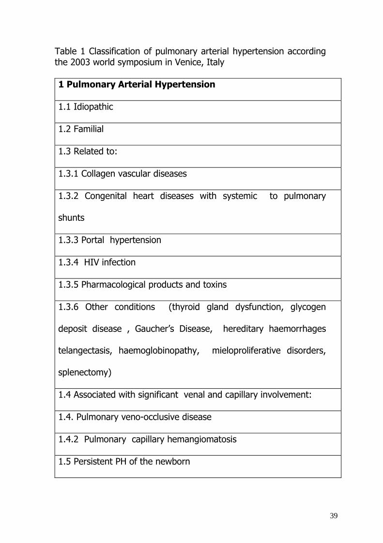
Table 1 Classification of pulmonary arterial hypertension according the 2003 world symposium in Venice, Italy 1 Pulmonary Arterial Hypertension
1.1 Idiopathic
1.2 Familial
1.3 Related to:
1.3.1 Collagen vascular diseases
1.3.2 Congenital heart diseases with systemic to pulmonary
shunts
1.3.3 Portal hypertension
1.3.4 HIV infection
1.3.5 Pharmacological products and toxins
1.3.6 Other conditions (thyroid gland dysfunction, glycogen
deposit disease , Gaucher’s Disease, hereditary haemorrhages
telangectasis, haemoglobinopathy, mieloproliferative disorders,
splenectomy)
1.4 Associated with significant venal and capillary involvement:
1.4. Pulmonary veno-occlusive disease
1.4.2 Pulmonary capillary hemangiomatosis
1.5 Persistent PH of the newborn
40
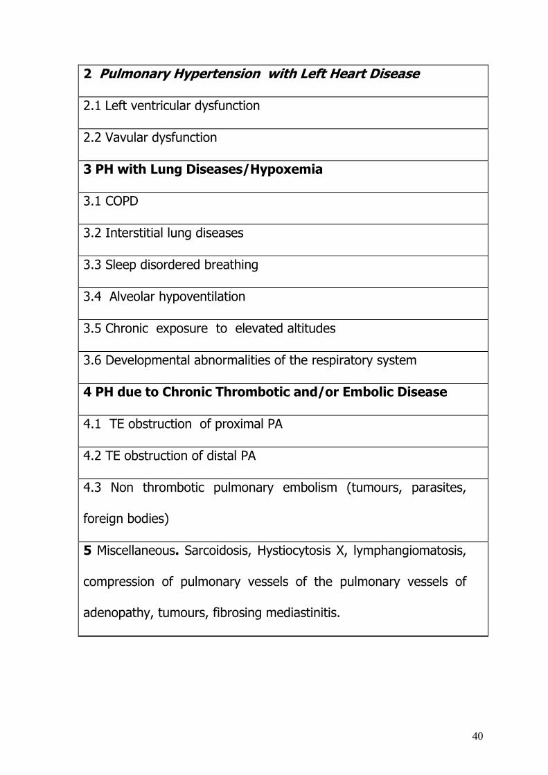
2 Pulmonary Hypertension with Left Heart Disease
2.1 Left ventricular dysfunction
2.2 Vavular dysfunction
3 PH with Lung Diseases/Hypoxemia
3.1 COPD
3.2 Interstitial lung diseases
3.3 Sleep disordered breathing
3.4 Alveolar hypoventilation
3.5 Chronic exposure to elevated altitudes
3.6 Developmental abnormalities of the respiratory system
4 PH due to Chronic Thrombotic and/or Embolic Disease
4.1 TE obstruction of proximal PA
4.2 TE obstruction of distal PA
4.3 Non thrombotic pulmonary embolism (tumours, parasites,
foreign bodies)
5 Miscellaneous. Sarcoidosis, Hystiocytosis X, lymphangiomatosis,
compression of pulmonary vessels of the pulmonary vessels of
adenopathy, tumours, fibrosing mediastinitis.
41
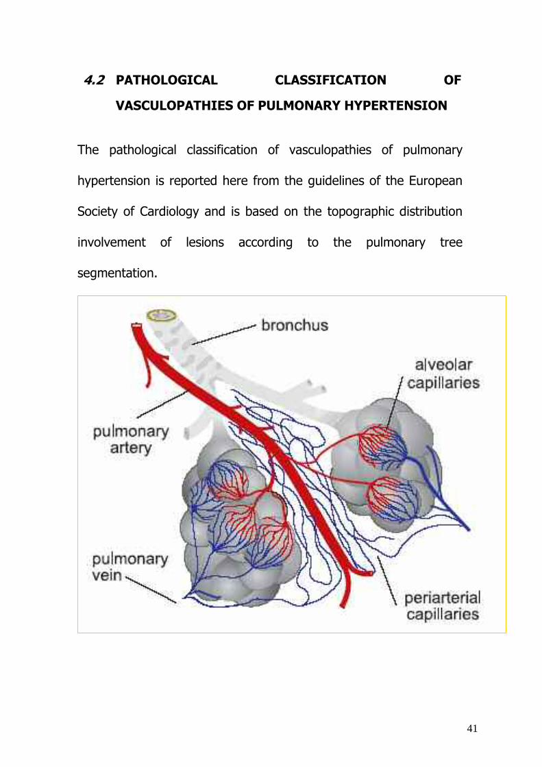
4.2 PATHOLOGICAL CLASSIFICATION OF
VASCULOPATHIES OF PULMONARY HYPERTENSION
The pathological classification of vasculopathies of pulmonary
hypertension is reported here from the guidelines of the European
Society of Cardiology and is based on the topographic distribution
involvement of lesions according to the pulmonary tree
segmentation.
42
Pathological Classification
(1) Pulmonary arteriopathy (pre-and intra-acinar arteries)
Subsets
-Pulmonary arteriopathy with isolated medial hypertrophy
- Pulmonary arteriopathy with medial hypertrophy and intimal
thickening (cellular, fibrotic)
– Concentric laminar
– Eccentric, concentric non-laminar
-Pulmonary arteriopathy with plexiform and/or dilatation lesions or
arteritis
- Pulmonary arteriopathy with isolated arteritis
(1a) As above but with coexisting venous-venular changesa (cellular
and/or fibrotic intimal thickening, muscularisation)
(2) Pulmonary occlusive venopathy (veins of various size and
venules) with or without coexisting arteriopathy
(3) Pulmonary microvasculopathyc with or without coexisting
arteriopathy and/or venopathy
(4) Unclassifiable
43
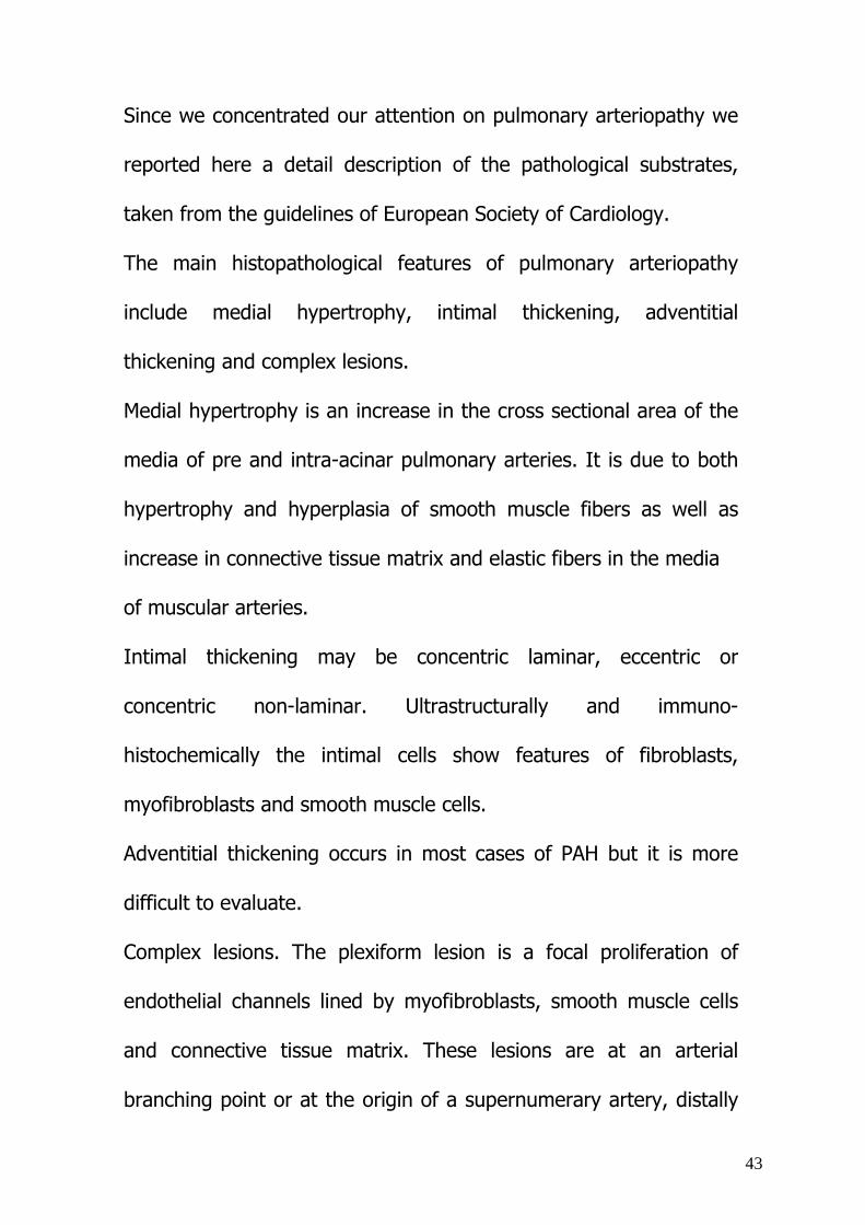
Since we concentrated our attention on pulmonary arteriopathy we
reported here a detail description of the pathological substrates,
taken from the guidelines of European Society of Cardiology.
The main histopathological features of pulmonary arteriopathy
include medial hypertrophy, intimal thickening, adventitial
thickening and complex lesions.
Medial hypertrophy is an increase in the cross sectional area of the
media of pre and intra-acinar pulmonary arteries. It is due to both
hypertrophy and hyperplasia of smooth muscle fibers as well as
increase in connective tissue matrix and elastic fibers in the media
of muscular arteries.
Intimal thickening may be concentric laminar, eccentric or
concentric non-laminar. Ultrastructurally and immuno-
histochemically the intimal cells show features of fibroblasts,
myofibroblasts and smooth muscle cells.
Adventitial thickening occurs in most cases of PAH but it is more
difficult to evaluate.
Complex lesions. The plexiform lesion is a focal proliferation of
endothelial channels lined by myofibroblasts, smooth muscle cells
and connective tissue matrix. These lesions are at an arterial
branching point or at the origin of a supernumerary artery, distally
44
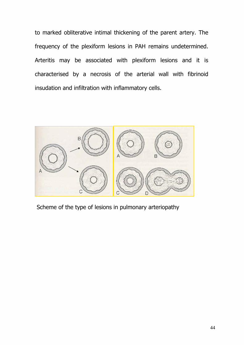
to marked obliterative intimal thickening of the parent artery. The
frequency of the plexiform lesions in PAH remains undetermined.
Arteritis may be associated with plexiform lesions and it is
characterised by a necrosis of the arterial wall with fibrinoid
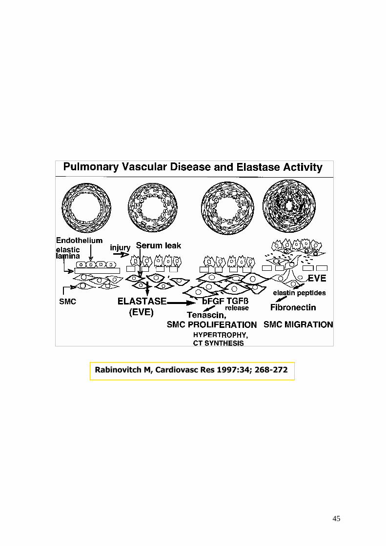
insudation and infiltration with inflammatory cells.
Scheme of the type of lesions in pulmonary arteriopathy
45
Rabinovitch M, Cardiovasc Res 1997:34; 268-272
46
5. PATHOBIOLOGICAL MECHANISMS OF PULMONARY
ARTERIAL HYPERTENSION
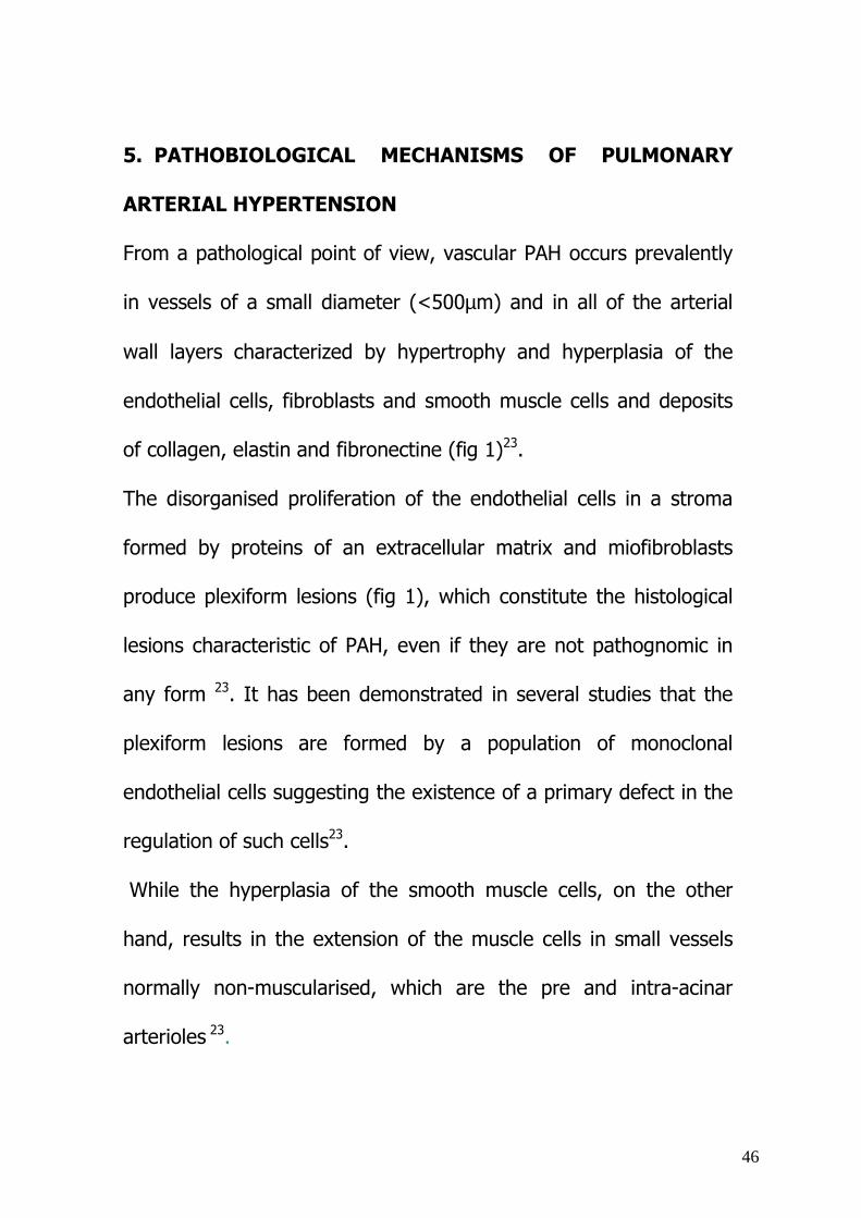
From a pathological point of view, vascular PAH occurs prevalently
in vessels of a small diameter (<500µm) and in all of the arterial
wall layers characterized by hypertrophy and hyperplasia of the
endothelial cells, fibroblasts and smooth muscle cells and deposits
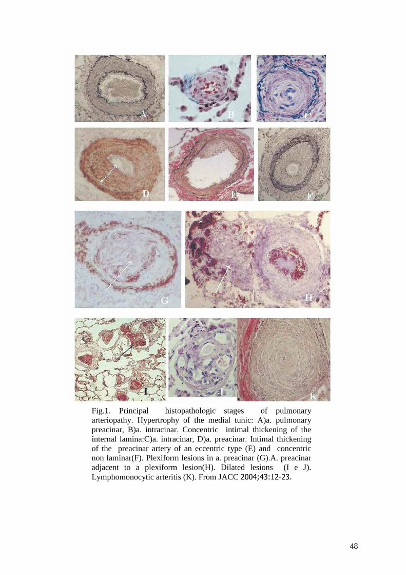
of collagen, elastin and fibronectine (fig 1)23.
The disorganised proliferation of the endothelial cells in a stroma
formed by proteins of an extracellular matrix and miofibroblasts
produce plexiform lesions (fig 1), which constitute the histological
lesions characteristic of PAH, even if they are not pathognomic in
any form 23. It has been demonstrated in several studies that the
plexiform lesions are formed by a population of monoclonal
endothelial cells suggesting the existence of a primary defect in the
regulation of such cells23.
While the hyperplasia of the smooth muscle cells, on the other
hand, results in the extension of the muscle cells in small vessels
normally non-muscularised, which are the pre and intra-acinar
arterioles 23.
47
Fibroblasts of the adventitial layer in severe PAH forms proliferate
and migrate and contribute, through the deposit of the extracellular
matrix, to the formation of a layer called neointima, between the
endothelium and the internal elastic lamina23.
Many factors are involved in the remodeling of the arterial wall.
Currently, the objects of intense research activity include the
vessel-active mediators: prostacycline, VIP, nitric oxide, endothelin,
ion channels , endothelial growth factors (VEGFR-A and VEGFR-B) ,
and the cytokines (activine and TGF-βs) 23. All of these appear to
act through: the proliferation and the dysfunction of the endothelial
cell, the thrombotic obliteration of the vascular lumen, the
alteration of the complex control mechanisms of the vascular tone,
the deposit of collagen and extracellular matrix, the inhibition of
apoptosis, and the modulation of the inflammatory response 23.
Recently, the mutation of the gene regulating the transcription of
the bone morphogenetic protein receptor type II” (BMPR2) has
been documented in around 50% of familial PAH cases and about
26% in the sporadic forms24. The progress made in understanding
the molecular mechanisms of this pathology, favoured the
introduction of new pharmacological drugs that improved the
negative outcome of these patients25.
48
Fig.1. Principal histopathologic stages of pulmonaryarteriopathy. Hypertrophy of the medial tunic: A)a. pulmonarypreacinar, B)a. intracinar. Concentric intimal thickening of theinternal lamina:C)a. intracinar, D)a. preacinar. Intimal thickeningof the preacinar artery of an eccentric type (E) and concentricnon laminar(F). Plexiform lesions in a. preacinar (G).A. preacinaradjacent to a plexiform lesion(H). Dilated lesions (I e J).Lymphomonocytic arteritis (K). From JACC 2004;43:12-23.

49
6. ENDOTHELIAL PROGENITOR CELLS
Stem cells can be defined as non-specialised cells, with a
clonogenic or replicating potential, capable of auto-renovation and
able to differentiate into diverse cellular lines. The division produces
two cells: a daughter cell with the same potential of the mother
cell (stem) and the other with different potential able only to
differentiate in a specialized line (progenitor). The progenitor cell
are not able of dividing indefinitely (loss of staminality) and all of its
progeny will differentiate into a single specialised cell or into
diverse types of mature cells.
The present knowledge regarding stem cells can be divided into
two groups: embryonic stem cells and adult stem cells 26.
The embryonic stem cells form the internal cellular mass
(embryoblast) of the blastocysts and are capable of generating
cells (both in-vitro and during the human embryogenesis) that
maintain their capacity to differentiate in any tissue of the
organism.
The adult stem cells, capable of being isolated during the fetal
period in organs and during the neonatal period in the umbilical
cord, are present in the adulthood in both the solid organ tissues
and in the blood.
50
These cells maintain their ability to replicate and are able to replace
differentiated cell loss so as to guarantee the physiological cell turn-
over and the cell damage repair.
For a long time it had been hypothesized that the adult stem cells
were capable of differentiating exclusively in mature cells of the
tissues where they were hosted. That is, it was thought that the
cells were organ specific or having a limited plasticity. However, the
classic paradigm of the organ specific differentiation of the adult
stem cells has recently been challenged, given the greater amount
of evidence that has confirmed the persistence in these cells which
possess a certain grade of plasticity or total potency 26, 27.
It was, also previously, believed that the endothelial cells and the
smooth muscle cells could originate from different cellular
precursors 28. In fact, it was held that during embryogenesis the
endothelial cells derived from two progenitors: the angioblast,
capable of differentiating in mature endothelial cells and the
haemoangioblast, capable of generating in both endothelial cells
and common blood cells. Even in adults the existence of a medullar
precursor has been documented. This stem cell has the
characteristics of a haemoangioblast, which is responsible for the
formation of blood and endothelial cells. This progenitor may
51
migrate from the stromal zone to the medullar vascular zone, and
then procede to the differentiation steps towards the endothelial or
haemopoietic precursor, which can occur initially in the medulla and
subequently in the circulation29. The vascular muscle cells, distinct
in vascular smooth muscle cells (muscle cells capable of presenting
in more layers in the vascular walls of the arteries proximal to the
heart) and in pericytes (muscle cells that present in mono-layers
in the vascular walls of the more distal section of the arteriolar
tree), can originate from diverse precursors: mesenchymal stem
cells (in both embryonic and adult), embryonic cells of the neural
crest, the epicardial embryonic cells and the endothelial
progenitors 30. Until a few years ago, the theory described above
represented the most widely shared explanation for the origin of
endothelial cells and smooth muscle cells of the arterial wall. The
studies of Yamashita and coll. 31 , in contrast, have showed the
existence of a new progenitor called “common vascular progenitor”
capable of differentiating in both mature endothelial cells and in
smooth muscle cells 31.
In particular, the exposure of the common vascular progenitor to
Vascular Endothelial Growth Factor (VEGF) may promote its
differentiation in endothelial cells, while the Platelet Derived Growth
52
Factor-BB (PDGF-BB) could stimulate differentiation in smooth
muscle cells or in pericytes31.
In this latter hypothesis, which dates back to the 1960’s, the cells
residing in the circulating blood of an individual adult are believed
to contribute to the renewal of the vascular endothelium. It has
been observed that the implantation of Dacron vascular prostheses
in animals was followed by a rapid recovering of a cell layer which
was identical to the vascular endothelium 32. At that time, however,
it was not yet clear if the endothelial recovering of the prosthesis
had been the result of a migration of the mature endothelial cells
adjacent to the prosthesis or had been derived from the
progenitor cell differentiation, medullar or circulating, in the mature
endothelial cells. Only later did it emerge that in the circulation it
was possible to isolate cellular elements having noteworthy
plasticity, with the capacity to actively proliferate and form
endothelial capillary structures under favourable conditions 33,34. In
fact, CD34+ stem cells of medullar derivation, which were
transplanted in experimental animals previously fitted with Dacron
vascular prostheses, were involved in the development of
endothelial recovering for the lumen surface of the prostheses35.
Through the markings of the transplanted cells, it was possible to
53
confirm that the endothelial recovering of the prostheses was
obtained by the radication in loco of the transplanted medullar
cells35.
In addition, again in experimental animal models, it was
demonstrated that the marrow cells can contribute not only to the
recovering of the vascular material of the prosthesis, but also to the
repairing of endothelial damage36.
The circulating mobilisation of CD133+/VEGFR-2+ endothelial
progenitors has been documented in humans exposed to significant
vascular trauma, such as extensive burns or invasive coronary
procedures36. In fact in these subjects the number of circulating
endothelial progenitors immediately increased by more than 50-
fold at a distance of 6-12 hr from the vascular trauma and return
to baseline levels within 72 hr36.
Important stimuli for the mobilisation of endothelial progenitors
include not only particularly serious vascular traumas but also
localized and extended ischemic tissue events36,37,38,39. For
example, a significant increase in the number of endothelial
progenitors in the acute phase of myocardial infarct has been
observed40,41,42,43,44,45.

54
Other studies carried out in different clinical situations have
confirmed the capacity of endothelial cell progenitors, expanding
numerically ex vivo, to regenerate ischemic tissue; including
myocardium in subjects with acute myocardium
infarct46,47,48,49,50,51,52.
55
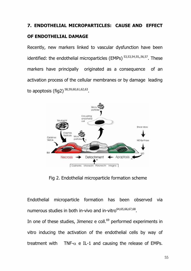
7. ENDOTHELIAL MICROPARTICLES: CAUSE AND EFFECT
OF ENDOTHELIAL DAMAGE
Recently, new markers linked to vascular dysfunction have been
identified: the endothelial microparticles (EMPs) 53,53,54,55,,56,57. These
markers have principally originated as a consequence of an
activation process of the cellular membranes or by damage leading
to apoptosis (fig2) 58,59,60,61,62,63.
Fig 2. Endothelial microparticle formation scheme
Endothelial microparticle formation has been observed via
numerous studies in both in-vivo and in-vitro64,65,66,67,68.
In one of these studies, Jimenez e coll.69 performed experiments in
vitro inducing the activation of the endothelial cells by way of
treatment with TNF-α e IL-1 and causing the release of EMPs.
56
Analogous results have also been obtained by Combes et al70 where
the release of EMPs was induced by treating the endothelial cells
with the serum of patients with antiphospholipid syndrome.
Furthermore, from a study by Laurence and coll. 71 the correlation
between EMPs and apoptosis showed changes in the apoptic
endothelial cells following exposure to TTP plasma.
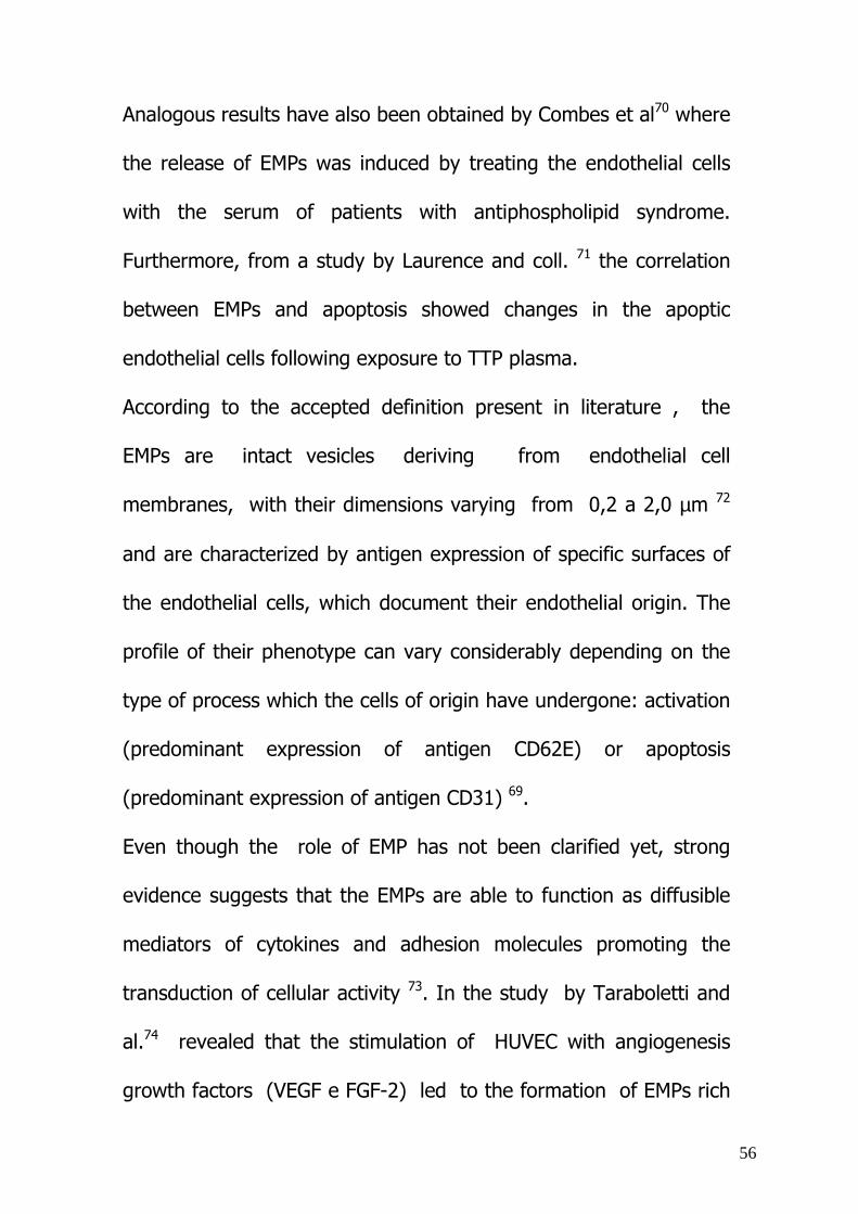
According to the accepted definition present in literature , the
EMPs are intact vesicles deriving from endothelial cell
membranes, with their dimensions varying from 0,2 a 2,0 µm 72
and are characterized by antigen expression of specific surfaces of
the endothelial cells, which document their endothelial origin. The
profile of their phenotype can vary considerably depending on the
type of process which the cells of origin have undergone: activation
(predominant expression of antigen CD62E) or apoptosis
(predominant expression of antigen CD31) 69.
Even though the role of EMP has not been clarified yet, strong
evidence suggests that the EMPs are able to function as diffusible
mediators of cytokines and adhesion molecules promoting the
transduction of cellular activity 73. In the study by Taraboletti and
al.74 revealed that the stimulation of HUVEC with angiogenesis
growth factors (VEGF e FGF-2) led to the formation of EMPs rich
57
in metalloproteinases matrix and capillary structures, suggesting
their potential role in angiogenesis. Furthermore, it has been
demonstrated that the EMPs have procoagulant activity, defined
through the action of platelet factor 3 and TF75. In the pro-
thrombotic state, in fact, a study by Shet e coll. 76 has evidenced
high values of TF-positive EMPs, which were strongly correlated
with procoagulant activity. It is also noteworthy to report also the
work by Jimenez and coll.77 where a severe endothelial dysfunction
in the aorta of mice after exposure to EMPs was evidenced. In this
study it was demonstrated that the EMPs are capable of
influencing the transduction pathway of NO, but not the
expression of NO synthesis, with a consequential induction of
endothelial dysfunction.
Further implications of the EMPs in the pathophysiology reside in
their pro-inflammatory activity and their tendency to bind the
activated monocytes. In the work by Boulanger and coll.78in fact ,
it has been demonstrated that the EMPs induce the release of
cytokines (TNF-α e IL-1β) with the consequential pacrine and
autocrine activations of the monocytes and the endothelium.
58
8. THE CREATION OF A PULMONARY HYPERTENSION
DATABASE AND THE CORRELATION BETWEEN DISEASE
GRADING AND INFLAMMATORY INDEXES
8.1 Materials and methods
Our research was, above all, aimed at identifying the patients
affected by pulmonary hypertension (PH), who had been admitted
to the Padua University Hospital, and re-evaluating their clinical
diagnoses on the basis of new classifications adopted at The 2003
World Symposium on Pulmonary Arterial Hypertension held in
Venice, Italy.
We create a database of patients, updated according to the new
nomenclature and on the basis of collected data we carry out our
study.
With this goal in mind, we obtained a lists of patients from the
administrative offices, admitted between 2000 and 2006 at
University Padua Hospital and discharged with the code 4160
(primary pulmonary hypertension) or with the code 4168 (other
forms of pulmonary hypertension) for the disease classification
ICD-9-CM79.
A Microsoft Excel file of 255 patients was created which
included: the first and last name of the patient, date of birth, type
59
of admission, medical chart number, dates of admission and
discharge, the departments of admission and discharge, the
principal diagnosis and 6 secondary diagnoses, the names of up the
departments in which the patient was moved during the hospital
stay.
According the ICD-9-CM codes, which specifically referred to the
primary form, it was impossible to distinguish between the familial
form and the idiopathic form, since the two were included in the
same code. The recoding was made on the basis of a thorough
evaluation of all the information available in the clinical charts.
The other important evaluation that was done at the time of the
medical chart analysis concerned the secondary form that the
international classification identifies generically as other forms of
pulmonary hypertension (PH) with the code 4168. The new
classification of PH distinguishes these latter into 5 classes.
To fulfil the privacy low, the names of the patients were transferred
from the excel file to an access file where the data of the patients
were registered. Furthermore, we created excel files for the
numerical values to perform statistical analysis.
For each medical chart , we identified these most significant data:
main diagnoses and up to 3 comorbities; relevant family
60
history;remote pathological anamnesis; NYHA classes at diagnosis
and after treatment; haematochemical exams including blood cell
count at admission and after therapy; ESR; serum protein
electrophoresis; plasma level of fibrinogen; C-reactive protein, uric
acid, albumin; electrocardiographic data signalling the absence or
presence of right ventricular hypertrophy; echocardiographic data
with a particular attention to the severity of pulmonary
hypertension, dimension and dynamics of right cardiac cavities;
ABG (arterial blood gas) values at baseline and after therapy;
pulmonary functioning test; 6 minute walking test; right cardiac
catherization data and finally medical treatment performed.
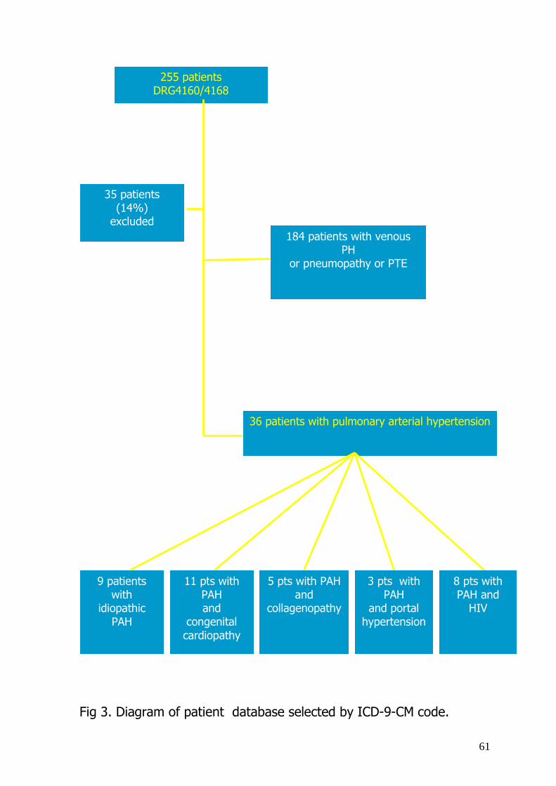
Two hundreds fifty-five patients were selected(fig 3). Thirty-five
(14%) were excluded because the clinical diagnosis of pulmonary
hypertension didn’t satisfy the definition of Venice Classification.
Thirthy-six patients were affected by PAH.
In our database the total number of patients affected by other
forms of PH was 184 subjects.
Since we had limited our research to forms of PAH, patients with
other forms were excluded.
61
Fig 3. Diagram of patient database selected by ICD-9-CM code.
255 patients DRG4160/4168
35 patients (14%)
excluded 184 patients with venous
PH or pneumopathy or PTE
36 patients with pulmonary arterial hypertension
9 patients with
idiopathic PAH
11 pts with PAH and
congenital cardiopathy
5 pts with PAH and
collagenopathy
3 pts with PAH
and portal hypertension
8 pts with PAH and
HIV

62
Idiopathic arterial hypertension
During the study period (2000-2006) 2±1 cases of idiopathic PAH
for year were reported.
Two of the nine patients with idiopathic PH presented positive
family hystory for PH and they were sent for genetic testing at the
University of Bologna and considered affected by familial PH.
The ratio men/women was 1.6:1, confirming the prevalence of
these disease in the female sex.
The average age of the subjects of both sexes at diagnosis was 54
±1 year. Only a boy of 12 years, showed an idiopathic PAH form, at
echo and cardiac catheterization.
Body mass index, calculated from reported chart data on height
and weight resulted normal with values of 25±4. (tab 2)
The time interval between onset of symptoms and the final
diagnosis was 12±6 months in 5 subjects, and 24±6 months in
the remaining 4 patients.
The clinical presentation was highly variable. Dyspnoea during
exercise and at rest was the symptom most frequently reported.
Moreover 3 patients suffered episodes of syncope. Angina at rest
and after exercise was reported by 4 patients. Aspecific symptoms

63
which included asthenia and abdominal discomfort were also
reported by all of the subjects.
At clinical examination, most of the subjects presented severe
symptoms at diagnosis, with none of the patients in NYHA I class,
6 patients in NYHA classes II/III and 3 patients in NYHA class
IV (tab2).
The mean values of the pulmonary artery systolic pressure values
were obtained by Doppler method from tricuspid regurgitation80,81,82
was 53±15 mmHg. The arterial pulmonary systolic pressure values
were higher in subjects in the advanced NYAH classes.
Five patients showed at ECG right ventricular hypertrophy.
Normal value of ABG analysis were present at rest and at room
temperature in all cases.
The distance covered during the 6MW test showed a correlation
with the NYHA classes of the patients: 6 subjects in NYHA classes
II and III travelled an average distance of 360 meters, 3 patients
in NYHA class IV exhibited a capacity to cover a much shorter
distance of 190 meters.
The respiratory function tests were in the normal range.
At cardiac catherization the mean values was PAP values was 56
±15 mmHg and normal capillary wedge pressures. and pulmonary
64
vascular resistance of 18±5 UI. Pharmacological tests 82 with NO
inhalators or intravenously delivered prostacyclin were carried out
in 4 out of 5 subjects examined and 2 subjects were responders.
All the patients were treated with oral anticoagulants. Calcium
antagonists (nifedipin, ditiazem or amlodipine) were taken by 3
patients. Seven patients were in therapy with receptor antagonists
of endothelin (bosentan). In three patients epoprostenol ev was
administered.
Six of the nine patients had repeated admissions to hospital.
Considering the lack of homogeneity in the tests performed during
readmission, it was not possible to carry out analyses on: response
to therapy, disease evolution and patient survival.

65
Table 2. Parameters of patients with idiopathic pulmonary hypertension Age 54 years ± 12 months
Sex Men/Women 1.6:1
NYHA classes at diagnosis 6 pt in NYHA classes II-III
3 pt. in NYHA classes II-IV
Time between symptom onset
and diagnosis
5 pt 12±6 months
4 pt 24±6 months.
ECG 5 pt right ventricular
hypertrophy
Eco PAPs 53±15 mmHg.
6MW test Median distance covered
306 meters
Median PAP 56±15 mmHg
PVR (pulmonary vascular
resistance)
18±5 UI
66
Associated forms of pulmonary arterial hypertension
Congenital heart disease
Our associated PAH cases consists of 27 patients of which eleven
presented congenital heart disease, five collagen disease, eight
AIDS, three portal hypertension correlated to hepatic pathologies
(two affected with hepatic cirrhosis and one had Budd-Chiari
syndrome). No cases of secondary farmacologic induced pulmonary
arterial hypertension could be identified.
Eleven patients affected by congenital heart disease had median
age of 25 years±6 months. The ratio men/ women was 1.5:1.
Six patients had an interventricular septum defect: three isolated,
one associated with patent ductus areriosus and atrial septum fossa
ovale type defect; one patient had a defect of atrial-ventricular
septum; a single case of pulmonary atresia with ventricular septal
defect and systemic pulmonary plurifocal vascolarization, two
subjects had a common truncus arteriosus, one had an non
obstructive total venous anomalus pulmonary vein drainage into the
coronary sinus.
The mean values of the PAPs at echocardiogram was 55±12
mmHg. At cardiac catherization the mean value of PAP was 56 ±15

67
mmHg, normal capillary wedge pressure along with pulmonary
vascular resistance 16±8 UI.
All patients of this series were under therapy with antagonist
receptors of endothelin (bosentan).
Other forms
Five patients with PAH associated with a collagen disease had a
mean age of 55 years±14 months. The ratio men/women was 1:2.
In this group of patients the most frequent disease was
scleroderma (two of which had the diffused form and one had the
limited form (CREST)). One patient was affected by lupus; one
presented an undeterminated mixed connective tissue disease.
All patients were in NYHA classes II or III. Most of the subjects
showed the phenomenon of Raynaud. The mean PAP values were
65±5 mmHg. Invasive haemodynamic investigation was carried out
in 3 of the patients from the group.
In our analyses we considered patients with all the forms of
pulmonary hypertension, included in the first group of the
International Classification held in Venice 2003, eventhough we
recognized that patients with asociated connective tissue disorders
could present higher indexes of inflammation.
68
Data are presented as means and standard error of the mean
(SEM). In order to compare the above listed variables between the
PH patients and the healthy controls, the Student’s t-test was
performed on the parametric variables. While the Wilcoxon test was
performed on the non-parametric variables. The Pearson and
Spearman correlation analyses were utilized to quantify the
statistical correlations, respectively among the parametric and non-
parametric variables. ANOVA was employed to compare the levels
of the various phlogistic indexes among the patients grouped in the
fourth NYHA class.

69
8.2 Results
The analysis of the data of the 36 patients with idiopathic or
associated PAH and the data obtained from the 36 healthy controls,
showed:
1. The patients affected by PAH compared to healthy control
subjects had white blood cell counts of 6700±113/mm3 vs
6360±156/mm3 and ESR levels of 21±2.5 mm vs 16±2.7 mm
respectively. However, a statistically significant difference was
found (p<0.05) for CRP (4.7±0.09 mg/l vs 1,6±0.04 mg/l),
fibrinogen (465±53 mg/dl vs 319±66 mg/dl), uric acid (5,1±0.8
mg/dl vs 3,2±0.7 mg/dl), which were higher in patients with
pulmonary arterial hypertension (fig 4). On the other hand, the
levels of albumin was lower in patients with pulmonary arterial
hypertension than in healthy controls (3.1±0.8 g/dl vs 4.1±1.1 g/dl,
p=0.026).

70
0
0,5
1
1,5
2
2,5
3
3,5
4
4,5
5
CRP mg/L
PAH
Controls
150
200
250
300
350
400
450
500
Fibrinogen mg/dl
PAH
Controls
0
1
2
3
4
5
6
Uric Acid mg/dl
PAH
Controls
Figure 4. CRP, fibrinogen and uric acid blood levels in patients with pulmonary arterial hypertension (PAH) and controls. Data are presented as means plus standard error of the mean (SEM).
4.7 (0.09)
1,6 (0.04)
465 (53)
319 (66)
5,1 (0,8)
3,2 (0,7)
71
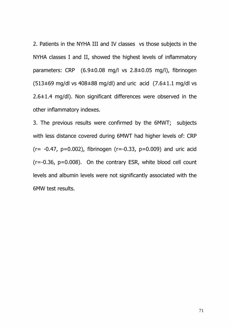
2. Patients in the NYHA III and IV classes vs those subjects in the
NYHA classes I and II, showed the highest levels of inflammatory
parameters: CRP (6.9±0.08 mg/l vs 2.8±0.05 mg/l), fibrinogen
(513±69 mg/dl vs 408±88 mg/dl) and uric acid (7.6±1.1 mg/dl vs
2.6±1.4 mg/dl). Non significant differences were observed in the
other inflammatory indexes.
3. The previous results were confirmed by the 6MWT; subjects
with less distance covered during 6MWT had higher levels of: CRP
(r= -0.47, p=0.002), fibrinogen (r=-0.33, p=0.009) and uric acid
(r=-0.36, p=0.008). On the contrary ESR, white blood cell count
levels and albumin levels were not significantly associated with the
6MW test results.

72
8.3 Discussion
A series of relevant observations could be drawn from the analysis
of clinical notes of patients with PH selected in our database.
Non specific symptoms characterized patients with pulmonary
hypertension. In fact the diagnosis needs a quantitative evaluation
by echocardiogram or cardiac catheterization. Moreover different
disease can produce the same haemodynamic findings. All these
facts produce bias in clinical diagnosis reported in the charts and
may justify the relative low number of patients recorded.
The nosologic modifications introduced at the Venice Convention
produced a significant improvement, because endorsed the concept
that clinical conditions with different ethiology but with the same
pathological structural alterations could be grouped togheter.
According to the Venice Classification we structured our database
enrolling patients with different ethiology but with the same
disease.
From the result of our analysis we can state that patients with PAH
present a sub-clinical activation of the inflammatory cascade, which
could be related to the grade of pulmonary vascular disease.
The role of uric acid in inflammation process is controversial.
However we can confirm the increase of uric acid blood levels in
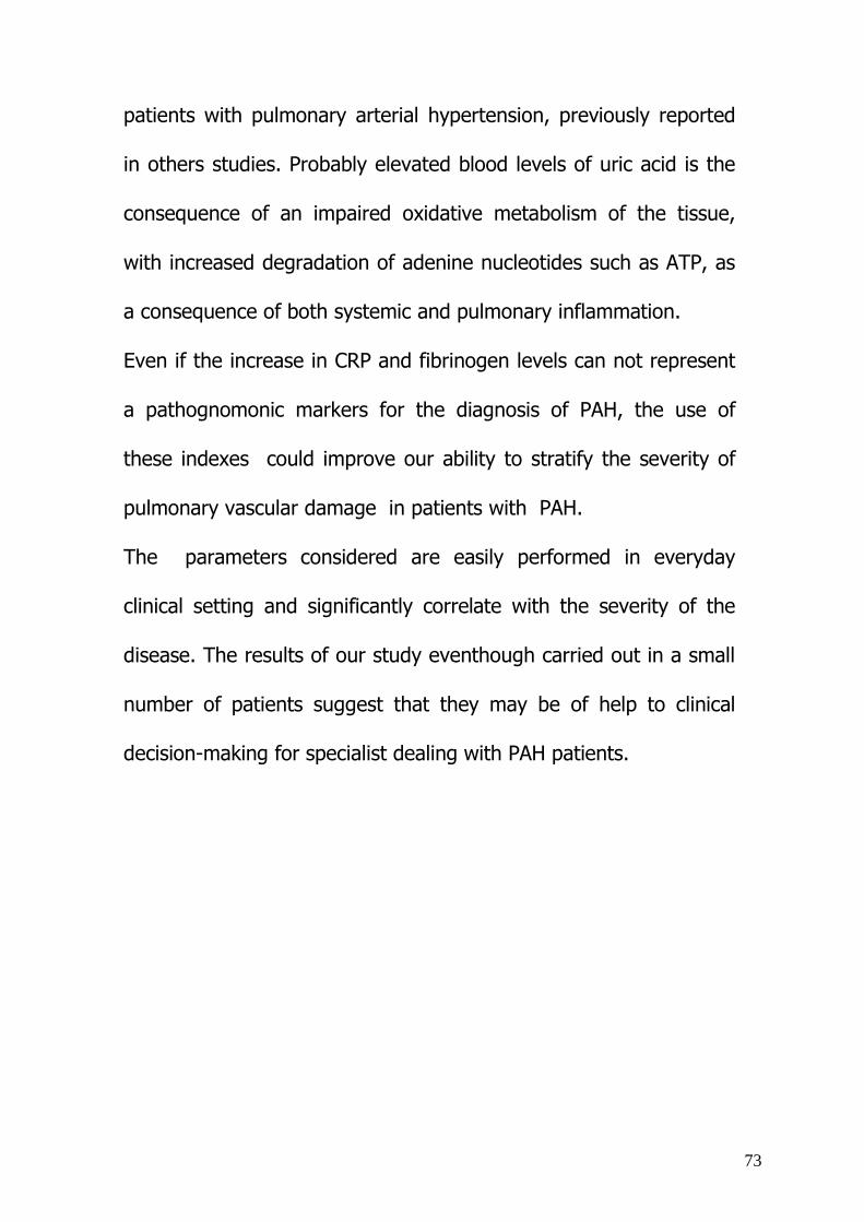
73
patients with pulmonary arterial hypertension, previously reported
in others studies. Probably elevated blood levels of uric acid is the
consequence of an impaired oxidative metabolism of the tissue,
with increased degradation of adenine nucleotides such as ATP, as
a consequence of both systemic and pulmonary inflammation.
Even if the increase in CRP and fibrinogen levels can not represent
a pathognomonic markers for the diagnosis of PAH, the use of
these indexes could improve our ability to stratify the severity of
pulmonary vascular damage in patients with PAH.
The parameters considered are easily performed in everyday
clinical setting and significantly correlate with the severity of the
disease. The results of our study eventhough carried out in a small
number of patients suggest that they may be of help to clinical
decision-making for specialist dealing with PAH patients.
74
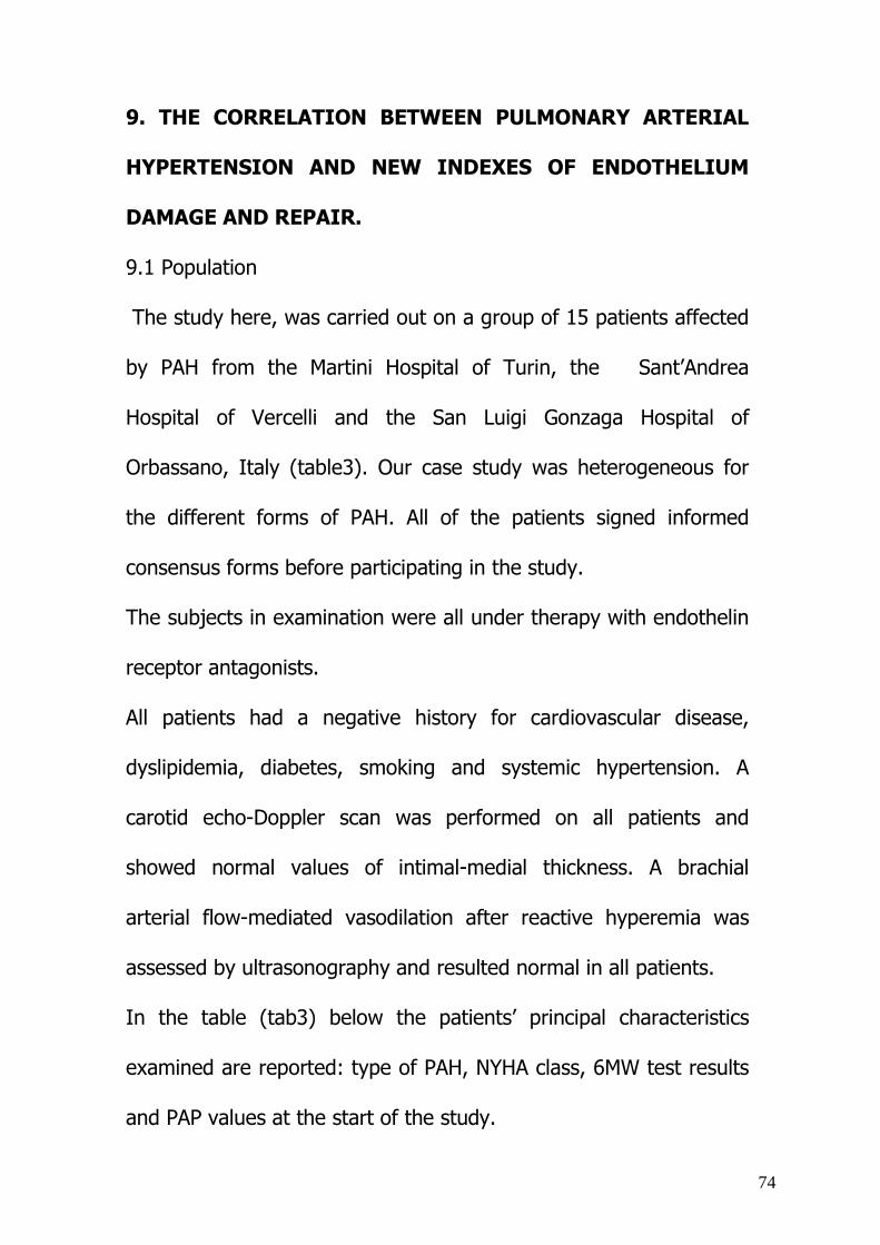
9. THE CORRELATION BETWEEN PULMONARY ARTERIAL
HYPERTENSION AND NEW INDEXES OF ENDOTHELIUM
DAMAGE AND REPAIR.
9.1 Population
The study here, was carried out on a group of 15 patients affected
by PAH from the Martini Hospital of Turin, the Sant’Andrea
Hospital of Vercelli and the San Luigi Gonzaga Hospital of
Orbassano, Italy (table3). Our case study was heterogeneous for
the different forms of PAH. All of the patients signed informed
consensus forms before participating in the study.
The subjects in examination were all under therapy with endothelin
receptor antagonists.
All patients had a negative history for cardiovascular disease,
dyslipidemia, diabetes, smoking and systemic hypertension. A
carotid echo-Doppler scan was performed on all patients and
showed normal values of intimal-medial thickness. A brachial
arterial flow-mediated vasodilation after reactive hyperemia was
assessed by ultrasonography and resulted normal in all patients.
In the table (tab3) below the patients’ principal characteristics
examined are reported: type of PAH, NYHA class, 6MW test results
and PAP values at the start of the study.
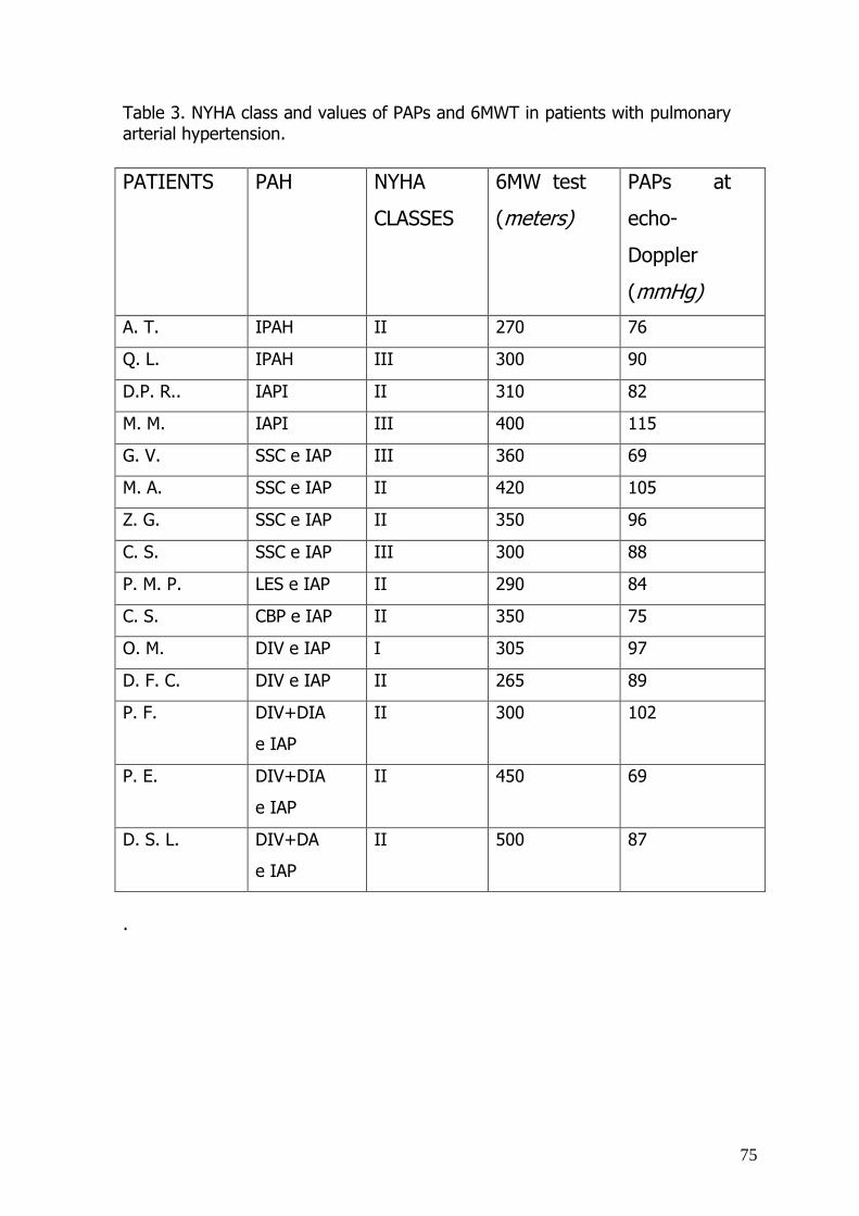
75
Table 3. NYHA class and values of PAPs and 6MWT in patients with pulmonary arterial hypertension. PATIENTS
PAH NYHA
CLASSES
6MW test
(meters)
PAPs at
echo-
Doppler
(mmHg)
A. T. IPAH II 270 76
Q. L. IPAH III 300 90
D.P. R.. IAPI II 310 82
M. M. IAPI III 400 115
G. V. SSC e IAP III 360 69
M. A. SSC e IAP II 420 105
Z. G. SSC e IAP II 350 96
C. S. SSC e IAP III 300 88
P. M. P. LES e IAP II 290 84
C. S. CBP e IAP II 350 75
O. M. DIV e IAP I 305 97
D. F. C. DIV e IAP II 265 89
P. F. DIV+DIA
e IAP
II 300 102
P. E. DIV+DIA
e IAP
II 450 69
D. S. L. DIV+DA
e IAP
II 500 87
.
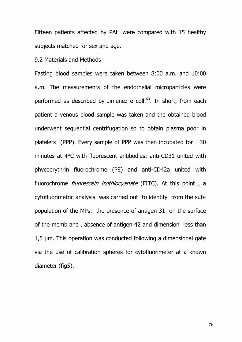
76
Fifteen patients affected by PAH were compared with 15 healthy
subjects matched for sex and age.
9.2 Materials and Methods
Fasting blood samples were taken between 8:00 a.m. and 10:00
a.m. The measurements of the endothelial microparticles were
performed as described by Jimenez e coll.69. In short, from each
patient a venous blood sample was taken and the obtained blood
underwent sequential centrifugation so to obtain plasma poor in
platelets (PPP). Every sample of PPP was then incubated for 30
minutes at 4°C with fluorescent antibodies: anti-CD31 united with
phycoerythrin fluorochrome (PE) and anti-CD42a united with
fluorochrome fluorescein isothiocyanate (FITC). At this point , a
cytofluorimetric analysis was carried out to identify from the sub-
population of the MPs: the presence of antigen 31 on the surface
of the membrane , absence of antigen 42 and dimension less than
1,5 µm. This operation was conducted following a dimensional gate
via the use of calibration spheres for cytofluorimeter at a known
diameter (fig5).

77
Events of∅ < 1,5 µm
PMPs
EMPs
Fig 5. Cytofluorimetric analysis of the EMPs
To determine the number of endothelial progenitors a blood sample
was taken, from every participant in the study. EDTA was used as
an anticoagulant in the test tube blood samples. Each sample of
whole blood underwent a centrifugation on density gradient so as
to separate the mononuclear cells (lymphocytes e monocytes).
After sequential washing with PBS, so to eliminate the eventual
platelet contamination, the obtained cells were incubated at 4°C for
30 minutes with antibodies anti-CD34-FITC and anti-KDR-PE. After
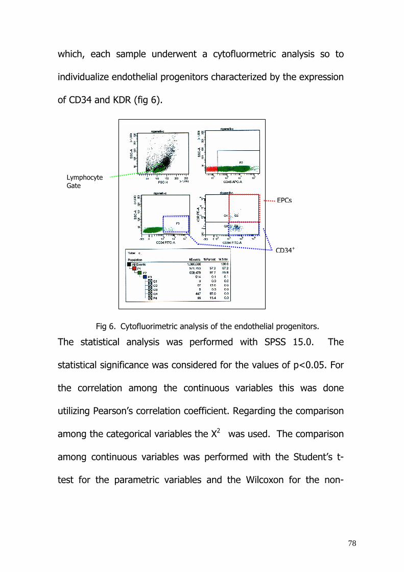
78
Lymphocyte Gate
EPCs
CD34+
which, each sample underwent a cytofluormetric analysis so to
individualize endothelial progenitors characterized by the expression
of CD34 and KDR (fig 6).
Fig 6. Cytofluorimetric analysis of the endothelial progenitors.
The statistical analysis was performed with SPSS 15.0. The
statistical significance was considered for the values of p<0.05. For
the correlation among the continuous variables this was done
utilizing Pearson’s correlation coefficient. Regarding the comparison
among the categorical variables the X2 was used. The comparison
among continuous variables was performed with the Student’s t-
test for the parametric variables and the Wilcoxon for the non-
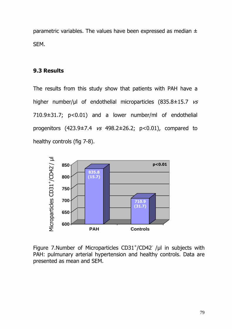
79
parametric variables. The values have been expressed as median ±
SEM.
9.3 Results
The results from this study show that patients with PAH have a
higher number/µl of endothelial microparticles (835.8±15.7 vs
710.9±31.7; p<0.01) and a lower number/ml of endothelial
progenitors (423.9±7.4 vs 498.2±26.2; p<0.01), compared to
healthy controls (fig 7-8).
600
650
700
750
800
850
PAH Controls
Figure 7.Number of Microparticles CD31+/CD42- /µl in subjects with PAH: pulmunary arterial hypertension and healthy controls. Data are presented as mean and SEM.
Mic
ropa
rtic
les
CD31
+/C
D42
- /µl
835.8 (15.7)
710.9 (31.7)
p<0.01
80
380
400
420
440
460
480
500
PAH Controls
Figure 8.Number of EPc/µl in subjects with PAH: pulmonary arterial Hypertension and healthy controls. Data are presented as mean and SEM.
The correlation analysis performed in the entire cohort between the
number of endothelial microparticles and the PAP levels assessed at
echocardiography demonstrates that there exists a significantly
positive relationship between these two variables (r=0.609;
p=0.004). Figure 9 shows the increase of endothelial microparticles
for increasing tertiles of PAPs (1st tertile: < 35 mmHg, 2nd tertile 35-
85 mmHg, 3d tertile >85 mmHg).
EPc/µl
423,9 (7,4)
498,2 (26,2)
p<0.01
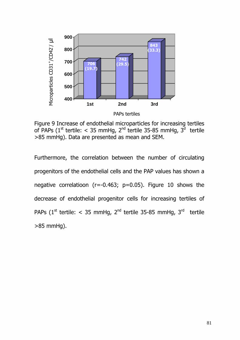
81
400
500
600
700
800
900
1st 2nd 3rd
Figure 9 Increase of endothelial microparticles for increasing tertiles of PAPs (1st tertile: < 35 mmHg, 2nd tertile 35-85 mmHg, 3d tertile >85 mmHg). Data are presented as mean and SEM.
Furthermore, the correlation between the number of circulating
progenitors of the endothelial cells and the PAP values has shown a
negative correlatioon (r=-0.463; p=0.05). Figure 10 shows the
decrease of endothelial progenitor cells for increasing tertiles of
PAPs (1st tertile: < 35 mmHg, 2nd tertile 35-85 mmHg, 3rd tertile
>85 mmHg).
Mic
ropa
rtic
les
CD31
+/C
D42
- /µl
PAPs tertiles
706 (19.7)
742 (29.5)
843 (33.3)

82
400
420
440
460
480
500
520
1st 2nd 3rd
Figure 9. Decrease of endothelial progenitors cells for increasing tertiles of PAPs (1st tertile: < 35 mmHg, 2nd tertile 35-85 mmHg, 3d
tertile >85 mmHg). Data are presented as mean and SEM.
A negative correlation between the number of microparticles and
the 6MW test results was found (r=-0.573, p=0.001). Figure 11
shows the increase of endothelial microparticles for decreasing
tertiles of distance travelled at the 6MWT (1st tertile >600 meters,
2nd tertile 350-600 meters, 3rd tertile <350 meters).
On the other hand, the number of endothelial progeniors cells
decreases with a reduction in the results from the 6MW test
(r=0.444; p=0.014). Figure 12 shows the decrease of endothelial
progenitor cells for decreasing tertiles of distance travelled at the
PAPs tertiles
EPc/µl
406,7 (13,4)
436,7 (11,6)
468,9 (31,3)

83
6MWT (1st tertile >600 meters, 2nd 350-600 meters, 3rd <350
meters).
400450500550600650700750800850
1st 2nd 3rd
Figure 11. Increase of endothelial microparticles for decreasing tertiles of distance travelled at the 6MWT (1st tertile >600 meters, 2nd tertile 350-600 meters, 3rd tertile <350 meters).
Mic
ropa
rtic
les
CD31
+/C
D42
- /µl
6 MW test tertiles
706 (24.8)
735 (32.6)
819 (25.6)

84
Figure 12. Decrease of endothelial progenitor cells for decreasing tertiles of distance travelled at the 6MWT (1st tertile >600 meters, 2nd tertile 350-600 meters, 3rd tertile<350 meters).
EPc/µl
6 MW test tertiles
400
410
420
430
440
450
460
470
480
1st 2nd 3rd
475 (23.6)
445 (16.5) 420
(14,6)
85
9.4 Discussion
In our study we report an increase in the number of circulating
endothelial microparticles and endothelial progenitor cells in
patients with pulmonary arterial hypertension when compared to
normal volunteers. We have shown these markers correlate with
the pulmonary artery pressure and the exercise capacity of the
individual.
We hypothesized that the shear stress could result in the shedding
of the involved endothelial cells, resulting in an increase of
circulating endothelial microparticles.
Although we were unable to identify the precise site of origin of the
endothelial microparticles in our patients, we suggest that the only
likely site of origin is the pulmonary vasculature. This hypotesis is
based on: 1) the microparticles demonstrate markers CD31+/CD42-
of endothelial origin (the pulmonary vasculature has the greatest
microvascular surface area in the body), 2) the number of
microparticles correlate significantly with the severity of pulmonary
hypertension, 3) in patients with PAH the pulmonary vascular bed is
the predominant site of pathology, 4) in our patients the inclusion
criteria exluded the presence of pathologies producing systemic
endothelial vascular damage .
86
On the other hand, we were unable to determine the homing and
differentiation of the endothelial progenitors cells, but we can affirm
that their number is impaired in patients with pulmonary vascular
disease.
Because we did not have lung biopsy samples in our patients, it was
not possible to correlate the angiogenetic properties of endothelial
progenitor cell with the presence of plexiform lesions.
However it was clear that these cells could play an important role in
the pathobiology of pulmonary arterial hypertension.
From the results of our study we think that endothelial microparticles
and endothelial progenitors can serve as a surrogate marker of the
disease process, and can provide a test for early diagnosis of severe
pulmonary hypertension.
87
10 CONCLUSIONS
Pulmonary arterial hypertension is listed among the rare diseases,
and is characterized by vascular obstruction, leading to increased
pulmonary vascular resistence and right side heart failure.
It occurs in women more often than man and, most important, has
a mean age at diagnosis of 36 years and is usually fatal within 3
years without therapy. Modern treatment has markedly improved
physical function and has extended survival.
We still do not understand what initiate the disease and what
allows it to progress. Recently genetic factors has been indentify
as key player in the development of this multifactorial disease.
Our interest in the inflammatory biomarkers reflect the current
momentum and excitement surrounding research on PAH.
The aim of our research was to determine the role of inflammation
in the pathobiology of the disease through the assessment of
inflammatory biomarkers and to indentify them as a possible clinical
useful biomarkers that allow non invasive diagnosis and monitoring
of PAH.
The balance between endothelial damage and repair is essential for
the maintenance of vascular homeostasis. We think that the
endothelial microparticles CD31+/CD42- represent a new and valid
88
indicator for damage produced at the level of the pulmonary
vasculature. On the other hand, greater evidence seems to point to
the fact that endothelial damage is a condition essential, but not
sufficient for the deterioration of the vascular function. It is clear
that the latter is the result not only of the vascular damage but also
to the repair mechanisms.
Currently, many evidence support that hypotesis that endothelial
progenitor cells play a key role in the repair function of damaged
endothelium. Taken this as it may, all the conditions that are
capable of influencing the number or the function of endothelial
progenitor cells are significantly involved in the onset of vascular
damage.
The results of this study demonstrate that PAH is the result of the
imbalance between damage and repair mechanisms, which
together guarantee normal vascular function: increased numbers of
endothelial microparticles as results of vascular damage and
decreased number endothelial progenitors cells as alteration of
repair mechanism.
In other words, PAH produces a fragmentation of mature
endothelial cells, increases formation of endothelial microparticles
and reduces the number of circulating endothelial progenitors. This

89
constitutes the functional expression of a vascular dysfunction
which in turn determines a structural and biological reshaping of
the pulmonary vascular tree and furthermore, also involves complex
systemic homeostatic mechanisms.
90
Bibliografia
1. Simonneau G, Galié N, Rubin LJ et al. Clinical Classification of
Pulmonary Hypertension. JACC 2004;43:5-12S.
2. Budhiraja R, Tuder RM and Hassoun PM. Endothelial
dysfunction in pulmonary hypertension. Circulation
2004;109:159-165.
3. M Humbert, NW Morrell, SL Archer, KR Stenmark, MR
MacLean, IM Lang et al. Cellular and molecular pathobiology
of pulmonary arterial hypertension. J Am Coll Cardiol
2004;43:12-23.
4. Humbert M, Monti G, Fartoukh et al. Increased interleukin-1
and interleukin-6 serum concentrations in severe primary
pulmonary hypertension. Am J Respir Crit Care Med
1995;151:1628-1631.
5. Dorfmuller P, Zarka V, Durand-Gasselin I et al. Chemokine
RANTES in severe pulmonary arterial hypertension. Am J
Respir Crit Care Med. 2002;165:534-539.
6. Fartoukh M, Emilie D, Le Gall C, et al. Chemokine macrophage
inflammatory protein -1α mRNA expression in lung biopsy
specimens of primari pulmonary hypertension. Chest 1998;
114 (1 suppl):50S-51S.

91
7. Pinto RFA, de Lourdes Higuchi M, Aiello VD. Decreased
numbers of T-lymphocytes and predominance of recently
recruited macrophages in the walls of peripheral pulmonary
arteries from 26 patients with pulmonary hypertension
secondary to congenital cardiac shunts. Cardiovascular
Pathology 2004.13.268-275.
8. Damas JK, Otterdal K, Yndestand A et al. Soluble CD40 ligand
in Pulmonary Arterial Hypertension. Possible pathogenetic
role of the interaction between platelets and endothelial cells.
Circulation 2004;110:999-1005.
9. Aubert J-K. Biochemical markers in the management of
pulmonary hypertension. Swiss Med wkly 2005;135:43-49
10. SG Haworth. Role of the endothelium in pulmonary arterial
hypertension. Vascular Pharmacology 45 (2006) 317-325.
11. KR Stenmark, KA Fagan, MG Frid. Hypoxia-induced
pulmonary vascular remodelling. Cellular and molecular
mechanism. Cir Res 2006;99:675-691.
12. Burke A and Virmani R. Mini-symposium:pulmonary
pathologu. Evalutaion of pulmonary arterial hupertension in
biopsies of the lung. Current Diagnostic Pathology.
2006;3:14-26.
13. NJ Davie, JT Crossno, MG Frid. Hypoxia-induced pulmonary
adventitial remodelling and neovascularization:contributor of
92
progenitor cells. Am J Physiol Lung cell Mol Physiol
2004;286:668-678.
14. Jimenez JJ, Jy W, Mauro LM, Horstman LL, Bidot CJ, Ahn YS.
Endothelial microparticles (EMP) as vascular disease markers.
Adv Clin Chem. 2005;39:131–157.
15. Koga H, Sugiyama S, Kugiyama K, Watanabe K, Fukushima
H, TanakaT, Sakamoto T, Yoshimura M, Jinnouchi H, Ogawa
H. Elevated levels of VE-cadherin-positive endothelial
microparticles in patients with type 2 diabetes mellitus and
coronary artery disease. J Am Coll Cardiol. 2005;45:1622–
1630.
16. Amabile N, Guerin AP, Leroyer A, Mallat Z, Nguyen C,
Boddaert J,London GM, Tedgui A, Boulanger CM. Circulating
endothelial microparticlesare associated with vascular
dysfunction in patients with end-stage renal failure. J Am Soc
Nephrol. 2005;16:3381–3388.
17. Asahara T, Murohara T, Sullivan A et al. Isolation of putative
progenitor endothelial cells for angiogenesis. Science
1997;275:964-7.
18. Masataka Sata. Role of circulating vascular progenitors in
angiogenesis, vascular healing, and pulmonary hypertension.
Lessons from animal models. Artheriosclerosis Thromb Vasc
Biol 2006;26:1008-1014.
19. N Nagaya, K kangawa, M Kanda et al. Hybrid cell-gene
therapy for pulmonary hypertension based on phagocytosing
action of endothelial progenitor cells. Circulation
2003;108:889-895.
93
20. Wang XX, Zhang FR and Shang YP et al. Trasplantation of
autologous endothelial progenitor cell may be benefical in
patients with idiopathic pulmonary arterial hypertension.
JACC 2007;491566-71.
21. BarstRJ, McGoon M, Torbicki A, Sitbon O, Krowka MJ,
Olschewski H et al. Diagnosis and differential assessment of
pulmonary arterial hypertension. J Am Coll Cardiol
2004;43:40-7.
22. Simonneau G, Galie N, Rubin LJ, Langleben D, Seeger W,
Domenighetti G et al. Clinical classification of pulmonary
hypertension.J Am Coll Cardiol 2004;43:5-12.
23. G Pietra, F Capron, S Stewart, O Leone, M Humbert, IM
Robbins et al. Pathologic assessment of vasculopathies in
pulmonary hypertension. J Am Coll Cardiol 2004;43:24-32.
24. Martin KB, Klinger JR and SIS Rounds. Pulmonary arterial
hypertension: new insighst and new hope. Respirology 2006;
11:6-17.
25. N Galié, W Seeger, R Naeije, G Simonneau, LJ Rubin.
Comparative analysis of clinical trials and evidence-based
treatment algorithm in pulmonary arterial hypertension. J Am
Coll Cardiol 2004;43:81-88.

94
26. Korbling M, Estrov Z. Adult stem cells for tissue repair – a
new therapeutic consept? N Engl J Med 2003;349:570-82.27.
27. Korbling M, Estrov Z, Champlin R. Adult stem cells and tissue
repair. Bone Marrow Transplant 2003;32(1):S23-4.
28. Carmeliet P. Developmental biology: One cell, two fates.
Nature 2000;408:43-45.
29. Hristov M, Erl W, Weber PC. Endothelial progenitor cells:
mobilization differentiation, and homing. Arterioscler Thromb
Vasc Biol 2003;23:1185-9.
30. Liu C, Nath KA, Katusic ZS, et al. Smooth muscle progenitor
cells in vascular disease. Trends Cardiovasc Med
2004;14:288-93.
31. Yamashita J, Itoh H, Hirashima M, et al. Flk1-positive cells
derived from embryonic stem cells serve as vascular
progenitors. Nature 2000;408:92-6.
32. Stump MM, Jordan GL, Debakay ME, et al. Endothelium
grown from circulating blood on isolated intravascular Dacron
Hub. Am J Pathol 1963;43:361-367.
33. Zammaretti P, Zisch, Andreas H. Adult endothelial progenitor
cells: Renewing vasculature. The International Journal of
Biochemistry and Cell Biology 2005;37:493-503.

95
34. Shi Q, Rafii S, Wu MH, et al. Evidence for circulating bone
marrow-derived endothelial cells. Blood 1998;92:362-7.
35. Ferrario M, Massa M, Rosti V et al. Early haemoglobin-
independent increase of plasma erythropoietin levels in
patients with acute myocardial infarction. Eur Heart J. 2007
28:1805-13.
36. Xu Q. The role of stem cells in atherosclerosis. Arch Mal
Coeur Vaiss. 2005;98:672-6.
37. Urbich C, Dimmeler S. Risk factors for coronary artery
disease, circulating endothelial progenitor cells, and the role
of HMG-CoA reductase inhibitors. Kidney Int. 2005;67:1672-
6.
38. Hillebrands JL, Onuta G, Rozing J.Role of progenitor cells in
transplant arteriosclerosis. Trends Cardiovasc Med.
2005;15:1-8.
39. Messina E, De Angelis L, Frati G et al. Isolation and
expansion of adult cardiac stem cells from human and
murine heart. Circ Res. 2004;95:911-21.
40. Heeschen C, Aicher A, Lehmann R et al. Erythropoietin is a
potent physiologic stimulus for endothelial progenitor cell
mobilization. Blood. 2003 Aug 15;102:1340-6.
96
41. Lu SJ, Feng Q, Caballero S et al. Generation of
functional hemangioblasts from human embryonic stem cells.
Nat Methods. 2007;4:501-9.
42. Rosenzweig A. Circulating endothelial progenitors--cells as
biomarkers. N Engl J Med. 2005;353(10):1055-7.
43. Leor J, Marber M. Endothelial progenitors: a new Tower of
Babel? J Am Coll Cardiol. 2006; 48(8):1588-90.
44. Uzan G. Therapeutic potential of circulating endothelial
cells. J Soc Biol. 2005;199:107-11.
45. Kocher AA, Schuster MD, Bonaros N et al. Myocardial
homing and neovascularization by human bone marrow
angioblasts is regulated by IL-8/Gro CXC chemokines. J Mol
Cell Cardiol. 2006;40:455-64.
46. Dignat-George F, Sampol J, Lip G, Blann AD. Circulating
endothelial cells: realities and promises in vascular disorders.
Pathophysiol Haemost Thromb. 2003;33:495-9.
47. Qin G, Ii M, Silver M, Wecker A et al. Functional disruption of
alpha4 integrin mobilizes bone marrow-derived endothelial
progenitors and augments ischemic neovascularization. J Exp
Med. 2006 ;203153-63.
97
48. Wojakowski W, Tendera M. Mobilization of bone marrow-
derived progenitor cells in acute coronary syndromes. Folia
Histochem Cytobiol. 2005;43229-32.
49. Romagnani P, Annunziato F, Liotta F et al.CD14+CD34low
cells with stem cell phenotypic and functional features are
the major source of circulating endothelial progenitors.Circ
Res. 2005 ;97:314-22.
50. Galli D, Innocenzi A, Staszewsky L et al. Mesoangioblasts,
vessel-associated multipotent stem cells, repair the infarcted
heart by multiple cellular mechanisms: a comparison with
bone marrow progenitors, fibroblasts, and endothelial cells.
Arterioscler Thromb Vasc Biol. 2005;25:692-7.
51. Bonafè F, Muscari C, Guarnieri C, Caldarera CM.
Regeneration of infarcted cardiac tissue: the route of stem
cells. Ital Heart J Suppl. 2003;4:299-305.
52. Schwartzenberg S, Deutsch V, Maysel-Auslender S et al.
Circulating apoptotic progenitor cells: a novel biomarker in
patients with acute coronary syndromes. Arterioscler Thromb
Vasc Biol. 2007 ;27:27-31.
98
53. Schillaci G, Pirro M. Endothelial microparticles and arterial
stiffness: casual coincidence or causative culprit? Am J
Hypertens. 2007;20:965-6.
54. Wang JM, Huang YJ, Wang Y et al. Increased circulating
CD31+/CD42- microparticles are associated with impaired
systemic artery elasticity in healthy subjects. Am J
Hypertens. 2007 ;20:957-64.
55. Ardoin SP, Shanahan JC, Pisetsky DS. The role of
microparticles in inflammation and thrombosis. Scand J
Immunol. 2007;66:159-65.
56. Lacroix R, Sabatier F, Mialhe A et al. Activation of
plasminogen into plasmin at the surface of endothelial
microparticles: a mechanism that modulates angiogenic
properties of endothelial progenitor cells in vitro. Blood.
2007;110:2432-9.
57. Chironi G, Simon A, Hugel B et al. Circulating leukocyte-
derived microparticles predict subclinical atherosclerosis
burden in asymptomatic subjects. Arterioscler Thromb Vasc
Biol. 2006;26:2775-80.
99
58. Morel O, Toti F, Hugel B et al. Procoagulant microparticles:
disrupting the vascular homeostasis equation?Arterioscler
Thromb Vasc Biol. 2006;26:2594-604.
59. Boulanger CM, Amabile N, Tedgui A. Circulating
microparticles: a potential prognostic marker for
atherosclerotic vascular disease.Hypertension 2006;48:180-6.
60. Baird AE, Wright VL.Vascular biology: cellular and molecular
profiling. Semin Neurol. 2006;26:65-74.
61. Tan KT, Lip GY.The potential role of platelet microparticles in
atherosclerosis. Thromb Haemost. 2005;94:488-92.
62. Sata M. Circulating vascular progenitor cells contribute to
vascular repair, remodeling, and lesion formation. Trends
Cardiovasc Med 2003;13:249-53.
63. Gill M, Dias S, Hattori K, et al. Vascular trauma induces rapid
but transient mobilization of VEGFR2(+)AC133(+) endothelial
precursor cells. Circ Res 2001;88:167-74.
64. Martínez MC, Tesse A, Zobairi F, Andriantsitohaina R. Shed
membrane microparticles from circulating and vascular cells
in regulating vascular function.Am J Physiol Heart Circ
Physiol. 2005;288:H1004-9.
100
65. Moreno PR, Fuster V. New aspects in the pathogenesis of
diabetic atherothrombosis. J Am Coll Cardiol. 2004;44:2293-
300.
66. Héloire F, Weill B, Weber S, Batteux F. Aggregates of
endothelial microparticles and platelets circulate in peripheral
blood. Variations during stable coronary disease and acute
myocardial infarction.Thromb Res. 2003;110:173-80.
67. Pirro M, Schillaci G, Bagaglia F., Mannarino E, et al.
Increased Ratio of CD31+/CD42- Microparticles to
Endothelial Progenitors as a Novel Marker of Atherosclerosis
in Hypercholesterolemia. Arterioscler Thromb Vasc Biol. 2006
Nov;26(11):2530-5. Epub 2006 Aug 31.
68. Goon PKY, Lip GYH, Boos CJ, Stonelake PS, Blann AD.
Circulating endothelial cells, endothelial progenitor cells and
endothelial microparticles in cancer. Neoplasia 2006;8:79-88.
69. Jimenez JJ, Jy W, Mauro LM, Soderland C, Horstmann LL,
Ahn YS. Endothelial cells release phenotypically and
quantitatively distinct microparticles in activation and
apoptosis. Thromb Res 2003;109:175–180.
70. Combes V, Simon A, Grau GE, Arnoux D, Camoin L, Sabatier
F, Mutin M, Sanmarco M, Sampol J, Dignat-George F. In vitro

101
generation of endothelial microparticles and possible
prothrombotic activity in patients with lupus anticoagulant. J
Clin Invest 1999;104:93–102.
71. Laurence J, Mitra D, Steiner M, Staiano-Coico L, Jaffe E.
Plasma from patients with idiopathic and human
immunodeficiency virus-associated thrombotic
thrombocytopenic purpura induces apoptosis in
microvascular endothelial cells. Blood 1996:3245–3254.
72. Piccin A, Murphy WG, Smith OP. Circulating microparticles:
pathophysiology and clinical implications. Blood reviews 2006
(in press).
73. Freyssinet JM. Cellular microparticles: what are they bad or
good for? J Thromb Haemost 2003;1:1655–1662.
74. Taraboletti G, D'Ascenzo S, Borsotti P, Giavazzi R, Pavan A,
Dolo V. Shedding of the matrix metalloproteinases MMP-2,
MMP-9 and MT1-MMP as membrane vesicle-associated
components by endothelial cells. Am J Pathol 2002;160:673–
680.
75. Casciola-Rosen L, Rosen A, Petri M, Petri M, Schlissel M.
Surface blebs on apoptotic cells are sites of enhanced
procoagulant activity: implications for coagulation events and
antigenic spread in systemic lupus erythematosus. Proc Nat
Acad Sci USA 1996;93:1624–1629.
76. Shet AS, Aras O, Gupta K, Hass MJ, Rausch DJ, Saba N,
Koopmeiners L, Key NS, Hebbel RP. Sickle blood contains
102
tissue factor-positive microparticles derived from endothelial
cells and monocytes. Blood 2003;102:2678–2683.
77. Jimenez JJ, Jy W, Mauro LM, Horstmann LL, Soderland C,
Ahn YS. Endothelial microparticles released in thrombotic
thrombocytopaenic purpura express von Willebrand factor
and markers of endothelial activation. Br J Haematol
2003;123:896–902.
78. Boulanger CM, Scoazec A, Ebrahimian T, Henry P, Mathieu E,
Tedgui A, Mallat Z. Circulating microparticles from patients
with myocardial infarction cause endothelial dysfunction.
Circulation 2001;104:2649–2652.
79. Classificazione delle malattie, traumatismi e cause di morte.
Xa revisione, 2002. Istituto Poligrafico dello Stato.
80. S. Provencher, S. Martel, X Jais, O Sitbon, M. Humbert, G.
Simonneau. Diagnostic et classification des hypertensions
arteriélles pulmonaires. Pneumologie 2005;2:192-203.
81. S Sciomer, R Badagliaca, F Fedele. Pulmonary Hypertension:
echocardiographic assessment. Italian Heart Journal 2005
Ottobre;6:840-845.
82. N Gallié, A Manes, A Branzi. Evaluation of Pulmonary arterial
Hypertension. Italian Heart Journal 2005 Ottobre;6:575-581.

103