Radiofrequency identification (rfid) market in 2014 – 2020: transparency market research
Upload
heba-abdellatifCategory
view
158download
0
Pulmonary AlveolarProteinosis

Overview
• (PAP) is a syndrome
Characterized by progressive
accumulation of surfactant
Phospholipids and proteins
within alveoli and terminal
airways.
• The disease is not associated with inflammation,
and lung architecture is typically preserved

Patho-physiology• The alveoli in PAP are filled with proteinaceous
material, that is found to be normal surfactant composed of 90% lipids and 10% surfactant-associated proteins A, B, C, and D (SP-A , SP-B .. )
Defect in surfactant Homeostatic mechanisms
Increase productionOf surfactant
Decreased clearance of surfactant
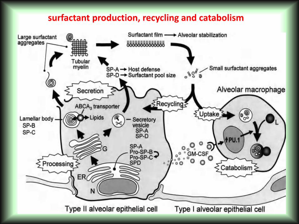
surfactant production, recycling and catabolism


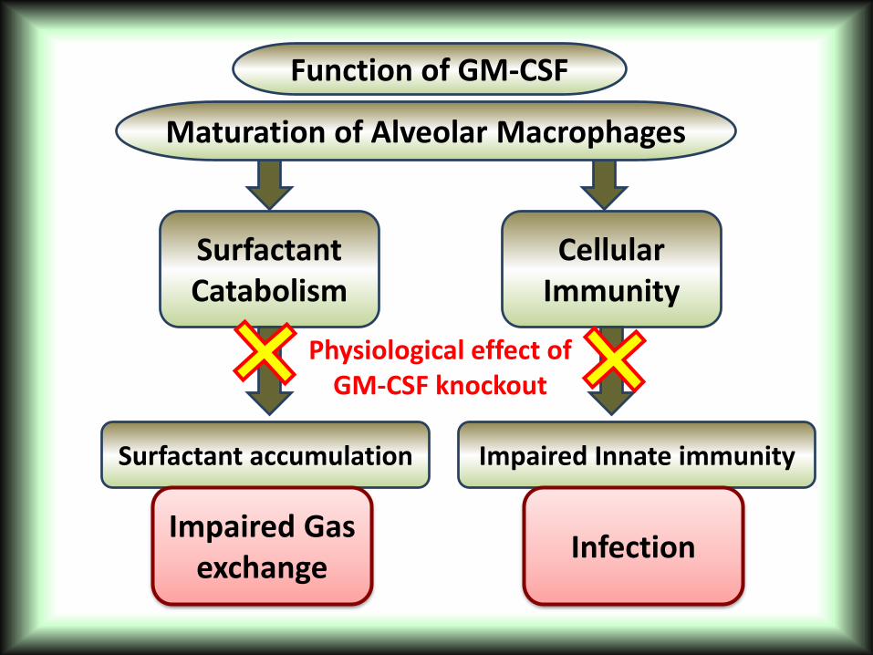
Function of GM-CSF
SurfactantCatabolism
CellularImmunity
Maturation of Alveolar Macrophages
Surfactant accumulation Impaired Innate immunity
InfectionImpaired Gas
exchange
Physiological effect ofGM-CSF knockout

Physiological effect of PAP
• This surfactant-derived alveolar fluid may cause increased work of breathing, a diminished surface area for gas diffusion, and, ultimately, respiratory failure.
• The development of superinfection, which is thought to be a relatively common consequence of pulmonary macrophage dysfunction, may further complicate the condition “Nocardia spp.”

PAP
Auto GM-CSF Antibodies
leukemia myelodysplastic
syndromes HIV
Occupational (silica)
(SP) B deficiency and GM-CSF receptor β chain
abnormality
1ry/autoimmune/ Idiopathic
Neonatal / Congenital
Secondary
Causes types
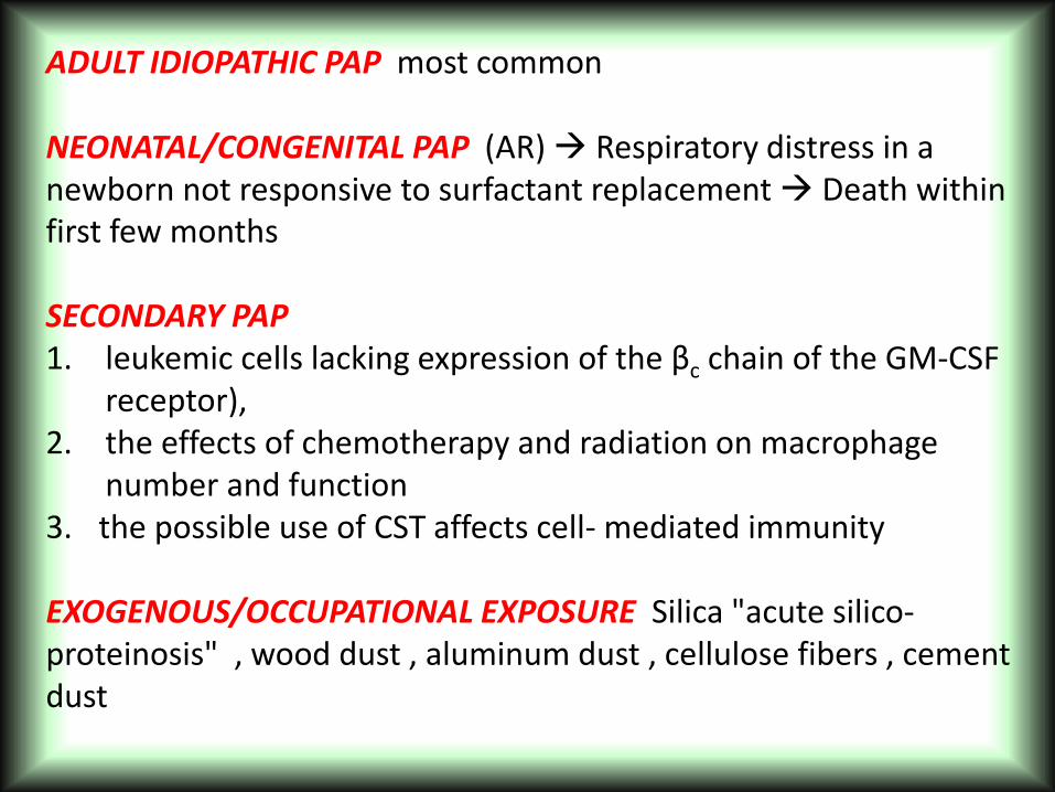
ADULT IDIOPATHIC PAP most common
NEONATAL/CONGENITAL PAP (AR) Respiratory distress in a newborn not responsive to surfactant replacement Death within first few months
SECONDARY PAP1. leukemic cells lacking expression of the βc chain of the GM-CSF
receptor),2. the effects of chemotherapy and radiation on macrophage
number and function3. the possible use of CST affects cell- mediated immunity
EXOGENOUS/OCCUPATIONAL EXPOSURE Silica "acute silico-proteinosis" , wood dust , aluminum dust , cellulose fibers , cement dust
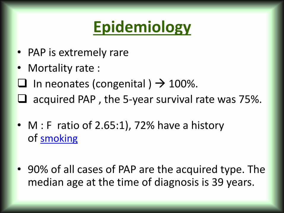
Epidemiology
• PAP is extremely rare
• Mortality rate :
In neonates (congenital ) 100%.
acquired PAP , the 5-year survival rate was 75%.
• M : F ratio of 2.65:1), 72% have a history of smoking
• 90% of all cases of PAP are the acquired type. The median age at the time of diagnosis is 39 years.

Clinical Presentation
• Progressive dyspnea
• cough ≈ 75 %
• occasional hemoptysis and fever ≈ 20%
• constitutional symptoms.
• Crackles ≈ 50%
• clubbing, and cyanosis have been reported.

Radiology
• a"butterfly" distributionTypically a bilateral, symmetrical alveolar filling pattern is seen. With
• interstitial, mixed, diffusely nodular, and focally dense patterns have been reported
DD• pulmonary edema (ARDS)• Pneumocystis jirovecii (carinii)
pneumonia. • Hypersensitivity pneumonitis

Figure PAP in a 61-raey- cinorhc htiw nam dlo
suonegoleym dna eugitaf fo tesno tnecer dna aimekuel
hpargoidar tsehc roiretnaoretsoP .hguocshows symmetric, perihilar
ground-glass and reticulonodular opacities evitaler eht etoN .
selgna cinerhpotsoc eht fo gniraps .

HRCT : crazy-paving

• ‘‘crazy-paving’’ pattern that consists of scattered or diffuse ground-glass attenuation with superimposed interlobular septal thickening and intralobular lines
• Changes correlate with the presence of a restrictive ventilatory defect, reduced diffusing capacity, and hypoxemia


Figure 6b. Crazy-paving in PAP.
Frazier A A et al. Radiographics 2008;28:883-899
©2008 by Radiological Society of North America


32-year-old man with HIV BAL showscystic forms of P carinii(arrows).
Diffuse mucinous BAC in a 78-year-old man open lung biopsy shows neoplastic cells with abundant intr-acytoplasmic mucin(arrows).
53-year-old woman with SLE andmassive hemoptysis. acute intraalveolarhemorrhage.

Methotrexate-induced NSIP in a 41-year-old woman with rheumatoid arthritis
Lipoid pneumonia in a 64-year-oldWoman open lung biopsy shows numerous lipid-laden macrophages that fill and distend the alveoli (arrow) and interstitium

PFTs
• Mild restrictive.
• reduction in diffusing capacity.
• mildly hypoxemic
• elevated alveolar-arterial PO2 difference along with a compensated respiratory alkalosis.
• The shunt fraction has been shown to be elevated as compared to patients with other diffuse lung diseases

LAB
• Serum LDH is frequently elevated ( 82% ) .
• Surfactant proteins SP-A, SP-B, and SP-D have been reported to be elevated in the sera of patients with PAP. ( nonspecific ALI , IPF )

BAL
• Grossly milky and opaque , forming sediment when left to settle.
Milky proteinaceous whole lung lavagate from a patient with PAP. Note the foamy surfactant layer.

Microscopic features of BALF -cytospin preparation
BALF is composed of granular, acellular, and amorphous material
and a small number of cells. Cell components consist of large,
(1) foamy alveolar macrophages
(2) small monocyte-like macrophages

• The most striking observation of the BAL sediment is the gross appearance of the alveolar macrophage. These cells are enlarged and engorged with lipid material with a foamy, vacuolated appearance under light microscopy .

3) Pathological FindingsMicroscopically, alveoli and terminal bronchioli are filled with a fine eosinophilic material stained strongly for surfactant proteins with periodic acid-Schiff reagent. The alveolar wall and interstitial architecture are relatively well-preserved.
Biopsy
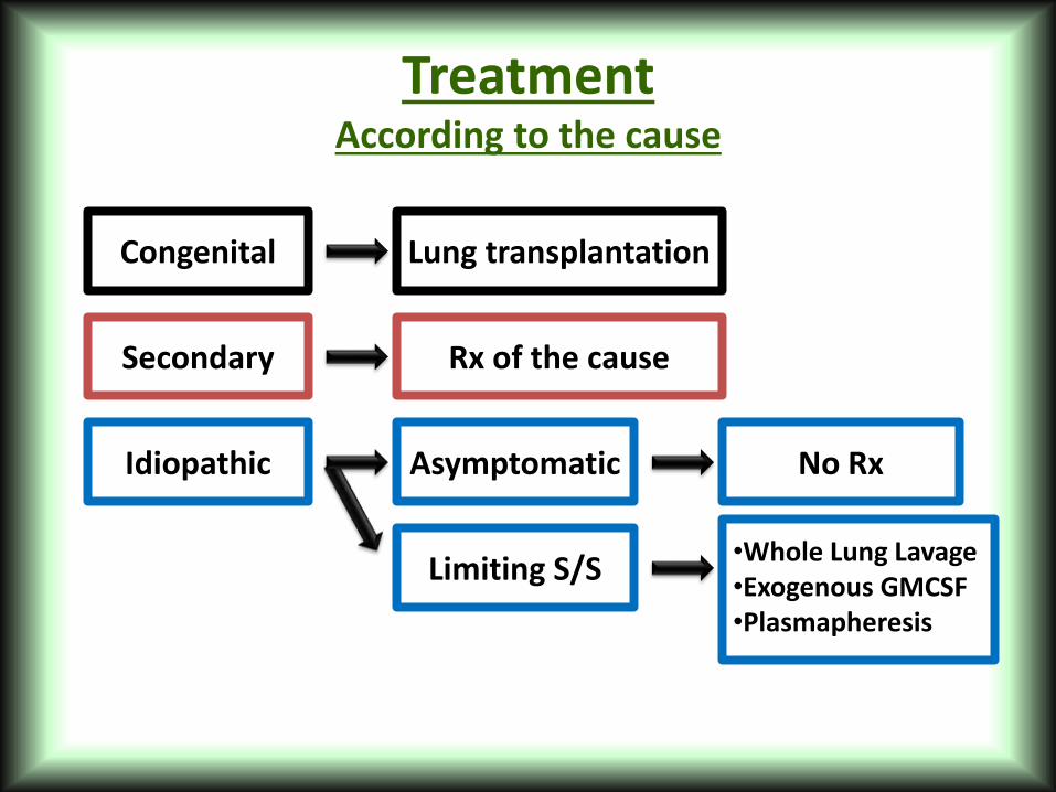
Treatment According to the cause
Congenital
Secondary
Idiopathic Asymptomatic
Rx of the cause
Lung transplantation
Limiting S/S
No Rx
•Whole Lung Lavage•Exogenous GMCSF•Plasmapheresis

•repeated segmental flooding with saline ( 20-40 L) under GA via a double lumen endotracheal tube significant clinical, physiologic, and radiologic improvements in up to 84% of cases after the first lavage ( repeat treatments commonly required )
•Recently, alternative techniques using fiberoptic bronchoscopy
WLL : Whole Lung Lavage
Whole lung lavage fluid. The retrieved fluid of the left lung in the first bottle is more milky and turbid. The turbidity and amount of sediment decreased gradually.

Parameter of improvement
Arterial PO2 (mm Hg)
(A - a)PO2 (mm Hg)
FEV1 (L)
Vital capacity (L)
DLCO (mL/mm Hg per min)
Sever Dyspnea
PaO2 < 60 mmHg
P ( A – a ) O2 > 40 mmHg
Shunt fraction > 10% to 12%
recommendations for WLL
Complications of WLL includehypoxemiaPneumoniaSepsishydropneumothorax, adult respiratory distress syndrome

The initial therapy included 5 – 20 µg/kg/day of GM-CSF subcutaneously with a rapid dose
GMCSF via aerosol
GM-CSF therapy is not curative, and some patients relapse after discontinuing therapy
GM-CSF ( experimental approaches)