PUBLICATIONS - Shodhgangashodhganga.inflibnet.ac.in/.../10603/11833/15/15_publications.pdf ·...
30
PUBLICATIONS 4-
Transcript of PUBLICATIONS - Shodhgangashodhganga.inflibnet.ac.in/.../10603/11833/15/15_publications.pdf ·...

PUBLICATIONS
4-

Published and communicated
• Samir Damare, Chandralata Raghukumar, S. Raghukumar (2006). Fungi in deep-sea
sediments of the Central Indian Basin. Deep-sea Research I, Vol. 53, 14-27.
• Samir Damare, Chandralata Raghukumar, Ushadevi Muraleedharan, S. Raghukumar
(2006). Deep-sea fungi as a source of alkaline and cold-tolerant proteases. Enzyme and
Microbial Technology, Vol.39, 172-181.
• Chandralata Raghukumar and Samir Damare. Deep-Sea Fungi. II High Pressure
Microbiology. American Society for Microbiology. Book chapter (In press).
• Samir Damare, Abhishek Mishra, Chandralata Raghukumar. Production and
characterization of alkaline serine protease of a deep-sea fungus by solid substrate
fermentation (Under revision)
• Samir Damare, Chandralata Raghukumar, S. Raghukumar. Fungi and
Macroaggregation in the Deep-sea Sediments (Communicated).
Manuscripts under preparation
• Samir Damare, Manju Nagarajan, Chandralata Raghukumar. "Induction of Spore
Germination Under Elevated Hydrostatic Pressure in Deep-sea and Terrestrial Fungi"
• Samir Damare and Chandralata Raghukumar. "Stress Response of Deep-sea Fungi as
Elucidated by Electrophoresis"
• Samir Damare, Varada Damare, Pankaj Verma, Yogesh Shouche, Chandralata
Raghukumar. "Diversity of Yeast from the Water Column of Equatorial Indian Ocean"
Patent Chandralata Raghukumar, Samir Damare, Ushadevi Muraleedharan. "A Process for Production
of Low Temperature Active Alkaline Protease from Deep-sea Fungus' has been filed in India (No.
0271NF), US and WO. European patent — EP 1692296.
Presentations at Symposia and Conferences
• Presented a paper (oral presentation) titled 'Deep-sea Fungi as a Source of Extremophilic
Enzymes for Industrial Applications' at the National Seminar on New Trends in
Biotechnology at Goa, India, 11 th & 12th Jan. 2007.
• Presneted a paper (oral presentation) titled 'Production of Alkaline Proteases from Deep-
sea Fungus by Solid Substrate Fermentation' at the National Seminar on Recent Trends

in Mycological Research at J.J. College of Arts and Science, Tamilnadu, India, Dec. 28 th
& 29th Dec. 2006.
• Presented a paper (oral presentation) titled 'Effect of Deep-sea Conditions on Fungal
Spores' at the 7th Asia Pacific Marine Biotechnology Conference at Cochin, India, Nov. 2
- 5, 2006.
• Presented a paper (oral presentation) titled 'Deep-sea Fungi as a Source of Cold-tolerant
Proteases' in the Conference "From Functional Genomics to Natural Products of Marine
Microorganisms" at Greifswald, Germany, June 21-24, 2006.
• Presented a paper (oral presentation) titled Deep-sea fungi as a source of alkaline, cold-
tolerant proteases' in the Conference on Microbiology of the Tropical Seas held at
National University of Oceanography, Goa during Dec.13-15, 2004.
• Presented a paper (oral presentation) titled 'Occurrence of fungi in deep sea sediments of
the Central Indian Basin' in the National Seminar on Recent Advances in Mycology held
at Mangalore University, Dec. 2-3, 2004.
• Poster titled Barotolerant Deep-sea Fungi and their Proteases' was presented in
National Symposium on Prospecting of Fungal Diversity and Emerging Technologies at
29th Annual Meeting of Mycological Society of India at Agharkar Research Institute,
Pune in Feb. 2003, and the abstract published in the proceedings of the same.

Europh!aches Patentamt
(19) European Patent Office
Office europeen des brevets
(11) VerOffentllchungsnummer:
(11) Publication number: EP 1 692 296 AO (11) Numero de publication:
Internationale Anmeldung verOffentlicht durch die
Weltorganisation filr geistiges Eigentum unter der Nummer:
WO 2005/045047 (art. 158 des EPU).
International application published by the World
Intellectual Property Organisation under number:
WO 2005/045047 (art. 158 of the EPC).
Demande internationale publiee par ('Organisation
Mondiale de la Propriete sous le numero:
WO 2005/045047 (art. 158 de la CBE).

%BO
120
100 Ze•
80
60 a
• 40
CL
▪
20
0 6 6.6 7 7.5 8 8.6 9 9.5 10 10.5 11 11.5 12
PH
WO
2005
/045
047
Al
80
60
40
15 75
120 F 100
Vis
i 20
et 0
5 30 45 60
Temperature (°C)
(12) INTERNATIONAL APPLICATION PUBLISHED UNDER THE PATENT COOPERATION TREATY (PCT)
(19) World Intellectual Property Organization
International Bureau
•
11111 1111111 11 11111011111111111111111111111111111M 11111111111111 N11111111111 III
(43) International Publication Date 19 May 2005 (19.05.2005) PCT
(10) International Publication Number WO 2005/045047 Al
(51) International Patent Classification: Cl2P 1/06, Cl2N 9/62
(21) International Application Number: PCT/IB2003/005000
(22) International Fffing Date: 7 November 2003 (07.11.2003)
(25) Filing Language: English
(26) Publication Language: English
(71) Applicant (for all designated States except US): COUN-CIL OF SCIENTIFIC AND INDUSTRIAL RE-SEARCH [IN/INJ; Rafi Marg, 110 001 New Delhi (IN).
(72) Inventors; and (75) Inventors/Applicants (for US only): RAGHUKUMAR,
Chandralata [IN/IN]; National Institute of Oceanogra-phy, Dona Paula, 403 004 Goa (IN). DAMARE,
Ravikant [IN/INJ; National Institute of Oceanography, Dona Paula, 403 004 Goa (IN). MURALEEDHARAN, Usha, Devi [IN/INJ; Department of Marine Biotechnology, Goa University, Taleigna Plateau, 403 206 Goa (IN).
(74) Agent: GABRIEL, Devadoss, Calab; K & S PART-NERS, 84-C, C-6 Lane, Off Central Avenue, Sainik Farms, 110 062 New Delhi (IN).
(81) Designated States (national): AE, AG, AL, AM, AT, AU, AZ, BA, BB, BG, BR, BY, BZ, CA, CH, CN, CO, CR, CU, CZ, DE, DK, DM, DZ, EC, EE, EG, ES, FI, GB, GD, GE, GH, GM, HR, HU, ID, IL, IN, IS, JP, KE, KG, KP, KR, KZ, LC, LK, LR, LS, LT, LU, LV, MA, MD, MG, MK, MN, MW, MX, MZ, NI, NO, NZ, OM, PG, PH, PL, PT, RO, RU, SC, SD, SE, SG, SK, SL, SY, TJ, TM, TN, TR, TT, TZ, UA, UG, US, UZ, VC, VN, YU, ZA, ZM, ZW.
(84) Designated States (regional): ARFPO patent (GH, GM, KE, LS, MW, MZ, SD, SL, SZ, TZ, UG, ZM, ZW), Eurasian patent (AM, AZ, BY, KG, KZ, MD, RU, TJ, TM), European patent (AT, BE, BG, CH, CY, CZ, DE, DK, EE,
[Continued on next page)
=---- (54) Title: A PROCESS FOR PRODUCING AN ALKALINE PROTEASE FROM A DEEP-SEA FUNGUS
(57) Abstract: Present invention particularly relates to production of alkaline protease for various industries and especially for "enzyme detergents" by a fungus Aspergillus sp. deposited in the microbial type culture collection of Institute of Microbial Technology, Chandigarh, India, under the accession number MTCC 5102 and the said fungus can be grown in conventional media with commercial brands of milk powder using distilled water at pH 7.0 at room temperature (28oC±2 °C), however, the said fungus grows well in seawater media too and also at 5°C; further, the protease enzyme produced by this fungus acts equally well in the pH range of 6 to 11 and shows 100% activity at temperature of 42-47°C but almost 90% of the activity is present at 30 °C, 50% of the activity at 15 °C and at 60°C The enzyme is thermostable up to 45 min at 450C.

WO 2005/045047 Al 1111111111111111111111111111111111111111111111111111111111111111111111111111112
ES, FI, FR, GB, GR, HU, 1E, IT, LU, MC, NL, PT, RO, SE, SL SK, TR), OAPI patent (BF, BJ, CF, CO, CI, CM, GA, GN, GQ, GW, ML, MR, NE, SN, TD, TG).
Declarations under Rule 4.17: - as to the applicant's entitlement to claim the priority of the
earlier application (Rule 4.17( iii)) for the following desig-nations AE, AG, AL, AM, Al; AU, AZ BA, BB, BG, BR, BZ BZ, CA, CH, CN, CO, CR, CU, CZ, DE, DK, DM, DZ, EC, EE, EG, ES, Fl, GB, GD, GE, GH, GM, HR, HU, ID, IL, IN, IS, JP, KE, KG, KR KR, KZ LC, LK, LR, LS, LT, LU, LIV, MA, MD, MG, MK, MN, MW, MX, MI NI, NO, NZ, OM, PG, PH, PL, PT; RO, RU, SC, SD, SE, SG, SK, SL, SY, TJ, TM, TN, TR, TT, TZ, UA, UG, UZ VC, VN, YU, ZA, ZM, ZW, ARIPO patent (GH, GM, ICE, LS, MW, MI SD, SL, SZ, TZ, UG, ZM, ZW), Eurasian patent (AM, AZ BY, KG, KZ, MD, RU, TJ, TM), European patent (Al; BE, BG, CH, CY, CZ, DE, DK, EE, ES, FI, FR, GB, GR, HU, 1E, IT, LU, MC, NL, PT; RO, SE, SI, SK, TR), OAPI patent (BF, BJ, CF, CG, CI, CM, GA, GN, GQ, GW, ML, MR, NE, SN, TD, TG)
- as to the applicant's entitlement to claim the priority of the earlier application (Rule 4.17(iii)) for the following desig-nations AE, AG, AL, AM, Al; AU, AZ, BA, BB, BG, BR, BY, BZ, CA, CH, CN, CO, CR, CU, CZ, DE, DK, DM, DZ, EC, EE, EG, ES, Fl, GB, GD, GE, GH, GM, HR, HU, ID, IL, IN, IS, JP, KE, KG, KR KR, KZ, LC, LK, LR, LS, LT, LU, LV, MA, MD, MG, MK, MN, MW, MX, Ml NI, NO, NZ, OM, PG, PH, PL, PT, RO, RU, SC, SD, SE, SG, SK, SL, SY, TI, TM, TN, TR, TT, TZ, UA, UG, UZ VC, VN, YU, ZA, ZM,
ZW, ARIPO patent (GH, GM, ICE, LS, MW, MZ SD, SL, SZ TZ, UG, ZM, ZW), Eurasian patent (AM, AZ, BY, KG, KZ, MD, RU, TJ, TM), European patent (AT, BE, BG, CH, CZ CZ, DE, DK, EE, ES, FI, FR, GB, GR, HU, 1E, IT, LU, MC, NL, PT, RO, SE, SI, SK, TR), OAPI patent (BF, BJ, CF, CG, CI, CM, GA, GN, GQ, GW, ML, MR, NE, SN, TD, TG)
- as to the applicant's entitlement to claim the priority of the earlier application (Rule 4.17(iii)) for the following desig-nations AE, AG, AL, AM, Al; AU, AZ, BA, BB, BG, BR, BY, BZ, CA, CH, CN, CO, CR, CU, CZ, DE, DK, DM, DZ EC, EE, EG, ES, FI, GB, GD, GE, GH, GM, HR, HU, ID, IL, IN. IS. JP, ICE, KG, Kp, KR, KZ, LC, LK, LR, ZS, LT, LU, LV, MA, MD, MG, MK, MN, MW, MX, MZ, NI, NO, Ni, OM, PG, PH, FL, PT, RO, RU, SC, SD, SE, SG, SK, SL, SY, TJ, TM, TN, TR, TT, TZ, UA, UG, UZ VC, VN, YU, Z4, ZM, ZW, ARIPO patent (GH, GM, KE, LS, MW, MI SD, SI, SZ, 7Z, UG, ZM, ZW), Eurasian patent (AM, AZ BY, KG, KZ MD, RU, TJ, TM), European patent (Al; BE, BG, CH, CZ Cl DE, DK, EE, ES, FI, FR, GB, GR, HU, 1E, IT, LU, MC, NL, PT, RO, SE, SI, SK, TR), OAPI patent (BF, BJ, CF, CG, CI, CM, GA, GN, GQ, GW, ML, MR, NE, SN, TD, TG)
Published: — with international search report
For two-letter codes and other abbreviations, refer to the "Guid-ance Notes on Codes and Abbreviations" appearing at the begin-ning of each regular issue of the PCT Gazette.

Available online at www.sciencedirect.com
SCIENCE
DIRECT•
Deep-Sea Research I 53 (2006) 14-27
DEEP-SEA RESEARCH PART I
ELSEVIER www.elsevier.com/locate/dsr -
Fungi in deep-sea sediments of the Central Indian Basin
Samir Damare, Chandralata Raghukumar*, S. Raghukumar National Institute of Oceanography, Dona Paula, Goa 403 004, India
Received 10 January 2005; received in revised form 17 September 2005; accepted 24 September 2005 Available online 10 November 2005
Abstract
Although a great amount of information is available on bacteria inhabiting deep-sea sediments, the occurrence of fungi in this environment has been poorly studied and documented. We report here the occurrence of fungi in deep-sea sediments from —5000 m depth in the Central Indian Basin (9—I6°S and 73-76°E). A total of 181 cultures of fungi, most of which belong to terrestrial sporulating species, were isolated by a variety of isolation techniques. Species of Aspergillus and non-sporulating fungi were the most common. Several yeasts were also isolated. Maximum species diversity was observed in 0-2 cm sections of the sediment cores. Direct staining of the sediments with Calcofluor, a fluorescent optical brightener, revealed the presence of fungal hyphae in the sediments. Immunofluorescence using polyclonal antibodies raised against a deep-sea isolate of Aspergillus terreus (# A 4634) confirmed its presence in the form of hyphae in the sub-section from which it was isolated. A total of 25 representative species of fungi produced substantial biomass at 200 bar pressure at 30° as well as at 5 °C. Many fungi showed abnormal morphology at 200 bar/5 °C. A comparison of terrestrial isolates with several deep-sea isolates indicated that the former could grow at 200 bar pressure when growth was initiated with mycelial inocula. However, spores of a deep-sea isolate A. terreus (# A 4634), but not the terrestrial ones, showed germination at 200 bar pressure and 30 °C. Our results suggest that terrestrial species of fungi transported to the deep sea are initially stressed but may gradually adapt themselves for growth under these conditions. © 2005 Elsevier Ltd. All rights reserved.
Keywords: Deep-sea fungi; Hydrostatic pressure; Diversity; Central Indian Basin
1. Introduction
Fungi play a crucial role as saprotrophs in the ecology of terrestrial sediments. They occupy a wide variety of niches on land by virtue of their highly versatile physiological adaptations. One of the least studied habitats of fungi is the deep sea, an environment characterized by low temperature, high hydrostatic pressure and a 'feast and famine'
*Corresponding author. Tel.: + 91 832 2450480; fax: + 91 832 2450602.
E-mail address: [email protected] (C. Raghukumar).
nutrient condition (Morita, 1982; Herbert and Codd, 1986). The presence and ecological impor-tance of deep-sea bacteria has been well recognized ever since Zobell and Morita (1957) isolated bacteria specifically adapted to grow under high pressures and termed them `barophiles'. Yayanos (1979) obtained barophilic bacteria for the first time in pure culture. Much progress has been made with deep-sea bacteria since then with respect to their diversity (cultured and uncultured), molecular phylogeny, growth and enzyme profiles and pres-sure-adaptations. In contrast, one of the major groups of eukaryotic microorganisms, the fungi, has
0967-0637/$ - see front matter © 2005 Elsevier Ltd. All rights reserved. doi:10.1016/j.dsr.2005.09.005

S. Damare et al. / Deep-Sea Research 153 (2006) 14-27
15
largely remained neglected. Roth et al. (1964) isolated marine fungi for the first time from oceanic waters of northwestern subtropical Atlantic Ocean down to a depth of 4450 m. Kohlmeyer and Kohlmeyer (1979) reported obligate marine fungi from wooden panels immersed at depths of 500-3000 m. However, these fungi were not cul-tured. One of the first reports of fungi in deep-sea sediments was provided by Raghukumar et al. (1992), who isolated fungi from calcareous sedi-ments of the Bay of Bengal at a depth of 965 m and demonstrated germination of spores of Aspergillus ustus under simulated deep-sea conditions. Subse-quently, cultivation of marine yeasts (Lorenz and Molitoris, 1992) and filamentous fungi and germi-nation of fungal spores (ZaunstOck and Molitoris, 1995) under simulated deep-sea conditions of low temperature and elevated hydrostatic pressure were reported. Takami et al. (1997) showed the presence of fungi and yeasts in sediment samples obtained from the Mariana Trench at a depth of 10,500 m in the Pacific Ocean. These were later identified to be Penicillium lagena and Rhodotorula mucilaginosa, respectively (Takami, 1999). The presence of fungi based on direct detection and isolation techniques in a 4.7 m long sediment core from the Chagos Trench in the Indian Ocean at a depth of —5000 m was reported recently (Raghukumar et al., 2004). How-ever, these have been sporadic reports and not comprehensive enough to prove the existence of fungi in deep-sea sediments. We have used the following approach to address the occurrence and diversity of fungi from deep-sea sediments at an average depth of 5000 m in the Central Indian Basin (CIB): (1) Isolation of fungi by different culturing techniques and their identification; (2) direct detection of fungal hyphae in deep-sea sediments in order to confirm their growth and (3) experiments to study their growth under simulated deep-sea conditions.
2. Methods
2.1. Sampling site and collection of deep-sea sediments
Sediment samples were obtained from depths of 4900 to 5390 in in the Central Indian Basin (9-16°S and 73 76°E) (Fig. 1) on board the Russian research vessel AA Sidorenko during 3 cruises. Samples were collected with an USNEL-type box corer of 50 cm 3
size. Sampling with a box corer was possible because
Fig. 1. Map of the Central Indian Basin showing within inset location of the sampling sites (9-16°S and 73-76°F) during cruises # AAS 34, # AAS 46 and # AAS 61.
of the more or less flat topography of the ocean floor in the sampling area. Eleven box core samples were collected during cruise # AAS 34 in April 2001 (10000/264"-10 0 10/364" S; 75°21'000" --76°05'160"E), 7 during Cruise # AAS 46 in June 2002 (10°00'237"-10°02'661"S; 75°59'498"-76°09'822" E) and 38 during cruise # AAS 61 in March 2003 (9°59'861"-16°00'047"S; 73°29'819" 76°30'559"E). Se-diment at the sampling sites was mainly radiolarian ooze, light to dark brown in color and intensely mottled indicating high bioturbation (Sharma et al., 2001). It was predominantly clayey-silt type with high water content and low shear strength and was loosely packed (Khadge, 2000). Subcores of sediments were collected from the center of the box corer with alcohol-sterilized PVC cylinders of 5 cm diameter.

16
S. Damare et al. / Deep-Sea Research 153 (2006) 14-27
Subsections of 2 cm down to 10 cm depth and thereafter every 5 cm length were extruded from these sediment cores of —30-40 cm length directly into sterile plastic bags to avoid any aerial contaminants. The bags were closed with rubber bands and carried to the laminar flow hood in the microbiology laboratory on board.
2.2. Isolation of fungi
A portion of the sediment from the middle of each sub-section that had not been in contact with the walls of the PVC cylinder was removed with a flame-sterilized spatula and placed in sterile vials for isolation of fungi (Raghukumar et al., 2004). The media used for isolations were malt extract agar (MEA), malt extract broth (MEB), corn meal agar (CMA), Sabaurauds dextrose agar (SDA), Czapek Dox agar (CDA) and Czapek Dox broth (HiMedia Pvt. Ltd., India). All the media were used at 1/5 strength to simulate the low nutrient condition in the deep sea. They were prepared in seawater and fortified with streptomycin (0.1 g in 100 ml medium) and penicillin (40,000 Units in 100 ml medium) to inhibit bacterial growth. Fungi were isolated by the following methods: (1) Dilution plating method, where —0.1 g of sediment was suspended in sterile sea water, vortexed for 1 min and 100 pl aliquots spread-plated. (2) Particle plating technique (Bills and Polishook, 1994), where approximately 1 g of sediment slurry was sieved successively through a mesh size of 200 and 100 gm screens. The particles that passed through 200 gm mesh but were retained on the 100 gm mesh were spread-plated. For both the above techniques, the plates were incubated at 5 °C at 1 bar pressure for 15-20 days. (3) Pressure incubation, in which approximately 0.5 g of sedi-ments were placed in sterile plastic bags containing 2 ml of sterile MEB and the open ends of the bags sealed with an electrical sealing machine (Quickseal, Sevana, India). The bags were placed in a deep-sea culture vessel (Tsurumi & Seiki Co., Japan) filled with sterile water and pressurized to 300 bar pressure. The pressure vessels were immediately placed at 5 °C and incubated for 30 days. At the end of this incubation period, 100 pi of the sediment was spread-plated on nutrient media and the plates were incubated at 1 bar pressure and 30 °C until fungal colonies appeared (within 8-10 days). Three repli-cate plates were maintained for each sediment sample, medium and isolation technique. Media plates were exposed to air for 10 min on the deck of
the research vessel where the cores were received, the microbiology laboratory on board and the laminar flow inoculation hood to check for the presence of aerial contaminants. This was repeated during every sampling station.
Fungi isolated from the deep-sea sediments were subcultured and maintained on MEA slants at 5 °C. Slides of fungi were prepared in lactophenol cotton blue and microscopically examined prior to photo-micrography and identification using the taxonomic keys (Domsch et al., 1980). Species of fungi isolated using all different media and techniques were pooled for each individual subsection of the cores and diversity measurements for each of these samples was calculated using the software PRIMER v5 (Clarke and Gorley, 2001). The results are expressed as species richness, Pielou's evenness index and Shanon Wiener diversity index (log 2). These in turn express the richness of biodiversity in each sample, the extent of even distribution of different species and proportion among total counts, respectively, in different depths of sediment cores.
For comparison, fungi were also isolated from sediments collected from shallow coral reef slopes at a depth of 30 m off Lakshadweep Island Kavaratti (10°35'N and 72°39'E) in the Arabian Sea by the particle plating technique. Two isolates of Aspergil-lus terreus (MTCC # 279 and MTCC # 479) and one Aspergillus sydowii (MTCC # 635) culture isolated from terrestrial environments obtained from Micro-bial Type Culture Collection (MTCC, Chandigarh, India) were included in this study for comparison.
2.3. Direct detection of fungi in deep-sea sediments
About 0.5 g of each sediment sample in sterile vials were fixed with 5% formalin solution and stored at 5 °C for direct detection of fungi according to the method described by Mueller and Sengbusch (1983). Aliquots of these fixed sediments were stained with 0.5% solution of sterile-filtered Calco-fluor, an optical brightener (Sigma Chemicals, USA). The excess stain was washed off by centrifugation with sterile seawater. Microscopic mounts of the sediment were then examined under ultraviolet light filter (excitation wave length 330-385 nm and barrier filter BA 420) of an epifluorescence microscope (Olympus BX 60, To-kyo, Japan) to detect fluorescing fungal hyphae. Fungal hyphae and spores were photographed with a digital camera (Olympus 4.1 Mp, Tokyo, Japan).

S. Darnare et al. / Deep-Sea Research 153 (2006) 14-27 17
Several sediment samples were scanned for the presence of fungi after each cruise. In addition, sediments with 0.5 ml sterile seawater were vortexed after addition of a drop of sterile detergent solution. The foam formed on the surface of trapped sediment material was pipetted out onto a sterile glass slide, stained and examined microscopically as described above.
2.4. Detection of Aspergillus terreus Thom (isolate # A 4634) in deep-sea sediments by immunofluorescence
The immunofluorescence technique, which has been widely used to detect specific fungi in terrestrial substrates (Jellison and Goodell, 1988; Friese and Allen, 1991; Banks et al., 1993), was employed for the purpose. Antibodies were raised commercially for A. terreus (# A 4634), one of the most frequently isolated fungi from the deep-sea sediments of the Central Indian Basin. This isolate was obtained from core # BC 12 at the subsurface depth of 15-20 cm during the cruise # AAS 46 (10°01'S; 76°00'E at a depth of 5400m). Antibodies were raised in New Zealand male white rabbits by Genei India Pvt. Ltd., Bangalore, by standard protocols (Johnson and Thorpe, 1987). Thus, about 2 mg of fungal pellet was crushed in 2 ml of 0.15 M NaC1 and centrifuged, and 1 ml supernatant was emulsified with 1 ml Freund's adjuvant. Fifty sg of this antigen was injected subcutaneously at multiple sites on the backs of the rabbits followed by booster doses on days 30, 45, 55 and 65. The antibody titre was monitored by Dot ELISA and yielded a value of 1:10,000.
The presence of A. terreus in natural sediment samples was studied by the following method: The antiserum containing the antibodies (as supplied by Genei India Pvt. Ltd., Bangalore) diluted to 1:10 with sterile phosphate buffered saline pH 7.0 (PBS) was the minimum concentration required for detec-tion of fluorescence of the fungus in these samples. About —0.1 g of the deep-sea sediment sample from which the fungus was isolated was stained with the antiserum at 25 °C for 60 min followed by 5 washes, each with 1000111 PBS. The sediment was further incubated with 100 p.1 of 1:10 diluted secondary antibody (Genei India Pvt. Ltd.), namely the goat anti-rabbit anti-serum tagged with fluorescein iso-thiocyanate (FITC) for 60 min at 25 °C. The excess stain was removed by washing the sediment 5 times with PBS. The sediment was spread evenly onto a
slide and observed under an epifluorescence micro-scope (excitation wavelength 450-480 nm and bar-rier filter BA 515).
The reactivity of the antiserum to A. terreus was confirmed by growing the fungus both at 1 bar and 200 bar pressure/5° and 30 °C and staining as described above. The antiserum containing the antibodies diluted to 1:100 with PBS was the minimum concentration required for detection of fluorescence of the fungus in culture. The absence of cross reactivity with other fungi was checked by staining an unidentified fungus, Aspergillus sp, a non-sporulating fungus and a terrestrial A. terreus (MTCC # 279) with antiserum similarly and examining for the presence or absence of immuno-fluorescence.
2.5. Growth of fungi under elevated hydrostatic pressure
Representative fungi isolated by different techni-ques and with different nutrient media were examined for spore germination and mycelial growth under elevated hydrostatic pressures by the following methods: (1) The selected fungi were grown in MEA plates at 1 bar pressure and 30 °C temperature, and the spores were collected by gently flooding the plates with sterile sea water. The spore suspension was appropriately diluted after haemo-cytometer counts, inoculated in MEB fortified with 1% glucose and 0.1% Tween 80 in pouches made with sterilized gas permeable polypropylene sheets and sealed without trapping any air bubbles. The pouches were suspended in a deep-sea culture vessel filled with sterile water and pressurized to 200 bar pressure and incubated at 30° and 5 °C. Similarly prepared pouches were incubated at 1 bar pressure at 30° and 5 °C for comparison. After 3 days of incubation, the deep-sea culture vessels were decompressed gradually (at the rate of 50 bar/ 15 min), and the percentage of germinating conidia was by counting in 20 microscope fields. For comparison, two isolates of A. terreus (MTCC # 279 and MTCC # 479) and one A. sydowii (MTCC # 635) culture isolated from terrestrial environments obtained from Microbial Type Culture Collection (MTCC, Chandigarh, India) were included in this study. (2) For raising mycelial biomass, cultures of 25 deep-sea fungi were grown in MEB for 3 days at 1 bar and 30 °C. Vegetative mycelium prior to the onset of sporulation was homogenized with sterile glass beads. A known weight of the finely
-4t

18 S. Damare et al. / Deep-Sea Research 153 (2006) /4-27
broken mycelial suspension was inoculated in 20 ml MEB and incubated at 30° and 5 °C/200 bar pressure as described above. After 20 days, the contents of the pouches were filtered over pre-weighed filter papers, dried to a constant dry weight and the difference between the initial and final biomass determined as mycelial dry weight (Raghu-kumar and Raghukumar, 1998). A similar experi-ment was carried out to compare growth of 8 deep-sea, 3 terrestrial and 3 shallow water fungi. The experiments were carried out with 5 ml of MEB medium.
Fungi grown under high hydrostatic pressure at 5° and 30 °C were stained with Calcofluor and lactophenol cotton blue, and their morphology under different culture conditions was recorded by photomicrography.
2.6. Gradual adaptation to growth under elevated hydrostatic pressure
A total of 109 of the 181 isolates obtained in this study failed to grow at 300 bar pressure. An experiment was carried out to examine if these could be gradually acclimatized to growth at elevated pressures. The cultures were initially grown at 50 bar/30 °C for 20 days, after which the pressure vessel was decompressed and the bags brought back to 1 bar pressure and checked for growth and viability upon culturing. Cultures that had grown at 50 bar pressure were transferred to fresh pouches under sterile conditions and incubated at 100 bar pressure. This process was continued at 200, 300 and 400 bar pressure. Viability of fungi after exposure to each pressure was tested by growing in MEA at 30 °C/1 bar pressure.
3. Results
3.1. Abundance and diversity
The percentages of culturable fungi obtained by dilution plating and direct incubation of deep-sea sediments under elevated pressure were similar (Table 1). Particle plating yielded a smaller number of fungi. Sediments from shallow coral reef waters yielded much higher numbers of fungi (Table 1). The highest number of species was often obtained at 0-2 cm depth of deep-sea sediment cores, while the numbers were much less below 25 cm depth (Table 2). Aspergillus species were the dominant fungi isolated followed by non-sporulating and unidentified sporulating fungi. A one-way analysis of variance (ANOVA) comparing the number of species isolated from different subsections of each core showed that differences between the subsections were not significant (F value 0.34, P-value 0.97, d.f. = 10,143). Shanon index, Pielou's evenness and species richness values were similar up to 20-25 cm depth, after which there was a marked reduction. Among the various media used for isolation of fungi, MEA and MEB followed by CMA were found to be better than the other media used. None of the media were selective for isolating specific fungi (data not shown). Details of the media used for some of the fungi are shown in Table 3.
Many of the aspergilli showed abnormal mor-phology immediately after isolation. These showed extremely long conidiophores with vesicles being covered by long hyphae, instead of phialides or metulae or conidia, as is typical of the genus Aspergillus (Fig. 2).
Table 1 Number of fungi isolated by various techniques
Source Particle plating Dilution plating Isolation following pressure incubation at 300 bar/5 °C
Deep sea
Total number of sediment samples used 376 72 224 Number of fungi isolated 65 28 88 % frequency of occurrence 17 39 39
Shallow water Total number of sediment samples used 16 Not done Number of fungi isolated 26 %frequency of occurrence 163

S. Damare et al. / Deep-Sea Research 153 (2006)
Table 2 Percentage distribution of fungi at different depths in the sediment core
14-27 19
Genera Depths (cm)
0-2 2-4 4-6 6-8 8-10 10-15 15-20 20-25 25-30 30-35 35-40
Total species isolated 25 19 20 16 14 17 16 22 11 13 8 Total sediment sections used 56 56 56 56 56 56 56 55 50 42 20
Aspergillus sp. 8 II 5 25 29 12 6 14 18 54 13 Aspergillus terreus 28 5 15 13 7 18 19 14 9 8 -
Aspergillus restrictus 12 26 10 19 21 12 13 9 - -
Aspergillus sydowii - 5 5 6 7 6 - -
Penicillium sp. 8 5 10 7 6 13 9 Cladosporium sp. 4 - 15 7 6 13 5 15 Curvularia sp. 4 - - - - - Fusarium sp. - - - - - 13 Non-sporulating fungi 16 11 10 13 7 18 6 18 36 15 13 Unidentified sporulating fungi 16 32 25 19 14 24 31 23 36 8 63 Unidentified Ascomycetes - - - - - 5 -
Aureobasidium sp. 4 - - - Unidentified yeasts - 5 5 6 - - 5 - - Shanon index 1.99 1.81 2.06 1.84 1.91 1.96 1.80 2.05 1.26 1.30 1.07 Pielou's eveness 0.90 0.87 0.94 0.95 0.92 0.94 0.93 0.93 0.91 0.81 0.77 Species richness 2.49 2.38 2.67 2.16 2.65 2.47 2.16 2.59 1.25 1.56 1.44
Table 3 Isolation details of deep-sea fungi used for experiments
Isolate # Fungi Cruise # Core # Latitude (South)
Longitude (East)
Depth (m)
Section of the core (cm)
Method of isolation
Medium used
A 4637 A. terreus AAS 46 BC 4 10° 01' 75° 59' 5305 0-2 P1 MEB
A 4636 A. terreus AAS 46 BC 12 10° 01' 76° 00' 5400 0-2 P1 MEB
A 4634 A. terreus AAS 46 BC 12 10° 01' 76° 00' 5400 15-20 P1 MEB A 4633 A. terreus AAS 46 BC 12 10° 01' 76° 00' 5400 10-15 P1 MEB A 4630 Aspergillus sp. AAS 46 BC 7 10° 02' 76° 00' 5296 4-6 P1 MEB
A 4628 A. terreus AAS 46 BC 12 10° 01' 76° 00' 5400 8-10 P1 MEB A 61 P10 A. terreus AAS 61 BC 14 14° 00' 75° 30' 5145 20-25 P1 MEB
A 61 P4 Unidentified AAS 61 BC 3 11° 59' 76° 29' 5280 0-2 P1 Artemia
A 4625 Unidentified AAS 46 BC 7 10° 02' 76° 00' 5296 8-10 P1 MEB A 614 A. terreus AAS 61 BC 17 13° 00' 73° 30' 4810 10-15 PP SDA A 6137 Unidentified AAS 61 3MBC 5 10° 01' 76° 00' 5280 15-20 PP CMA A 3457 Fusarium sp. AAS 34 BC 3 10° 00' 76° 01' 5294 15-20 PP MEA
A 3449 Fusarium sp. AAS 34 BC 5 10" 03' 76° 01' 5294 20 25 PP MEA
A 3441 Unidentified AAS 34 BC 5 10° 03' 76° 01' 5294 8 10 PP MEA A 348 Non-sporulating AAS 34 BC A I/B 10° 10' 76° 05' 5250 & 10 DP ZMA A 3428 Curvularia sp. AAS 34 BC 8 10° 09' 75° 21' 5180 0-2 DP ZMA A 61 P63 Yeast AAS 61 BC 23 10° 59' 73° 29' 5100 20-25 P1 Artemia A 3426 Unidentified AAS 34 BC 14 10° 02' 76° 00' 5280 8 10 DP MEA A 6136 Aspergillus sp. AAS 61 3MBC 11 10° 02' 76° 01' 5320 20-25 PP MEA A 6139 Unidentified AAS 61 3MBC 12 10° 01' 76° 00' 5280 15-20 PP CDA A 61 P64 Yeast AAS 61 BC 12 15° 00' 74° 30' 5390 8-10 P1 MEB A 6128 Aspergillus sp. AAS 61 BC 19 12° 59' 75° 29' 5070 30- 35 PP CMA A 6126 Aspergillus sp. AAS 61 BC 19 12° 59' 75° 29' 5070 30-35 PP SDA A 3415 Unidentified AAS 34 BC 8 10° 09' 75° 21' 5180 25-30 DP ZMA A 3412 Aspergillus sp. AAS 34 BC 8 10° 09' 75° 21' 5180 30-35 DP ZMA
P1-Pressure Incubation, PP-Particle Plating, DP-Dilution Plating, MEB-Malt Extract Broth, MEA-Malt Extract Agar, ZMA-Zobell Marine Agar, CDA-Czapek Dox Agar, SDA-Sabourauds Dextrose Agar, Artemia-Autoclaved Anemia larvae suspended in seawater.

20
S. Damare et al / Deep-Sea Research I53 (2006) 14-27
Fig. 2. Aspergillus sp. isolated from deep-sea sediments with abnormal morphology, showing hyphae in place of metulae and conidia-bearing phialides on the surface of the vesicle. Bar represents 10 gm.
3.2. Direct detection of fungi in sediments
An actively germinating fungal spore was de-tected in sediment samples placed in dilute nutrient medium and incubated at 300 bar pressure and 5 °C (Fig. 3). Fungal hyphae were directly detected by staining the deep-sea sediments with Calcofluor (Fig. 4). A total of 35 and 13 out of 165 and 90 sediment samples collected during the cruises # AAS 61 and # AAS 46, respectively, showed presence of fungi by this method.
3.3. Detection of fungi in sediments by immunofluorescence
Sediment samples of the core # BC 12 (subsection 15-20 cm, Table 3) stained with FITC-tagged antiserum against A. terreus # A 4634 revealed fluorescing hyphae (Figs. 5a and b). An unidentified organic particle in these sediments was densely colonized by the fungus. These showed positive fluorescent reaction to the antiserum (Fig. 5c). The antiserum did not react with other fungi thus showing specificity for the fungus against which the antibodies were raised in rabbits. The fungus showed positive fluorescence after growth at 1 and 200 bar/5° and 30 °C.
Fig. 3. An epifluorescence microscopy photograph of a Calco-fluor-stained germinating spore from deep-sea sediments (0-2 cm subsection of a core) incubated under 300 bar pressure and 5°C. Bar represents 10 gm.
Fig. 4. An epifluorescence microscopy photograph of a fungal hypha from a deep-sea sediment sample stained with Calcofluor. Bar represents 10 gm.
3.4. Growth under simulated deep-sea conditions
Out of a total of 181 fungi isolated from deep-sea sediments, a representative 25, which showed a

S. Damare et al. / Deep-Sea Research 153 (2006) 14-27 21
Fig. 5. (a) Immunofluorescence detection of a hypha of Aspergillus terreus (isolate # A 4634) from 15 to 10 cm depth of a core of the deep-sea sediment treated with FITC-tagged polyclonal antibodies raised against the fungus. Bar represents 10µm. (b) Bright-field photomicrograph of the same hypha as in (a). Bar represents 10 pm. (c) Immunofluorescence detection of a dense cluster of hyphae of Aspergillus terreus on an organic particle from 15 to 20 cm core of a deep-sea sediment treated with FITC-tagged polyclonal antibodies raised against the fungus. Bar represents 10 pm.
capability for growth at 300 bar upon initial isola-tion, were selected for growth under different pressures and temperatures. Details of their isolation are given in Table 3. All the fungi showed growth at 200 bar pressure, both at 5° and 30 °C (Table 4). Eleven fungi grew better at 200 bar/5 °C, than at 200 bar/30 °C. Five of these belong to Aspergillus species. All the fungi grew best at 1 bar and 30 °C, growth being 3-4 times greater than under elevated hydrostatic pressure (200 bar) at 5° and 30 °C.
Several fungi showed abnormal features when grown under elevated pressure either at 5° or 30 °C. For example, A. terreus, # A 4634 showed normal mycelial growth at 1 bar pressure at 30° and 5 °C (Fig. 6a), while hyphal swellings and constrictions occurred at 30° and 5 °C/200 bar pressure (Fig. 6b). An orange-pigmented yeast showed equally good growth at 30 °C/1 bar pressure and 5 °C/200 bar pressure as well (Figs. 7a, b). The conidia of Penicillium sp. (isolate # 4615) showed regular with germination at 30 °C/100 bar pressure, while those of Aspergillus sp. (isolate # 3454) showed swollen conidia (Figs. 8 and 9). The conidia of these species showed normal germination at 30 "Cj 1 bar pressure.
3.5. Comparison of the growth of deep-sea fungi with that of terrestrial isolates under elevated hydrostatic pressure
Spores of the terrestrial isolates A. terreus and A. sydowii totally failed to germinate at 200 bar/30 °C (Table 5). However, 66% of the spores of the deep-sea isolate A. terreus (isolate # A 4634) germinated under these conditions. None of these showed germination at 5 °C/200 bar (Table 5).
Table 4 Biomass produced by various deep-sea fungi under different pressure and temperature conditions
Isolate # Fungi Biomass produced (mg dry wt.)
200 bar/30 °C 200 bar/5 °C 1 bar/30 °C
A 4637 A. terreus 30.0 31.2a 134.5 A 4636 A. terreus 38.4 7.7 132.7 A 4634 A. terreus 18.3 19.6a 156.3 A 4633 A. terreus 20.8 19.8 121.3 A 4630 Aspergillus sp. 25.6 9.0 126.7 A 4628 A. terreus 10.0 13.7a 118.3 A 61 P10 A. terreus 20.6 12.9 125.7 A 61 P4 Unidentified 17.8 27.1 8 128.2 A 4625 Unidentified 65.2 13.8 140.2 A 614 A. terreus 8.1 2.9 59.6 A 6137 Unidentified 15.4 6.5 71.6 A 3457 Fusarium sp. 21.1 29.6a 228.0 A 3449 Fusariwn sp 14.2 10.1 125.8 A 3441 Unidentified 31.5 38.98 178.0 A 348 Non-sporulating 4.7 5.48 64.3 A 3428 Curvulanasp. 4.5 19.3' 175.7 A 61 P63 Yeast 23.4 9.1 249.2 A 3426 Unidentified 21.0 22.2' 234.6 A 6136 Aspergillus sp. 20.0 9.1 169.2 A 6139 Unidentified 17.5 15.4 171.4 A 61 P64 Yeast 12.2 16.88 150.7 A 6128 Aspergillus sp. 9.3 9.3 165.7 A 6126 Aspergillus sp. 9.8 8.9 203.4 A 3415 Unidentified 1.7 11.38 105.3 A 3412 Aspergillus sp. 3.1 1.7 140.0
'Fungi showing better growth at 5 °C than at 30 °C under 200 bar pressure.
Contrary to spores, both terrestrial and deep-sea isolates of fungi showed growth at 5 °C/200 bar and 30 °C/200 bar when mycelia were used as inocula (Table 6). The deep-sea isolates A. terreus (# A

22 S. Damare et al. / Deep-Sea Research 153 (2006) 14-27
Fig. 6. Photomicrographs of Aspergillus terreus (isolate # A 4634) showing (a) normal hyphae during growth at 1 bar/30 °C, and (b) abnormal hyphae with swellings during growth at 200 bar/5 °C. Bars represents 10 pm (b) was photographed under phase contrast.
Fig. 7. Nomarski Differential Interference Contrast photomicroagraphs of the yeast (isolate # A 344) showing (a) normal growth at 1 bar/ 30 °C, and (b) normal growth with abundant pseudomycelia at 200 bar/5 °C (arrow). Scale 10 pm.
Fig. 8. Spores of Penicillium sp. (isolate # 4615) showing normal germination at 300 bar/30 °C. Scale 10 pm.
Fig. 9. Spores of Aspergillus sp. (isolate # 3454) germinating a 300 bar/30 °C. Note the swollen conidia (arrow). Scale 10µm.

S. Damare et al. / Deep-Sea Research 153 (2006) 14-27
23
Table 5 Germination of spores of the deep-sea isolate Aspergillus terreus (# A 4634) and the terrestrial Aspergillus species from Microbial Type Culture Collection, Chandigarh, India
Culture 'Yo germination
200 bar/30 °C
200 bar/5 °C
1 bar/30 °C 1 bar/5 °C
Aspergillus terreus MTCC # 279 1+3.1 Aspergillus terreus MTCC # 479 0.5±5.6 Aspergillus sydowii MTCC # 635 0 Aspergillus terreus-deep sea isolate # A 4634
66+26.7
No germination 92± 7.2
No Germination 94+6.3 91 ±6.7 94+8.1
Table 6 Comparison of biomass produced by the deep-sea isolates and the terrestrial species obtained from Microbial Type Culture Collection, '4 Chandigarh, India and some shallow water cultures isolated from Lakshadweep coral reef slope
Culture Biomass produced by the fungi (mg)
1 bar/30 °C 200 bar/30 °C 1 bar/5 °C 200 bar/5 °C
Deep-sea isolates A. terreus (# A 4634) 11.2 8.9 5 9.6 Aspergillus sp. (#A 61 P4) 4.6 3.0 2.5 2.0 Unidentified (#A 3415) 10.5 8.0 6.2 5.0 Aspergillus sp. (#A 6128) 10.0 3.3 1.9 1.6 Cladosporium sp. (#A 6136) 3.7 0.9 1.4 1.5 Non-sporulating (# A 3428) 1.9 0.4 0.7 0.6 Orange yeast (#A 61 P63) 1.9 1.3 1.5 1.3 Off-white yeast (# A 344) 6.6 1.0 3.3 1.7
Terrestrial and shallow-water isolates Aspergillus terreus MTCC # 279 12.7 6.2 4.7 6.6 Aspergillus terreus MTCC # 479 10.6 7.7 4.3 10.4 Aspergillus sydowii MTCC # 635 12.6 7.6 9.3 6.3 Shallow water non-sporulating form 1. 16.1 0 5.0 9.3 Shallow water non-sporulating form 2. 17.1 7.7 10.1 5.7 Shallow water non-sporulating form 3. 13.3 4.9 6.7 3.4
Mycelial inocula were used.
4634), Aspergillus sp. (# A 61 P4) and the unidentified isolate # A 3415 did not show significant difference in biomass production at 30 °C/1 bar or 200 bar pressure. The orange yeast (# A 61 P63) showed almost similar growth under all combinations of temperature and pressure. Several of the filamentous fungi showed equally good growth at 5 °C/1 bar or 200 bar pressure (Table 6).
3.6. Gradual adaptation of fungi to increasing hydrostatic pressure
A large number of the 109 fungi that did not grow at 300 bar pressure grew at 50 bar pressure (Table 7). Decreasing numbers of fungi showed
viability and growth when they were gradually /1° subjected to higher pressure. Only 2 strains of A.
terreus and the yeast # A 344 grew at 300 bar and only the latter grew at 400 bar pressure.
4. Discussion
Studies on fungi in deep-sea sediments are fraught with the danger of contamination by fungal structures (both spores and mycelia) in the sampling devices, as well as from air on deck and in the laboratory. We have attempted to reduce this risk by exercising utmost caution, as detailed under Section 2. One of the culture techniques that we used for enhancing recovery of native fungi was the particle plating method, which employs culturing

24 S. Darnare et al. / Deep-Sea Research 153 (2006) 14-27
Table 7 Number of fungi surviving and showing active growth after exposure to sequential increase of hydrostatic pressure
Incubation pressure Total fungi tested-109 (bar)
Viable, but not
Actively growinga
growing
50 32 74 100 39 42 200 6 16 300 8 3 400 2
All experiments were carried out at 30 °C. aViability tested by plating on MEA medium.
from particles in the 100-200 tun size range, thus considerably removing loose spores (Bills and Polishook, 1994). Further, sediment samples were placed in a diluted nutrient medium under elevated hydrostatic pressure and low temperature immedi-ately upon recovery of sediments on board and incubated for 30 days prior to isolating fungi from them. We believe that this would minimize possible `deep-sea non-adapted' aerial contaminants.
The most direct evidence for the occurrence of fungi in the deep-sea sediments from -5000 m depth was obtained by staining sediment samples with the optical brightener Calcofluor, which enhances fluorescence of cellulose and chitin, the latter being a characteristic fungal cell wall component (Mueller and Sengbusch, 1983). The mycelial state of fungi represents an actively growing vegetative condition within or interspersed among organic particles. We believe that the presence of organic particles in the deep-sea sediment samples (Fig. 5c) allows growth of fungal mycelia therein. The total organic matter (TOM) in these sediments was in the range of 4-12 mg g-1 dry sediment. Labile organic matter (LOM) comprising carbohydrate, protein and lipids varied from 0.5 to 1.5 mg g -1 dry sediment (Raghu-kumar et al., 2001). Evidence for the presence of several megafaunal species and faecal casts of benthic animals have been observed in the Central Indian Basin using deep-sea video-photography (Rodrigues et al., 2001). Hence, presence of fungi in the deep-sea sediments may be expected in view of the abundant organic material present therein.
The frequency of fungal species recovered from various depths in the sediment core was not significantly different (Table 2), indicating the homogenous nature of the sediment in the areas
sampled. Raghukumar et al. (2001) have earlier reported the homogenous distribution of TOM and LOM in these sediments. This might be the result of high bioturbation by sediment in-fauna (Ingole et al., 2001). The homogeneous nature of sediments at different depths was further indicated by the similar Shanon index and species richness values (Table 2).
The identity of fungi present in the sediments cannot be determined without culturing. All the different culture techniques that we employed yielded filamentous fungi, belonging to the genera such as Aspergillus, Penicillium, Cladosporium, Curvularia, Fusarium and several non-sporulating forms which are known from terrestrial habitats. A. terreus was one of the most common fungi isolated. Colonies of the terrestrial fungi in our isolations from the sediments might have resulted either from dormant spores or actively growing mycelia. Hence, presuming that the fungi that we found in the deep-sea sediments originated from land, the following sequence of events can be deduced from our experimental and observational approach to understand if such fungi were capable of an active mycelial growth under the elevated hydrostatic pressures and cold temperatures of the deep sea.
Fungi may be transported to the sea both in the form of hyphae growing on organic particles from land and in the form of spores transported to the sea by wind. There are several ways in which fungi can be transported in their mycelial forms to the deep sea. Large particulate organic matter, such as decaying leaves and wood, may be carried offshore and eventually sink. Turner (1973) has described the presence of 'islands of wood' in the deep-sea, which seem to be due to sinking of waterlogged wood washed offshore during monsoons in the tropics or spring runoff in high latitudes. She further specu-lated that such "persistent but constantly shifting `islands' of wood might bring in saprophytic species that serve as dispersal centers and contribute to habitat diversity, niche specialization and enrich-ment". Even substrates of purely oceanic origin, such as marine aggregates in surface waters, may not be devoid of fungi. We have observed fungal colonization of transparent exopolysacchar-ides (TEPS) collected from coastal Arabian Sea waters (Figs. 10a, b) and also oceanic waters. Many species of fungi, when transported to the deep-sea from land in the form of hyphae, may be capable of growth under the prevalent high hydrostatic

S. Damare et al. / Deep-Sea Research 153 (2006) 14-27
25
pressures and low temperature. Thus, in one experiment, all the fungi that we tested, irrespective of whether they were isolated from deep-sea sediments, shallow coral reef lagoon waters or terrestrial sources, grew and produced substantial biomass when mycelial inocula were used (Table 6). Contrary to hyphae, spores may be poor candidates for propagation of fungi in the deep-sea sediments. None of the four fungi that we tested, including 3 terrestrial and 1 deep-sea isolate, germinated at 5 °C at 1 or 200 bar. The terrestrial isolates germinated very poorly at 200 bar/30 °C, while the marine isolate showed substantial germination (Table 5). Thus, pressure appears to have different effects on mycelium and spores of fungi. Alternatively, their germination under deep-sea conditions may be substantially delayed, as observed by ZaunstOck and Molitoris (1995).
Many of the fungi that reach the sea floor as mycelia may initially be highly stressed by the extreme conditions existing therein. Thus, several fungi showed swellings in their hyphae and other abnormalities when grown at 200 bar/5 °C (Fig. 2). The abnormalities were much less at 200 bar/30 °C, suggesting that the low temperature was more adverse than the high pressure itself. Lorenz (1993) has shown abnormal growth of A. ustus under elevated hydrostatic pressure. Those fungi that adapt themselves may eventually be able to grow normally under deep-sea conditions. Thus, the isolate # A 3415, an unidentified fungus, grew in the form of normal hyphae under simulated deep-sea conditions. Use of the immunofluorescence techni-que revealed normal hyphae of A. terreus # 4634 in the deep-sea sediment collected from a 15-20 cm
core (Fig. 4). Aspergilli are physiologically very versatile and are some of the most successful fungi in colonizing a variety of substrates on land (Domsch et al., 1980). The fungi that we isolated corresponded morphologically to known terrestrial species. However, deep-sea adaptations might have resulted in genetic modifications. This aspect needs to be addressed in the future.
Based on studies on Escherichia coli, Sato et al. (1995) hypothesized that since life originated in the deep-sea environment, a high pressure gene expres-sion system is conserved in living organisms, even if they are presently adapted to atmospheric pressure. Bartlett (2002) also reported that bacterial and archaeal piezophiles in culture are closely related to shallow-water microbes which are not piezophilic. Thus it appears that "high-pressure selection has not required the evolution of dramatically different lineages of life". These studies further support our observation on growth of terrestrial fungi under elevated hydrostatic pressure.
The effect of hydrostatic pressure varies accord-ing to the species being considered. While the biomass of A. terreus (# A 4634) was not significantly reduced after cultivation under elevated hydrostatic pressure (Table 6), the yeast # A 344 showed reduction in biomass under such conditions. Morphologically, the yeast cells showed pseudomy-celium formation under the simulated deep-sea conditions and A. terreus showed abnormal hyphal swellings (Fig. 6b). Nagahama et al. (2001) have reported 99 yeast strains from the deep-sea floor in the northwest Pacific Ocean, but pressure tolerance and growth of these under elevated hydrostatic pressure is not known. Lorenz and Molitoris (1997)
Fig. 10. a) Epifluorescence microscopy of a fungal hypha in a transparent exopolymeric particle (TEP) collected from 30 m depth in the Arabian Sea. TEP was stained with Calcofluor and photographed under blue light with an epifluorescence microscope. Scale 101.1m. (b) The same hypha photographed under bright field. Scale 10µm.

26 S. Damare et al. / Deep-Sea Research 153 (2006) 14-27
have shown growth of the basidiomycetous yeast Rhodosporidium sphaerocarpum at 400 bar pressure with some abnormalities.
We also found that numerous isolates obtained from the deep sea did not grow at 200 bar/5 °C, even with mycelial inocula. Hence, it is possible that not all fungi that reach the deep sea show growth under these conditions. However, a few of them may still be capable of gradual adaptation to the deep-sea conditions. Thus, out of the 109 fungi that did not grow at 300 bar pressure, a yeast could be gradually adapted to grow at 400 bar pressure and three fungi were able to grow at 300 bar pressure (Table 7). These were once again a strain of A. terreus and a yeast. It has been shown that yeast cells show increased barotolerance after heat-shock treatment, and it has been proposed that the effect of hydrostatic pressure on yeast is analogous to high temperatures and that a heat-shock pretreatment is able to induce barotolerance (Iwahashi et al., 1991).
In conclusion, we demonstrate in this study the active presence of fungi in deep-sea sediments by direct detection using Calcofluor stain, immuno-fluorescence detection using polyclonal antibodies, culturing and experimentation under simulated deep-sea conditions. Some of the cultures obtained, grew under elevated hydrostatic pressure, while some did so following a period of adaptation. A few others grew under elevated pressure but showed abnormalities in their morphology. The present study confirms the earlier hypothesis (Raghukumar and Raghukumar, 1998) that terrestrial fungi blown to the sea surface and sinking to the deep-sea sediments have adapted to the alien environmental conditions. Besides, it also suggests that these are some of the hardiest forms that can adapt and survive under the most extreme conditions.
Acknowledgments
C. Raghukumar is thankful to the Department of Biotechnology, New Delhi, for the grant BT/ PR1193/AAQ/03/102/2000. The first author is thankful for the fellowship provided by DBT and to the Director, MO, for the facility. The oceanic cruises to the Central Indian Ocean were financed by the Department of Ocean Development, New Delhi. We are indebted to Dr. Rahul Sharma, the project leader of the program on "Environmental Impact Assessment of polymetallic nodule mining in the Central Indian Basin" for all the support and logistics during the research cruises. We acknowl-
edge the help and cooperation of the Captain and crew of the Russian research vessel AA Sidorenko during long voyages to the Central Indian Ocean. This is NIO's contribution No. 4032.
References
Banks, J.N., Cox, S.J., Northway, B.J., 1993. Polyclonal and monoclonal antibodies to field and storage fungi. Interna-tional Biodeterioration and Biodegradation 32, 137-144.
Bartlett, D.H., 2002. Pressure effects on in vivo microbial processes. Biochemica et Biophysica Acta 1595, 367-381.
Bills, G.F., Polishook, J.D., 1994. Abundance and diversity of microfungi in leaf litter of a lowland rain forest in Costa Rica. Mycologia 86, 187-198.
Clarke, K.R., Gorky, R.N., 2001. PRIMER v5 (Plymouth Routines in Multivariate Ecological Research).
Domsch, K.H., Gams, W., Anderson, T.-H., 1980. Compendium of Soil Fungi. Academic Press, London.
Friese, C.F., Allen, M.F., 1991. Tracking fates of exotic and local VA mycorrhizal fungi: methods and patterns. Agriculture, Ecosystems and Environment 34, 87-96.
Herbert, R.A., Codd, G.A., 1986. Microbes in Extreme Environments. Academic Press, London.
Ingole, B.S., Ansari, Z.A., Rathod, V., Rodrigues, N., 2001. Response of deep-sea macrobenthos to a small-scale environ-mental disturbance. Deep-Sea Research II 48, 3401-3410.
Iwahashi, H., Kaul, S.C., Obuch, K., Komatsu, Y., 1991. Induction of barotolerance by heat shock treatment in yeast. FEMS Microbiology Letters 80, 325-328.
Jellison, J., Goodell, B.S., 1988. Immunological characterization of lignocellulose degradation. Biomass 15, 109-116.
Johnson, A., Thorpe, R., 1987. Immunochemistry in Practice. Blackwell Scientific Publication, Oxford.
Khadge, N.H., 2000. Geotechnical properties of surface sedi-ments in the INDEX area. Marine Georesources and Geotechnology 18, 251-258.
Kohlmeyer, J., Kohlmeyer, E., 1979. Marine Mycology. The Higher Fungi. Academic Press, New York.
Lorenz, R., 1993. Kultivierung mariner Pilze unter erh6htem hydrostatischen Druck. Ph.D. Thesis, University of Regens-burg, Germany.
Lorenz, R., Molitoris, H.P., 1992. High-pressure cultivation of marine fungi: cultivation experiments. In: Balny, C., Hayashi, R., Masson, P. (Eds.), High Pressure and Biotechnology. John Libbey & Co., London, pp. 315-319.
Lorenz, R., Molitoris, H.P., 1997. Cultivation of fungi under simulated deep-sea conditions. Mycological Research 101, 1355-1365.
Morita, R.Y., 1982. Starvation survival of heterotrophs in the marine environment. Advances in Microbial Ecology 6, 171-198.
Mueller, V., Sengbusch, P.V., 1983. Visualization of aquatic fungi (Chytridiales) parasitizing on algae by means of induced fluorescence. Archiv fuer Hydrobiologie 97, 471-485.
Nagahama, T., Hamamoto, M., Nakase, T., Takami, H., Horikoshi, K., 2001. Distribution and identification of red yeasts in deep-sea environments around the northwest Pacific Ocean. Antonie Van Leeuwenhoek 80, 101-110.

S. Damare et al. / Deep-Sea Research 153 (2006) 14-27 27
Raghukumar, C., Raghukumar, S., 1998. Barotolerance of fungi isolated from deep-sea sediments of the Indian Ocean. Aquatic Microbial Ecology 15, 153-163.
Raghukumar, C , Raghukumar, S., Sharma, S., Chandramohan, D., 1992. Endolithic fungi from deep-sea calcareous substrata: isolation and laboratory studies. In: Desai, B.N. (Ed.), Oceanography of the Indian Ocean. Oxford IBH Publication, New Delhi.
Raghukumar, C., Loka Bharathi, P.A., Ansari, Z.A., Nair, S., Ingole, B., Sheelu, G., Mohandass, C., Nath, B.N., Rodri-gues, N., 2001. Bacterial standing stock, meiofauna and sediment-nutrient characteristics: indicators of benthic dis-turbance in the Central Indian Ocean. Deep-Sea Research II, 3381-3399.
Raghukumar, C., Raghukumar, S., Sheelu, G., Gupta, S.M., Nath, B.N., Rao, B.R., 2004. Buried in time: culturable fungi in a deep-sea sediment core from the Chagos Trench, Indian Ocean. Deep-Sea Research I 51, 1759-1768.
Rodrigues, N., Sharma, S., Nath, B.N., 2001. Impact of benthic disturbance on megafauna in the Central Indian Basin. Deep-Sea Research II, 3411-3426.
Roth, F.J., Orpurt, P.A., Ahearn, D.J., 1964. Occurrence and distribution of fungi in a subtropical marine environment. Canadian Journal of Botany 42, 375-383.
Sato, T., Kato, C., Horikoshi, K., 1995. Effect of high pressure on gene expression by lac and tac promoters in E. coli. Journal of Marine Biotechnology 3, 89-92.
Sharma, R., Nath, B.N., Parthiban, G., Jai Sankar, S., 2001. Sediment redistribution during simulated benthic disturbance and its implications on deep seabed mining. Deep-Sea Research II 48, 3363-3380.
Takami, H., 1999. Isolation and characterization of microorgan-isms from deep-sea mud. In: Horikoshi, K., Tsujii, K. (Eds.), Extremophiles in Deep-Sea Environments. Springer, Tokyo, pp. 3-26.
Takami, H., Inoue, A., Fuji, F., Horikoshi, K., 1997. Microbial flora in the deepest sea mud of the Mariana Trench. FEMS Microbiology Letters 152, 279-285.
Turner, R.D., 1973. Wood-boring bivalves, opportunistic species in the deep-sea. Science 180, 1377-1379.
Yayanos, A.A., 1979. Isolation of a deep-sea barophilic bacterium and some of its growth characteristics. Science 205, 808-810.
Zaunstiick, B., Molitoris, H.P., 1995. Germination of fungal spores under deep-sea conditions. Abstr. VI International Marine Mycology Symposium, Portsmouth.
ZoBell, C.E., Morita, R.Y., 1957. Barophilic bacteria in some deep-sea sediments. Journal of Bacteriology 73, 563-568.

Available online at www.sciencedirect.com
SCIENCE 6DIRECT•
ENZYME AND MICROBIAL TECHNOLOGY ELSEVIER Enzyme and Microbial Technology 39 (2006) 172-181
www.elsevier.com/locate/emt
Rapid communication
Deep-sea fungi as a source of alkaline and cold-tolerant proteases
Samir Damare a , Chandralata Raghukumar a ' * , Usha Devi Muraleedharan b , Seshagiri Raghukumar a a National Institute of Oceanography, Dona Paula, Goa 403 004, India
b Department of Biotechnology. Goa University, Taleigao Plateau, Bambolim, Goa 403 006, India
Received 29 July 2005; received in revised form 14 February 2006; accepted 24 March 2006
Abstract
Fungi from coastal environments have been widely studied with respect to the production of secondary metabolites and biotechnologically useful lignocellulolytic enzymes. A few studies on mycology of deep-sea sediments, however, have been carried out. This paper reports a study on alkaline, cold-tolerant proteases from deep-sea fungi. A total of 221 deep-sea isolates of fungi from 5000 m in the Central Indian Basin were screened for the enzyme. Many of these grew and produced alkaline protease at 5 and 30 °C and 1 bar pressure. Aspergillus uslus (NIOCC #20) producing the highest amounts of the enzyme was selected for further studies. The growth yield was substantial at 30 and 5 °C at 1 bar and elevated hydrostatic pressures. The fungus produced alkaline, cold-tolerant protease when grown at 30 °C and 1 bar pressure. The enzyme was active at combinations of 30, 5 °C and 50 and 300 bar pressure. However, protease production was negligible when the fungus was grown at 5 °C, under 1 bar or elevated hydrostatic pressures. The enzyme produced at 30 °C and 1 bar pressure was further characterized. The fungus produced a maximum of 1639 ACU mL-I of protease by day 7. The enzyme, with molecular mass of 32 kDa and p/ values of 6.6 and 6.9 showed several interesting properties. It had a broad pH range of 6-10, with an optimum at pH 9. The optimum temperature for protease activity was 45 °C and approximately 10% of the activity was retained at 2 °C. The enzyme was totally inhibited in the presence of 2 mM PMSF suggesting it to be a serine protease. It was active in the presence of several commercial detergents at 2 g 1, -1 concentration and in the presence of 0.5 M NaCI, equivalent to 29 parts per thousand salinity. In the presence of stabilizing agents such as glycerol, CaC12 its thermostability at 60 °C was enhanced. Heavy metal ions Cu, Hg, Fe, Ni and Zn did not inhibit the enzyme activity considerably. This study indicates that fungi from deep-sea sediments could be a useful source of proteases. 0 2006 Elsevier Inc. All rights reserved.
Keywords: Central Indian Basin; Sediments: Hydrostatic pressure; Deep-sea fungi; Proteases
1. Introduction
Fungi from marine habitats have received much attention in recent years for the production of useful secondary metabolites [1-3]. Research on marine fungi and biotechnologically useful enzymes produced by them, however, has been restricted to those isolated from coastal habitats and to lignocellulose degrading enzymes for application in bioremediation and paper industries [4,5]. Fungi and their enzymes from the deep-sea environment have received scant attention. Proteins and peptides constitute a substantial portion of the organic nutrients present in the deep-sea sediments as well as suspended particulate matter [6,7]. Therefore, extracellular proteases would play a pivotal role in the physiology of deep-sea fungi and in remineralization pro-cesses. Raghukumar and Raghukumar [8] reported production
* Corresponding author. Tel.: +91 832 2450480; fax: +91 832 2450602. _As E-mail addresses: [email protected] , [email protected] (C. Raghukumar).
0141-0229/$ — see front matter () 2006 Elsevier Inc. All rights reserved. doi:10.1016/j.enzmictec.2006.03.032
of protease enzyme under simulated deep-sea conditions by two filamentous fungi isolated from deep-sea calcareous sediments. About 25 and 75% of fungi isolated from the deep-sea sediments of the Central Indian Basin showed hydrolysis of casein and gelatin [9]. These studies suggested that deep-sea fungi could be a potential source of proteases.
Proteases are of immense interest in food, dairy, detergent, pharmaceutical and leather industries [10]. More than 25% of the worldwide sale of enzymes is contributed by proteases alone, where mainly alkaline proteases are used [10]. Alkaline pro-teases of bacteria from a variety of marine substrates, such as the hemolymph of a polychaete [11], the stomach of Antarctic krill [12], marine crab [13] and deep-sea sediments [14,15] have been described in recent literature. Extremophi les are an impor-tant source of enzymes and their specific properties are expected to be useful for novel applications [16]. In the light of the above, we have carried out a study on the production of alkaline, cold-tolerant proteases from fungi isolated from deep-sea sediments of the Central Indian Basin. The enzyme produced by one of the

S. Damare et al. / Enzyme and Microbial Technology 39 (2006) 172-181 173
isolates was characterized in detail, and the results are presented in this paper.
2. Materials and methods
2.1. Isolation of fiingi
Sediment samples were obtained from depths of around 5000 m in the Central Indian Basin (9-16°S and 73-76°E) on board the Russian research vessel AA Sidorenko, using USNEL-type box corer of 50 cm; size. Using alcohol-sterilized PVC cylinders of 5 cm diameter, subcores of sediments were collected from the center of the box corer. Subsections of 2 cm were extruded from these sediment cores of ^-30 cm length directly into sterile plastic bags to avoid any aerial contaminants. The bags were closed with rubber bands and transported to the laminar flow hood in the laboratory on board. A portion of the sediment from the middle of each sub sample that had not been in contact with the walls of the PVC cylinder was removed using an alcohol flame sterilized spatula and placed in sterile vials for isolation of fungi [17]. Fungi were isolated using different techniques [18].
For a comparison between deep-sea fungal isolates and shallow-water ones, fungi were also isolated from sediments collected from 30 m depth of a coral lagoon of the Lakshadweep Islands in the Arabian Sea (11°N and 71°E) using similar technique [18].
2.2. Protease production
All the 221 deep sea and 22 shallow water fungal cultures isolated during the study were tested for their ability to produce alkaline protease using a qualitative plate assay on Czapek Dox agar (CDA) supplemented with 1% skimmed milk powder (Trade name Sagar, India). Clearance zone produced around the fungal colonies in plates indicated protease positive reaction [19]. The protease positive cultures were used for further quantification of the enzyme. This was done by growing the cultures in CD broth (Czapek Dox broth without agar) with 0.3% skimmed milk powder. Protease production was compared at 5 and 30 °C by growing the cultures at these temperatures. The culture supernatants collected by centrifugation at 10,000 rpm and 5 °C were used to assay the protease enzyme.
2.3. Protease assay
The protease activity was assayed with 150 pi- of crude culture filtrate and 250 p.L of the substrate azocasein (Sigma Chemicals, USA) at 2% concentration prepared in 0.1 M boric acid-borax buffer at pH 9. Protease activity was mea-sured by incubating the reaction mixture at 30 and 5'C for 30 min. The reaction was stopped by addition of 1.2 mL of 10% trichloroacetic acid solution. The contents were centrifuged at 8000 rpm for 10 min. To the supernatant, 1.4 mL of 1 N NaOH was added and the absorbance read immediately at 440 nm against appropriate blanks in a spectrophotometer (Shimadzu, Model 1210, Japan). One ACU (Azocasein Digestion Unit) is defined as the increase in absorbance by 0.001 min -i under the assay conditions [14]. One tyrosine unit is equivalent to p.g tyrosine released rid, I min -1 [20]. Accordingly, 1 ACU is equivalent to 0.05 tyrosine units.
Protease activity under elevated hydrostatic pressures was assayed in Epperi dorf tubes of 0.5 mL capacity. The lids of the tubes were snapped off and sealed with parafilm after adding the reaction mixture, which contained the substrate azocasein, buffer at pH 9 and appropriately diluted enzyme. Care was taken to avoid trapping of air bubbles while sealing. The tubes were suspended in deep-sea culture vessel pressurized to the desired hydrostatic pressure and incu-bated under desired temperature for 30 min. At the end of the incubation period, the vessels were depressurized and without any delay the enzyme activity was arrested by adding TCA. Protease activity was measured as described above.
2.4. Characterization of enzyme
One of the cultures NIOCC #20, identified as Aspergillus ustus (Bain.) Thom and Church (deposited at the Institute for Microbial Technology, Chandigarh, India under the reference number MTCC 5102) showed the highest protease
activity at pH 9, both at 5 and 30 °C temperatures among all the deep-sea fungal cultures and therefore was selected for further studies. The conditions for obtain-ing maximum protease production were partially optimized with reference to the pH of the medium, protein sources and the time of harvesting. On the basis of these results, the culture filtrate was collected by filtering first through glass fibre (GF/F) filters (Whatmann, USA) and then by durapore 0.22 p.m (Milli-pore, USA) on day 7 and was concentrated in a speed vacuum concentrator (Biotron, Korea). The concentrate was passed through an anion exchange col-umn 'Resource Q . (Amersham Biosciences, Uppsala, Sweden). It was eluted using a gradient of NaCI (0-0.25 M) prepared with 10 mM phosphate buffer at pH 7. The flow rate was adjusted to 0.1 mL min - I and the eluted fractions of I mL each were collected. Fractions showing protease activity were pooled, concentrated and was further subjected to gel filtration column chromatogra-phy using Superdex 200 column (Amersham Biosciences, Uppsala, Sweden). The enzyme was eluted with 0.2 M NaCI prepared in 100 mM acetate buffer at pH 4.5. The flow rate was adjusted to 0.5 mL min -t and the eluted fractions of 2 mL each were collected. Fractions showing protease activity were pooled and used for the characterization of the enzyme. The homogeneity of the fraction-ated enzyme was confirmed by running a native PAGE with 10% acrylamide at 60 V. The gel was blotted on 1.5% agarose containing 1% casein for 1 h. The agarose blot was stained with 0.15% amido black (Sigma Chemicals, USA) and 0.3% coomasie blue in methanol:acetic acid:water at a ratio of 4: I :5 [21]. The molecular weight of the purified enzyme was determined using SDS-PAGE. The electrophoresis was carried out on 12% resolving gel at a constant voltage of 60 V. The protein was detected using silver staining method [22]. The p/ of the purified enzyme was estimated by running an isoelectric focussing tube gel. Broad range ampholytes of pH 3-10 were used. The pH gradient was formed by running the gel at 250 V for 30 min following which the sample was loaded onto the gel. The isoelectric focussing was carried out at 500 V for 4 h.
The optimum pH of the purified enzyme activity was determined using eight different buffers (100 mM): sodium acetate (pH 5), citrate phosphate (pH 5-7), phosphate (pH 6-8), Tris-HCI (pH 8 and 9), borax-boric acid (pH 8 and 9), glycine-NaOH (pH 9 and 10), borax-NaOH (pH 10) and carbonate-bicarbonate (pH 10 and II) at 45 °C. Optimum temperature of the protease activity was determined at pH 9 by assaying at 2, 10, 15, 30, 45, 50, 60, 70, 80 and 90 °C. The Km constant and Vmax were determined from Lineweaver Burke plot at pH 9 and 45 °C using the substrate azocasein with purified enzyme. Thermostability of protease at different temperatures was assayed by incubating the enzyme samples at 40, 50, 60, and 70 °C for I 0 min at pH 9 and then the residual activity was measured. Thermostability of purified enzyme at its optimum temperature (45 °C) was estimated by incubating the enzyme at 45 °C and the residual activity was assayed at an interval of 10 min. The stability of purified enzyme at different pHs was carried out by incubating 25 pi of enzyme with 75 pi of corresponding buffer for 1 h and then the residual protease activity was measured. Different additives were used to increase the thermostability of the enzyme at 60 °C. The effect of various inhibitors, heavy metals, reducing agents, ionic strength, bleaching agents and detergents on protease activity of this isolate was carried out at pH 9 and 45 °C. All chemicals used were of ultrapure quality (Sigma Chemicals from USA, Merck and Qualigens from India).
2.5. Growth and pmtease production under elevated pressures
NIOCC #20 was also tested for its ability to grow and produce protease at elevated hydrostatic pressures. Sterile plastic pouches containing 45 mL of Czapek Dox broth with 0.3% skimmed milk solution were inoculated with 5 mL of culture suspension (finely broken mycelia and spores in seawater). These were suspended in deep-sea culture vessels (Tsurumi & Seiki Co., Japan) filled with sterile distilled water. These vessels were pressurized at 50 and 100 bar (10 bar = 1 MPa) and incubated at 30 and 5 `C for 20 days. Three replicates were maintained for each treatment. After 20 days, the biomass was separated from culture broth by centrifugation and further filtered through sterile 0.22 p.m hydrophilic durapore membrane filters (Millipore, USA). Protease activity of the filtrate was estimated using azocasein as substrate. The assay was carried out at the optimal conditions of the enzyme (pH 9; 45 °C and 1 bar pressurq. The activity was also assayed under 50 and 100 bar pressure at pH 9 and 30 °
The extracellular protease produced by the culture when grown under atmo-spheric pressure (I bar pressure) and 30 °C and 50 bar/30 °C was compared by assaying its activity under elevated hydrostatic pressure of 50, 100, 200 and
Protease activity ACU mL 1
■ 30 to 40 ACU
D20 to 30 ACU
1 - Deep sea fungi grown at 30 °C
2 - Deep sea fungi grown at 5 °C
3 - Shallow water fungi grown at 30 °C
4 - Shallow water fungi grown at 5°C
2 3 Culture conditions
174
S. Damare et al. /Enzyme and Microbial Technology 39 (2006) 172-181
Table 1 Growth and alkaline (pH 9) protease production by deep-sea and shallow water isolates of fungi
Source of fungi Total number isolated Culture condition
Number and (%) isolates
Cultures with protease Cultures with protease showing growth
activity at 30 °Ca activity at 5 °Ca
Deep sea 221 I bar and 30 °C
221(100%)
105 (48%)
73 (33%) 1 bar and 5°C
113(51%)
15 (13%)
12(11%)
Shallow water 22 I bar and 30 °C
22(100%)
10(45%)
3(14%) 1 bar and 5 °C
7 (32%)
2 (29%)
2 (29%)
a Percentage of cultures with protease activity was calculated from the total number of cultures showing growth (numbers in the 4th column).
300 bar at pH 9 and 30 °C. The Km constants of the protease activity from cul-tures grown at 30 °C and I bar pressure were measured at I bar/5 °C and 50, 100, 200 and 300 bar/45 °C using the substrate azocasein at pH 9.
3. Results
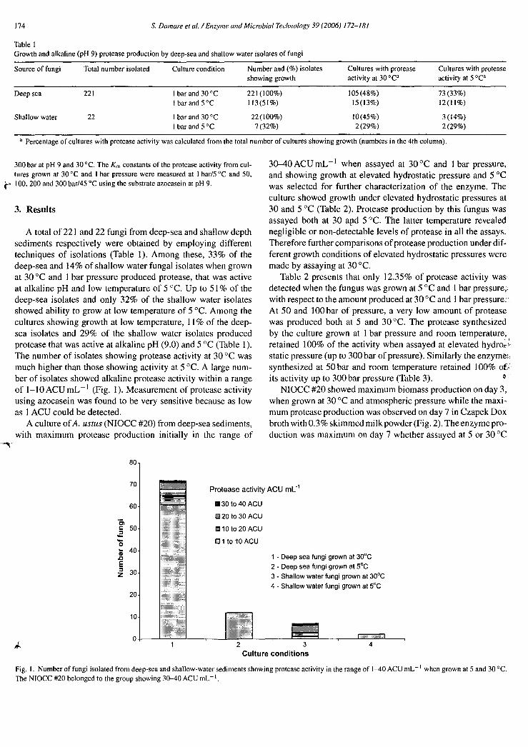
A total of 221 and 22 fungi from deep-sea and shallow depth sediments respectively were obtained by employing different techniques of isolations (Table 1). Among these, 33% of the deep-sea and 14% of shallow water fungal isolates when grown at 30 °C and 1 bar pressure produced protease, that was active at alkaline pH and low temperature of 5 °C. Up to 51% of the deep-sea isolates and only 32% of the shallow water isolates showed ability to grow at low temperature of 5 °C. Among the cultures showing growth at low temperature, 11% of the deep-sea isolates and 29% of the shallow water isolates produced protease that was active at alkaline pH (9.0) and 5 °C (Table 1). The number of isolates showing protease activity at 30 °C was much higher than those showing activity at 5 °C. A large num-ber of isolates showed alkaline protease activity within a range of 1-10 ACU mL-1 (Fig. 1). Measurement of protease activity using azocasein was found to be very sensitive because as low as 1 ACU could be detected.
A culture of A. ustus (NIOCC #20) from deep-sea sediments, with maximum protease production initially in the range of
30-40 ACU mL —I when assayed at 30 °C and 1 bar pressure, and showing growth at elevated hydrostatic pressure and 5 °C was selected for further characterization of the enzyme. The culture showed growth under elevated hydrostatic pressures at 30 and 5 °C (Table 2). Protease production by this fungus was assayed both at 30 and 5 °C. The latter temperature revealed negligible or non-detectable levels of protease in all the assays. Therefore further comparisons of protease production under dif-ferent growth conditions of elevated hydrostatic pressures were made by assaying at 30 °C.
Table 2 presents that only 12.35% of protease activity was detected when the fungus was grown at 5 °C and 1 bar pressure, with respect to the amount produced at 30 °C and 1 bar pressure: At 50 and 100 bar of pressure, a very low amount of protease was produced both at 5 and 30 °C. The protease synthesized by the culture grown at 1 bar pressure and room temperature, retained 100% of the activity when assayed at elevated hydro r ,
static pressure (up to 300 bar of pressure). Similarly the enzyme‘:, synthesized at 50 bar and room temperature retained 100% ofi; its activity up to 300 bar pressure (Table 3).
NIOCC #20 showed maximum biomass production on day 3, when grown at 30 °C and atmospheric pressure while the maxi-mum protease production was observed on day 7 in Czapek Dox broth with 0.3% skimmed milk powder (Fig. 2). The enzyme pro-duction was maximum on day 7 whether assayed at 5 or 30 °C
c 50 13110 to 20 ACU
CI tO 10 ACU ■- 40
30
20
10
Fig. I. Number of fungi isolated from deep-sea and shallow-water sediments showing protease activity in the range of 1-40 ACU mL -1 when grown at 5 and 30 °C. The NIOCC #20 belonged to the group showing 30-40 ACU mL-1.

O di 60
40
120 E
100
160 = Biomass (mg.)
140 -Ai- Protease activity ACU rnL-1
80
20
0
- 100
- 90 7 -J
- 80 E
70 B _ 60 < - 50 :5
- 40 VI (1) V) - 30
- 20
-10
0
0 a-
.1 -f
E
I'', 30 w U) G9 20 0 a-
70
60 t 30 C
5 6 7 8 9 10 11 121, Number of days
5
40
S. Damare etal. / Enzyme and Microbial Technology 39 (2006) 172-181 175
Table 2 Characteristics of the deep-sea isolate NIOCC #20 when grown for 20 days in CD broth with 0.3% skimmed milk powder under various pressure and temperature conditions
Growth temperature (°C) 1 bar 50 bar 100 bar Parameters
30 208.2 69.4 44.1 Biomass produced (mg dry weight) 5 121.4 93.7 85.8
30 36.02 3.67 0.78 Protease activity (ACU mL ) ) assayed at 30°C and 1 bar 5 4.45 0.45 0.22
30 100 10.2 2.17 Percentage activity against the highest value 5 12.35 1.25 0.61
30 0.173 0.15 0.014 Protease activity/mg dry biomass 5 0.037 0.013 0.007
The biomass represents the dry weight of the fungal mycelium. The liquid culture was filtered over a pre-weighed Whatmann No. 1 filter paper, dried to a constant weight at 50 °C and the weight determined. The values are mean of two replicates.
Table 3 Activity of the protease of the deep-sea fungus NIOCC #20 assayed under ele-vated hydrostatic pressures and 30 °C
Pressure (bar) Protease activity (ACU mL t )
A
B
427.33+4.51
ND 50 356 ± 2.12 3.67± 0
100 363.33 ± 4.72 3.33± 0 200 417.33 ± 10.24 4.33 f 0.24 300 428.33 ± 4.95 4.67 ±
A: protease produced when grown at 1 bar pressure/30 °C; B: protease produced when grown at 50 bar pressure/30 °C.
(Fig. 3). The culture showed maximum enzyme production in the medium adjusted to pH 9. Protease was produced in Czapek Dox medium without an organic nitrogen supplement (Fig. 4). Addition of milk, soy meal, malt extract, Tween 80, and corn steep liquor (CSL) increased the enzyme production by several folds (Fig. 4).
The protease was purified from 980 mL of culture super-natant using a three-step procedure that included speed vacuum concentration, anion exchange and gel filtration column chro-
0.5
1 3 5
7 Number of days
Fig. 2. Temporal production of biomass and protease by NIOCC #20 grown in CD broth with 0.3% skimmed milk powder. The protease assay was carried out at pH 9 and 45 °C with azocasein as substrate. The biomass was determined by fil-tering the cultures (in triplicates) on specified days over pre-weighed Whatmann filter papers and dry weight of the fungal mycelium was determined.
matography. The enzyme eluted out as unbound fractions from Resource Q anion exchange column with 0.27-fold purification (Table 4). After gel filtration on superdex-200 column, the purity of the enzyme was 4.05-fold (Table 4). The homogeneity of the enzyme was further confirmed by activity staining on non-denaturing-PAGE. One distinct protease band was visualized on native gel (Fig. 5a). The molecular weight of the purified enzyme fraction obtained by gel filtration chromatography was 32 kDa as determined by SDS-PAGE (Fig. 5b). The purified enzyme had p/ of 6.6 and 6.9 (Fig. 5c). This might be isozymes of protease which were resolved due to the presence of ampholytes in the gel. They might have been merged in the native gel where only one activity band was noticed.
The enzyme exhibited pH optima of 9 with >80% of activity between pH 6 and 10 at 45 °C (Fig. 6a). The protease activity was tested at temperatures ranging from 5 to 90 °C at pH 9 (Fig. 6b). The maximum activity was at 45 °C and about 45% of the maximum activity was detected at 20 °C and about 10% at 2 °C (Fig. 6b). Thermostability studies showed that the enzyme retained 80 and 55% of its activity at 50 and 60 °C respectively for 10 min at pH 9 (Fig. 6c). At its optimum temperature of activity (45 °C), the enzyme retained 100% of its activity for 30 min after which it steadily decreased (data not shown). The
Fig. 3. Protease production by NIOCC #20 when grown at 5 °C (broken line) and 30 °C (solid line). Protease was assayed as described in Fig. 2.

176 S. Damare et al. / Enzyme and Microbial Technology 39 (2006) 172-181
Pro
teas
e ac
tivity
AC
U m
1:1
120
100
80
60
40
20
0 CD
CD +
Malt extract CD + Soy CD + mi k CD + Tween CD + CSL molasses
broth meal 80 + milk
Growth media
Fig. 4. Protease production by NIOCC #20 when grown at 30°C in different media. CD= Czapek Dox broth, CSL =corn steep liquor.
Table 4 Purification of protease of the deep-sea fungus NIOCC #20 grown at pH 9.0 and 30°C
Total enzyme units (ACU) Total protein (mg) Sp. activity (ACU mg —I protein) Purification (fold) Recovery (%)
Crude filtrate 981000 131.08 7484 1 100 Vacuum concentrated 205800 93.06 2212 0.3 21 Resource Q 166786 82.61 2019 0.27 17 Superdex-200 206800 6.82 30323 4.05 21
Protein was estimated using Folin phenol reagent [281.
enzyme was most stable at pH 4.5 in the absence of the substrate (Fig. 6d).
The protease activity was totally inhibited by 2 mM PMSF suggesting it to be a serine protease. EDTA at 5 and 100 mM concentration hardly inhibited the protease activity (Table 5)
indicating that it was not a metalloprotease. The protease of NIOCC #20 is thiol-independent because reducing agents as DTT and mercaptoethanol did not affect the activity con-siderably (Table 5). At 1 mM concentration of Hg only 45% inhibition of the enzyme was observed. Addition of Fe, Ni and
(c)
6.6 6.9
Fig. 5. (a) Zymogram of the active fraction on native page showed activity staining for protease. The clear zone in casein as a result of protease activity appears as a band. (b) The SDS-PAGE of the protease: lane I =crude culture filtrate; lane 2 = vacuum-concentrated culture filtrate; lane 3 = Resource Q-pooled fraction; lane 4= pooled active fraction obtained by gel filtration using Superdex 200 showing single band of 32 kDa (arrow). (c) IEF gel showing isozymes with p/ values of 6.6 and 6.9.

S. Damare et al. / Enzyme and Microbial Technology 39 (2006)172-18I
120
100
80
60
40
20
% R
esi
dua
l act
ivity
177
% R
esid
ual
act
ivity
100-
80-
60-
•
120-
% R
ela
tive
ac
tiv
ity
20
O
5 6 7 8
Assay pH
10 11 12
50 60
Temperature °C
40
(c)
70
40-
0 4
(a)
80 100
Assay temperature °C
20 40 60 0
(b)
6.5 7 7.5 8 8.5
pH of buffer (d)
9 9.5 10 10.5 11
120
100
80
60
40
20
% R
ela
tive
activ
ity
Fig. 6. (a) Relative activity of protease of NIOCC #20 at different pHs and 45 °C. Protease activity was measured as mentioned under Section 2: (A) acetate buffer; ( ■ ) citrate phosphate buffer; (A) phosphate buffer; ( x ) Tris—HCI buffer; (A) boric—borax buffer; ( ❑) Glycine—NaOH buffer; (+) borax—NaOH; (Q) carbonate-bicarbonate buffer. (b) Relative activity of protease at different temperatures measured at pH 9. (c) Residual activity of protease was measured after incubating the enzyme sample at pH 9 for 10 min at different temperatures. (d) Stability of protease at different pHs was carried out by incubating the enzyme at specified pHs without the substrate for 60 min. Activity was measured at its optimum temperature (45 °C) and expressed as percent residual activity.
Zn at 1 mM hardly inhibited the enzyme activity. On the other hand, Cu at 1 mM concentration inhibited the protease activity totally (Table 5). About 70% of the activity was retained in the presence of sodium chloride of 0.5 M concentration which equals to 29 ppt salinity of seawater (Table 5). Chemical surfactants like Triton X-100 and Tween 80 (both at 1.0%) and commercial detergents at 2 g L -1 concentrations did not reduce the protease activity, although SDS (0.1%) totally inhibited its activity (Table 6). About 50% of the enzyme activity was lost after treatment at 60 °C for 10 min. However, thcrmostability of the enzyme at 60 °C increased on addition of glycerol, CaC12, PEG 6000, sucrose, mannitol, sorbitol and starch during enzyme assay (Table 7). The protease of the culture grown at 1 bar pressure showed a K m constant of 2 mg m1_, -1 when measured at pH 9 and 45 °C and this increased marginally at elevated hydrostatic pressures of 50 and 100 bar (Table 8). There was a marked increase in Km constant at 5 °C/1 bar, 45 °C/200 and 300 bar pressure. The Vmax values were not much affected by the elevated hydrostatic pressures except a
substantial drop at 50 bar pressure from that at 1 bar pressure (Table 8).
4. Discussion
The major aim of this study was to explore deep-sea fungi for production of cold-tolerant and alkaline protease. The isolate N1OCC #20 obtained after the screening produced substantial growth at 5 °C, under I and 100 bar pressure (corresponding to 1000 in). Under the above two conditions, it attained a biomass equivalent to 58 and 41% respectively of that produced at 30 °C/ I bar pressure (Table 2). However, when the fungus was grown at 5 °C under 1, 50 or 100 bar and the enzyme activity assayed at 5 or 30 °C under different hydrostatic pressures, lit-tle or negligible amount of cold-tolerant protease was detected. In contrast to the above, the enzyme produced by the fuck gus at 30 °C and 1 bar was much more versatile and retained about 10% of activity at 2 °C at 1 bar compared to its activ-ity at optimum temperature (Fig. 6b). The enzyme produced at

178
S. Damare et al. /Enzyme and Microbial Technology 39 (2006) 172-181
Table 5
Effect of various inhibitors, heavy metals, varying ionic strengths (sodium chlo-
ride) and reducing agents on protease activity of the deep-sea fungus NIOCC
#20
Table 7
Effect of additives on thermostability of the crude enzyme of the deep-sea fungus
NIOCC #20 with reference to 10 min of heat treatment at 60 °C without any
additive
Inhibitors
Residual activity (%)
Additive present during the heat treatment Residual activity (To)
Protease inhibitors (mM)
None (control)
100 PMSF (2)
0 EDTA (5)
99
EDTA (100)
93
Heavy metals (mM)
NiC12 (1)
72
ZnCl2 ( 1 )
81
HgC12 (1)
55 CuC12 (1)
0
FeC13 (1 )
74
Sodium chloride (M)
0
100 0.1
88.6 0.25
80
0.50
70
0.75
61.4 1.0
40
Reducing agents (%)
None
100 DTT dithiothereitol (0.1)
91
DTT (0.5)
73 3-Mercaptoethanol (0.1)
77
p-Mercaptoethanol (0.5)
75 Sodium thioglycolate (0.1)
45
Sodium thioglycolate (0.5)
27 Sodium thioglycolate (1.0)
27
Glutathione (0.1)
0
30 °C under 1 or 50 bar also retained its activity when assayed under elevated hydrostatic pressures up to 300 bar (Table 3). However, the increasing Km constants of protease at elevated hydrostatic pressures of 200 and 300 bar (Table 8) indicates low enzyme-substrate affinity under these extreme conditions. The total enzyme activity per se is not affected by elevated pressures (Table 3), which is also indicated by the almost constant V nia, values (Table 8).
Table 6
Effect of detergents and bleaching agents on protease activity of the deep-sea fungus NIOCC #20
Compound (concentration in %)
Residual specific activity (%)
None
100 Tween 80 (1.0)
142 Triton X-100 (1.0)
139 SDS (0.1)
0 Ala (commercial fabric bleach) (2.0)
20
Sodium hypochlorite (1.0)
2
H202 (2.0)
40 Commercial detergent wheel (2 g
91
Mr. White
85 1M)i Shakti
64 Tide
62 Surf Excel
161 Ariel Compact
73
Control (untreated)
100
No additives
51 Glycerol ( I%)
101.5 CaCl2 (1 mM)
108.5 CaCl2 (5 mM)
110.6 CaCl2 (10 mM)
102.4 PEG 6000 (1%)
111.1 Sucrose (1%)
84.8 Mannitol (1%)
79.2 Sorbitol (1%)
85.6 Starch (1%)
98.8
Thus, although growth was fairly unaffected by elevated pres-sure and low temperature, protease production under similar conditions was reduced 10-100 folds. This appears to be a general trend with all the deep-sea isolates that we obtained (Table 1). Thus, a higher percentage of deep-sea isolates (51%) were capable of growing at 5 °C, when compared to those isolated from shallow waters (32%). Besides, a high number of deep-sea isolates produced low-temperature active protease when grown at 1 bar and 30 °C, but very few of these did so when grown at 1 bar and 5 °C (Table 1). Compared to the deep-sea isolates, fewer isolates from shallow water produced cold-tolerant protease when grown at 1 bar pressure and 30 °C and their growth was also poor at 5 °C. NIOCC #20 appears to be a strain adapted to deep-sea conditions, since it grew better at 5 °C/100 bar than at 30 °C/100 bar, although its production of protease under the former conditions was low (Table 2). This is in contrast to the report on a deep-sea bacterial strain DB6705, which grew better at higher rather than low tempera-tures when cultured at 500 bar hydrostatic pressure [23]. Growth and metabolic activities under extreme conditions are known to be low [24]. Our results with NIOCC #20 also show similar trends.
Enzymes from cold-adapted organisms have been classified into three groups [10]. Group I enzymes are heat-sensitive, but the other enzymatic characteristics are similar to mesophilic enzymes. Group II enzymes are heat-sensitive and relatively more active than mesophilic enzymes at a low temperature. Group III enzymes have same thermostability as mesophilic enzymes but are more active than mesophilic enzymes at a low
Table 8
Km constant and Vrna, values of the purified protease of the deep-sea fungus
NIOCC #20 measured under different conditions
Enzyme activity assayed at Km constant Vina. (ACU -1 )
(mg mL-1 azocasein)
1 bar and 45 °C
2.0
26.3
50 bar and 45 °C
2.2
19.2
100 bar and 45 °C
2.4
24.4
200 bar and 45 °C
3.3
25.0
300 bar and 45 °C
5.0
25.0

Table 9 Comparison of NIOCC #20 with other alkalir.e and low-temperature active proteases
Organism Source of isolation Substrate used Maximum U Definition of U Optimum pH Optimum temperature
Other properties Reference
(°C)
Alteromonas haloplanktis Marine bacterium MCA nmol substrate released min -1 8-9 20 74 kDa, thiol protease [15]
Vibrio sp. Deep-sea sediments Casein 353 U inL -1 1.Lg tyrosine min -1 8.5-9.0 40 35 kDa [14]
Azospirillum sp. Cold mountain soil Casein 2 UmL-1 Increase in OD of 0.1 min -1 8.5 40 48.6 kDa, cysteine [21 ]
Vibrio sp. Cold marine source in Azocasein 47 kDa, serine [13]
Iceland Paecilomyces marquandii
(fungus) Not known Casein, haemoglobin 0.016-0.018 U mL- 12 45 38 kDa [29]
Nocardiopsis dassonvillei Not known Casein, haemoglobin 0.053 AU mL- I CPU = mM serine min-1 8-9 60 Serine [30]
Sphingomonas paucimobilis Stomach of Antarctic krill
A variety of substrates Tyrosine released 6.5-7.0 30 (47% of the activity at
Metalloprotease [12]
0 °C)
Conidiobolus coronatus Soil Casein 30 U mL-1 1 Abs mL-1 min -I =U 9.7 37 [31]
Rhizophus oryzae Not known Azocasein 3370 U mL-1 0.1 Abs h-1 10.5 60 [32]
Bacillus mojavensis Soil Casein mL- I min-1 of tyrosine 10.5 60 [20]
Nocardiopsis sp. Not known Azocasein 50 U mg-1 protein Abs h-1 8.0 50 Serine protease [33]
Paecilomyces lilacinus Biocontrol agent Azocasein [34]
Bacillus sp. Korean polychnete Casein 1 µg tyrosine min -1 10.0 45-50 Serine protease [11]
A. ustus NIOCC #20 Deep-sea sediment Azocasein 1639 ACU mL-1 Abs 0.001 min -1 6.5-10 45 (45% of the activity at
32 kDa, serine protease The present work [35]
20°C)
S. D
atna
re e
t al. /E
nz ym
e a
nd
Mi c
robia
l Techn
olo
gy 3
9 (2
006
) 172-1
81

180 S. Damare et al. / Enzyme and Microbial Technology 39 (2006) 172-181
temperature. Accordingly, the protease of our isolate falls into the group II. The protease of mesophilic Penicillium sp. did not show any activity below 20 °C [25]. The alkaline protease from another mesophilic species of Penicillium did not show any activity below 35 °C [26]. On the other hand, NIOCC #20 showed about 45, 26 and 12% of the maximum activity at 20, 15 and 5 °C. It looses about 50% of its activity at 60 °C (Fig. 6c). Moreover, at its optimum temperature of activity (45 °C), it is stable only for 30 min after which it rapidly looses activity (data not shown). The enzyme is stable for more than 24 h at 5 °C and pH 4.5. Its half-life under these conditions is about 196 h. These results suggest its placement in the group II. The effect
rpf temperature on the enzyme activity is determined by the tem-perature coefficient Q10 [27]. It is the factor by which the rate increases when the temperature is raised by 10°. If the rate of the reaction is completely temperature-independent, the resulting Qi 0 will be 1.0. If the reaction rate increases with increas-ing temperature, Q10 will be greater than 1. Thus, the more temperature-dependent a process is, the higher will be its Q10 value. The calculated Qio value of 1 between 2 and 10 °C in the present work indicates temperature-independent enzyme activ-ity at lower temperatures. On the other hand, the increased Qio values (above 1) at 15-45 °C indicate direct relation to increas-ing temperatures. Above 45 °C, the Qio value is less than one suggesting negative effect of temperature on the enzyme activity.
The present strain produced a maximum of 1639 ACU mL —I of the enzyme, as estimated by the azocasein method. Appli-
cation of different methods and the definition of enzyme units to estimate the enzyme by various authors using different sub-strates such as casein, haemoglobin, methylcoumarylaminoacid (MCA), azocasein or gelatin make the comparison difficult. However, our estimations have shown that the protease activity per mL in the crude culture filtrate of NIOCC #20 corresponds to 84 p,g of tyrosine released. The protease production by NIOCC #20 appears to be as good as or even better than those reported
4Table 9). The protease of NIOCC #20 is a serine protease because it
was totally inhibited by PMSF which is a known inhibitor of serine protease. It was not inhibited by EDTA and therefore it is not a metalloprotease. It was not substantially inhibited by the heavy metals Ni and Zn which are the inhibitors of cys-teine protease. Its activity did not increase in the presence of thiol compounds, indicating that it was thiol-independent ser-ine protease. Protease produced by this isolate showed several special features. It was active in the presence of several commer-cial detergents when assayed under its optimum conditions. The enzyme was active in the presence of 0.5 M NaCI equivalent to 29 ppt of seawater salinity. Its stability at 60 °C increased in the presence of various stabilizing agents. Such features in proteases are often much sought after for use as additives in detergents for low-temperature wash [10]. Most alkaline proteases have been reported to he significantly stabilized in the presence of additives such as those used by us [20]. ALNIOCC #20 secreted maximum extracellular protease among
all the deep-sea fungi isolated and when grown in a medium pre-pared with seawater at pH 8 and 9 and thus is truly alkalophilic. Protease production occurred in Czapek Dox medium without an
organic nitrogen source also and thus the enzyme appears to be constitutive (Fig. 4). Its production, however, could be enhanced in the presence of milk, soy meal, and corn steep liquor.
Protease of A. ustus (NIOCC #20) with an optimum pH of 9 and showing about 45 and 25% of its activity at 20 and 15 °C respectively is an alkaline protease. Its performance in terms of activity can be further improved by optimization of growth and other parameters. Terrestrial isolate of A. ustus (isolate # MTCC 2200) obtained from IMTECH culture collection showed extremely low protease production (23 ACU mL — I ) under sim-ilar assay conditions. This study shows that deep-sea fungi are useful candidates in the search for alkaline proteases.
Acknowledgements
We are grateful to Department of Biotechnology, New Delhi for the financial grant BT/PR 1193/AAQ/03/102/2000. SD is thankful to DBT for a junior research fellowship. We thank Ms. Asha Nadurmath (Goa University) and Ms. Vinda Shirgaonkar for technical assistance. We also wish to thank Department of Ocean Development for providing the ship facility on board the Russian research vessel. NIO's contribution # 4120.
References
[1] Liberra K, Lindequist U. Marine fungi-a prolific resource of biologically active natural products. Pharmazie 1995;50:583-8.
[2] Yu VB, Verigina NS, Soya VV, Pivkin MV, Zvyagintseva TN. Fila-mentous marine fungi as producers of O-glucosyl hydrolases: 13-1,3 glucanase from Chaetomium indicum. Mar Biotechnol 2003;5:349-59.
[3] Jensen PR, Fenical W. Secondary metabolites from marine fungi. In: Hyde KD, editor. Fungi in marine environments, Fungal Diversity Res. Ser. 2002;7:293-315.
[4] Raghukumar C. Fungi from marine habitats: an application in bioreme-diation. Mycol Res 2000;104:1222-6.
[5] Raghukumar C. Bioremediation of coloured pollutants by terrestrial ver-sus facultative marine fungi. In: Hyde KD, editor. Fungi in marine environments, Fungal Diversity Research Series 2002;7:317-44.
[6] Banse K. Some views on the degradation and disposition of organic particles as collected by sediment traps in the open sea. Deep-Sea Res 11990;37:1177-95.
[7] Smith DC, Simon M, Alldredge AL, Azam F. Intense hydrolytic enzyme activity on marine aggregates and implications for rapid particle disso-lution. Nature 1992;359:139-42.
[8] Raghukumar C, Raghukumar S. Barotolerance of fungi isolated from deep-sea sediments of the Indian Ocean. Aquat Microb Ecol 1998;15:53-63.
[9] Raghukumar C, Sheelu G, Loka Bharathi PA, Nair S, Mohandass C. Microbial biomass and organic nutrients in the deep-sea sediments of the Central Indian Ocean Basin. Mar Geores Geotech 2001;19:1-16.
[10] Ohgiya S, Hoshino 1, Okuyama H, Tanaka S, Ishizaki K. Biotechnol-ogy of enzymes from cold-adapted microorganisms. In: Biotechnological applications of cold-adapted organisms. Springer-Verlag; 1999. p. 17-34.
[11] Joo HS, Kumar GC, Park GC, Park SR, Chang CS. Bleach resistant alkaline protease produced by a Bacillus sp. isolated from the Korean polychaete, Periserrula leucophryna. Proc Biochem 2004;39:1441-7.
[12] Turkiewicz M, Gromek E, Kalinowska H, Zielinska M. Biosynthesis and properties of an extracellular metalloprotease from the Antarctic marine bacterium Sphingornonas paucimobilis. J Biotechnol 1999;70:53-60.
[13] Alfredson GA, Gudmundson HM, Xiang JY, Kristjansson HM. Subtilisin-like serine protease from psychrophilic marine bacteria. J Mar Biotechnol 1995;3:71-2.

S. Damare et al. / Enzyme and Microbial Technology 39 (2006) /72-181 181
[14] Hamamato T, Kaneda M, Kudo T, Horikoshi K. Characterization of a protease from a psychrophilic Vibrio sp. Strain 5709. J Mar Biotechnol 1995;2:219-22.
[15] Suzuki S, Odagami T. Low-temperature-active thiol protease from marine bacterium Alteromonas haloplanktis. 1 Biotechnol 1997;5: 230-3.
[16] Kumar GC, Takagi H. Microbial alkaline proteases: from bioindustrial viewpoint. Biotechnol Adv 1999;17:561-94.
[17] Raghukumar C, Raghukumar S, Sheelu G, Gupta SM, Nath BN, Rao BR. Buried in time: culturable fungi in a deep-sea sediment core from the Chagos Trench, Indian Ocean. Deep-Sea Res I 2004;51:1759-68.
[18] Damare S, Raghukumar C, Raghukumar S. Fungi in deep-sea sediments of the Central Indian Basin. Deep-Sea Res I 2006;53:14-27.
[19] Molitoris HR. Methods for determination of enzymatic activities of marine fungi. Czech Mycol 2000;52:97-124.
[20] Beg QK, Gupta R. Purification and characterization of an oxidation-stable, thiol-dependent serine alkaline protease from Bacillus mojavensis. Enzyme Microb Technol 2003;32:294-304.
[21] Oh KH, Seong CS, Lee SW, Kwon OS, Park YS. Isolation of a psychrotrophic Azospirillum sp. and characterizaion of its extracellular protease. FEMS Microbiol Lett 1999;174:173-8.
[22] Heukeshovan J, Dernick R. Simplified method for silver staining of proteins in polyacrylamide gels and the mechanism of silver staining. Electrophoresis 1985;6:103-12.
[23] Kato C, Masui N, Horikoshi K. Properties of obligately barophilic bac-teria isolated from a sample of deep-sea sediment from the Izu-Bonin trench. J Mar Biotechnol 1996;4:96-9.
[24] Wirsen CO, Jannasch HW. Activity of marine psychrophilic bacte-ria at elevated hydrostatic pressures and low temperatures. Mar Biol 1975;31:201-8.
[25] Germano 5, Pandey A, Osaku CA, Rocha SN, Soccol CR. Charac-terization and stability of proteases from Penicillium sp. produced by solid-state fermentation. Enzyme Microb Technol 2003;32:246-51.
[26] Agrawal D, Patidar P, Banerjee T, Patil S. Production of alkaline protease by Penicillium sp. under SSF conditions and its application to soy protein hydrolysis. Process Biochem 2004;39:977-81.
[27] http://www.csupomona.edu/—seskandari/Q I 0.1ttml. [28] Lowry OH, Rosenbrough NJ, Farr AL, Randall RJ. Protein measurement
with the Folin phenol reagent. J Biol Chem 1951;193:265-75. [29] Beck CM, Strobel RJ. Low temperature active alkaline protease from
Paecilomyces marquandii and its preparation, EP Patent 290569 (1987). [30] Liu CL, Beck CM, Strobel RI. Low-temperature active alkaline protease
from Nocardiopsis dassonvillei and its preparation, EP Patent 290567 (1998).
[31] Bhosale SH, Rao MB, Deshpande VV, Srinivasan MC. Thermostability of high-activity alkaline protease from Conidiobolus coronatus (NCE1' 86.8.20). Enzyme Microb Technol 1995;17:136-9.
[32] Banerjee R, Bhattacharya BC. Extracellular alkaline protease of newly isolated Rhizopus oryzae, optimization of production conditions. Biotechnol Lett 1992;14:301-4.
[33] Moreiera KA, Porto TS, Teixeira MFS, Porto ALF, Filho JLL. New alkaline protease from Nocardiopsis sp.: partial purification and charac-terization. Proc Biochem 2003;39:67-72.
[34] Khan A, Williams K, Molloy MP, Nevalainen H. Purification and char-acterization of a serine protease and chitinases from Paecilomyces liliac-inus and detection of chitinase activity on 2D gels. Protein Exp Purificat 2003;32:210-20.
[35] Raghukumar C, Damare S, Muraleedharan C. A process for production of low temperature-active alkaline protease from a deep-sea fungus, Patent filed Ref. No. 2003/P2 (NF 271/2003).
7