Psych 56L/ Ling 51: Acquisition of Language Lecture 15 Language in Special Populations.
Upload
briana-sandersCategory
view
220download
2
Psych 56L/ Ling 51:Acquisition of Language
Lecture 4
Biological Bases of Language II

Announcements
Be working on HW1 (due 1/20/11)
Be working on bio bases review questions
Check out the reference material on the webpage

Anatomy & Language

The Human Vocal Tract: A Finely Honed Instrument
Speech is produced when air from the lungs exits the larynx and is filtered by the vocal tract above the larynx: glottis, pharynx, uvula, velum, hard palate, tongue, nasal cavity, alveolar ridge, teeth, lips.

Human Speech Apparatus: Pros and Cons
Larynx: most speech-specific feature of the human vocal tract.
Compared to other mammals, human larynx is very low.
The good: Low larynx helps produce a wider variety of speech sounds.
The bad: Humans are more likely to get food caught in the trachea and choke.
Lower mouth shape: accommodate the lower larynx
The good: Help support lower larynx.
The bad: Lead to overcrowded teeth and impacted wisdom teeth.
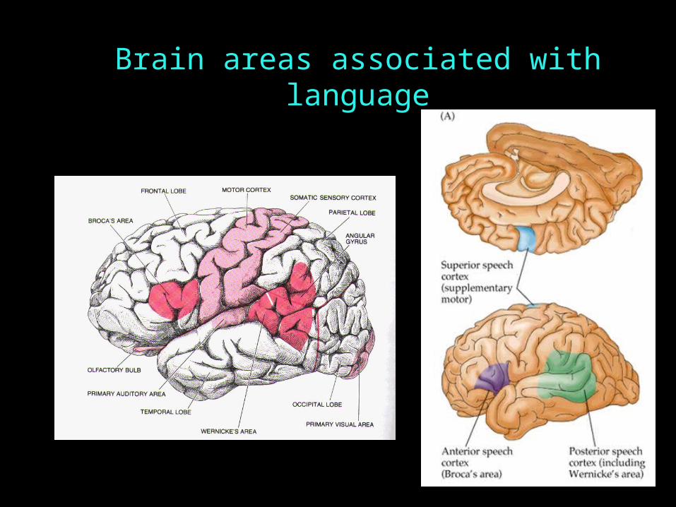
Brain areas associated with language

Functional Architecture
Functional architecture: how the brain is organized to do what it does (that is, how it is organized to accomplish some function)
Neurolinguistics: study of the brain with relation to language functioning. One big question: is there a separate chunk of brain (or dedicated brain activity = a functional “organ”) specifically for language?

Methods of Neurolinguistic Investigation
Lesion studies: correlate missing bits of brain (lesions) with missing bits of psychological functioning.
One very interesting kind of missing brain bit: split or damaged corpus callosum, found in split brain patients

Methods of Neurolinguistic Investigation
Left Brain Right Brain
Left Body Right Body
Contralateral connections Contralateral connections in the 1860s: investigators in the 1860s: investigators apply electric currents to brains of anesthetized animals apply electric currents to brains of anesthetized animals and made an interesting discovery.and made an interesting discovery.
Note on connections:
Contralateral: acrossIpsalateral: same side

Left Visual Field Right Visual Field
Left Brain Right Brain
Hemispheres & Visual Field
corpus callosum

Information Flow:
LVF RH LH
RVF LH RH

Methods of Neurolinguistic Investigation
Dichotic listening tasks: use the fact that contralateral connections from the ears to the brain are stronger than ipsalateral connections. Experimenters present two tasks at the same time, one to each ear, and ask subjects which one is perceived.
If they say the left ear’s
stimulus, then the right side of the brain processes that signal. If they say the right ear’s stimulus, then the left side of the brain processes that signal.

Dichotic Listening

Methods of Neurolinguistic Investigation
ERPs: Event-related brain potentials, gauged via electrode caps. The location of ERPs associated with different mental activities is taken as a clue to the area of the brain responsible for those activities.
Good: non-invasive, relatively
undemanding on the subject, provide precise timing on brain events
Bad: poor information on exact location of ERP since just monitoring the scalp

Methods of Neurolinguistic Investigation
Brain-imaging techniques: gauge what part of the brain is active as subjects perform certain tasks
PET scans: Positron emission topography scans
- subjects inhale low-level radioactive gas or injected with glucose tagged with radioactive substance
- experimenters can see which parts of the brain are using more glucose (requiring the most energy)
http://www.learner.org/vod/vod_window.html?pid=1615

Methods of Neurolinguistic Investigation
Brain-imaging techniques: gauge what part of the brain is active as subjects perform certain tasks
fMRI scans: functional magnetic resonance imaging
- subjects have to be very still inside MRI machine, which is expensive to operate
- experimenters can see which parts of the brain are getting more blood flow or consuming more oxygen

Methods of Neurolinguistic Investigation
Brain-imaging techniques: gauge what part of the brain is active as subjects perform certain tasks
MEG: Magnetoencephalography
- subjects have to be very still
- experimenters can see which parts of the brain are active
Video of word recognition in brain (10 sec long):http://www.mrc-cbu.cam.ac.uk/facilities/meg/

Methods of Neurolinguistic Investigation
Brain-imaging techniques: gauge what part of the brain is active as subjects perform certain tasks
Optical Topography: Near-infrared spectroscopy (NIRS)
- transmission of light through the tissues of the brain is affected by hemoglobin concentration changes, which can be detected

Where is language located? Left hemisphere evidence
From brain injury and aphasia (when language is severely impaired):
Paul Broca’s lesion studies
- “Tan”, who had left hemisphere lesion and loss of language abilities
Functional asymmetry: damage to the left hemisphere seems to cause language problems (whether it is spoken or signed) while damage to the right hemisphere seems to cause non-linguistic visual-spatial information processing problems.

Broca’s Aphasia
Parietal LobeFrontal Lobe
Occipital Lobe

Broca’s Aphasia
Patients have trouble producing speech, mostly content words (nouns and verbs) with few grammatical morphemes
“Yes… ah… Monday… er… Dad and Peter H… [his own name], and Dad…. er… hospital… and… ah… Wednesday… Wednesday, nine o’clock…”
Videos of sample speech from a Broca’s aphasic: http://www.youtube.com/watch?v=f2IiMEbMnPMhttp://www.learner.org/vod/vod_window.html?pid=1574
(from 2:58-6:16)

Broca’s Aphasia
Broca’s aphasics & comprehension:
Relatively good comprehension of some sentences:
Can understand sentences like these:
The dog bit the woman.
The apple that the boy is eating is red.
…but not these (because their meaning can’t be inferred from
the meaning of the nouns and verbs alone):
The car is pushed by the truck.
The girl whom the boy is pushing is tall.

Wernicke’s Aphasia
Patients with posterior lesions in the left hemispherePatients with posterior lesions in the left hemisphere Speech is fluentSpeech is fluent But comprehension is impairedBut comprehension is impaired
Occipital Lobe
Frontal Lobe

Wernicke’s Aphasia
Patients have speech that is “syntactically full but semantically empty”
“I feel very well. My hearing, writing been doing well. Things that I couldn’t hear from. In other words, I used to be able to work cigarettes I didn’t know how…”
Video of sample speech from a Wernicke’s aphasic: http://www.youtube.com/watch?v=aVhYN7NTIKU
Comprehension is very low.

Where is language located?

Where is language located? Left hemisphere evidence
From split-brain patients (with severed corpus callosum - no communication between hemispheres)

Where is language located? Left hemisphere evidence
From split-brain patients (with severed corpus callosum - no communication between hemispheres)
Can’t say what they saw on the left side, but can draw with their left hand.

General Testing Setup
Testing Split Brain Patients

Testing Split Brain Patients
Patient says: “Spoon!”
Name that object Name that object (picture in RVF)(picture in RVF)

Testing Split Brain Patients
Patient: (says nothing)Researcher: “Did you see anything?”Patient: “Nope.”
Name that object Name that object (picture in LVF)(picture in LVF)

Testing Split Brain Patients
Right Hand: Pulls out spoonLeft Hand does nothing
Pick up Pick up the object the object displayeddisplayed(picture in (picture in RVF)RVF)

Testing Split Brain Patients
Pick up Pick up the object the object displayeddisplayed(picture in (picture in LVF)LVF)
Left Hand: Pulls out spoon!Right hand does nothing

Left Hemisphere rationalizing behavior of Right Hemisphere

Typical Split Brain Patient
Left Brain:Left Brain:– Normal Language UseNormal Language Use– No easily detectable deficits.No easily detectable deficits.
Right Brain:Right Brain:– Some rudimentary word recognition.Some rudimentary word recognition.

Where is language located? Left hemisphere evidence
From normal adults: dichotic-listening experiments
ba ga
Normal adults have a right-ear advantage

Deaf Signers with Left Hemisphere Damage:Deaf Signers with Left Hemisphere Damage:– Language Deficit. Aphasic.Language Deficit. Aphasic.
Deaf Signers with Right Hemisphere Damage:Deaf Signers with Right Hemisphere Damage:– Visuo-Spatial Deficits.Visuo-Spatial Deficits.– No easily detectable language deficits.No easily detectable language deficits.
Left Hemisphere implicated in languageLeft Hemisphere implicated in language
Poizner, Klima, & Bellugi (1987)
Evidence for Left Hemisphere Lateralization from American Sign Language

Hickok et al. 1998: ASL lateralization evidence
Left hemisphere damage leads to language damage

Why the left hemisphere?
Left hemisphere may process information more analytically.
Trained musicians process music in the left hemisphere. Normal (untrained) people process it on the right.
Left hemisphere may be better at executing well-practiced routines, while right is better at responding to novel stimuli.
Language, for adults, is a well-practiced routine.

Where is language located? Not-just-left hemisphere evidence
Sometimes, aphasia doesn’t result when there is left hemisphere damage.
Sometimes, aphasia results when there is right hemisphere damage.
In some people (usually left-handed people), language is controlled by the right hemisphere.

Where is language located? Not-just-left hemisphere evidence
Right hemisphere contributions to language: tone contour, emotional tone, jokes, sarcasm, figurative language interpretation, following indirect requests
(much of this falls under pragmatics)
Evidence: right hemisphere lesion patients
Right hemisphere activated by semantic processing, while left hemisphere activated primarily by syntactic processing
Evidence: ERP studies
Evidence: late language learners who aren’t as proficient with syntax, and have language located primarily in right hemisphere

How does a left hemisphere specialization for language develop?
Equipotentiality hypothesis: left and right hemispheres have equal potential at birth
Prediction: dichotic listening and brain injury in children show less specialization for language than adults
Invariance hypothesis: left hemisphere specialization available at birth
Prediction: dichotic listening and brain injury data from children should look like the corresponding data from adults

How does a left hemisphere specialization for language develop?
fMRI studies: newborns and 3-month-old infants show greater left-hemisphere than right-hemisphere activation in response to speech stimuli (as do adults)
- But also greater left-hemisphere activity in response to non-speech sounds, suggesting general bias to process sounds in left hemisphere (older children [10-month-olds] and adults process non-speech sounds with right hemisphere)

How does a left hemisphere specialization for language develop?
Summary from experimental studies:
Language processing appears to be specialized to the left hemisphere as early as researchers can test it.
But the infant brain is not the same as the adult brain - specialization/lateralization continues to increase as the brain matures.

How does a left hemisphere specialization for language develop?
Childhood aphasia: Aphasia nearly always results from left hemisphere damage and rarely from right hemisphere damage (Woods & Teuber 1978)
However, immature brain is not organized the same way as the mature brain.
- children more likely to suffer Broca’s aphasia (non-fluent aphasia) than Wernicke’s
- children tend to recover better from brain damage, with younger children recovering better than older children

Neural plasticity in children
Plasticity: the ability of parts of the brain to take over functions they ordinarily would not serve - ex: right hemisphere taking over language functions if left hemisphere is damaged.
However, plasticity isn’t the perfect solution - ex: subtle syntactic impairments in these cases suggest that the right hemisphere isn’t as good at parts of language as the left hemisphere is.

Neural plasticity in children
How plasticity works:
The child’s brain has much redundancy (extra synaptic connections.)
Maturation = pruning unnecessary connections
What’s necessary: what gets used (where child’s brain activity is).
Once connections are pruned, redundancy is lost and particular functions become localized.

Neural plasticity in children
But wait - young children use their right hemisphere (somewhat) for language. Since there’s language activity, why does the right hemisphere lose its language functionality?
Maturation hypothesis: adult language brain structures develop in the left hemisphere and take over (specialization is genetically determined)
Process change hypothesis: children change the way they process language, and the new way is more in line with the left hemisphere’s natural capacities. (specialization is result of process change)

Neural plasticity in adults, too?Rasmussen & Milner (1977)
Normal Speakers
Speakers with left hemisphere damageCan recover somewhat by using right hemisphere for language?

Genetic Basis of Language Development

Heritability of individual differences
Twin studies: assess how similar/different monozygotic (identical) and dizygotic (fraternal) twins are
Stromswold (2001): heritable factors account for 25-50% of variance in normal children’s language abilities; 50-60% of variance in impaired children’s language abilities

Heritability of individual differences
Twin studies: assess how similar/different monozygotic (identical) and dizygotic (fraternal) twins are
Difference between grammatical and lexicon development:
genetic factors account for 25% of syntactic differences and 5% of variance among vocabulary (Stromswold 2006). In general, biological contribution to syntactic development is greater than biological contribution to lexical development.

Genetics of language impairment
Language impairment runs in families.
- language-impaired children are far more likely to have language-impaired family members
- monozygotic twins are more likely to share a language impairment

Genetics of language impairment
Language impairment runs in families.
- KE family (16 of 30 members had language impairment)
- affected members had poor language abilities and severe difficulties with the motor skills involved with speech production
- single dominant gene appeared to be the cause: mutation on gene that affects encoding of protein FOXP2 (Fisher 2006)
…however, this is only one genetic part of language development
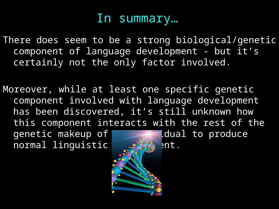
In summary…
There does seem to be a strong biological/genetic component of language development - but it’s certainly not the only factor involved.
Moreover, while at least one specific genetic component involved with language development has been discovered, it’s still unknown how this component interacts with the rest of the genetic makeup of an individual to produce normal linguistic development.

Questions?
You should be able to do all the homework questions in HW1, and review questions for bio bases up through (24)