Pseudomonas Products ModifyTransferrin Lactoferrin to...
11
Pseudomonas and Neutrophil Products Modify Transferrin and Lactoferrin to Create Conditions That Favor Hydroxyl Radical Formation Bradley E. Britigan*" and Brian L. Edekert *Research Service and Department ofInternal Medicine, Veterans Administration Medical Center, and tDepartment ofInternal Medicine, University ofIowa College ofMedicine, Iowa City, Iowa 52242 Abstract In vivo most extracellular iron is bound to transferrin or lacto- ferrin in such a way as to be unable to catalyze the formation of hydroxyl radical from superoxide (7O-) and hydrogen peroxide (H202). At sites of Pseudomonas aeruginosa infection bacterial and neutrophil products could possibly modify transferrin and/ or lactoferrin forming catalytic iron complexes. To examine this possibility, diferrictransferrin and diferriclactoferrin which had been incubated with pseudomonas elastase, pseudo- monas alkaline protease, human neutrophil elastase, trypsin, or the myeloperoxidase product HOCI were added to a hypoxan- thine/xanthine oxidase *02-/H202 generating system. Hy- droxyl radical formation was only detected with pseudomonas elastase treated diferrictransferrin and, to a much lesser extent, diferriclactoferrin. This effect was enhanced by the combina- tion of pseudomonas elastase with other proteases, most promi- nently neutrophil elastase. Addition of pseudomonas elastase- treated diferrictransferrin to stimulated neutrophils also resulted in hydroxyl radical generation. Incubation of pseudo- monas elastase with transferrin which had been selectively iron loaded at either the NH2- or COOH-terminal binding site yielded iron chelates with similar efficacy for hydroxyl radical catalysis. Pseudomonas elastase and HOCI treatment also de- creased the ability of apotransferrin to inhibit hydroxyl radical formation by a Fe-NTA supplemented hypoxanthine/xanthine oxidase system. However, apotransferrin could be protected from the effects of HOCI if bicarbonate anion was present dur- ing the incubation. Apolactoferrin inhibition of hydroxyl radi- cal generation was unaffected by any of the four proteases or HOC. Alteration of transferrin by enzymes and oxidants pres- ent at sites of pseudomonas and other bacterial infections may increase the potential for local hydroxyl radical generation thereby contributing to tissue injury. (J. Clin. Invest. 1991. 88:1092-1102.) Key words: iron * elastase - myeloperoxidase . alkaline protease - trypsin Introduction Sites of Pseudomonas aeruginosa and many other bacterial in- fections are notable for an intense infiltration of neutrophils which generate a variety of toxic oxygen species as an impor- This work was presented in part at the Interscience Conference on Antimicrobial Agents and Chemotherapy, Atlanta, GA, 24 October 1990. Address correspondence to Bradley E. Britigan, M.D., Dept. of In- ternal Medicine, University of Iowa, SW54, GH, Iowa City, IA 52242. Receivedfor publication 2 October 1990 and in revisedform 4 June 1991. tant aspect of their antimicrobial armamentarium (1, 2). Mem- brane attachment of bacteria activates a unique membrane as- sociated NADPH-dependent oxidase system which generates superoxide CO-)' and hydrogen peroxide (H202), the latter via the spontaneous dismutation of *°- (1, 2). Interaction of H202 with myeloperoxidase (MPO) released from neutrophil cyto- plasmic granules generates additional microbicidal oxidants such as hypochlorous acid (HOC1) and chloramines (3, 4). Fi- nally, in the presence of an exogenous iron catalyst, neutrophil- derived *0- and H202 can also react to form the extremely toxic hydroxyl radical (5). Neutrophil oxidant formation has been implicated as an important contributor to tissue injury at sites of inflammation (2). A variety of in vitro and in vivo studies have suggested that hydroxyl radical may be one of the reactive oxygen species involved (2). Because neutrophils do not possess an endoge- nous iron catalyst for hydroxyl radical generation (5-1 1), in order for this species to be produced in vivo, an exogenous catalytic iron complex must be present. In vivo almost all ex- tracellular iron is tightly bound to host iron-binding proteins (12-14). Transferrin, a 76,000-80,000-D protein which is capa- ble of binding two molecules of ferric iron, is the principal iron-binding protein in serum. It is also present in low concen- trations at mucosal surfaces, but the related iron-binding pro- tein lactoferrin provides the majority of the iron-chelating ca- pacity at these locations (14). Although occasional reports to the contrary have appeared (15-17), it is generally believed that iron bound to either transferrin or lactoferrin is unable to par- ticipate as a hydroxyl radical catalyst (18-21). This feature of the iron-(lacto)transferrin complex may allow it to function as an inhibitor of oxidant-mediated tissue injury by decreasing the potential for the generation of hydroxyl radical (22, 23). A variety of other neutrophil- and bacteria-derived prod- ucts, particularly proteases, have been suggested to contribute to tissue injury at sites of P. aeruginosa infection (24-31). A number of these proteases have been reported to alter the physi- cochemical nature of transferrin and lactoferrin. Doring and colleagues (32) have shown that the P. aeruginosa secretory products pseudomonas elastase and pseudomonas alkaline protease cleave both transferrin and lactoferrin into smaller protein fragments. Pseudomonas elastase cleavage of diferric- transferrin enhances the removal of iron from transferrin by the pseudomonas siderophore pyoverdin (32). Neutrophil elas- tase, trypsin, chymotrypsin, and pepsin also appear to be able to cleave transferrin and lactoferrin (32-37). In addition, expo- sure of transferrin to oxidant products of the MPO/H202 reac- tion (e.g., HOCl) has been shown to interfere with the subse- quent ability of the protein to bind 59Fe without causing release of previously bound iron (38-42). The Journal of Clinical Investigation, Inc. Volume 88, October 1991, 1092-1102 1. Abbreviations used in this paper: MPO, myeloperoxidase; *°2, SU- peroxide; TBA, thiobarbituric acid. 1092 B. E. Britigan and B. L. Edeker
Transcript of Pseudomonas Products ModifyTransferrin Lactoferrin to...
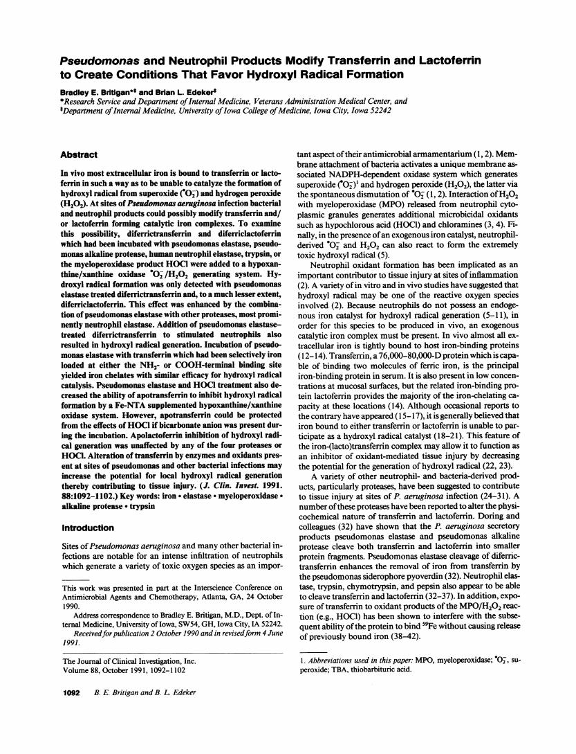
Pseudomonas and Neutrophil Products Modify Transferrin and Lactoferrinto Create Conditions That Favor Hydroxyl Radical FormationBradley E. Britigan*" and Brian L. Edekert*Research Service and Department of Internal Medicine, Veterans Administration Medical Center, andtDepartment of Internal Medicine, University of Iowa College of Medicine, Iowa City, Iowa 52242
Abstract
In vivo most extracellular iron is bound to transferrin or lacto-ferrin in such a way as to be unable to catalyze the formation ofhydroxyl radical from superoxide (7O-) and hydrogen peroxide(H202). At sites of Pseudomonas aeruginosa infection bacterialand neutrophil products could possibly modify transferrin and/or lactoferrin forming catalytic iron complexes. To examinethis possibility, diferrictransferrin and diferriclactoferrinwhich had been incubated with pseudomonas elastase, pseudo-monas alkaline protease, human neutrophil elastase, trypsin, orthe myeloperoxidase product HOCI were added to a hypoxan-thine/xanthine oxidase *02-/H202 generating system. Hy-droxyl radical formation was only detected with pseudomonaselastase treated diferrictransferrin and, to a much lesser extent,diferriclactoferrin. This effect was enhanced by the combina-tion of pseudomonas elastase with other proteases, most promi-nently neutrophil elastase. Addition of pseudomonas elastase-treated diferrictransferrin to stimulated neutrophils alsoresulted in hydroxyl radical generation. Incubation of pseudo-monas elastase with transferrin which had been selectively ironloaded at either the NH2- or COOH-terminal binding siteyielded iron chelates with similar efficacy for hydroxyl radicalcatalysis. Pseudomonas elastase and HOCI treatment also de-creased the ability of apotransferrin to inhibit hydroxyl radicalformation by a Fe-NTA supplemented hypoxanthine/xanthineoxidase system. However, apotransferrin could be protectedfrom the effects of HOCI if bicarbonate anion was present dur-ing the incubation. Apolactoferrin inhibition of hydroxyl radi-cal generation was unaffected by any of the four proteases orHOC.Alteration of transferrin by enzymes and oxidants pres-ent at sites of pseudomonas and other bacterial infections mayincrease the potential for local hydroxyl radical generationthereby contributing to tissue injury. (J. Clin. Invest. 1991.88:1092-1102.) Key words: iron * elastase - myeloperoxidase .alkaline protease - trypsin
Introduction
Sites of Pseudomonas aeruginosa and many other bacterial in-fections are notable for an intense infiltration of neutrophilswhich generate a variety of toxic oxygen species as an impor-
This work was presented in part at the Interscience Conference onAntimicrobial Agents and Chemotherapy, Atlanta, GA, 24 October1990.
Address correspondence to Bradley E. Britigan, M.D., Dept. of In-ternal Medicine, University of Iowa, SW54, GH, Iowa City, IA 52242.
Receivedfor publication 2 October 1990 and in revisedform 4 June1991.
tant aspect of their antimicrobial armamentarium (1, 2). Mem-brane attachment of bacteria activates a unique membrane as-sociated NADPH-dependent oxidase system which generatessuperoxide CO-)' and hydrogen peroxide (H202), the latter viathe spontaneous dismutation of *°- (1, 2). Interaction of H202with myeloperoxidase (MPO) released from neutrophil cyto-plasmic granules generates additional microbicidal oxidantssuch as hypochlorous acid (HOC1) and chloramines (3, 4). Fi-nally, in the presence of an exogenous iron catalyst, neutrophil-derived *0- and H202 can also react to form the extremelytoxic hydroxyl radical (5).
Neutrophil oxidant formation has been implicated as animportant contributor to tissue injury at sites of inflammation(2). A variety of in vitro and in vivo studies have suggested thathydroxyl radical may be one of the reactive oxygen speciesinvolved (2). Because neutrophils do not possess an endoge-nous iron catalyst for hydroxyl radical generation (5-1 1), inorder for this species to be produced in vivo, an exogenouscatalytic iron complex must be present. In vivo almost all ex-tracellular iron is tightly bound to host iron-binding proteins(12-14). Transferrin, a 76,000-80,000-D protein which is capa-ble of binding two molecules of ferric iron, is the principaliron-binding protein in serum. It is also present in low concen-trations at mucosal surfaces, but the related iron-binding pro-tein lactoferrin provides the majority of the iron-chelating ca-pacity at these locations (14). Although occasional reports tothe contrary have appeared (15-17), it is generally believed thatiron bound to either transferrin or lactoferrin is unable to par-ticipate as a hydroxyl radical catalyst (18-21). This feature ofthe iron-(lacto)transferrin complex may allow it to function asan inhibitor of oxidant-mediated tissue injury by decreasingthe potential for the generation of hydroxyl radical (22, 23).
A variety of other neutrophil- and bacteria-derived prod-ucts, particularly proteases, have been suggested to contributeto tissue injury at sites of P. aeruginosa infection (24-31). Anumber of these proteases have been reported to alter the physi-cochemical nature of transferrin and lactoferrin. Doring andcolleagues (32) have shown that the P. aeruginosa secretoryproducts pseudomonas elastase and pseudomonas alkalineprotease cleave both transferrin and lactoferrin into smallerprotein fragments. Pseudomonas elastase cleavage of diferric-transferrin enhances the removal of iron from transferrin bythe pseudomonas siderophore pyoverdin (32). Neutrophil elas-tase, trypsin, chymotrypsin, and pepsin also appear to be ableto cleave transferrin and lactoferrin (32-37). In addition, expo-sure of transferrin to oxidant products of the MPO/H202 reac-tion (e.g., HOCl) has been shown to interfere with the subse-quent ability of the protein to bind 59Fe without causing releaseof previously bound iron (38-42).
The Journal of Clinical Investigation, Inc.Volume 88, October 1991, 1092-1102
1. Abbreviations used in this paper: MPO, myeloperoxidase; *°2, SU-peroxide; TBA, thiobarbituric acid.
1092 B. E. Britigan and B. L. Edeker

The tertiary structure of transferrin and lactoferrin is likelyimportant to their ability to bind ferric iron in a form incapableof acting as an hydroxyl radical catalyst. Therefore, the possibil-ity that exposure of these iron-binding proteins to pseudo-monas or neutrophil proteases, or HOCI, could lead to genera-tion of new iron chelates capable of catalyzing hydroxyl radicalformation was examined. In the present communication, wereport the results of these studies and discuss them in the con-text of potential mechanisms of inflammatory tissue injurywith particular emphasis on that tissue injury associated withP. aeruginosa infection.
Methods
Reagents. Pseudomonas elastase and pseudomonas alkaline proteasewere kindly made available by Dr. Charles Cox, University of Iowa,Department of Microbiology, and Dr. Robert Fick, Jr., University ofIowa, Department of Internal Medicine. Human neutrophil elastasewas purchased from Elastin Products, Owensville, MO. Trypsin, hypo-xanthine, 2-deoxyribose, thiobarbituric acid, 5,5 dimethyl-l-pyrroline-1-oxide (DMPO), a-phenyl-n-tertbutyl-nitrone (PBN), CuZn superox-ide dismutase (SOD), catalase, nitrilotriacetic acid (NTA), and diethy-lenetriaminepentaacetic acid (DTPA) were all obtained from SigmaChemical Co., St. Louis, MO. Xanthine oxidase was purchased fromBoehringer-Mannheim Biochemicals, Indianapolis, IN, dimethyl sulf-oxide (DMSO) from Fisher Scientific Co., Fairlawn, NJ, and hypochlo-rous acid (HOCI) as 5% sodium hypochlorite (Regal Liquid Bleach)from Iowa Prison Industries, Anamosa, IA.
Iron-loading of lactoferrin and transferrin. Apotransferrin and apo-lactoferrin were purchased from Sigma Chemical Co. or Calbiochem-Behring Corp., La Jolla, CA. In some cases diferric complexes of lacto-ferrin and transferrin were also obtained from the same commercialsources. Alternatively, apolactoferrin (20 mg/ml) or apotransferrin (20mg/ml) were incubated in 200 mMNH4HCO3(pH 8.2) containingsufficient ferric-NTA to achieve 70-80% saturation of the lactoferrin/transferrin iron binding sites present. After 1 h at 37°C, the prepara-tions were placed in 12,000-14,000 mol wt membrane exclusion tub-ing (Spectropor, Spectrum Medical Industries, Inc., Los Angeles, CA)and dialyzed 24 h against 0.5 liters PBSwith 1 mMNaHCO3and 8 gchelex 100 beads (Bio-Rad Laboratories, Inc., Richmond, CA).
Transferrin in which the NH2- or COOH-terminal iron-binding sitewas selectively loaded was prepared using previously described meth-ods (43, 44). In each case successful selective loading was confirmed bydetermining sample migration in a 6 Murea gel (see below).
Protease cleavage of lactoferrin and transferrin. For experimentsperformed with iron-loaded transferrin or lactoferrin, the protein wasloaded according to the above protocol. For experiments in which apo-lactoferrin or apotransferrin were employed, commercial preparations(Sigma Chemical Co. or Calbiochem-Behring Corp.) were used withoutfurther modification. Lactoferrin or transferrin mixtures (20 mg/ml)were incubated in the presence of pseudomonas elastase (0.2-200 psg/ml), pseudomonas alkaline protease (200 Mg/ml), neutrophil elastase(200 gg/ml), or trypsin (50 yg/ml). The standard duration for mostexperiments was 48-72 h at 370C. In some cases shorter durations ofincubation with pseudomonas elastase were performed. Control sam-ples were incubated in parallel in the absence of added proteases. Insome cases, after protease incubation, samples were placed in 12,000-14,000 mol wt membrane exclusion tubing and dialyzed at 4°C for 3 hagainst 0.5 liters PBScontaining 1 mMNaHCO3and which had previ-ously been treated with chelating resin (Sigma Chemical Co.) to re-move adventitious metals. Extent of protease cleavage was assessed bySDS-PAGE(see below).
Hypochlorous acid treatment of lactoferrin and transferrin. Trans-ferrin (apo or diferric) or lactoferrin (apo or diferric) prepared as abovewere incubated in I mMNaHCO3containing HOC1at a 20-fold molarexcess of transferrin or lactoferrin (250 uMHOC:12.5 ,uM transferrin
or lactoferrin) for 30 min at 370C. In some cases, incubations withapotransferrin were performed in the absence of NaHCO3. The pH ofbuffer containing NaHCO3was titrated with HCGso that it was identi-cal with nonbicarbonate containing buffer (pH 7.5).
SDS-PAGE. Desired transferrin or lactoferrin preparations weresolubilized by boiling in Laemmli solubilizing buffer (45) containing2-mercaptoethanol (1%) for 5 min. Samples (7.5-12 yg protein/lane)were electrophoresed (150 volt hours) into a 10%polyacrylamide mini-gel after which the proteins were stained with silver according to themethod of Wray et al. (46).
Urea gels. 6 Multrapure grade urea (Sigma Chemical Co.) wasadded to the standard 6%polyacrylamide gel preparation gel formula-tion (1.93 ml polyacrylamide, 4.3 ml 3 MTris [pH 8.92], 0.1 ml 5%TEMED, and 0.24 ml 1.5% ammonium persulfate) with H20 substi-tuted for SDS. Transferrin or lactoferrin was mixed with 7 Murea and10:1 with glycerol. A small amount of bromophenol blue was some-times added to monitor migration of the gel front. The running bufferused was 6 Murea, 0.1 MTris, 0.01 Mboric acid, and 0.05 MEDTA(47). Gels were run 5 h at 100 V (constant voltage) on ice, and thenstained with Coomassie blue (48).
Formation of TBA-reactive 2-deoxyribose oxidation products. Thisassay was performed using a slight modification (49) of previously de-scribed methods (9, 50). Briefly, desired l-ml reaction mixtures wereprepared containing hypoxanthine (2 mM)and 2-deoxyribose (5 mM),+/- exogenous iron chelates such that the final iron concentration was25 MM. Xanthine oxidase (15 mU/ml) was then added and the reactionmixture incubated for 15 min at 37°C (rotating incubator). 0.5 ml of1%thiobarbituric acid (TBA) and 1 ml 6%TCAwas then added follow-ing which the mixture was heated (100°C X 10 min). The protein wasthen pelleted (1,000 g, 5 min) and the absorbance of the resulting super-natant at 532 nmdetermined using a model DU-30 spectrophotometer(Beckman Instruments, Inc., Irvine, CA).
Spin trapping. Spin trapping was performed using two previouslydeveloped methods (5, 1 1). Xanthine oxidase (31 mU/ml) was addedto a solution containing hypoxanthine (2 mM), DTPA (0.1 mM),DMSO(140 mM), and DMPO(100 mM)or PBN(I 0 mM), +/- exoge-nous iron chelate (0.1-0.2 mM). The reaction mixture was transferredto a flat quartz EPR cell and the EPRspectrum determined using amodel E104A EPRspectrometer (Varian Associates, Palo Alto, CA),which was kindly made available by Dr. Harold Goff (University ofIowa, Department of Chemistry). In some cases SOD(30 U/ml) orcatalase (500 U/mi) were also included in the reaction mixture. Unlessotherwise indicated EPR spectrometer settings were: microwavepower, 20 mW;modulation amplitude, I G; time constant, 1 s; modula-tion frequency, 100 kHz; sweep rate 12.5 G/min; and receiver gain, 5.0x 10⁴.
Statistical analysis. A paired Student's t test was used for all statisti-cal analyses. Results were considered significant at P < 0.05. To en-hance the effectiveness of presentation, in some cases data are ex-pressed as a percentage of appropriate control. However, original (raw)data were used for all statistical comparisons.
Results
Protease cleavage of diferrictransferrin and diferriclactoferrin.The tertiary structures of lactoferrin and transferrin are likelyimportant to their ability to bind iron in a form incapable ofacting as a hydroxyl radical catalyst. Because previous studies(32-37) demonstrated the ability of several proteases to cleavethese iron-binding proteins, the ability of the resulting pro-tease-induced cleavage products to function as hydroxyl radi-cal catalysts was assessed. Four proteases were targeted forstudy: pseudomonas elastase, pseudomonas alkaline protease,human neutrophil elastase, and trypsin. The two pseudomonasproteases were selected because they have previously been sug-gested as virulence factors for pseudomonas-associated tissue
Modification of (Lacto)transferrin 1093

injury (24-27, 32). Neutrophil elastase was chosen as a modelfor phagocyte-derived serine proteases and because it has beenlinked to some forms of inflammatory tissue injury (51). Fi-nally, trypsin was felt to be representative of local mucosalproteases.
First the relative ability of each protease to cleave diferric-transferrin and diferriclactoferrin was assessed. Diferrictrans-ferrin or diferriclactoferrin were incubated with pseudomonaselastase, pseudomonas alkaline protease, neutrophil elastase,or trypsin for 48-72 h at 370C. The number and apparentmolecular weight of the cleavage products differed with each ofthe proteases employed (Figs. 1-4). Similarly the extent ofcleavage of the native protein also varied (Figs. 1-4). For di-ferrictransferrin the extent of cleavage was pseudomonas elas-tase > trypsin > neutrophil elastase > alkaline protease (Figs.1-4). For diferriclactoferrin the hierarchy was similar (Figs.1-4). Relative to diferrictransferrin, diferriclactoferrin ap-peared to be more resistant to proteolysis by trypsin (Fig. 4).
Ability of protease-cleaved diferrictransferrin and diferric-lactoferrin to catalyze hydroxyl radical production. Havingconfirmed the ability of the various proteases to cleave the twoiron-binding proteins, the ability of the protease-treated di-ferrictransferrin and diferriclactoferrin complexes to functionas a hydroxyl radical catalyst was examined. The oxidation ofhypoxanthine by xanthine oxidase generates *°- and H202 butnot hydroxyl radical (52, 53). Thus, detection of hydroxyl radi-cal production by this reaction in the presence of an iron che-late is an indication of the ability of that chelate to function as ahydroxyl radical catalyst. Accordingly, diferrictransferrin, di-ferriclactoferrin, or these proteins which had been previouslyincubated with one of the four proteases described above wereadded to a mixture of hypoxanthine and 2-deoxyribose. Xan-thine oxidase was then added to initiate 02- and H202 genera-tion after which production of hydroxyl radical was quanti-tated by measuring the formation of TBA-reactive 2-deoxyri-bose oxidation products (Table I). Evidence for hydroxylradical production was apparent with the addition of pseudo-monas elastase-treated diferrictransferrin to the hypoxan-thine/xanthine oxidase system (Table I). This was not relatedto release of free iron from the protein as dialysis of the pseudo-monas elastase cleaved transferrin in 12,000-14,000 mol wtexclusion tubing against chelating resin failed to reduce the
Apo TF ADo LF Fe TF Fe LF+ - + _ + - +
200kD- t. .
97.4 -
68-
43
Apo TF
+-200 kD -
97.4 -
68 -
43 -
Apo LF Fe TF Fe LF_ + - +
! ,UU !P
25.7-257i. .".
18.4 - -ai;14.3 -
Figure 2. SDS-PAGEof apotransferrin (apoTF), apolactoferrin(apoLF), diferrictransferrin (FeTF), and diferriclactoferrin (FeLF)incubated 48 h in the absence (-) or presence (+) of 200 jg/ml hu-man neutrophil elastase.
amount of hydroxyl radical generated with its subsequent addi-tion to the hypoxanthine/xanthine oxidase system (notshown). A small but statistically significant amount of hy-droxyl radical was also observed with pseudomonas elastase-treated diferriclactoferrin (Table I). Hydroxyl radical catalysiswas not detected with use of untreated diferrictransferrin ordiferriclactoferrin (not shown), or with either of these two com-pounds which had been preincubated with pseudomonas alka-line protease, neutrophil elastase, or trypsin (Table I). Additionof pseudomonas elastase to the hypoxanthine/xanthine oxi-dase system in the absence of diferrictransferrin or diferriclac-toferrin did not suggest that the protease itself functioned as ahydroxyl radical catalyst (data not shown). Similarly, use ofapotransferrin which had been cleaved by pseudomonas elas-tase did not result in generation of hydroxyl radical confirmingthat iron associated with the transferrin was the source of hy-droxyl radical catalysis.
Confirmation of the ability of pseudomonas elastase-in-duced cleavage products to act as a hydroxyl radical catalyst.Although the formation of TBA-reactive deoxyribose oxida-tion products appears to be relatively specific for hydroxyl radi-cal, additional confirmation that pseudomonas elastase cleav-age of diferrictransferrin yields catalytic iron chelates wassought using spin trapping techniques. Free radicals by defini-
Apo TF Apo LF_
Fe TF Fe LF- -
97.4 -
68-
43 -
25.7-
18.4-14.3-
Figure 1. SDS-PAGEof apotransfermn (apoTF), apolactoferrin(apoLF), diferrictransferrin (FeTF), and diferriclactoferrin (FeLF)incubated 48 h in the absence (-) or presence (+) of 200 ,g/ml pseu-domonas elastase.
25.7 -
18.4- .:
14.3
.ft ..&.
Figure 3. SDS-PAGEof apotransferrin (apoTF), apolactoferrin(apoLF), diferrictransferrin (FeTF), and diferriclactoferrin (FeLF)incubated 48 h in the absence (-) or presence (+) of 200 ,g/ml pseu-domonas alkaline protease.
1094 B. E. Britigan and B. L. Edeker

Apo TF Apo LF Fe TF Fe LF_ + _ + - + - +
200 kD-
97.4 -
68 -
43 - ~~~~~~~~~~~~~~~~~~~~~~~~~~~~~~~~~~~~~~~~~~.........._E25.7-
18.4 -
14.3 -I
Figure 4. SDS-PAGEof apotransferrin (apoTF), apolactoferrin(apoLF), diferrictransferrin (FeTF), and diferriclactoferrin (FeLF)incubated 48 h in the absence (-) or presence (+) of 50 ,g/ml trypsin.
tion contain an unpaired electron which generates a small mag-netic moment detectable by EPR spectrometry. Because oftheir short half-life and reasons inherent to EPRspectrometrymost oxygen-centered radicals such as O and hydroxyl radi-cal are not detectable at physiologic temperatures using thistechnique. However, *02 and hydroxyl radical will react with
some nitrone compounds, termed spin traps, to generate stablenitroxide free radicals readily detectable by EPR. The uniquehyperfine splitting pattern of the EPRspectrum of these speciesallows identification of the original free radical species. Wehave previously employed spin trapping systems for the detec-tion of hydroxyl radical in biologic systems (5, 1 1). In the pres-ence of the spin trap DMPOand DMSOhydroxyl radical ismanifested as the methyl radical spin adduct of DMPO,DMPO/"CH3 (5). Whenhydroxyl radical is generated in a sys-tem containing DMSOand a different spin trap, PBN, thePBN(OCH3spin adduct results (1 1).
Consistent with earlier studies ( 18-22), when diferrictrans-ferrin or diferriclactoferrin was added to a hypoxanthine/xanthine oxidase system containing DMSOand either DMPOor PBN, no evidence of hydroxyl radical generation was de-tected (Figs. 5-7). Addition of diferrictransferrin which hadbeen previously exposed to pseudomonas elastase to the hypo-xanthine/xanthine oxidase system resulted in the generation ofspin adducts of DMPOand PBNexpected from the formationof hydroxyl radical, DMPO(CH3and PBN(OCH3, respec-tively (Figs. 5 and 6). Detection ofthese spin adducts was appro-priately inhibited 80-100% by the inclusion of catalase (Figs. 5and 6) or SOD(not shown) in the reaction mixture. Consistentwith the results of the deoxyribose oxidation assay, addition ofpseudomonas elastase cleaved diferriclactoferrin to the hypo-xanthine/xanthine oxidase reaction yielded spin trap evidenceof a small amount of hydroxyl radical formation (Fig. 7).
Table I. Generation of Catalytic Iron Chelates by Treatment ofDiferrictransferrin and Diferriclactoferrin with Different Proteases
Absorbance (532 nm)
Fe-TF Fe-LF
PAE [200] 0.128±0.018 (24)§ 0.034±0.010 (7)*[20] x 48 h 0.073±0.005 (6)§ 0.048±0.019 (3)[20] x 24 h 0.119±0.022 (5)t ND[20] x 12 h 0.048±0.016 (5)* ND[20] x 6 h 0.034±0.009 (5)* ND[2] 0.028±0.006 (3) 0.034±0.024 (3)[0.2] 0.012±0.005 (3) 0.028±0.003 (3)
PAP 0.003±0.003 (3) 0.005±0.005 (3)HNE 0.016±0.007 (9) 0.001±0.001 (3)Trypsin 0.015±0.011 (3) 0.009±0.003 (3)
Formation of TBA-reactive 2-deoxyribose oxidation products (532nmabsorbance) reflecting formation of hydroxyl radical by reactionmixtures composed of hypoxanthine, xanthine oxidase, and diferric-transferrin (Fe-TF) or diferriclactoferrin (Fe-LF), which were un-treated or had been previously exposed to pseudomonas elastase(PAE), pseudomonas alkaline protease (PAP), human neutrophilelastase (HNE), or trypsin. The absorbance expressed represents themean of results obtained (A532) with the addition to the hypoxan-thine/xanthine oxidase system of protease-treated diferrictransferrinor diferriclactoferrin less the A532 obtained with a paired control(untreated) iron chelate. Numbers in parenthesis after each set of dataindicate the number of separate experiments performed. Incubationswere for 48-72 h except where noted with exposure of diferric trans-ferrin to 20 ,g/ml PAE. Shown in brackets are the concentrations ofPAE (pg/ml) used in specific sets of experiments. The final concen-tration of iron present was 25 MM. Statistically different from the hy-poxanthine/xanthine oxidase diferric(lacto)transferrin control, * P< 0.05, $ P < 0.01, and § P < 0.0001, respectively.
HoG-i
HX / XO
HX/ XO+ F.TF
HX / XO+FOTF/ PE
HX / XO+ F.TF /PE+ Cat
CHl, OHOHI
CH, OH OOH
Figure 5. EPRspectra representative of three to five separate experi-ments obtained with the addition of xanthine oxidase to DMPO,DMSO,DTPA, and hypoxanthine (HX/XO) as well as the same re-action mixture supplemented with diferrictransferrin (0.1 mMFe,+ FeTF) or the same concentration of diferrictransferrin previouslypreincubated with 200 ug/ml pseudomonas elastase (+ FeTF/PE).Also shown is the spectrum obtained with the pseudomonas elastasecleaved diferrictransferrin if catalase (500 U/ml) was also included(+ FeTF/PE, + Cat). High- and low-field peaks corresponding to thespin adducts DMPO/'CH3, DMPO/OH,and DMPO/OOHaredesignated CH3, OH, and OOH, respectively.
Modification of (Lacto)transferrin 1095
i

HX / XO lOG
HX / XO+ FeTF
HX / XO+ F.TF PE + Cat
Figure 6. EPRspectra representative of three separate experimentsobtained after the addition of xanthine oxidase to PBN, DMSO,DTPA, and hypoxanthine (HX/XO) as well as the same reactionmixture supplemented with diferrictransferrin (0.1 mMFe, + FeTF)or the same concentration of diferrictransferrin previously cleavedwith pseudomonas elastase (+ FeTF/PE). Also shown is the effect ofcatalase (500 U/ml) on the FeTF/PE-induced spectrum (FeTF/PE+ Cat). The spectrum observed with FeTF/PE is that of PBN/0OCH3.
Effect of the pseudomonas elastase concentration and thecombination of different proteases on the generation of a hy-droxyl radical catalyst from diferrictransferrin and diferriclac-toferrin. There exists considerable discrepancy in the literatureas to the concentration of pseudomonas elastase which mightbe encountered in vivo (26, 54-59). To obtain greater insightinto the potential biologic relevance of the above observationswe varied the concentration of pseudomonas elastase to whichdiferrictransferrin and diferriclactoferrin were exposed beforeits addition to the hypoxanthine/xanthine oxidase system. Asshown in Table I, evidence of hydroxyl radical catalysis wasdetectable after diferrictransferrin incubation with concentra-tions of pseudomonas elastase as low as 2 ,g/ml. In contrast tothese results, statistically significant hydroxyl radical genera-tion was not detected using diferriclactoferrin exposed to con-centrations of pseudomonas elastase 20 Ag/ml (Table I).
In addition to assessing the effect of pseudomonas elastaseconcentration, the effect of the duration of diferrictransferrinexposure to pseudomonas elastase exposure on the generationof catalytic iron chelates was also examined. Diferrictransferrinincubated with 20 ,ug/ml pseudomonas elastase for as short as 6h was able to catalyze hydroxyl radical formation (Table I). Themagnitude of hydroxyl radical production was increased withthe duration of incubation (Table I). Incubations beyond 24 hdid not appear to increase the overall yield of catalytic ironchelate(s).
As discussed earlier sites of pseudomonas infection likelycontain a variety of different proteases. Accordingly, the im-pact of a multiple protease mixture on diferrictransferrin anddiferriclactoferrin was examined. Diferrictransferrin and di-ferriclactoferrin were incubated simultaneously in the presenceof pseudomonas elastase, pseudomonas alkaline protease, hu-manneutrophil elastase, and trypsin for 48-72 h. This reactionmixture was then added to the hypoxanthine/xanthine oxidasesystem and magnitude of hydroxyl radical formation assessed
I IoI I I ICH3,ONOOH OH CH3,
Figure 7. (A) EPRspectra obtained after the addition of xanthine ox-
idase to DMPO,DMSO,DTPA, and hypoxanthine (HX/XO) as wellas the same reaction mixture supplemented with diferriclactoferrin(0.1 mMFe, + FeLF) or the same concentration of diferriclactoferrinpreviously cleaved with pseudomonas elastase (+ FeLF/PE). High-and low-field peaks corresponding to the spin adducts DMPO/tCH3,DMPO/OH,and DMPO/OOHare designated CH3, OH, and OOH,respectively. Note the small increase in DMPO/-CH3peak amplitudeobserved with FeLF/PE relative to the other two conditions.
by the deoxyribose oxidation assay. As shown in Table II, in thecase of diferrictransferrin incubation in the presence of the fourproteases enhanced the resulting formation of hydroxyl radicalto a level almost five times that observed with incubation withpseudomonas elastase alone. This combination, however, hadno effect on diferriclactoferrin (Table II). To assess which ofthethree proteases was responsible for enhancing the effect ofpseudomonas elastase incubations of diferrictransferrin were
performed with pseudomonas elastase paired with only one ofthe other three proteases. The combination of pseudomonaselastase with human neutrophil elastase appeared to accountfor much, but not all, of the enhanced effect of the proteasecombination noted above (Table II). Furthermore, as can beascertained by comparing results in Tables I and II, neutrophilelastase markedly enhanced the ability of pseudomonas elas-tase to induce catalytic iron chelates at all concentrations ofpseudomonas elastase studied (0.2-20 ,g/ml). In contrast, ex-posure of diferrictransferrin to the combination of 20 ;&g/mlpseudomonas elastase and pseudomonas alkaline protease or
trypsin yielded hydroxyl radical production which was some-what less than that induced by pseudomonas elastase alone(Table II), although the decrease was not statistically signifi-cant.
Is the ability ofpseudomonas elastase to generate catalyticiron chelatesfrom diferrictransferrin restricted to only one bind-ing site? Because transferrin has two iron-binding sites itseemed possible that the iron chelate resulting from pseudo-monas elastase cleavage of diferrictransferrin was generated
1096 B. E. Britigan and B. L. Edeker
I-1o-j
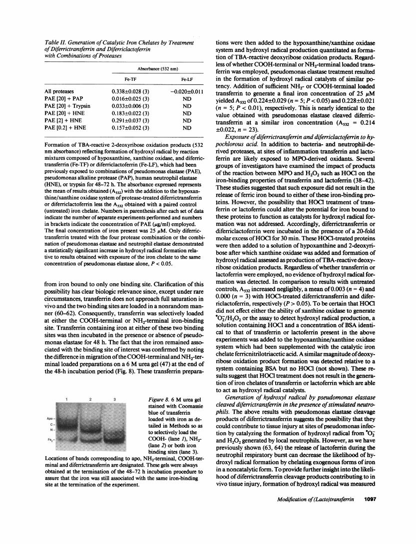
Table II. Generation of Catalytic Iron Chelates by Treatmentof Diferrictransferrin and Diferriclactoferrinwith Combinations of Proteases
Absorbance (532 nm)
Fe-TF Fe-LF
All proteases 0.338±0.028 (3) -0.020±0.011PAE [20] + PAP 0.016±0.025 (3) NDPAE [20] + Trypsin 0.033±0.006 (3) NDPAE [20] + HNE 0.183±0.022 (3) NDPAE [2] + HNE 0.291±0.037 (3) NDPAE [0.2] + HNE 0.157±0.052 (3) ND
Formation of TBA-reactive 2-deoxyribose oxidation products (532nmabsorbance) reflecting formation of hydroxyl radical by reactionmixtures composed of hypoxanthine, xanthine oxidase, and diferric-transferrin (Fe-TF) or diferriclactoferrin (Fe-LF), which had beenpreviously exposed to combinations of pseudomonas elastase (PAE),pseudomonas alkaline protease (PAP), human neutrophil elastase(HNE), or trypsin for 48-72 h. The absorbance expressed representsthe mean of results obtained (A532) with the addition to the hypoxan-thine/xanthine oxidase system of protease-treated diferrictransferrinor diferriclactoferrin less the A532 obtained with a paired control(untreated) iron chelate. Numbers in parenthesis after each set of dataindicate the number of separate experiments performed and numbersin brackets indicate the concentration of PAE (,ug/ml) employed.The final concentration of iron present was 25 MM. Only diferric-transferrin treated with the four protease combination or the combi-nation of pseudomonas elastase and neutrophil elastase demonstrateda statistically significant increase in hydroxyl radical formation rela-tive to results obtained with exposure of the iron chelate to the sameconcentration of pseudomonas elastase alone, P < 0.05.
from iron bound to only one binding site. Clarification of thispossibility has clear biologic relevance since, except under rarecircumstances, transferrin does not approach full saturation invivo and the two binding sites are loaded in a nonrandom man-ner (60-62). Consequently, transferrin was selectively loadedat either the COOH-terminal or NH2-terminal iron-bindingsite. Transferrin containing iron at either of these two bindingsites was then incubated in the presence or absence of pseudo-monas elastase for 48 h. The fact that the iron remained asso-ciated with the binding site of interest was confirmed by notingthe difference in migration ofthe COOH-terminal and NH2-ter-minal loaded preparations on a 6 Murea gel (47) at the end ofthe 48-h incubation period (Fig. 8). These transferrin prepara-
1 2 3 Figure 8. 6 Murea gelstained with Coomassie
T,Jblue of transferrinAp*-_| loaded with iron as de-
tailed in Methods so asto selectively load the
Fe2- _ COOH-(lane 1), NH2-(lane 2) or both ironbinding sites (lane 3).
Locations of bands corresponding to apo, NH2-terminal, COOH-ter-minal and diferrictransferrin are designated. These gels were alwaysobtained at the termination of the 48-72 h incubation procedure toassure that the iron was still associated with the same iron-bindingsite at the termination of the experiment.
tions were then added to the hypoxanthine/xanthine oxidasesystem and hydroxyl radical production quantitated as forma-tion of TBA-reactive deoxyribose oxidation products. Regard-less of whether COOH-terminal or NH2-terminal loaded trans-ferrin was employed, pseudomonas elastase treatment resultedin the formation of hydroxyl radical catalysts of similar po-tency. Addition of sufficient NH2- or COOH-terminal loadedtransferrin to generate a final iron concentration of 25 1AMyielded A532 of 0.224±0.029 (n = 5; P< 0.05) and 0.228±0.021(n = 5; P < 0.01), respectively. This is nearly identical to thevalue obtained with pseudomonas elastase cleaved diferric-transferrin at a similar iron concentration (A532 = 0.214±0.022, n = 23).
Exposure of diferrictransferrin and diferriclactoferrin to hy-pochlorous acid. In addition to bacteria- and neutrophil-de-rived proteases, at sites of inflammation transferrin and lacto-ferrin are likely exposed to MPO-derived oxidants. Severalgroups of investigators have examined the impact of productsof the reaction between MPOand H202 such as HOC1on theiron-binding properties of transferrin and lactoferrin (38-42).These studies suggested that such exposure did not result in therelease of ferric iron bound to either of these iron-binding pro-teins. However, the possibility that HOCI treatment of trans-ferrin or lactoferrin could alter the potential for iron bound tothese proteins to function as catalysts for hydroxyl radical for-mation was not addressed. Accordingly, diferrictransferrin ordiferriclactoferrin were incubated in the presence of a 20-foldmolar excess of HOC1for 30 min. These HOCl-treated proteinswere then added to a solution of hypoxanthine and 2-deoxyri-bose after which xanthine oxidase was added and formation ofhydroxyl radical assessed as production of TBA-reactive deoxy-ribose oxidation products. Regardless of whether transferrin orlactoferrin were employed, no evidence of hydroxyl radical for-mation was detected. In comparison to results with untreatedcontrols, A532 increased negligibly, a mean of 0.003 (n = 4) and0.000 (n = 3) with HOCG-treated diferrictransferrin and difer-riclactoferrin, respectively (P > 0.05). To be certain that HOCGdid not effect either the ability of xanthine oxidase to generate*O0-/H202 or the assay to detect hydroxyl radical production, asolution containing HOCGand a concentration of BSA identi-cal to that of transferrin or lactoferrin present in the aboveexperiments was added to the hypoxanthine/xanthine oxidasesystem which had been supplemented with the catalytic ironchelate ferricnitrilotriacetic acid. A similar magnitude of deoxy-ribose oxidation product formation was detected relative to asystem containing BSA but no HOCG(not shown). These re-sults suggest that HOC1treatment does not result in the genera-tion of iron chelates of transferrin or lactoferrin which are ableto act as hydroxyl radical catalysts.
Generation of hydroxyl radical by pseudomonas elastasecleaved diferrictransferrin in the presence of stimulated neutro-phils. The above results with pseudomonas elastase cleavageproducts of diferrictransferrin suggests the possibility that theycould contribute to tissue injury at sites of pseudomonas infec-tion by catalyzing the formation of hydroxyl radical from '02and H202 generated by local neutrophils. However, as we havepreviously shown (63, 64) the release of lactoferrin during theneutrophil respiratory burst can decrease the likelihood of hy-droxyl radical formation by chelating exogenous forms of ironin a noncatalytic form. To provide further insight into the likeli-hood of diferrictransferrin cleavage products contributing to invivo tissue injury, formation of hydroxyl radical was measured
Modification of (Lacto)transferrin 1097

after stimulation of human neutrophils with PMAin the pres-ence of diferrictransferrin which had been previously cleavedwith pseudomonas elastase. As assessed with the deoxyriboseoxidation assay, hydroxyl radical was generated in the presenceof pseudomonas elastase-treated diferrictransferrin (5 A532= 0.075±0.010, n = 7, P < 0.05) but not with untreated di-ferrictransferrin.
Protease and hypochlorous acid treatment of apotransferrinand apolactoferrin. Based on the work of other investigators(33-41), it seemed possible that the iron-binding properties oftransferrin and lactoferrin might be more susceptible to pro-tease or HOC1modification if the iron-binding proteins wereexposed to these agents before their association with iron. Con-sequently, we incubated apotransferrin or apolactoferrin in thepresence of pseudomonas elastase, pseudomonas alkaline pro-tease, human neutrophil elastase, trypsin; or HOC1under thesame conditions as the earlier experiments with diferric che-lates (Figs. 1-4). In general, the relative susceptibility of di-ferrictransferrin and diferriclactoferrin to each of the proteaseswas similar to that noted with the apo forms of the molecules(Figs. 1-4). However, the nature and relative proportion ofcleavage products generated by most proteases varied with useof apo as compared to diferric forms of transferrin or lacto-ferrin (Figs. 1-4). Most notably, both apotransferrin and apo-lactoferrin were cleaved to a much greater extent than the di-ferric forms by pseudomonas alkaline protease (Fig. 3). Apo-transferrin was similarly more susceptible to neutrophilelastase than its diferric counterpart (Fig. 2). Finally, apolacto-ferrin appeared to be more resistant than apotransferrin tocleavage by trypsin and neutrophil elastase (Figs. 2 and 4).
The functional consequence of exposure of apotransferrinand apolactoferrin to each of the proteases was then examinedby assessing the ability of the various protease cleavage prod-ucts to inhibit iron catalyzed hydroxyl radical production.Each reaction product was added to a mixture of hypoxan-thine, Fe-NTA, and deoxyribose. The resulting ability of thesystem to generate hydroxyl radical upon the addition of xan-thine oxidase was then determined relative to the hypoxan-thine/xanthine oxidase system in the presence of the untreatediron-binding proteins or in their absence. Pretreatment of apo-transferrin with HOCl or pseudomonas elastase (20 or 200 ,ug/ml) but not the other proteases interfered with the ability of thecompound to inhibit formation of hydroxyl radical (Table III).Analogous to results with diferrictransferrin, the combinationof pseudomonas elastase with the other three proteases or justhuman neutrophil elastase (but not other combinations) en-hanced the effect of pseudomonas elastase (Table III). Some-what surprisingly, the deleterious effect of HOC1was not notedif bicarbonate anion (1 mM)was present during the period ofHOCl exposure (Table III). The final pH under both condi-tions was adjusted to 7.5, eliminating that factor as the explana-tion for the observation. In contrast to the results with apo-transferrin, the ability of apolactoferrin to inhibit formation ofhydroxyl radical was not altered by exposure to any of the fourproteases (alone or in combination) or HOC1(Table III).
Discussion
Recent evidence suggests that proteases likely present at sites ofpseudomonas infection could potentially alter the iron chelat-ing properties of transferrin and lactoferrin (32-37), proteinswhich may play an important role in decreasing the likelihood
Table III. Effect of Protease and HOCi Treatment on the Abilityof Apo-Transferrin and Apo-Lactoferrin to Prevent Iron-catalyzedHydroxyl Radical Formation
Percentage of controlhydroxyl radical generation
ApoTF ApoLF
No treatment 7 (10) 5 (10)PAE [200] 36 (4) 5 (3)
[20] 40 (7) 7 (7)[2] 5 (5) ND[0.2] 6 (5) ND
PAP 3 (4) 5 (3)HNE 10 (4) 6 (3)Trypsin 9 (4) 7 (3)
All proteases 57 (3) 2 (3)PAE [20] + PAP 21 (3) 2 (3)PAE [20] + Trypsin 50 (5) 5 (3)PAE [20] + HNE 63 (4) 6 (3)HOCI 66 (4) 7 (3)HOC1/HCO3 8 (3) ND
Median inhibition of hydroxyl radical formation by the reaction ofhypoxanthine and xanthine oxidase in the presence of Fe-NTA (10jiM) with the inclusion of 1 mg/ml apotransferrin (ApoTF) or apo-lactoferrin (ApoLF) which had been treated previously with pseudo-monas elastase (PAE), pseudomonas alkaline protease (PAP, 200 jig/ml), human neutrophil elastase (HNE, 200 jg/ml), trypsin (50 jg/ml),or HOCL. Concentration of PAEused (jig/ml) is shown in brackets.In the case of HOCI results are shown for exposure to HOCI in thepresence or absence of I mMbicarbonate anion. In each case dataare expressed as the percentage of formation of TBA-reactive deoxy-ribose oxidation products (A532) detected relative to that obtained onthe same day with the hypoxanthine/xanthine oxidase/Fe-NTA mix-ture in the absence of apotransferrin or apolactoferrin-derived pro-teins. Treatment of apotransferrin with pseudomonas elastase (20 or200 jig/ml) or with HOCI (in the absence of bicarbonate) resulted ina significant loss of the protein's ability to inhibit hydroxyl radicalgeneration (P < 0.01). No other treatment with a single proteaseyielded statistically significant loss of apotransferrin inhibitory activ-ity. The combination of the four proteases or HNEand PAE en-hanced the effect observed with PAE-treated apotransferrin (P < 0.05).
of in vivo formation of hydroxyl radical (22, 23, 63, 64). Conse-quently, we assessed the impact of in vitro exposure of lacto-ferrin and transferrin to four different proteases (pseudomonaselastase, pseudomonas alkaline protease, human neutrophilelastase, and trypsin) on the potential for generation of thehighly cytotoxic hydroxyl radical. Initial studies used the reac-tion of hypoxanthine and xanthine oxidase was used as a conve-nient model for neutrophil oxidant formation to avoid poten-tial confounding influences of the proteases on various aspectsof neutrophil function.
Incubation with any of the above proteases alone or in com-bination resulted in the cleavage of transferrin or lactoferrininto multiple subunits. Consistent with a previous report of itshigh resistance to protease cleavage (33) we found that humanlactoferrin exhibited a greater overall resistance to proteolysisthan transferrin. The number and apparent molecular weightof these cleavage products varied with the iron-binding proteinand protease examined and are in general agreement with the
1098 B. E. Britigan and B. L. Edeker

work of other investigators (32-37). However, in contrast toour results (Fig. 1), Doring et al. (32) stated that no cleavageproducts above 14,000 Dwere detected with the incubation ofpseudomonas elastase with transferrin. The difference may re-late to the fact that in the studies of Doring and colleagues (32)the ratio of pseudomonas elastase to transferrin was 30-foldhigher than in our studies. Finally, others have noted that rela-tive to their iron loaded form, apolactoferrin and apotrans-ferrin may be more resistant to protease digestion (33, 37). Wedid not find this to be the case, but the marked differences inexperimental conditions used amongthe studies make compar-isons difficult.
In the present work, only the products of the interaction ofpseudomonas elastase and diferrictransferrin resulted in thegeneration of large amounts of hydroxyl radical when added tothe hypoxanthine/xanthine oxidase *O/H202 generating sys-tem. Pseudomonas elastase-cleaved diferrictransferrin wasalso able to catalyze formation of hydroxyl radical when amore physiologically relevant source of O/H202, stimulatedneutrophils, was substituted for the hypoxanthine/xanthineoxidase system.
There did not appear to be a clear correlation between themagnitude of protease-induced cleavage and generation of cata-lytic iron chelates. For example, diferrictransferrin was exten-sively cleaved by both pseudomonas and neutrophil elastase.Yet only the pseudomonas elastase diferrictransferrin cleavageproducts catalyzed hydroxyl radical formation. As demon-strated by experiments in which transferrin was selectivelyloaded at only one iron-binding site, pseudomonas elastase ap-parently cleaves the transferrin molecule in such a way thatiron bound to either the COOH-or NH2-terminal binding sitebecomes equally capable of acting as a hydroxyl radical cata-lyst. Dialysis of the pseudomonas elastase-cleaved transferrinindicated that these results were not related to simple release ofbound iron. In addition, autoradiography of '9Fe-transferrinexposed to pseudomonas elastase has revealed multiple lowermolecular weight fragments which have 59Fe associated withthem (Britigan, B. E., B. L. Edeker, and M. L. McCormick,unpublished observation). Although pseudomonas elastasecleavage of diferriclactoferrin also yielded products with somecapacity to act as a hydroxyl radical catalyst, this required highconcentrations of pseudomonas elastase. Even then, the mag-nitude of hydroxyl radical generated was quite small and there-fore may not have biologic significance.
Wide variations have been reported in the concentration ofenzymatically active pseudomonas elastase present at sites ofinfection with this organism. Most of this work has centered onstudies of airway secretions obtained from patients with cysticfibrosis whose airways were infected/colonized with P. aeru-ginosa. Fick et al. detected pseudomonas elastase activity of
- 2 Ag/ml in bronchoalveolar lavage samples from such pa-tients (54). Taking into account the 100-fold dilution such sam-ples are usually felt to represent (65) this would represent aconcentration of pseudomonas elastase in the airway of nearly200 ,ug/ml, close to the maximal concentration used in ourstudy. Similarly, significant levels of pseudomonas elastase ac-tivity were reported by Bruce et al. (55). In contrast much loweror absent levels of pseudomonas elastase activity were detectedin a similar group of patients in studies by others (26, 56-58).These groups have suggested that pseudomonas elastase maynot be active in such individuals due to the presence in theairway of pseudomonas elastase-specific immunoglobulins
which develop over time in such patients. However, as pointedout by Bruce et al. (55), these studies also vary in the way inwhich samples were collected and processed.
To our knowledge there is no data on levels of pseudo-monas elastase present at sites of human pseudomonas infec-tion in conditions other than cystic fibrosis. Using a rat modelsystem in which P. aeruginosa was injected into a "granulomapouch," Doring et al. (59) reported pseudomonas elastase andalkaline protease concentrations in inflammatory exudate of0-185 and 0-742 ng/ml, respectively. These contrasted levelsin in vitro culture supernatant using similar concentrations oforganism where concentrations were in some cases 100-1,000-fold higher than those measured in vivo. This difference wasfound to be in part explainable by a rapid clearance of activeenzyme from the exudate fluid suggesting that the rate of elas-tase and alkaline protease production by the organism in vivowas considerably greater than would be predicted by steady-state levels. In addition, neither this study nor the ones men-tioned above optimally address what the local concentration ofenzyme might be in close proximity to sites where organismsare adherent to host cells as would be expected to occur inpulmonary airways.
In vivo sites of P. aeruginosa infection are characterized bythe presence of multiple proteases. When, diferrictransferrinwas simultaneously incubated with multiple proteases the gen-eration of catalytic iron chelates was enhanced relative to thatobserved with pseudomonas elastase alone. Humanneutrophilelastase although inactive alone enhanced the effect of pseudo-monas elastase. This was associated with increased proteolysisobserved with SDS-PAGE(not shown). In the presence of hu-man neutrophil elastase the concentration of pseudomonaselastase required for the generation of catalytic iron chelateswas reduced markedly to that which would be likely achievableunder in vivo conditions. These results are in accordance withsuggestions by other investigators (26, 66) that these two elas-tases could enhance the pathogenic potential of one another.Because the combination of pseudomonas elastase and neutro-phil elastase were still not as effective in generating catalyticiron chelates as the combination of four proteases it is likelythat the actions of multiple mucosal proteases could synergisti-cally enhance the breakdown of transferrin in vivo.
In contrast to the results with human neutrophil elastase,there was a suggestion that both trypsin and pseudomonas al-kaline protease may decrease the effectiveness of pseudomonaselastase in the generation of catalytic iron chelates from di-ferrictransferrin. Such an observation could result from inacti-vation of pseudomonas elastase by the other protease or poten-tially modification of transferrin cleavage resulting in productswhich retain their ability to bind iron in a noncatalytic form.However, because these decreases did not reach statistical sig-nificance and the results of experiments using a combination ofall four proteases showed the greatest enhancement of catalyticiron formation, the above observation is probably of only mar-ginal importance.
Similar to the results obtained with iron-loaded forms oftransferrin, exposure of apotransferrin to pseudomonas elas-tase, but not the other three proteases, decreased its ability toinhibit hydroxyl radical generation by the hypoxanthine/xanthine oxidase/Fe-NTA system which depends on the abilityof apotransferrin to bind iron in a non-catalytic form. Thiscould be due to either a loss of the overall ability of the pseudo-monas elastase-induced transferrin cleavage products to bind
Modification of (Lacto)transferrin 1099

iron or an alteration in the redox properties of bound iron. Asnoted with diferrictransferrin, the effect of pseudomonas elas-tase was enhanced if human neutrophil elastase and to a lesserextent trypsin was also present during the incubation. Analo-gous to results with diferriclactoferrin, exposure to either of thefour proteases (or their combinations) did not appear to haveany significant impact on the ability of apolactoferrin to inhibithydroxyl radical formation by the same system. These resultsalso did not appear to be explainable solely on the basis of themagnitude of proteolysis observed with each of the four pro-teases.
These data suggest that pseudomonas elastase maybe some-what unique among proteases in its ability to generate ferriccomplexes from transferrins which are capable of acting as hy-droxyl radical catalysts or interfere with the subsequent abilityof apo forms of these proteins to bind microenvironmentaliron in a noncatalytic form. Lactoferrin, however, appears tobe much more resistant than transferrin to such modificationby pseudomonas elastase. These data are very consistent withthe work of Doring et al. (32) which demonstrated that pseudo-monas elastase treatment of iron-loaded transferrin but notlactoferrin allowed subsequent chelation of that iron by thepseudomonas siderophore pyoverdin. In the same study (32),exposure of transferrin or lactoferrin to either neutrophil elas-tase or pseudomonas alkaline protease did not result in en-hanced removal of iron from these proteins by pyoverdin.
Transferrin and lactoferrin have been reported to be suscep-tible to oxidation by products of the reaction of MPOand H202such as HOC1(38-42). Nevertheless exposure of either diferric-transferrin or diferriclactofenin to HOC1did not generate ironchelates capable of catalyzing hydroxyl radical generation.However, exposure of apotransferrin but not apolactoferrin toHOC1decreased the ability of that iron-binding protein to in-hibit hydroxyl radical formation by the hypoxanthine/xan-thine oxidase/Fe-NTA system. These data support and extendthe recent work of both Clark and Pearson (39) and Winter-bourn and Malloy (38). The former investigators reported (39)that exposure of transferrin to stimulated neutrophils or a cell-free MPO/H202 system in the presence of iodide did not in-duce release of previously bound iron but did interfere with theability of unsaturated transferrin to subsequently form a stableiron complex. Similarly, Winterbourn and Malloy (38) notedthat both ferrilactoferrin and ferritransfenin did not releaseiron to any great extent upon exposure to either HOC1or aMPO/H202/C- system. Exposure of the apo forms of theseproteins to either system decreased the subsequent ability oflactoferrin and transferrin to bind iron. However, lactoferrinwas much more resistant to inactivation.
In contrast to our data, Halliwell and colleagues reported(42) that exposure of either apotransferrin or apolactoferrin toHOC1had no effect on the ability of these proteins to limitformation of hydroxyl radical generated by iron-dependent hy-droxyl radical generating systems. Specifics of the HOCGincu-bation are not described in their report making direct compari-son of their results and ours difficult. Of interest, we too failedto note an effect of HOC1on apotransferrin if bicarbonate ionwas present during the period of HOCGexposure. Clark andPearson found (39) evidence that amino acids (particularly ty-rosine) near the iron-binding sites of transferrin may beuniquely sensitive to MPO-mediated iodination. Bicarbonateis felt to bind in close proximity to the iron binding site oftransferrin, most likely to an arginine residue (13, 67, 68).
There is evidence that bicarbonate binding to some types oftransferrins mayalter protein conformation (69). It seems possi-ble that such a process could decrease the susceptibility of theiron binding site to halogination, contributing to the protectiveeffects of bicarbonate on HOCGinduced decreases in apotrans-ferrin iron-binding capacity.
Pseudomonas and other bacterial infections frequently be-gin at mucosal surfaces where their secretory products contrib-ute to an array of proteases already present. The arrival of neu-trophils with their release of granule proteases and generationof MPO-derived oxidants would provide further potential forinactivation of local iron-binding proteins. Thus, it is perhapsnot surprising that lactoferrin whose role it is to function in thismilieu of oxidants and proteases is more resistant to damagethan transferrin which serves primarily as an iron transportprotein in serum, a much less hostile environment.
Nevertheless, transferrin does contribute to the iron-bind-ing potential at mucosal sites such as the pulmonary airways(54) where P. aeruginosa is an important pathogen. P. aerugin-osa pulmonary infections are highly destructive and have anassociated mortality approaching 70% even with appropriateantibiotic intervention (24). In addition, P. aeruginosa coloni-zation/infection of the airways of patients with cystic fibrosis istemporally associated with the onset of the progressive deterio-ration of lung function which eventually is responsible for thedeath of > 90% of these individuals (24). A wide variety of P.aeruginosa-derived enzymes including pseudomonas elastasehave been implicated as virulence factors in infections with thisorganism (24-27). Other products such as bacterial sidero-phores have also been implicated (24). Wehave recently shownthat iron bound to the P. aeruginosa siderophore pyochelin canfunction as a hydroxyl radical catalyst possibly leading to localtissue injury (49). It seems possible that alterations of ferric-transferrin products by pseudomonas elastase or neutrophil-derived MPOcould contribute to some of the local tissue in-jury associated with P. aeruginosa infections.
Acknowledaments
Wewish to thank Drs. Charles Cox, Robert Fick, Jr., and Robert Clarkfor their gifts of reagents and helpful suggestions, Michelle Railsbackand Tedmund Roeder for technical assistance, and Naomi Erickson forher help with preparation of the manuscript.
This work was supported through the Veterans Administration Re-search Service, Public Health Service grants HL 44275 and Al 28412,the Cystic Fibrosis Foundation, the Pfizer Scholars Program, and theSandoz Foundation for Gerontologic Research. It was performed dur-ing the tenure of B. E. Britigan as a Veterans Administration ResearchAssociate.
References
1. Root, R. K., and M. S. Cohen. 1981. The microbicidal mechanisms ofhuman neutrophils and eosinophils. Rev. Infect. Dis. 3:565-598.
2. Weiss, S. J. 1986. Oxygen, ischemia and inflammation. Acta Physiol.Scand. 548(Suppl.):9-37.
3. Klebanoff, S. J., and C. B. Hamon. 1972. Role of myeloperoxidase-me-diated antimicrobial systems in intact leukocytes. J. Reticuloendothel Soc.12:170-196.
4. Weiss, S. J., M. D. Lampert, and S. T. Test. 1983. Long-lived oxidantsgenerated by human neutrophils: characterization and bioactivity. Science(Wash. DC). 222:625-628.
5. Britigan, B. E., G. M. Rosen, Y. Chai, and M. S. Cohen. 1986. Do humanneutrophils make hydroxyl radical? Detection of free radicals generated by hu-man neutrophils activated with a soluble or particulate stimulus using electronparamagnetic resonance spectrometry. J. Biol. Chem. 261:4426-4431.
1100 B. E. Britigan and B. L. Edeker
6. Winterbourn, C. C. 1986. Myeloperoxidase as an effective inhibitor ofhydroxyl radical production: implications for the oxidative reactions of neutro-phils. J. Clin. Invest. 78:545-550.
7. Thomas, M. J., P. S. Shirley, C. Hedrick, and L. R. DeChatelet. 1986. Roleof free radical processes in stimulated human polymorphonuclear leukocytes.Biochemistry. 25:8042-8048.
8. Kaur, H., Z. Fagerheim, M. Grootveld, A. Puppo, and B. Halliwell. 1988.Aromatic hydroxylation of phenylalanine as an assay for hydroxyl radicals: appli-cation to activated neutrophils and heme protein leghemoglobin. Anal. Biochem.172:360-367.
9. Greenwald, R. A., S. W. Rush, S. A. Mark, and Z. Weitz. 1989. Conversionof superoxide generated by polymorphonuclear leukocytes to hydroxyl radical: adirect spectrophotometric detection system based on degradation of deoxyribose.Free Radical Biol. & Med. 6:385-392.
10. Pou, S., M. S. Cohen, B. E. Britigan, and G. M. Rosen. 1989. Spin trappingand human neutrophils: limits of detection of hydroxyl radical. J. Biol. Chem.264: 12299-12302.
11. Britigan, B. E., T. J. Coffman, and G. R. Buettner. 1990. Spin trap evi-dence for the lack of significant hydroxyl radical production during the respira-tion burst of human phagocytes using a spin adduct resistant to superoxide me-diated destruction. J. Biol. Chem. 265:2650-2656.
12. Bullen, J. J., H. J. Rogers, and E. Griffiths. 1978. Role of iron in bacterialinfection. Curr. Top. Microbiol. Immunol. 80:1-35.
13. Legrand, D., J. Mazurier, J. Montreuil, and G. Spik. 1988. Structure andspatial conformation of the iron-binding sites of transferrins. Biochimie.70:1185-1195.
14. Masson, P. L., J. F. Heremans, and C. H. Dive. 1966. Studies on lacto-ferrin, an iron-binding protein commonto many external secretions. Clin. Chim.Acta. 14:735-739.
15. Ambruso, D. R., and R. B. Johnston, Jr. 1981. Lactoferrin enhanceshydroxyl radical production by human neutrophils, neutrophil particulate frac-tions and an enzymatic generating system. J. Clin. Invest. 67:352-360.
16. Bannister, J. V., W. H. Bannister, H. A. 0. Hill, and P. J. Thornalley.1982. Enhanced production of hydroxyl radicals by the xanthine-xanthine oxi-dase reaction in the presence of lactoferrin. Biochim. Biophys. Acta. 715:116-120.
17. Motohasi, N., and I. Mori. 1983. Superoxide dependent formation ofhydroxyl radical catalyzed by transferrin. FEBS(Fed. Eur. Biochem. Soc) Lett.157:197-199.
18. Aruoma, 0. I., and B. Halliwell. 1987. Superoxide-dependent and ascor-bate-dependent formation of hydroxyl radicals from hydrogen peroxide in thepresence of iron are lactoferrin and transferrin promoters of hydroxyl-radicalgeneration? Biochem. J. 241:273-278.
19. Winterbourn, C. C. 1983. Lactoferrin-catalyzed hydroxyl radical produc-tion: additional requirements for a chelating agent. Biochem. J. 210:15-19.
20. Baldwin, D. A., E. R. Jenny, and P. Aisen. 1984. The effect of humanserum transferrin and milk lactoferrin on hydroxyl radical formation from super-oxide and hydrogen peroxide. J. Biol. Chem. 259:13391-13394.
21. Buettner, G. R. 1987. The reaction of superoxide, formate radical, andhydrated electron with transferrin and its model compound, Fe(III)-ethylenedi-amine-N,N'-bis [242-hydroxyphenyl) acetic acid] as studied by pulse radiolysis. J.Biol. Chem. 262:11995-11998.
22. Gutteridge, J. M. C., S. K. Paterson, A. W. Segal, and B. Halliwell. 1981.Inhibition of lipid peroxidation by the iron-binding protein lactoferrin. Biochem.J. 199:259-261.
23. Ward, P. A., G. 0. Till, R. Kunkel, and C. Beauchamp. 1983. Evidence forthe role of hydroxyl radical in complement and neutrophil-dependent tissue in-jury. J. Clin. Invest. 72:789-801.
24. Fick, R. B., Jr. 1989. Pathogenesis of the Pseudomonas lung lesion incystic fibrosis. Chest. 96:158-164.
25. Pier, G. B. 1985. Pulmonary disease associated with Pseudomonas aeru-ginosa in cystic fibrosis: current status of the host bacterium interaction. J. Infect.Dis. 151:575-580.
26. Suter, S., 0. B. Schaad, L. Roux, U. E. Nydegger, and F. A. Waldvogel.1984. Granulocyte neutral proteases and Pseudomonas elastase as possible causesof airway damage in patients with cystic fibrosis. J. Infect. Dis. 149:523-531.
27. Dunn, M. M., M. Dunne, and D. W. Kamp. 1990. Polymorphonuclearleukocyte- and Pseudomonas aeruginosa-induced damage to a human pulmo-nary epithelial cell line. J. Infect. Dis. 162:172-177.
28. Mohammed, J. R., B. S. Mohammed, L. J. Pawluk, D. M. Bucci, andW. B. Davis. 1988. Purification and cytotoxic potential of myeloperoxidase incystic fibrosis sputum. J. Lab. Clin. Med. 112:711-720.
29. Holder, I. A. 1983. Experimental studies of the pathogenesis of infectionsdue to Pseudomonas aeruginosa: effect of treatment with protease inhibitors.Rev. Infect. Dis. 5:S914-S921.
30. Kreger, A. S. 1983. Pathogenesis of Pseudomonas aeruginosa ocular dis-ease. Rev. Infect. Dis. 5:S93 1-S935.
31. Wretlind, B., and 0. R. Pavlovskis. 1983. Pseudomonas elastase and itsrole in pseudomonas infections. Rev. Infect. Dis. 5:S998-S1004.
32. Doring, G., M. Pfestorf, K Botzenhart, and M. A. Abdallah. 1988. Impactof proteases on iron uptake of Pseudomonas aeruginosa pyoverdin from trans-ferrin and lactoferrin. Infect. Immun. 56:291-293.
33. Brines, R. D., and J. H. Brock. 1983. The effect oftrypsin and chymotryp-sin on the in vitro antimicrobial iron-binding properties of lactoferrin in humanmilk and bovine colostrom. Biochim. Biophys. Acta. 759:229-235.
34. Line, W. F., D. A. Sly, and A. Bezkorovainy. 1976. Limited cleavage ofhuman lactoferrin with pepsin. Int. J. Biochem. 9:203-208.
35. Bluard-Deconinck, J.-M., J. Williams, R. W. Evans, J. van Snick, P. A.Osinski, and P. L. Masson. 1978. Iron-binding fragments from the N-terminaland C-terminal regions of human lactoferrin. Biochem. J. 171:321-327.
36. Evans, R. W., and J. Williams. 1978. Studies of the binding of differentiron donors to human serum transferrin and isolation of iron-binding fragmentsfrom the N- and C-terminal regions of the protein. Biochem. J. 173:543-552.
37. Esparza, I., and J. H. Brock. 1980. The effect of trypsin digestion on thestructure and iron-donating properties of transferrins from several species. Bio-chim. Biophys. Acta. 622:297-307.
38. Winterbourn, C. C., and A. L. Malloy. 1988. Susceptibilities of lactoferrinand transferrin to myeloperoxidase-dependent loss of iron-binding capacity. Bio-chem. J. 250:613-616.
39. Clark, R. A., and D. W. Pearson. 1989. Inactivation of transferrin ironbinding capacity by the neutrophil myeloperoxidase system. J. Biol. Chem.264:9420-9427.
40. Penner, M. H., R. B. Yamasaki, D. T. Osuga, D. R. Babin, C. F. Meares,and R. E. Feeney. 1983. Comparative oxidations oftyrosines and methionines intransferrins: human serum transferrin, human lactotransferrin, and chicken ovo-transferrin. Arch. Biochemn. Biophys. 225:740-747.
41. Geoghegan, K. F., J. L. Dallas, and R. E. Feeney. 1980. Periodate inactiva-tion of ovotransferrin and human serum transferrin. J. Biol. Chem. 242:2810-2815.
42. Halliwell, B., 0. 1. Aruoma, M. Wasil, and J. M. C. Gutteridge. 1988. Theresistance of transferrin, lactoferrin: caeruloplasm to oxidative damage. Biochem.J. 256:311-312.
43. Baldwin, D. A., and D. M. R. de Sousa. 1981. The effect of salts on thekinetics of iron release from N- and C-terminal monoferrictransferrins. Biochem.Biophys. Res. Commun. 99:1101-1107.
44. Bates, G. W., C. Billups, and P. Saltman. 1967. The kinetics and mecha-nism of iron (III) exchange between chelates and transferrin. I. The complexes ofcitrate and nitrilotriacetic acid. J. Biol. Chem. 242:2810-2815.
45. Laemmli, U. K. 1970. Cleavage of structural proteins during the assem-blage of the head of bacteriophage T4. Nature (Lond.). 227:680-685.
46. Wray, W., T. Boulikas, V. P. Wray, and R. Hancock. 1981. Silver stain ofproteins in polyacrylamide gels. Anal. Biochem. 118:197-203.
47. Makey, D. G., and U. S. Seal. 1976. Thedetection offourmolecularformsof human transferrin during the iron binding process. Biochim. Biophys. Acta.453:250-256.
48. Nauseef, W. M. 1987. Posttranslational processing of a human myeloidlysosomal protein, myeloperoxidase. Blood. 70:1143-1150.
49. Coffman, T. J., C. D. Cox, B. L. Edeker, and B. E. Britigan. 1990. Possiblerole of bacterial siderophores in inflammation. Iron bound to the Pseudomonassiderophore pyochelin can function as a hydroxyl radical catalyst. J. Clin. Invest.86:1030-1037.
50. Halliwell, B., andJ. M. C. Gutteridge. 1981. Formation ofathiobarbituricacid-reactive substance from deoxyribose in the presence of iron salts. The role ofsuperoxide and hydroxyl radicals. FEBS (Fed. Eur. Biochem. Soc.) Lett.128:347-351.
51. Weiss, S. J. 1989. Tissue destruction by neutrophils. N. Engl J. Med.320:365-376.
52. Diguiseppi, J., and I. Fridovich. 1980. Ethylene from 2-keto-4-thiomethylbutyric acid: the Haber-Weiss reaction. Arch. Biochem. Biophys. 295:323-329.
53. Britigan, B. E., S. Pou, G. M. Rosen, D. M. Lilleg, and G. R. Buettner.1990. Hydroxyl radical is not a product of the reaction of xanthine oxidase andxanthine. The confounding problem of adventitious iron bound to xanthine oxi-dase. J. Biol. Chem. 265:17533-17538.
54. Fick, R. B., Jr., G. P. Naegel, S. U. Squier, R. E. Wood, J. B. L Gee, andH. Y. Reynolds. 1984. Proteins of the cystic fibrosis respiratory tract fragmentedimmunoglobulin G opsonic antibody causing defective opsonophagocytosis. J.Clin. Invest. 74:236-248.
55. Bruce, M. C., L. Poncz, J. D. Klinger, R. C. Stern, J. Tomashefski, andD. G. Dearborn. 1985. Biochemical and pathological evidence for proteolyticdestruction of lung connective tissue in the cystic fibrosis patient Am. Rev. Re-spir. Dis. 132:529-535.
56. Suter, S., 0. B. Schaad, H. Tegner, K. Ohlsson, D. Desgrandchamps, andF. A. Waldvogel. 1986. Levels of free granulocyte elastase in bronchial secretionsfrom patients with cystic fibrosis. J. Infect. Dis. 153:902-909.
57. Goldstein, W., and G. Doring, 1986. Lysosomal enzymes from polymor-phonuclear leukocytes and proteinase inhibitors in patients with cystic fibrosis.Am. Rev. Respir. Dis. 134:49-56.
58. Jackson, A. H., S. L. Hill, S. C. Afford, and R. A. Stockley. 1984. Sputum
Modification of(Lacto)transferrin 1101
sol-phase proteases and elastase activity in patients with cystic fibrosis. Eur. J.Respir. Dis. 65:114-124.
59. Doring, G., A. Dalhoff, O. Vogel, H. Brunner, U. Droge, and K. Botzen-hart. 1984. In vivo activity of proteases of Pseudomonas aeruginosa in a ratmodel. J. Infect. Dis. 149:532-537.
60. Williams, J., and K Moreton. 1980. The distribution of iron between themetal binding sites of transferrin in human serum. Biochem. J. 185:483-488.
61. Leibman, A., and P. Aisen. 1979. Distribution of iron between the bindingsites of transferrin in serum. Methods and results in normal human subjects.Blood. 53:1058-1065.
62. Zak, O., and P. Aisen. 1985. Iron is not randomly distributed between thebinding sites of circulating human transferrin. In Proteins of Iron Storage andTransport. G. Spik, J. Montreuil, R. R. Crichton, and J. Mazurier, editors. Else-vier Science Publishers, NewYork. 61-64.
63. Britigan, B. E., G. M. Rosen, B. Y. Thompson, Y. Chai, and M. S. Cohen.1986. Stimulated neutrophils limit iron-catalyzed hydroxyl radical formation asdetected by spin trapping techniques. J. Biol. Chem. 261:17026-17032.
64. Britigan, B. E., D. J. Hassett, G. M. Rosen, D. R. Hamill, and M. S.Cohen. 1989. Neutrophil degranulation inhibits potential hydroxyl radical for-
mation: differential impact of myeloperoxidase and lactoferrin release on hy-droxyl radical production by iron supplemented neutrophils assessed by spintrapping. Biochem. J. 264:447-455.
65. Reynolds, H. Y. 1987. Bronchoalveolar lavage. Am. Rev. Respir. Dis.135:250-263.
66. Suter, S. 1989. The imbalance between granulocyte neutral proteases andantiproteases in bronchial secretions from patients with cystic fibrosis. Antibiot.Chemother. 42:158-168.
67. Zwier, J. L., J. B. Wooten, and J. S. Cohen. 1981. Studies of anion bindingby transferrin using Carbon-1 3 nuclear magnetic resonance spectroscopy. Bio-chemistry. 20:3505-3510.
68. MacGillivray, R. T. A., E. Mendez, J. G. Shewale, S. K. Sinha, J. Line-back-Zins, and K. Brew. 1983. The primary structure of human serum trans-ferrin. The structures of the seven cyanogen bromide fragements and the assem-bly of the complete structure. J. Biod. Chem. 258:3543-3553.
69. Oe, H., N. Takahasi, E. Doi, and M. Hirose. 1989. Effectsofanion bindingon the confirmations of the two domains of ovotransferrin. J. Biochem. 106:858-863.
1102 B. E. Britigan and B. L. Edeker