
Pseudomonas Jinki Yeom, Yunho Lee, and Woojun Park ATP-dependent RecG helicase is required for the...
23
1 ATP-dependent RecG helicase is required for the transcriptional regulator OxyR function in Pseudomonas species Jinki Yeom, Yunho Lee, and Woojun Park Department of Environmental Science and Ecological Engineering, Korea University, Seoul, Republic of Korea. 136-713 *Running head: RecG functions in bacterial transcription To whom correspondence should be addressed: Woojun Park, Division of Environmental Science and Ecological Engineering, Korea University, Seoul, Republic of Korea. 136-713, Tel.: +82 2 3290 3067; Fax: +82 2 953 0737; E-mail: [email protected] Keywords: OxyR; RecG; Pseudomonas; oxidative stress; transcriptional regulation Background: The oxyR and recG genes appeared to be located within the same operon in many bacteria. Results: OxyR and RecG might work together to control the expression of oxidative stress- related genes. Conclusion: RecG might be required for the induction of the OxyR regulon by unwinding palindromic DNA for transcription. Significance: This provides novel insights into the oxidative stress transcriptional machinery in Pseudomonas SUMMARY The oxyR gene appears to reside in an operon with the recG helicase gene in many bacteria, including pathogenic Pseudomonas aeruginosa and P. putida. Analysis of P. putida transcriptomes shows that many OxyR- controlled genes are regulated by the ATP- dependent RecG helicase, and that RecG alone modulates the expression of many genes. We found that purified RecG binds to the promoters of many OxyR-controlled genes and that expression of these genes was not induced under conditions of oxidative stress in recG mutants of P. aeruginosa, P. putida, and Escherichia coli. In vitro data revealed that promoters containing palindromic sequences are essential for RecG binding and that single-strand binding proteins and ATP are also needed for RecG to promote transcription, whereas magnesium ion has the opposite effect. The OxyR tetramer preferentially binds to promoters after RecG has generated linear DNA in the presence of ATP; otherwise, the OxyR dimer has higher affinity. This study provides new insights into the mechanism of bacterial transcription by demonstrating that RecG might be required for the induction of the OxyR regulon by unwinding palindromic DNA for transcription. This work describes a novel bacterial transcriptional function by RecG helicase with OxyR and may provide new targets for controlling Pseudomonas species pathogen. OxyR regulates many genes involved in defense against hydrogen peroxide (H 2 O 2 )- induced oxidative stress in Escherichia coli and Salmonella enterica (1-3). E. coli genes regulated by OxyR include trxB (encoding a thioredoxin reductase), katG (encoding hydroperoxidase I), gorA (encoding a glutathione reductase), and ahpCF (encoding an alkyl hydroperoxide reductase). Structural and biochemical studies have identified the molecular mechanism underlying OxyR activation in E. coli (3, 4). OxyR is redox- sensitive and forms an intra-molecular disulfide bond in the presence of H 2 O 2 . This oxidized form of OxyR acts as a transcriptional activator by binding to its cognate consensus promoter, which contains palindromic sequences. Interestingly, the OxyR dimer has higher affinity for its in vitro binding sites than the tetramer, despite the fact that the OxyR tetramer is considered the active form. However, the reason for this observation is not fully understood (3). Little is known about the oxidative stress response in Pseudomonas putida (an organism that is very abundant in soils) as transcriptome analysis under oxidative stress has not been reported for this species. Additionally, the function of P. putida OxyR is not well http://www.jbc.org/cgi/doi/10.1074/jbc.M112.356964 The latest version is at JBC Papers in Press. Published on May 23, 2012 as Manuscript M112.356964 Copyright 2012 by The American Society for Biochemistry and Molecular Biology, Inc. by guest on June 23, 2018 http://www.jbc.org/ Downloaded from
Transcript of Pseudomonas Jinki Yeom, Yunho Lee, and Woojun Park ATP-dependent RecG helicase is required for the...

1
ATP-dependent RecG helicase is required for the transcriptional regulator OxyR function in
Pseudomonas species
Jinki Yeom, Yunho Lee, and Woojun Park
Department of Environmental Science and Ecological Engineering, Korea University,
Seoul, Republic of Korea. 136-713
*Running head: RecG functions in bacterial transcription
To whom correspondence should be addressed: Woojun Park, Division of Environmental Science
and Ecological Engineering, Korea University, Seoul, Republic of Korea. 136-713, Tel.: +82 2 3290
3067; Fax: +82 2 953 0737; E-mail: [email protected]
Keywords: OxyR; RecG; Pseudomonas; oxidative stress; transcriptional regulation
Background: The oxyR and recG genes
appeared to be located within the same operon in
many bacteria.
Results: OxyR and RecG might work together
to control the expression of oxidative stress-
related genes.
Conclusion: RecG might be required for the
induction of the OxyR regulon by unwinding
palindromic DNA for transcription.
Significance: This provides novel insights into
the oxidative stress transcriptional machinery in
Pseudomonas
SUMMARY
The oxyR gene appears to reside in an
operon with the recG helicase gene in many
bacteria, including pathogenic Pseudomonas
aeruginosa and P. putida. Analysis of P. putida
transcriptomes shows that many OxyR-
controlled genes are regulated by the ATP-
dependent RecG helicase, and that RecG
alone modulates the expression of many genes.
We found that purified RecG binds to the
promoters of many OxyR-controlled genes
and that expression of these genes was not
induced under conditions of oxidative stress
in recG mutants of P. aeruginosa, P. putida,
and Escherichia coli. In vitro data revealed
that promoters containing palindromic
sequences are essential for RecG binding and
that single-strand binding proteins and ATP
are also needed for RecG to promote
transcription, whereas magnesium ion has the
opposite effect. The OxyR tetramer
preferentially binds to promoters after RecG
has generated linear DNA in the presence of
ATP; otherwise, the OxyR dimer has higher
affinity. This study provides new insights into
the mechanism of bacterial transcription by
demonstrating that RecG might be required
for the induction of the OxyR regulon by
unwinding palindromic DNA for
transcription. This work describes a novel
bacterial transcriptional function by RecG
helicase with OxyR and may provide new
targets for controlling Pseudomonas species
pathogen.
OxyR regulates many genes involved in
defense against hydrogen peroxide (H2O2)-
induced oxidative stress in Escherichia coli and
Salmonella enterica (1-3). E. coli genes
regulated by OxyR include trxB (encoding a
thioredoxin reductase), katG (encoding
hydroperoxidase I), gorA (encoding a
glutathione reductase), and ahpCF (encoding an
alkyl hydroperoxide reductase). Structural and
biochemical studies have identified the
molecular mechanism underlying OxyR
activation in E. coli (3, 4). OxyR is redox-
sensitive and forms an intra-molecular disulfide
bond in the presence of H2O2. This oxidized
form of OxyR acts as a transcriptional activator
by binding to its cognate consensus promoter,
which contains palindromic sequences.
Interestingly, the OxyR dimer has higher affinity
for its in vitro binding sites than the tetramer,
despite the fact that the OxyR tetramer is
considered the active form. However, the reason
for this observation is not fully understood (3).
Little is known about the oxidative stress
response in Pseudomonas putida (an organism
that is very abundant in soils) as transcriptome
analysis under oxidative stress has not been
reported for this species. Additionally, the
function of P. putida OxyR is not well
http://www.jbc.org/cgi/doi/10.1074/jbc.M112.356964The latest version is at JBC Papers in Press. Published on May 23, 2012 as Manuscript M112.356964
Copyright 2012 by The American Society for Biochemistry and Molecular Biology, Inc.
by guest on June 23, 2018http://w
ww
.jbc.org/D
ownloaded from

2
characterized. The oxyR gene appears to reside
in an operon with the recG helicase gene in
many bacteria, including P. putida and
pathogenic Pseudomonas aeruginosa; however,
only the DNA repair function of RecG has been
examined under conditions of oxidative stress
(5). RecG is a helicase (6), but its function in
conjunction with OxyR has never been
addressed.
DNA helicases are motor proteins that
transiently catalyze the unwinding of stable
duplex DNA molecules using ATP hydrolysis as
the energy source (6, 7). They play an essential
role in nearly all aspects of nucleic acid
metabolism, such as DNA replication, repair,
recombination, and transcription (5, 6). The
RecG protein of E. coli is a double-stranded
DNA helicase that targets a variety of branched
DNA substrates, including Holliday junctions,
three-strand junctions, and various loop
structures (5, 8, 9). RecG homologues are found
in most bacterial species (5) and play important
roles in the control of chromosome replication
and segregation (5, 10). It has been suggested
that some helicases may sense redox status
directly (11, 12). Therefore, the functions of
helicases in bacteria may be quite diverse.
Our data show that RecG directly binds to
palindromic sequences of OxyR binding sites
and is required for OxyR-mediated activation of
many oxidative stress defense genes. RecG
alone also influences the expression of many
genes whose promoters exhibit palindromic
features. Furthermore, we demonstrate that
RecG requires Mg2+
and ATP for its function. By
identifying a novel mechanism of RecG action,
we provide a new perspective on mechanisms of
bacterial transcription.
EXPERIMENTAL PROCEDURES
Bacterial strains, plasmids, and growth
conditions-The bacterial strains and plasmids are
shown in supplemental Table S1. The mutant
strains of P. aeruginosa are purchased from
Washington University Genome Center. The
mutant strains of E. coli K-12 were purchased
from NBRP-E.coli at NIG (Japan). Antibiotics
(kanamycin, 100 μg/ml; rifampicin, 200 g/ml)
were added where necessary. The open reading
frames (ORF) of the oxyR and recG genes were
PCR-amplified using OxyR-OE F/OxyR-OE R
and RecG-OE F/RecG-OE R primer pairs,
respectively. The amplified fragments, harboring
the oxyR and recG genes, were cloned into the
BamHI/HindIII sites of pET-28a(+), yielding
pET-oxyR and pET-recG. pET-oxyR and pET-
recG were transformed into E. coli BL21 (DE3)
cells via electroporation. E. coli BL21 (DE3)
cells were grown with moderate shaking at
various temperatures in 2-YT medium
supplemented with kanamycin (100 μg/ml). The
cells were grown to mid-log phase (OD600 of
approximately 0.5) at 37°C with aeration, and
then induced by adding 0.25 mM isopropyl thio-
D-galactoside (IPTG) for 5–7 h at 30°C. Wild-
type P. putida cells were cultured in Luria-
Bertani (LB) medium (10 g of tryptone, 5 g of
yeast extract and 10 g of NaCl per liter
deionized water) at 30°C with aeration for
transcriptome analysis. All chemicals were
acquired from Sigma (Sigma Chemical Co., St.
Louis, MO, USA) unless otherwise stated.
Microarray experiments-The cells were
grown to early exponential phase (OD600 of
approximately 0.2) at 30°C with aeration. The
cells were then treated with oxidative stress
agents (paraquat, 0.5 mM; cumen hydroperoxide,
3 mM) for 10 min. Total RNA was isolated using
an RNeasy Mini Kit (Qiagen, Valencia, CA) in
accordance with the manufacturer’s
recommendations. The integrity of the bacterial
total RNA was assessed by capillary
electrophoresis using an Agilent 2100
Bioanalyzer (Agilent, Palo Alto, CA) and further
purified using an RNeasy Mini Kit (Qiagen).
cDNA probes for cDNA microarray analysis
were prepared by reverse-transcription of total
RNA (50 μg) in the presence of aminoallyl-
dUTP and 6 μg of random primers (Invitrogen,
Carlsbad, CA) for 3 h. The cDNA probes were
cleaned up using a Microcon YM-30 column
(Millipore, Bedford, MA) followed by coupling
to Cy3 dye (for the reference) or Cy5 dye (for
the test sample) (Amersham Pharmacia, Uppsala,
Sweden). The dried Cy3 or Cy5-labeled cDNA
probes were then resuspended in hybridization
buffer containing 30% formamide (v/v), 5× SSC,
0.1% SDS (w/v) and 0.1 mg/ml salmon sperm
DNA. The Cy3- or Cy5-labeled cDNA probes
were mixed together and hybridized to a
microarray slide. The hybridization images on
the slides were scanned using an Axon 4000B
(Axon Instrument, Union City, CA) and
analyzed with GenePix Pro 3.0 software (Axon
Instrument, Union City, CA) to determine the
gene expression ratios (control vs. test sample).
by guest on June 23, 2018http://w
ww
.jbc.org/D
ownloaded from

3
Gene expression log 2 expression ratios were
normalized using GenePix Pro 3.0 software
(Axon Instrument, Union City, CA). The
microarray data were deposited in NCBI, GEO
site (Accession number: GSE34409 and
GSE34410).
Northern blot analysis and quantitative real-
time RT-PCR (qRT-PCR)-Total RNA was
isolated from 5 ml of exponentially growing
cells using the RNeasy Mini kit (Qiagen)
according to the manufacturer’s instructions.
RNA concentrations were estimated using
absorbance at 260 nm. Samples of total RNA (5
μg) were run on denaturing agarose gels
containing 0.25 M formaldehyde and the gels
were stained with ethidium bromide to visualize
23S and 16S rRNA. The fractionated RNA was
transferred to nylon membranes (Schleicher &
Schuell, USA) using a Turboblotter (Schleicher
& Schuell). The mRNA levels were determined
by hybridizing the membrane with a gene-
specific, 32
P-labeled probe (Takara) prepared by
PCR amplification with their respective primer
pair as indicated in supplemental Table S1.
Autoradiography was conducted using an IP
plate (Fujifilm) and a Multiplex Bio-Imaging
System (Fujifilm). For quantitative real-time RT-
PCR (qRT-PCR), total RNA was isolated from 5
ml of exponentially growing cells using the
RNeasy Mini kit (Qiagen) according to the
manufacturer’s instructions. Ten micrograms of
total RNA were treated with DNase I for 1 h at
37°C. cDNAs were synthesized from DNase-
treated total RNA to obtain first strand cDNA
suitable for PCR amplification by using
RevertAid H Minus First Strand cDNA
Synthesis kit (Fermentas). cDNAs were
synthesized with the primer pair, trxB F/trxB R.
Quantitative real-time RT-PCR (qRT-PCR) was
performed using the iCycler iQ real-time PCR
detection system (Bio-Rad). cDNA was
produced from the same RNA used for RT PCR.
For real-time RT-PCR, 1 μl template cDNA, 5
pmol primers, 0.5× SYBR Green and 1 U Taq
polymerase (Fermentas) were used.
Fluorescence was measured at the end of each
72°C incubation and analyzed with iCycler iQ
software (version 3.0). Melting curve analysis
(60–95°C in 0.5°C increments) was performed
to ensure PCR specificity. For quantification, the
16S rRNA gene was used to obtain reference
expression data. Four independent experiments
were performed and means and standard
deviations are shown.
Protein purification-All purification steps
were conducted at 4°C using an FPLC system
(AKTA FPLC, Unicorn 4.0, Amersham
Bioscience). E. coli cell pellets were
resuspended in Buffer A (50 mM Tris-Cl and 1
mM dithiothreitol, pH 7.5) and disrupted via
sonication. After the removal of cell debris by 30
min of centrifugation at 14,000 g, the soluble
fraction was loaded onto an anion exchange
column (1 ml, DEAE-cellulose, Amersham
Bioscience) equilibrated with Buffer A and the
proteins eluted with a 20 ml linear gradient of 0–
1 M NaCl in Buffer A (pH 7.5). The fractions
(1.0 ml each) were collected and concentrated
by ultrafiltration in a Centricon (2 ml YM-10,
Amicon). The concentrates were then applied to
a Ni-NTA column (1 ml, His-trap, Amersham
Bioscience), equilibrated with binding buffer (20
mM sodium phosphate, 0.5 M NaCl, 40 mM
imidazole, pH 7.4), and the proteins eluted with
15 ml of elution buffer (20 mM sodium
phosphate, 0.5 M NaCl, 250 mM imidazole, pH
7.4). The fractions were dialyzed via
ultrafiltration in a Centricon chamber (2 ml YM-
10, Amicon) and stored at -80°C in 10%
glycerol. Sodium dodecyl sulfate
polyacrylamide gel electrophoresis (SDS-PAGE,
10%) was used to track the progress of OxyR
and RecG purification and the gel was stained
using Coomassie Blue G-250.
DNA unwinding assay-Sets of junction and
duplex DNA were constructed using
oligonucleotides as previously described (13).
The assays for DNA unwinding by RecG were
performed at 30°C in 20 mM Tris–HCl (pH 7.5),
2 mM dithiothreitol, 100 μg/ml bovine serum
albumin and the indicated concentrations of
RecG, SSB, ATP and MgCl2 and 0.1 nM DNA.
The optimum MgCl2 and ATP concentrations
were estimated in 20 μl reaction volumes. The
reactions were started by the addition of RecG
and, after 30 min, the reactions were stopped by
adding 5 μl of 100 mM Tris–HCl (pH 7.5), 2.5%
(w/v) SDS, 200 mM EDTA, 10 mg/ml
proteinase K and incubating at 37°C for 10 min.
The samples were analyzed by electrophoresis
on 10% polyacrylamide gels.
Electrophoretic mobility shift assay (EMSA)-
EMSA was conducted as described previously
(14). The PtrxB DNA probes were generated
using the trxB pro full, trxB pro-1 R, trxB pro-2
F, trxB pro-3 F, trxB pro-4 F, and trxB pro-5 R
by guest on June 23, 2018http://w
ww
.jbc.org/D
ownloaded from

4
primers (supplemental Table S1). Also, DNA
probes for other genes were produced using their
respective primer pairs (supplemental Table S1).
The reaction mixture (20 μl final volume),
containing the PtrxB probe, purified OxyR and
RecG proteins (0–0.2 μg), SSB (Promega, 0.05-
0.1 μg), loading buffer and poly dI-dC(1 μg) in
binding buffer (10 mM Tris, pH 7.5, 2 mM
MgCl2, 10%, v/v, glycerol, and 75 mM KCl),
was incubated for 30 min at 4°C. The resulting
complexes were then analyzed by
electrophoresis on 4.5% polyacrylamide gels for
2 h at 120V. Autoradiography was conducted
using an IP plate (Fujifilm) and a Multiplex Bio-
Imaging System (Fujifilm). Plasmid EMSA was
performed under the same conditions, except for
agarose gel electrophoresis. The sample
mixtures were analyzed by electrophoresis on
1.5% agarose gels for 1 h at 135V. Agarose gels
were stained with EtBr (1μg/ml) for 15 minutes
and then destained with distilled water for 15
minutes.
Nuclease sensitivity assay-T7 endonuclease I
(New England Bio-Labs) was performed at
room temperature for 1 h in the appropriate
buffers. Ten units of T7 endonuclease were used
to digest 1 μg of plasmid. After digestion, the
plasmid DNA was subjected to 0.8% agarose gel
electrophoresis. To map the cleavage sites,
nuclease-digested plasmids were purified,
digested with EcoRI, and then subjected to 2%
agarose gel electrophoresis. The fragments were
purified and sequenced using internal primers.
In vitro transcription assay-In vitro run-off
transcription assays were performed using a
modified version of a previously reported
method (15). Briefly, RNAP (1.5 pmol) was
incubated at 30°C for 5 min in 15 μl
transcription buffer (40 mM Tris pH 7.9, 0.5
mM MgCl2, 0.6 mM EDTA, 0.4 mM potassium
phosphate, 1.5 mM DTT, 0.25 mg/ml BSA and
20% (v/v) glycerol) with 0.15 pmol of template
DNA. RNA synthesis was initiated by the
addition of 3 μl of substrate mixture containing
0.5 μCi [α-32P]CTP (1000 Ci/mmol) and 0.4
mM each of UTP, ATP and GTP. After
incubating for 30 min at room temperature, the
reaction was terminated by adding 50 μl of stop
solution (375 mM sodium acetate pH 5.2, 15
mM EDTA, 0.15% SDS and 0.1 mg/ml salmon
sperm DNA). Transcripts were precipitated with
ethanol, resuspended in formamide sample
buffer (80% (v/v) formamide, 8% glycerol, 0.1%
SDS, 8mM EDTA, 0.01% bromophenol blue
and 0.01% xylene cyanol) and analyzed on a
10% polyacrylamide gel containing 7 M urea.
RESULTS
Transcriptome analyses of P. putida and the
recG mutant under oxidative stress-
Transcriptome analysis of P. putida under
oxidative stress has never been reported.
Superoxide-generating paraquat (PQ) drastically
altered the expression profile, which was
categorized into different functional groups in
our microarray study (data not shown,
supplemental Table S2). The microarray results
showed that trxB, lsfA, katA, ahpC, ahpF, and
PP2441 genes induced under PQ oxidative stress
conditions (supplemental Table S2). Microarray
analysis was also conducted under stress
conditions induced by cumen hydroperoxide
(CHP) to characterize peroxide-mediated
oxidative stress in P. putida (data not shown,
supplementary Table S3). Studies with H2O2
have been reported in Pseudomonas species (16),
but not with organic peroxide. Many oxidative
stress defense-related genes (e.g. lsfA, katA,
sodA, ohr, ahpC, ahpF, trx-2, and PP2441) were
highly up-regulated under CHP oxidative stress
(supplemental Table S3). Eleven genes were
highly induced by both PQ and CHP treatment
(supplemental Table S2 and S3), suggesting that
they play important roles in defense against
oxidative stress in P. putida. These genes include
the oxidative stress-related genes, ahpC, ahpF,
katA, lsfA, and PP3639, the metabolism-related
genes, aceA and PP3832, a transporter gene,
cysP, and a regulatory gene, rpoX. Of the
identified oxidative stress-related genes, ahpC,
ahpF and katA are induced under oxidative
stress in P. aeruginosa (16).
OxyR and SoxRS regulate many genes
involved in oxidative stress defense in E. coli
and S. typhimurium in response to H2O2 and
superoxide, respectively (17). Therefore, we
examined the expression of genes from the oxyR
and soxRS regulons of E. coli in our P. putida
microarray after the induction of oxidative stress
(supplemental Table S4 and S5). The gene
expressions of E. coli oxyR and soxRS regulon in
P. putida seem to be different from those of E.
coli. Some E. coli oxyR regulon genes were
induced by CHP, but the expression of soxRS
regulon genes absolutely did not match with the
soxRS regulon in P. putida under PQ treatment.
by guest on June 23, 2018http://w
ww
.jbc.org/D
ownloaded from
5
This is consistent with reports indicating that E.
coli-SoxR homologues function differently in
Pseudomonas species (18). It is worth noting
that several PQ-induced genes, including trxB
(encoding a thioredoxin reductase), belong to the
H2O2-induced OxyR regulon in E. coli (3, 17).
Interestingly, the oxyR and recG genes
appeared to be located within the same operon in
Pseudomonas and other types of bacteria
(supplemental Fig. S1), and were co-expressed
as an operon in P. putida and P. aeruginosa (16)
(supplemental Fig. S1C). Thus, we sought to
understand why the recG gene belongs to the
same operon as the oxyR regulator. We
hypothesized that recG may be involved in the
expression of oxyR regulon genes. To test this,
transcriptome analyses using the recG mutant
strain were conducted in the presence or absence
of oxidative stress. When we compared the
transcriptome data of wild-type bacteria with
those of recG mutants under oxidative stress,
almost all of the genes in the wild type that were
up-regulated by oxidative stress were not
induced in the recG mutant strain under PQ
stress (Table 1). This suggests that that OxyR
and RecG may work together to control the
expression of many oxidative stress-related
genes in P. putida.
RecG and OxyR co-regulate the trxB gene in
P. putida-The trxB and gor genes were chosen
for further analysis because their gene
expression was regulated by OxyR under
peroxide-induced oxidative stress in E. coli (1).
Thioredoxin reductase, encoded by the trxB gene,
and glutathione reductase, encoded by the gor
gene are reducing agents used for defense
against oxidative stress in bacteria (1, 17);
however, expression of trxB and gor has not
been studied in P. putida. In contrast to results
obtained with E. coli, trxB gene expression was
induced to a greater extent by PQ-induced stress
than by CHP-induced stress (supplemental Table
S4), whereas no change was observed in gor
expression (data not shown). We verified the
microarray data by performing northern blot
analysis of trxB and gor gene expression under
various oxidative stress conditions (data not
shown). We then examined trxB gene regulation
in response to oxidative stress in P. putida.
Expression of trxB increased in response to
superoxide-generating materials such as PQ and
menadione (MD), but not to agents that induce
peroxide-mediated oxidative stress (Fig. 1A).
Expression of gor barely increased under the
different oxidative stress conditions (data not
shown). qRT-PCR and northern blot analyses
confirmed that the expression of trxB was
induced maximally after 10 min treatment with
0.5 mM of the superoxide oxidative stress
inducer, PQ, compared with H2O2 treatment (Fig.
1B). The P. putida trxB mutants were sensitive
to superoxide oxidative stress-inducing agents
such as PQ and MD (data not shown) and their
growth rate (0.56 ± 0.08 h-1
) was reduced
profoundly compared with that of the wild type
(1.03 ± 0.16 h-1
). However, the motility of the
wild type and mutant strains was similar (data
not shown).
Electrophoretic mobility shift assay (EMSA)
using purified proteins showed that both OxyR
and RecG bound to the trxB promoter region
[ptrxB (full) region] (Fig. 1C and D). We
performed EMSA using various fragments of the
trxB promoter (ptrxB (full), ptrxB pro-1, ptrxB
pro-2, ptrxB pro-3, ptrxB pro-4, and ptrxB pro-5)
(Fig. 2). The OxyR-binding site was already
characterized by previous study on E. coli OxyR
function (1, 17). Our data showed that OxyR
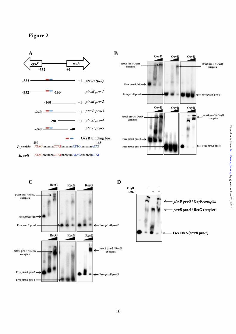
bound to the -200 to -163 promoter region (Fig.
2B) and RecG bound to the -200 to -90 promoter
region of the trxB in P. putida (Fig. 2C). Binding
studies using a mixture of RecG and OxyR
proteins showed that the both proteins could
bind to the trxB gene promoter region (ptrxB
pro-5) (Fig. 2D). Expression of the trxB gene in
the recG mutants was not induced under
conditions of oxidative stress (Fig. 3A),
indicating that RecG may be important for
regulating trxB gene expression within the oxyR
regulon.
RecG regulates other oxyR regulon genes-
We further analyzed seven genes that putatively
belong to the E. coli oxyR regulon and also have
promoters that contain an OxyR-binding
consensus sequence (supplemental Fig. S2A).
Interestingly, the expression of five of these
genes (katA, ahpC, trx-2, hslO, and PP0877) was
reduced in the recG mutant under oxidative
stress (Fig. 3A). The expression of katA and the
ahpC-ahpF operon in the recG mutant was
significantly reduced even under untreated
conditions (Fig. 3A); while the expression of
trxB, trx-2, hslO, and PP0877 was reduced under
both untreated and treated conditions (Fig. 3A).
Purified RecG and OxyR both bound to the
promoters of these genes (Fig. 3B), suggesting
by guest on June 23, 2018http://w
ww
.jbc.org/D
ownloaded from

6
that RecG also regulates other OxyR-regulated
genes.
OxyR-binding sites generate cruciform DNA
structures-In this study, we show that RecG
functions as a helicase and can unwind four and
three junction DNAs, but it cannot unwind
duplex DNA structure (supplemental Fig. S3).
Because RecG functions as a helicase and binds
to Holliday junctions, R-loops, D-loops and
cruciform DNA structures (8, 9), we examined
whether the promoter region of the trxB gene
forms non-linear DNA structures in vitro which
can be recognized by RecG (supplemental Fig.
S2A-C). OxyR-binding sites contain
palindromic sequences that bind the OxyR
protein in the form of a tetramer (3, 4). Two
OxyR dimers bind to similar palindromic sites,
resulting in tetramer formation (3). Interestingly,
we discovered that the OxyR-binding sequences
might form a cruciform DNA structure
(supplemental Fig. S2B, red box and S2C).
EMSA was then performed using various
fragments of the trxB gene promoter in the
presence or absence of the OxyR-binding sites
(Fig. 4A and B). When one palindromic OxyR-
binding site was truncated, RecG was unable to
bind to the trxB promoter (Fig. 4B); however,
the OxyR dimer, but not the tetramer, might be
able to still bind to the truncated site, which was
judged by the EMSA pattern (Fig. 4B, second
panel). Since cruciform DNA structures
commonly contain single strand DNA regions,
we examined the effect of adding purified single
strand binding protein (SSB) from E. coli to our
EMSA assays. The data showed that SSB also
bound to the trxB gene promoter (Fig. 4C).
Helicase RecG binds to the C-terminal region of
SSB (19, 20), which was verified in P. putida
using EMSA. SSB might facilitate the binding
of RecG to the trxB promoter (Fig. 4D). In
previous study, it was known that C-terminal of
SSB could bind to some helicases such as RecG
(20) and SSB acts as a DNA maintenance hub at
active chromosomal forks for stabilization of
DNA replication. In the binding studies
described earlier, an excess of RecG protein was
used (Fig. 1D and Fig. 3B); if low concentration
of RecG (0.05μg) was used, no band shift was
observed (Fig. 4D, left panel). However, in the
presence of SSB, only a low concentration of
RecG protein was required to bind to the
promoter (Fig. 4D, right panel and E). Also, if
OxyR-binding sites were truncated, both RecG
and SSB could not bind to the trxB promoter
(Fig. 4F).
To analyze the DNA structure of the trxB
promoter region, various DNA fragments of the
trxB promoter regions were used for EMSA
using SSB proteins (supplemental Fig. S4A). An
upstream 20 nucleotide from pro-5 was
truncated in the pro-6 fragment, and a
downstream-110 nucleotide from pro-6 was
truncated in pro-7. The OxyR-binding sites of
pro-6 and pro-7 are present at the edge of these
fragments. The OxyR-binding sites of promoter-
1 and promoter-2 are located at the center of the
fragments. Interestingly, SSB bound to the pro-5
region, but not to OBD-1, OBD-2, and OBD-3
(supplemental Fig. S4B). Therefore, the OxyR-
binding sites are necessary for binding of SSB.
More importantly, SSB could not bind to the
pro-6 and pro-7 regions (supplemental Fig. S4A
and B), which suggests that the OxyR-binding
sites at the edges of the DNA may not form
precise cruciform DNA structures. The location
of the OxyR-binding sites in the linear DNA
fragment is very important for the proper
formation of cruciform DNA structures.
Subsequently, SSB bound to the long promoter
regions (promoter-1 and promoter-2) that had
OxyR-binding sites at their centers. Interestingly,
the EMSA data from promoter-1 and promoter-2
showed a number of band shifts (supplemental
Fig. S4B), suggesting that the promoter-1 and
promoter-2 regions may contain different
cruciform structures. The sub 2, 3 and 4 DNA
fragments were generated by nucleotide cascade
substitutions within the OxyR-binding sites
(supplemental Fig. S4C); therefore, the sub 2, 3
and 4 regions may not form proper DNA
structures. Indeed, SSB did not bind to the sub 2,
sub3 and sub4 regions (supplemental Fig. S4D).
The presence of cruciform DNA structures
was further examined using T7 endonuclease I,
which cleaves cruciform structures (13). We
mapped the T7 endonuclease I cleavage sites
using restriction enzyme digestion and
sequencing of the DNA fragments (13). First, we
predicted the most thermodynamically stable
DNA structure for the trxB promoter region
using the mfold program.
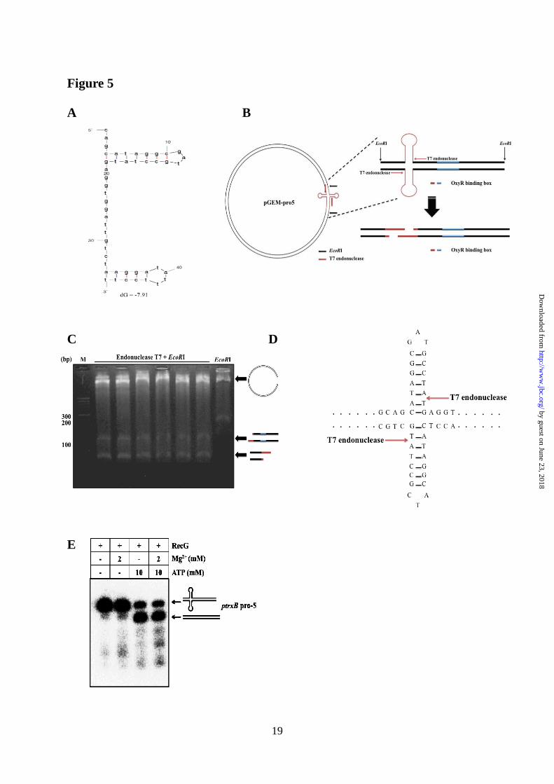
(http://mfold.rna.albany.edu/?q=mfold) (13). We
found that the OxyR-binding sites generated a
main hairpin DNA structure within the trxB
promoter region (Fig. 5A). When incubated with
T7 endonuclease I, a considerable amount of the
by guest on June 23, 2018http://w
ww
.jbc.org/D
ownloaded from

7
pGEM-pro5 vector harboring the trxB promoter
was digested into linear fragments. To further
map the cleavage sites, we performed sequence
analysis of each nuclease-cleaved fragment (Fig.
5B and C). Two EcoRI-T7 endonuclease
fragments were sequenced and the cleavage sites
were localized to one position (Fig. 5C and D).
By contrast, EcoRI-treated samples generated
only one fragment (apart from the vector
backbone, Fig. 5D; last lane). Taken together,
these data suggest that OxyR-binding sites may
generate cruciform DNA structures within linear
DNA.
Both Mg2+
and ATP affect the expression of
OxyR-regulated genes-RecG function is
modulated by Mg2+
and ATP (21, 22). Mg2+
stabilizes cruciform structures (23), whereas
ATP is used as an energy source for unwinding
by RecG helicase (22). Therefore, depending on
the Mg2+
:ATP ratio, cruciform DNA structures
can be altered by RecG helicase (22). We found
that addition of Mg2+
induced strong binding of
RecG to the trxB promoter, while the addition of
ATP weakened it (Fig. 6A). When the Mg2+
:ATP
molar ratio was changed to 1:5, RecG binding to
the promoter region decreased because RecG
used the ATP to create linear DNA. This ratio is
the typical molar ratio of mobile cruciform DNA
(22, 24) and the result was confirmed using a
helicase unwinding assay (Fig. 5E).
DNA binding studies were performed using
RecG, OxyR, SSB, Mg2+
and ATP, and the
results showed that binding of the OxyR dimer
was stronger than that of the OxyR tetramer. In
the presence of Mg2+
, the OxyR dimer bound to
the promoter more strongly than the OxyR
tetramer (Fig. 6B, lanes 3 and 4; Fig. 6C),
perhaps due to the tight cruciform DNA
structures formed. However, when ATP was
added to the reaction, the OxyR tetramer bound
to the promoter region more strongly than the
OxyR dimer. These data suggest that RecG,
along with SSB, binds to the cruciform DNA
structure of the oxyR regulon-promoters and
RecG then creates linear DNA by hydrolyzing
ATP. Subsequently, the OxyR tetramer binds to
the linearized DNA, and oxidized OxyR induces
expression of the oxyR regulon.
When size exclusion chromatography was
performed using FPLC, the purified OxyR was
eluted predominantly as a tetramer (data not
shown). It has been known that the active form
of OxyR is homotetrameric, consisting of a
dimer where each dimer is made up of 34 kDa
monomers (25). Many regulatory proteins such
as CbnR, DntR form the tetramer as a dimer of
dimers (26). In Fig. 6B, RecG and SSB
concentrations were 5 and 10-fold less than
those in the Fig. 6C so that binding of those two
proteins were not detected in the first lane of Fig.
6B in the presence of RecG plus SSB. When
high concentration of OxyR was added (the
second lane in Fig 6B), two different sizes of
bands were observed, which might be a dimer
and a tetramer of OxyR bound to the DNA. We
cannot rule out the possibility that different
forms of DNAs and different oligomeric OxyR
proteins might exist in our experiments because
there were more than two OxyR-DNA bands in
many EMSA experiments (Fig. 1C, 2B, 2D).
When ATP was added, RecG was released and
more OxyR tetramer-bound DNA was formed.
We speculated that more free-DNA became
available because more OxyR proteins were
bound to the less amounts of DNA (Fig 6B, lane
7).
DNA binding studies using a supercoiled
plasmid harboring the trxB promoter (pGEM-
pro5) supported this conclusion (Fig. 6D and E).
In the presence of Mg2+
, the OxyR dimer bound
to the promoter more strongly than the OxyR
tetramer (Fig. 6E, lane 6). The reverse occurred
when ATP was added to the reaction (Fig. 6E,
lane 7). By contrast, control samples containing
vector alone did not show a shift under the same
conditions (Fig. 6D). We performed the in vitro
transcription analysis for investigation of
transcriptional effect of SSB, RecG, and ATP
(15). Data obtained from the in vitro
transcription assay were consistent with the
EMSA data (Fig. 6F), which showed that
presence and absence of those components
changed OxyR-dependent in vitro transcription.
Highest in vitro transcription was observed only
in the presence of all three components (Fig. 6F
lanes 6 and 7). In addition, in vitro transcription
decreased when an excess of DTT was used (Fig.
6F, lanes 11 and 12). We also observed that
when the amount of OxyR used in the reaction
decreased, or the amount of RecG or Mg2+
increased, gene expression was reduced,
whereas increasing the amount of ATP had the
opposite effect (Fig. 6F). 0.25 mM of Mg2+
was
in the in vitro transcription reaction because the
transcription buffer (0.5 mM MgCl2) was diluted
when reaction mixture was prepared. Extra 2
by guest on June 23, 2018http://w
ww
.jbc.org/D
ownloaded from

8
mM Mg2+
was added to see the effect of Mg2+
stabilization for cruciform DNA. However,
many in vitro transcription assays used 0.25-5
mM Mg2+
concentration for RNA polymerase
activity so that dramatic effect of Mg2+
addition
was not observed in the Fig 6F (lane 6, and 7),
but when more Mg2+
(10 mM) was added to the
assay (Fig. 6G), transcriptional activity
decreased because of Mg2+
stabilized the
cruciform DNA.
RecG acts as a global regulator-To examine
whether recG acts as a global regulator, we
performed transcriptome analysis using the recG
mutant under oxidative stress. Almost all genes
that were induced by oxidative stress in wild-
type cells were down-regulated in the recG
mutant under oxidative stress (Table 1). RecG
deletion could reduce expression of some genes
in the absence of oxidative stress. When PQ- and
CHP-induced genes (39 and 60, respectively)
were analyzed, 13 of 39 and 8 of 60 genes were
reduced in the recG mutant without oxidative
stress (Table S2 and S3, ΔrecG/WT column).
Among 51 PQ and 54 CHP down-regulated
genes, only 3 and 9 genes were affected by
RecG deletion in the absence of oxidative stress,
respectively. No PQ-down-regulated genes were
induced in the recG mutant. Thus, RecG can
modulate gene expression even in the absence of
oxidative stress. The presence of palindromic
sequences within their promoters was also
examined, since RecG acts as a regulator of loop
or cruciform DNA structures. Of the 36 genes
whose expression was reduced in the recG
mutant under PQ-treatment, 30 contained a
palindromic site within their promoter regions
(representatives are shown in supplemental Fig.
S5). Notably, the oxidative stress-related genes
ahpC, katA, trx-2, hslO and lsfA, which are
induced by oxidative stress and contain the
OxyR-binding sequence, had palindromic
sequences in their promoter regions. Other
oxidative stress-related genes that also contain a
palindromic sequence include: cysP, a
transporter related gene induced in response to
chromate-oxidative stress in Shewanella
oneidensis (27); rpoX, which has a similar
function as rpoS and regulates various genes
under oxidative stress in Vibrio alginolyticus
(28); acnB, which encodes an aconitase and is
sensitive to oxidative stress in E. coli (29); and
bkdB, which is annotated as the branched-chain
alpha-keto acid dehydrogenase subunit, E2, and
is induced by cold stress in Bacillus subtilis (30).
We next compared the transcriptomes of the
recG mutant and wild type strains in the absence
of oxidative stress and found that the expression
of more than 99 genes (including the previously
mentioned oxyR-regulated genes) in the recG
mutant were reduced 2-fold compared to that in
the wild type (supplemental Table S6).
Therefore, RecG may induce structural changes
in DNA regardless of OxyR function, and
appears to be involved in global bacterial
transcription. RecG is important for gene expression in P.
aeruginosa and E. coli-The P. putida oxyR gene
mutant appears to be lethal, as we were unable
to generate this mutant successfully, despite
several attempts. Additionally, oxyR deletion
mutants of P. putida have not been reported,
with the exception of a single amino acid mutant
(31). Therefore, P. aeruginosa was used to study
the in vivo role of OxyR in Pseudomonas. The
oxyR-recG operon of P. aeruginosa is similar to
that of P. putida; however, there are two trxB
genes in the P. aeruginosa genome, one of
which (trxB2) has a sequence similar to that of
the trxB gene of P. putida. The trxB2 gene is
located next to the PA0848 gene, which, together,
may comprise an operon in P. aeruginosa.
Additionally, an oxyR box is present in the
PA0848 promoter. Interestingly, the expression
of these genes was severely reduced in oxyR and
recG mutants in P. aeruginosa (Fig. 7A). To
investigate whether OxyR and RecG bind to the
promoters of these two genes, EMSA was
performed using purified OxyR and RecG from
P. putida. The results showed that both proteins
bound to the PA0848 promoter (Fig. 7B) of P.
aeruginoas. The PA0848 and trxB2 genes exist
in an operon and the promoter region exist in
front of PA0848 gene. Furthermore, OxyR and
RecG bound to the promoters of OxyR-regulated
genes of E. coli (i.e. ahpC, dsbG, fhuF, fur, trxC
and yfdI) and regulated their expression (Fig. 7C
and D).
DISCUSSION
Although the function of RecG in the
recombination and repair of DNA has been
studied (5), we demonstrated for the first time
that RecG also plays an important role in
bacterial transcription by binding to promoters
containing palindromic sequences. This function
is specific to RecG, because other helicases
by guest on June 23, 2018http://w
ww
.jbc.org/D
ownloaded from

9
(dinG, PA3272, rep, ruvA and recQ) and the recJ
gene, encoding a nuclease, which is known to be
involved in RecQ helicase activity of P.
aeruginosa, did not influence gene expression of
OxyR-regulated genes (supplemental Fig. S6).
Our microarray data also suggest that RecG may
function alone in transcriptional regulation.
Genes whose expression levels were
significantly reduced in the recG mutant were
classified based on COG function (data not
shown, supplemental Table S6). Thus, the
unwinding function of RecG may be required for
transcription of these regions.
Cruciform formation within palindromic
regions of linear DNA carries an energy cost
because bases must to be unpaired to initiate
extrusion of the cruciform arms (9, 10). Thus,
cruciform DNA will branch-migrate back to
form duplex linear stable molecules. Cruciform
DNA formation can be driven by negative
supercoiling, but their formation in linear DNA
is highly unlikely. However, our data showed
that the trxB promoter generated cruciform DNA
within linear DNA if the DNA regions next to
the palindromic regions were long enough
(supplemental Fig. S4). Although the binding
site for SSB is 35 or 65 nucleotides, depending
on the salt conditions, other results suggest that
SSB can bind to a 21-nucleotide region in
Salmonella enterica DNA (depending on the salt
concentration) (32). The EMSAs were
conducted under low-salt conditions, which
would have allowed SSB to bind to such small
nucleotide regions. Also, other studies indicate
that extrusion of linear DNA generates hairpin
and cruciform structures (33, 34). Although little
is known about the physical DNA structure of
the trxB-like promoters, the data provided
evidence that SSB may bind to trxB promoters
in both linear and supercoiled DNA.
The data also showed that the pattern of
transcriptional expression affected by RecG
varied depending on the location of the
cruciform DNA. The promoter region of the
trxB gene produces cruciform DNA within
OxyR-binding sites, but the palindromic
sequences of the ahpC and katA genes are
located upstream of the OxyR-binding sites.
Although expression of the ahpC and katA genes
was reduced overall in the recG mutant
compared with that in wild-type cells,
expression of these genes was still induced in
response to oxidative stress (Fig. 3A). Thus, we
speculate that RecG influences DNA stability
which, in turn, may affect the expression of
ahpC and katA regardless of oxidative stress.
Because RecG does not directly affect the
OxyR-binding sites of the ahpC and katA genes,
unlike the trxB gene, the tetramer form of OxyR
may still be functional at these sites. Other oxyR-
regulated genes (trx-2, hslO, and trxB) contain
cruciform DNA within their OxyR-binding sites.
The data suggest that the function of RecG may
correlate with the position of the cruciform DNA
on the target gene.
We also found that OxyR senses superoxide,
but not hydrogen peroxide. The mechanism of
OxyR activation may vary in different types of
bacteria. In the case of Porphyromonas
gingivalis, the OxyR regulator does not sense
hydrogen peroxide, but activates various
oxidative stress-related genes (35). Additionally,
OxyR functions as a negative regulator in some
types of bacteria (36). Previous studies have
shown that the OxyR dimer has stronger DNA
binding affinity than the tetramer (3, 4).
Although the observed interactions with the
tetramer appear to be weak and may require
other proteins to be stabilized in solution, the
tetramer is still required for the activation of
OxyR-regulated genes. The data indicate that
DNA is linearized by RecG helicase, and that the
addition of SSB and ATP provides stability for
the binding of the OxyR tetramer, which then
promotes full activation of OxyR-regulated
genes (Fig. 8). Taken together, the results reveal
a novel mechanism underlying RecG-mediated
regulation of transcription in Pseudomonas
species, and demonstrate that the OxyR tetramer
may preferentially bind to its cognate
palindromic promoter after RecG generates
linear DNA.
REFERENCES
1. Imlay, J. A. (2008) Annu. Rev. Biochem. 77, 755-776
2. Zheng, M., Wang, X., Templeton, L. J., Smulski, D. R., LaRossa, R. A., and Storz, G. (2001) J.
Bacteriol. 183, 4562-4570
by guest on June 23, 2018http://w
ww
.jbc.org/D
ownloaded from

10
3. Lee, C., Lee, S. M., Mukhopadhyay, P., Kim, S. J., Lee, S. C., Ahn, W. S., Yu, M. H., Storz, G.,
and Ryu, S. E. (2004) Nat. Struct. Mol. Biol. 11, 1179-1185
4. Zaim, J., and Kierzek, A. M. (2003) Nucleic. Acids Res. 31, 1444-1454
5. Rudolph, C. J., Upton, A. L., Briggs, G. S., and Lloyd, R. G. (2010) DNA Repair (Amst) 9, 210-
223
6. Sharples, G. J., Ingleston, S. M., and Lloyd, R. G. (1999) J. Bacteriol. 181, 5543-5550
7. Hall, M. C., and Matson, S. W. (1999) Mol. Microbiol. 34, 867-877
8. Gregg, A. V., McGlynn, P., Jaktaji, R. P., and Lloyd, R. G. (2002) Mol. Cell 9, 241-251
9. Briggs, G. S., Mahdi, A. A., Wen, Q., and Lloyd, R. G. (2005) J. Biol. Chem. 280, 13921-13927
10. Rudolph, C. J., Upton, A. L., Harris, L., and Lloyd, R. G. (2009) Mol. Microbiol. 73, 352-366
11. Ren, B., Duan, X., and Ding, H. (2009) J. Biol. Chem. 284, 4829-4835
12. Voloshin, O. N., and Camerini-Otero, R. D. (2007) J. Biol. Chem. 282, 18437-18447
13. Kurahashi, H., Inagaki, H., Yamada, K., Ohye, T., Taniguchi, M., Emanuel, B. S., and Toda, T.
(2004) J. Biol. Chem. 279, 35377-35383
14. Yeom, S., Yeom, J., and Park, W. (2010) Microbiology 156, 1487-1496
15. Kang, J. G., Hahn, M. Y., Ishihama, A., and Roe, J. H. (1997) Nucleic Acids Res. 25, 2566-2573
16. Ochsner, U. A., Vasil, M. L., Alsabbagh, E., Parvatiyar, K., and Hassett, D. J. (2000) J. Bacteriol.
182, 4533-4544
17. Imlay, J. A. (2006) Mol. Microbiol. 59, 1073-1082
18. Park, W., Peña-Llopis, S., Lee, Y., and Demple, B. (2006) Biochem. Biophys. Res. Commun. 341,
51-56
19. Cadman, C. J., and McGlynn, P. (2004) Nucleic Acids Res. 32, 6378-6387
20. Costes, A., Lecointe, F., McGovern, S., Quevillon-Cheruel, S., and Polard, P. (2010) PLoS Genet.
6, e1001238
21. Singleton, M. R., Scaife, S., and Wigley, D. B. (2001) Cell 107, 79-89
22. McGlynn, P., and Lloyd, R. G. (1999) Nucleic Acids Res. 27, 3049-3056
23. Vologodskaia, M. Y., and Vologodskii, A. V. (1999) J. Mol. Biol. 289, 851-859
24. Shida, T., Iwasaki, H., Saito, A., Kyogoku, Y., and Shinagawa, H. (1996) J. Biol. Chem. 271,
26105-26109
25. Knapp, G. S., Tsai, J. W., and Hu, J. C. (2009) Protein Sci. 18, 101-107
26. Zeller, T., and Klug, G. (2004) Microbiology 150, 3451-3462
27. Brown, S. D., Thompson, M. R., Verberkmoes, N. C., Chourey, K., Shah, M., Zhou, J., Hettich,
R. L., and Thompson, D. K. (2006) Mol. Cell Proteomics 5, 1054-1071
28. Zhao, J. J., Chen, C., Zhang, L. P., and Hu, C. Q. (2009) J. Biomed. Biotechnol. 2009, 126986
29. Kumar, J. K., Tabor, S., and Richardson, C. C. (2004) Proc. Natl. Acad. Sci. U S A 101, 3759-
3764
30. Kaan, T., Homuth, G., Mäder, U., Bandow, J., and Schweder, T. (2002) Microbiology 148, 3441-
3455
31. Hishinuma, S., Ohtsu, I., Fujimura, M., and Fukumori, F. (2008) FEMS Microbiol. Lett. 289,
138-145
32. Huang, Y. H., Lee, Y. L., and Huang, C. Y. (2011) Protein J. 30, 102-108
33. Taylor, A. F., Smith, G. R. (1992) Proc. Natl. Acad. Sci. U S A 89, 5226-5230
34. Dai, X., Rothman-Denes, L. B. (1998) Genes Dev. 12, 2782-2790
35. Ohara, N., Kikuchi, Y., Shoji, M., Naito, M., and Nakayama, K. (2006) Microbiology 152, 955-
966
36. Panmanee, W., and Hassett, D. J. (2009) FEMS Microbiol. Lett. 295, 238-244
FOOTNOTES
This work was supported by the National Research Foundation (NRF) grant funded by Korea
Government (MEST) (No. 2012-0005277), Republic of Korea
To whom correspondence should be addressed: Woojun Park, Department of Environmental
by guest on June 23, 2018http://w
ww
.jbc.org/D
ownloaded from
11
Science and Ecological Engineering, Korea University, Seoul, Republic of Korea. 136-713, Tel.:
+82 2 3290 3067; Fax: +82 2 953 0737; E-mail: [email protected]
The abbreviations used are: PQ, paraquat; MD, menadione; H2O2, hydrogen peroxide; CHP, cumen
hydroperoxide; EMSA, electrophoretic mobility shift assay; OBD, OxyR-binding site deletion; SSB,
single stranded binding protein
FIGURE LEGENDS
FIGURE 1. Analysis of trxB gene expression under various oxidative stress conditions. (A)
Quantitative real-time RT-PCR (qRT-PCR, graph) analyses of trxB gene expression. Four qRT-
PCRexperiments were performed and means and standard deviations are shown. C, no treatment;
PQ, paraquat (0.5 mM); MD, menadione (1.0 mM); H2O2, hydrogen peroxide (1.0 mM); and CHP,
cumen hydroperoxide (3mM) for 10 min. (B) Northern blot analysis shows that increasing
concentrations of PQ and H2O2 induced trxB gene expression. (C) In vitro EMSA analysis. Lanes 1–
4: increasing amounts of purified OxyR (0, 0.05, 0.1, and 0.2 μg). Lanes 5–7, labeled as cold DNA,
represent increasing amounts of unlabeled trxB (full) promoter (0.3, 0.7, and 1.2 μM unlabeled
probe) while the amount of OxyR was fixed at 0.2 μg. (D) In vitro EMSA analysis. Lanes 1–6
represent increasing amounts of purified RecG (0, 0.02, 0.05, 0.10, 0.15 and 0.20 μg). Lanes 7–8,
labeled as cold DNA, correspond to increasing amounts of unlabeled trxB (full) promoter (0.4 and
1.2 μM unlabeled probe) while the amount of RecG was fixed at 0.2 μg. At least, three independent
experiments were performed in all experiments.
FIGURE 2. (A) Schematic representation of the trxB promoter region that are used for EMSA in
our OxyR binding studies. (B) In vitro EMSA using OxyR on various the trxB promoter regions.
The concentration of purified OxyR was used in 0.05 and 0.2 μg. (C) In vitro EMSA using RecG on
various the trxB promoter regions. The concentration of purified RecG was used in 0.05 and 0.2 μg.
(D) In vitro EMSA using OxyR and RecG on the ptrxB-pro5 promoter regions. The concentrations
of purified OxyR and RecG were fixed in 0.2 μg. At least, three independent experiments were
performed in all experiments.
FIGURE 3. Effect of OxyR and RecG on the transcriptional regulation of the oxyR regulon. (A)
Expression analysis for oxidative stress related genes under oxidative conditions in P. putida wild-
type and recG gene mutant cells. C, no treatment; PQ, paraquat (0.5 mM) for 10 min. (B) In vitro
EMSA analysis. Purified OxyR and RecG (both 0.2 μg) were used. Autoradiography was conducted
using an IP plate (Fujifilm) and a Multiplex Bio-Imaging System (Fujifilm). At least, three
independent experiments were performed in all experiments.
FIGURE 4. Studies on the trxB gene promoter region. (A) Schematic representation of the trxB
promoter region which were used for EMSA in the presence or absence of the OxyR-binding
sequence. (B) In vitro EMSA analysis showed that OxyR and RecG proteins bind to various regions
of the trxB promoter in P. putida. A schematic representation of the truncated promoter is shown in
(A) panel. Purified OxyR and RecG (both 0.2 μg) were used. (C) In vitro EMSA analysis with SSB
protein and ptrxB pro-5. Lanes 1–6 represent increasing amounts of purified SSB (0, 0.01, 0.02, 0.05,
0.08 and 0.10 μg). Lanes 7–8 correspond to 2.0 and 4.0 mg, respectively, of unlabeled trxB promoter
in the presence of 0.1 μg of SSB. (D, E) Assessment of SSB and RecG binding to the ptrxB pro-5
promoter using in vitro EMSA analysis. (D) Lanes 1–5 represent increasing amounts of purified
RecG (0, 0.01, 0.02, 0.04 and 0.05 μg). Lanes 6–9 correspond to increasing amounts of SSB (0.01,
0.02, 0.04 and 0.05 μg) while the amount of RecG was fixed at 0.05 μg. (E) Lanes 1–4 represent
increasing amounts of purified RecG (0, 0.02, 0.05 and 0.10 μg). Lanes 5–7 correspond to
increasing amounts of RecG (0.02, 0.05 and 0.10 μg), while the amount of SSB was fixed at 0.05 μg.
(F) Assessment of SSB and RecG binding to the ptrxB pro-5 promoter in the presence or absence of
the OxyR-binding sequence using in vitro EMSA. RecG (0.05 μg) and SSB (0.05 μg) were used for
by guest on June 23, 2018http://w
ww
.jbc.org/D
ownloaded from

12
EMSA. At least, three independent experiments were performed in all experiments.
Figure 5. Identification of cruciform DNA structure in the trxB promoter region using T7
endonuclease I. (A) Prediction of thermodynamic stable cruciform DNA structure using the OxyR
binding sequence using mfold program. (B) The arrow mark means cleavage site by T7
endonuclease I. Schematic representations of EcoRI plus T7 endonuclease cleavage sites mapping
on the trxB promoter region. (C) The plasmid containing the trxB promoter region (pGEM-pro5)
were digested with EcoRI T7 endonuclease and were subjected to agarose gel (2.0%) electrophoresis.
Lanes 1, DNA ladder; lanes 2-7, EcoRI+T7 endonuclease treated samples; lane 8, EcoRI treated
sample. (D) The fragments were sequenced and results were represented on the cruciform DNA
structure using arrows mark. (E) Helicase unwind assay was performed with RecG, Mg2+
and ATP in
the trxB gene promoter region (ptrxB pro-5). The concentrations of purified RecG was fixed in 0.2
μg. At least, two independent experiments were performed in all experiments.
FIGURE 6. The effect of SSB, Mg2+
and ATP on the regulatory mechanism of RecG and OxyR. (A)
EMSA analysis showing that MgCl2 and ATP affected the binding of RecG to the trxB gene
promoter (ptrxB-pro5). Binding affinity was monitored using the indicated concentrations of ATP
and MgCl2. RecG concentration was fixed at 0.05 μg. (B) In vitro EMSA analysis demonstrated that
OxyR protein binding was dependent upon the concentration (mM) of MgCl2 and ATP. RecG, SSB
and OxyR concentrations were added at 0.02 μg, 0.01 μg, and 0.2 μg, respectively. The numbers
below each lane show the intensity of the band relative to the intensity of control band (First lane),
which were analyzed by ProXPRESS 2D (Perkin Elmer, USA) and Total Lab 2.0 Software
(Nonlinear Dynamics, BioSystematica, UK). (C) Assessment of the binding of various components
to the trxB promoter using in vitro EMSA. RecG, SSB, OxyR, MgCl2 and ATP concentrations were
added at 0.1 μg, 0.1 μg, 0.2 μg, 2 mM, and 10 mM, respectively. (D) Control EMSA experiment
using pGEM-T easy plasmid. The reaction mixture (20 μl final volume), containing the pGEM-T
easy, purified OxyR and RecG proteins (both 0.1 μg), SSB (Promega, 0.1 μg), BSA (0.5 μg), loading
buffer and poly dI-dC(1 μg) in binding buffer (10 mM Tris, pH 7.5, 2 mM MgCl2, 10%, v/v,
glycerol, and 75 mM KCl), was incubated for 30 min at 4°C. (E) Plasmid EMSA analysis performed
with 2 mM MgCl2, 10 mM ATP, SSB, RecG and OxyR. RecG, SSB and OxyR concentrations were
added at 0.2 μg, 0.05 μg, and 0.2 μg, respectively. SC and OC refer to the supercoiled plasmid and
open circular plasmid, respectively. (F) Effect of various components on runoff transcription assay
from promoters of the trxB genes. An autoradiogram of denaturing 10% PAGE of radiolabeled RNA
products is shown. Transcription reactions were conducted at 30°C for 30 min under standard
reaction conditions (see Materials and Methods). RecG, SSB, OxyR, MgCl2 and ATP concentrations
were added at 0.1 μg, 0.1 μg, 0.2 μg, 2 mM, and 10 mM, respectively. DTT concentrations were
added at 10 and 50 mM. (G) A runoff transcription assay was performed with various concentrations
of RecG, OxyR, SSB, MgCl2 and ATP. The amount of reaction components was the same as in (F),
except for the following: Lanes 1–4 represent decreasing amounts of purified OxyR (0, 0.10, 0.05
and 0.01 μg), Lanes 5–7 represent decreasing amounts of purified RecG (0.05, 0.1 and 0.2 μg),
Lanes 8–9 represent increasing amounts of MgCl2 (5 and 10 mM), and Lanes 10–11 represent
increasing amounts of ATP (20 and 50 mM). At least, three independent experiments were
performed in all experiments.
FIGURE 7. Investigation of whether RecG functions as global regulator. (A) Expression analysis of
oxidative stress-related genes from supplemental Fig. S3A under oxidative conditions in P.
aeruginosa. PA0848 and trxB2 genes belong to the same operon in P. aeruginosa. An image of the
ethidium bromide-stained gel was used to verify consistent loading. Cells were treated for 10 min
with C, no treatment; PQ, paraquat (0.5 mM); or CHP, cumen hydroperoxide (3 mM). The numbers
below each lane show the intensity of the band relative to the intensity of control band (First lane),
which were analyzed by ProXPRESS 2D (Perkin Elmer, USA) and Total Lab 2.0 Software
(Nonlinear Dynamics, BioSystematica, UK). (B) In vitro EMSA showed that OxyR and RecG bound
to the promoter of the PA0848-trxB2 operon in P. aeruginosa. Purified OxyR and RecG (0.2 μg of
by guest on June 23, 2018http://w
ww
.jbc.org/D
ownloaded from

13
each) were used. (C) Expression analysis of oxidative stress-related genes under oxidative
conditions in E. coli. An image of the ethidium bromide-stained gel was used to verify consistent
loading. Cells were treated for 10 min with C, no treatment; PQ, paraquat (0.5 mM). The numbers
below each lane show the intensity of the band relative to the intensity of control band (First lane),
which were analyzed by ProXPRESS 2D (Perkin Elmer, USA) and Total Lab 2.0 Software
(Nonlinear Dynamics, BioSystematica, UK). (D) In vitro EMSA analysis showed that OxyR and
RecG bound to the promoters of ahpC, dsbG, fhuF, fur, trxC and yfdI of E. coli. Purified OxyR and
RecG (0.2 μg) were used. At least three independent experiments were performed in all experiments.
FIGURE 8. Model of RecG helicase function during bacterial transcription. RecG helicase, in
conjunction with SSB and ATP, linearizes hairpin DNA. When oxidized, the OxyR tetramer stably
binds to the linearized promoter and activates transcription. The thickness of the arrows represents
the preferences of potential helicase function. RNAP: RNA polymerase enzyme.
by guest on June 23, 2018http://w
ww
.jbc.org/D
ownloaded from

14
Table 1. List of genes whose expression was reduced among PQ-induced genes in P. putida recG
mutant
PP number WT+PQa ΔrecG+PQ ΔrecG/WT Palindoromeb Description
PP2439 (ahpC) 17.39 ± 1.09 1.17 ± 0.05 0.46 ± 0.02 + alkyl hydroperoxide reductase, C subunit
PP0481 (katA) 14.83 ± 1.12 1.04 ± 0.02 0.48 ± 0.01 + catalase
PP2334 12.13 ± 0.09 1.34 ± 0.17 0.94 ± 0.05 + carboxyvinyl-carboxyphosphonate
PP2440 (ahpF) 10.27 ± 1.11 1.84 ± 0.06 0.41 ± 0.07 ahpC operon alkyl hydroperoxide reductase, F subunit
PP0786 (trxB) 6.92 ± 0.11 1.27 ± 0.13 0.50 ± 0.02 + thioredoxin reductase
PP2339 (acnB) 5.62 ± 0.09 1.43 ± 0.13 0.50 ± 0.08 + aconitate hydratase 2
PP4403 (bkdB) 4.96 ± 0.04 1.12 ± 0.43 0.97 ± 0.01 + 2-oxoisovalerate dehydrogenase, lipoamide
PP5215 (trx-2) 3.41 ± 0.33 0.40 ± 0.05 0.95 ± 0.09 + thioredoxin
PP5172 3.41 ± 0.33 0.40 ± 0.05 0.99 ± 0.01 + conserved hypothetical protein
PP0951 (rpoX) 3.41 ± 0.41 1.38 ± 0.12 1.24 ± 0.03 + sigma54 modulation protein
PP4621 (hmgA) 3.39 ± 0.09 1.02 ± 0.09 1.07 ± 0.02 + homogentisate 1,2-dioxygenase
PP0234 (oprE) 3.27 ± 0.77 1.19 ± 0.19 0.95 ± 0.02 + porin E
PP1206 (oprD) 3.18 ± 0.11 1.02 ± 0.07 0.47 ± 0.01 - porin D
PP0222 3.14 ± 0.35 1.05 ± 0.28 0.93 ± 0.03 + monooxygenase, DszA family
PP2335 3.07 ± 0.12 1.15 ± 0.04 0.95 ± 0.03 + methylcitrate synthase, putative
PP0170 3.05 ± 0.01 1.13 ± 0.30 0.96 ± 0.04 + ABC transporter, periplasmic binding protein
PP4194 (gltA) 2.97 ± 0.22 1.34 ± 0.11 0.96 ± 0.13 + citrate synthase
PP0883 2.89 ± 0.12 0.93 ± 0.05 0.46 ± 0.06 + porin, putative
PP0238 (ssuD) 2.87 ± 0.08 1.17 ± 0.21 0.99 ± 0.01 + organosulfonate monooxygenase
PP2441 2.79 ± 0.11 1.23 ± 0.00 0.24 ± 0.02 + hypothetical protein
PP5278 2.77 ± 0.29 1.03 ± 0.07 0.98 ± 0.01 + aldehyde dehydrogenase family protein
PP3148 2.77 ± 0.19 1.04 ± 0.28 0.50 ± 0.04 - glutamine synthetase, putative
PP0235 (lsfA) 2.73 ± 0.09 1.31 ± 0.09 0.99 ± 0.01 + antioxidant protein LsfA
PP3122 2.73 ± 0.17 0.94 ± 0.24 1.00 ± 0.01 - CoA-transferase, subunit A, putative
PP0885 2.69 ± 0.11 0.99 ± 0.09 0.95 ± 0.06 - dipeptide ABC transporter, periplasmic
PP4402 (bkdA2) 2.69 ± 0.08 0.99 ± 0.08 0.96 ± 0.01 + 2-oxoisovalerate dehydrogenase, beta subunit
PP4064 (ivd) 2.66 ± 0.07 0.97 ± 0.21 0.50 ± 0.09 + isovaleryl-CoA dehydrogenase
PP1071 2.62 ± 0.02 0.98 ± 0.15 0.95 ± 0.05 + amino acid ABC transporter, periplasmic amino acid
PP5171 (cysP) 2.58 ± 0.09 0.98 ± 0.15 0.49 ± 0.00 + sulfate ABC transporter, periplasmic
PP3123 2.57 ± 0.12 0.98 ± 0.15 0.97 ± 0.02 + CoA-transferase, subunit B, putative
PP0223 2.50 ± 0.09 1.18 ± 0.04 1.04 ± 0.02 + monooxygenase, DszC family
PP2333 2.33 ± 0.01 1.12 ± 0.40 1.01 ± 0.00 - transcriptional regulator, GntR family
PP0252 (hslO) 2.28 ± 0.02 1.15 ± 0.04 1.07 ± 0.06 + chaperonin, 33 kDa
PP4620 2.27 ± 0.12 0.95 ± 0.12 0.97 ± 0.01 + fumarylacetoacetase
PP2337 2.17 ± 0.07 1.09 ± 0.23 0.96 ± 0.02 + conserved hypothetical protein
PP4185 (sucD) 2.16 ± 0.01 1.26 ± 0.26 0.50 ± 0.01 - succinyl-CoA synthetase, alpha subunit
a Two independent experiments were performed using microarray analysis and the average values with standard deviation
are shown. Genes whose expressions were changed under PQ compared with wild-type in absent to PQ. bPalindromic sites were determined using a palindrome search program (http://bioinfo.cs.technion.ac.il/projects/Engel-
Freund/new.html) and the maximum number of mismatches allowed within palindromic sites was two bases.
by guest on June 23, 2018http://w
ww
.jbc.org/D
ownloaded from

15
Figure 1
A
B
C D
by guest on June 23, 2018http://w
ww
.jbc.org/D
ownloaded from
16
Figure 2
A B
C D
by guest on June 23, 2018http://w
ww
.jbc.org/D
ownloaded from

18
Figure 4
A B
C D
E F
by guest on June 23, 2018http://w
ww
.jbc.org/D
ownloaded from
19
Figure 5
A B
C D
E
by guest on June 23, 2018http://w
ww
.jbc.org/D
ownloaded from

20
Free DNA (ptrxB pro-5)
RecG
0.0 1.1 0.6 0.3 0.2
1.0 0.0 0.2 0.6 0.5
Figure 6
A B C
D E
F G
by guest on June 23, 2018http://w
ww
.jbc.org/D
ownloaded from

21
Figure 7
A B
C
D
by guest on June 23, 2018http://w
ww
.jbc.org/D
ownloaded from

Jinki Yeom, Yunho Lee and Woojun Parkfunction in Pseudomonas species
ATP-dependent RecG helicase is required for the transcriptional regulator OxyR
published online May 23, 2012J. Biol. Chem.
10.1074/jbc.M112.356964Access the most updated version of this article at doi:
Alerts:
When a correction for this article is posted•
When this article is cited•
to choose from all of JBC's e-mail alertsClick here
Supplemental material:
http://www.jbc.org/content/suppl/2012/05/24/M112.356964.DC1
by guest on June 23, 2018http://w
ww
.jbc.org/D
ownloaded from