Pseudomonas aeruginosa Exotoxin Y Is a Promiscuous Cyclase That Increases Endothelial Tau
13
Pseudomonas aeruginosa Exotoxin Y Is a Promiscuous Cyclase That Increases Endothelial Tau Phosphorylation and Permeability * □ S Received for publication, October 6, 2011, and in revised form, May 15, 2012 Published, JBC Papers in Press, May 25, 2012, DOI 10.1074/jbc.M111.301440 Cristhiaan D. Ochoa ‡§ , Mikhail Alexeyev §¶ , Viktoriya Pastukh ‡§ , Ron Balczon §¶ , and Troy Stevens ‡§1 From the Departments of ‡ Pharmacology, Medicine, and ¶ Cell Biology and Neuroscience and the § Center for Lung Biology, College of Medicine, University of South Alabama, Mobile, Alabama 36688 Background: ExoY induces inter-endothelial gaps, although the mechanisms by which this occurs are poorly understood. Results: ExoY synthesized cAMP and cGMP, which caused endothelial Tau hyperphosphorylation, accumulation of insoluble Tau, inter-endothelial cell gaps, and increased permeability. Conclusion: ExoY is a promiscuous cyclase and an edema factor. Significance: Acute Pseudomonas infections cause a pathophysiological sequela in endothelium previously recognized only in chronic neurodegenerative diseases. Exotoxin Y (ExoY) is a type III secretion system effector found in 90% of the Pseudomonas aeruginosa isolates. Although it is known that ExoY causes inter-endothelial gaps and vascular leak, the mechanisms by which this occurs are poorly under- stood. Using both a bacteria-delivered and a codon-optimized conditionally expressed ExoY, we report that this toxin is a dual soluble adenylyl and guanylyl cyclase that results in intracellular cAMP and cGMP accumulation. The enzymatic activity of ExoY caused phosphorylation of endothelial Tau serine 214, accumu- lation of insoluble Tau, inter-endothelial cell gap formation, and increased macromolecular permeability. To discern whether the cAMP or cGMP signal was responsible for Tau phosphorylation and barrier disruption, pulmonary microvas- cular endothelial cells were engineered for the conditional expression of either wild-type guanylyl cyclase, which synthe- sizes cGMP, or a mutated guanylyl cyclase, which synthesizes cAMP. Sodium nitroprusside stimulation of the cGMP-generat- ing cyclase resulted in transient Tau serine 214 phosphorylation and gap formation, whereas stimulation of the cAMP-generat- ing cyclase induced a robust increase in Tau serine 214 phos- phorylation, gap formation, and macromolecular permeability. These results indicate that the cAMP signal is the dominant stimulus for Tau phosphorylation. Hence, ExoY is a promiscu- ous cyclase and edema factor that uses cAMP and, to some extent, cGMP to induce the hyperphosphorylation and insolu- bility of endothelial Tau. Because hyperphosphorylated and insoluble Tau are hallmarks in neurodegenerative tauopathies such as Alzheimer disease, acute Pseudomonas infections cause a pathophysiological sequela in endothelium previously recog- nized only in chronic neurodegenerative diseases. Pulmonary microvascular endothelium forms a highly re- strictive barrier to allow for proper gas exchange (1– 6). Inflam- matory mediators and vascular permeability-increasing com- pounds cause retraction of cell borders and inter-endothelial gaps by reorganizing the endothelial cytoskeleton, cell-cell, and cell matrix interactions (reviewed in Ref. 7). When this barrier is disrupted, exudative alveolar edema occurs, and gas exchange is compromised. In endothelial cells, receptors coupled to adenylyl and gua- nylyl cyclases generate membrane-limited 3-5 cyclic adeno- sine monophosphate (cAMP) and 3-5 cyclic guanosine mono- phosphate (cGMP), respectively, which activate effectors to stabilize the cortical actin rim, insert adherens junction pro- teins into the plasma membrane, and strengthen overall inter- endothelial cell contacts (8 –13). Therefore, compounds that generate subplasmalemmal cAMP and cGMP contribute to the prevention and reduction of inflammation by averting inter- endothelial gap formation and the subsequent increase in per- meability (14 –16). In contrast, soluble adenylyl and guanylyl cyclases that generate cytosolic cAMP and cGMP signals are endothelial barrier disruptive and thus contribute to inflamma- tory edema (17–19). Of note, bacteria like Bacillus anthracis and Pseudomonas aeruginosa promote edema formation by introducing soluble cyclases into host cells (20, 21). P. aeruginosa is a leading cause of lung injury, particularly in critically ill patients that need mechanical ventilation (22). Importantly, exotoxin Y (ExoY) 2 is found in 90% of P. aerugi- nosa clinical isolates (23). ExoY is a soluble adenylyl cyclase that is introduced into host cells via the type III secretion system and increases the cytoplasmic levels of cAMP (24), mediates the hyperphosphorylation of endothelial Tau protein (25), impairs microtubule and microfilament stability 3 (26), induces inter- * This work was supported, in whole or in part, by National Institutes of Health Grants HL-60024 and HL-66299 (to T. S.), HL-76125 and HL-107122 (to C. D. O.), and 1RO1RR031286 (to M. A.). □ S This article contains supplemental “Experimental Procedures,” Codon- optimized Sequences, Figs. S1–S3, and Movies 1–3. 1 To whom correspondence should be addressed: Dept. of Pharmacology, College of Medicine, University of South Alabama, Mobile, AL 36688. Tel.: 251-460-7086; Fax: 251-460-7452; E-mail: [email protected]. 2 The abbreviations used are: ExoY, exotoxin Y; m.o.i., multiplicity of infec- tion; bis-Tris, 2-[bis(2-hydroxyethyl)amino]-2-(hydroxymethyl)propane- 1,3-diol; SNP, sodium nitroprusside; ANOVA, analysis of variance; sGC, sol- uble guanylyl cyclase; sAC, soluble adenylyl cyclase. 3 R. Balczon, N. Prasain, C. D. Ochoa, J. Prater, B. Zhu, M. Alexeyev, S. Sayner, D. W. Frank, and T. Stevens, submitted for publication. THE JOURNAL OF BIOLOGICAL CHEMISTRY VOL. 287, NO. 30, pp. 25407–25418, July 20, 2012 © 2012 by The American Society for Biochemistry and Molecular Biology, Inc. Published in the U.S.A. JULY 20, 2012 • VOLUME 287 • NUMBER 30 JOURNAL OF BIOLOGICAL CHEMISTRY 25407 by guest on January 23, 2019 http://www.jbc.org/ Downloaded from
Transcript of Pseudomonas aeruginosa Exotoxin Y Is a Promiscuous Cyclase That Increases Endothelial Tau
Pseudomonas aeruginosa Exotoxin Y Is a Promiscuous CyclaseThat Increases Endothelial Tau Phosphorylation andPermeability*□S
Received for publication, October 6, 2011, and in revised form, May 15, 2012 Published, JBC Papers in Press, May 25, 2012, DOI 10.1074/jbc.M111.301440
Cristhiaan D. Ochoa‡§, Mikhail Alexeyev§¶, Viktoriya Pastukh‡§, Ron Balczon§¶, and Troy Stevens‡§�1
From the Departments of ‡Pharmacology, �Medicine, and ¶Cell Biology and Neuroscience and the §Center for Lung Biology, Collegeof Medicine, University of South Alabama, Mobile, Alabama 36688
Background: ExoY induces inter-endothelial gaps, although the mechanisms by which this occurs are poorly understood.Results: ExoY synthesized cAMP and cGMP, which caused endothelial Tau hyperphosphorylation, accumulation of insolubleTau, inter-endothelial cell gaps, and increased permeability.Conclusion: ExoY is a promiscuous cyclase and an edema factor.Significance: Acute Pseudomonas infections cause a pathophysiological sequela in endothelium previously recognized only inchronic neurodegenerative diseases.
Exotoxin Y (ExoY) is a type III secretion system effector foundin� 90% of the Pseudomonas aeruginosa isolates. Although it isknown that ExoY causes inter-endothelial gaps and vascularleak, the mechanisms by which this occurs are poorly under-stood. Using both a bacteria-delivered and a codon-optimizedconditionally expressed ExoY, we report that this toxin is a dualsoluble adenylyl and guanylyl cyclase that results in intracellularcAMP and cGMP accumulation. The enzymatic activity of ExoYcaused phosphorylation of endothelial Tau serine 214, accumu-lation of insoluble Tau, inter-endothelial cell gap formation,and increased macromolecular permeability. To discernwhether the cAMP or cGMP signal was responsible for Tauphosphorylation and barrier disruption, pulmonary microvas-cular endothelial cells were engineered for the conditionalexpression of either wild-type guanylyl cyclase, which synthe-sizes cGMP, or a mutated guanylyl cyclase, which synthesizescAMP. Sodiumnitroprusside stimulation of the cGMP-generat-ing cyclase resulted in transient Tau serine 214 phosphorylationand gap formation, whereas stimulation of the cAMP-generat-ing cyclase induced a robust increase in Tau serine 214 phos-phorylation, gap formation, and macromolecular permeability.These results indicate that the cAMP signal is the dominantstimulus for Tau phosphorylation. Hence, ExoY is a promiscu-ous cyclase and edema factor that uses cAMP and, to someextent, cGMP to induce the hyperphosphorylation and insolu-bility of endothelial Tau. Because hyperphosphorylated andinsoluble Tau are hallmarks in neurodegenerative tauopathiessuch as Alzheimer disease, acute Pseudomonas infections causea pathophysiological sequela in endothelium previously recog-nized only in chronic neurodegenerative diseases.
Pulmonary microvascular endothelium forms a highly re-strictive barrier to allow for proper gas exchange (1–6). Inflam-matory mediators and vascular permeability-increasing com-pounds cause retraction of cell borders and inter-endothelialgaps by reorganizing the endothelial cytoskeleton, cell-cell, andcell matrix interactions (reviewed in Ref. 7). When this barrieris disrupted, exudative alveolar edemaoccurs, and gas exchangeis compromised.In endothelial cells, receptors coupled to adenylyl and gua-
nylyl cyclases generate membrane-limited 3�-5� cyclic adeno-sinemonophosphate (cAMP) and 3�-5� cyclic guanosinemono-phosphate (cGMP), respectively, which activate effectors tostabilize the cortical actin rim, insert adherens junction pro-teins into the plasma membrane, and strengthen overall inter-endothelial cell contacts (8–13). Therefore, compounds thatgenerate subplasmalemmal cAMP and cGMP contribute to theprevention and reduction of inflammation by averting inter-endothelial gap formation and the subsequent increase in per-meability (14–16). In contrast, soluble adenylyl and guanylylcyclases that generate cytosolic cAMP and cGMP signals areendothelial barrier disruptive and thus contribute to inflamma-tory edema (17–19). Of note, bacteria like Bacillus anthracisand Pseudomonas aeruginosa promote edema formation byintroducing soluble cyclases into host cells (20, 21).P. aeruginosa is a leading cause of lung injury, particularly in
critically ill patients that need mechanical ventilation (22).Importantly, exotoxin Y (ExoY)2 is found in �90% of P. aerugi-nosa clinical isolates (23). ExoY is a soluble adenylyl cyclase thatis introduced into host cells via the type III secretion systemandincreases the cytoplasmic levels of cAMP (24), mediates thehyperphosphorylation of endothelial Tau protein (25), impairsmicrotubule and microfilament stability3 (26), induces inter-
* This work was supported, in whole or in part, by National Institutes of HealthGrants HL-60024 and HL-66299 (to T. S.), HL-76125 and HL-107122 (toC. D. O.), and 1RO1RR031286 (to M. A.).
□S This article contains supplemental “Experimental Procedures,” Codon-optimized Sequences, Figs. S1–S3, and Movies 1–3.
1 To whom correspondence should be addressed: Dept. of Pharmacology,College of Medicine, University of South Alabama, Mobile, AL 36688. Tel.:251-460-7086; Fax: 251-460-7452; E-mail: [email protected].
2 The abbreviations used are: ExoY, exotoxin Y; m.o.i., multiplicity of infec-tion; bis-Tris, 2-[bis(2-hydroxyethyl)amino]-2-(hydroxymethyl)propane-1,3-diol; SNP, sodium nitroprusside; ANOVA, analysis of variance; sGC, sol-uble guanylyl cyclase; sAC, soluble adenylyl cyclase.
3 R. Balczon, N. Prasain, C. D. Ochoa, J. Prater, B. Zhu, M. Alexeyev, S. Sayner,D. W. Frank, and T. Stevens, submitted for publication.
THE JOURNAL OF BIOLOGICAL CHEMISTRY VOL. 287, NO. 30, pp. 25407–25418, July 20, 2012© 2012 by The American Society for Biochemistry and Molecular Biology, Inc. Published in the U.S.A.
JULY 20, 2012 • VOLUME 287 • NUMBER 30 JOURNAL OF BIOLOGICAL CHEMISTRY 25407
by guest on January 23, 2019http://w
ww
.jbc.org/D
ownloaded from

endothelial gap formation, and increases vascular permeability(27, 28).Recently, Göttle et al. (29) reported that bacterial soluble
cyclases similar to ExoY are capable of synthesizing more thanone cyclic nucleotide simultaneously (i.e. cAMP, cIMP, andcUMP). This suggested to us that ExoY could also synthesizeother cyclic nucleotides in addition to cAMP and that, if so, thiswould have important implications for the understanding oflung endothelial permeability and the pathophysiology ofP. aeruginosa-mediated acute lung injury. Here, we report thatP. aeruginosa ExoY is sufficient to increase intracellular levelsof both cAMP and cGMP in endothelial cells. We show thatboth cytosolic cAMP and, to a lesser degree, cGMPmediate thehyperphosphorylation of endothelial Tau Ser-214. We alsoshow that P. aeruginosa ExoY intoxication leads to accumula-tion of insoluble Tau. Finally, we demonstrate that accumula-tion of cytosolic cAMP, and not cGMP, leads to large inter-endothelial gaps and increased permeability in pulmonarymicrovascular endothelial cells. Because hyperphosphorylatedand insoluble Tau are hallmarks of neurodegenerative tauopa-thies such as Alzheimer disease (30, 31), these findings suggestthat acuteP. aeruginosa infections and chronic neurodegenera-tive diseases share Tau hyperphosphorylation and insolubilityas a common pathophysiological mechanism.
EXPERIMENTAL PROCEDURES
Cell Culture—Pulmonary microvascular endothelial cells(internal identification: PMVECR1)were obtained from the cellculture core at the University of South Alabama Center forLung Biology. The isolation and characterization of these cellshas been described previously in detail (1, 32, 33). Cells werecultured in Dulbecco’s modified Eagle’s medium (DMEM)with10% heat-inactivated fetal bovine serum (catalogue No. 10082,Invitrogen) and 1% penicillin/streptomycin (catalogue No.15140, Invitrogen) at 37 °C in 21% oxygen and 5% carbon diox-ide. Pulmonary artery smoothmuscle cells were the kind gift ofDr. Celina Gairhe (Department of Pharmacology, University ofSouthAlabama) andwere cultured as described previously (34).DNA and Viral Constructs—Retrovirus 2641 (rv2641) and
pulmonary microvascular endothelial cells infected with thisvirus (MV/2641) were described previously (35). Constructionof the lentiviral vector for doxycycline-inducible C-terminalfusions to a destabilizing variant of FKBP12 (F36V, E31G,R71G, and K105E), pMA3174, will be described separately.4The full-length synthetic gene for P. aeruginosa, exoY, wascodon-optimized commercially for expression in rat cells (ratcodon adaptation index, 0.99) (see supplemental material forsequence). The termination codon was removed from the full-length codon-optimized exoY gene by PCR using primersExoYRIf (GCGAATTCGCCACCATGAGGATCGACGGC-CACAG) and ExoYdTerHpa (RGCGTTAACCAGCTCCAC-CTTCCTCTGGA). pMA3200was constructed by inserting thefull-lengthwild-type codon-optimized exoY genewithout a ter-mination codon into pMA3174 in-frame with a destabilizingvariant of FKBP-12 (36–38). To codon-optimize for expression
inmammalian cells, themutant (exoYK81M) catalytic domain ofexoY (amino acids 1–207; codon adaptation index � 0.94), wasalso generated commercially (BioBasic, Markham, Ontario,Canada). pMA3228 was generated by replacing the 5�-terminalEcoRI-BsrGI fragment of the WT exoY gene in pMA3200 withthe corresponding fragment of the gene encoding the codon-optimized mutant catalytic domain.cDNAs encoding rat sGC1�3 and sGC1�3 were purchased
fromOpen Biosystems (Huntsville, AL; catalogue No. 7104600and 7190419). sGC1�3 was modified to introduce a Kozaksequence at the 5�-end and a Myc tag plus XhoI site at the3�-end by PCR using primers GC1a3-A-Mlu (FGTCAACGCG-TGCCACCATGTTCTGCAGGAAGTTCAA) and GC1a3-B-mycXho (CCTCGAGTCACAGGTCCTCCTCGCTGATG-AGTTTCTGCTCATCTACCCCTGATGCTTTGC). In thePCR product, 6 bp at the 5�-terminus were lost due to a PCR/cloning incident, thus destroying the MluI site. To generatepMA3379, the cDNA for sGC1�3 was inserted into pMA3211,a lentiviral vector for doxycycline-inducible expression derivedfrom pMA2780 (35), from which an extra SalI site near the3�-LTR was removed. The cDNA for sGC1�3 was modified byPCR to introduce an EcoRI site plus a Kozak sequence at the5�-end and to truncate 3�-UTR and introduce a XbaI site at the3�-end using primers GC1b3-A-Rif (GCGAATTCGCCACCA-TGTACGGTTTTGTGAACCA) and GC1b3-B-XbaR (GCT-CTAGATTCAGTTTTCATCCTGGTTTG). To generatepMA3383 this modified cDNA was inserted into hygromycinresistance-encoding retroviral vector pMA1662. The mutationR592Q was introduced into sGC1�3 by overlap extension PCR(39) using primers GC1a3592QF (GAGTGAAGATGCCCCA-GTATTGCCTGTTTG) and GC1a3592QR (CAAACAGGCA-ATACTGGGGCATCTTCACTC). The resulting mutantcDNA was inserted into pMA3211, thus generating pMA3431.Mutations E473K and C541D were introduced into sGC1�3the same way using primers GC1b3D541F (GGATGCCAC-GGTATGATCTCTTCGGAAATA), GC1b3D541R (TAT-TTCCGAAGAGATCATACCGTGGCATCC), GC1b3K473F(CATTTGTTTACAAGGTGAAAACAGTTGGTG), andGC1b3K473R (CACCAACTGTTTTCACCTTGTAAACAA-ATG). To generate pMA3411, this double mutant cDNA wasinserted into pMA1662. Retrovirus- and lentivirus-containingsupernatants were produced by CaPO4-mediated transfectionof the Phoenix ampho and HEK293FT cell lines, respectively,using established protocols (35). Gag, Pol, andEnv functions forlentiviral constructs were provided in trans by cotransfection ofthe vector plasmid with two helper plasmids, psPAX2 andpMD2.G. In all instances when PCR was employed to amplifyDNA fragments, the fidelity of amplification was confirmed bysequencing.Bacterial Strains—P. aeruginosa strains have been described
in detail elsewhere (24, 27). Two strains of P. aeruginosa wereused, one with an active ExoY toxin (PA103 exoUexoT::TcpUCPexoY or P. aeruginosa ExoY) and one with an inactiveExoY exotoxin (PA103�exoUexoT::Tc pUCPexoYK81M orP. aeruginosa ExoYK81M). Bacteria were taken from frozenexplants, grown overnight on solid agar/carbenicillin (400�g/ml), and resuspended in PBS to an optical density (OD540)of 0.25. This was previously determined to equal 2 � 108 bac-
4 I. N. Shokolenko, R. Fayzulin, V. Pastukh, J. Hill, and M. Alexeyev, unpublisheddata.
Exotoxin Y and Endothelial Tau Hyperphosphorylation
25408 JOURNAL OF BIOLOGICAL CHEMISTRY VOLUME 287 • NUMBER 30 • JULY 20, 2012
by guest on January 23, 2019http://w
ww
.jbc.org/D
ownloaded from

teria/ml (27). Bacteria were subsequently diluted in PBS toachieve the desired multiplicity of infection (m.o.i.).For bacterial infection, endothelial cells were trypsinized
and counted using a Coulter counter (Beckman Coulter) asreported previously (40). Endothelial cells were grown to 12–24h post-confluence and then infectedwithP. aeruginosaExoYorP. aeruginosa ExoYK81M at an m.o.i. of 20:1 and incubated forup to 6 h at 37 °C in 21% oxygen and 5% carbon dioxide asdescribed previously (27).Measurement of 3�-5�-cAMP and 3�-5�-cGMP—Cyclic
nucleotides levels were assessed by standard radioimmunoas-say (catalogue No. BT-300 for cAMP and BT-340 for cGMP,Biomedical Technologies, Inc., Stoughton, MA) following themanufacturer’s protocol. After bacterial infection, endothelialcells were lysedwith 1 NHCl in the presence of 500�M 3-isobu-tyl-1-methylxanthine. The reaction was neutralized with 1 N
NaOH. Lysates were stored at �70 °C for further analyses (27).Pulmonary microvascular endothelial cells were grown to
12–24 h post-confluence. Cells were incubated for up to 6 h at37 °C and 5% CO2. Cell integrity was determined by the pres-ence or absence of inter-endothelial cells gaps using phase con-trast microscopy (Nikon IX70) at the end of this time period.When required, time lapse microscopy (SPOT Advanced soft-ware) (27) was also used. For this purpose, cells were main-tained under environmentally controlled conditions (37 °C, 5%CO2), and micrographs were taken using a Nikon 80i uprightmicroscope. NIS-Elements was used to process imagesequences (41).Antibodies, Immunoblot, Immunoprecipitation, and Den-
sitometry—Cell lysates were generated as described previously(33). Briefly, cells were rinsed with cold (4 °C) 1� PBS followedby lysis with radioimmune precipitation assay buffer (catalogueNo. BP-115, Boston Bioproducts, Worcester, MA) with a 1:100protease inhibitor mixture (catalogue No. P8340, Sigma-Al-drich) and 1:100 phosphatase inhibitors (phosphatase inhibitormixture I (catalogue No. BP-479) and phosphatase inhibitormixture II (catalogue No. BP-480), Boston Bioproducts). Celllysates were normalized for protein concentration using theLowry protein assay kit (procedure P5656, Sigma-Aldrich),resolved in 4–12% bis-Tris polyacrylamide gels (catalogue No.NP0321, Invitrogen), and then transferred to 0.2-mm nitrocel-lulose membranes (catalogue No. 162–0213, Bio-Rad). Mem-branes were incubated with the appropriate antibodies (phos-pho-Tau Ser-214 at 1:500, catalogue No. 44-742G, Invitrogen;pan-Tau (TAU-5) at 1:1000, catalogue No. AT-5004, MBLInternational Corp., Woburn, MA; Myc-Tag (9B11) at 1:1000,catalogue No. 2276, Cell Signaling, Danvers, MA). All antibod-ies were diluted in 5% bovine serum albumin and incubatedovernight. Membranes were probed with species-appropriateHRP-conjugated secondary antibody (1:6000 for 1 h at roomtemperature) and developed using SuperSignal West Femtochemiluminescent substrate (catalogue No. 34096, ThermoScientific).For immunoprecipitation (catalogue No 26149, Thermo
Scientific) the manufacturer’s instructions were followed.P. aeruginosa ExoY�-infected cells were washed with ice-coldPBS and lysed using Pierce immunoprecipitation lysis bufferwith protease and phosphatase inhibitors (1:100). Protein con-
centration was measured, and 80 �l of cell lysate (�20 �g ofprotein) was incubated with 40 �l of phospho-Tau Ser-214antibody overnight. Samples were eluted and analyzed byWestern blot (see above) using a TAU-5 antibody.Western blot densitometries were measured using ImageJ
software (National Institutes of Health, Bethesda, MD) by themethod published previously (33). Densitometries are ex-pressed as area in arbitrary units.PKA and PKG Inhibition—PKI-(6–22)-amide, a short syn-
thetic PKI-derived peptide (catalogue no. sc-201160, SantaCruz Biotechnology, Santa Cruz, CA) (42), and PKGI, a shortsynthetic PKG inhibitory peptide (catalogue No. sc-201161,Santa Cruz Biotechnology) (43), were delivered using the pro-tein delivery system ChariotTM (catalogue No. 30025, ActiveMotif North America, Carlsbad, CA) following the manufac-turer’s instructions at 1 h prior to Pseudomonas infection.SodiumNitroprusside (SNP), Doxycycline, and Shield1—SNP
(catalogue No. 228710, Sigma-Aldrich) was diluted followingthe manufacturer’s instructions to aliquots of 10 mM. On theday of treatment, pulmonary microvascular endothelial cellswere washed with 1� PBS and treated with SNP diluted to 100�M in 1� Tyrode’s buffer. Doxycycline and Shield1 were pur-chased from Clontech (Mountain View, CA) and used accord-ing to the manufacturer’s instructions.Macromolecular Permeability Assays—Endothelial permea-
bility was assayed by measuring the transmonolayer flux to aFITC-labeled dextran tracer (catalogue No. FD40S, Sigma-Al-drich) using a modified version of the protocol described byLampugnani and Dejana (44). Pulmonary microvascular endo-thelial cells engineered to conditionally overexpress eithersGC�1-�1 or sGC�Q-�-KD were grown on clear polyesterTranswell insert membranes (catalogue No. 3470, CorningInc.) in phenol red-free DMEM (catalogue No. 21063, Invitro-gen) with 10% heat-inactivated fetal bovine serum and 1% pen-icillin/streptomycin at 37 °C in 21% oxygen and 5% carbondioxide. Twenty-four hours before the experiment day, cellswere treated with doxycycline at 1 �g/ml. On experiment day(12–24 h post-confluency), cells were incubated with the tracerand treated with 100 �M SNP for up to 2 h. Fluorescence wasread using the Expectra Max M5 plate reader (MolecularDevices, Sunnyvale, CA) at emission 485 nm and excitation 539nm. Data were acquired as fluorescence units with Softmax ProV5 (Molecular Devices) and arbitrarily reported as normalizedfluorescence (x/baseline fluorescence).Sarkosyl Extraction—Purification of Sarkosyl-insoluble Tau
was based on a previously published protocol (45). Briefly, fol-lowing Pseudomonas infection, endothelial cells were homoge-nized in a 6� volume of the following (in mM): 50 Tris base, pH8.0, 274NaCl, 5 KCl, 2 EGTA, and 2 EDTAwith protease inhib-itor mixture (catalogue No. P8340, Sigma-Aldrich) and phos-phatase inhibitors (phosphatase inhibitor mixture I, catalogueNo. BP-479, and phosphatase inhibitor mixture II, catalogueNo. BP-480, Boston Bioproducts). The extract (homogenate)was spun for 15min at 13,000� g, and supernatant was used asa total fraction. The amount of starting material was adjustedfor protein concentration. The supernatant was then centri-fuged further at 150,000� g for 15min to separate proteins intosoluble and insoluble (pellet) fractions. A pellet was re-ex-
Exotoxin Y and Endothelial Tau Hyperphosphorylation
JULY 20, 2012 • VOLUME 287 • NUMBER 30 JOURNAL OF BIOLOGICAL CHEMISTRY 25409
by guest on January 23, 2019http://w
ww
.jbc.org/D
ownloaded from

tracted and centrifuged at 150,000 � g for 15 min. The pelletwas discarded, and the supernatant was incubated with 1%Sarkosyl at 37 °C for 1 h and centrifuged at 150,000 � g for 30min, washed briefly with the same buffer, and centrifugedagain. The pellet (solubilized in Tris-EDTA buffer) containedSarkosyl-insoluble Tau.Statistical Analyses—Data are presented asmean� S.E. Data
were analyzed by one-way ANOVA, two-way ANOVA, andBonferroni’s multiple comparisons test as appropriate. A valueof p 0.05 was considered statistically significant. GraphPad
Prism 4.0 software (GraphPad Software, Inc., La Jolla, CA) wasused for statistical analysis.
RESULTS
Pseudomonas aeruginosa ExoY Increased Intracellular Levelsof Both cAMPand cGMP—Arecent report resolved that otherbacterial adenylyl cyclase exotoxins similar to ExoY arecapable of simultaneously synthesizing more than one cyclicnucleotide (29). Therefore, we tested whether ExoY wouldincrease the levels of cGMP in addition to those of cAMP.
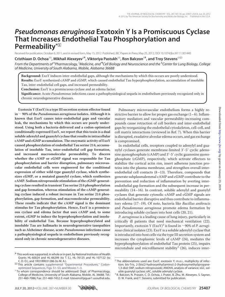
FIGURE 1. P. aeruginosa ExoY is sufficient to increase the intracellular accumulation of cAMP and cGMP. A, schematic representation of ExoYk81M.B, schematic representation of ExoY. C, mean � S.E. of global cAMP levels in pulmonary microvascular endothelial cells 6 h post-P. aeruginosa ExoY� infection.D, mean � S.E. of global cGMP levels in pulmonary microvascular endothelial cells 6 h post-P. aeruginosa ExoY� infection. E and F, general maps of vectors usedto deliver codon-optimized ExoY and ExoYK81M. LTR, retroviral/lentiviral long-terminal repeat; HIV RRE, human immunodeficiency virus response element; Ptet,doxycycline-regulated promoter; FKBP, protein destruction domain (FKBP); PSV40, SV40 promoter; PAC, puromycin resistance gene. G, conditional expressionof codon-optimized ExoYK81M. H, conditional expression of codon-optimized ExoY. I, intracellular cAMP levels of pulmonary microvascular endothelial cellsconditioned to express codon-optimized ExoY-myc-FKBP or ExoYK81M-myc-FKBP 2 h after induction with 2 �M Shield1 and 2 �g/ml doxycycline. J, intracellularcGMP levels of pulmonary microvascular endothelial cells conditioned to express codon-optimized ExoY-myc-FKBP or ExoYK81M-myc-FKBP 2 h after inductionwith 2 �M Shield1 and 2 �g/ml doxycycline. *, p 0,001 versus P. aeruginosa ExoYK81M; §, p 0.05 versus control. Data represent five independent experiments.Statistical significance was determined by one-way ANOVA followed by Bonferroni’s multiple comparisons test.
Exotoxin Y and Endothelial Tau Hyperphosphorylation
25410 JOURNAL OF BIOLOGICAL CHEMISTRY VOLUME 287 • NUMBER 30 • JULY 20, 2012
by guest on January 23, 2019http://w
ww
.jbc.org/D
ownloaded from

We found that both cAMP and cGMP increased pulmonarymicrovascular endothelial cells infected with the P. aerugi-nosa ExoY� but not P. aeruginosa ExoYK81M strain (Fig. 1, Cand D). Of note, ExoY� generated roughly 10-fold morecGMP than cAMP.To test whether ExoYwas sufficient to increase the cytoplas-
mic levels of both cAMP and cGMP, we engineered pulmonarymicrovascular endothelial cells to conditionally express ExoY.First, we tried to express the full-length exoY gene using anadenoviral delivery system in pulmonary microvascular endo-thelial cells, but we could not detect the expression of ExoYprotein. Even though degeneracy is a fundamental attribute ofthe genetic code, bacterial genes use codons that are rarelyemployed by mammalian cells (37). Suspecting codon mis-matching, we next engineered pulmonary microvascular endo-thelial cells to conditionally express a codon-optimizedP. aeruginosa ExoY with a hemagglutinin (HA) tag at the Cterminus (see supplemental material for ExoY codon-opti-mized sequence). Although the expression of ExoY-HA wassuccessful (supplemental Fig. S1), we were not able to detectenzymatic activity. We then tested whether HA orMyc (ExoY-myc) tags in the C terminus of bacteria-delivered ExoY reducedExoY enzymatic activity when compared with wild-type ExoYand found that ExoY-HA had the least activity, with ExoY-mychaving the most (although intermediate when compared withthe wild type (data not shown)). Thereafter, we conditionallyexpressed a codon-optimized P. aeruginosa ExoY with a Myctag but detected enough background expression of the trans-gene to intoxicate the cells, suggesting that enzymatic activitywas present. This result revealed the need to systematically reg-ulate gene and protein expression.
To control gene and protein expression, we developed a two-tier control protein expression system to conditionally expresscodon-optimized ExoY by combining doxycyline-induciblegene expression (35) with protein stability regulation by smallmolecules (36–38). Two constructs were developed: ExoY-myc-FKBP (Fig. 1E) and ExoYK81M-myc-FKBP (Fig. 1F). Fol-lowing induction with both doxycycline and Shield1, wedetected both ExoY-myc-FKBP and ExoYK81M-myc-FKBP atthe predicted molecular mass of �55 kDa (Fig. 1, G and H).ExoY was catalytically active, as pulmonary microvascularendothelial cells expressing codon-optimized ExoY-myc-FKBP, but not cells expressing ExoYK81M-myc-FKBP, accumu-lated both cAMP and cGMP (Fig. 1, I and J); once again, ExoYgenerated more cGMP than cAMP. Hence, ExoY is enzymati-cally active in the absence of bacteria and in the absence ofintoxication through the type III secretion system; expressionwithin the host cell is sufficient for ExoY to bind its necessarycofactor and synthesize cAMP and cGMP.Pseudomonas aeruginosa ExoY Was Sufficient to Induce
Inter-endothelial Cell Gaps—Consistent with a previous reportfrom our group (27), pulmonary microvascular endothelialcells monolayers developed gaps after infection with aP. aeruginosa strain that expressed an active ExoY toxin(P. aeruginosa ExoY�), but not when they were infected witha P. aeruginosa strain that expressed an inactive toxin(P. aeruginosa ExoYK81M) (Fig. 2, A and B). To determinewhether ExoY, in the absence of bacteria, was sufficient toinduce inter-endothelial gap formation in pulmonary micro-vascular endothelial cells, we tested the response in cellsengineered to express codon-optimized ExoY-myc-FKBPand codon-optimized ExoYK81M-myc-FKB. Induction of
FIGURE 2. P. aeruginosa ExoY is sufficient to induce inter-endothelial gap formation. A, pulmonary microvascular endothelial cells 6 h after P. aeruginosaExoYK81M infection at m.o.i. 20:1 in serum-free medium. Magnification, �20. No endothelial gap formation was observed. B, pulmonary microvascular endo-thelial cells 6 h after P. aeruginosa ExoY� infection at m.o.i. 20:1 in serum-free medium. Magnification, �20. Note the disruption of the monolayers. Photographare representative of 10 or more experiments. C, pulmonary microvascular endothelial cells that conditionally expressed codon-optimized ExoYK81M-myc-FKBP2 h after induction with 2 �M Shield1 and 2 �g/ml doxycycline. No inter-endothelial cell gaps were observed. These photographs are representative of fiveexperiments. D, pulmonary microvascular endothelial cells that conditionally expressed codon-optimized ExoY-myc-FKBP 2 h after induction with 2 �M Shield1and 2 �g/ml doxycycline. White arrows point to inter-endothelial cell gaps. These photographs are representative of four experiments.
Exotoxin Y and Endothelial Tau Hyperphosphorylation
JULY 20, 2012 • VOLUME 287 • NUMBER 30 JOURNAL OF BIOLOGICAL CHEMISTRY 25411
by guest on January 23, 2019http://w
ww
.jbc.org/D
ownloaded from
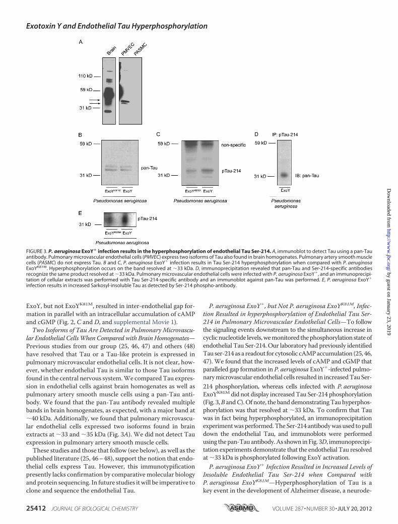
ExoY, but not ExoYK81M, resulted in inter-endothelial gap for-mation in parallel with an intracellular accumulation of cAMPand cGMP (Fig. 2, C and D, and supplemental Movie 1).Two Isoforms of Tau Are Detected in Pulmonary Microvascu-
lar Endothelial CellsWhenCompared with BrainHomogenates—Previous studies from our group (25, 46, 47) and others (48)have resolved that Tau or a Tau-like protein is expressed inpulmonary microvascular endothelial cells. It is not clear, how-ever, whether endothelial Tau is similar to those Tau isoformsfound in the central nervous system.We comparedTau expres-sion in endothelial cells against brain homogenates as well aspulmonary artery smooth muscle cells using a pan-Tau anti-body. We found that the pan-Tau antibody revealed multiplebands in brain homogenates, as expected, with a major band at�40 kDa. Additionally, we found that pulmonary microvascu-lar endothelial cells expressed two isoforms found in brainextracts at �33 and �35 kDa (Fig. 3A). We did not detect Tauexpression in pulmonary artery smooth muscle cells.These studies and those that follow (see below), as well as the
published literature (25, 46–48), support the notion that endo-thelial cells express Tau. However, this immunotypificationpresently lacks confirmation by comparativemolecular biologyandprotein sequencing. In future studies it will be imperative toclone and sequence the endothelial Tau.
P. aeruginosa ExoY�, but Not P. aeruginosa ExoYK81M, Infec-tion Resulted in hyperphosphorylation of Endothelial Tau Ser-214 in Pulmonary Microvascular Endothelial Cells—To followthe signaling events downstream to the simultaneous increase incyclicnucleotide levels,wemonitored thephosphorylationstateofendothelial Tau Ser-214. Our laboratory had previously identifiedTau ser-214 as a readout for cytosolic cAMPaccumulation (25, 46,47). We found that the increased levels of cAMP and cGMP thatparalleled gap formation in P. aeruginosa ExoY�-infected pulmo-narymicrovascular endothelial cells resulted in increasedTauSer-214 phosphorylation, whereas cells infected with P. aeruginosaExoYK81M did not display increasedTau Ser-214 phosphorylation(Fig. 3,B andC).Of note, the banddemonstratingTauhyperphos-phorylation was that resolved at �33 kDa. To confirm that Tauwas in fact being hyperphosphorylated, an immunoprecipitationexperimentwasperformed.TheSer-214antibodywasused topulldown the endothelial Tau, and immunoblots were performedusing thepan-Tauantibody.As shown inFig. 3D, immunoprecipi-tation experiments demonstrate that the endothelial Tau resolvedat �33 kDa is phosphorylated following ExoY activation.P. aeruginosa ExoY� Infection Resulted in Increased Levels of
Insoluble Endothelial Tau Ser-214 when Compared withP. aeruginosa ExoYK81M—Hyperphosphorylation of Tau is akey event in the development of Alzheimer disease, a neurode-
FIGURE 3. P. aeruginosa ExoY� infection results in the hyperphosphorylation of endothelial Tau Ser-214. A, immunoblot to detect Tau using a pan-Tauantibody. Pulmonary microvascular endothelial cells (PMVEC) express two isoforms of Tau also found in brain homogenates. Pulmonary artery smooth musclecells (PASMC) do not express Tau. B and C, P. aeruginosa ExoY� infection results in Tau Ser-214 hyperphosphorylation when compared with P. aeruginosaExoYK81M. Hyperphosphorylation occurs on the band resolved at �33 kDa. D, immunoprecipitation revealed that pan-Tau and Ser-214-specific antibodiesrecognize the same product resolved at �33 kDa. Pulmonary microvascular endothelial cells were infected with P. aeruginosa ExoY�, and an immunoprecipi-tation of cellular extracts was performed with Tau Ser-214-specific antibody and an immunoblot against pan-Tau was performed. E, P. aeruginosa ExoY�
infection results in increased Sarkosyl-insoluble Tau as detected by Ser-214 phospho-antibody.
Exotoxin Y and Endothelial Tau Hyperphosphorylation
25412 JOURNAL OF BIOLOGICAL CHEMISTRY VOLUME 287 • NUMBER 30 • JULY 20, 2012
by guest on January 23, 2019http://w
ww
.jbc.org/D
ownloaded from
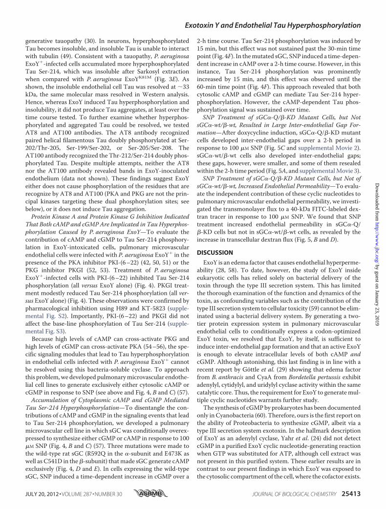
generative tauopathy (30). In neurons, hyperphosphorylatedTau becomes insoluble, and insoluble Tau is unable to interactwith tubulin (49). Consistent with a tauopathy, P. aeruginosaExoY�-infected cells accumulated more hyperphosphorylatedTau Ser-214, which was insoluble after Sarkosyl extractionwhen compared with P. aeruginosa ExoYK81M (Fig. 3E). Asshown, the insoluble endothelial cell Tau was resolved at �33kDa, the same molecular mass resolved in Western analysis.Hence, whereas ExoY induced Tau hyperphosphorylation andinsolubility, it did not produce Tau aggregates, at least over thetime course tested. To further examine whether hyperphos-phorylated and aggregated Tau could be resolved, we testedAT8 and AT100 antibodies. The AT8 antibody recognizedpaired helical filamentous Tau doubly phosphorylated at Ser-202/Thr-205, Ser-199/Ser-202, or Ser-205/Ser-208. TheAT100 antibody recognized the Thr-212/Ser-214 doubly phos-phorylated Tau. Despite multiple attempts, neither the AT8nor the AT100 antibody revealed bands in ExoY-inoculatedendothelium (data not shown). These findings suggest ExoYeither does not cause phosphorylation of the residues that arerecognize by AT8 and AT100 (PKA and PKG are not the prin-cipal kinases targeting these dual phosphorylation sites; seebelow), or it does not induce Tau aggregation.Protein Kinase A and Protein Kinase G Inhibition Indicated
That Both cAMP and cGMPAre Implicated in Tau Hyperphos-phorylation Caused by P. aeruginosa ExoY—To evaluate thecontribution of cAMP and cGMP to Tau Ser-214 phosphory-lation in ExoY-intoxicated cells, pulmonary microvascularendothelial cells were infected with P. aeruginosa ExoY� in thepresence of the PKA inhibitor PKI-(6–22) (42, 50, 51) or thePKG inhibitor PKGI (52, 53). Treatment of P. aeruginosaExoY�-infected cells with PKI-(6–22) inhibited Tau Ser-214phosphorylation (all versus ExoY alone) (Fig. 4). PKGI treat-ment modestly reduced Tau Ser-214 phosphorylation (all ver-sus ExoY alone) (Fig. 4). These observations were confirmed bypharmacological inhibition using H89 and KT-5823 (supple-mental Fig. S2). Importantly, PKI-(6–22) and PKGI did notaffect the base-line phosphorylation of Tau Ser-214 (supple-mental Fig. S3).Because high levels of cAMP can cross-activate PKG and
high levels of cGMP can cross-activate PKA (54–56), the spe-cific signaling modules that lead to Tau hyperphosphorylationin endothelial cells infected with P. aeruginosa ExoY� cannotbe resolved using this bacteria-soluble cyclase. To approachthis problem,we developed pulmonarymicrovascular endothe-lial cell lines to generate exclusively either cytosolic cAMP orcGMP in response to SNP (see above and Fig. 4, B and C) (57).Accumulation of Cytoplasmic cAMP and cGMP Mediated
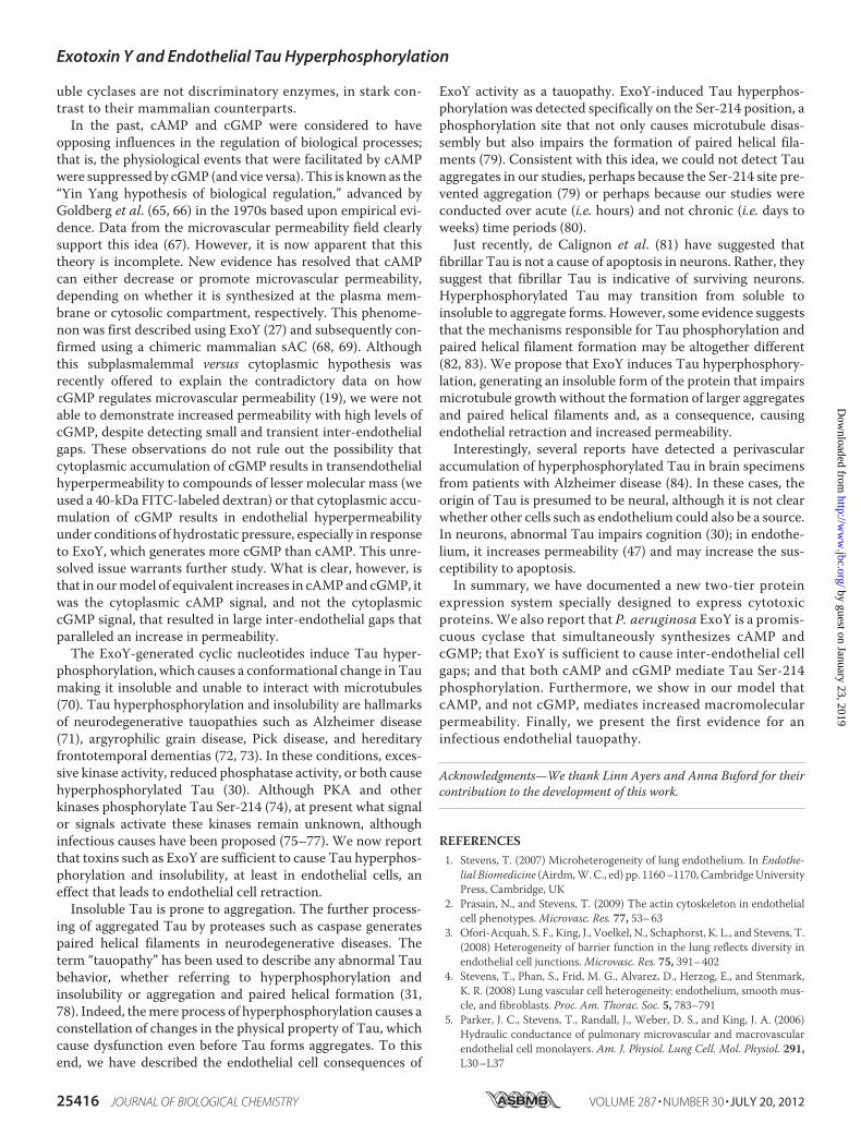
Tau Ser-214 Hyperphosphorylation—To disentangle the con-tributions of cAMP and cGMP in the signaling events that leadto Tau Ser-214 phosphorylation, we developed a pulmonarymicrovascular cell line in which sGC was conditionally overex-pressed to synthesize either cGMP or cAMP in response to 100�M SNP (Fig. 4, B and C) (57). Three mutations were made tothe wild-type rat sGC (R592Q in the �-subunit and E473K aswell as C541D in the�-subunit) thatmade sGC generate cAMPexclusively (Fig. 4, D and E). In cells expressing the wild-typesGC, SNP induced a time-dependent increase in cGMP over a
2-h time course. Tau Ser-214 phosphorylation was induced by15 min, but this effect was not sustained past the 30-min timepoint (Fig. 4F). In themutated sGC, SNP induced a time-depen-dent increase in cAMP over a 2-h time course. However, in thisinstance, Tau Ser-214 phosphorylation was prominentlyincreased by 15 min, and this effect was observed until the60-min time point (Fig. 4F). This approach revealed that bothcytosolic cAMP and cGMP can mediate Tau Ser-214 hyper-phosphorylation. However, the cAMP-dependent Tau phos-phorylation signal was sustained over time.SNP Treatment of sGC�-Q/�-KD Mutant Cells, but Not
sGC�-wt/�-wt, Resulted in Large Inter-endothelial Gap For-mation—After doxycycline induction, sGC�-Q/�-KD mutantcells developed inter-endothelial gaps over a 2-h period inresponse to 100 �M SNP (Fig. 5C and supplemental Movie 2).sGC�-wt/�-wt cells also developed inter-endothelial gaps;these gaps, however, were smaller, and some of them resealedwithin the 2-h time period (Fig. 5A, and supplementalMovie 3).SNP Treatment of sGC�-Q/�-KD Mutant Cells, but Not of
sGC�-wt/�-wt, Increased Endothelial Permeability—To evalu-ate the independent contribution of these cyclic nucleotides topulmonary microvascular endothelial permeability, we investi-gated the transmonolayer flux to a 40-kDa FITC-labeled dex-tran tracer in response to 100 �M SNP. We found that SNPtreatment increased endothelial permeability in sGC�-Q/�-KD cells but not in sGC�-wt/�-wt cells, as revealed by theincrease in transcellular dextran flux (Fig. 5, B and D).
DISCUSSION
ExoY is an edema factor that causes endothelial hyperperme-ability (28, 58). To date, however, the study of ExoY insideeukaryotic cells has relied solely on bacterial delivery of thetoxin through the type III secretion system. This has limitedthe thorough examination of the function and dynamics of thetoxin, as confounding variables such as the contribution of thetype III secretion system to cellular toxicity (59) cannot be elim-inated using a bacterial delivery system. By generating a two-tier protein expression system in pulmonary microvascularendothelial cells to conditionally express a codon-optimizedExoY toxin, we resolved that ExoY, by itself, is sufficient toinduce inter-endothelial gap formation and that an active ExoYis enough to elevate intracellular levels of both cAMP andcGMP. Although astonishing, this last finding is in line with arecent report by Göttle et al. (29) showing that edema factorfrom B. anthracis and CyaA from Bordetella pertussis exhibitadenylyl, cytidylyl, and uridylyl cyclase activity within the samecatalytic core. Thus, the requirement for ExoY to generatemul-tiple cyclic nucleotides warrants further study.The synthesis of cGMPby prokaryotes has been documented
only in Cyanobacteria (60). Therefore, ours is the first report onthe ability of Proteobacteria to synthesize cGMP, albeit via atype III secretion system exotoxin. In the hallmark descriptionof ExoY as an adenylyl cyclase, Yahr et al. (24) did not detectcGMP in a purified ExoY cyclic nucleotide-generating reactionwhen GTP was substituted for ATP, although cell extract wasnot present in this purified system. These earlier results are incontrast to our present findings in which ExoY was exposed tothe cytosolic compartment of the cell, where the cofactor exists.
Exotoxin Y and Endothelial Tau Hyperphosphorylation
JULY 20, 2012 • VOLUME 287 • NUMBER 30 JOURNAL OF BIOLOGICAL CHEMISTRY 25413
by guest on January 23, 2019http://w
ww
.jbc.org/D
ownloaded from

Indeed, ExoY becomes active only when it binds to an unknownmammalian cofactor (24); it remains to be seen whether thiscofactor can modify the substrate specificity of ExoY. This ideais consistent with the recent findings of Göttle et al. (29), whoreport that cells display different substrate specificities foredema factor-induced cyclic nucleotides. Another possibility isthat the ExoY catalytic core is nonspecific for nucleotides andbinds to those that are readily available. In the case of endothe-lial cells, ATP and GTP are both key physiological substrates.
Both transmembrane and soluble mammalian cyclases dis-play substrate specificity, yet two to three residue substitutionsin their catalytic cores reverse this specificity (61). The abilityof ExoY, edema factor, and CyaA to synthesize multiplecyclic nucleotides suggests evolutionary specification inmammalian enzymes. Several lines of evidence suggest ver-tical soluble cyclase gene transfer from bacteria to mammals(62), and reports indicate that both sAC and sGC could haveevolved separately (63). However, Roelofs et al. (64) identi-
FIGURE 4. cAMP and cGMP independently mediate Tau Ser-214 hyperphosphorylation. A, the specific PKA inhibitor PKI-(6 –22) (10 �M) prevented theincrease in Tau Ser-214 phosphorylation promoted by P. aeruginosa ExoY�. PKG inhibition with 10 �M PKGI modestly reduced Tau Ser-214 phosphorylation.B and C, general maps of vectors used to deliver the �- and �-subunits of rat sGC. These vectors were delivered to endothelial cells already expressing theadvanced Tet-on system as described in Ref. 35. Therefore, these were triple mutant pulmonary microvascular endothelial cells. LTR, retroviral/lentiviral longterminal repeat; HIV RRE, human immunodeficiency virus response element; Ptet, doxycycline-regulated promoter; �-wt, wild-type �-1 subunit of rat sGC;PSV40, SV40 promoter; PAC, puromycin resistance gene; MoMuLV, Moloney murine leukemia virus; �, packaging psi; CAG �-wt, wild-type �-1 subunit of rat sGC;Hyg, hygromycin resistance cassette; �-Q, �-1 R592Q subunit of sGC; �-KD, E473K/C541D subunit of sGC. Black dashed lines show the sequence translated byribosome. D, cGMP levels in both sGC �-wt/�-wt and sGC �-Q/�-KD after 24 h of doxycycline induction and 2 h of treatment with 100 �M SNP. E, cAMP levelsin both sGC �-wt/�-wt and sGC �-Q/�-KD after 24 h of doxycycline induction and 2 h of treatment with 100 �M SNP. *, p 0.001 versus control. Data representfour independent experiments. Statistical significance was determined by one-way ANOVA followed by Bonferroni’s multiple comparisons test. F, immunoblotblot analysis of pulmonary microvascular endothelial cells conditionally overexpressing rat sGC �-Q/�-KD, induced with doxycycline, and treated with 100 �M
SNP. G, densitometries revealed average response from three independent experiments. H, immunoblot blot analysis of pulmonary microvascular endothelialcells (PMVEC) conditionally overexpressing rat sGC �-wt/�-wt, induced with doxycycline, and treated with 100 �M SNP. I, densitometry confirms a sustainedincrease in Tau Ser-214 phosphorylation. Images are representative of three independent experiments. A.U., arbitrary units.
Exotoxin Y and Endothelial Tau Hyperphosphorylation
25414 JOURNAL OF BIOLOGICAL CHEMISTRY VOLUME 287 • NUMBER 30 • JULY 20, 2012
by guest on January 23, 2019http://w
ww
.jbc.org/D
ownloaded from

fied an ortholog of sAC in Dictyostelium that is an sGC,suggesting, at least in this case, that sGC may have evolvedfrom sAC. In consequence, one could argue that the nucle-otide specificity of soluble cyclases is a trait that could have
evolved from bacteria (via eukaryotic microorganisms) tomammals. Although the evolutionary pressures that deter-mined the specificity of mammalian soluble cyclases areunclear, our studies indicate that prokaryotic exotoxin-sol-
FIGURE 4 —continued
FIGURE 5. Intracellular accumulation of cAMP, but not cGMP, results in large inter-endothelial gap formation and increased endothelial permeability.A, sGC �-wt/�-wt mutant cells induced with doxycycline and treated with 100 �M developed smaller inter-endothelial gaps (see also supplemental Movie 3).B, doxycycline induction and treatment with 100 �M sGC �-wt/�-wt mutant cells did not increase endothelial cell permeability as measured by macromolecularflux to a 40-kDa FITC-labeled dextran tracer. n � 7 independent experiments. C, sGC �-Q/�-KD mutant cells induced with doxycycline and treated with 100 �M
SNP developed large inter-endothelial gaps over a 2-h time period (see also supplemental Movie 2). D, doxycycline induction and treatment with 100 �M SNPin sGC �-Q/�-KD mutant cells increased endothelial cell permeability as measured by macromolecular flux to a 40-kDa FITC-labeled dextran tracer. n � 8independent experiments. One-way ANOVA was used to assess significance for each condition over the 120-min time course. Two-way ANOVA was used tomake comparisons between treatments. *, p 0.05 versus vehicle control; [caret], p 0.05 versus base line.
Exotoxin Y and Endothelial Tau Hyperphosphorylation
JULY 20, 2012 • VOLUME 287 • NUMBER 30 JOURNAL OF BIOLOGICAL CHEMISTRY 25415
by guest on January 23, 2019http://w
ww
.jbc.org/D
ownloaded from
uble cyclases are not discriminatory enzymes, in stark con-trast to their mammalian counterparts.In the past, cAMP and cGMP were considered to have
opposing influences in the regulation of biological processes;that is, the physiological events that were facilitated by cAMPwere suppressed by cGMP (and vice versa). This is known as the“Yin Yang hypothesis of biological regulation,” advanced byGoldberg et al. (65, 66) in the 1970s based upon empirical evi-dence. Data from the microvascular permeability field clearlysupport this idea (67). However, it is now apparent that thistheory is incomplete. New evidence has resolved that cAMPcan either decrease or promote microvascular permeability,depending on whether it is synthesized at the plasma mem-brane or cytosolic compartment, respectively. This phenome-non was first described using ExoY (27) and subsequently con-firmed using a chimeric mammalian sAC (68, 69). Althoughthis subplasmalemmal versus cytoplasmic hypothesis wasrecently offered to explain the contradictory data on howcGMP regulates microvascular permeability (19), we were notable to demonstrate increased permeability with high levels ofcGMP, despite detecting small and transient inter-endothelialgaps. These observations do not rule out the possibility thatcytoplasmic accumulation of cGMP results in transendothelialhyperpermeability to compounds of lesser molecular mass (weused a 40-kDa FITC-labeled dextran) or that cytoplasmic accu-mulation of cGMP results in endothelial hyperpermeabilityunder conditions of hydrostatic pressure, especially in responseto ExoY, which generates more cGMP than cAMP. This unre-solved issue warrants further study. What is clear, however, isthat in ourmodel of equivalent increases in cAMPand cGMP, itwas the cytoplasmic cAMP signal, and not the cytoplasmiccGMP signal, that resulted in large inter-endothelial gaps thatparalleled an increase in permeability.The ExoY-generated cyclic nucleotides induce Tau hyper-
phosphorylation, which causes a conformational change in Taumaking it insoluble and unable to interact with microtubules(70). Tau hyperphosphorylation and insolubility are hallmarksof neurodegenerative tauopathies such as Alzheimer disease(71), argyrophilic grain disease, Pick disease, and hereditaryfrontotemporal dementias (72, 73). In these conditions, exces-sive kinase activity, reduced phosphatase activity, or both causehyperphosphorylated Tau (30). Although PKA and otherkinases phosphorylate Tau Ser-214 (74), at present what signalor signals activate these kinases remain unknown, althoughinfectious causes have been proposed (75–77). We now reportthat toxins such as ExoY are sufficient to cause Tau hyperphos-phorylation and insolubility, at least in endothelial cells, aneffect that leads to endothelial cell retraction.Insoluble Tau is prone to aggregation. The further process-
ing of aggregated Tau by proteases such as caspase generatespaired helical filaments in neurodegenerative diseases. Theterm “tauopathy” has been used to describe any abnormal Taubehavior, whether referring to hyperphosphorylation andinsolubility or aggregation and paired helical formation (31,78). Indeed, themere process of hyperphosphorylation causes aconstellation of changes in the physical property of Tau, whichcause dysfunction even before Tau forms aggregates. To thisend, we have described the endothelial cell consequences of
ExoY activity as a tauopathy. ExoY-induced Tau hyperphos-phorylation was detected specifically on the Ser-214 position, aphosphorylation site that not only causes microtubule disas-sembly but also impairs the formation of paired helical fila-ments (79). Consistent with this idea, we could not detect Tauaggregates in our studies, perhaps because the Ser-214 site pre-vented aggregation (79) or perhaps because our studies wereconducted over acute (i.e. hours) and not chronic (i.e. days toweeks) time periods (80).Just recently, de Calignon et al. (81) have suggested that
fibrillar Tau is not a cause of apoptosis in neurons. Rather, theysuggest that fibrillar Tau is indicative of surviving neurons.Hyperphosphorylated Tau may transition from soluble toinsoluble to aggregate forms. However, some evidence suggeststhat the mechanisms responsible for Tau phosphorylation andpaired helical filament formation may be altogether different(82, 83). We propose that ExoY induces Tau hyperphosphory-lation, generating an insoluble form of the protein that impairsmicrotubule growth without the formation of larger aggregatesand paired helical filaments and, as a consequence, causingendothelial retraction and increased permeability.Interestingly, several reports have detected a perivascular
accumulation of hyperphosphorylated Tau in brain specimensfrom patients with Alzheimer disease (84). In these cases, theorigin of Tau is presumed to be neural, although it is not clearwhether other cells such as endothelium could also be a source.In neurons, abnormal Tau impairs cognition (30); in endothe-lium, it increases permeability (47) and may increase the sus-ceptibility to apoptosis.In summary, we have documented a new two-tier protein
expression system specially designed to express cytotoxicproteins.We also report that P. aeruginosa ExoY is a promis-cuous cyclase that simultaneously synthesizes cAMP andcGMP; that ExoY is sufficient to cause inter-endothelial cellgaps; and that both cAMP and cGMP mediate Tau Ser-214phosphorylation. Furthermore, we show in our model thatcAMP, and not cGMP, mediates increased macromolecularpermeability. Finally, we present the first evidence for aninfectious endothelial tauopathy.
Acknowledgments—We thank Linn Ayers and Anna Buford for theircontribution to the development of this work.
REFERENCES1. Stevens, T. (2007) Microheterogeneity of lung endothelium. In Endothe-
lial Biomedicine (Airdm,W.C., ed) pp. 1160–1170, CambridgeUniversityPress, Cambridge, UK
2. Prasain, N., and Stevens, T. (2009) The actin cytoskeleton in endothelialcell phenotypes.Microvasc. Res. 77, 53–63
3. Ofori-Acquah, S. F., King, J., Voelkel, N., Schaphorst, K. L., and Stevens, T.(2008) Heterogeneity of barrier function in the lung reflects diversity inendothelial cell junctions.Microvasc. Res. 75, 391–402
4. Stevens, T., Phan, S., Frid, M. G., Alvarez, D., Herzog, E., and Stenmark,K. R. (2008) Lung vascular cell heterogeneity: endothelium, smooth mus-cle, and fibroblasts. Proc. Am. Thorac. Soc. 5, 783–791
5. Parker, J. C., Stevens, T., Randall, J., Weber, D. S., and King, J. A. (2006)Hydraulic conductance of pulmonary microvascular and macrovascularendothelial cell monolayers. Am. J. Physiol. Lung Cell. Mol. Physiol. 291,L30–L37
Exotoxin Y and Endothelial Tau Hyperphosphorylation
25416 JOURNAL OF BIOLOGICAL CHEMISTRY VOLUME 287 • NUMBER 30 • JULY 20, 2012
by guest on January 23, 2019http://w
ww
.jbc.org/D
ownloaded from

6. Jacobson, J. R., and Garcia, J. G. N. (2010) Chapter 6 appendix: endothelialcell function. In Murray and Nadel’s Textbook of Respiratory Medicine(Mason, R. J., Broaddus, B. C., Martin, T. R., King, T. E., Jr., Schraufnagel,E., Murray, J. F., and Nadel, J. A., eds) 5th Ed., p. 1–6, W. B. Saunders Co.,Philadelphia, PA
7. Mehta, D., and Malik, A. B. (2006) Signaling mechanisms regulating en-dothelial permeability. Physiol. Rev. 86, 279–367
8. Adamson, R. H., Ly, J. C., Sarai, R. K., Lenz, J. F., Altangerel, A., Drenck-hahn, D., and Curry, F. E. (2008) Epac/Rap1 pathway regulates microvas-cular hyperpermeability induced by PAF in rat mesentery. Am. J. Physiol.Heart Circ. Physiol. 294, H1188–H1196
9. Fukuhara, S., Sakurai, A., Sano, H., Yamagishi, A., Somekawa, S.,Takakura, N., Saito, Y., Kangawa, K., and Mochizuki, N. (2005) CyclicAMP potentiates vascular endothelial cadherin-mediated cell-cell contactto enhance endothelial barrier function through an Epac-Rap1 signalingpathway.Mol. Cell. Biol. 25, 136–146
10. Stelzner, T. J.,Weil, J. V., andO’Brien, R. F. (1989) Role of cyclic adenosinemonophosphate in the induction of endothelial barrier properties. J. Cell.Physiol. 139, 157–166
11. Adamson, R. H., Liu, B., Fry, G. N., Rubin, L. L., and Curry, F. E. (1998)Microvascular permeability and number of tight junctions are modulatedby cAMP. Am. J. Physiol. 274, H1885–H1894
12. Rentsendorj, O., Mirzapoiazova, T., Adyshev, D., Servinsky, L. E., Renné,T., Verin, A. D., and Pearse, D. B. (2008) Role of vasodilator-stimulatedphosphoprotein in cGMP-mediated protection of human pulmonary ar-tery endothelial barrier function. Am. J. Physiol. Lung Cell. Mol. Physiol.294, L686–L697
13. Moldobaeva, A., Welsh-Servinsky, L. E., Shimoda, L. A., Stephens, R. S.,Verin, A. D., Tuder, R. M., and Pearse, D. B. (2006) Role of protein kinaseG in barrier-protective effects of cGMP in human pulmonary artery en-dothelial cells. Am. J. Physiol. Lung Cell. Mol. Physiol. 290, L919–L930
14. Berthiaume, Y., Staub, N. C., and Matthay, M. A. (1987) �-Adrenergicagonists increase lung liquid clearance in anesthetized sheep. J. Clin. In-vest. 79, 335–343
15. Ahluwalia, A., MacAllister, R. J., and Hobbs, A. J. (2004) Vascular actionsof natriuretic peptides. Cyclic GMP-dependent and -independent mech-anisms. Basic Res. Cardiol. 99, 83–89
16. Kemp, S. F., Lockey, R. F., Simons, F. E., and World Allergy Organizationad hoc Committee on Epinephrine in Anaphylaxis (2008) Epinephrine:the drug of choice for anaphylaxis. A statement of the World AllergyOrganization. Allergy 63, 1061–1070
17. Sayner, S. L. (2011) Emerging themes of cAMP regulation of the pulmo-nary endothelial barrier. Am. J. Physiol. Lung Cell. Mol. Physiol. 300,L667–L678
18. Shen, Q., Rigor, R. R., Pivetti, C. D., Wu, M. H., and Yuan, S. Y. (2010)Myosin light chain kinase in microvascular endothelial barrier function.Cardiovasc. Res. 87, 272–280
19. Kuebler, W. M. (2011) The Janus-faced regulation of endothelial perme-ability by cyclic GMP. Am. J. Physiol. Lung Cell. Mol. Physiol. 301,L157–L160
20. Lory, S. (2004) Themulti-talented bacterial adenylate cyclases. Int. J. Med.Microbiol. 293, 479–482
21. Ahuja, N., Kumar, P., and Bhatnagar, R. (2004) The adenylate cyclasetoxins. Crit. Rev. Microbiol. 30, 187–196
22. Weber, D. J., Rutala, W. A., Sickbert-Bennett, E. E., Samsa, G. P., Brown,V., and Niederman, M. S. (2007) Microbiology of ventilator-associatedpneumonia compared with that of hospital-acquired pneumonia. Infect.Control Hosp. Epidemiol. 28, 825–831
23. Feltman, H., Schulert, G., Khan, S., Jain,M., Peterson, L., andHauser, A. R.(2001) Prevalence of type III secretion genes in clinical and environmentalisolates of Pseudomonas aeruginosa.Microbiology 147, 2659–2669
24. Yahr, T. L., Vallis, A. J., Hancock, M. K., Barbieri, J. T., and Frank, D. W.(1998) ExoY, an adenylate cyclase secreted by the Pseudomonas aerugi-nosa type III system. Proc. Natl. Acad. Sci. U.S.A. 95, 13899–13904
25. Sayner, S. L., Balczon, R., Frank, D. W., Cooper, D. M., and Stevens, T.(2011) Filamin A is a phosphorylation target of membrane but not cyto-solic adenylyl cyclase activity. Am. J. Physiol. Lung Cell. Mol. Physiol. 301,L117–L124
26. Cowell, B. A., Evans, D. J., and Fleiszig, S. M. (2005) Actin cytoskeletondisruption by ExoY and its effects on Pseudomonas aeruginosa invasion.FEMS Microbiol. Lett. 250, 71–76
27. Sayner, S. L., Frank, D. W., King, J., Chen, H., VandeWaa, J., and Stevens,T. (2004) Paradoxical cAMP-induced lung endothelial hyperpermeabilityrevealed by Pseudomonas aeruginosa ExoY. Circ. Res. 95, 196–203
28. Lowe, K., Alvarez, D. F., King, J. A., and Stevens, T. (2010) Perivascularfluid cuffs decrease lung compliance by increasing tissue resistance. Crit.Care Med. 38, 1458–1466
29. Göttle, M., Dove, S., Kees, F., Schlossmann, J., Geduhn, J., König, B., Shen,Y., Tang, W. J., Kaever, V., and Seifert, R. (2010) Cytidylyl and uridylylcyclase activity of Bacillus anthracis edema factor and Bordetella pertussisCyaA. Biochemistry 49, 5494–5503
30. Querfurth, H. W., and LaFerla, F. M. (2010) Alzheimer’s disease. N. Engl.J. Med. 362, 329–344
31. Bouchard, M., and Suchowersky, O. (2011) Tauopathies: one disease ormany? Can. J. Neurol. Sci. 38, 547–556
32. King, J., Hamil, T., Creighton, J., Wu, S., Bhat, P., McDonald, F., and Ste-vens, T. (2004) Structural and functional characteristics of lung macro-and microvascular endothelial cell phenotypes. Microvasc. Res. 67,139–151
33. Ochoa, C. D., Stevens, T., and Balczon, R. (2011) Cold exposure revealstwo populations of microtubules in pulmonary endothelia. Am. J. Physiol.Lung Cell. Mol. Physiol. 300, L132–L138
34. Gairhe, S., Bauer, N. N., Gebb, S. A., and McMurtry, I. F. (2011) Myoen-dothelial gap junctional signaling induces differentiation of pulmonaryarterial smooth muscle cells. Am. J. Physiol. Lung Cell. Mol. Physiol. 301,L527–L535
35. Alexeyev, M. F., Fayzulin, R., Shokolenko, I. N., and Pastukh, V. (2010) Aretro-lentiviral system for doxycycline-inducible gene expression andgene knockdown in cells with limited proliferative capacity.Mol. Biol. Rep.37, 1987–1991
36. Banaszynski, L. A., Chen, L. C., Maynard-Smith, L. A., Ooi, A. G., andWandless, T. J. (2006) A rapid, reversible, and tunable method to regulateprotein function in living cells using synthetic small molecules. Cell 126,995–1004
37. Lorimer, D., Raymond, A., Walchli, J., Mixon, M., Barrow, A., Wallace, E.,Grice, R., Burgin, A., and Stewart, L. (2009) Gene composer: databasesoftware for protein construct design, codon engineering, and gene syn-thesis. BMC Biotechnol. 9, 36
38. Grimley, J. S., Chen, D. A., Banaszynski, L. A., and Wandless, T. J. (2008)Synthesis and analysis of stabilizing ligands for FKBP-derived destabilizingdomains. Bioorg. Med. Chem. Lett. 18, 759–761
39. Horton, R. M., Hunt, H. D., Ho, S. N., Pullen, J. K., and Pease, L. R. (1989)Engineering hybrid genes without the use of restriction enzymes: genesplicing by overlap extension. Gene 77, 61–68
40. Parra-Bonilla, G., Alvarez, D. F., Al-Mehdi, A. B., Alexeyev, M., and Ste-vens, T. (2010) Critical role for lactate dehydrogenase A in aerobic glycol-ysis that sustains pulmonary microvascular endothelial cell proliferation.Am. J. Physiol. Lung Cell. Mol. Physiol. 299, L513–L522
41. Creighton, J., Jian, M., Sayner, S., Alexeyev, M., and Insel, P. A. (2011)Adenosinemonophosphate-activated kinase alpha1 promotes endothelialbarrier repair. FASEB J. 25, 3356–3365
42. Murray, A. J. (2008) Pharmacological PKA inhibition: all may not be whatit seems. Sci. Signal. 1, re4
43. Wei, J. Y., Jin, X., Cohen, E. D., Daw, N. W., and Barnstable, C. J. (2002)cGMP-induced presynaptic depression and postsynaptic facilitation atglutamatergic synapses in visual cortex. Brain Res. 927, 42–54
44. Lampugnani, M. G., and Dejana, E. (2004) Endothelial cell permeabilityassays is culture. InMethods in Endothelial Cell Biology (Augustin, H. G.,ed) pp. 103–112, Springer, New York
45. Berger, Z., Roder, H., Hanna, A., Carlson, A., Rangachari, V., Yue, M.,Wszolek, Z., Ashe, K., Knight, J., Dickson, D., Andorfer, C., Rosenberry,T. L., Lewis, J., Hutton, M., and Janus, C. (2007) Accumulation of patho-logical Tau species and memory loss in a conditional model of tauopathy.J. Neurosci. 27, 3650–3662
46. Creighton, J., Zhu, B., Alexeyev, M., and Stevens, T. (2008) Spectrin-an-chored phosphodiesterase 4D4 restricts cAMP from disrupting microtu-
Exotoxin Y and Endothelial Tau Hyperphosphorylation
JULY 20, 2012 • VOLUME 287 • NUMBER 30 JOURNAL OF BIOLOGICAL CHEMISTRY 25417
by guest on January 23, 2019http://w
ww
.jbc.org/D
ownloaded from

bules and inducing endothelial cell gap formation. J. Cell Sci. 121,110–119
47. Zhu, B., Zhang, L., Creighton, J., Alexeyev,M., Strada, S. J., and Stevens, T.(2010) Protein kinase A phosphorylation of tau-serine 214 reorganizesmicrotubules and disrupts the endothelial cell barrier.Am. J. Physiol. LungCell. Mol. Physiol. 299, L493–L501
48. Birukova, A. (2004) Novel role of microtubules in thrombin-induced en-dothelial barrier dysfunction. FASEB J. 18, 1879–1890
49. Hirata-Fukae, C., Li, H. F., Ma, L., Hoe, H. S., Rebeck, G. W., Aisen, P. S.,andMatsuoka, Y. (2009) Levels of soluble and insoluble Tau reflect overallstatus of Tau phosphorylation in vivo. Neurosci. Lett. 450, 51–55
50. Hidaka, H., Inagaki, M., Kawamoto, S., and Sasaki, Y. (1984) Isoquinoline-sulfonamides, novel and potent inhibitors of cyclic nucleotide dependentprotein kinase and protein kinase C. Biochemistry 23, 5036–5041
51. Cheli, Y., Luciani, F., Khaled,M., Beuret, L., Bille, K., Gounon, P., Ortonne,J. P., Bertolotto, C., and Ballotti, R. (2009) �MSH and cyclic AMP-elevat-ing agents control melanosome pH through a protein kinase A-indepen-dent mechanism. J. Biol. Chem. 284, 18699–18706
52. Kase, H., Iwahashi, K., Nakanishi, S., Matsuda, Y., Yamada, K., Takahashi,M., Murakata, C., Sato, A., and Kaneko, M. (1987) K-252 compounds,novel and potent inhibitors of protein kinase C and cyclic nucleotide-de-pendent protein kinases. Biochem. Biophys. Res. Commun. 142, 436–440
53. Komalavilas, P., Shah, P. K., Jo, H., and Lincoln, T. M. (1999) Activation ofmitogen-activated protein kinase pathways by cyclic GMP and cyclicGMP-dependent protein kinase in contractile vascular smooth musclecells. J. Biol. Chem. 274, 34301–34309
54. White, R. E., Kryman, J. P., El-Mowafy, A. M., Han, G., and Carrier, G. O.(2000) cAMP-dependent vasodilators cross-activate the cGMP-depen-dent protein kinase to stimulate BK(Ca) channel activity in coronary ar-tery smooth muscle cells. Circ. Res. 86, 897–905
55. Khaled, M., Larribere, L., Bille, K., Aberdam, E., Ortonne, J. P., Ballotti, R.,and Bertolotto, C. (2002) Glycogen synthase kinase 3� is activated bycAMP and plays an active role in the regulation of melanogenesis. J. Biol.Chem. 277, 33690–33697
56. Hucho, T. B., Dina, O. A., and Levine, J. D. (2005) Epac mediates a cAMP-to-PKC signaling in inflammatory pain: an isolectin B4(�) neuron-spe-cific mechanism. J. Neurosci. 25, 6119–6126
57. Sunahara, R. K., Beuve, A., Tesmer, J. J., Sprang, S. R., Garbers, D. L., andGilman, A. G. (1998) Exchange of substrate and inhibitor specificitiesbetween adenylyl and guanylyl cyclases. J. Biol. Chem. 273, 16332–16338
58. Prasain, N. (2009) Soluble adenylyl cyclases disassemblemicrotubules anddisrupt the pulmonary endothelial barrier. Ph.D. dissertation, Universityof South Alabama, Mobile, AL
59. Sawa, T., andWiener-Kronish, J. (2004) A therapeutic strategy against theshared virulence mechanism utilized by both Yersinia pestis and Pseu-domonas aeruginosa. Anesthesiol. Clin. North Am. 22, 591–606
60. Rauch, A., Leipelt, M., Russwurm, M., and Steegborn, C. (2008) Crystalstructure of the guanylyl cyclase Cya2. Proc. Natl. Acad. Sci. U.S.A. 105,15720–15725
61. Tucker, C. L., Hurley, J. H., Miller, T. R., and Hurley, J. B. (1998) Twoamino acid substitutions convert a guanylyl cyclase, RetGC-1, into anadenylyl cyclase. Proc. Natl. Acad. Sci. U.S.A. 95, 5993–5997
62. Roelofs, J., and Van Haastert, P. J. (2002) Deducing the origin of solubleadenylyl cyclase, a gene lost in multiple lineages. Mol. Biol. Evol. 19,2239–2246
63. Baker, D. A., and Kelly, J. M. (2004) Structure, function and evolution ofmicrobial adenylyl and guanylyl cyclases.Mol. Microbiol. 52, 1229–1242
64. Roelofs, J., Meima, M., Schaap, P., and Van Haastert, P. J. (2001) TheDictyostelium homologue ofmammalian soluble adenylyl cyclase encodesa guanylyl cyclase. EMBO J. 20, 4341–4348
65. George, W. J., Polson, J. B., O’Toole, A. G., and Goldberg, N. D. (1970)Elevation of guanosine 3�,5�-cyclic phosphate in rat heart after perfusionwith acetylcholine. Proc. Natl. Acad. Sci. U.S.A. 66, 398–403
66. Goldberg, N. D., Haddox, M. K., Nicol, S. E., Glass, D. B., Sanford, C. H.,Kuehl, F. A., Jr., and Estensen, R. (1975) Biologic regulation through op-posing influences of cyclicGMP and cyclic AMP: the Yin Yang hypothesis.Adv. Cyclic Nucleotide Res. 5, 307–330
67. Michel, C. C., and Curry, F. E. (1999)Microvascular permeability. Physiol.Rev. 79, 703–761
68. Sayner, S. L., Alexeyev,M., Dessauer, C.W., and Stevens, T. (2006) Solubleadenylyl cyclase reveals the significance of cAMP compartmentation onpulmonary microvascular endothelial cell barrier. Circ. Res. 98, 675–681
69. Prasain, N., Alexeyev, M., Balczon, R., and Stevens, T. (2009) Soluble ad-enylyl cyclase-dependent microtubule disassembly reveals a novel mech-anism of endothelial cell retraction.Am. J. Physiol. Lung Cell. Mol. Physiol.297, L73–L83
70. Matenia, D., andMandelkow, E.M. (2009) The Tau ofMARK: a polarizedview of the cytoskeleton. Trends Biochem. Sci. 34, 332–342
71. Grundke-Iqbal, I., Iqbal, K., Tung, Y. C., Quinlan, M., Wisniewski, H. M.,and Binder, L. I. (1986) Abnormal phosphorylation of the microtubule-associated protein Tau (tau) in Alzheimer cytoskeletal pathology. Proc.Natl. Acad. Sci. U.S.A. 83, 4913–4917
72. Frank, S., Clavaguera, F., and Tolnay, M. (2008) Tauopathy models andhuman neuropathology: similarities and differences. Acta Neuropathol.115, 39–53
73. Spillantini, M. G., Goedert, M., Crowther, R. A., Murrell, J. R., Farlow,M. R., and Ghetti, B. (1997) Familial multiple system tauopathy with pre-senile dementia: a disease with abundant neuronal and glial Tau filaments.Proc. Natl. Acad. Sci. U.S.A. 94, 4113–4118
74. Hanger, D. P., Anderton, B. H., and Noble, W. (2009) Tau phosphoryla-tion: the therapeutic challenge for neurodegenerative disease.TrendsMol.Med. 15, 112–119
75. Balin, B. J., Little, C. S., Hammond, C. J., Appelt, D.M.,Whittum-Hudson,J. A., Gérard, H. C., andHudson, A. P. (2008)Chlamydophila pneumoniaeand the etiology of late-onset Alzheimer’s disease. J. Alzheimers Dis. 13,371–380
76. Kinoshita, J. (2004) Pathogens as a cause ofAlzheimer’s disease.Neurobiol.Aging 25, 639–640
77. Itzhaki, R. F., Wozniak, M. A., Appelt, D. M., and Balin, B. J. (2004) Infil-tration of the brain by pathogens causes Alzheimer’s disease. Neurobiol.Aging 25, 619–627
78. Williams, D. R. (2006) Tauopathies: classification and clinical update onneurodegenerative diseases associated with microtubule-associated pro-tein tau. Intern. Med. J. 36, 652–660
79. Schneider, A., Biernat, J., vonBergen,M.,Mandelkow, E., andMandelkow,E. M. (1999) Phosphorylation that detaches Tau protein from microtu-bules (Ser-262, Ser-214) also protects it against aggregation into Al-zheimer paired helical filaments. Biochemistry 38, 3549–3558
80. King, M. E., Gamblin, T. C., Kuret, J., and Binder, L. I. (2000) Differentialassembly of human Tau isoforms in the presence of arachidonic acid.J. Neurochem. 74, 1749–1757
81. de Calignon, A., Fox, L. M., Pitstick, R., Carlson, G. A., Bacskai, B. J.,Spires-Jones, T. L., and Hyman, B. T. (2010) Caspase activation precedesand leads to tangles. Nature 464, 1201–1204
82. Sato, S., Tatebayashi, Y., Akagi, T., Chui, D. H,, Murayama, M., Miyasaka,T., Planel, E., Tanemura, K., Sun, X., Hashikawa, T., Yoshioka, K., Ishiguro,K., and Takashima, A. (2002) Aberrant Tau phosphorylation by glycogensynthase kinase-3� and JNK3 induces oligomeric Tau fibrils in COS-7cells. J. Biol. Chem. 277, 42060–42065
83. Bandyopadhyay, B., Li, G., Yin, H., and Kuret, J. (2007) Tau aggregationand toxicity in a cell culture model of tauopathy. J. Biol. Chem. 282,16454–16464
84. Williams, S., Chalmers, K., Wilcock, G. K., and Love, S. (2005) Relation-ship of neurofibrillary pathology to cerebral amyloid angiopathy in Alz-heimer’s disease. Neuropathol. Appl. Neurobiol. 31, 414–421
Exotoxin Y and Endothelial Tau Hyperphosphorylation
25418 JOURNAL OF BIOLOGICAL CHEMISTRY VOLUME 287 • NUMBER 30 • JULY 20, 2012
by guest on January 23, 2019http://w
ww
.jbc.org/D
ownloaded from
StevensCristhiaan D. Ochoa, Mikhail Alexeyev, Viktoriya Pastukh, Ron Balczon and Troy
Endothelial Tau Phosphorylation and Permeability Exotoxin Y Is a Promiscuous Cyclase That IncreasesPseudomonas aeruginosa
doi: 10.1074/jbc.M111.301440 originally published online May 25, 20122012, 287:25407-25418.J. Biol. Chem.
10.1074/jbc.M111.301440Access the most updated version of this article at doi:
Alerts:
When a correction for this article is posted•
When this article is cited•
to choose from all of JBC's e-mail alertsClick here
Supplemental material:
http://www.jbc.org/content/suppl/2012/05/25/M111.301440.DC1
http://www.jbc.org/content/287/30/25407.full.html#ref-list-1
This article cites 80 references, 21 of which can be accessed free at
by guest on January 23, 2019http://w
ww
.jbc.org/D
ownloaded from