Pseudomonas aeruginosa accentuates epithelial-to ...
11
Pseudomonas aeruginosa accentuates epithelial-to-mesenchymal transition in the airway L.A. Borthwick*, S.S. Sunny*, V. Oliphant*, J. Perry # , M. Brodlie*, G.E. Johnson # , C. Ward*, K. Gould # , P.A. Corris* ,# , A. De Soyza* ,# and A.J. Fisher* ,# ABSTRACT: Epithelial-to-mesenchymal transition (EMT) has been implicated in the dysregulated epithelial wound repair that contributes to obliterative bronchiolitis (OB) after lung transplantation. Acquisition of Pseudomonas aeruginosa in the transplanted airway has been shown to be a risk factor for the development of OB. We investigated the potential of P. aeruginosa to drive EMT in primary bronchial epithelial cells (PBECs) isolated from lung transplant recipients. Changes in the expression of epithelial and mesenchymal markers was assessed in cells challenged with clinical isolates of P. aeruginosa or co-cultured with P. aeruginosa-activated monocytic cells (THP-1) in the presence or absence of transforming growth factor (TGF)-b1. P. aeruginosa did not drive or accentuate TGF-b1-driven EMT directly. Co-culturing P. aeruginosa-activated THP-1 cells with PBECs did not drive EMT. However, co-culturing P. aeruginosa-activated THP-1 cells with PBECs significantly accentuated TGF-b1-driven EMT. P. aeruginosa, via the activation of monocytic cells, can accentuate TGF-b1-driven EMT. These in vitro observations may help explain the in vivo clinical observation of a link between acquisition of P. aeruginosa and an increased risk of developing OB. KEYWORDS: Bronchiolitis obliterans syndrome, lung transplantation, macrophages, obliterative bronchiolitis, transforming growth factor-b1 O ver the last 25 yrs, lung transplantation has evolved from an experimental inter- vention to an accepted therapeutic option for selected patients with end-stage lung diseases. To date, ,28,000 lung transplants have been reported to the international registry and, although early outcomes have significantly improved, long-term survival remains disap- pointing, being limited to a median of ,5 yrs. The major cause of late allograft loss is due to the development of bronchiolitis obliterans syn- drome (BOS) [1]. The pathological lesion of BOS is obliterative bronchiolitis (OB), which is characterised by inflammation and fibrosis in small and medium- sized airways, leading to airflow obstruction [2]. The pathogenesis of the disease remains unclear, but it is hypothesised that repeated epithelial injury from both alloimmune and nonalloim- mune mechanisms, and particularly a failure to re-establish an intact epithelium, contribute to fibrotic repair in the airway and disease progres- sion [2–4]. Recently, several groups have suggested a potential role for epithelial-to-mesenchymal transition (EMT) in the pathogenesis of OB [5–8]. EMT is a process by which an epithelial cell alters both its phenotype and function to that of a mesenchymal cell, including acquiring the ability to produce matrix metalloproteinases and deposit extracellular matrix [9]. Transforming growth factor (TGF)-b1 is regarded as the ‘‘master switch’’ for fibrosis in several organs, including the lung [6, 10–14], and has been shown to be elevated in the bronchoalveolar lavage (BAL) of patients with BOS [15–17]. TGF- b1 is a pleiotropic molecule, and its ability to drive EMT is dependent on both the amount of activated growth factor and the microenviron- ment in which it acts. There is established evidence that acute inflammatory cytokines are elevated in BAL of patients with BOS, including interleukin (IL)-8, tumour necrosis factor (TNF)-a AFFILIATIONS *Applied Immunobiology and Transplantation Research Group, Institute of Cellular Medicine, Newcastle University, and # Dept of Cardiopulmonary Transplantation, Freeman Hospital, Newcastle upon Tyne, UK. CORRESPONDENCE A.J. Fisher Professor of Respiratory Transplant Medicine, Institute of Cellular Medicine, Medical School Newcastle University William Leech Building Framlington Place Newcastle upon Tyne NE2 4HH UK E-mail: [email protected] Received: June 08 2010 Accepted after revision: Aug 26 2010 First published online: Sept 16 2010 European Respiratory Journal Print ISSN 0903-1936 Online ISSN 1399-3003 This article has supplementary material available from www.erj.ersjournals.com EUROPEAN RESPIRATORY JOURNAL VOLUME 37 NUMBER 5 1237 Eur Respir J 2011; 37: 1237–1247 DOI: 10.1183/09031936.00088410 CopyrightßERS 2011 c
Transcript of Pseudomonas aeruginosa accentuates epithelial-to ...
Pseudomonas aeruginosa accentuates
epithelial-to-mesenchymal transition
in the airwayL.A. Borthwick*, S.S. Sunny*, V. Oliphant*, J. Perry#, M. Brodlie*, G.E. Johnson#,C. Ward*, K. Gould#, P.A. Corris*,#, A. De Soyza*,# and A.J. Fisher*,#
ABSTRACT: Epithelial-to-mesenchymal transition (EMT) has been implicated in the dysregulated
epithelial wound repair that contributes to obliterative bronchiolitis (OB) after lung
transplantation. Acquisition of Pseudomonas aeruginosa in the transplanted airway has been
shown to be a risk factor for the development of OB. We investigated the potential of P.
aeruginosa to drive EMT in primary bronchial epithelial cells (PBECs) isolated from lung
transplant recipients.
Changes in the expression of epithelial and mesenchymal markers was assessed in cells
challenged with clinical isolates of P. aeruginosa or co-cultured with P. aeruginosa-activated
monocytic cells (THP-1) in the presence or absence of transforming growth factor (TGF)-b1.
P. aeruginosa did not drive or accentuate TGF-b1-driven EMT directly. Co-culturing P.
aeruginosa-activated THP-1 cells with PBECs did not drive EMT. However, co-culturing P.
aeruginosa-activated THP-1 cells with PBECs significantly accentuated TGF-b1-driven EMT.
P. aeruginosa, via the activation of monocytic cells, can accentuate TGF-b1-driven EMT. These
in vitro observations may help explain the in vivo clinical observation of a link between acquisition
of P. aeruginosa and an increased risk of developing OB.
KEYWORDS: Bronchiolitis obliterans syndrome, lung transplantation, macrophages, obliterative
bronchiolitis, transforming growth factor-b1
Over the last 25 yrs, lung transplantationhas evolved from an experimental inter-vention to an accepted therapeutic
option for selected patients with end-stage lungdiseases. To date, ,28,000 lung transplants havebeen reported to the international registry and,although early outcomes have significantlyimproved, long-term survival remains disap-pointing, being limited to a median of ,5 yrs.The major cause of late allograft loss is due to thedevelopment of bronchiolitis obliterans syn-drome (BOS) [1].
The pathological lesion of BOS is obliterativebronchiolitis (OB), which is characterised byinflammation and fibrosis in small and medium-sized airways, leading to airflow obstruction [2].The pathogenesis of the disease remains unclear,but it is hypothesised that repeated epithelialinjury from both alloimmune and nonalloim-mune mechanisms, and particularly a failure tore-establish an intact epithelium, contribute to
fibrotic repair in the airway and disease progres-sion [2–4]. Recently, several groups have suggesteda potential role for epithelial-to-mesenchymaltransition (EMT) in the pathogenesis of OB [5–8].EMT is a process by which an epithelial cell altersboth its phenotype and function to that of amesenchymal cell, including acquiring the abilityto produce matrix metalloproteinases and depositextracellular matrix [9].
Transforming growth factor (TGF)-b1 is regardedas the ‘‘master switch’’ for fibrosis in severalorgans, including the lung [6, 10–14], and hasbeen shown to be elevated in the bronchoalveolarlavage (BAL) of patients with BOS [15–17]. TGF-b1 is a pleiotropic molecule, and its ability todrive EMT is dependent on both the amount ofactivated growth factor and the microenviron-ment in which it acts. There is establishedevidence that acute inflammatory cytokines areelevated in BAL of patients with BOS, includinginterleukin (IL)-8, tumour necrosis factor (TNF)-a
AFFILIATIONS
*Applied Immunobiology and
Transplantation Research Group,
Institute of Cellular Medicine,
Newcastle University, and#Dept of Cardiopulmonary
Transplantation, Freeman Hospital,
Newcastle upon Tyne, UK.
CORRESPONDENCE
A.J. Fisher
Professor of Respiratory Transplant
Medicine, Institute of Cellular
Medicine, Medical School
Newcastle University
William Leech Building
Framlington Place
Newcastle upon Tyne
NE2 4HH
UK
E-mail: [email protected]
Received:
June 08 2010
Accepted after revision:
Aug 26 2010
First published online:
Sept 16 2010
European Respiratory Journal
Print ISSN 0903-1936
Online ISSN 1399-3003This article has supplementary material available from www.erj.ersjournals.com
EUROPEAN RESPIRATORY JOURNAL VOLUME 37 NUMBER 5 1237
Eur Respir J 2011; 37: 1237–1247
DOI: 10.1183/09031936.00088410
Copyright�ERS 2011
c
and IL-1b [18, 19]. Our group has recently shown that TNF-aand IL-1b accentuate TGF-b1-driven EMT, and cause dysre-gulated wound repair of injured epithelium, suggesting that apro-inflammatory microenvironment may play a role inairway remodelling [5].
The cause of the insult responsible for injuring the epitheliumis likely to be multifactorial, including oxidative stress [20],gastro-oesophageal reflux disease [21] and infections withviruses or bacteria [22–24]. Recurrent bacterial infections arecommon in the transplanted lung and may contribute to bothairway epithelial injury and repeated activation of the innateimmune system. We have previously shown evidence thatacquisition of Pseudomonas aeruginosa in the post-transplantlung is associated with an increased risk of BOS [22]. However,how low-grade infection with P. aeruginosa leads to the airwayremodelling characteristic of OB remains to be determined.
Based on these observations, we hypothesised that low-gradeinfection with P. aeruginosa in the transplanted lung maycontribute to the airway remodelling characteristic of OB bydriving or accentuating EMT.
MATERIALS AND METHODSThis study was performed in accordance with approval from thelocal research ethics committee (Newcastle and North TynesideLocal Regional Ethics Committee, Newcastle upon Tyne, UK) andinformed written consent was obtained from all study patients.
Cell culturePrimary bronchial epithelial cells (PBECs) from stable lungtransplant recipients were isolated by airway brushing atbronchoscopy, as described previously [25]. Cells werecultured in Small Airway Growth Medium (Lonza, Slough,UK) on collagen coated flasks. A549 cells were maintained inDulbecco’s modified Eagle’s medium (Sigma, Poole, UK).THP-1 cells (0.56106 cells?mL-1) and alveolar macrophagesisolated from the BAL of stable lung transplant recipients werecultured in RPMI-1640 medium (Sigma).
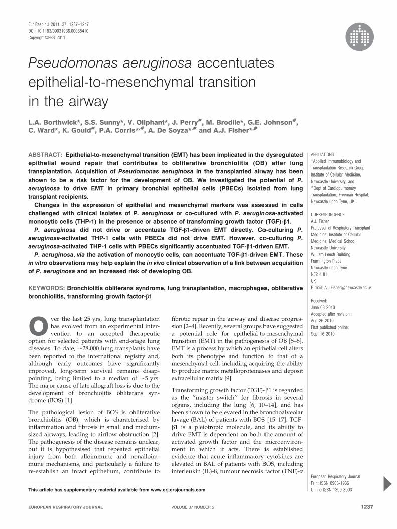
Co-culture experimentsTHP-1 cells (16106 cells?mL-1) activated with P. aeruginosalysate (12.5 mL?mL-1) were added to PBECs with or withoutTGF-b1 (10 ng?mL-1) for 72 h and EMT was assessed, or THP-1
100PA lysate
E-cadherin
Fibronectin
Vimentin
β-tubulin
Fibronectin TCA
LPS
120 kDa
220 kDa
42 kDa
55 kDa
220 kDa
TGF-β1
a) c)
b)
80
60
40
20
4000
3000
2000
1000
0 10PA lysate concentration µL·mL-1
50 1000
0
*
*
- - - + +
- - + - -
- + - - +
Cel
l via
bilit
y %
IL-8
con
cent
ratio
n pg
·mL-
1
FIGURE 1. Effect of Pseudomonas aeruginosa (PA) on primary bronchial epithelial cells. Stimulation of primary bronchial epithelial cells (PBECs) with PA lysate (10, 50 or
100 mL?mL-1) had no affect on a) cell viability (p.0.05; n53) and induced a dose-dependent increase in b) the secretion of interleukin (IL)-8, with a maximal response at
50 mL?mL-1. (p,0.05; n53). c) Stimulation of PBECs with PA lysate (50 mL?mL-1) or Escherichia coli-derived lipopolysaccharide (LPS; 10 mg?mL-1) had no affect on protein
expression or fibronectin deposition compared with control (p.0.05; n56). Treatment with transforming growth factor (TGF)-b1 (10 ng?mL-1) downregulated E-cadherin
expression, increased fibronectin and vimentin expression, and increased fibronectin deposition compared with control cells (p,0.05; n56). Stimulation with PA lysate in the
presence of TGF-b1 had no additional effect on protein expression or fibronectin deposition compared with TGF-b1 alone (p.0.05; n56). b-tubulin is shown as a loading
control. TCA: trichloroacetic acid. Data are presented as mean¡SEM. *: p,0.05.
LUNG CELL BIOLOGY L.A. BORTHWICK ET AL
1238 VOLUME 37 NUMBER 5 EUROPEAN RESPIRATORY JOURNAL
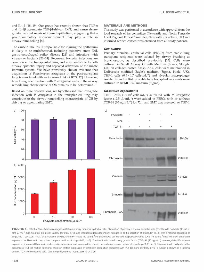
cells (16106 cells?mL-1) were activated with P. aeruginosalysate (12.5 mL?mL-1) for 6 h and then either the THP-1 cellsthemselves or the conditioned medium from THP-1 cells wasadded to PBECs with or without TGF-b1 (10 ng?mL-1) for 72 hand EMT was assessed.
P. aeruginosa whole-cell lysate preparationP. aeruginosa whole-cell lysates were prepared from a referencestrain (NCTC10662) and nine clinical isolates from our localrepository of post-transplant patients (A–I). Strains weregrown overnight on 1% horse blood agar plates, harvestedinto PBS and standardised to an optical density at 600 nm of0.2. Bacterial suspensions were disrupted (using a BransonDigital Sonifier (Branson Ultrasonics BV, Eemnes, theNetherlands) at an amplitude of 10% for 3 min on ice) andincubated with deoxyribonuclease II (200 mg?mL-1) at 37uC for1 h. Lysates were then treated with proteinase K (2 mg?mL-1)at 60uC for 2 h, boiled for 20 min (inactivating proteinase K)and stored at -80uC prior to use.
Flow cytometryFollowing treatment, cells were pelleted and resuspended in300 mL Phenol Red-negative RPMI media (Invitrogen, Paisley,
UK). Cells were incubated at 37uC for 30 min, then 30 mLpropidium iodide (1 mg?mL-1) was added prior to assessmentof cell viability.
ELISACytokine levels in supernatants were measured by sandwichELISA performed with commercially available matched anti-body pairs. The following cytokines were measured: IL-8, IL-1b and TNF-a (R&D Systems, Abingdon, UK).
Western BlottingProtein concentrations were determined using the BCA proteinassay kit (Perbio, Cramlington, UK).
Total cell lysates (10 mg) were separated on 4–12% bis-(hydroxyethyl)-amino-tris-(hydroxymethyl)-methane gels (Invi-trogen) and electrophoretically blotted onto HyBond-Ppolyvinylidene difluoride (Amersham, Hatfield, UK). Mem-branes were incubated with primary antibodies and detectedwith horseradish peroxidase-labelled immunoglobulin G con-jugates (Abcam, Cambridge, UK). Antibody complexes werevisualised using SuperSignal West Pico chemiluminescent kit(Perbio). Results are normalised to b-tubulin as appropriate.
100 80000
70000
60000
50000
40000
30000
20000
10000
4500
4000
3500
3000
2500
2000
1500
1000
500
0
0
a)
c)
90
80
60
70
50
30
40
20
10
2500
2000
1500
1000
500
0 12.5 25 50PA lysate concentration μL·mL-1
100 200 400 0 12.5 25 50PA lysate concentration μL·mL-1
100 200 400
0
Cel
l via
bilit
y %
IL-1β
conc
entra
tion
pg·m
L-1
b)
d)
IL-8
con
cent
ratio
n pg
·mL-
1TN
F-α
conc
entra
tion
pg·m
L-1
*
FIGURE 2. Cytokine release by THP-1 cells stimulated with Pseudomonas aeruginosa (PA). THP-1 cells (16106 cells?mL-1) were stimulated with PA lysate (at 0, 12.5, 25,
50, 100, 200 or 400 mL?mL-1) for 24 h (n53). Cell viability was assessed by propidium iodide staining, and the production of tumour necrosis factor (TNF)-a, interleukin (IL)-8
and IL-1b was analysed by ELISA. a) Results showed a dose-dependent decrease in cell viability from 96% in control to 72% at a concentration of 400 mL?mL-1 PA lysate.
b) PA lysate induced a dose-dependent increase in the release of IL-8. PA lysate induced a dose-dependent increase in c) IL-1b and d) TNF-a release up to a concentration of
50 mL?mL-1, after which no further increase was observed. Data are presented as mean¡SEM. *: p,0.05.
L.A. BORTHWICK ET AL LUNG CELL BIOLOGY
cEUROPEAN RESPIRATORY JOURNAL VOLUME 37 NUMBER 5 1239
Trichloroacetic acid protein precipitationTrichloroacetic acid (100% w/v) was added to culturemedia at a 1:4 ratio and incubated at 4uC for 10 min. Theprotein precipitate was pelleted by centrifugation at14,0006g for 5 min. The protein pellet was washed twicein 200 mL cold acetone and dried by heating to 95uC for10 min. The pellet was resuspended and separated underdenaturing conditions by sodium dodecylsulfate polyacry-lamide gel electrophoresis (SDS-PAGE). Membranes wereincubated with primary antibodies and detected as describedearlier.
ImmunofluorescenceCells fixed in 4% paraformaldehyde were incubated with primaryantibodies and detected using appropriate fluorochrome-linkedsecondary antibodies. 49,6-diamidino-2-phenylindole (DAPI) wasused as a nuclear counterstain. Images acquired using a LeicaTCS-SP-2UV microscope (Leica, Milton Keynes, UK) laserscanning confocal microscope (636 magnification).
Statistical analysisThe response of cells from each subject to a range of treatmentswere assessed and compared with untreated control cells.
100000 1800
1600
1400
1200
1000
800
600
400
200
200
1600
1200
800
400
0
0
a)
c)
80000
60000
40000
20000
4500
3500
4000
3000
2500
1500
2000
1000
500
Con
trol
NC
TC10
662 A B C D E F G H I
PA patient isolates
Con
trol
NC
TC10
662 A B C D E F G H I
PA patient isolates PA patient isolatesCon
trol
NC
TC10
662 A B C D E F G H I
0 0 100 200 400
400
PA lysate12.5 μL·mL-1LPS 100 ng·mL-1
Anti-CD14 antibody concentration ng·mL-10 100 200 400
0
IL-8
con
cent
ratio
n pg
·mL-
1TN
F-α
conc
entra
tion
pg·m
L-1
b)
d)IL
-1β
conc
entra
tion
pg·m
L-1
TNF-α
conc
entra
tion
pg·m
L-1
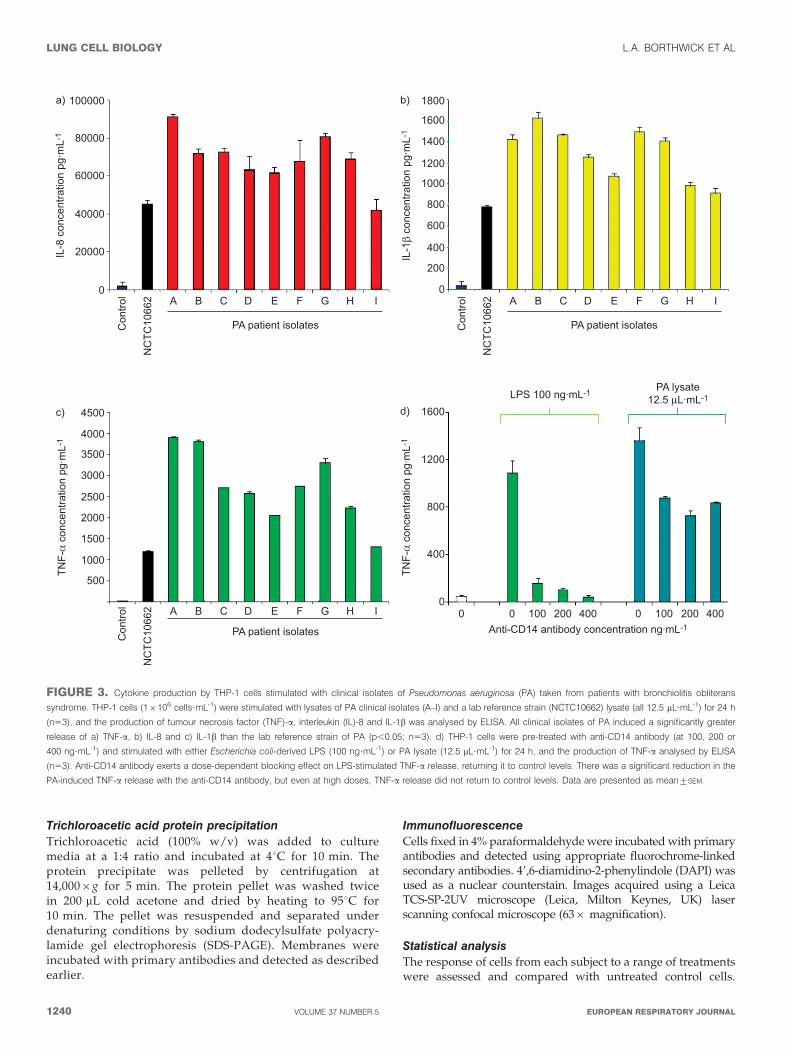
FIGURE 3. Cytokine production by THP-1 cells stimulated with clinical isolates of Pseudomonas aeruginosa (PA) taken from patients with bronchiolitis obliterans
syndrome. THP-1 cells (16106 cells?mL-1) were stimulated with lysates of PA clinical isolates (A–I) and a lab reference strain (NCTC10662) lysate (all 12.5 mL?mL-1) for 24 h
(n53), and the production of tumour necrosis factor (TNF)-a, interleukin (IL)-8 and IL-1b was analysed by ELISA. All clinical isolates of PA induced a significantly greater
release of a) TNF-a, b) IL-8 and c) IL-1b than the lab reference strain of PA (p,0.05; n53). d) THP-1 cells were pre-treated with anti-CD14 antibody (at 100, 200 or
400 ng?mL-1) and stimulated with either Escherichia coli-derived LPS (100 ng?mL-1) or PA lysate (12.5 mL?mL-1) for 24 h, and the production of TNF-a analysed by ELISA
(n53). Anti-CD14 antibody exerts a dose-dependent blocking effect on LPS-stimulated TNF-a release, returning it to control levels. There was a significant reduction in the
PA-induced TNF-a release with the anti-CD14 antibody, but even at high doses, TNF-a release did not return to control levels. Data are presented as mean¡SEM.
LUNG CELL BIOLOGY L.A. BORTHWICK ET AL
1240 VOLUME 37 NUMBER 5 EUROPEAN RESPIRATORY JOURNAL
Changes in protein expression (relative band density) orprotein secretion were quantified relative to untreated controlsand expressed as mean¡SEM.
The significance of differences between groups was assessedby a one-way ANOVA using SPSS 14.0 (IBM, Feltham, UK).Differences with a p-value of ,0.05 were considered statisti-cally significant.
RESULTS
Characterisation of patient study groupCultures of PBECs were obtained from six (two males and fourfemales) lung transplant recipients with stable allograftfunction between 3 and 48 months post-transplant. No subjectsshowed any evidence of acute rejection (grade A2 or above,according to the International Society for Heart and LungTransplantation classification) or infection, and were BOS stage0 (.90% baseline forced expiratory volume in 1 s) at the timeof sampling.
Investigating the effect of clinical isolates of P. aeruginosaon PBECs from lung transplant patientsPBECs stimulated with P. aeruginosa lysate showed nosignificant reduction in cell viability compared with untreatedcontrols (p.0.05; n53; fig. 1a) and demonstrated a dose-dependent increase in the release of IL-8 up to 50 ml?mL-1
(p,0.05; n53), after which no significant increase wasobserved (p.0.05; n53; fig. 1b).
Stimulating PBECs with P. aeruginosa lysate or Escherichia coliO55 lipopolysaccharide (LPS) (Sigma) had no significant effecton epithelial phenotype (p.0.05; n56). In contrast, treatmentwith TGF-b1 (10 ng?mL-1) significantly reduced the expressionof the epithelial marker E-cadherin, increased the expression ofthe mesenchymal markers vimentin and fibronectin, andincreased the deposition of fibronectin (p,0.05; n56), con-sistent with the induction of EMT. Stimulation with P.aeruginosa lysate in the presence of TGF-b1 had no significanteffect on fibronectin expression or deposition compared withTGF-b1 alone (p.0.05; n56; fig. 1c). These results suggest thatalthough P. aeruginosa lysate provides pathogen-associatedmolecular patterns (PAMPs) to PBECs, as seen by the increasedIL-8 secretion, it does not drive EMT by acting on epithelialcells directly, either alone or in synergy with TGF-b1.
Cytokine release from THP-1 cells and alveolarmacrophages induced by clinical isolates of P. aeruginosaAs P. aeruginosa does not directly affect EMT, we proceeded todetermine if it could act indirectly via activation of immunecells and the generation of a pro-inflammatory microenviron-ment. Treatment of the THP-1 monocytic cell line with12.5 mL?mL-1 P. aeruginosa lysate induced a significant increasein the release of IL-8 (fig. 2b), IL-1b (fig. 2c) and TNF-a (fig. 2d)
5000
4000
3000
2000
1000
8000
6000
4000
2000
0
0THP-1
Control 12.5 25
PA lysate μL·mL-1
50
Primarymacrophages
THP-1 Primarymacrophages
THP-1 Primarymacrophages
a)
d)
b) c)
TNF-α
conc
entra
tion
pg·m
L-1
TNF-α
conc
entra
tion
pg·m
L-1
140000
120000
100000
80000
60000
40000
20000
0IL
-8 c
once
ntra
tion
pg·m
L-1
1600
1200
800
400
0
IL-1β
conc
entra
tion
pg·m
L-1
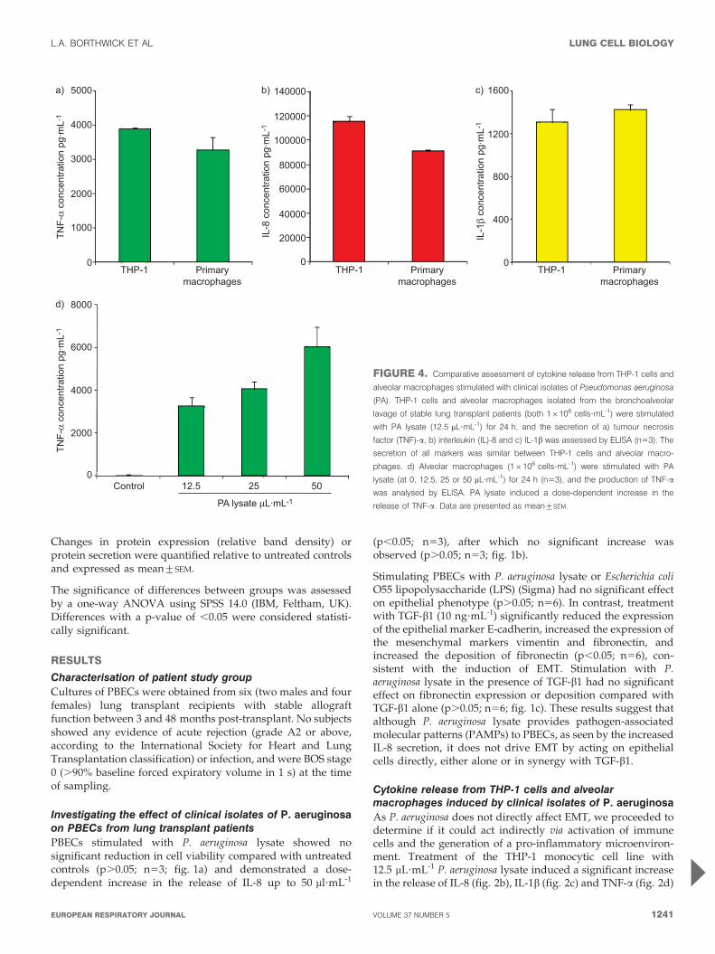
FIGURE 4. Comparative assessment of cytokine release from THP-1 cells and
alveolar macrophages stimulated with clinical isolates of Pseudomonas aeruginosa
(PA). THP-1 cells and alveolar macrophages isolated from the bronchoalveolar
lavage of stable lung transplant patients (both 16106 cells?mL-1) were stimulated
with PA lysate (12.5 mL?mL-1) for 24 h, and the secretion of a) tumour necrosis
factor (TNF)-a, b) interleukin (IL)-8 and c) IL-1b was assessed by ELISA (n53). The
secretion of all markers was similar between THP-1 cells and alveolar macro-
phages. d) Alveolar macrophages (16106 cells?mL-1) were stimulated with PA
lysate (at 0, 12.5, 25 or 50 mL?mL-1) for 24 h (n53), and the production of TNF-a
was analysed by ELISA. PA lysate induced a dose-dependent increase in the
release of TNF-a. Data are presented as mean¡SEM.
L.A. BORTHWICK ET AL LUNG CELL BIOLOGY
cEUROPEAN RESPIRATORY JOURNAL VOLUME 37 NUMBER 5 1241
(p,0.05; n53) while maintaining cell viability .80%.Concentrations .12.5 mL?mL-1 P. aeruginosa lysate inducedlittle additional cytokine release and compromised cellviability. We therefore used a P. aeruginosa lysate concentrationof 12.5 mL?mL-1 in subsequent experiments.
We compared the relative pro-inflammatory cytokine releasefrom THP-1 cells in response to a laboratory reference strain of P.aeruginosa (NCTC10662) and nine clinical transplant isolates of P.aeruginosa (A–I). Stimulation with clinical isolates of P. aeruginosainduced a significantly greater secretion of IL-8 (mean¡SEM
68,815¡4,498 versus 45,194¡2,345 pg?mL-1; fig. 3a), IL-1b(1,294¡83 versus 780¡20 pg?mL-1; fig. 3b) and TNF-a (2,739¡
280 versus 1,202¡20 pg?mL-1; fig. 3c) than the laboratoryreference strain. Furthermore, pre-treatment of THP-1 cells withan anti-CD14 antibody (400 ng?mL-1) blocked LPS-inducedTNF-a release (p,0.01; n53), returning levels to those compar-able to control (p50.69; n53; fig. 3d). However, the anti-CD14antibody (400 ng?mL-1) was able to inhibit only 39¡3.6%(p,0.01; n53) of TNF-a release from THP-1 cells stimulatedwith P. aeruginosa lysate, suggesting P. aeruginosa is acting onmultiple Toll-like receptors (TLRs) and providing us with a morerepresentative model to that seen during infection in vivo.Hereafter, strain G was used as a representative P. aeruginosaclinical isolate.
To confirm the suitability of the THP-1 monocytic cell line as asurrogate macrophage model in our co-culture system, it wasimportant to compare the secretion of cytokines in response toactivation with P. aeruginosa between THP-1 cells and alveolarmacrophages isolated from the BAL of stable lung transplantrecipients (fig. 4). IL-8 secretion was slightly higher in THP-1
cells compared with alveolar macrophages (116¡4 versus91¡14 ng?mL-1; p50.02; n53). However, we found nosignificant difference in the levels of TNF-a (3,898¡31 versus3,283¡372 pg?mL-1) and IL-1b (1,309¡118 versus 1,425¡
48 pg?mL-1) secreted from THP-1 cells and alveolar macro-phages, respectively (p,0.05; n53). We therefore proceeded touse THP-1 cells in all subsequent experiments.
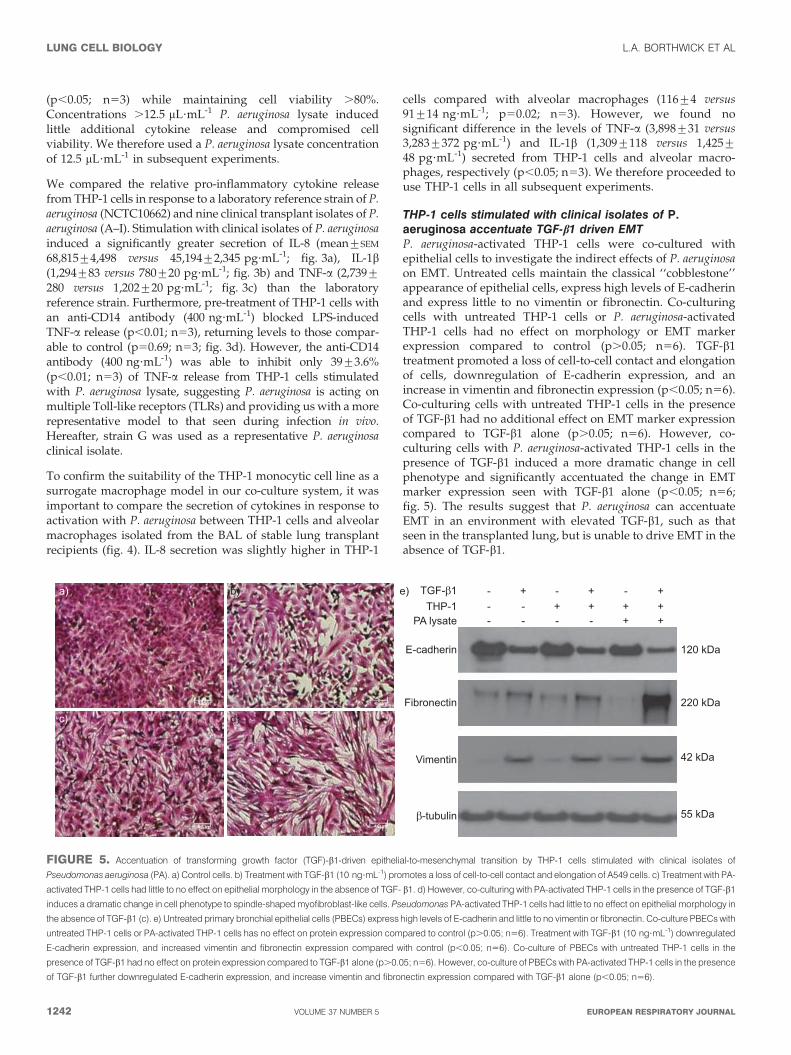
THP-1 cells stimulated with clinical isolates of P.aeruginosa accentuate TGF-b1 driven EMTP. aeruginosa-activated THP-1 cells were co-cultured withepithelial cells to investigate the indirect effects of P. aeruginosaon EMT. Untreated cells maintain the classical ‘‘cobblestone’’appearance of epithelial cells, express high levels of E-cadherinand express little to no vimentin or fibronectin. Co-culturingcells with untreated THP-1 cells or P. aeruginosa-activatedTHP-1 cells had no effect on morphology or EMT markerexpression compared to control (p.0.05; n56). TGF-b1treatment promoted a loss of cell-to-cell contact and elongationof cells, downregulation of E-cadherin expression, and anincrease in vimentin and fibronectin expression (p,0.05; n56).Co-culturing cells with untreated THP-1 cells in the presenceof TGF-b1 had no additional effect on EMT marker expressioncompared to TGF-b1 alone (p.0.05; n56). However, co-culturing cells with P. aeruginosa-activated THP-1 cells in thepresence of TGF-b1 induced a more dramatic change in cellphenotype and significantly accentuated the change in EMTmarker expression seen with TGF-b1 alone (p,0.05; n56;fig. 5). The results suggest that P. aeruginosa can accentuateEMT in an environment with elevated TGF-b1, such as thatseen in the transplanted lung, but is unable to drive EMT in theabsence of TGF-b1.
E-cadherin 120 kDa
220 kDa
42 kDa
55 kDa
Fibronectin
Vimentin
PA lysateTHP-1
- - - - + +- - + + + +- + - + - +TGF-β1
β-tubulin
a) b) e)
c) d)
FIGURE 5. Accentuation of transforming growth factor (TGF)-b1-driven epithelial-to-mesenchymal transition by THP-1 cells stimulated with clinical isolates of
Pseudomonas aeruginosa (PA). a) Control cells. b) Treatment with TGF-b1 (10 ng?mL-1) promotes a loss of cell-to-cell contact and elongation of A549 cells. c) Treatment with PA-
activated THP-1 cells had little to no effect on epithelial morphology in the absence of TGF- b1. d) However, co-culturing with PA-activated THP-1 cells in the presence of TGF-b1
induces a dramatic change in cell phenotype to spindle-shaped myofibroblast-like cells. Pseudomonas PA-activated THP-1 cells had little to no effect on epithelial morphology in
the absence of TGF-b1 (c). e) Untreated primary bronchial epithelial cells (PBECs) express high levels of E-cadherin and little to no vimentin or fibronectin. Co-culture PBECs with
untreated THP-1 cells or PA-activated THP-1 cells has no effect on protein expression compared to control (p.0.05; n56). Treatment with TGF-b1 (10 ng?mL-1) downregulated
E-cadherin expression, and increased vimentin and fibronectin expression compared with control (p,0.05; n56). Co-culture of PBECs with untreated THP-1 cells in the
presence of TGF-b1 had no effect on protein expression compared to TGF-b1 alone (p.0.05; n56). However, co-culture of PBECs with PA-activated THP-1 cells in the presence
of TGF-b1 further downregulated E-cadherin expression, and increase vimentin and fibronectin expression compared with TGF-b1 alone (p,0.05; n56).
LUNG CELL BIOLOGY L.A. BORTHWICK ET AL
1242 VOLUME 37 NUMBER 5 EUROPEAN RESPIRATORY JOURNAL
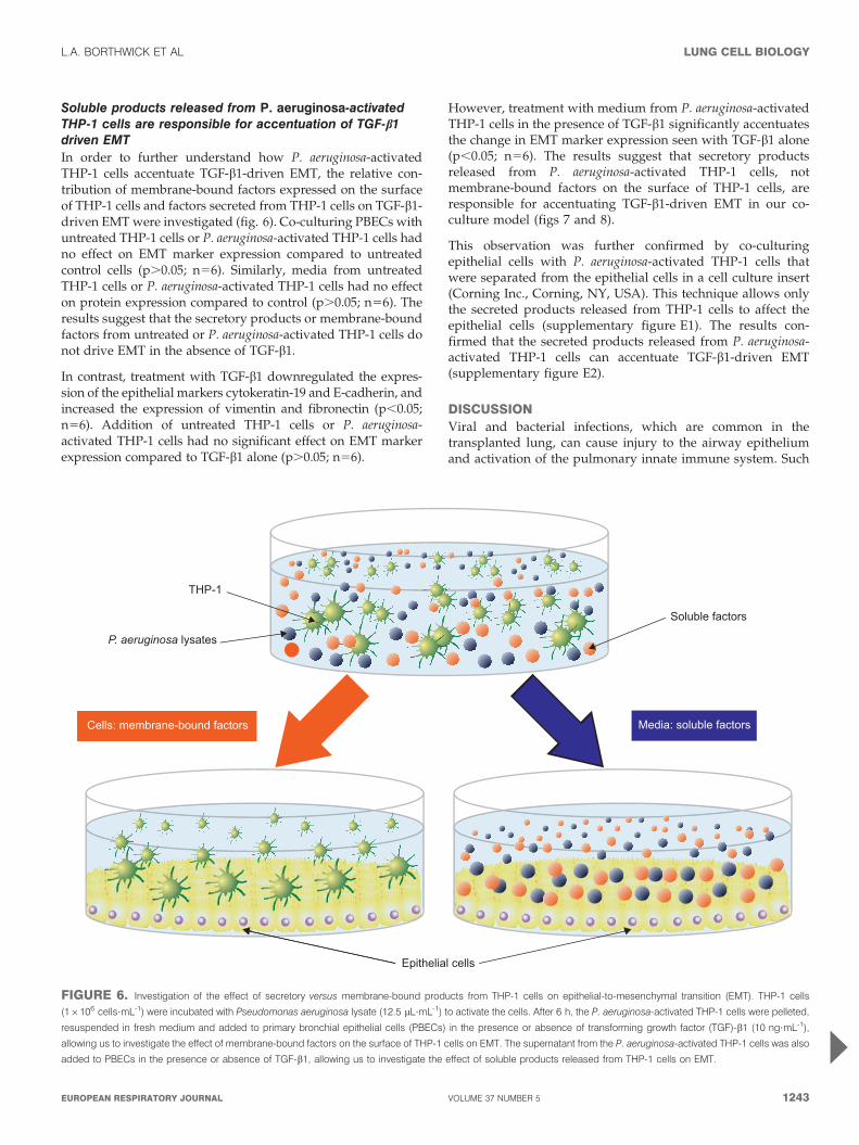
Soluble products released from P. aeruginosa-activatedTHP-1 cells are responsible for accentuation of TGF-b1driven EMTIn order to further understand how P. aeruginosa-activatedTHP-1 cells accentuate TGF-b1-driven EMT, the relative con-tribution of membrane-bound factors expressed on the surfaceof THP-1 cells and factors secreted from THP-1 cells on TGF-b1-driven EMT were investigated (fig. 6). Co-culturing PBECs withuntreated THP-1 cells or P. aeruginosa-activated THP-1 cells hadno effect on EMT marker expression compared to untreatedcontrol cells (p.0.05; n56). Similarly, media from untreatedTHP-1 cells or P. aeruginosa-activated THP-1 cells had no effecton protein expression compared to control (p.0.05; n56). Theresults suggest that the secretory products or membrane-boundfactors from untreated or P. aeruginosa-activated THP-1 cells donot drive EMT in the absence of TGF-b1.
In contrast, treatment with TGF-b1 downregulated the expres-sion of the epithelial markers cytokeratin-19 and E-cadherin, andincreased the expression of vimentin and fibronectin (p,0.05;n56). Addition of untreated THP-1 cells or P. aeruginosa-activated THP-1 cells had no significant effect on EMT markerexpression compared to TGF-b1 alone (p.0.05; n56).
However, treatment with medium from P. aeruginosa-activatedTHP-1 cells in the presence of TGF-b1 significantly accentuatesthe change in EMT marker expression seen with TGF-b1 alone(p,0.05; n56). The results suggest that secretory productsreleased from P. aeruginosa-activated THP-1 cells, notmembrane-bound factors on the surface of THP-1 cells, areresponsible for accentuating TGF-b1-driven EMT in our co-culture model (figs 7 and 8).
This observation was further confirmed by co-culturingepithelial cells with P. aeruginosa-activated THP-1 cells thatwere separated from the epithelial cells in a cell culture insert(Corning Inc., Corning, NY, USA). This technique allows onlythe secreted products released from THP-1 cells to affect theepithelial cells (supplementary figure E1). The results con-firmed that the secreted products released from P. aeruginosa-activated THP-1 cells can accentuate TGF-b1-driven EMT(supplementary figure E2).
DISCUSSIONViral and bacterial infections, which are common in thetransplanted lung, can cause injury to the airway epitheliumand activation of the pulmonary innate immune system. Such
THP-1
P. aeruginosa lysates
Cells: membrane-bound factors Media: soluble factors
Soluble factors
Epithelial cells
FIGURE 6. Investigation of the effect of secretory versus membrane-bound products from THP-1 cells on epithelial-to-mesenchymal transition (EMT). THP-1 cells
(16106 cells?mL-1) were incubated with Pseudomonas aeruginosa lysate (12.5 mL?mL-1) to activate the cells. After 6 h, the P. aeruginosa-activated THP-1 cells were pelleted,
resuspended in fresh medium and added to primary bronchial epithelial cells (PBECs) in the presence or absence of transforming growth factor (TGF)-b1 (10 ng?mL-1),
allowing us to investigate the effect of membrane-bound factors on the surface of THP-1 cells on EMT. The supernatant from the P. aeruginosa-activated THP-1 cells was also
added to PBECs in the presence or absence of TGF-b1, allowing us to investigate the effect of soluble products released from THP-1 cells on EMT.
L.A. BORTHWICK ET AL LUNG CELL BIOLOGY
cEUROPEAN RESPIRATORY JOURNAL VOLUME 37 NUMBER 5 1243
insults are believed to be important in driving crosstalk withalloimmune mechanisms and contributing to aberrant epithe-lial repair in the development of BOS. In particular, our groupand others have shown that acquisition of the Gram-negative,opportunistic bacterium P. aeruginosa in the transplantedairway is associated with an increased risk of developingBOS [22–24]. In this study, we explored the mechanism bywhich this organism might drive aberrant epithelial repair inthe airway. Our data demonstrate that clinical isolates of P.aeruginosa cultured from the transplanted lung can, via theaction of secretory products from activated immune cells,accentuate TGF-b1-driven EMT in airway epithelial cellsisolated from lung transplant recipients. These in vitro findingsmay help explain the clinical observation that acquisition of P.aeruginosa in the transplanted airway is associated with anincreased risk of developing BOS.
It is recognised that repeated sub-clinical injury and persistentinflammation in the airway epithelium coinciding withdefective regeneration will favour excessive fibroproliferation,and obliteration of small and medium-sized airways in OB[26]. In clinical practice, BOS is often first diagnosed (orprogresses more rapidly) following a bacterial or viralinfection, and these sources of acute inflammation may bevery important in accentuating the processes already driving
chronic lung allograft dysfunction. Our group has previouslyshown that TNF-a and IL-1b can cause dysregulated woundrepair of injured epithelium by accentuating TGF-b1-drivenEMT, demonstrating the link between acute inflammation andepithelial remodelling [5]. Previous data from VOS et al. [27]suggest that even transient infection with P. aeruginosa in lungtransplant recipients is associated with a significant increase ininflammation. We therefore hypothesised that acquisition of P.aeruginosa in the transplanted lung might produce sufficientinflammatory stimulus to accentuate dysregulated repair viaEMT in the airway epithelium.
Our data shows that Pseudomonas aeruginosa does not driveEMT directly but can accentuate TGF-b1 driven EMT indirectlyvia activation of innate immune cells. Airway epithelial cellsare able to respond to the presence of micro-organisms byexpression of TLRs on their surface, yet these cells commonlyshow a hyporesponsiveness to TLR ligands [28], whereaspulmonary innate immune cells show a much more vigorousinflammatory response to TLR ligands. When activated, lungmacrophages produce a multitude of growth factors andcytokines, including proteins that are present in elevated levelsin the BAL of patients with BOS [18, 19], such as IL-8, TNF-aand IL-1b [29]. There is growing interest in the role of themacrophage as an effector cell in allograft injury and fibrosis.
Cyt
oker
atin
-19
Control TGF-β1 TGF-β1+THP-1
Medium Cells
TGF-β1+PA-activated THP-1 TGF-β1+THP-1
TGF-β1+PA-activated THP-1
Vim
entin
Fibr
onec
tin
a) b) c) d) e) f)
g) h) i) j) k) l)
m) n) o) p) q) r)
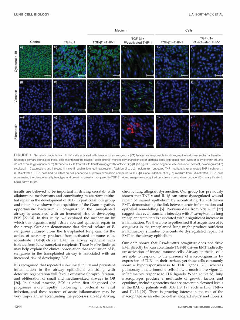
FIGURE 7. Secretory products from THP-1 cells activated with Pseudomonas aeruginosa (PA) lysates are responsible for driving epithelial-to-mesenchymal transition.
Untreated primary broncial epithelial cells maintained the classic ‘‘cobblestone’’ morphology characteristic of epithelial cells, expressed high levels of a) cytokeratin 19, and
do not express g) vimentin or m) fibronectin. Cells treated with transforming growth factor (TGF)-b1 (10 ng?mL-1) alone began to lose cell-to-cell contact, downregulated b)
cytokeratin-19 expression, and increase h) vimentin and n) fibronectin expression. Addition of c, j, o) medium from untreated THP-1 cells, e, k, q) untreated THP-1 cells or f, l,
r) PA-activated THP-1 cells had no affect on cell phenotype or protein expression compared to TGF-b1 alone. Addition of d, j, p) medium from PA-activated THP-1 cells
accentuated the change in cell phenotype and protein expression compared to TGF-b1 alone. Images were acquired on a Leica confocal microscope (636 magnification).
Scale bars548 mm.
LUNG CELL BIOLOGY L.A. BORTHWICK ET AL
1244 VOLUME 37 NUMBER 5 EUROPEAN RESPIRATORY JOURNAL
In the murine heterotopic tracheal transplant model, depletionof recipient macrophages significantly abrogates obliteration ofthe transplanted airway [30]. In this study, we used the THP-1monocytic cell line as a model for lung innate immune cells, asthe use of human macrophages isolated from either healthycontrols or stable lung transplant patients was not feasible dueto the large number of cells required for the co-cultureexperiments. Importantly, as part of the validation of ourmodel, we showed that undifferentiated THP-1 cells show asimilar inflammatory response to P. aeruginosa as alveolarmacrophages isolated from the BAL of stable lung transplantrecipients (fig. 4). It is also well established that THP-1 cellscan be differentiated using phorbol myristate acetate intoadherent macrophage-like cells [31]. However, we required theTHP-1 cells to remain in suspension to perform our co-cultureexperiments and, therefore, used undifferentiated cells.
Another important aspect of our model was the use of clinicalisolates of P. aeruginosa. The whole bacterial lysates used in thisstudy contain multiple PAMPs that signal via different TLRsand provides a more clinically relevant stimulus. This isfurther demonstrated by the observation that clinical isolates ofP. aeruginosa induce a greater inflammatory response than thelaboratory reference strain (NCTC10662).
The observations noted above suggest that targetting persis-tence of P. aeruginosa or its inflammatory consequences bylimiting innate immune activation in the transplanted lungmay be a potential therapy to limit dysregulated epithelialrepair. Our group and others have shown that azithromycintreatment can reverse the decline in lung function in somepatients with BOS [32–35]. Azithromycin does not have anysignificant anti-pseudomonal activity but has been shown to
E-cadherin
PA lysateTHP-1
TGF-β1
Fibronectin
Vimentin
β-tubulin
- - - - + + - - + +- - + + + + + + + +- + - + - + - + - +
120 kDa
220 kDa
42 kDa
55 kDa
0.40b)a)
0.35
0.30
0.25
0.20
0.15
0.10
0.05
0
Rel
ativ
e ba
nd d
ensi
ty
7c)
6
5
4
3
2
1
0Control
Medium Cells
TGF-β1 TGF-β1+PA-activated
THP-1
TGF-β1+PA-activated
THP-1
TGF-β1+THP-1
TGF-β1+THP-1
Rel
ativ
e ba
nd d
ensi
ty
*
*
*
2.0d)
1.8
1.6
1.4
1.2
1.0
0.6
0.4
0.8
0.2
0
Rel
ativ
e ba
nd d
ensi
ty
*
*
*
Medium Cells
Control
Medium Cells
TGF-β1 TGF-β1+PA-activated
THP-1
TGF-β1+PA-activated
THP-1
TGF-β1+THP-1
TGF-β1+THP-1
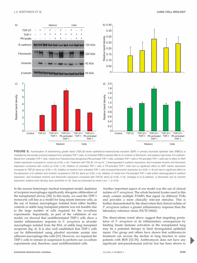
FIGURE 8. Accentuation of transforming growth factor (TGF)-b1-driven epithelial-to-mesenchymal transition (EMT) in primary bronchial epithelial cells (PBECs) is
mediated by the soluble products released from activated THP-1 cells. a) Untreated PBECs express little to no vimentin or fibronectin, and express high levels of E-cadherin.
Media from untreated THP-1 cells, media from Pseudomonas aeruginosa (PA)-activated THP-1 cells, untreated THP-1 cells or PA-activated THP-1 cells had no effect on EMT
marker expression compared to control (p.0.05; n56). Treatment with TGF-b1 (10 ng?mL-1) downregulated E-cadherin expression, and increased vimentin and fibronectin
expression compared with control (p,0.05; n56). Addition of untreated THP-1 cells or PA-activated THP-1 cells had no significant effect on EMT marker expression
compared to TGF-b1 alone (p.0.05; n56). Addition of medium from untreated THP-1 cells increased fibronectin expression (p,0.05; n56) but had no significant effect on
the expression of E-cadherin and vimentin compared to TGF-b1 alone (p.0.05; n56). Addition of media from PA-activated THP-1 cells further downregulated E-cadherin
expression, and increased vimentin and fibronectin expression compared with TGF-b1 alone (p,0.05; n56). Changes in b) E-cadherin, c) fibronectin and d) vimentin
expression (relative band density) were quantified (n56). Data are presented as mean¡SEM. *: p,0.05.
L.A. BORTHWICK ET AL LUNG CELL BIOLOGY
cEUROPEAN RESPIRATORY JOURNAL VOLUME 37 NUMBER 5 1245
exhibit extensive inhibition of the quorum-sensing systems,diminished virulence factor production and impairment of theoxidative stress response [36–38]. However, recent datasuggest that although azithromycin exhibits activity againstP. aeruginosa biofilms, resistant mutants are readily selected,and tailored anti-pseudomonal therapies may be required toeradicate infection in patients receiving azithromycin treat-ment [39]. This suggests that azithromycin is more likely to beexerting its benefits via its anti-inflammatory actions than by adirect action on P. aeruginosa colonisation.
Finally, it remains unclear in the literature if P. aeruginosa canbe a primary driver of airway remodelling in the transplantedlung or is in fact accelerating an already established process.The data we present in this paper suggests that an elevatedbackground level of TGF-b1 is required for P. aeruginosa tohave its accentuating effects on EMT. This supports thehypothesis that P. aeruginosa may be having an accentuatingeffect on an already smouldering alloimmune injury to theairway epithelium.
SUPPORT STATEMENTThis work was supported by a research grant from the MedicalResearch Council UK (G0700861). A. De Soyza is supported by aHEFCE Senior Lectureship. A.J. Fisher is supported by aGlaxoSmithKline clinical fellowship award.
STATEMENT OF INTERESTStatements of interest for A. De Soyza and A.J. Fisher can be found atwww.erj.ersjournals.com/site/misc/statements.xhtml
REFERENCES1 Christie JD, Edwards LB, Aurora P, et al. The Registry of the
International Society for Heart and Lung Transplantation: Twenty-sixth Official Adult Lung and Heart-Lung Transplantation Report:2009. J Heart Lung Transplant 2009; 28: 1031–1049.
2 Estenne M, Hertz MI. Bronchiolitis obliterans after human lungtransplantation. Am J Respir Crit Care Med 2002; 166: 440–444.
3 Boehler A, Estenne M. Post-transplant bronchiolitis obliterans. Eur
Respir J 2003; 22: 1007–1018.
4 Yousem SA, Berry GJ, Cagle PT, et al. Revision of the 1990 workingformulation for the classification of pulmonary allograft rejection:Lung Rejection Study Group. J Heart Lung Transplant 1996; 15:1–15.
5 Borthwick LA, McIlroy EI, Gorowiec MR, et al. Inflammation andepithelial to mesenchymal transition in lung transplant recipients:role in dysregulated epithelial wound repair. Am J Transplant 2010;10: 498–509.
6 Borthwick LA, Parker SM, Brougham KA, et al. Epithelial tomesenchymal transition (EMT) and airway remodelling afterhuman lung transplantation. Thorax 2009; 64: 770–777.
7 Hodge S, Holmes M, Banerjee B, et al. Posttransplant bronchiolitisobliterans syndrome is associated with bronchial epithelial tomesenchymal transition. Am J Transplant 2009; 9: 727–733.
8 Ward C, Forrest IA, Murphy DM, et al. Phenotype of airwayepithelial cells suggests epithelial to mesenchymal cell transitionin clinically stable lung transplant recipients. Thorax 2005; 60:865–871.
9 Kalluri R, Neilson EG. Epithelial–mesenchymal transition and itsimplications for fibrosis. J Clin Invest 2003; 112: 1776–1784.
10 Kasai H, Allen JT, Mason RM, et al. TGF-b1 induces humanalveolar epithelial to mesenchymal cell transition (EMT). Respir Res
2005; 6: 56.
11 Willis BC, Borok Z. TGF-b-induced EMT: mechanisms andimplications for fibrotic lung disease. Am J Physiol 2007; 293:L525–L534.
12 Willis BC, Liebler JM, Luby-Phelps K, et al. Induction of epithelial-mesenchymal transition in alveolar epithelial cells by transforminggrowth factor-b1: potential role in idiopathic pulmonary fibrosis.Am J Pathol 2005; 166: 1321–1332.
13 Wu Z, Yang L, Cai L, et al. Detection of epithelial to mesenchymaltransition in airways of a bleomycin induced pulmonary fibrosismodel derived from an a-smooth muscle actin–Cre transgenicmouse. Respir Res 2007; 8: 1.
14 Zhang M, Zhang Z, Pan HY, et al. TGF-b1 induces humanbronchial epithelial cell-to-mesenchymal transition in vitro. Lung
2009; 187: 187–194.
15 Bergmann M, Tiroke A, Schafer H, et al. Gene expression ofprofibrotic mediators in bronchiolitis obliterans syndrome afterlung transplantation. Scand Cardiovasc J 1998; 32: 97–103.
16 El-Gamel A, Sim E, Hasleton P, et al. Transforming growth factorbeta (TGF-b) and obliterative bronchiolitis following pulmonarytransplantation. J Heart Lung Transplant 1999; 18: 828–837.
17 Elssner A, Jaumann F, Dobmann S, et al. Elevated levels ofinterleukin-8 and transforming growth factor-b in bronchoalveolarlavage fluid from patients with bronchiolitis obliterans syndrome:proinflammatory role of bronchial epithelial cells. Munich LungTransplant Group. Transplantation 2000; 70: 362–367.
18 Riise GC, Williams A, Kjellstrom C, et al. Bronchiolitis obliteranssyndrome in lung transplant recipients is associated withincreased neutrophil activity and decreased antioxidant status inthe lung. Eur Respir J 1998; 12: 82–88.
19 Vanaudenaerde BM, De Vleeschauwer SI, Vos R, et al. The role ofthe IL23/IL17 axis in bronchiolitis obliterans syndrome after lungtransplantation. Am J Transplant 2008; 8: 1911–1920.
20 Hirsch J, Elssner A, Mazur G, et al. Bronchiolitis obliteranssyndrome after (heart–)lung transplantation. Impaired antipro-tease defense and increased oxidant activity. Am J Respir Crit Care
Med 1999; 160: 1640–1646.
21 Blondeau K, Mertens V, Vanaudenaerde BA, et al. Gastro-oesophageal reflux and gastric aspiration in lung transplantpatients with or without chronic rejection. Eur Respir J 2008; 31:707–713.
22 Botha P, Archer L, Anderson RL, et al. Pseudomonas aeruginosa
colonization of the allograft after lung transplantation and the riskof bronchiolitis obliterans syndrome. Transplantation 2008; 85:771–774.
23 Valentine VG, Gupta MR, Walker JE Jr, et al. Effect of etiology andtiming of respiratory tract infections on development of bronch-iolitis obliterans syndrome. J Heart Lung Transplant 2009; 28:163–169.
24 Vos R, Vanaudenaerde BM, Geudens N, et al. Pseudomonal airwaycolonisation: risk factor for bronchiolitis obliterans syndrome afterlung transplantation? Eur Respir J 2008; 31: 1037–1045.
25 Forrest IA, Murphy DM, Ward C, et al. Primary airway epithelialcell culture from lung transplant recipients. Eur Respir J 2005; 26:1080–1085.
26 Al-Githmi I, Batawil N, Shigemura N, et al. Bronchiolitis obliteransfollowing lung transplantation. Eur J Cardiothorac Surg 2006; 30:846–851.
27 Vos R, Vanaudenaerde BM, Dupont LJ, et al. Transient airwaycolonization is associated with airway inflammation after lungtransplantation. Am J Transplant 2007; 7: 1278–1287.
28 Mayer AK, Muehmer M, Mages J, et al. Differential recognition ofTLR-dependent microbial ligands in human bronchial epithelialcells. J Immunol 2007; 178: 3134–3142.
29 Kelley J. Cytokines of the lung. Am Rev Respir Dis 1990; 141:765–788.
30 Oyaizu T, Okada Y, Shoji W, et al. Reduction of recipientmacrophages by gadolinium chloride prevents development of
LUNG CELL BIOLOGY L.A. BORTHWICK ET AL
1246 VOLUME 37 NUMBER 5 EUROPEAN RESPIRATORY JOURNAL
obliterative airway disease in a rat model of heterotopic trachealtransplantation. Transplantation 2003; 76: 1214–1220.
31 Park EK, Jung HS, Yang HI, et al. Optimized THP-1 differentiationis required for the detection of responses to weak stimuli. Inflamm
Res 2007; 56: 45–50.32 Verleden GM, Dupont LJ. Azithromycin therapy for patients with
bronchiolitis obliterans syndrome after lung transplantation.Transplantation 2004; 77: 1465–1467.
33 Yates B, Murphy DM, Forrest IA, et al. Azithromycin reversesairflow obstruction in established bronchiolitis obliterans syn-drome. Am J Respir Crit Care Med 2005; 172: 772–775.
34 Gerhardt SG, McDyer JF, Girgis RE, et al. Maintenanceazithromycin therapy for bronchiolitis obliterans syndrome:results of a pilot study. Am J Respir Crit Care Med 2003; 168:121–125.
35 Gottlieb J, Szangolies J, Koehnlein T, et al. Long-term azithromycinfor bronchiolitis obliterans syndrome after lung transplantation.Transplantation 2008; 85: 36–41.
36 Nalca Y, Jansch L, Bredenbruch F, et al. Quorum-sensing antag-onistic activities of azithromycin in Pseudomonas aeruginosa PAO1: aglobal approach. Antimicrob Agents Chemother 2006; 50: 1680–1688.
37 Ichimiya T, Takeoka K, Hiramatsu K, et al. The influence ofazithromycin on the biofilm formation of Pseudomonas aeruginosa in
vitro. Chemotherapy 1996; 42: 186–191.38 Favre-Bonte S, Kohler T, Van Delden C. Biofilm formation by
Pseudomonas aeruginosa: role of the C4-HSL cell-to-cell signal andinhibition by azithromycin. J Antimicrob Chemother 2003; 52: 598–604.
39 Mulet X, Macia MD, Mena A, et al. Azithromycin in Pseudomonas
aeruginosa biofilms: bactericidal activity and selection of nfxB
mutants. Antimicrob Agents Chemother 2009; 53: 1552–1560.
L.A. BORTHWICK ET AL LUNG CELL BIOLOGY
EUROPEAN RESPIRATORY JOURNAL VOLUME 37 NUMBER 5 1247