Provincial Reciprocity Attainment Program Abdominal.
79
Provincial Reciprocity Attainment Program Provincial Reciprocity Attainment Program Abdominal
-
Upload
colin-pearson -
Category
Documents
-
view
219 -
download
0
Transcript of Provincial Reciprocity Attainment Program Abdominal.

Provincial Reciprocity Attainment ProgramProvincial Reciprocity Attainment Program
Abdominal
Abdominal Organs
ADUKPIEADUKPIE

Types of Abdominal Organs
O V A R IE S
K ID N E Y S
P A N C R E A S
S P L E E N
L IV E R
S O L ID O R G A N S
B L A D D E R
L A R G E IN TE S TIN E
S M A L L IN TE S TIN E
G A L L B L A D D E R
S TO M A C H
H O L L O W O R G A N S
A B D O M IN A L O R G A N S

Abdominal Organs
Spleen
Stomach
Kidney
Part of the Liver
Kidney
Large Intestine
Small Intestine
Part of the Pancreas
Lt Ureter
Large Intestine
Small Intestine
Femoral Artery/Vein to Left Leg
Liver
Gall Bladder
Kidney
Part of the Pancreas
Large Intestine
Appendix
Large Intestine
Rt Ureter
Small Intestine
Femoral Artery/Vein to Rt Leg
LUQRUQ
RLQ LLQ

Traumatic Injuries
Abdominal Trauma
May be difficult to evaluate in the prehospital setting due to: Wide spectrum of potential injuries to
multiple organs Physical findings that are sometimes
lacking or exaggerated

Abdominal Trauma
Assessment may be compromised by: Use of alcohol and/or recreational drugs Injury to brain, spinal cord Injury to ribs, spine, pelvis
Exercise a high degree of suspicion based on mechanism of injury and kinematics
Boundaries of the Abdomen
Diaphragm Anterior abdominal wall Pelvic bones Vertebral column Muscles of the abdomen and flanks
Surface Anatomy of Abdomen
Quadrants Upper - right, left Lower - right, left
Xiphoid Symphysis pubis Umbilicus

Peritoneal Cavity
Also called the “true” abdominal cavity Quadrants
Upper - right, left Lower - right, left
Contents-liver, spleen, stomach, small intestine, colon, gallbladder, female reproductive organs
Pelvic Cavity
Surrounded by the pelvic bones Lower part of retroperitoneal space Contents:
Rectum Bladder Urethra Iliac vessels In women, internal genitalia

Retroperitoneal Space
Potential space behind the “true” abdominal cavity Contents (ADUCKPIE):
Abdominal aorta Duodenum Ureter Colon Kidneys Pancreas Inferior vena cava Esophagus

Mechanisms of Abdominal Injury
Blunt trauma Compression or crushing forces Shearing forces Deceleration forces
Degree of injury is usually related to: Quantity and duration of force applied Type of abdominal structure injured (fluid
filled, gas filled, solid, hollow)

Blunt Trauma
Motor vehicle collisions Motorcycle collisions Pedestrian injuries Falls Assault Blast injuries

Penetrating Trauma
Energy imparted to the body Low velocity - knife, ice pick Medium velocity - gunshot wounds, shotgun
wounds High velocity - high-power hunting rifles,
military weapons Ballistics Trajectory Distance

Solid & Hollow Organs
Solid Organs Liver Spleen Pancreas Kidneys Adrenals Ovaries (female)
Hollow Organs Stomach Intestines Gallbladder Urinary bladder Uterus (female)
Solid Organ Injury
Liver
Largest organ in the abdominal cavity Located in the right upper quadrant of
abdomen Commonly injured from trauma to the:
Eighth through twelfth ribs on right side of body Upper central part of abdomen Damaged in 19% of blunt ABD trauma 37% of penetrating trauma

Liver
Suspect liver injury in any patient with: Steering wheel injury Lap belt injury History of epigastric trauma
After injury, blood and bile escape into peritoneal cavity Produces signs and symptoms of shock
and peritoneal irritation, respectively

Spleen
Lies in upper left quadrant of abdomen Rich blood supply Slightly protected by organs surrounding it
medially and anteriorly and by lower portion of rib cage Most commonly injured organ from blunt trauma
(41%) Associated intraabdominal injuries common 40% of patients do not show symptoms

Spleen
Suspect splenic injury in: Motor vehicle crashes Falls or sport injuries in which there was an
impact to lower left chest, flank, or upper left abdomen
Kehr’s sign Left upper quadrant pain with radiation to left
shoulder Common complaint associated with splenic
injury

Kidneys
Located high on posterior wall of abdominal cavity in retroperitoneal space Held in place by renal fascia Cushioned by a generous layer of
adipose tissue Partially enclosed and protected by lower
rib cage
Kidneys
Injuries may involve fracture and laceration Resulting in hemorrhage, urine
extravasation, or both Contusions usually are self-limiting
Heal with bed rest and forced fluids Fractures and lacerations may require
surgical repair

Hollow Organ Injury

Stomach
Not commonly injured after blunt trauma because of its protected location in abdomen
Penetrating trauma may cause gastric transection or laceration
Patients exhibit signs of peritonitis rapidly from leakage of gastric contents
Diagnosis confirmed during surgery unless nasogastric drainage returns blood

Colon and Small Intestine
Injury is usually the result of penetrating trauma
Large and small intestine may also be injured by compression forces High-speed motor vehicle crashes Deceleration injuries associated with wearing
personal restraints Bacterial contamination common problem
with these injuries

Retroperitoneal Organ Injury
May occur because of blunt or penetrating trauma to the: Anterior abdomen Posterior abdomen (particularly the flank
area) or Thoracic spine

Ureters
Hollow organs Rarely injured in blunt trauma because
of their flexible structure Injury usually occurs from penetrating
abdominal or flank wounds (stab wounds, firearm injuries)

Pancreas
Solid organ that lies in the peritoneal space
Blunt injury usually occurs from a crushing injury of the pancreas between the spine and a steering wheel, handlebar, or blunt weapon
Most pancreatic injuries are due to penetrating trauma
Duodenum
Lies across the lumbar spine Seldom injured due to its location in the
retroperitoneal area, near pancreas May be crushed or lacerated when
great force of blunt trauma or penetrating injury occurs Usually associated with concurrent
pancreatic trauma
Pelvic Organ Injury
Usually results from motor vehicle crashes that produce pelvic fractures
Less frequent causes: Penetrating trauma Straddle-type injuries from falls Pedestrian accidents Some sexual acts

Urinary Bladder
Hollow organ May be ruptured by blunt or
penetrating trauma or pelvic fracture Rupture more likely if bladder is
distended at time of injury Suspect bladder injury in inebriated
patients subjected to lower abdominal trauma
Vascular Structure Injury
Intraabdominal arterial and venous injuries may be life-threatening
Injury usually occurs from penetrating trauma May also occur from compression or
deceleration forces applied to abdomen Usually presents as hypovolemia Occasionally associated with a palpable
abdominal mass

Vascular Structure Injury
Major vessels most frequently injured: Aorta Inferior vena cava Renal, mesenteric, and iliac arteries and
veins

Pelvic Fractures
Disruption of the pelvis may occur from: Motorcycle crashes Pedestrian-vehicle collisions Direct crushing injury to the pelvis Falls from heights greater than 12 feet
Blunt or penetrating injury may result in: Fracture Severe hemorrhage Associated injury to urinary bladder and urethra
Most common injured organs are the urinary bladder and urethra
Mortality rate 6.4 – 19% Structural damage to the pelvis Room to empty large quantity of blood (shock)
Inability to urinate Gross hematuria suspect bladder Blood at the meatus, suspect urethral damage
Pelvic injury
Pelvic Fractures
Suspicion of pelvic injury should be based on: Mechanism of injury Presence of tenderness on palpation of
iliac crests Force may be direct or indirect Assessment findings Management
Evisceration
Protrusion of an internal organ through a wound or surgical incision, especially in the abdominal wall Common finding with stab wounds May be seen to a lesser degree with gunshot
wounds Do not replace organs back into abdomen
Protect organs from further damage Cover with sterile saline moistened dressing
Transport
Focused History and Physical
Head injury and/ or intoxicants (drugs/alcohol) mask signs and symptoms
Hemoperitoneum (solid organ/vascular injuries) Adult abdomen will accommodate 1.5 liters
with no abdominal distention Often present even with normal abdominal exam
Unexplained shock Shock out of proportion to known injuries
Peritonitis – S/S
Pain (subjective symptom from patient) Tenderness (objective sign with percussion/palpation) Guarding/rigidity Distention (late finding) Abrasions Ecchymosis Visible wounds Mechanism of injury Unexplained shock

Critical Findings
Rapid assessment and transport Detailed assessment On-going assessment
Noncritical Findings
Focused history and physical examination
Other interventions and transport considerations

Comprehensive Assessment
Vital signs Inspection Auscultation Percussion Palpation

Comprehensive Assessment
Absence of signs and symptoms does not rule out abdominal injuries Not necessary to determine definitively if
abdominal injuries are present Remember to examine the back Differential diagnosis Continued management

Management/Treatment Plan
Surgical intervention only effective therapy Rapid evaluation Initiation of shock resuscitation Rapid packaging and transport to nearest
appropriate facility Facility must have immediate surgical capability Rapid transport
Defeated if hospital cannot provide immediate surgical intervention
Crystalloid fluid replacement en route to hospital
Indications for Rapid Transport
Critical findings Surgical intervention required to control
hemorrhage and/ or contamination High index of suspicion for abdominal
injury Unexplained shock Physical signs of abdominal injury

Indications for Rapid Transport
Hemorrhage continues until controlled in OR
Survival determined by length of time from injury to definitive surgical control of hemorrhage Any delay in the field negatively impacts
this time period

ABD and Renal Disease

Hiatal Hernia
Herniation of the stomach through the diaphragmatic opening
S/S Chest pain (especially when lying down) Difficulty swallowing Reflux Burping Possible hemorrhage May see signs of shock if severe

Hiatal Hernia
Treatment ABC’s Position of comfort O2 Rule out ischemia Treat for shock if applicable Transport

Inguinal Hernia
Herniation of intestine into inguinal canal S/S
Pain and/or discomfort Mass may increase with strenuous activity N/V
Treatment ABC’s O2 Position of comfort

Umbilical Hernia
Herniation of intestines or fluids into the umbilicus
S/S: May increase with crying, strains or is upright Usually no pain associated with tightening
Treatment: ABC’s Pt comfort O2 if necessary

Bowel Obstruction
Blockage of the intestines due to tumor, feces, adhesions or hernias
S/S: N/V Distention Pain (Crampy and intermittent) Diarrhea (early)/Constipation (Late) Fever (late) Absent bowel sounds (late) BAD Breath Signs of shock

Bowel Obstruction
Treatment: ABC’s O2 Position of comfort IV ALS ? (May need gravol or pain relief)
Diverticulitis
Inflammation of the diverticula S/S:
Maybe asymptomatic Abdominal pain (usually LLQ) Febrile N/V Cramps Chills Constipation/diarrhea Bright red blood Signs of shock

Diverticulitis
Treatment: ABC’s Position of comfort Treat for shock IV ? ALS ? (Pain, N/V)
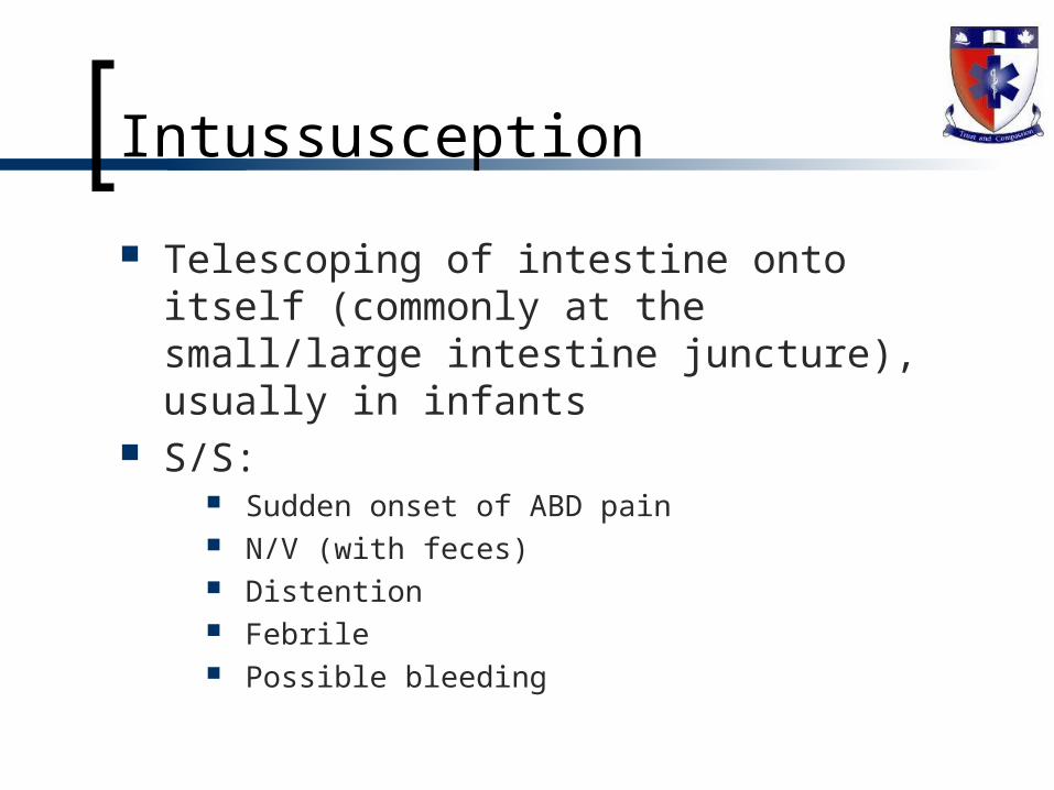
Intussusception
Telescoping of intestine onto itself (commonly at the small/large intestine juncture), usually in infants
S/S: Sudden onset of ABD pain N/V (with feces) Distention Febrile Possible bleeding

Intussusception
Treatment: ABC’s O2 Position of comfort ALS ?
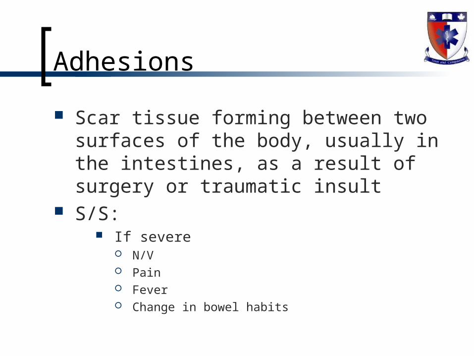
Adhesions
Scar tissue forming between two surfaces of the body, usually in the intestines, as a result of surgery or traumatic insult
S/S: If severe
N/V Pain Fever Change in bowel habits

Reflux
Weakness of esophageal sphincter allowing gastric contents to enter esophagus
S/S: Heartburn Burning sensation Burping N/V etc

IBS
Spastic colon S/S:
Stress Change in bowel habits ABD pain or cramping Excessive gas Decrease in appetite
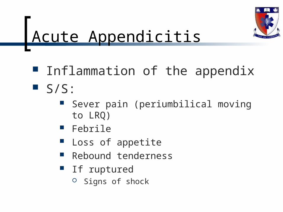
Acute Appendicitis
Inflammation of the appendix S/S:
Sever pain (periumbilical moving to LRQ) Febrile Loss of appetite Rebound tenderness If ruptured
Signs of shock
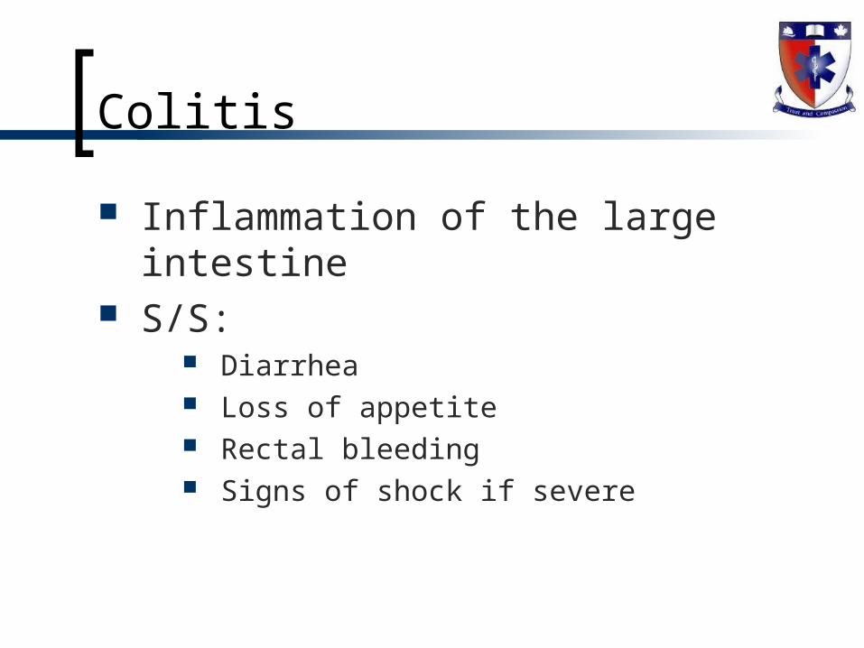
Colitis
Inflammation of the large intestine S/S:
Diarrhea Loss of appetite Rectal bleeding Signs of shock if severe

Chrone’s Disease
Chronic inflammatory disease causing ulcerations in the small intestines (but may affect large and other regions of the tract)
S/S: Diarrhea ABD pain N/V Anorexia Dependant on area and amount of damage

Acute Peritonitis
Acute inflammation of the peritoneum S/S:
ABD pain Tenderness Guarding Is severe signs of shock

Anorexia & Bulemia
Eating disorders usually connect to the psychology of the patient
S/S: Obsession with weight loss May be purging, using laxatives, diuretics… Dehydration Signs of shock (metabolic and hypovolemia)

Acute Pancreatitis
Inflammation of the pancreas due to stones, necrosis, infections…
S/S: Severe epigastric pain N/V If severe
Infection Hemorrhage Complications to other organs Acites

Renal Calculi
Kidney stones S/S:
Abdominal pain starting in back and radiating to groin
Infection Hematurea Severe may show signs of sepsis
Hepatitis
Inflammation of the liver S/S:
Fatigue Anorexia General malaise N/V Photophobia Muscle and joint pain Dark urine RUQ pain Clay colored stools Jaundice

Hepatic Failure
Liver failure due to disease or insult S/S:
Jaundice Fatigue Edema Metabolic changes (expect EKG changes) Hepatomegaly Febrile Severe may show shock

Cirrhosis
Necrosis of the liver cells S/S:
Fatigue Anorexia GI bleed Ascites Jaundice Signs of shock (late)

Cholecystitis
Inflammation of the gall bladder S/S:
URQ pain radiating to the right shoulder History of gall stones Febrile Fatty food intolerance N/V Severe may be shocky
Renal Failure
Kidney failure S/S:
Oliguria leading to anurea Edema Acidosis Metabolic changes Leading to MOF May see
LOC changes N/V…..
Pelvic Inflammatory Disease
Inflammation of the female pelvic organs
S/S: ABD pain with rebound Guarding Febrile Pain with intercourse Changes in menstruation Painful urination

Testicular Torsion
Twisting of spermatic cord depleting supply of blood
S/S: Swelling SEVERE PAIN N/V Hematuria

Glomerulonephritis
Inflammation of the glomerulus S/S:
N/V Edema Decrease in output (may be absent) Hypertension

Nephrotic Syndrome
Increase in permeability of nephrons S/S:
Proteinuria Edema Swelling of the scrotum Distention May see signs of shock

Flank Pain
N/V