Protein-Peptide Interactions Revolutionize Drug...
24
Chapter 3 © 2012 Olmez and Akbulut, licensee InTech. This is an open access chapter distributed under the terms of the Creative Commons Attribution License (http://creativecommons.org/licenses/by/3.0), which permits unrestricted use, distribution, and reproduction in any medium, provided the original work is properly cited. Protein-Peptide Interactions Revolutionize Drug Development Elif Ozkirimli Olmez and Berna Sariyar Akbulut Additional information is available at the end of the chapter http://dx.doi.org/10.5772/48418 1. Introduction Protein-protein interactions form the basis of many cellular processes. Disruption or deregulation of these complex interactions is the main cause of a significant number of human ailments. Consequently, there is intense research effort to design inhibitors that target specific protein-protein interactions. This places intricate protein-protein interactions in the heart of the development for novel drug leads. The emergence of ‘omic’ technologies, namely genomics, transcriptomics and proteomics, has greatly accelerated our understanding of the protein-protein interaction networks leading to the discovery of a number of proteins and their interaction interface as potential drug targets. The “druggable proteins” are targeted by commercially viable, and preferably orally bioavailable, therapeutics [1]. These drugs are usually small organic molecules that function as competitive or noncompetitive proteins inhibitors [2]. On the other hand, many “undruggable” proteins are important targets in various disease states. These proteins are considered undruggable because they lack a cavity for the small organic inhibitors to bind and they interact with their protein partners through extensive and flat surfaces. The use of protein based therapeutics expands the repertoire of “druggable proteins” by targeting those proteins that cannot be inhibited by the available small molecules [3]. Besides their improved specificity offered by their high compatibility with the target proteins, the major advantage of peptide therapeutics is their reduced immunogenicity and improved safety. On the other hand, low oral bioavailability, low protease/peptidase resistance, low cellular uptake, high rate of hepatic and renal clearance, high biodegradability and high flexibility are some limitations of peptides as therapeutics [4]. Peptide drugs take advantage of the highly specific and selective interaction between proteins. The peptide is usually based on the sequence of the binding region between the
Transcript of Protein-Peptide Interactions Revolutionize Drug...
Chapter 3
© 2012 Olmez and Akbulut, licensee InTech. This is an open access chapter distributed under the terms of the Creative Commons Attribution License (http://creativecommons.org/licenses/by/3.0), which permits unrestricted use, distribution, and reproduction in any medium, provided the original work is properly cited.
Protein-Peptide Interactions Revolutionize Drug Development
Elif Ozkirimli Olmez and Berna Sariyar Akbulut
Additional information is available at the end of the chapter
http://dx.doi.org/10.5772/48418
1. Introduction
Protein-protein interactions form the basis of many cellular processes. Disruption or
deregulation of these complex interactions is the main cause of a significant number of
human ailments. Consequently, there is intense research effort to design inhibitors that
target specific protein-protein interactions. This places intricate protein-protein interactions
in the heart of the development for novel drug leads. The emergence of ‘omic’ technologies,
namely genomics, transcriptomics and proteomics, has greatly accelerated our
understanding of the protein-protein interaction networks leading to the discovery of a
number of proteins and their interaction interface as potential drug targets.
The “druggable proteins” are targeted by commercially viable, and preferably orally
bioavailable, therapeutics [1]. These drugs are usually small organic molecules that
function as competitive or noncompetitive proteins inhibitors [2]. On the other hand,
many “undruggable” proteins are important targets in various disease states. These
proteins are considered undruggable because they lack a cavity for the small organic
inhibitors to bind and they interact with their protein partners through extensive and flat
surfaces. The use of protein based therapeutics expands the repertoire of “druggable
proteins” by targeting those proteins that cannot be inhibited by the available small
molecules [3]. Besides their improved specificity offered by their high compatibility with
the target proteins, the major advantage of peptide therapeutics is their reduced
immunogenicity and improved safety. On the other hand, low oral bioavailability, low
protease/peptidase resistance, low cellular uptake, high rate of hepatic and renal
clearance, high biodegradability and high flexibility are some limitations of peptides as
therapeutics [4].
Peptide drugs take advantage of the highly specific and selective interaction between
proteins. The peptide is usually based on the sequence of the binding region between the
Binding Protein 50
two proteins. The linear sequences might originate from a loop within a structured domain,
or from a disordered region in protein termini or between defined domains. In order to
achieve desired efficacy, following delivery to the correct cellular compartment, the
designed peptide needs to target the appropriate site and bind it. A perfect therapeutic
agent is then a short protein sequence that will reach, bind and modulate the function of a
target protein for the required amount of time and efficacy. These linear sequences are
difficult to discover due to their short length and a tendency to reside in disordered regions
in proteins. Increase in the available structural information on the protein – protein
interactions has spurred the design of novel peptide therapeuticals. Furthermore, it is now
possible to screen and select high affinity peptides for these targets with the advent of
peptide libraries and array techniques.
Peptide drugs may function by interacting with different targets such as proteins, lipids,
nucleotides or metabolites. Particularly, there is significant research focused on
antimicrobial peptides that target lipid cell membranes. This review focuses on the
challenges and opportunities in the design and development of peptide based drugs that
bind and inhibit some important protein targets.
2. Protein–peptide interactions
Diverse cellular events such as protein and vesicle trafficking, gene expression, DNA repair,
control of the cytoskeleton and targeted protein degradation as well as signaling cascades
are regulated through dynamic protein interactions [5-7]. Enhancing the efficacy of a
peptide therapeutic addressing one of these processes is tightly bound to basic principles
governing protein-peptide interactions. Despite their significance and estimated abundance,
a large fraction of protein-peptide interactions lack detailed characterization and some
questions of scientific and commercial interest remain: How does a peptide overcome the
energetic cost involved in switching from an unstructured, flexible peptide to a rigid, well-
defined bound structure? What is the recognition process for the binding event? What
stabilizes these interactions? If a peptide binds to a protein, what is the spatial configuration
and what is the strength of this interaction? If they don’t bind each other, can they be made
to bind by modifications? The increasing number of resolved protein-peptide structures
sheds light into the mechanistic details of binding.
2.1. Protein – Peptide structures in the PDB
The rational design of peptide drugs is stimulated by the availability of structural
information on protein – protein complexes. Peptides derived from the binding region of an
inhibitor protein usually serve as a starting point in the design of peptide inhibitors against
the protein – protein interaction. Coordinate and distance information about the binding
interface can be based on X-ray crystallography or NMR methods. Other experimental
methods that identify interface residues include alanine scanning mutagenesis [8], chemical
modification, mass spectrometry and phage display [9].
Protein-Peptide Interactions Revolutionize Drug Development 51
We have filtered the Protein Data Bank [10] for the keyword “peptide” in the structure
description and with chain length between 5 to 35 amino acids and found a total of 1816
crystal structures and 307 structures determined by solution NMR in March 2012.
Additionally, two electron microscopy structures of the Escherichia coli 70S ribosome in the
presence of the leader peptide were also reported. A 2010 study clustered the complex
structures determined by crystallography in the Pep-X database (http://pepx.switchlab.org)
[11]. This database contains 505 nonredundant protein-peptide interface complexes. 14% of
these complexes are with the Major Histocompatibility Complex, 12% of them are with
thrombin and 8% are with alpha-ligand binding domain. Another nonredundant protein –
peptide database of 103 structures was reported in 2010 by the Schueler-Furman group [12].
The peptide binding site is usually a large and shallow pocket on the protein surface and it
does not change its conformation upon peptide binding. In addition, hydrogen bonds with
the peptide backbone and interactions with hot spot residues provide the enthalpic
contribution to protein – peptide recognition. The protein – peptide interface is enriched in
Leu and Ile as well as aromatic residues. The protein – peptide interface was shown to
resemble the core of the protein, with more hydrophobic residues than the protein surface
and with the structural motifs found in protein folds [12, 13].
2.2. Protein interaction domains in peptide recognition
It has become apparent that a significant number of protein interactions are commonly
formed between conserved protein recognition domains and short linear peptide motifs,
often less than 10 amino acids in length [14-17]. Members of a given protein domain family
usually recognize a consensus motif but they may recognize different variations of this motif
and they may possess unique binding specificities [17-24].
Peptides can interact with globular protein domains in very diverse ways. These include
binding of a peptide onto a protein domain by forming an additional beta-sheet, binding to
clefts in extended beta or proline type II helical conformations or adoption of a helical
conformation. For example, SH2 and phosphotyrosine-binding (PTB) domains recognize
phosphotyrosine motifs [6, 25-27], while polyProline helices are recognized by SH3, WW
and EVH1 domains [14, 28, 29] (Figure 1). 14-3-3 proteins, FHA and WD40 domains
recognize phosphothreonine/serine-containing elements [30]; bromo and chromo domains
recognize acetylated or methylated lysine [31, 32]; VHL proteins recognize hydroxyproline
motifs [33]. On the other hand, short amino acid motifs at the carboxyl termini of target
proteins, such as ion channels, are important for recognition by PDZ domains [34].
Design of peptide based inhibitors against proteins with such modules is hampered by the
similarity between the recognized peptide sequences. However the structural information
available clarifies many ambiguities regarding protein-peptide interactions. The specificity
and selectivity of the protein modules in the cell suggest the presence of a mechanism
whereby a selective peptide drug can be designed that interferes with the binding of protein
domains to their respective partners.

Binding Protein 52
Figure 1. SH3 domain (gray) in complex with a polyproline peptide (PDB code: 1n5z), SH2 domain
(gray) in complex with a phosphotyrosine peptide (PDB code: 1sps)
3. Identification/development of peptide ligand drugs
Figure 2 illustrates the primary steps involved in the design and development of peptide
ligand drugs. The initial step in peptide drug design is the identification of the protein
target. This is usually a protein that is implicated in a disease state. If possible (and/or
available), the interaction partners of the target protein are also determined. Information
from structure-activity relationship studies is then used for rational design. Structural
information of the protein – protein interface is fundamental for rational drug design. If
there is no information about the interacting partner, combinatorial approaches, such as
phage display, peptide arrays or peptide aptamers, should be used to screen tight binding
peptide sequences. Rational design may follow combinatorial approaches to design a
peptide sequence with improved specificity and higher affinity. Once a tight binding
potential peptide sequence is identified, the peptide is usually modified to enhance stability,
uptake and delivery. These may include alteration of amino acids to nonnatural amino
acids, cyclization of the peptide or constraining the peptide so that it forms an alpha helix.
This modified peptide is a peptidomimetic, which has the properties of the peptide with
respect to binding mechanism but also has higher stability and uptake potential than a
natural peptide ligand. After in vitro tests of the modified peptide, in vivo tests and clinical
trials are performed. Peptide may undergo further modifications during these tests. The
ones that pass clinical trials are then marketed.
3.1. Rational design
Increase in the availability of crystallographic structures of protein complexes has conveyed
valuable information for rational drug design efforts [36, 37]. Given a known (or predicted)
protein – protein complex structure, inhibitors that target the interface between the two

Protein-Peptide Interactions Revolutionize Drug Development 53
proteins can interfere with this interaction. Design of peptides based on the interface has
also been an area of intense research [38, 39].
Figure 2. Schematic diagram of drug design (Target protein is beta-lactamase (PDB code: 1zg4, [35]) ,
photo of the patient in hospital by Randy Glasbergen (www.glasbergen.com, Copyright 2002).
Docking of small organic molecules to protein targets has shown good progress with the
advent of docking, virtual screening and pharmacophore building algorithms [40].
However, the prediction of the complex structure between a peptide ligand and its protein
partner is not easy due to the flexible nature of peptides. The solution structure and the
bound structure of peptides are usually different, with the peptide adopting its bound
conformation only in the presence of the protein [41]. In addition, protein – peptide docking
studies are further complicated by the absence of a cavity for peptide binding, because
protein – peptide interaction sites are usually shallow pockets on the surface [12]. Several
algorithms have been proposed for protein – flexible peptide docking. Three recent
algorithms are the molecular dynamics based Dynadock [42], the Monte Carlo based
FlexPepDock [43] and PepCrawler, which uses the protein – protein interaction interface
structure and the Rapidly-exploring Random Trees approach [39].
With the advent of high-throughput technologies, rational drug design led to the
development of combinatorial chemistry to provide diverse libraries and arrays for drug
discovery [44]. Combined with the screening of libraries and arrays against target proteins,
protein target
rational
clinical trials
in vivo peptide evaluation
MARKET
combinatorial approches
phage display peptide array
peptide improvement

Binding Protein 54
rational drug design is a powerful tool for discovering novel pharmacologically active small
peptide leads. These drug leads can further be engineered for the development of future
generations of novel therapeuticals.
3.2. Peptide phage display
There are a number of display technologies (phage, ribosome, mRNA, bacterial, etc.) to
select peptides for defined proteins targets. In this review, the discussion of display
technologies will be restricted to phage display, the most widely utilized display method.
Phage display technique is based on displaying peptides on the surface of a bacteriophage
by expressing the peptides as fusions to capsid proteins [45]. Using either lytic or
filamentous phage or phagemid vectors, various phage-displayed libraries have been
designed but the most common systems are based on filamentous phages in which peptides
are fused to coat proteins. The choice of the coat protein is an important factor in
modulating the display valency of the fusion protein on the phage particle which can vary
between less than one and several thousand copies per virion on average [44]. The fact that a
large number of virions occupy a small volume makes it possible to express billions of
peptides on phage particles for constructing libraries of the required diversity. In such
libraries, each phage displays a unique random peptide. In cases where the peptides are
critically big to disrupt the integrity of the capsid at high copies, they can be constrained by
cyclization through incorporating pairs of cysteine residues forming intramolecular
disulfide bonds [44, 45]. Affinity purification is often used to screen phage displaying
peptides of interest. Several rounds of screening might be necessary in order to isolate target
specific binders. Finally, the tight binding peptides are identified by rapid sequence analysis
[37, 45]. Unlike rational design, screening phage displayed libraries for bioactive ligands
requires no prior knowledge of the target structure [44].
Mirror image phage display is an elegant approach to obtain peptide ligands in the D-
conformation which are resistant to gut and serum proteases. In principle, the selection is
carried out against a target protein synthesized in the D-amino acid configuration (the
mirror image of the original target) using a phage library of peptides in the naturally
occurring L-conformation. For reasons of symmetry, the mirror images of these phage-
displayed peptides interact with the target protein of the natural handedness [46].
3.3. Peptide arrays
Systematically arranged peptides on a solid support, peptide arrays, show great promise in
screening lead drugs [47]. Peptide arrays synthesized on cellulose membranes are very
versatile and their preparation is very rapid and cost-effective [48]. Peptide arrays are
primarily classilified based on the method used for assembly of peptides on the surface of
the solid support. The in situ peptide array has peptides directly synthesized on the solid
surface. In contrast, spotting peptide array relies on immobilization of presynthesized
peptides onto a suitably derivatized solid surface [47, 49].
Protein-Peptide Interactions Revolutionize Drug Development 55
The two techniques used in situ peptide synthesis are the photolithographic synthesis (light-
directed parallel chemical synthesis) and the SPOT synthesis. The former approach, first
reported by Fodor et al [50], uses photolabile protecting groups to simultaneously
synthesize thousands of spots, each with a unique peptide sequence. Improvements to this
work have been reported by McGall et al [51], Pellois et al [52] and Li et al [53, 54]. In the
SPOT technique, first reported by Frank [55, 56], peptides are synthesized by sequential
spotting of small volumes of activated amino acids to a porous membrane. Advances have
made rapid synthesis of a large number of peptides possible [57]. The advantage of in situ
technique is that it avoids conventional synthesis of each peptide sequence found on the
array.
The spotting array technique is preferable when small numbers of peptides are needed in
the array or when the peptides will be used to prepare large numbers of identical arrays.
There are currently a variety of methods for slide derivatization and immobilization of
peptides to the surface [49]. In any application, chemical surfaces should allow efficient
immobilization using the appropriately chosen functional groups present in a peptide.
Additionally, the protocol to introduce the functional group, the tag, to the peptide should
not be tedious.
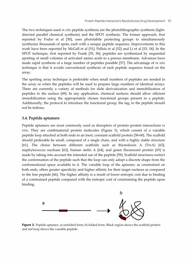
3.4. Peptide aptamers
Peptide aptamers are most commonly used as disrupters of protein–protein interactions in
vivo. They are combinatorial protein molecules (Figure 3), which consist of a variable
peptide loop attached at both ends to an inert, constant scaffold protein [58-60]. The scaffold
should preferable be small, composed of a single chain, and with a highly stable structure
[61]. The choice between different scaffolds such as thioredoxin A (TrxA) [62],
staphylococcus nuclease [63], human stefin A [64], and green fluorescent protein [65] is
made by taking into account the intended use of the peptide [59]. Scaffold structures restrict
the conformation of the peptide such that the loop can only adopt a discrete shape from the
conformational space available to it. The variable loop of the aptamer, as constrained on
both ends, offers greater specificity and higher affinity for their target surfaces as compared
to the free peptide [66]. The higher affinity is a result of lower entropic cost due to binding
of a constrained peptide compared with the entropic cost of constraining the peptide upon
binding.
Figure 3. Peptide aptamer, a) unfolded form, b) folded form. Black region shows the scaffold protein
and red loop shows the variable peptide.
Binding Protein 56
Once the scaffold has been chosen, the three basic principles followed in aptamer design are
(i) generation of a pool of peptides commonly using combinatorial approaches, (ii) selection
to find best candidates, and (iii) amplification by expression in bacterial cells, such as
Escherichia coli [59].
4. Toward a peptide drug with better bioavailability and stability
Peptides are gaining increasing attention as drug leads over small molecule drugs featured
by their high affinity and specificity to interact with their targets together with their low
toxicity profiles [67]. Unfortunately, the major limitations encountered in stability and
delivery, overshadow their remarkable success. To be competitive and profitable, the lead
peptide, which is usually designed based on the protein – protein interface, needs to be
improved for better cell membrane permeability and stability and ADME (Absorption,
Distribution, Metabolism, Excretion) properties [4, 68, 69]. Initial attempts were mainly
focused on the improvement of existing peptide leads but the need for peptides with better
physicochemical properties and pharmacokinetic profiles eventually has given rise to the
field of peptidomimetics, the development of small peptide like compounds with the ability
to mimic the structure or action of the raw peptide. The engineering of the desired property
carries the new small molecule beyond the capabilities of the raw peptide lead [70].
4.1. Improvement of half life and stability
Peptides can display half lives as short as a few minutes, which is usually too short deliver
sufficient drug amounts to target tissues. Consequently, many peptide drugs with exciting
pharmacological activities prove to be ineffective in vivo. The short half life, which renders
the peptide ineffective, is primarily due to its fast renal clearance, connected to its hydrophilic
property and small size, and its poor metabolic stability and biodegradability as a result of
enzymatic degradation by proteolytic enzymes (proteases and peptidases) of the blood,
liver, and kidney. Hence different strategies for targeted modifications of peptide drugs in
order to prolong their plasma half lives are highly demanded to improve drugs’
pharmacokinetic profiles. [67, 71].
In performing a modification to a peptide drug to protect it from proteolytic cleavage, each
peptide drug should be considered as a separate entity since, based on the sequence, each is
a target for a different group of enzymes. This makes the detailed knowledge of proteases,
their tissue localization and cleavage specificity very essential [71]. Only then can
modifications on a particular drug be imposed to improve its susceptibility towards
proteolytic enzymes targeting it. Protease resistance can be conferred by substituting the
natural amino acids by unnatural amino acids (D-), an N-methyl-alpha-amino acid, or a
beta-amino acid. The amide bond between two amino acids may be replaced. The N- or C-
termini may be blocked or carbohydrate chains can be added. N-terminus may be esterified
or pegylated. In addition, controlled release parenteral delivery, mucosal delivery and
transdermal delivery have emerged as alternative strategies to oral delivery which exposes
the peptide to stomach acid [4].

Protein-Peptide Interactions Revolutionize Drug Development 57
Constraining a peptide from an unstructured ensemble of many configurations to a fixed
conformation serves two purposes; the conformational heterogeneity of the peptide in the
unbound form is reduced, hence reducing the entropic cost associated with binding and more
importantly the protease resistance of the peptide is enhanced [72]. Some example strategies
of constraining the peptide are to “staple” or crosslink the peptide to assume an alpha helical
shape [72] (Figure 4A), to cyclize the beta hairpin form [73] (Figure 4B), or to change the
backbone such that it is nonrotatable [74]. Another similar modification mimics the structure
of plant derived cyclotides, which contain a cyclic cysteine knot [75] (Figure 4C).
Figure 4. Examples of constrained peptides A) Cyclotide with three disulfide bridges shown in gold
[76] 2k7g B) Cyclic beta-hairpin 2ns4 [77] C) Stapled peptide with the so-called staple, or hydrocarbon
link shown in gold [78] 2yja
In many cases, modification of the peptide drug significantly increases enzymatic stability,
but activity loss is always an issue. Therefore it is important that improved stability
counterbalances activity loss. For this reason, co-administration of peptides with inhibitors
of enzymes that target the peptide might be offered as an attractive alternative tool to
chemical modifications to increase peptide stability [79-81].
4.2. Enhancing uptake and delivery
An important challenge in the design and development of peptide based drugs is their size
and hydrophilic character, preventing their spontaneous uptake by the cell. For peptides
which target membrane receptors, delivery to the target sites may be made possible with the
application of liposomes or nano- and microparticles. On the other hand, in case of peptide
drugs that target intracellular proteins, intracellular delivery through the biological
membrane is crucial for their efficacy. Since poor uptake and limited delivery has been an
important drawback hampering the acceptance of peptide drugs in the pharmaceutical
market, different approaches have been proposed to address this problem.
Sustained delivery systems based on biodegradable polymers from renewable resourses
such as chitosan and its derivatives, from petroleum resources such as PLGA (poly-lactic-co-
glycolic acid) or PGA (polyglycolide) or blends of these have been receiving increasing
attention following the nanotechnological advances applicable to peptide delivery [82-84].
As an example, progress made in the use of chitosan in peptide delivery is detailed below.
Binding Protein 58
The polysaccharide based chitosan is a nontoxic linear polymer composed of -1,4 linked
D-glucosamine derived from the deacetylation of the naturally occurring polymer chitin.
The biodegradable, biocompatible, bioadhesive, and permeation enhancing properties have
made chitosan and its derivatives, such as N-trimethyl chitosan, outstanding polymers for
delivery [85]. In addition to nasal, pulmonary, transdermal, and parenteral delivery routes
using chitosan-based nano- and microparticle carriers, chitosan coated particles or
pegylated chitosan particles receive particular interest for the delivery of peptides [83, 86].
Use of chitosan-based nano- and microparticles for peptide antigens based on luteinizing
hormone-releasing hormone [87], peptide hormone insulin [88], glutathione [89, 90],
heparin [91], and calcitonin [92] are just a few examples on the application of chitosan in
peptide delivery.
Liposomes, regarded as drug delivery vehicles, are also widely used as carriers of peptides.
They also enhance the local availability of peptides, protecting them from proteolytic
action. Liposomes are artificially prepared microscopic vesicles composed of a lipid
bilayer. The therapeutic peptide is encapsulated inside the aqueous compartment
surrounded by the lipid membrane. Various types of liposome formulations have been
prepared with different dimensions, composition, surface charge and structure to induce
specificity and cell targeting [93, 94]. Different liposome formulations were tested for the
administration of peptides such as insulin [95], calcitonin [96] and vasoactive intestinal
peptide (VIP) [97].
There is significant discussion regarding the use of cell-penetrating peptides (CPPs) as tools
to carry peptides to desired targets [37]. CPPs are usually 10-30 amino acids long and harbor
a hydrophobic and a basic region. The major advantage of CPPs over antimicrobial
peptides, which simply target the lipid membrane, is that CPPs help carry cargo into the cell
in an energy independent manner without disrupting the cell membrane, hence can target
intracellular enzymes and machinery [98, 99]. Two CPPs that have been studied in detail are
the TAT peptide (GRKKRRQRRRPPQ) [100, 101] and penetratin (RQIKIWFQNRRMKWKK)
[102, 103] . The mechanism of uptake has been proposed to be by endosomes at low
concentrations [98]. Transient pore formation, and resulting direct penetration, was also
proposed as a mechanism of uptake for Tat peptide at high concentrations [104]. In direct
penetration, the membrane is transiently destabilized by the interaction of the basic residues
(Arg) and the negatively charged components of the cell membrane. Another CPP, TP10
(also known as transportan, AGYLLGKINLKALAALAKKIL), lacks arginine residues and
has been suggested to be delivered by the endocytotic pathway or by the interaction of the
positively charged Lysine residues with the membrane [105, 106]. Structure activity
relationship studies on another CPP, pVEC (LLIILRRRIRKQAHAHSK) showed that
mutation of arginines to alanine did not abolish uptake, but scrambling the sequence of the
peptide or mutating the first five hydrophobic residues to alanine did [107]. Conjugation of
peptide drugs to CPPs is particularly relevant in the treatment of diseases which require the
relevant peptide to traverse the blood-brain barrier.

Protein-Peptide Interactions Revolutionize Drug Development 59
5. Therapeutical peptides, present and future
5.1. Current status of peptide drugs
Although the synthesis and clinical use of the first synthetic peptide, insulin, dates back to
1920s [108, 109], it had not been possible to consider peptides as potential drugs before the
introduction in 1960s by the 1984 Nobel Chemistry Prize Laureate Bruce Merrifield, of solid
phase peptide synthesis, which lowered the production costs and time [110, 111]. Currently,
the pharmaceutical industry and its contract manufacturers express their willingness to go
into larger scale production using both solid- and solution-phase strategies. Today, there are
more than 50 peptide drugs that have been approved for clinical use and the increasing
number of peptides entering clinical trials now supports the notion that peptide drugs have
a long and secure future. The targeted therapeutic areas of the present peptides include but
are not limited to oncology, metabolic, cardiovascular and infectious diseases, all of which
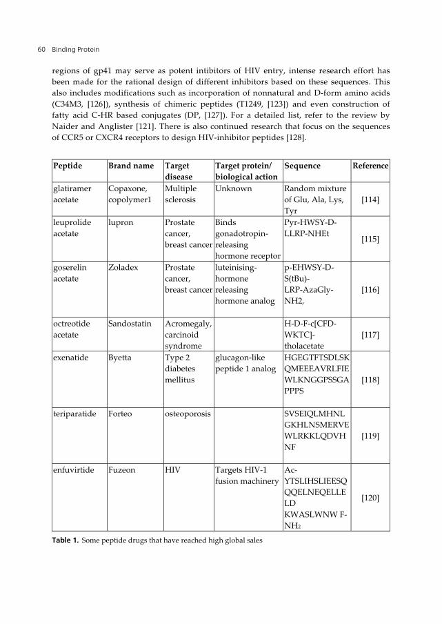
represent important markets. Table 1 includes a list of some of the peptide drugs that have
reached high global sales (Pechon et al. development trends for peptide therapeutics,
peptide therapeutics foundation, 2010 report).
Currently, most of the peptide drugs are peptide hormones (such as insulin) or peptides that
mimic hormones [4]. However, the number of peptide drugs that act as enzyme inhibitors
[112] or as antimicrobial peptides [113] is increasing.
5.2. Protein targets for potential peptide drugs
New peptide drugs are currently under development for a variety of protein targets. Here
we focus on three major disease states, namely HIV infection, cancer and Alzheimer’s
disease and discuss some of the ongoing research toward the design and development of
peptide drugs against these diseases.
Primary HIV infection starts through recognition of its envolope glycoproteins (gp120 and
gp41) by the CD4 receptors and CCR5 (macrophage) or CXCR4 (T cell) co-receptors on its
target. Upon entry, the envelope protein undergoes a major conformational change and
juxtaposes the viral and host membranes. Finally, the viral genome integrates into the
host genome. The envelope proteins is the site of primary infection, therefore fusion
inhibitor peptides blocking their interaction with the protein targets in the host might be
regarded as potential drugs [121]. With this motive, initial efforts of the early 1990s have
eventually lead to the development of the first approved anti HIV-agent originally
designated DP-178, later T-20 now FUZEON or Enfuviritide, which is a synthetic peptide
based on the C-terminal heptad repeat region (C-HR) sequence of HIV-1 gp41 (Table 1)
[120]. Although it is highly effective in vitro, its limited use due to difficulties encountered
in its administration has shown that this drug in this from is not the ultimate solution for
HIV treatment. Continued research, supported with structural studies, has shown C34,
also derived from the C-HR sequence of gp41, can compete with gp41 [122, 123]. T21 and
N36, derived from the N-terminal heptad repeat region (N-HR) were also reported to be
potent inhibitors [124, 125]. Based on the fact that peptides derived from C-HR and N-HR
Binding Protein 60
regions of gp41 may serve as potent intibitors of HIV entry, intense research effort has
been made for the rational design of different inhibitors based on these sequences. This
also includes modifications such as incorporation of nonnatural and D-form amino acids
(C34M3, [126]), synthesis of chimeric peptides (T1249, [123]) and even construction of
fatty acid C-HR based conjugates (DP, [127]). For a detailed list, refer to the review by
Naider and Anglister [121]. There is also continued research that focus on the sequences
of CCR5 or CXCR4 receptors to design HIV-inhibitor peptides [128].
Peptide Brand name Target
disease
Target protein/
biological action
Sequence Reference
glatiramer
acetate
Copaxone,
copolymer1
Multiple
sclerosis
Unknown Random mixture
of Glu, Ala, Lys,
Tyr
[114]
leuprolide
acetate
lupron Prostate
cancer,
breast cancer
Binds
gonadotropin-
releasing
hormone receptor
Pyr-HWSY-D-
LLRP-NHEt [115]
goserelin
acetate
Zoladex Prostate
cancer,
breast cancer
luteinising-
hormone
releasing
hormone analog
p-EHWSY-D-
S(tBu)-
LRP-AzaGly-
NH2,
[116]
octreotide
acetate
Sandostatin Acromegaly,
carcinoid
syndrome
H-D-F-c[CFD-
WKTC]-
tholacetate
[117]
exenatide Byetta Type 2
diabetes
mellitus
glucagon-like
peptide 1 analog
HGEGTFTSDLSK
QMEEEAVRLFIE
WLKNGGPSSGA
PPPS
[118]
teriparatide Forteo osteoporosis SVSEIQLMHNL
GKHLNSMERVE
WLRKKLQDVH
NF
[119]
enfuvirtide Fuzeon HIV Targets HIV-1
fusion machinery
Ac-
YTSLIHSLIEESQ
QQELNEQELLE
LD
KWASLWNW F-
NH2
[120]
Table 1. Some peptide drugs that have reached high global sales

Protein-Peptide Interactions Revolutionize Drug Development 61
Despite current progress, there is continued challenge in developing anti-HIV peptides due
to their rapid renal clearance, poor distribution, and susceptibility to peptidase degradation.
Hence search for HIV fusion inhibitors has been extended to screening a wide range of
different sources such as red algae Griffithsia. From the algal lectin Griffithsin, a small HIV-
1 entry inhibitor of 18-residues, Grifonin-1, was derived and this peptide was found to bind
the HIV surface glycoprotein gp120 and block its binding with host surface receptors [129].
Envelope glycoproteins and cell surface receptors do not constitute the only target to
prevent HIV infections. The cell-surface expressed nucleolin, which is one of the major RNA
binding proteins of the nucleolus and serves as a binding protein for different ligands
including HIV, might be another possible target in HIV treatment. The pentameric
pseudopeptide HB-19 was found to inhibit HIV infection by binding to the nucleolin and to
block the attachment of virus particles to cells. Hence HB-19 represents a potential anti-HIV
drug [130, 131]. Since nucleoin at the cell surface is also a binding site for a variety of ligands
implicated in tumorigenesis and angiogenesis, its potential as an anticancer drug has also
been evaluated [131].
Loss of apoptotic control has been implicated in many disease states ranging from cancer
[132] to autoimmune diseases . Caspases, a group of proteases implicated in apoptosis and
inflammatory response, are therefore an important drug target. The WEHD tetrapeptide
was found to be an optimal peptide sequence for caspase inhibitors using positional
scanning synthetic combinatorial library [133] and many variants of this sequence have been
designed. These peptidomimetic lead compounds for caspase inhibition, such as
Pralnacasan, VX-765, emricasan and NCX-1000, have been recently reviewed by MacKenzie
et al. [134]. The structure of caspase-2 in complex with a pentapeptide [135] is shown in
Figure 5.
Estrogen receptors have been a drug target in breast and endometrial cancers since they
regulate reproduction, maintain bone density and are important for central nervous system
function. Peptide inhibitors that mimic and compete with the leucine-rich pentapeptide
motif (LXXLL, where X is any residue) of the ER coactivator are promising lead compounds
in the design of selective peptide inhibitors [136]. One modification to these lead
compounds is using a hydrocarbon link to stabilize them in the alpha-helix form, also
known as a stapled peptide [78]. The structure of the estrogen receptor in complex with a
staple peptide is shown in Figure 5.
Bcl-2 is another protein involved in apoptosis and it has been the target of many drug
design efforts [3]. However, the shallow groove which interacts with its binding partners
renders it “undruggable” and therefore peptide based drug design against Bcl-2 has
emerged as a promising approach [137]. The structure of Bcl-xL in complex with a Bad
peptide is shown in Figure 5.
The phosphotyrosine recognition domain, SH2, is a subunit of many kinases, which are the
key players in important signal transduction events. In Src kinases, SH2 – kinase domain
intramolecular interaction keeps the kinase in its downregulated form [138], while in Stat3
(signal transducer and activator of transcription 3), the SH2 domain serves as a binding site

Binding Protein 62
for downstream signaling [68]. Loss of kinase regulation or constitutive kinase activation
has been implicated in cancer and autoimmune diseases. As such, SH2 is an important drug
target and SH2 inhibitor design is an area of intense research. The Stat3 SH2 recognition
sequence pYXXQ (where pY is the phosphotyrosine and X is any residue) was modified into
a lead peptidomimetic, nanomolar affinity was attained [68]. The structure of Grb2 SH2
domain in complex with a pYXN-derivative [139] is shown in Figure 5.
The proline rich AMAP-1 protein interacts with the SH3 domain of cortactin and this
complex formation is implicated in tumor invasion [140]. This interface is therefore a drug
target in breast cancer invasion and metastasis. The proline rich domain of AMAP-1 was
used to design a proline rich peptide [141], which was then made cell-permeable by the
addition of the HIV Tat sequence [142].
Figure 5. Some of the protein targets discussed in this review and their interaction with their designed
peptide ligands. Caspase (PDB code: 3r6l), estrogen receptor (2yja), Bcl-2 (1g5j) and Grb2 SH2 domain
(3ov1) are shown in silver surface representation and the peptide ligand is red.

Protein-Peptide Interactions Revolutionize Drug Development 63
The molecular origins of a number of neurodegenerative disorders such as Alzheimer’s,
Huntington’s, Parkinson’s and Creutzfeldt-Jakob diseases have been associated with the
aggregation of proteins [143, 144]. In particular, the pathological event in Alzheimer’s disease
(AD) is the progressive accumulation of 42 residue -amyloid peptides and resulting
formation of insoluble -amyloid fibrils [145]. Presently there is no cure for the treatment of
AD but novel peptides that can inhibit and or reverse this abnormal conformational change
are the subject of many recent reports. -amyloid fibrillogenesis involves the conversion of -
helix/random coil to -sheet motifs and proceeds via oligomeric and protofibrillar
intermediates therefore it can be inhibited by destabilizing the -sheet-rich -amyloid
intermediates, using -sheet breaker peptides [146-149]. One example of a -sheet breaker
peptide, which comprises a short fragment of -amyloid peptide (KLVFF; residues 16-20), can
bind full-length -amyloid peptide and prevent its assembly into amyloid fibrils [150]. The
peptide LPFFD derived from another fragment of the -amyloid peptide (LVFFA; residues
17-21) is another -sheet breaker that abolishes fibril formation [146, 147] . Permanne et al
further modified this peptide to extend the serum half-life and increase the blood-brain
barrier permeability [151] . Rationally designed hybrid molecules composed of β-breaker
elements combined with aromatic moieties (e.g. D-Trp--aminoisobutyric acid) have also
emerged as promising leads [152]. Development of beta-sheet breaker peptides have also
been reported toward diabetes 2, which is another amyloidogenic disease state [153].
6. Conclusion
As our understanding of complex biological networks increases, different proteins emerge as
potential targets for peptide based drug development. Peptides are gaining increasing
importance as drug leads over small molecule drugs featured by their high affinity and
specificity to interact with the protein targets together with their low toxicity profiles. Many
peptides that are currently administered do not meet the required criteria in cost, stability,
long serum half-life and delivery. Nevertheless they serve as a source of inspiration for the
development future generation peptide drugs. Evaluating the overall success, the increasing
number of peptide leads going into clinical trials is a significant triumph for structural biology.
Author details
Elif Ozkirimli Olmez
Bogazici University, Chemical Engineering Department, Istanbul, Turkey
Berna Sariyar Akbulut
Marmara University, Bioengineering Department, Goztepe Campus, Istanbul, Turkey
Acknowledgement
We gratefully acknowledge funding Marmara University Nihad Sayar Foundation for
Education, the Bogazici University Research Fund (09HA504P) and Tubitak Research Grant
(109M229).
Binding Protein 64
7. References
[1] Hopkins, A.L. and C.R. Groom, The druggable genome. Nat Rev Drug Discov, 2002. 1(9):
p. 727-30.
[2] Boran, A.D.W. and R. Iyengar, Systems Pharmacology. Mt Sinai J Med, 2010. 77(4): p. 333-
344.
[3] Verdine, G.L. and L.D. Walensky, The Challenge of Drugging Undruggable Targets in
Cancer: Lessons Learned from Targeting BCL-2 Family Members. Clinical Cancer Research,
2007. 13(24): p. 7264-7270.
[4] Vlieghe, P., et al., Synthetic therapeutic peptides: science and market. Drug Discov Today,
2010. 15(1-2): p. 40-56.
[5] Pawson, T., Protein modules and signalling networks. Nature, 1995. 373(6515): p. 573-80.
[6] Pawson, T., M. Raina, and P. Nash, Interaction domains: from simple binding events to
complex cellular behavior. FEBS Letters, 2002. 513(1): p. 2-10.
[7] Pawson, T., Assembly of Cell Regulatory Systems Through Protein Interaction Domains.
Science, 2003. 300(5618): p. 445-452.
[8] DeLano, W.L., Unraveling hot spots in binding interfaces: progress and challenges. Curr Opin
Struct Biol, 2002. 12(1): p. 14-20.
[9] Shoemaker, B.A. and A.R. Panchenko, Deciphering protein-protein interactions. Part I.
Experimental techniques and databases. PLoS Comput Biol, 2007. 3(3): p. e42.
[10] Berman, H.M., et al., The Protein Data Bank. Nucleic Acids Research, 2000. 28(1): p. 235-
42.
[11] Vanhee, P., et al., PepX: a structural database of non-redundant protein-peptide complexes.
Nucleic Acids Research, 2010. 38(Database): p. D545-D551.
[12] London, N., D. Movshovitz-Attias, and O. Schueler-Furman, The structural basis of
peptide-protein binding strategies. Structure, 2010. 18(2): p. 188-99.
[13] Vanhee, P., et al., Protein-peptide interactions adopt the same structural motifs as monomeric
protein folds. Structure, 2009. 17(8): p. 1128-36.
[14] Ren, R., et al., Identification of a ten-amino acid proline-rich SH3 binding site. Science, 1993.
259(5098): p. 1157-61.
[15] Castagnoli, L., et al., Selectivity and promiscuity in the interaction network mediated by
protein recognition modules. FEBS Letters, 2004. 567(1): p. 74-9.
[16] Encinar, J.A., et al., ADAN: a database for prediction of protein-protein interaction of modular
domains mediated by linear motifs. Bioinformatics, 2009. 25(18): p. 2418-24.
[17] Gorelik, M. and A.R. Davidson, Distinct Peptide Binding Specificities of Src Homology 3
(SH3) Protein Domains Can Be Determined by Modulation of Local Energetics across the
Binding Interface. J Biol Chem, 2012. 287(12): p. 9168-77.
[18] Zarrinpar, A., R.P. Bhattacharyya, and W.A. Lim, The structure and function of proline
recognition domains. Sci STKE, 2003. 2003(179): p. RE8.
[19] Zarrinpar, A., S.-H. Park, and W.A. Lim, Optimization of specificity in a cellular protein
interaction network by negative selection. Nature, 2003. 426(6967): p. 676-80.

Protein-Peptide Interactions Revolutionize Drug Development 65
[20] Stollar, E.J., et al., Structural, Functional, and Bioinformatic Studies Demonstrate the Crucial
Role of an Extended Peptide Binding Site for the SH3 Domain of Yeast Abp1p. Journal of
Biological Chemistry, 2009. 284(39): p. 26918-26927.
[21] Lim, W.A., F.M. Richards, and R.O. Fox, Structural determinants of peptide-binding
orientation and of sequence specificity in SH3 domains. Nature, 1994. 372(6504): p. 375-9.
[22] Dalgarno, D.C., M.C. Botfield, and R.J. Rickles, SH3 domains and drug design: ligands,
structure, and biological function. Biopolymers, 1997. 43(5): p. 383-400.
[23] Mayer, B.J., SH3 domains: complexity in moderation. J Cell Sci, 2001. 114(Pt 7): p. 1253-63.
[24] Gfeller, D., et al., The multiple-specificity landscape of modular peptide recognition domains.
Mol Syst Biol, 2011. 7: p. 484.
[25] Kavanaugh, W.M. and L.T. Williams, An alternative to SH2 domains for binding tyrosine-
phosphorylated proteins. Science, 1994. 266(5192): p. 1862-5.
[26] Blaikie, P., et al., A region in Shc distinct from the SH2 domain can bind tyrosine-
phosphorylated growth factor receptors. J Biol Chem, 1994. 269(51): p. 32031-4.
[27] Liu, B.A., et al., The human and mouse complement of SH2 domain proteins-establishing the
boundaries of phosphotyrosine signaling. Mol Cell, 2006. 22(6): p. 851-68.
[28] Chen, H.I. and M. Sudol, The WW domain of Yes-associated protein binds a proline-rich
ligand that differs from the consensus established for Src homology 3-binding modules. Proc
Natl Acad Sci USA, 1995. 92(17): p. 7819-23.
[29] Tonikian, R., et al., Bayesian modeling of the yeast SH3 domain interactome predicts
spatiotemporal dynamics of endocytosis proteins. PLoS Biol, 2009. 7(10): p. e1000218.
[30] Yaffe, M.B. and A.E. Elia, Phosphoserine/threonine-binding domains. Curr Opin Cell Biol,
2001. 13(2): p. 131-8.
[31] Owen, D.J., et al., The structural basis for the recognition of acetylated histone H4 by the
bromodomain of histone acetyltransferase gcn5p. EMBO J, 2000. 19(22): p. 6141-9.
[32] Bannister, A.J., et al., Selective recognition of methylated lysine 9 on histone H3 by the HP1
chromo domain. Nature, 2001. 410(6824): p. 120-4.
[33] Jaakkola, P., et al., Targeting of HIF-alpha to the von Hippel-Lindau ubiquitylation complex by
O2-regulated prolyl hydroxylation. Science, 2001. 292(5516): p. 468-72.
[34] Doyle, D.A., et al., Crystal structures of a complexed and peptide-free membrane protein-
binding domain: molecular basis of peptide recognition by PDZ. Cell, 1996. 85(7): p. 1067-76.
[35] Stec, B., et al., Structure of the wild-type TEM-1 beta-lactamase at 1.55 angstrom and the
mutant enzyme Ser70Ala at 2.1 angstrom suggest the mode of noncovalent catalysis for the
mutant enzyme. Acta Crystallographica Section D-Biological Crystallography, 2005. 61:
p. 1072-1079.
[36] Sinskey, A., S. Finkelstein, and S. Cooper, Getting to Rational Drug Design - at Last.
Pharmagenomics, 2002: p. 18-22.
[37] Mason, J.M., Design and development of peptides and peptide mimetics as antagonists for
therapeutic intervention. Future Med Chem, 2010. 2(12): p. 1813-22.
[38] London, N., et al., Can self-inhibitory peptides be derived from the interfaces of globular
protein-protein interactions? Proteins, 2010. 78(15): p. 3140-9.
Binding Protein 66
[39] Donsky, E. and H.J. Wolfson, PepCrawler: a fast RRT-based algorithm for high-resolution
refinement and binding affinity estimation of peptide inhibitors. Bioinformatics, 2011. 27(20):
p. 2836-42.
[40] Plewczynski, D., et al., Can we trust docking results? Evaluation of seven commonly used
programs on PDBbind database. J. Comput. Chem., 2011. 32(4): p. 742-55.
[41] Post, C.B., Exchange-transferred NOE spectroscopy and bound ligand structure determination.
Curr Opin Str Biol, 2003. 13: p. 581-588.
[42] Antes, I., DynaDock: A new molecular dynamics-based algorithm for protein-peptide docking
including receptor flexibility. Proteins, 2010. 78(5): p. 1084-104.
[43] Raveh, B., N. London, and O. Schueler-Furman, Sub-angstrom modeling of complexes
between flexible peptides and globular proteins. Proteins, 2010. 78(9): p. 2029-40.
[44] Molek, P., B. Strukelj, and T. Bratkovic, Peptide Phage Display as a Tool for Drug Discovery:
Targeting Membrane Receptors. Molecules, 2011. 16(1): p. 857-887.
[45] Smith, G.P. and V.A. Petrenko, Phage Display. Chem. Rev., 1997. 97(2): p. 391-410.
[46] Schumacher, T.N., et al., Identification of D-peptide ligands through mirror-image phage
display. Science, 1996. 271(5257): p. 1854-7.
[47] Panicker, R.C., X. Huang, and S.Q. Yao, Recent advances in peptide-based microarray
technologies. Comb Chem High Throughput Screen, 2004. 7(6): p. 547-56.
[48] Briant, D.J., et al., Rapid identification of linear protein domain binding motifs using peptide
SPOT arrays. Methods Mol Biol, 2009. 570: p. 175-85.
[49] Min, D.-H. and M. Mrksich, Peptide arrays: towards routine implementation. Curr Opin
Chem Biol, 2004. 8(5): p. 554-8.
[50] Fodor, S.P., et al., Light-directed, spatially addressable parallel chemical synthesis. Science,
1991. 251(4995): p. 767-73.
[51] McGall, G., et al., Light-directed synthesis of high-density oligonucleotide arrays using
semiconductor photoresists. Proc Natl Acad Sci USA, 1996. 93(24): p. 13555-60.
[52] Pellois, J.P., et al., Individually addressable parallel peptide synthesis on microchips. Nat
Biotechnol, 2002. 20(9): p. 922-6.
[53] Li, S., et al., Photolithographic synthesis of peptoids. J Am Chem Soc, 2004. 126(13): p. 4088-
9.
[54] Li, S., et al., Photolithographic synthesis of cyclic peptide arrays using a differential deprotection
strategy. Chem Commun (Camb), 2005(5): p. 581-3.
[55] Frank, R., Spot synthesis – an easy technique for the positionally addressable, parallel chemical
synthesis on a membrane support. Tetrahedron, 1992. 48: p. 9217-9232.
[56] Frank, R., The SPOT-synthesis technique. Synthetic peptide arrays on membrane supports--
principles and applications. J Immunol Methods, 2002. 267(1): p. 13-26.
[57] Winkler, D.F.H., H. Andresen, and K. Hilpert, SPOT synthesis as a tool to study protein-
protein interactions. Methods Mol Biol, 2011. 723: p. 105-27.
[58] Colas, P., et al., Genetic selection of peptide aptamers that recognize and inhibit cyclin-
dependent kinase 2. Nature, 1996. 380(6574): p. 548-50.

Protein-Peptide Interactions Revolutionize Drug Development 67
[59] Mascini, M., I. Palchetti, and S. Tombelli, Nucleic acid and peptide aptamers: fundamentals
and bioanalytical aspects. Angew Chem Int Ed Engl, 2012. 51(6): p. 1316-32.
[60] Hoppe-Seyler, F., et al., Peptide aptamers: specific inhibitors of protein function. Curr Mol
Med, 2004. 4(5): p. 529-38.
[61] Nygren, P.-A. and A. Skerra, Binding proteins from alternative scaffolds. J Immunol
Methods, 2004. 290(1-2): p. 3-28.
[62] Borghouts, C., C. Kunz, and B. Groner, Peptide aptamers: recent developments for cancer
therapy. Expert Opin Biol Ther, 2005. 5(6): p. 783-97.
[63] Norman, T.C., et al., Genetic selection of peptide inhibitors of biological pathways. Science,
1999. 285(5427): p. 591-5.
[64] Woodman, R., et al., Design and validation of a neutral protein scaffold for the presentation of
peptide aptamers. Journal of Molecular Biology, 2005. 352(5): p. 1118-33.
[65] Abedi, M.R., G. Caponigro, and A. Kamb, Green fluorescent protein as a scaffold for
intracellular presentation of peptides. Nucleic Acids Research, 1998. 26(2): p. 623-30.
[66] Crawford, M., R. Woodman, and P. Ko Ferrigno, Peptide aptamers: tools for biology and
drug discovery. Brief Funct Genomic Proteomic, 2003. 2(1): p. 72-9.
[67] Jenssen, H. and S.I. Aspmo, Serum stability of peptides. Methods Mol Biol, 2008. 494: p.
177-86.
[68] Mandal, P.K., et al., Structure-affinity relationships of glutamine mimics incorporated into
phosphopeptides targeted to the SH2 domain of signal transducer and activator of transcription
3. J. Med. Chem., 2009. 52(19): p. 6126-41.
[69] Mandal, P.K., W.S.-L. Liao, and J.S. McMurray, Synthesis of phosphatase-stable, cell-
permeable peptidomimetic prodrugs that target the SH2 domain of Stat3. Org Lett, 2009.
11(15): p. 3394-7.
[70] Liskamp, R.M.J., et al., Peptides and proteins as a continuing exciting source of inspiration for
peptidomimetics. Chembiochem, 2011. 12(11): p. 1626-53.
[71] Werle, M. and A. Bernkop-Schnürch, Strategies to improve plasma half life time of peptide
and protein drugs. Amino Acids, 2006. 30(4): p. 351-67.
[72] Henchey, L.K., A.L. Jochim, and P.S. Arora, Contemporary strategies for the stabilization of
peptides in the α-helical conformation. Curr Opin Chem Biol, 2008. 12(6): p. 692-697.
[73] Phichith, D., et al., Novel peptide inhibiting both TEM-1 β-lactamase and penicillin-binding
proteins. FEBS J, 2010. 277(23): p. 4965-72.
[74] Ward, J.M., et al., Constraining binding hot spots: NMR and molecular dynamics simulations
provide a structural explanation for enthalpy-entropy compensation in SH2-ligand binding. J
Am Chem Soc, 2010. 132(32): p. 11058-70.
[75] Poth, A.G., et al., Discovery of cyclotides in the fabaceae plant family provides new insights
into the cyclization, evolution, and distribution of circular proteins. ACS Chem Biol, 2011.
6(4): p. 345-55.
[76] Wang, C.K., et al., Combined X-ray and NMR Analysis of the Stability of the Cyclotide
Cystine Knot Fold That Underpins Its Insecticidal Activity and Potential Use as a Drug
Scaffold. Journal of Biological Chemistry, 2009. 284(16): p. 10672-10683.

Binding Protein 68
[77] Athanassiou, Z., et al., Structure-guided peptidomimetic design leads to nanomolar beta-
hairpin inhibitors of the Tat-TAR interaction of bovine immunodeficiency virus. Biochemistry,
2007. 46(3): p. 741-51.
[78] Phillips, C., et al., Design and structure of stapled peptides binding to estrogen receptors. J Am
Chem Soc, 2011. 133(25): p. 9696-9.
[79] Pauly, R.P., et al., Improved glucose tolerance in rats treated with the dipeptidyl peptidase IV
(CD26) inhibitor Ile-thiazolidide. Metab Clin Exp, 1999. 48(3): p. 385-9.
[80] Deacon, C.F., T.E. Hughes, and J.J. Holst, Dipeptidyl peptidase IV inhibition potentiates the
insulinotropic effect of glucagon-like peptide 1 in the anesthetized pig. Diabetes, 1998. 47(5): p.
764-9.
[81] Ahrén, B., et al., Inhibition of dipeptidyl peptidase IV improves metabolic control over a 4-week
study period in type 2 diabetes. Diabetes Care, 2002. 25(5): p. 869-75.
[82] Keijzer, C., et al., PLGA, PLGA-TMC and TMC-TPP nanoparticles differentially modulate the
outcome of nasal vaccination by inducing tolerance or enhancing humoral immunity. PLoS
ONE, 2011. 6(11): p. e26684.
[83] Fonte, P., et al., Chitosan-coated solid lipid nanoparticles for insulin delivery. Meth Enzymol,
2012. 508: p. 295-314.
[84] Morishita, M. and N.A. Peppas, Is the oral route possible for peptide and protein drug
delivery? Drug Discov Today, 2006. 11(19-20): p. 905-10.
[85] Garcia-Fuentes, M., N. Csaba, and M.J. Alonso, Nanostructured Chitosan Carriers for Oral
Protein and Peptide Delivery. TOUCH BRIEFINGS, 2007: p. 16-19.
[86] Amidi, M., et al., Chitosan-based delivery systems for protein therapeutics and antigens. Adv
Drug Deliv Rev, 2010. 62(1): p. 59-82.
[87] Chua, B.Y., et al., Chitosan microparticles and nanoparticles as biocompatible delivery vehicles
for peptide and protein-based immunocontraceptive vaccines. Mol Pharm, 2012. 9(1): p. 81-90.
[88] Mahjub, R., et al., Preparation, statistical optimization, and in vitro characterization of insulin
nanoparticles composed of quaternized aromatic derivatives of chitosan. AAPS PharmSciTech,
2011. 12(4): p. 1407-19.
[89] Ieva, E., et al., Analytical characterization of chitosan nanoparticles for peptide drug delivery
applications. Anal Bioanal Chem, 2009. 393(1): p. 207-15.
[90] Trapani, A., et al., A comparative study of chitosan and chitosan/cyclodextrin nanoparticles as
potential carriers for the oral delivery of small peptides. Eur J Pharm Biopharm, 2010. 75(1):
p. 26-32.
[91] Krauland, A.H. and M.J. Alonso, Chitosan/cyclodextrin nanoparticles as macromolecular
drug delivery system. Int J Pharm, 2007. 340(1-2): p. 134-42.
[92] Prego, C., et al., Chitosan-PEG nanocapsules as new carriers for oral peptide delivery. Effect of
chitosan pegylation degree. J Control Release, 2006. 111(3): p. 299-308.
[93] Ali, M. and N. Manolios, Peptide Delivery Systems. Letters in Peptide Science, 2002. 8: p.
289-294.
[94] Tan, M.L., P.F.M. Choong, and C.R. Dass, Recent developments in liposomes, microparticles
and nanoparticles for protein and peptide drug delivery. Peptides, 2010. 31(1): p. 184-93.

Protein-Peptide Interactions Revolutionize Drug Development 69
[95] Jain, A.K., et al., Muco-adhesive multivesicular liposomes as an effective carrier for
transmucosal insulin delivery. J Drug Target, 2007. 15(6): p. 417-27.
[96] Werle, M. and H. Takeuchi, Chitosan-aprotinin coated liposomes for oral peptide delivery:
Development, characterisation and in vivo evaluation. Int J Pharm, 2009. 370(1-2): p. 26-32.
[97] Hajos, F., et al., Inhalable liposomal formulation for vasoactive intestinal peptide. Int J Pharm,
2008. 357(1-2): p. 286-94.
[98] Madani, F., et al., Mechanisms of Cellular Uptake of Cell-Penetrating Peptides. Journal of
Biophysics, 2011. 2011: p. 1-10.
[99] Guterstam, P., et al., Elucidating cell-penetrating peptide mechanisms of action for membrane
interaction, cellular uptake, and translocation utilizing the hydrophobic counter-anion
pyrenebutyrate. Biochimica Et Biophysica Acta-Biomembranes, 2009. 1788(12): p. 2509-
2517.
[100] Vivès, E., P. Brodin, and B. Lebleu, A truncated HIV-1 Tat protein basic domain rapidly
translocates through the plasma membrane and accumulates in the cell nucleus. J Biol Chem,
1997. 272(25): p. 16010-7.
[101] Futaki, S., Arginine-rich peptides: potential for intracellular delivery of macromolecules and
the mystery of the translocation mechanisms. International Journal of Pharmaceutics, 2002.
245(1-2): p. 1-7.
[102] Derossi, D., et al., The third helix of the Antennapedia homeodomain translocates through
biological membranes. J Biol Chem, 1994. 269(14): p. 10444-50.
[103] Alves, I.D., et al., Cell biology meets biophysics to unveil the different mechanisms of
penetratin internalization in cells. Biochim Biophys Acta, 2010. 1798(12): p. 2231-9.
[104] Herce, H.D. and A.E. Garcia, Molecular dynamics simulations suggest a mechanism for
translocation of the HIV-1 TAT peptide across lipid membranes. Proceedings of the National
Academy of Sciences of the United States of America, 2007. 104(52): p. 20805-20810.
[105] Padari, K., et al., Cell transduction pathways of transportans. Bioconjug Chem, 2005. 16(6):
p. 1399-410.
[106] Yandek, L.E., et al., Mechanism of the Cell-Penetrating Peptide Transportan 10 Permeation
of Lipid Bilayers. Biophysical Journal, 2007. 92(7): p. 2434-2444.
[107] Elmquist, A., M. Hansen, and Ü. Langel U, Structure-activity relationship study of the cell-
penetrating peptide pVEC. Biochim Biophys Acta, 2006. 1758(6): p. 721-9.
[108] Banting, F.G., et al., Pancreatic Extracts in the Treatment of Diabetes Mellitus. Can Med
Assoc J, 1922. 12(3): p. 141-6.
[109] Thomson, A.P., THE CLINICAL USE OF INSULIN. Br Med J, 1924. 1(3298): p. 457-60.
[110] Albericio, F., Developments in peptide and amide synthesis. Curr Opin Chem Biol, 2004.
8(3): p. 211-21.
[111] Merrifield, B., Solid phase synthesis. Science, 1986. 232(4748): p. 341-7.
[112] Lovshin, J.A. and D.J. Drucker, Incretin-based therapies for type 2 diabetes mellitus. Nature
Reviews Endocrinology, 2009. 5(5): p. 262-269.
[113] Nguyen, L.T., E.F. Haney, and H.J. Vogel, The expanding scope of antimicrobial peptide
structures and their modes of action. Trends in Biotechnology, 2011. 29(9): p. 464-72.

Binding Protein 70
[114] Johnson, K.P., et al., Copolymer 1 reduces relapse rate and improves disability in relapsing-
remitting multiple sclerosis: results of a phase III multicenter, double-blind placebo-controlled
trial. The Copolymer 1 Multiple Sclerosis Study Group. Neurology, 1995. 45(7): p. 1268-76.
[115] Arnold, A.J. and A.D. Desmond, Gonadotrophin hormone releasing analogues open new
doors in cancer treatment. Br Med J (Clin Res Ed), 1987. 295(6612): p. 1565.
[116] Furr, B.J. and R.I. Nicholson, Use of analogues of luteinizing hormone-releasing hormone for
the treatment of cancer. J Reprod Fertil, 1982. 64(2): p. 529-39.
[117] Maurer, R., et al., Opiate antagonistic properties of an octapeptide somatostatin analog. Proc
Natl Acad Sci USA, 1982. 79(15): p. 4815-7.
[118] DeFronzo, R.A., et al., Effects of exenatide (exendin-4) on glycemic control and weight over
30 weeks in metformin-treated patients with type 2 diabetes. Diabetes Care, 2005. 28(5): p.
1092-100.
[119] Neer, R.M., et al., Effect of parathyroid hormone (1-34) on fractures and bone mineral density
in postmenopausal women with osteoporosis. N Engl J Med, 2001. 344(19): p. 1434-41.
[120] Kilby, J.M., et al., Potent suppression of HIV-1 replication in humans by T-20, a peptide
inhibitor of gp41-mediated virus entry. Nat Med, 1998. 4(11): p. 1302-7.
[121] Naider, F. and J. Anglister, Peptides in the treatment of AIDS. Curr Opin Struct Biol,
2009. 19(4): p. 473-482.
[122] Chan, D.C. and P.S. Kim, HIV entry and its inhibition. Cell, 1998. 93(5): p. 681-4.
[123] Liu, S., S. Wu, and S. Jiang, HIV entry inhibitors targeting gp41: from polypeptides to small-
molecule compounds. Curr Pharm Des, 2007. 13(2): p. 143-62.
[124] Wild, C., et al., A synthetic peptide inhibitor of human immunodeficiency virus replication:
correlation between solution structure and viral inhibition. Proc Natl Acad Sci USA, 1992.
89(21): p. 10537-41.
[125] Lu, M. and P.S. Kim, A trimeric structural subdomain of the HIV-1 transmembrane
glycoprotein. J Biomol Struct Dyn, 1997. 15(3): p. 465-71.
[126] Gaston, F., et al., Development and characterization of peptidic fusion inhibitors derived from
HIV-1 gp41 with partial D-amino acid substitutions. ChemMedChem, 2009. 4(4): p. 570-81.
[127] Wexler-Cohen, Y. and Y. Shai, Demonstrating the C-terminal boundary of the HIV 1 fusion
conformation in a dynamic ongoing fusion process and implication for fusion inhibition. FASEB
J, 2007. 21(13): p. 3677-84.
[128] Dogo-Isonagie, C., et al., Peptides from the second extracellular loop of the C-C chemokine
receptor type 5 (CCR5) inhibit diverse strains of HIV-1. J Biol Chem, 2012.
[129] Micewicz, E.D., et al., Grifonin-1: a small HIV-1 entry inhibitor derived from the algal lectin,
Griffithsin. PLoS ONE, 2010. 5(12): p. e14360.
[130] Callebaut, C., et al., Inhibition of HIV infection by pseudopeptides blocking viral envelope
glycoprotein-mediated membrane fusion and cell death. Virology, 1996. 218(1): p. 181-92.
[131] Krust, B., et al., The anti-HIV pentameric pseudopeptide HB-19 is preferentially taken up in
vivo by lymphoid organs where it forms a complex with nucleolin. Proc Natl Acad Sci USA,
2001. 98(24): p. 14090-5.

Protein-Peptide Interactions Revolutionize Drug Development 71
[132] Kerr, J.F., C.M. Winterford, and B.V. Harmon, Apoptosis. Its significance in cancer and
cancer therapy. Cancer, 1994. 73(8): p. 2013-26.
[133] Thornberry, N.A., et al., A combinatorial approach defines specificities of members of the
caspase family and granzyme B. Functional relationships established for key mediators of
apoptosis. J Biol Chem, 1997. 272(29): p. 17907-11.
[134] MacKenzie, S.H., J.L. Schipper, and A.C. Clark, The potential for caspases in drug
discovery. Curr Opin Drug Discov Devel, 2010. 13(5): p. 568-76.
[135] Tang, Y., J.A. Wells, and M.R. Arkin, Structural and enzymatic insights into caspase-2
protein substrate recognition and catalysis. J Biol Chem, 2011. 286(39): p. 34147-54.
[136] Chang, C.y., et al., Dissection of the LXXLL nuclear receptor-coactivator interaction motif
using combinatorial peptide libraries: discovery of peptide antagonists of estrogen receptors
alpha and beta. Mol Cell Biol, 1999. 19(12): p. 8226-39.
[137] Petros, A.M., et al., Rationale for Bcl-xL/Bad peptide complex formation from structure,
mutagenesis, and biophysical studies. Protein Sci, 2000. 9(12): p. 2528-34.
[138] Moarefi, I., et al., {Activation of the Src-family tyrosine kinase Hck by SH3 domain
displacement}. Nature, 1997. 385: p. 650-653.
[139] Myslinski, J.M., et al., Protein-ligand interactions: thermodynamic effects associated with
increasing nonpolar surface area. J Am Chem Soc, 2011. 133(46): p. 18518-21.
[140] Sabe, H., et al., ArfGAP family proteins in cell adhesion, migration and tumor invasion. Curr
Opin Cell Biol, 2006. 18(5): p. 558-64.
[141] Onodera, Y., et al., Expression of AMAP1, an ArfGAP, provides novel targets to inhibit
breast cancer invasive activities. EMBO J, 2005. 24(5): p. 963-973.
[142] Hashimoto, S., et al., Targeting AMAP1 and cortactin binding bearing an atypical src
homology 3/proline interface for prevention of breast cancer invasion and metastasis. Proc Natl
Acad Sci USA, 2006. 103(18): p. 7036-41.
[143] Selkoe, D.J., Folding proteins in fatal ways. Nature, 2003. 426(6968): p. 900-4.
[144] Wetzel, R., Nucleation of huntingtin aggregation in cells. Nat Chem Biol, 2006. 2(6): p. 297-
8.
[145] Haass, C., The molecular significance of amyloid beta-peptide for Alzheimer's disease. Eur
Arch Psychiatry Clin Neurosci, 1996. 246(3): p. 118-23.
[146] Soto, C., et al., Inhibition of Alzheimer's amyloidosis by peptides that prevent beta-sheet
conformation. Biochem Biophys Res Commun, 1996. 226(3): p. 672-80.
[147] Soto, C., et al., Beta-sheet breaker peptides inhibit fibrillogenesis in a rat brain model of
amyloidosis: implications for Alzheimer's therapy. Nat Med, 1998. 4(7): p. 822-6.
[148] Adessi, C., et al., Pharmacological profiles of peptide drug candidates for the treatment of
Alzheimer's disease. J Biol Chem, 2003. 278(16): p. 13905-11.
[149] Bieler, S. and C. Soto, Beta-sheet breakers for Alzheimer's disease therapy. Curr Drug
Targets, 2004. 5(6): p. 553-8.
[150] Tjernberg, L.O., et al., Arrest of beta-amyloid fibril formation by a pentapeptide ligand. J Biol
Chem, 1996. 271(15): p. 8545-8.

Binding Protein 72
[151] Permanne, B., et al., Reduction of amyloid load and cerebral damage in a transgenic mouse
model of Alzheimer's disease by treatment with a beta-sheet breaker peptide. FASEB J, 2002.
16(8): p. 860-2.
[152] Frydman-Marom, A., et al., The generic amyloid formation inhibition effect of a designed
small aromatic β-breaking peptide. Amyloid, 2011. 18(3): p. 119-27.
[153] Porat, Y., et al., Inhibition of islet amyloid polypeptide fibril formation: a potential role for
heteroaromatic interactions. Biochemistry, 2004. 43(45): p. 14454-62.