Protein modification and trafficking There are two types ... · 1 Protein modification and...
8
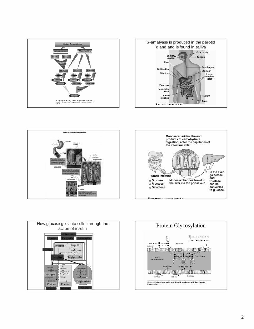
1 Protein modification and trafficking There are two types of glycosylation N-glycosidic bonds form via an N-glycosidic linkage is through the amide group of asparagine and the carboxyl group of N-acetylglucosamine Protein Glycosylation
Transcript of Protein modification and trafficking There are two types ... · 1 Protein modification and...

1
Protein modification and trafficking
There are two types of glycosylation
N-glycosidic bonds form via an N-glycosidic linkage is through the amide group of asparagine and the carboxyl group of N-acetylglucosamine
Protein Glycosylation
2
α-amalyase is produced in the parotid gland and is found in saliva
How glucose gets into cells through the action of insulin
Glucose Fatty acids Amino acids
Liver
Most cells AdiposeMuscle
Insulin
Intestine
Glycogen
Triglycerides
Glycogen
TriglyceridesProteins Proteins
Protein Glycosylation

3
Where does Dolichol come from?
• Dolichol is an isoprenoid compound synthesized by the same metabolic route as cholesterol. In vertebrate tissues, dolichol contains 18-20 isoprenoid units (90-100 carbons total). Dolichol is phosphorylated by a kinase that uses CTP to form dolichol Phosphate. Dolichol phosphate is the structure upon which the carbohydrate moieties of N-linked glycoproteins are built. After assembly on dolicholphosphate, the carbohydrate structure is transferred to an asparagine residue of a target protein having the sequence Asn-x-Ser/Thr, where X is any amino acid.
Cholesterol Biosynthesis
Dolichol

4
Disulfide bonds form between cysteines
• PDI protein disulfide isomerase works in the ER. In the cytosol most Cystines are in the reduced state partly because of active oxygen radical scavengers. In the ER PDI works by forming disulfide bonds with the target protein and then transferring that bond to another cystine within the target protein.
Further protein modification
Why glycosylation?– Aids in proper protein folding.– Provides protection against proteases (e.g. lysosomal
membrane proteins)– Employed for signaling.
• Most soluble and membrane-bound proteins made in the ER are glycoproteins, in contrast to cytsolic proteins.
• Glycoprotein synthesis is a 3-part process:1. Assembly of the precursor oligosaccharide2. En-bloc transfer to the protein3. Modification of the oligosaccharide by removal of sugars
Where does glucose come from?• Starch is the major source of dietary glucose. The enzymes
responsible for starch degradation are called amylases. Other sources of glucose are sucrose, a disaccharide glucose-fructose from fruits, and lactose, a glucose-galactose disaccharide from milk. Only monosaccharide species like glucose, fructose and galactose can be absorbed via active membrane transport systems. Special intestinal glucosidases split the disaccharides into their monosaccharide components. Maltose is hydrolyzed by isomaltase (oligo-1,6-glucosidase, E.C. 3.2.1.10) and, with lower efficacy, by sucrase (sucrose alpha-glucosidase, E.C. 3.2.1.48). Lactose intolerance comes from a lack of lactase in many adults, causing an accumulation of milk sugar with consequences such as dehydration.
Protein glycosylation1. Assembly of the precursor
oligosaccharide…
IMPORTANT POINTS:– Assembly takes place on the carrier
lipid dolichol, anchored in the ER membane.
– A pyrophosphate bridge joins the 1st sugar to the dolichol.
– Sugars are added singly and sequentially.
– After the two N-acetylglucos-amines are added, the assembly flips from the cytosolic side to the ER lumen.
– Nine mannose and three glucose molecules are added, totaling 14 sugars.
Now in the second step, attachment
2. En-bloc transfer of the oligosaccharide to the protein
• One step transfer, catalyzed by oligosaccharyl transferase, which is bound to the membrane at the translocator.
• Covalently attached to certain asparagines in the polypeptide chain (said to be “N-linked” glycosylation).
• Attaches to NH2 side chain of Asn but only in the context:
Asn-x-Ser or Asn-x-Thr

5
Finally modification of oligosaccharide
Modification of the oligosaccharide by removal of sugars…
• Three glucoses and one mannose are removed sequentially in the ER.
Transport from the ER to Golgi• Appropriately modified proteins leave the ER and travel to
the Golgi Apparatus.• They travel in membrane vesicles that arise from special
regions of membranes that are coated by proteins. • There are of three types of coated vesicles that are well
characterized, clathrin-coated, COPI-coated and COPII-coated vesicles.
• COPI and COPII act mainly in ER or Golgi cisternae.• Clathrin acts in Golgi or plasma membranes.
Clathrin coated vesicles
TGN is trans Golgi network
Clathrin cycle
Clathrin adaptins and dynaminWhen the protein is properly folded, COPII coated vesicles transport the proteins via the vesicular tubular cluster (vtc) to the cis-Golgi network.
•The COPII coating is removed (Sar1 hydolyzes GTP) and the vesicles fuse with each other to form the vtc. •The vtc is motored along microtubules that function like railroad tracks.•The vtc fuses with the cis-Golgi network.
Transport from the ER to the Golgi

6
Some proteins exiting the ER are returned to the ER by COPI coated vesicles. These proteins are identified by the presence of specific signal sequences that interact with the COPI vesicles or associate with specific receptors.
Example of retrieved protein:ER chaperones like BiP that are mistakenly transported.
This example describes the situation of BiP. BiP has the signal sequence, KDEL. When BiP escapes the ER, it associates with the KDEL receptor. The slightly acid environment of the vtc and Golgi favor this association. When the returning vesicle fuses with the ER, the neutral pH of the ER causes BiP to dissociate from the receptor.
Proteins exiting the ER join the Golgi apparatus at the cis Golgi network. The Golgi apparatus consists of a collection of stacked compartments.
nucleus
Cell membrane
The Golgi Apparatus has two major functions:1. Modifies the N-linked oligosaccharides and adds O-linked oligosaccharides.2. Sorts proteins so that when they exit the trans Golgi network, they are delivered to the correct destination.
Modification of the N-linked oligosaccharides is done by enzymes in the lumen of various Golgi compartments.
While N-linked glycosylation appears to function in protein-folding in the lumen of the ER, the function of the oligosaccharide modifications occurring in the Golgi is largely unknown.
One ultimate destination of some proteins that arrive in the TGN is the lysosome. These proteins include acid hydrolases.
Lysosomes are like the stomach of the cell. They are organelles surrounded by a single membrane and filled with enzymes called acid hydrolases that digest (degrade) a variety of macromolecules. A vacular H+ ATPase pumps protons into the lysosome causing the pH to be ~5.
The macromolecules that are degraded in the lysosomearrive by endocytosis, phagocytosis, or autophagy.

7
The acid hydrolases in the lysosome are sorted in the TGN based on the chemical marker mannose 6-phosphate.
This was first attached in the ER.
The phosphate is added in the Golgi
Adaptins bridge the M6P receptor to clathrin.
Hydrolases are transported to the late endosome which later matures into a lysosome.
Acidic pH causes hydrolase to dissociate from the receptor.
The creation of the M6P marker in the Golgi relies on recognition of a signal patch in the tertiary structure of the hydrolase.
Patients with a disease called inclusion-cell disease have cells lacking hydrolases in their lysosomes. Instead, the hydrolases are found in the blood. These patients lack GlcNAc phophotransferase. Without the M6P-tag, the acid hydrolases are transported to the plasma membrane instead of the late endosome.
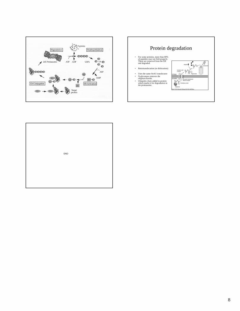
Ubiquitin pathway for protein degradation
E1 + ATP + Ub -----------> E1.Ub-AMP + PPi
E1.Ub-AMP + Ub ----------> E1-s-co-Ub.AMP-Ub
E1-s-co-Ub.AMP-Ub + E2-SH -----> E2-s-co-Ub + E1.AMP-Ub
E2-s-co-Ub + Protein-NH2 -------> E2-SH + Protein-NH-CO-Ub
Thioester bond Isopeptide bond
8
Protein degradation• For some proteins, more than 80%
of peptides may not fold properly. These are removed from the ER and degraded.
• Retrotranslocation (or dislocation)
• Uses the same Sec61 translocator• N-glycanase removes the
oligosaccharide.• Ubiquitin chain added to protein
which marks it for degradation in the proteasome.
END


![1 General Aspects of the Glycosidic Bond · PDF fileof the leaving-group development: ... and novel thio- [59,60] and O-imidates ... 4j 1 General Aspects of the Glycosidic Bond Formation](https://static.fdocuments.us/doc/165x107/5aae30607f8b9a6b308bb5cb/1-general-aspects-of-the-glycosidic-bond-the-leaving-group-development-and.jpg)