Protein mass spectrometry for bioprocess development...
205
Protein mass spectrometry for bioprocess development & monitoring A thesis submitted to University College London for the degree of DOCTOR OF ENGINEERING Alex Berrill September, 2009
Transcript of Protein mass spectrometry for bioprocess development...

Protein mass spectrometry for bioprocess
development & monitoring A thesis submitted to University College London for the degree of
DOCTOR OF ENGINEERING
Alex Berrill
September, 2009

Protein mass spectrometry for bioprocess development & monitoring
2
I, Alex Berrill confirm that the work presented in this thesis is my own. Where
information has been derived from other sources, I confirm that this has been
indicated in the thesis.

Protein mass spectrometry for bioprocess development & monitoring
3
ABSTRACT Bioprocesses for therapeutic protein production typically require significant
resources to be invested in their development. This development could be
improved with technologies that can elucidate the physicochemical properties of
process stream components with small sample volumes in a rapid and readily
performed manner. This is especially true in early phase development when
material and established analytical methods are limiting.
This thesis has investigated various process materials but was focussed
mainly on those existing in an ApolipoproteinA-IM (ApoA-IM) process, produced
using an Eschericia coli (E. coli) host.
Using a mass spectrometric technique this project began by monitoring the
product and contaminant during the ApoA-IM process and how this analytical
approach compares to traditional analytical methods such as high performance
liquid chromatography (HPLC). Results showed that, unlike many other analytical
methods, surface enhanced laser desorption ionisation mass spectrometry (SELDI-
MS) can handle early process samples that contain complex mixtures of biological
molecules with limited sample pre-treatment and thereby provide meaningful
process-relevant information.
The change in material during the flocculation/centrifugation stage of the
process was then examined. When only a change in cellular debris was observed an
existing methodology developed at University College London (UCL) was
implemented to maximise cellular debris removal. The predictive scale down
methodology enabled rapid optimization of the operating conditions for a
flocculation followed with a centrifugation step using only small volumes (20mL) of
a high solids (~20% w/w) E. coli heat extract. These experiments suggested that
adding a higher level of a cationic polymer could substantially increase the strength
of the flocculated particles produced, thereby enhancing overall clarification
performance in a large scale centrifuge. This was subsequently validated at pilot
scale.

Protein mass spectrometry for bioprocess development & monitoring
4
The proteins remaining from this flocculation/centrifugation stage were
then compared using the mass spectrometric technique to calculate the difficulty of
removing each protein contaminant from the ApoA-IM product and suggested
conditions for future sorbent scouting runs.

Protein mass spectrometry for bioprocess development & monitoring
5
ACKNOWLEDGEMENTS
This work would not have been possible without the invaluable advice, and
support of my supervisors both at UCL and Pfizer: Daniel Bracewell, Nigel Titchener-
Hooker & Sa Ho. I particularly want to thank my supervisors for the opportunity
they gave me to do part of my EngD at Pfizer in St. Louis, an experience that I will
never forget. I would also like to thank my family and friends for all their
encouragement and support over the course of my studies.
This project would also not have been possible without the support from the
Engineering and the Physical Sciences Research Council (EPSRC) under the
Innovative Manufacturing Research Initiative along with the support from Pfizer Inc.

Protein mass spectrometry for bioprocess development & monitoring
6
CONTENTS
ABSTRACT ........................................................................................................... 3
ACKNOWLEDGEMENTS ........................................................................................ 5
LIST OF FIGURES ................................................................................................ 11
LIST OF TABLES .................................................................................................. 14
LIST OF EQUATIONS ........................................................................................... 15
CHAPTER 1 INTRODUCTION ............................................................................. 16
1.1 PROTEIN PRODUCTION PROCESSES ................................................................................................ 16
1.1.1 Protein recovery ........................................................................................................... 18
1.1.2 Protein purification ....................................................................................................... 19
1.2 ULTRA SCALE-DOWN (USD) TECHNOLOGIES TO AID PROCESS DEVELOPMENT ......................................... 26
1.2.1 Determining the shear sensitivity of process material ................................................. 27
1.2.2 Determining the physicochemical properties of proteins ............................................ 28
1.2.3 Sorbent screening ......................................................................................................... 34
1.3 SELDI-MS .............................................................................................................................. 37
1.3.1 Surface enhanced affinity capture ................................................................................ 38
1.3.2 Laser desorption/ionisation .......................................................................................... 42
1.3.3 Time of flight mass spectrometry ................................................................................. 43
1.3.4 Potential uses ............................................................................................................... 46
1.3.5 Advantages ................................................................................................................... 48
1.3.6 Limitations .................................................................................................................... 49
1.4 HOST ORGANISM ....................................................................................................................... 49
1.5 PROCESS MATERIALS .................................................................................................................. 49
1.5.1 Hen egg white ............................................................................................................... 50
1.5.2 Antibody fragment process material ............................................................................ 50
1.5.3 ApolipoproteinA-IM dimer process material ................................................................ 51
1.6 PROJECT OBJECTIVES .................................................................................................................. 53
1.6.1 Objective 1: Develop the use of a mass spectrometry technology for bioprocess materials ....................................................................................................................... 54
1.6.2 Objective 2: Application of the approach to an industrial process in conjunction with a USD device .................................................................................................................... 54
1.6.3 Objective 3: Develop novel approaches for acquisition and analysis of mass spectra to expedite bioprocess development and monitoring ...................................................... 54
CHAPTER 2 CONFIGURATION OF A PROTEIN MASS SPECTROMETER FOR BIOPROCESS MATERIALS.................................................................................... 56
2.1 INTRODUCTION ......................................................................................................................... 56
2.2 MATERIALS & METHODS ............................................................................................................ 57

Protein mass spectrometry for bioprocess development & monitoring
7
2.2.1 Protein samples ............................................................................................................ 57
2.2.2 Mass spectrometry ....................................................................................................... 58
2.3 RESULTS .................................................................................................................................. 60
2.3.1 Sample analysis with mass spectrometry ..................................................................... 60
2.3.2 Width of mass spectra peaks ........................................................................................ 62
2.3.3 Alignment of mass spectra peaks ................................................................................. 64
2.3.4 The effect of protein concentration and laser strength on mass spectra peak resolution and intensity ................................................................................................ 65
2.4 SUMMARY ............................................................................................................................... 71
CHAPTER 3 PROTEIN MASS SPECTROMETRY FOR MONITORING THE APOLIPOPROTEINA-IM PROCESS ........................................................................ 73
3.1 INTRODUCTION ......................................................................................................................... 73
3.2 MATERIALS & METHODS ............................................................................................................ 75
3.2.1 Protein samples ............................................................................................................ 75
3.2.2 Mass spectrometry ....................................................................................................... 75
3.2.3 Reversed Phase HPLC .................................................................................................... 77
3.2.4 Size Exclusion HPLC ....................................................................................................... 77
3.2.5 Host cell protein analysis .............................................................................................. 77
3.3 RESULTS .................................................................................................................................. 78
3.3.1 Initial capture chromatography .................................................................................... 78
3.3.2 Ion-exchange chromatography ..................................................................................... 83
3.3.3 Oxidation ...................................................................................................................... 84
3.3.4 Anion-exchange chromatography & final step ............................................................. 84
3.3.5 Host cell protein removal ............................................................................................. 85
3.3.6 Mass spectrometry analysis during flocculation process ............................................. 86
3.4 SUMMARY ............................................................................................................................... 88
CHAPTER 4 DEFINING & IMPROVING THE FLOCULATION & CENTRIFUGATION STAGE OF THE APOLIPOPROTEINA-IM PROCESS USING A USD DEVICE ................ 90
4.1 INTRODUCTION ......................................................................................................................... 90
4.2 MATERIALS & METHODS ............................................................................................................ 91
4.2.1 Bench-top studies ......................................................................................................... 91
4.2.2 Pilot plant studies ......................................................................................................... 93
4.2.3 Microscope images ....................................................................................................... 94
4.3 THEORETICAL CONSIDERATIONS .................................................................................................... 94
4.3.1 Sigma theory ................................................................................................................. 94
4.3.2 Hindered settling .......................................................................................................... 95
4.4 RESULTS .................................................................................................................................. 97
4.4.1 Evaluation of shear ....................................................................................................... 97

Protein mass spectrometry for bioprocess development & monitoring
8
4.4.2 Adoption of a hindered settling correction factor ........................................................ 99
4.4.3 Comparing % clarification between pilot scale and USD mimic ................................... 99
4.4.4 Varying flocculating agent addition rate for both scales ............................................ 100
4.4.5 Varying flocculating agent addition ratio with USD mimic ......................................... 103
4.4.6 Validation of USD flocculating agent addition ratio at pilot scale .............................. 106
4.5 SUMMARY ............................................................................................................................. 107
CHAPTER 5 DEVELOPING AN EXPERIMENTAL METHODOLOGY TO DEFINE PRODUCT AND CONTAMINANT RELATIONSHIPS .............................................. 109
5.1 INTRODUCTION ....................................................................................................................... 109
5.2 MATERIALS & METHODS .......................................................................................................... 110
5.2.1 Reducing pH below the pI of the ApoA-IM monomer ................................................ 110
5.2.2 Mass spectrometry ..................................................................................................... 110
5.3 RESULTS ................................................................................................................................ 112
5.3.1 Selection of wash step solutions for hydrophobic ProteinChips ................................ 112
5.3.2 Selection of final wash step solutions for all of the selective ProteinChips ............... 117
5.3.3 Final decision on which buffers to use for selective ProteinChips ............................. 120
5.3.4 Selective adsorption profiles for the ApoA-IM post centrifuge supernatant proteins120
5.4 SUMMARY ............................................................................................................................. 123
CHAPTER 6 DATABASE FOR HANDLING PROTEIN MASS SPECTRA ................... 125
6.1 INTRODUCTION ....................................................................................................................... 125
6.2 MATERIALS & METHODS .......................................................................................................... 126
6.2.1 Mass spectrometry ..................................................................................................... 126
6.2.2 Data treatment ........................................................................................................... 127
6.3 RESULTS ................................................................................................................................ 127
6.3.1 Organisation & storage of mass spectrometry data ................................................... 127
6.3.2 Program execution...................................................................................................... 129
6.3.3 Treatment of raw mass spectrometry data ................................................................ 129
6.3.4 Reproducibility ............................................................................................................ 135
6.4 SUMMARY ............................................................................................................................. 139
CHAPTER 7 MASS SPECTROMETRY TO DESCRIBE PRODUCT AND CONTAMINANT RELATIONSHIPS FOR BIOPROCESS DEVELOPMENT............................................ 140
7.1 INTRODUCTION ....................................................................................................................... 140
7.2 MATERIALS & METHODS .......................................................................................................... 141
7.2.1 Protein samples .......................................................................................................... 141
7.2.2 Mass spectrometry ..................................................................................................... 141
7.2.3 Chromatography ......................................................................................................... 142
7.2.4 Data visualisation techniques ..................................................................................... 143

Protein mass spectrometry for bioprocess development & monitoring
9
7.3 RESULTS ................................................................................................................................ 145
7.3.1 Measurement of contaminants during purification ................................................... 145
7.3.2 Generating basic physicochemical properties using mass spectrometry ................... 147
7.3.3 Visualising nearest neighbour contaminants using mass spectrometry data ............ 149
7.3.4 Identifying conditions for separating nearest neighbour contaminants using mass spectrometry .............................................................................................................. 152
7.3.5 Nearest neighbour removal analysis .......................................................................... 154
7.4 SUMMARY ............................................................................................................................. 157
CHAPTER 8 CONCLUSIONS & RECOMMENDATIONS FOR FUTURE WORK ........ 159
8.1 REVIEW OF PROJECT OBJECTIVES ................................................................................................. 159
8.1.1 Objective 1: Develop the use of a mass spectrometry technology for bioprocess materials ..................................................................................................................... 159
8.1.2 Objective 2: Application of the approach to an industrial process in conjunction with a USD device .................................................................................................................. 159
8.1.3 Objective 3: Develop novel approaches for acquisition and analysis of mass spectra to expedite bioprocess development and monitoring .................................................... 160
8.2 PUBLICATIONS ........................................................................................................................ 160
8.3 FINAL COMMENTS AND FUTURE WORK ......................................................................................... 161
8.3.1 Related to objective 1: Develop the use of a mass spectrometry technology for bioprocess materials ................................................................................................... 161
8.3.2 Related to objective 2: Application of the approach to an industrial process in conjunction with a USD device ................................................................................... 162
8.3.3 Related to objective 3: Develop novel approaches for acquisition and analysis of mass spectra to expedite bioprocess development and monitoring................................... 162
CHAPTER 9 VALIDATION OF BIOPROCESSES: IMPLICATION OF SCALE-DOWN METHODOLOGIES ............................................................................................ 166
9.1 PROCESS VALIDATION ............................................................................................................... 166
9.2 SCALE-DOWN MODELS .............................................................................................................. 166
9.2.1 Process validation scenario: Effect of contaminant properties on purification ......... 167
9.2.2 Process validation scenario: A new protein candidate for a process development team .................................................................................................................................... 167
ABBREVIATIONS .............................................................................................. 169
NOMENCLATURE ............................................................................................. 171
APPENDIX - MATLAB CODE .............................................................................. 173
READING SPECTRA DATA ....................................................................................................................... 173
Reading processed data ............................................................................................................ 180
Reading raw data ....................................................................................................................... 181
PEAK SELECTION ................................................................................................................................. 187
NEAREST NEIGHBOUR PREDICTOR ........................................................................................................... 188

Protein mass spectrometry for bioprocess development & monitoring
10
REFERENCES .................................................................................................... 192

Protein mass spectrometry for bioprocess development & monitoring
11
LIST OF FIGURES
FIGURE 1.1 TYPICAL PROTEIN BIOPROCESS OPTIONS (LIENQUEO AND ASENJO,2000). ............................................ 16
FIGURE 1.2 OPERATIONAL SEQUENCE SELECTION FOR PROTEIN PURIFICATION (ASENJO AND ANDREWS,2004). .......... 20
FIGURE 1.3 A THEORETICAL MECHANISM FOR HYDROPHOBIC INTERACTIONS. ........................................................ 24
FIGURE 1.4 THE THEORY BEHIND GEL FILTRATION (GE GEL FILTRATION PRINCIPLES AND METHODS,2003). ................. 26
FIGURE 1.5 MAPPING THE PHYSICOCHEMICAL PROPERTIES OF PROTEINS (WALL ET AL. 2002).................................. 28
FIGURE 1.6 PREDICTING DIMENSIONLESS RETENTION TIME (LIENQUEO ET AL. 2007).............................................. 31
FIGURE 1.7 A TWO DIMENSIONAL POLYACRYLAMIDE GEL (OU ET AL. 2001). ........................................................ 33
FIGURE 1.8 EXPERIMENTAL PROCEDURE FOR MICRO-BATCH ADSORPTION. ........................................................... 36
FIGURE 1.9 A MICRO-PIPETTE COLUMN (WENGER ET AL. 2007). ....................................................................... 36
FIGURE 1.10 COMPARISON OF DIFFERENT CHROMATOGRAPHY SCALES (WENGER ET AL. 2007). .............................. 37
FIGURE 1.11 SELDI SELECTIVE PROTEINCHIP PREPARATION. ............................................................................. 39
FIGURE 1.12 USING THE SELDI BIOPROCESSOR. ............................................................................................. 40
FIGURE 1.13 THE SELDI PROTEINCHIP READER.............................................................................................. 43
FIGURE 1.14 THE SELDI MASS SPECTROMETER (MERCHANT AND WEINBERGER,2000). ........................................ 44
FIGURE 1.15 TIME OF FLIGHT IN A MASS SPECTROMETER (MERCHANT AND WEINBERGER,2000). ............................ 44
FIGURE 1.16 FAB’ PRODUCTION PROCESS. ..................................................................................................... 51
FIGURE 1.17 APOA-IM AMINO ACID SEQUENCE. ............................................................................................ 52
FIGURE 1.18 THE STRUCTURE OF TRUNCATED HUMAN APOLIPOPROTEINA-I (APOA-I) (BORHANI ET AL. 1997) ......... 52
FIGURE 1.19 APOA-IM DIMER PRODUCTION PROCESS. .................................................................................... 53
FIGURE 2.1 REPRESENTATIVE MASS SPECTRA FOR ADSORBED PROTEIN SAMPLES. ................................................... 61
FIGURE 2.2 RELATIONSHIP BETWEEN M/Z AND PEAK WIDTH. ............................................................................. 63
FIGURE 2.3 THE EFFECT OF PEAK ALIGNMENT ON PARTITION REPRODUCIBILITY. ..................................................... 65
FIGURE 2.4 THE EFFECT OF PROTEIN CONCENTRATION ON PEAK INTENSITY. .......................................................... 67
FIGURE 2.5 THE EFFECT OF LASER STRENGTH ON PEAK INTENSITY. ....................................................................... 69
FIGURE 2.6 THE EFFECT OF PROTEIN CONCENTRATION AND LASER STRENGTH ON RESOLUTION. ................................. 70
FIGURE 3.1 APOA-IM DIMER PRODUCTION PROCESS SAMPLES. ......................................................................... 74
FIGURE 3.2 CALCULATION OF SUMMED PEAK INTENSITY. ................................................................................... 76

Protein mass spectrometry for bioprocess development & monitoring
12
FIGURE 3.3 MASS SPECTRA FOR APOA-IM DIMER PRODUCTION PROCESS SAMPLES. .............................................. 79
FIGURE 3.4 APOA-IM MONOMER QUANTIFICATION USING MASS SPECTROMETRY AND RP-HPLC. ........................... 81
FIGURE 3.5 MASS SPECTRA OF USD FLOCCULATION SAMPLES. ........................................................................... 87
FIGURE 4.1 MICROSCOPE IMAGES OF FLOCCULATED APOA-IM MATERIAL EXPOSED TO SHEAR. ................................ 98
FIGURE 4.2 DEVELOPING A MIMIC OF THE APOA-IM DIMER RECOVERY PROCESS ................................................... 98
FIGURE 4.3 COMPARING THE USD % CLARIFICATION WITH PILOT SCALE ............................................................ 100
FIGURE 4.4 VARYING FLOCCULATING AGENT ADDITION RATE AT BOTH SCALES. .................................................... 102
FIGURE 4.5 USING THE USD MIMIC TO STUDY THE EFFECT OF VARYING THE RATIO OF FLOCCULATING AGENT ADDITION ON
THE LABORATORY CENTRIFUGE INDEX. ......................................................................................................... 104
FIGURE 4.6 USING THE USD MIMIC TO STUDY THE EFFECT OF VARYING THE RATIO OF FLOCCULATING AGENT ADDITION ON
THE % CLARIFICATION. ............................................................................................................................. 105
FIGURE 4.7 VALIDATING AT PILOT SCALE THE USD PREDICTIONS ON THE EFFECT OF FLOCCULATING AGENT ADDITION
RATIO.................................................................................................................................................... 106
FIGURE 5.1 STEPS FOR SELECTIVE PROTEINCHIP PREPARATION. ........................................................................ 111
FIGURE 5.2 COMPETITIVE ADSORPTION OF BSA AND LYSOZYME TO A HYDROPHOBIC PROTEINCHIP USING REVERSE PHASE
SOLVENTS. ............................................................................................................................................. 113
FIGURE 5.3 NON-COMPETITIVE ADSORPTION OF BSA TO A HYDROPHOBIC PROTEINCHIP USING TFA OR AMMONIUM
SULPHATE. ............................................................................................................................................. 114
FIGURE 5.4 COMPETITIVE ADSORPTION OF APOA-IM POST CENTRIFUGE SUPERNATANT PROTEINS TO A HYDROPHOBIC
PROTEINCHIP WITH ACN OR AMMONIUM SULPHATE. .................................................................................... 116
FIGURE 5.5 NON-COMPETITIVE ADSORPTION OF BSA TO A HYDROPHOBIC PROTEINCHIP BY VARYING AMMONIUM
SULPHATE CONCENTRATION AND INCUBATION TIME. ...................................................................................... 118
FIGURE 5.6 VARYING THE FINAL WASH STEP FOR ADSORBED APOA-IM POST CENTRIFUGE SUPERNATANT PROTEINS. .. 119
FIGURE 5.7 CONDITION ARRAY FOR PROFILING POST CENTRIFUGE SUPERNATANT ................................................. 121
FIGURE 5.8 MASS SPECTRA FOR ADSORBED APOA-IM POST CENTRIFUGE SUPERNATANT PROTEINS USING CONDITIONS IN
FIGURE 5.7. ........................................................................................................................................... 122
FIGURE 6.1 OPTIONS FOR MASS SPECTRA DATA TREATMENT. ........................................................................... 126
FIGURE 6.2 IDENTIFICATION CODE FOR MASS SPECTRA FILES. ........................................................................... 128
FIGURE 6.3 UPLOADING MASS SPECTRA DATA INTO THE MATLAB ENVIRONMENT. ................................................ 129
FIGURE 6.4 PROCESSING MASS SPECTRA DATA WITHIN THE MATLAB ENVIRONMENT. ........................................... 130
FIGURE 6.5 RAW MASS SPECTRA FOR ADSORBED APOA-IM POST CENTRIFUGE SUPERNATANT PROTEINS. ................. 131
FIGURE 6.6 MASS SPECTRA SIGNAL NOISE FOR ADSORBED APOA-IM POST CENTRIFUGE SUPERNATANT PROTEINS. ..... 132

Protein mass spectrometry for bioprocess development & monitoring
13
FIGURE 6.7 DENOISED MASS SPECTRA FOR ADSORBED APOA-IM POST CENTRIFUGE SUPERNATANT PROTEINS. .......... 133
FIGURE 6.8 DENOISED & BASELINED MASS SPECTRA FOR ADSORBED APOA-IM POST CENTRIFUGE SUPERNATANT
PROTEINS. .............................................................................................................................................. 134
FIGURE 6.9 PEAK REPRODUCIBILITY BETWEEN SPOT PARTITIONS FOR ADSORBED APOA-IM POST CENTRIFUGE
SUPERNATANT PROTEINS. .......................................................................................................................... 136
FIGURE 6.10 PEAK REPRODUCIBILITY BETWEEN PROTEINCHIPS FOR ADSORBED APOA-IM POST CENTRIFUGE
SUPERNATANT PROTEINS. .......................................................................................................................... 138
FIGURE 7.1 A STAR PLOT EXAMPLE FROM THE MATHWORKS INC. (NATICK, MA, U.S.A.) ..................................... 143
FIGURE 7.2 DENDROGRAM FORMATION ...................................................................................................... 145
FIGURE 7.3 MASS SPECTRA FOR PROCESS CHROMATOGRAPHY SAMPLES. ............................................................ 146
FIGURE 7.4 NON-COMPETITIVE ADSORPTION OF OVALBUMIN TO ELECTROSTATICALLY CHARGED SURFACES. .............. 148
FIGURE 7.5 COMPETITIVE ADSORPTION OF HEN EGG WHITE PROTEINS TO ELECTROSTATICALLY CHARGED SURFACES. ... 149
FIGURE 7.6 STAR PLOTS FOR THIRTY PROTEINS CONTAINED WITHIN POST CENTRIFUGE SUPERNATANT. ..................... 151
FIGURE 7.7 DENDROGRAM TO SHOW RELATIONSHIPS BETWEEN POST CENTRIFUGE SUPERNATANT PROTEINS. ............ 152
FIGURE 7.8 SELECTIVITY MAPS FOR PRODUCT AND NEAREST NEIGHBOUR CONTAMINANTS. .................................... 154
FIGURE 7.9 IMPURITY REMOVAL ANALYSIS FROM CHROMATOGRAPHY ELUTION FRACTIONS. ................................... 156
FIGURE 8.1 DEFINING CUT-OFF TO CLUSTER HARD TO REMOVE PROTEINS ........................................................... 164
FIGURE 9.1 THEORETICAL FRAMEWORK FOR THE RAPID EVALUATION OF PROTEIN CANDIDATES AT EARLY STAGE PROCESS
DEVELOPMENT. ....................................................................................................................................... 168

Protein mass spectrometry for bioprocess development & monitoring
14
LIST OF TABLES
TABLE 1.1 SEPARATION PRINCIPLES AND UNIT OPERATIONS USED IN DOWNSTREAM PROCESSING (LIENQUEO AND
ASENJO,2000). ........................................................................................................................................ 17
TABLE 1.2 INFLUENCE OF PROTEIN STABILITY ON ITS PURIFICATION (GE PROTEIN PURIFICATION,2003). ..................... 21
TABLE 1.3 COMPARING CHROMATOGRAPHY TECHNIQUES (GE PROTEIN PURIFICATION,2003). ................................ 22
TABLE 1.4 TECHNIQUES FOR DETERMINING PHYSICOCHEMICAL PROPERTIES. ......................................................... 29
TABLE 1.5 CHROMATOGRAPHY MODE SELECTION BASED ON NEW PROTEOMIC TECHNIQUES OR CONVENTIONAL
CHROMATOGRAPHY (WENGER ET AL. 2007). ................................................................................................. 35
TABLE 1.6 FACTORS IN SPA FORMULATION AND APPLICATION (CORDINGLEY ET AL. 2003). .................................... 41
TABLE 1.7 THE FOUR MAIN PROTEINS IN HEN EGG WHITE (VACHIER ET AL. 1995). ................................................. 50
TABLE 2.1 SETTINGS USED WHEN PROCESSING DATA WITH CIPHERGEN EXPRESS 3.0 .............................................. 60
TABLE 3.1 PEAKS MANUALLY DETECTED IN MASS SPECTRA. ................................................................................ 76
TABLE 3.2 QUANTIFICATION OF MONOMER AND TRUNCATED SPECIES USING MASS SPECTROMETRY AND RP-HPLC. ..... 80
TABLE 3.3 QUANTIFICATION OF MONOMER, DIMER AND AGGREGATES USING MASS SPECTROMETRY AND SE-HPLC. .... 82
TABLE 3.4 QUANTIFICATION OF HOST CELL PROTEINS USING MASS SPECTROMETRY AND ELISA. ............................... 83
TABLE 3.5 RP-HPLC ANALYSIS BEFORE AND AFTER THE PILOT SCALE FLOCCULATION. ............................................. 88
TABLE 5.1 FACTORS IN SELDI PROTEINCHIP PREPARATION. ............................................................................ 109
TABLE 5.2 EQUILIBRATION AND FINAL WASH BUFFERS FOR THE SELECTIVE PROTEINCHIPS. ..................................... 120

Protein mass spectrometry for bioprocess development & monitoring
15
LIST OF EQUATIONS
EQUATION 1.1 EMPIRICAL DIMENSIONLESS RETENTION TIME (DRT).................................................................... 30
EQUATION 1.2 THEORETICAL DIMENSIONLESS RETENTION TIME (DRT) (LIENQUEO ET AL. 2007). ............................ 31
EQUATION 1.3 EMPIRICAL PARTITION COEFFICIENTS, KI.. ................................................................................... 32
EQUATION 1.4 TIME OF FLIGHT MASS SPECTROMETER CALIBRATION. ................................................................... 45
EQUATION 2.1 WIDTH OF MASS SPECTRA PEAKS. ............................................................................................ 63
EQUATION 4.1 % CLARIFICATION FOR CENTRIFUGATION. .................................................................................. 92
EQUATION 4.2 SIGMA FOR A SWING-OUT LABORATORY CENTRIFUGE. .................................................................. 94
EQUATION 4.3 SIGMA FOR A DISC-STACK CENTRIFUGE. ..................................................................................... 95
EQUATION 4.4 COMPARING TWO DIFFERENT CENTRIFUGES. .............................................................................. 95
EQUATION 4.5 HINDERED SETTLING CORRECTION FACTOR ................................................................................. 96
EQUATION 7.1 EUCLIDEAN DISTANCE .......................................................................................................... 144
EQUATION 7.2 CALCULATING THE NEW DISTANCE FROM CLUSTER AB TO PEAK MASS C USING UPGMA. ................. 144

Protein mass spectrometry for bioprocess development & monitoring
16
CHAPTER 1 INTRODUCTION
1.1 Protein production processes
There are many different routes in the biopharmaceutical industry for
recovering and purifying protein products. Whether the protein is produced
extracellular or intracellular is critical to the route chosen (Figure 1.1). Extracellular
proteins are generally easier to purify because they can be separated from whole
cells without the need for cell breakage. This avoids the release of many host cell
proteins that would be additional impurities to remove downstream. Methods for
separation include centrifugation, microfiltration or expanded bed adsorption
(EBA). Intracellular proteins require cell disruption to be released which could
involve lysis or homogenisation (Table 1.1).
Figure 1.1 Typical protein bioprocess options (Lienqueo and Asenjo,2000).
ucylepr
TextBox
Image removed due to copyright

Protein mass spectrometry for bioprocess development & monitoring
17
Table 1.1 Separation principles and unit operations used in downstream
processing (Lienqueo and Asenjo,2000).
One of the notable difficulties with process development is the inherent
complexity caused by interactions between unit operations. Optimization of
biopharmaceutical processes tend to be based on yield or product quality (Groep et
al. 1997). Biopharmaceutical processing steps are highly interactive and optimising
individual steps is unlikely to lead to an optimal process (Dyr and
Suttnar,1997;Groep et al. 1997). Groep et al. demonstrated this by varying
conditions in the aerobic fermentation and homogenization for an alcohol
dehydrogenase (ADH) production process grown intracellularly in Saccharomyces
cerevisiae (Groep et al. 1997). An interaction was demonstrated between growth
conditions, cell strength and cell disruption.
ucylepr
Inserted Text
I
ucylepr
Inserted Text
ucylepr
TextBox
Table removed due to copyright

Protein mass spectrometry for bioprocess development & monitoring
18
1.1.1 Protein recovery
There are many varied operations for protein recovery but this thesis will
focus on flocculation and centrifugation. Flocculation allows cellular debris to form
strong, stable aggregates which can then be removed from the liquid using a single
unit operation such as centrifugation.
1.1.1.1 Flocculation
Flocculation describes the aggregation of individual particles in a suspension
due to the addition of a flocculating agent. Early work in 1969 by McGregor et al.
highlighted the factors that affect the flocculation of homogenous cultures. These
were (i) bacteria genus; (ii) suspending media; (iii) temperature; (iv) physiological
age; (v) flocculating agent (type, concentration & rate of addition); (vi) surface
shear (McGregor and Finn,1969). Most of these variables were shown to be related
to the release of proteins, polysaccharides, or nucleic acids.
Flocculation with natural polymers from micro-organisms, mineral
flocculating agents such as aluminium sulphate or synthetic polymers are
traditionally used in waste water treatment to remove microorganisms from the
effluent of activated sludge (McGregor and Finn,1969). Additives such as aluminium
sulphate have also been found to aid protein recovery from pure cultures such as
those in the biopharmaceutical industry (Richardson et al. 1990). Gasner et al.
found that strong polyelectrolytes as well as mineral hydrocolloids were the most
effective (Gasner and Wang,1970). These mineral flocculating agents however tend
to form low mechanical strength flocs rendering these agents useless for
centrifugation (Shan et al. 1996).
Synthetic flocculating agents, such as the one used in this thesis consist of
long polymeric chains that adsorb different particles in a suspension, aiding their
aggregation. These carry various charged groups that cause adsorption of the
particles. Once adsorbed the particles either bridge together or their charges are
neutralised. The polymer used in this thesis was cationic, binding to negatively
charged cellular debris to form flocs.

Protein mass spectrometry for bioprocess development & monitoring
19
Dosing of these agents is very important and there is usually an optimum
level of addition where the maximum amount of flocculation occurs. If insufficient
agent is added only some of the particles will flocculate. An over dose of flocculant,
however means many of the particles are coated with flocculant, causing the
particles to have the same charge, leading to particle repulsions and preventing
flocculation.
Floc particle strength is heavily influenced by surface shear during its
formation. During floc formation a steady-state between floc growth and breakage
is achieved. Mixing increases floc growth by increasing collision frequency but also
breaks down the large floc particles due to shear force effects (Kim et al. 2001).
The main downside of adding a flocculating agent to a bioprocess is that
regulators such as the FDA require evidence that the flocculant has been removed
downstream prior to drug formulation. Theoretically, if the flocculation is carried
out correctly most of the flocculant will be removed with the flocs during
clarification. However, undoubtedly some flocculant will remain in the product
stream and analytical strategies have to be devised to demonstrate its removal.
1.1.1.2 Centrifugation
Centrifugation uses centrifugal force to separate mixtures such as
flocculated process material. It’s frequently used in biopharmaceutical processes
mainly due to its operational robustness and low running costs. Separation is
achieved by density differences between heavy components (flocs) and lighter
components (liquor) in the process material. Centrifuge designs include multi-
chamber, tubular bowl and disc-stack. Disc-stack centrifuges have particular
benefits as they allow intermittent solids discharge removing the need for manual
dismantling of the centrifuge bowl to remove solids.
1.1.2 Protein purification
Purification typically defines the profitability of a process consuming around
60-70% of the total process operating costs (Dyr and Suttnar,1997). The extent of

Protein mass spectrometry for bioprocess development & monitoring
20
purification in a biopharmaceutical process is ultimately dictated by the product
and its end use. An injectable drug will necessitate a great deal more purification
than a food product. This is reflected in their respective manufacturing costs, which
ultimately dictate their selling price. Hence, a biopharmaceutical company is
constantly trying to meet drug purity requirements while minimising costs. This
results in a constant trade-off between resolution, capacity and speed. Product will
be lost at each step of the process so it is important to limit the number of steps to
keep the overall yield high (Bonnerjea et al. 1986). The route with the least steps to
give a higher yield is rarely found and non-optimal conditions are chosen in favour
of increased speed to market (Dyr and Suttnar,1997).
Limited knowledge of the molecular properties of process material and how
to handle such data has made the creation of a rational method to separate target
proteins from impurities difficult (Asenjo and Andrews,2004). There are many
factors involved in the decision on which combination of techniques (Figure 1.2) to
purify a target protein including the properties of the process material (Table 1.2).
Chromatography techniques are frequently used due to their high purification
power and relatively low cost.
Figure 1.2 Operational sequence selection for protein purification (Asenjo and
Andrews,2004).
ucylepr
TextBox
Image removed due to copyright

Protein mass spectrometry for bioprocess development & monitoring
21
Usually a chromatography sequence is designed so that minimal treatment
is required between chromatography steps. For example, during ion-exchange the
target protein is usually eluted in high salt conditions. It makes sense therefore to
have a hydrophobic column following this step since binding in these columns also
occurs with high salt concentrations.
Chromatography resins now exist that allow mixed-mode separation. These
resins have both hydrophobic and electrostatic properties, allowing hydrophobic
binding and electrostatic elution. This reduces the number of steps in a process by
having a single step that can take the place of both hydrophobic interaction and
ion-exchange chromatography.
Sample and target protein properties
Influence on purification strategy
Temperature stability Need to work rapidly at lowered temperature
pH stability Selection of buffers for extraction ad purification. Selection of conditions for ion exchange, affinity or reversed phase chromatography
Organic solvents stability Selection of conditions for reversed phase chromatography
Detergent requirement Consider effects on chromatographic steps and the need for detergent removal. Consider choice of detergent.
Salt (ionic strength) Selection of conditions for precipitation techniques, ion exchange and hydrophobic interaction chromatography
Co-factors for stability or activity Selection of additives, pH, salts, buffers
Protease sensitivity Need for fast removal of proteases or addition of inhibitors
Sensitivity to metal ions Need to add EDTA or EGTA to buffers
Redox sensitivity Need to add reducing agents
Molecular weight Selection of gel filtration media
Charge Selection of ion exchange conditions
Biospecific affinity Selection of ligand for affinity medium
Post-translational modifications Selection of group-specific affinity medium
Hydrophobicity Selection of medium for hydrophobic interaction chromatography
Table 1.2 Influence of protein stability on its purification (GE protein
purification,2003).

Protein mass spectrometry for bioprocess development & monitoring
22
1.1.2.1 Chromatography
Chromatography exploits differences in the physicochemical properties of
molecules present in process feeds. The three most common types of
chromatography utilise differences in charge (ion-exchange chromatography),
hydrophobicity (hydrophobic interaction chromatography) and size (size exclusion
chromatography). The choice of which type to use and at which stage of the
process in dependant on a number of factors some of which are highlighted in
Table 1.3.
Technique Main features Capture Intermediate Polish Sample start condition
Sample end condition
IEX High resolution High capacity High speed
Low ionic strength Sample volume Not limiting
High ionic strength or pH change concentrated
HIC High resolution High capacity High speed
High ionic strength Sample volume not limiting
Low ionic strength concentrated
SEC High resolution
Limited sample volume (<5% total column volume) and flow rate range
Buffer exchanged (if required) Diluted
Table 1.3 Comparing chromatography techniques (GE protein purification,2003).
1.1.2.1.1 Ion-exchange
Ion-exchange (IEX) chromatography is potentially a very high resolution
separation that has a high sample loading capacity (GE protein purification,2003).
The separation is based on the reversible adsorption of a charged protein onto an
oppositely charged chromatographic medium. Proteins are loaded onto a column
and then conditions are altered so that bound substances are eluted differentially.
This elution is usually performed by increases in salt concentration or changes in
pH. Changes are made stepwise or with a continuous gradient. Most commonly,

Protein mass spectrometry for bioprocess development & monitoring
23
samples are eluted with salt (NaCl), using a gradient elution. Target proteins are
concentrated during adsorption and collected in a purified, concentrated form.
The net surface charge of proteins varies according to the surrounding pH.
When above its isoelectric point (pI) a protein will bind to an anion-exchanger,
when below its pI a protein will bind to a cation-exchanger. Typically, IEX
chromatography is used to adsorb the target molecule, but can also be used to bind
impurities. IEX chromatography can be repeated at different pH values to separate
several proteins which have distinctly different charge properties.
1.1.2.1.2 Hydrophobic interaction
Protein binding in hydrophobic interaction chromatography (HIC) is
promoted by moderately high concentrations of anti-chaotropic salts, which also
have a stabilizing influence on protein structure (Figure 1.3). Elution can be
achieved by a linear or stepwise decrease in the salt concentration of the mobile
phase.
HIC works well in combination with ion-exchange and size exclusion
chromatography because its principle of protein adsorption is complementary.
Since it requires a minimum of sample pre-treatment steps it can also be well
utilised in processes with preceding precipitation steps. Its sensitivity is exhibited by
the fact that even non-polar groups deep within the tertiary structure of a molecule
can interact if they are incorrectly folded or damaged. This is particularly valuable
when it is necessary to purify the native form of a molecule from close derivatives.
Early developed adsorbents appeared to show a mixed mode of
hydrophobic and ionic interaction but these were later improved by removing
charged groups present in the sorbents allowing high binding of neutral proteins at
high salt concentrations to be seen. Elution was also as expected by using a salt-free
buffer or by decreasing the polarity of the eluent.
HIC sorbents are usually composed of cellulose, dextran, or cross-linked
agarose. Sorbents on the market include Phenyl and Octyl Sepharose fast flow,

Protein mass spectrometry for bioprocess development & monitoring
24
Sepharose high performance and Superose. These meet various demands on
chromatographic productivity, selectivity and efficiency.
Possible theories for hydrophobic interaction:
1. A “salting-out” effect in hydrophobic adsorption with the driving force
being the entropy gained from structure changes in the water surrounding
the interacting hydrophobic groups. The displacement of the ordered
water molecules surrounding the hydrophobic ligands and the proteins
leads to an increase in entropy (S) resulting in a negative value for the
change in free energy (G) of the system. This implies that the
hydrophobic ligand-protein interaction is thermodynamically favourable
(Figure 1.3).
2. The increase in surface tension of the water arising from the structure –
forming salts dissolved in it.
3. Van der Waals attraction forces between protein and ligand increase as
the ordered structure of water increases in the presence of salting out
salts.
Figure 1.3 A theoretical mechanism for hydrophobic interactions.
Close to the surface of the hydrophobic ligand and solute (L and H), the water
molecules are more highly ordered than in the bulk water and appear to “shield off”
the hydrophobic ligand and solute molecules. Added salt interacts strongly with the
water molecules leaving less water available for the “shielding off” effect, which is
H S
L H S L
W P=Polymer Matrix S=Solute Ligand L=Ligand attached to polymer matrix H=Hydrophobic patch on surface of solute molecule W=Water molecules in the bulk solution

Protein mass spectrometry for bioprocess development & monitoring
25
the driving force for L and H to interact with each other (GE HIC principle and
methods,2003).
1.1.2.1.3 Immobilised metal affinity
Immobilized metal affinity is when amino acids covalently bind to
immobilised metals ions. An amino acid that does this readily is histidine. In a
chromatography format the immobilised metal affinity sorbent is first charged with
metal ions such as nickel (Ni2+) and copper (Cu2+). Proteins with an affinity for these
ions interact with them and are retained by the sorbent while proteins with low
affinity flow through. Changes in pH or competitive molecules like imidazole can
then elute these proteins from the sorbent.
Proteins that do not possess an affinity for metal ions can be engineered
using recombinant DNA techniques in order to acquire this property. The DNA
coding the protein of interest is inserted in a vector adjacent to a polyhistidine
coding DNA sequence. During translation, a polyhistidine tag is therefore added to
the C-terminal or N-terminal or the protein of interest giving it an affinity for metal
ions.
1.1.2.1.4 Size exclusion
Size exclusion (SE) chromatography separates molecules according to
differences in molecular weight (MW) (Figure 1.4) (GE gel filtration principles and
methods,2003). Size exclusion is ideal for bio-molecules that maybe sensitive to pH,
metal ion or co-factor concentration, and harsh environmental conditions. In
contrast to IEX and HIC, there is no adsorption between molecules and the media,
so buffer composition does not affect resolution. This means SE can be used for
buffer exchange and separations can be performed in the presence of essential
ions, cofactors, detergents, urea, guanidine hydrochloride, under variable ionic
strengths and temperatures.
The media for size exclusion are porous spherical particles selected for their
physicochemical stability, and inertness.

Protein mass spectrometry for bioprocess development & monitoring
26
A major disadvantage of using size exclusion chromatography in a process is
that only the small volumes can be loaded, typically less than 10%, meaning larger
volumes of resin/buffers and longer operating times are required. For this reason,
SEC is usually only used as a final step in a process.
Figure 1.4 The theory behind gel filtration (GE gel filtration principles and
methods,2003).
Equilibration is carried out with buffer which fills the inter- and intra-particle spaces.
Intra-particle fluid (stationary phase) is in equilibrium with inter-particle fluid
(mobile phase). Samples are eluted isocratically (constant buffer composition).
1.2 Ultra scale-down (USD) technologies to aid process development
USD technologies are beneficial as they can simulate large scale equipment
while only requiring millilitre volumes of material. Thus, predictive performance of
Bu
ffer
Diffusion
Void Volume
Vo
Total Column Volume
Vt
Media Volume Vt -Vo
Sam
ple
inje
ctio
n
Hig
h m
ole
cula
r w
eigh
t
Inte
rmed
iate
mo
lecu
lar
wei
ght
Low
mo
lecu
lar
wei
ght
Inte
ract
ing
wit
h m
ediu
m
Vo
Ve
Vt
Vt -Vo

Protein mass spectrometry for bioprocess development & monitoring
27
large scale operations can be obtained at a fraction of the time and volume of
material required compared with conventional approaches (Boychyn et al.
2004;Boychyn et al. 2000;Neal et al. 2003;Reynolds et al. 2003;Willoughby et al.
2004). USD technologies are particularly beneficial in early development when
typically only small quantities of material are available.
The USD approach for determining the sensitivity of the flocculated
suspensions used in this thesis was a rotating disc device developed at UCL. The
USD technology used for determining physicochemical properties of proteins used
in this thesis was surface enhanced laser desorption ionisation mass spectroscopy
(SELDI-MS).
1.2.1 Determining the shear sensitivity of process material
Biopharmaceutical processes typically exert a lot of stress on process
particulates which can lead to cell breakage or protein aggregation (Cromwell et al.
2006). Determining the shear sensitivity of these particulates can guide equipment
selection and the conditions used during process development. Shear cells are
ultra-scale down devices that can determine shear sensitivity (Biddlecombe et al.
2007;Boychyn et al. 2001;Boychyn et al. 2004;Boychyn et al. 2000;Hutchinson et al.
2006;Maybury et al. 2000;Salte et al. 2005;Tustian et al. 2007). The bioprocess
material is placed in the shear device and exposed to different levels of shear. The
amount of particulate damage is then analysed using techniques such as UV
spectroscopy. This can then be modelled in the pilot scale.
A pilot scale centrifuge can be modelled in the laboratory by using a shear
device to simulate the shear experienced by particulates as they enter the pilot
scale centrifuge feed zone followed by their separation with a laboratory
centrifuge.

Protein mass spectrometry for bioprocess development & monitoring
28
1.2.2 Determining the physicochemical properties of proteins
It has been the aim of many researchers to predict the physicochemical
properties of protein populations (Wall et al. 2002;Xu and Glatz,2009). Typical
properties investigated are size, charge and hydrophobicity (Figure 1.5).
Figure 1.5 Mapping the physicochemical properties of proteins (Wall et al. 2002).
The common properties are charge, molecular weight and hydrophobicity measured
respectively as the pI, MW and the percentage of acetonitrile required to elute each
protein from an RP-HPLC column. The relative shades of the data points are related
to electrospray ionisation-time of flight-mass spectrometry (ESI-TOF-MS) intensity.
ESI-TOF-MS can give relative quantification between peaks in different samples.
There are many techniques for determining physicochemical properties of
proteins (table 1.4).
ucylepr
TextBox
Image removed due to copyright

Protein mass spectrometry for bioprocess development & monitoring
29
Method Derived Throughput Material required per run
Impurity ID Cost per run
Reproducibility
Structure models
Theoretically High N/A Yes Low N/A
SELDI Empirically High Low Yes High Medium
Gel Techniques
Empirically Low Medium Yes (low resolution)
Medium Medium
Microwell /Resin
Empirically Medium Medium No Medium Medium
Packed Column
Empirically Low High No Low High
Table 1.4 Techniques for determining physicochemical properties.
1.2.2.1 Theoretically derived
Many groups are working on linking a proteins known structure with its
physicochemical properties. In 2002, Berggren et al., investigated the link between
the number and type of surface exposed amino acids in a protein with its
partitioning in an aqueous two-phase system (Berggren et al. 2002). The
partitioning was between two EO30PO70-dextran aqueous phases of varying
concentration. It was found that aromatic amino acids had the biggest effect,
moving to the upper EO30PO70 phase and charged proteins tended to move into
the lower dextran phase.
Cramer et al. have looked at quantitative structure property relationship
(QSPR) models (Chen et al. 2007;Chen et al. 2008;Chen and Cramer,2007;Cramer
and Jayaraman,1993;Ladiwala et al. 2005;Ladiwala et al. 2006;Yang et al.
2007a;Yang et al. 2007b). This approach uses non-linear support vector machine
(SVM) techniques to correlate experimental data with physicochemical attributes.
This method has had some success in predicting the retention of proteins in linear
gradient hydrophobic interaction chromatography (Chen et al. 2008) and high salt
binding on ion exchange resins (Yang et al. 2007a). The technique generates
descriptors of a protein by its three dimensional structure. These descriptors are
then grouped and ranked on their ability to predict experimental data for a training
set of proteins. A priori predictions are then made based on these descriptors for a

Protein mass spectrometry for bioprocess development & monitoring
30
test set of proteins not included in the models. A group at GE Healthcare has also
used QSPR to predict retention times (Malmquist et al. 2006).
Asenjo et al. used structural data to predict the empirical protein retention
time (Equation 1.1) for hydrophobic interaction chromatography (Lienqueo et al.
2002;Lienqueo et al. 2003;Lienqueo et al. 2006;Lienqueo et al. 2007;Mahn et al.
2004;Mahn et al. 2005;Mahn et al. 2007;Mahn and Asenjo,2005;Salgado et al.
2005a;Salgado et al. 2005b;Salgado et al. 2006a;Salgado et al. 2006b;Salgado et al.
2008).
𝐷𝑅𝑇 =𝑡𝑟 − 𝑡0
𝑡𝑓 − 𝑡0
Equation 1.1 Empirical dimensionless retention time (DRT).
tr is the time corresponding to the retention time of the target protein; t0 is the time
corresponding to the start of the elution gradient; tf is the time corresponding to the
end of the salt gradient. If a protein is not retained by the resin, DRT is equal to 0,
and if a protein elutes only after the gradient has been completed, its DRT is equal
to 1.
They calculated various surface hydrophobicity measurements based on the
three dimensional structure and the hydrophobicity of the exposed amino acid
residues to predict the dimensionless retention time (DRT). Depending on the
information available and the hydrophobicity distribution, different models were
used for calculating theoretical DRT (Figure 1.6). An example of one of the formulas
used is shown in Equation 1.2.

Protein mass spectrometry for bioprocess development & monitoring
31
Figure 1.6 Predicting dimensionless retention time (Lienqueo et al. 2007).
The different methodologies for computational experiments to determine the
suitable purification of a target protein.Where LH is local hydrophobicity; HCA is
hydrophobic contact area; HI is hydrophobic imbalance; DRT III is the linear
estimation of the amino acid surface composition.
Equation 1.2 Theoretical dimensionless retention time (DRT) (Lienqueo et al.
2007).
Where Φsurface is the average surface hydrophobicity value; saai is the solvent
accessible area occupied by amino acid ‘i’; φaai is the hydrophobicity value for amino
ucylepr
TextBox
Image removed due to copyright
ucylepr
TextBox
Image removed due to copyright

Protein mass spectrometry for bioprocess development & monitoring
32
acid ‘i’; sp is the total solvent accessible area of the protein; A, B and C are constants
for each set of operating conditions (e.g. varying resins).
1.2.2.2 Empirically derived
Techniques to derive physicochemical properties experimentally include
aqueous biphasic systems (ABS), two dimensional-polyacrylamide gel
electrophoresis (2D-PAGE) and reverse phase (RP) HPLC.
1.2.2.2.1 Aqueous Biphasic Systems
Aqueous biphasic systems are produced when two polymers, one polymer
and a kosmotropic salt, or a chaotrope and kosmotrope are mixed together at an
appropriate concentration or temperature. The proteins to be separated are then
added. The greater a proteins hydrophobicity, the greater it’s abundance in the
more hydrophobic phase and hence the greater its partition coefficient, K (Equation
1.3). Unlike non-polar organic phase systems these two immiscible phases are
water based and avoid the introduction of volatile organic compounds which can
damage and denature biological molecules. Aqueous systems maintain protein
stability; can partition individual proteins in mixtures; and give hydrophobic
information (Gu and Glatz,2007).
𝐾𝑖 =𝐶𝑖
𝑡𝑜𝑝
𝐶𝑖𝑏𝑜𝑡𝑡𝑜𝑚
Equation 1.3 Empirical partition coefficients, Ki..
Where C is the concentration of protein i in the top and bottom phases. Relatively
hydrophobic proteins have Log(K)>0.
Glatz et al. used these systems to determine hydrophobic descriptors for a
number of proteins (Xu and Glatz,2009). The upper phase they used was formed by
polyethylene glycol (PEG) which was more hydrophobic than their lower phase
solution of sodium sulphate and sodium chloride.

Protein mass spectrometry for bioprocess development & monitoring
33
1.2.2.2.2 Two dimensional gel electrophoresis
Glatz et al., used 2D-PAGE (an example of a 2D-PAGE gel is shown in Figure
1.7) to determine the MW and pI of proteins. This information along with aqueous
biphasic hydrophobicity data was used to construct a three dimensional property
map of all the proteins in a corn extract sample.
Typically, SDS-PAGE alone can resolve about one hundred distinct proteins
but with the addition of a second dimension (charge) many more proteins can be
observed. One of the main advantages of 2D-PAGE analysis is its ability to resolve
proteins in very complex materials (Figure 1.7).
The 2D-PAGE approach relies on proteins first undergoing isoelectric
focussing using a tube or strip gel with a pH gradient to deduce the first dimension
of pI. This gel is then laid horizontally next to an SDS slab gel and an electric current
applied to get the second dimension of MW.
Figure 1.7 A two dimensional polyacrylamide gel (Ou et al. 2001).
The 2D-PAGE gel shows the protein complexity of a human hepatocellular
carcinoma cell line.
ucylepr
TextBox
Image removed due to copyright

Protein mass spectrometry for bioprocess development & monitoring
34
1.2.2.2.3 RP-HPLC
The elution time for proteins in RP-HPLC is related to their hydrophobicity.
The percent acetonitrile at time of elution is related to the ratio of. non-polar to
polar amino acids (Wall et al. 2002).
1.2.3 Sorbent screening
Instead of determining physicochemical properties to predict
chromatography behaviour, proteins can be run directly on a variety of sorbents
and conditions using USD methods. This can be done using micro-batch adsorption
or micro-pipette columns (Table 1.5).

Protein mass spectrometry for bioprocess development & monitoring
35
Table 1.5 Chromatography mode selection based on new proteomic techniques or
conventional chromatography (Wenger et al. 2007).
1.2.3.1 Micro-batch adsorption
Companies use many different sorbents and comparison studies are timely
due to the running of many columns, analysis of many fractions and the cost of the
sorbents used (Weinberger et al. 2002a). Filter plates can be filled with various
sorbents and mixed with the sample under various binding and elution conditions
ucylepr
TextBox
Table removed due to copyright

Protein mass spectrometry for bioprocess development & monitoring
36
(Figure 1.8) (Guerrier et al. 2007;Rege et al. 2006). Flow-throughs, washes and
eluates can be extracted through the filter using a vacuum or centrifuge and then
analysed using UV spectroscopy or HPLC (Weinberger et al. 2002a).
Figure 1.8 Experimental procedure for micro-batch adsorption.
1.2.3.2 Micro-pipette column
Small scale columns (less than 1mL) are available that can improve the
throughput of sorbent scouting while maintaining dynamic conditions. An
adaptation of this technique includes pipette tips packed with sorbent (PhyTipsTM)
(Figure 1.9). Using these tips in conjunction with a robotic device, Wenger et al.,
achieved a 10-fold increase in throughput (Wenger et al. 2007).
Figure 1.9 A micro-pipette column (Wenger et al. 2007).
A driving force for this 1000-fold reduction in scale was to increase
throughput and reduce labour. Conditions in a Saccharomyces cerevisiae
fermentation for a recombinant human papillomavirus vaccine were altered and a
96 Well Plate
Protein Solution
& Sorbent
Orbital Mixing
Solid/Liquid Separation
UV/HPLC
ucylepr
TextBox
Image removed due to copyright

Protein mass spectrometry for bioprocess development & monitoring
37
following cation exchange chromatography step carried out in a laboratory column
and micro-pipette tip format. The elution fractions were analysed and fermentation
productivity was expressed as the total protein recovered after chromatography
per input of cell weight. There was a good correlation for productivity between the
laboratory column and the micro-pipette tips (Figure 1.10).
Figure 1.10 Comparison of different chromatography scales (Wenger et al. 2007).
Cation exchange purification was used with an automated micro-scale and a
laboratory purification to assess the fermentation productivity (total protein
recovery after chromatography per cell weight input) across a wide range of cell
growth and induction conditions.
1.3 SELDI-MS
Surface enhanced laser desorption/ionisation time of flight mass
spectroscopy (SELDI-TOF-MS) is a new high throughput analytical method
developed by Hutchens and Yip (Hutchens and Yip,1993). It promotes the
revolutionary concept of derivatizing the mass spectrometric probe surface with
functional groups to allow surface enhanced affinity capture (SEAC) of components
ucylepr
TextBox
Image removed due to copyright

Protein mass spectrometry for bioprocess development & monitoring
38
in a sample (Merchant and Weinberger,2000). These retained components are then
analysed using mass spectrometry. This allows a simple all-in-one platform for
reactions along with a highly sensitive identifiable detection (Caputo et al. 2003).
1.3.1 Surface enhanced affinity capture
The probe surface plays an active role in the extraction, presentation,
structural modification, and/or amplification of components in the sample
(Merchant and Weinberger,2000). Biomolecules bind to these surfaces due to
hydrophobic, electrostatic, coordinate covalent bond or Lewis-acid/base
interactions (Merchant and Weinberger,2000). Creation of specific biochemical
surfaces is possible for SEAC, including antibodies, receptors, enzymes, DNA, small
molecules, ligands and lectins (Merchant and Weinberger,2000). This allows
compounds with shared physicochemical properties to be retained; thus, the
various analytes are likely to have a more equal probability of becoming
incorporated in the matrix crystal, which in turn reduces analyte signal suppression
(Merchant and Weinberger,2000). Each ProteinChip has several of these active
surfaces, all possessing the same functional group (Barzaghi et al. 2004;Weinberger
et al. 2002a). ProteinChips can mimic chromatography media by altering their
selectivity using different binding buffers for application and washing (Figure 1.11).

Protein mass spectrometry for bioprocess development & monitoring
39
Figure 1.11 SELDI selective ProteinChip preparation.
Four ProteinChip arrays, each with distinct affinity capture properties are aligned
side by side. Each array has eight specific regions or “spots” with defined surface
chemistry allowing eight different experimental conditions. Different proteins
(shapes) are retained on different arrays due to the specific surface chemistries.
Different adsorption conditions (e.g. pH and/or [NaCl]) influence selectivity on a
given array (Merchant and Weinberger,2000).
The SELDI ProteinChip arrays are produced by Bio-Rad Laboratories Inc.
(Hercules, CA, U.S.A.) and consist of thin pieces of stainless steel with chemically
active 1mm diameter spots (Figure 1.12). Cation-exchange (CM10, a carboxy methyl
surface), anion-exchange (Q10, a quaternary amine surface), hydrophobic (H50)
and immobilised metal affinity (IMAC10) are known as selective ProteinChips and
the normal phase (NP20) as non-selective. The normal phase (NP20) arrays have a
silicon oxide surface that hydrophilic and charged protein residues can bind to by
direct pipetting of the sample to the surface (Favre-Kontula et al. 2008). The
selective ProteinChips are prepared by exposing them to the sample in a
bioprocessor (Figure 1.12).

Protein mass spectrometry for bioprocess development & monitoring
40
Figure 1.12 Using the SELDI bioprocessor.
Selective ProteinChips are inserted into a plastic cassette. A bottomless plastic 96
well reservoir is then placed over the top that fits neatly over each spot on the
ProteinChip. An aluminium frame clips the whole assembly into place. This complete
unit is called a bioprocessor and it allows the isolation of each spot for the
application of different binding buffers.

Protein mass spectrometry for bioprocess development & monitoring
41
Washing steps on the ProteinChip allow the removal of unbound or weakly
bound molecules and salts that can interfere with ProteinChip analysis. Once the
ProteinChips have undergone washing they can be air-dried ready for the addition
of the energy absorbing molecule (EAM). The EAM is added in excess to the
ProteinChips and causes crystallization of proteins present allowing better protein
ionisation during the laser desorption/ionisation (LDI) stage.
Selection of the EAM depends on the sample under investigation but it is
commonly sinapinic acid (SPA) dissolved in trifluoracetic (TFA) acid and acetonitrile
(ACN). The application of EAM allows better protein ionisation, enhances signal
sensitivity, reproducibility and quantification (Weinberger et al. 2002a).
Work by Cordingley et al. looked at the effects of factors involved in EAM
addition in a fractional factorial design of experiments approach on several
developed metrics reflecting SELDI trace quality and reproducibility (Cordingley et
al. 2003). This allowed them to investigate each factor and the interactions
between factors in order to select the optimised level of each for the best possible
traces. The factors chosen are shown in Table 1.6
Table 1.6 Factors in SPA formulation and application (Cordingley et al. 2003).
They found that the best conditions for SPA addition were increased time
between last wash and SPA application, TFA/ACN mixture made just prior to
ucylepr
TextBox
Table removed due to copyright

Protein mass spectrometry for bioprocess development & monitoring
42
application, high precision pipetting, and two applications of a relatively low
volume of SPA. The age of the SPA did not seem to have a significant effect.
1.3.2 Laser desorption/ionisation
LDI analysis has been around since the 1960s. Initially it was used to study
small inorganic salts and organic molecules but was later extended to large
biopolymers with the arrival of matrix assisted LDI (MALDI) (Merchant and
Weinberger,2000;Tang et al. 2004;Weinberger et al. 2002b;Weinberger et al.
2002a;Weinberger et al. 2002c). Unlike SELDI, the sample probe used with MALDI is
passive, its sole purpose is to facilitate presentation of all of the molecules in the
sample to the mass spectrometer (Merchant and Weinberger,2000).
LDI occurs when the sample directly absorbs energy from the laser and
heats up via direct or secondary thermal changes, thus producing desorbed gaseous
ions (Merchant and Weinberger,2000). Energy is typically provided for desorption
as a pulsed UV laser (Merchant and Weinberger,2000). Commonly a nitrogen laser
(lambda – 337nm) is used, due to a simple footprint and relatively low cost
(Merchant and Weinberger,2000). An optical attenuator controls laser fluence, and
an optical beam splitter is used to divert some of the laser beam to a high speed
photodetector that is used to trigger time of flight (TOF) measurement (Merchant
and Weinberger,2000).
A ProteinChip spot is divided into pixels and these are grouped into
partitions. Typically the spot is divided into four of these partitions allowing
different acquisition settings to be used for each. This means, for the first partition
the laser fires at every fourth pixel starting with the first and for the second
partition the laser fires at every fourth pixel starting with the second and so on. The
advantage of partitioning is that four different acquisition settings can be used to
get four data sets representative of the whole spot surface. For instance, two
partitions can be used with different laser strengths to get low and high m/z data
leaving two partitions for other acquisition settings if deemed necessary.

Protein mass spectrometry for bioprocess development & monitoring
43
1.3.3 Time of flight mass spectrometry
The SELDI ProteinChip reader used in this thesis is shown in Figure 1.13
along with schematics on its operation (Figure 1.14 & Figure 1.15). Once the laser
has generated the gaseous ions they enter an acceleration stage (Figure 1.15). This
acceleration stage consists of an ion acceleration region containing an ion optic
assembly, and an ion-free flight region comprised of an ion drift tube (Merchant
and Weinberger,2000). Two different ions with respective masses, m1 and m2
(m2>m1) are created at the same time and location with the same charge, z
(Merchant and Weinberger,2000). The repeller is raised to some potential V while
the ground aperture is held at ground potential (Merchant and Weinberger,2000).
If m1 and m2 are cations then the repeller plate is raised to a positive potential and
a negative potential if they are anions (Merchant and Weinberger,2000). In this
regard, only a single polarity of ions can be analysed at any one time (Merchant and
Weinberger,2000). Due to the creation of this electric field, ions are accelerated to
a constant final energy along distance s, through the opening in the ground
aperture, and out into the drift tube region of the mass spectrometer (Merchant
and Weinberger,2000). Ions continue to travel through free flight distance x prior to
striking the system’s detector (Merchant and Weinberger,2000).
Figure 1.13 The SELDI ProteinChip Reader

Protein mass spectrometry for bioprocess development & monitoring
44
Figure 1.14 The SELDI mass spectrometer (Merchant and Weinberger,2000).
Figure 1.15 Time of flight in a mass spectrometer (Merchant and
Weinberger,2000).
Signal processing is promoted by a high-speed analog-to-digital converter
(A/D) typically linked to a personal computer running a system control and data
ucylepr
TextBox
Image removed due to copyright
ucylepr
TextBox
Image removed due to copyright

Protein mass spectrometry for bioprocess development & monitoring
45
reduction software program (Merchant and Weinberger,2000). Detected analyte is
displayed as a peak whose amplitude or area is proportional to abundance. Time of
flight (TOF) is related to the ion analyte mass-to-charge ratio (m/z) (Merchant and
Weinberger,2000). The individual mass spectral peaks generated correspond to the
molecular weights of components retained on each spot; the effective molecular
mass range of the instrument is 1-500kDa (Weinberger et al. 2002a).
Calibration is achieved by correlating the flight time for a number of well-
characterized analytes with their established m/z values (Merchant and
Weinberger,2000). A least-squares fit algorithm is applied to determine slope and
intercept values for the linear relationship between m/z and the square of total
flight time (Merchant and Weinberger,2000). A calibration expression similar to
that of Equation 1.4 is subsequently used to determine the m/z for all unknown
samples.
batz
mt /
2
Equation 1.4 Time of flight mass spectrometer calibration.
Where m is the mass of the ion; z is the ion charge; t is the time of flight; a and b are
calibration constants.
Two calibration strategies are typically employed: internal and external
standard calibration (Merchant and Weinberger,2000). For internal calibration, the
sample of interest is spiked with at least one calibrant and the sample of interest is
simultaneously analysed with the added calibrants in a single experiment
(Merchant and Weinberger,2000). The resultant spectrum from this measurement
is calibrated using known m/z values for each calibrant, allowing a highly accurate
m/z determination for the unknown (Merchant and Weinberger,2000). During
external calibration, calibrants and unknowns are analyzed in independent
experiments (Merchant and Weinberger,2000). The calibration function derived
during the calibration experiment is applied to the unknown spectrum to provide
an accurate determination of m/z (Merchant and Weinberger,2000). Due to finite

Protein mass spectrometry for bioprocess development & monitoring
46
differences in ion acceleration potential, sample location, and free flight
differences, internal calibration generally provides an error of less than 100ppm
compared to an error of 500-1000ppm with the external calibration(Merchant and
Weinberger,2000).
1.3.4 Potential uses
The majority of SELDI-MS publications are related to biomarker discovery
but there is also literature on its use in fermentation/cell-culture optimization (Park
et al. 2006); purification development (Merchant and Weinberger,2000); process
monitoring (Kumar et al. 2008) and product analysis (Woolley and Al Rubeai,2009)
including on-chip digestion (Caputo et al. 2003;Lodish et al. 1999).
1.3.4.1 Fermentation/cell culture optimisation
SELDI-MS has been used to measure the production of an anti-botulinum
neurotoxin antibody fragment (bt-Fab) during its fermentation (Park et al. 2006).
Linked with a statistical experimental design IMAC (Ni2+) ProteinChips could be used
to find fermentation conditions that could improve bt-Fab production. An immuno-
affinity assay corroborated these conclusions. SELDI-MS has also been used to
select the best strategy for the optimal expression of a recombinant S-LAT mouse
protein and were able to discriminate between a 23.9 and a 24.1 kDa -N and -C
terminal construct of the protein (Brenac et al. 2006).
1.3.4.2 Process monitoring and product analysis
SELDI-MS can be used for analysis of proteins in process liquid streams
(Weinberger et al. 2002a). Normal phase ProteinChips have silicate surfaces that
retain proteins through hydrogen bonding and/or Van der Waals forces
(Weinberger et al. 2002a). During a process, SELDI-MS gives a highly sensitive
analysis of process components and can also reveal important information about
protein integrity (e.g. proteolytic degradation, glycosylation) that can be exploited

Protein mass spectrometry for bioprocess development & monitoring
47
for the purpose of process optimisation, lot acceptance criteria, or product
specifications (Weinberger et al. 2002a).
One group has investigated using SELDI-MS in parallel with SDS-PAGE to
analyze fractions in order to optimize chromatography runs (Brenac, V et al. 2008).
In this work it was possible to track both the target protein and host cell proteins
(Brenac, V et al. 2008;Weinberger et al. 2002a). Other groups have also used SELDI-
MS to evaluate purification steps (Fortis et al. 2006;Guerrier et al. 2005;Guerrier et
al. 2007;Wierling et al. 2007).
1.3.4.3 Purification development
Adsorption to SELDI surfaces may reveal the biochemical property of the
analyte, including chemical properties such as hydrophobicity, total charge, pI,
phosphorylation, glycosylation, and primary composition (Merchant and
Weinberger,2000). In situ clean-up diminishes sample loss by eliminating non-
specific binding and dilutions inherent with traditional column chromatography
(Merchant and Weinberger,2000). SELDI-MS also eliminates fractional analysis
present with conventional chromatography due to proteins on the ProteinChips
being selected and analysed directly (Weinberger et al. 2002a).
Chromatography mode decisions can be made and scaled up to
commercially available sorbents with similar chemical properties as the
ProteinChips (Weinberger et al. 2002a). A study by Weinberger et al, used SELDI
ProteinChips to predict the chromatography step for endostatin purification from
Pichia pastoris (Weinberger et al. 2002a). They predicted that cation exchange
chromatography at pH 5 promoted optimal binding of endostatin and full
desorption occurred at greater or equal to 300mM of NaCl. Scale up of these
conditions to a lab-scale column with CM HyperZ sorbent showed that endostatin
purity was increased from less than 10% in the initial feedstock to greater than 90%
after the column (Weinberger et al. 2002a). They also found polishing conditions
with an IMAC (Cu2+) array that could reduce the small molecular weight
components that were still present after cation exchange (Weinberger et al. 2002a).

Protein mass spectrometry for bioprocess development & monitoring
48
1.3.5 Advantages
Historically, protein analysis has been carried out with 2D-PAGE. However
2D-PAGE lacks reproducibility, fails to resolve most proteins outside 5-100kDa and
the molecular weight measurement is typically only accurate to -/+5%. Many
protein spots need further analysis with MS and the technique has difficulty in
separating membrane proteins.
The advantage of using SELDI over other mass spectrometry techniques
such as MALDI is selectivity. This allows easy wash removal of salts, detergent,
buffer, and organics so interference common with MALDI is reduced with minimal
lose of sample (Caputo et al. 2003). This also means that samples incompatible with
MALDI can still be used with SELDI with very little sample modification (Caputo et
al. 2003;Favre-Kontula et al. 2008;Weinberger et al. 2002a).
The energy absorbing molecule (EAM) is applied to the proteins already
homogenously presented on the ProteinChips surface (Weinberger et al. 2002a).
MALDI however mixes the EAM with the sample before sample application to an
inert surface, this inherently causes non-homogeneity of the sample (Weinberger et
al. 2002a). This results in less efficient desorption and consequently makes
reproducible detection, identification and quantification of the proteins in complex
mixtures difficult (Weinberger et al. 2002a). SELDI-MS is sensitive enough for
detection and quantification of proteins in the range of 1-50 femtomoles per
protein with remarkably fine molecular weight based resolution (Weinberger et al.
2002a).
SELDI-MS data acquisition and analysis can be as little as 30 minutes,
providing an analytical technique that can match high throughput sorbent screening
reducing the time for chromatography sorbent evaluation (Brenac, V et al. 2008).
Weinberger et al., took less than 5 days and 10mL of sample to screen 200 sorbents
some of which showed target protein or impurity capture (Weinberger et al.
2002a).

Protein mass spectrometry for bioprocess development & monitoring
49
1.3.6 Limitations
Ion suppression, sample matrix, coeluting compounds, and cross-talk can all
affect the quantitative performance of a mass detector (Annesley,2003). Ion
suppression is caused by non-volatile material such as salts, ion-pairing agents,
endogenous compounds and drugs/metabolites. These materials change the
efficiency of droplet formation and/or surface ionization. Consequently, this affects
the amount of charged ions in the gas phase that ultimately reach the detector.
Only relative quantification with SELDI-MS can be achieved unless the
ProteinChip is spiked with known concentrations of the protein under investigation.
1.4 Host organism
The recombinant host used in this thesis was Escherichia coli (E.coli). E.coli is
gram negative, motile and rod-shaped belonging to the Enterobacteriaceae family.
The bacterium is a well established host within the pharmaceutical industry and has
become one of the most comprehensively studied free-living, single celled
organisms. E.coli is used as the host for many bioprocesses due to it being less
complex and easier to grow and maintain in minimal media compared to other cells
such as those derived from mammals.
An E.coli population can double in less than an hour allowing rapid
population growth. As a bacterium, E.coli has an additional segment of DNA known
as plasmid DNA that complements the main segment of DNA and is found in the
cytoplasm. A desired gene can be inserted into the plasmid to enable E.coli to
produce recombinant proteins.
1.5 Process materials
A variety of different protein solutions were used in this thesis including
single protein solutions, hen egg white, a fab lysate and ApoA-IM process samples.
The latter two materials were produced in E.coli.

Protein mass spectrometry for bioprocess development & monitoring
50
1.5.1 Hen egg white
Lysozyme is important in the pharmaceutical and food industry due to its
effective anti-microbial properties (Vachier et al. 1995). Ovotransferrin is an iron
transport glycoprotein which exhibits anti-microbial activity (Vachier et al. 1995).
Ovalbumin is a glycoprotein that is the main cause of egg white gelling and has
nutritional value as a protein supplement (Vachier et al. 1995). The abundance of
the four main proteins in HEW, along with their pI and MW are shown in Table 1.7.
Table 1.7 The four main proteins in hen egg white (Vachier et al. 1995).
Laboratory techniques to purify these proteins include precipitation by salts
or solvents, ionic strength reduction or chromatography (Vachier et al. 1995).
Chromatography particularly with ion-exchange has been the most promising due
to other techniques results in protein denaturation and low purities (Vachier et al.
1995).
1.5.2 Antibody fragment process material
An antibody fragment (fab’) was produced in a high cell density batch/fed-
batch industrial fermentation of a recombinant E.coli strain W3110. The E.coli was
harvested from the fermentation broth by centrifugation before undergoing an
alkali heat lysis step to extract the fab’ from the periplasm. The cells were then
removed by centrifugation and the remaining periplasmic extract purified using ion-
exchange chromatography (Figure 1.16).
ucylepr
TextBox
Table removed due to copyright

Protein mass spectrometry for bioprocess development & monitoring
51
Figure 1.16 Fab’ production process.
1.5.3 ApolipoproteinA-IM dimer process material
ApolipoproteinA-IM (ApoA-IM) is a naturally occurring variant of ApoA-I that
appears to confer protection against cardiovascular disease to those that carry the
mutated gene (Suurkuusk and Hallen,1999). Apolipoprotein A-I (ApoA-I) is a 28 kDa
protein present in human plasma in low concentration. The native form of the
protein is a 56 kDa covalent dimer. ApoA-I is the major protein component of high
density lipoprotein, which serves to maintain cholesterol homoeostasis and
eliminate excess cholesterol. The mutation results in an arginine residue at position
173 being substituted with cysteine (Vitello and Scanu,1976). Figure 1.17 & Figure
1.18 show the amino acid sequence of ApoA-IM and the structure of ApoA-I
respectively. The cysteine substitution in the mutated form (ApoA-IM) allows a
disulphide bond to be formed between two monomers to create an ApoA-IM
dimer. This can be prevented during certain stages of production using a reducing
agent such as DTT. A “simplified” process diagram for ApoA-IM dimer generation is
shown in Figure 1.19.
Centrifugation
RECOVERY
Heat
Extraction
IEX
Column
PURIFICATION
Centrifugation
Fermentation

Protein mass spectrometry for bioprocess development & monitoring
52
Figure 1.17 ApoA-IM amino acid sequence.
ApoA-IM is the naturally occurring variant of ApoA-I comprising of 243 amino acids
with a cysteine substitution for arginine at position 173. The monomer & dimer have
respective molecular weights of 28 & 56 kDas. The pI of ApoA-IM is between 5.1 and
5.3.
Figure 1.18 The structure of truncated human apolipoproteinA-I (ApoA-I) (Borhani
et al. 1997)
ApoA-IM is a major protein component of high density lipoprotein (HDL). The
molecule consists of a pseudo-continuous alpha-helix with kinks at regularly spaced
proline residues. It forms a horseshoe shape with dimensions (125 x 80 x 40 Å). Four
molecules in an asymmetric unit associate via their hydrophobic faces to form an
anti-parallel four helix bundle with an elliptical ring shape.
ucylepr
TextBox
Image removed due to copyright

Protein mass spectrometry for bioprocess development & monitoring
53
Figure 1.19 ApoA-IM dimer production process.
After the E.coli cells are ready for harvesting they are heat lysed to release ApoA-IM
into solution. Reducing conditions are then introduced to keep ApoA-IM in its
monomer form. The remaining cellular debris is flocculated ready to be removed by
centrifugation. Diluting the material at this stage improves the yield of ApoA-IM
during centrifugation. After centrifugation the ApoA-IM containing supernatant
passes through a series of chromatography columns to reduce impurity levels and
any non-reducible forms of ApoA-IM. The ordering of the columns is a common
example of IEX being followed with a HIC column due to the IEX elution and HIC
loading occurring with salt. After three columns ApoA-IM is oxidised to its dimer
form and non-oxidisable ApoA-IM levels lowered with the Q column before final
fill/finish steps.
1.6 Project objectives
In the demanding world of biopharmaceutical manufacture it is highly
advantageous for a company to be able to rapidly develop well performing and cost
effective processes with minimal resource requirements. This thesis investigates
the use of ultra-scale down (USD) technologies for this purpose.
Centrifugation
Dilution DTT
Fermentation
RECOVERY
Heat
Extractio
n
Flocculation
IEX
Column
Q
Column
HIC
Column
UF/DF
1
UF/DF
2
UF/DF
3
Sterile
Filtration
PURIFICATION
Oxidation
+ charged polymer
Capture
Column

Protein mass spectrometry for bioprocess development & monitoring
54
1.6.1 Objective 1: Develop the use of a mass spectrometry technology for bioprocess materials
In order to gain a greater insight into processes during and after their
development improved analytical techniques are required. Protein mass
spectrometry can now be carried out in a rapid and cost-effective manner and has
varied application. Chapter two will apply a protein mass spectrometry technique
to the bioprocess materials used in this thesis and investigate ways to improve their
quality.
1.6.2 Objective 2: Application of the approach to an industrial process in conjunction with a USD device
To validate the use of the mass spectrometric technique for this project it
will be used alongside traditional assays to analyse process streams in an existing
process. Chapter three attempts to validate the mass spectrometry approach by
applying it as an analytical technique for process monitoring of ApoA-IM dimer
process development. Comparisons between the different approaches can then be
drawn. The mass spectrometric technique will then be used to monitor the
flocculation/centrifugation stage of the ApoA-IM process.
In chapter four another ultra-scale down technique for improving the
flocculation/centrifugation in terms of cellular debris removal will also be applied.
1.6.3 Objective 3: Develop novel approaches for acquisition and analysis of mass spectra to expedite bioprocess development and monitoring
Further expansion on the mass spectrometry approach is necessary to gain
further insights into proteins that exist in process streams. The aim is to develop a
platform that can expedite data treatment while adding increased flexibility on data
handling to improve bioprocess development.
Chapter five investigates the mass spectrometry protocols that will be used
to characterise the relationship between product and contaminant proteins present
in the ApoA-IM material after flocculation/centrifugation.

Protein mass spectrometry for bioprocess development & monitoring
55
Chapter six describes the generation of the platform to handle the large
volume of mass spectrometry data that was produced in chapter five. The Matlab
code used is included as an appendix to this thesis.
Chapter seven introduces a novel method for using the mass spectrometry
data produced in chapter five to elucidate some of the physicochemical properties
of the proteins present in process materials and how the information can be used
for process development.
The overall conclusions and future work for this research are summarized in
chapter eight.

Protein mass spectrometry for bioprocess development & monitoring
56
CHAPTER 2 CONFIGURATION OF A PROTEIN MASS SPECTROMETER FOR BIOPROCESS MATERIALS
2.1 Introduction
This engineering doctorate (EngD) was focussed at using surface enhanced
laser desorption ionisation - time of flight - mass spectrometry (SELDI-TOF-MS) as a
process monitoring tool and to aid purification decisions. Before this could be done,
it was necessary to address the many issues inherent with this chosen experimental
approach.
This chapter seeks to investigate how spectra resolution is affected by
sample type, sample concentration, and laser strength. This is to give the SELDI-MS
user the necessary awareness and knowledge to control these input variables in
order to produce high resolution mass spectra peaks. This chapter will also evaluate
the use of the mass spectrometry approach for later work in this EngD project as
well as highlight some of the issues that will need to be addressed in later chapters.
Summary of chapter aims:
Provide information about SELDI-MS spectra characteristics that can be used in
future chapters.
Provide guidelines for using the mass spectrometry technique for future
chapters.

Protein mass spectrometry for bioprocess development & monitoring
57
2.2 Materials & Methods
All chemicals, unless specified otherwise, were obtained from Sigma
Chemical Co. Ltd. (St. Louis, MO, U.S.A.) and were of analytical grade.
2.2.1 Protein samples
Various protein samples were used in this EngD project. Single purified
proteins were selected to get mass accuracy for the mass spectrometer and for
initial case studies. Hen egg white (HEW) was chosen as a relatively simple mixture
of a few abundant proteins making it ideal for deciding upon initial data treatment
routines. A clarified E.coli heat lysate containing fab’ produced at UCL and samples
from a E.coli ApoA-IM dimer process used at Pfizer provided the industrially realistic
feed materials to further develop the data treatment routines.
2.2.1.1 Single purified proteins
Purified cytochrome c horse, lysozyme, soy bean trypsin inhibitor, bovine
serum albumin, and ovalbumin were used in this thesis. They were each dissolved
in 10mM PBS, pH 7.4.
2.2.1.2 Hen egg white
HEW consists mainly of four proteins (Table 1.7) (Vachier et al. 1995). HEW
was diluted with 2 volumes of 50mM Tris at pH 8 and stirred for 1 hour at 4oC. Any
precipitate was removed by centrifugation at 27,000g for 15 minutes. The
remaining supernatant aliquots were frozen at -20oC ready for use.
2.2.1.3 Fab’ lysate
The humanised fab’ lysate was produced by batch fermentation of E.coli
W3310 transformed with the plasmid pAGP-4 (Bowering et al. 2002). The strain and
plasmid were kindly provided by Celltech R&D Ltd., Slough, U.K.. For periplasmic

Protein mass spectrometry for bioprocess development & monitoring
58
extraction, fermentation cell broth was centrifuged in a CARR Powerfuge and cell
paste heat lysed to release periplasmic proteins into supernatant (Figure 1.16). Cells
were removed using a Beckman centrifuge at 166.7rps for 1h 40 min at 4oC.
2.2.1.4 ApoA-IM dimer process samples
Samples were collected during different stages of Pfizer’s (Chesterfield, MO,
U.S.A.) ApoA-IM dimer process (Figure 3.1). After collection, the samples were
immediately frozen at -20oC. When required for use, the samples were defrosted at
room temperature and used immediately. All work was carried out at Pfizer
(Chesterfield, MO, U.S.A.).
2.2.2 Mass spectrometry
All reagents and equipment specific to SELDI-MS except acetonitrile (ACN)
and trifluoroacetic (TFA) acid were supplied by Bio-Rad Laboratories Inc. (Hercules,
CA, U.S.A.).
2.2.2.1 Energy absorbing molecule preparation
The energy absorbing molecule (EAM) used was sinapinic acid (SPA). It was
prepared by reconstitution with 200µl of 1% TFA in ultrapure water and 200µl of
pure acetonitrile into a pre-supplied SPA vial. The vial was vortexed until dissolved
for 10 mins and centrifuged at 220rps for 5 mins to pellet any undissolved SPA.
2.2.2.2 Non-Selective ProteinChip preparation
All samples were diluted ten-fold with 10mM PBS, pH 7.4 before deposition
of 5L onto the NP20 (normal phase) ProteinChip surface. After air drying for 20
minutes the spots were washed three times with 5L of deionised water to
eliminate non-adsorbed proteins. After 20 minutes from the last wash being
removed, the spots were dry and 1L of the SPA was applied. After 5 minutes the

Protein mass spectrometry for bioprocess development & monitoring
59
EAM application was repeated before a final 5 minute dry step prior to ProteinChip
analysis.
2.2.2.3 Selective ProteinChip preparation
The selective ProteinChips used in this chapter (CM10 & Q10) were
prepared following identical protocols. Spots were equilibrated three times with
150L of equilibration buffer for 5 minutes under shaking (8.3rps). The spots were
then loaded with 150L of the sample previously diluted in equilibration buffer. The
sample was incubated with the spot surface for 30 minutes while being shaken
(8.3rps). Each spot was then washed three times with 150L of the equilibration
buffer for 5 minutes under shaking (8.3rps) to eliminate non-adsorbed proteins.
This was followed by a quick rinse of water to remove any salts and then the spots
were left to dry at room temperature.
After 20 minutes the spots were dry and 1L of the EAM is applied. After 5
minutes the EAM application was repeated before a final 5 minute dry step prior to
ProteinChip analysis.
2.2.2.4 Data acquisition
All arrays were analyzed using a PCS4000 Reader (Figure 1.13) in a positive
ion mode, with a source voltage of 25kV. The m/z range investigated was
dependent on the sample. Focus mass was set to the m/z of the ApoA-IM monomer
(28.1kDa/e). Each spot was divided into four partitions allowing the possibility of
four different acquisition protocols to be carried out for each spot. Laser strength
responsible for the desorption-ionization of proteins on the spot surface was
adjusted based on the mass spectra intensity observed for each sample. Adsorption
of sample proteins on arrays appeared as a signal at the appropriate molecular
masses.

Protein mass spectrometry for bioprocess development & monitoring
60
2.2.2.5 Data treatment
SELDI-MS data in this chapter was treated with Ciphergen Express 3.0
software using settings in Table 2.1.
Analysis setting tab
Options Description Value set
Baseline
Calculates electrical and
chemical (EAM) noise.
Smooth before fitting baseline
Can improve baseline accuracy by fitting data to moving average before baseline calculation
On
Window Specifies width of the moving average filter used to smooth baseline
25 points
Width Selecting automatic assigns the automatic fitting width
Auto
Filtering
Filtering parameters
Removes high frequency noise to improve signal to noise ratio.
On
Average
Width Excessive filter widths can distort peaks and reduce resolution
0.2 times expected
width
Noise
Calculates noise as
variation in signal, not
signal itself.
Start measuring noise from:
Sets lower mass limit for noise calculation. Typically matrix m/z excluded.
Minimum m/z
Measure noise to:
Sets upper mass limit for noise calculation. End at end is maximum mass in spectrum.
End at end
Spot Correction
On/Off Corrects for slight systematic shifts in TOF data when spectra are collected from
different spots on same array.
Off
Table 2.1 Settings used when processing data with Ciphergen Express 3.0
2.3 Results
2.3.1 Sample analysis with mass spectrometry
Throughout this EngD different samples have been analyzed using SELDI-MS.
One of the goals of this project was to create a framework that could be used for a
variety of different samples including those realistic to bioprocess streams. It was
therefore important to evaluate how the SELDI mass spectra would look for these
different materials (Figure 2.1). From previous knowledge of our samples, small and
large m/z regions were defined respectively as any peak less than or equal to
22.5kDa/e and any peak greater than 22.5kDa/e. The laser strength used was

Protein mass spectrometry for bioprocess development & monitoring
61
higher for the large m/z region because these proteins required more energy to
ionise.
The mass spectra of the samples presented in Figure 2.1 are clearly very
different. They are shown in ascending order of expected complexity. Hen egg
white (HEW) the least complex, exhibiting relatively few peaks compared to the fab’
and ApoA-IM samples.
One of the novelties of using SELDI-MS was the ability to identify an array of
proteins in the mixtures tested as opposed to total protein and specific protein
assays for the product, which are the common analytical techniques used for
microwell and chromatography experiments.
Figure 2.1 Representative mass spectra for adsorbed protein samples.
The samples investigated were hen egg white (HEW), and two clarified E.coli heat
lysates; one containing an antibody fragment (fab’) produced at UCL and the other
containing ApoA-IM produced at Pfizer. The ProteinChips used in this study were
normal phase (NP20) arrays which have a silicon oxide surface to bind hydrophilic
and charged protein residues (Favre-Kontula et al. 2008).
Lysozyme, ovomucoid, ovalbumin, & conalbumin are four of the most
abundant proteins in HEW and they have respective molecular weights (MW) of
14.3, 28, 44.5 and 77.7kDa (Vachier et al. 1995). In Figure 2.1, peaks corresponding
to these MWs can be seen in the HEW mass spectra, although ovalbumin was
14.3kDa/e
44.5kDa/e
77.7kDa/e
47.5kDa/e
28.15kDa/e
56.3kDa/e
28kDa/e

Protein mass spectrometry for bioprocess development & monitoring
62
observed as a relatively small peak. These MWs were confirmed by using purified
forms of the three proteins. The broadness of the peaks is due to the highly
glycosylated nature of hen proteins compared to the unglycosylated proteins in
E.coli (Mine,1995).
Figure 2.1 highlights the issues faced with using SELDI-MS to quantify
proteins. It is noticeable in the HEW spectra that lysozyme (14.3kDa/e) was the
largest peak, followed by conalbumin (77.7kDa/e) and lastly a very small peak for
ovalbumin (44.5kDa/e). This is the opposite expected based on the naturally
occurring v/v quantities of these proteins in HEW. Typically lysozyme, ovalbumin, &
conalbumin constitute 3, 13 & 54% (v/v) of HEW respectively (Vachier et al. 1995).
Fab’ has a MW of 47.5kDa and ApoA-IM exists as a monomer at 28.15kDa
and a dimer at 56.3kDa, these can also be seen in their respective spectra (Figure
2.1). Some of the other peaks in these three spectra maybe multi-charged versions
of the proteins present. Ciphergen Express 3.0 software (BioRad, Hercules, U.S.A.)
has a peak wizard tool that may reduce these mistakes but since only a few samples
will be used in this thesis it should be possible to manually select only true peaks.
2.3.2 Width of mass spectra peaks
As mentioned in the previous section, the peaks in the HEW spectra are
significantly broader than in other samples and that is due to them being heavily
glycosylated. It is harder to observe with this sample but as the other two samples
show there is a drop in resolution at higher masses due to a proportional increase
in peak width (Figure 2.1) (Dijkstra et al. 2006). This indicates that peak area rather
than peak height maybe more accurate at measuring the amount of protein
adsorbed.
At the time of this study it was reported by Ciphergen and the literature that
peak accuracy was typically +/-0.3% of m/z (Brozkova et al. 2008;Panicker et al.
2009). This suggests that peak width typically increased by 0.6% of m/z. This value
could be very useful for calculating peak area. ApoA-IM post centrifuge supernatant
was chosen to validate this number due to the large number of detectable peaks.

Protein mass spectrometry for bioprocess development & monitoring
63
Figure 2.2 shows the effect of m/z on peak width at half peak height using
the ApoA-IM post centrifuge supernatant proteins (Figure 3.1) bound to a CM10
ProteinChip using 20mM sodium citrate, pH 3, 0M NaCl. This condition was chosen
because it showed high levels of adsorption. Figure 2.2 shows a linear relationship
from 10-30kDa/e. From this relationship we were able to derive Equation 2.1.
Figure 2.2 Relationship between m/z and peak width.
ApoA-IM post centrifuge supernatant was incubated on the CM10 ProteinChip with
20mM sodium citrate, pH 3, 0M NaCl. The total protein concentration incubated
with the surface was 0.05mg/mL. Peaks were detected using the Ciphergen peak
detection wizard at a setting of 3 times S/N ratio. Peak widths were recorded as
peak width at half peak height. A laser strength of 1750nJ was used.
𝑤ℎ ≈ 0.006𝑚/𝑧
Equation 2.1 Width of mass spectra peaks.
Where wh is peak width at half peak height; m is mass; and z is charge

Protein mass spectrometry for bioprocess development & monitoring
64
2.3.3 Alignment of mass spectra peaks
The Ciphergen Express software comes with a number of tools to align
corresponding peaks that can have percentage deviations of up to 0.3% due to
different ProteinChip readers, aging of the readers or slight variations during
acquisition (Jeffries,2005). Due to the need to selectively bind different proteins
under different conditions it would be impractical to use these tools due to there
being no single protein existing in all spectra to align others. It was therefore
necessary to measure what effect alignment has on reproducibility. To do this,
peaks where automatically selected from ApoA-IM post centrifuge supernatant
proteins adsorbed to a Q10 ProteinChip at pH 8.0, 0M NaCl using the Ciphergen
peak detection wizard. This adsorption condition was chosen because it bound a
large number of proteins over a wide range of m/z values. Peaks were determined
as those with a signal to noise (s/n) greater than 3. The coefficient of variation
(%CV) was then calculated for the peaks across 3 out of 4 partitions on a spot
before and after manual alignment.
Figure 2.3 shows that there was a slight difference before and after
alignment which was magnified at the low masses. From this result it was believed
that calculating an area under the peak rather than peak intensity may improve
reproducibility between aligned and unaligned peaks. The benefit of using peak
area to improve reproducibility will be shown later in Figure 6.9 & Figure 6.10.

Protein mass spectrometry for bioprocess development & monitoring
65
m/z (kDa/e)
6 8 20 40 60 8010
% C
V
6
8
10
12
14
16
18
20
22
24
Figure 2.3 The effect of peak alignment on partition reproducibility.
(○) before manual peak alignment of automatically selected peaks, (●) after
manual peak alignment of automatically selected peaks. The ApoA-IM post
centrifuge supernatant proteins were adsorbed onto a Q10 ProteinChip with 50mM
Tris, pH 8, 0M NaCl. Peaks were found using the Ciphergen peak detection wizard at
a setting of 3 times S/N ratio. A laser strength of 1750nJ was used on 3 out of 4
partitions.
2.3.4 The effect of protein concentration and laser strength on mass spectra peak resolution and intensity
From an experimental standpoint it was now of interest to find the
sensitivity limits of the mass spectrometer and investigate what effect protein
concentration and laser strength had on resolution.
ApoA-IM monomer (28.1kDa/e)
ApoA-IM dimer (56.3kDa/e)

Protein mass spectrometry for bioprocess development & monitoring
66
2.3.4.1 Protein concentration
As shown previously with the HEW sample in Figure 2.1 it would be
impossible to achieve absolute quantification using SELDI-MS without spiking
samples with known standards. This would not be feasible in this thesis due to the
complex nature of binding that will be experienced under the varying conditions
used and the limited prior knowledge of which proteins are present in the samples
examined. Even if it were possible to do this, it would no longer make this method
high throughput which is one of the main advantages of using SELDI-MS over other
techniques.
It was decided that to understand further the effect protein size has on peak
intensity a predefined sample was required. The sample chosen for this was a
mixture of five proteins; cytochrome c horse, lysozyme, soy bean trypsin inhibitor,
bovine serum albumin, & conalbumin. This mixture was made by creating 1 mg/mL
solutions of each protein in 10mM PBS, pH 7.4 and then combining all five in equal
volume. This produced a clearly defined stock solution of proteins that covered a
wide range of MWs from cytochrome c horse (12.43kDa) to conalbumin (77.7kDa).
The stock solution was diluted to give eight different concentrations and these
were then analyzed by spotting directly onto an NP20 ProteinChip. It was now
possible to quantify the effect MW had on peak intensity.
Figure 2.4 shows that even when proteins are in equal proportions peak
intensity is higher at lower masses. At higher masses intensity drops significantly.
The biggest shift is observed between the two smallest proteins; cytochrome c
(12.4kDa/e) and lysozyme (14.3kDa/e) and the remaining proteins. This suggests
that different acquisition settings are required for smaller proteins.
The reason that smaller proteins have higher peak intensities is thought to
be due to the relative ease at which smaller proteins ionize to form smaller ion
clouds increasing their chance of hitting the ion detector (Dijkstra et al. 2007). This
combined with a noticeable drop in resolution again supports the use of peak area
rather than peak height for quantifying proteins. This observation will be used later
in chapter six.

Protein mass spectrometry for bioprocess development & monitoring
67
It can also be seen from Figure 2.4 that all the proteins have sigmoidal
profiles with an approximate linear region between 5–100fM. This supports
BioRads and others observations that the mass spectrometer is sensitive to
femtomole (fM) quantities (Brenac, V et al. 2008;Weinberger et al. 2002a). All of
these proteins are detectable above 1fM but to get strong signals the operator
should use concentrations greater than 10fM.
Figure 2.4 The effect of protein concentration on peak intensity.
A mixture of five proteins was used: (●) cytochrome c horse (cyt), 12.4kDa; (○)
lysozyme (lys), 14.3kDa; (▼) soy bean trypsin inhibitor (sbti), 19.7kDa; (□) bovine
serum albumin (bsa), 66.8kDa; (■) conalbumin (con), 77.7kDa. There is an

Protein mass spectrometry for bioprocess development & monitoring
68
approximate linear range between 1-102 fM for all five proteins. Analysis was
carried out directly on an NP20 ProteinChip with a laser strength of 2000nJ.
2.3.4.2 Laser strength
It was clear during analysis that the strength of the nitrogen laser used to
ionize the proteins from the ProteinChip surface had a clear impact on peak
intensity and resolution. Therefore the same five protein mixture was bound to one
spot on an NP20 ProteinChip and the four partitions each analyzed with different
laser strengths.
Figure 2.5 shows a hyperbolic relationship between laser strength and peak
intensity. It would now be necessary to see what combinations of protein
concentration and laser strength would give high peak resolutions.

Protein mass spectrometry for bioprocess development & monitoring
69
Figure 2.5 The effect of laser strength on peak intensity.
A mixture of five standard proteins was used: (●) cytochrome c horse (cyt), 12.4kDa;
(○) lysozyme (lys), 14.3kDa; (▼) soy bean trypsin inhibitor (sbti), 19.7kDa; (□)
bovine serum albumin (bsa), 66.8kDa; (■) conalbumin (con), 77.7kDa. The stock
solution was diluted to 0.01mg/mL in 10mM PBS, pH 7.4 prior to NP20 ProteinChip
application.
2.3.4.3 Optimising protein concentration and laser strength
Some proteins of interest in this thesis are fairly large (ApoA-IM dimer,
56.3kDa/e). These large proteins have been shown previously to have lower peak
resolution (Figure 2.2). Effort was therefore made to reduce the impact that protein

Protein mass spectrometry for bioprocess development & monitoring
70
concentration and laser strength would have on peak resolution particularly with
these large proteins (Figure 2.6).
Figure 2.6 The effect of protein concentration and laser strength on resolution.
(A) cytochrome C horse and (B) conalbumin were adsorbed to an NP20 ProteinChip.
Peak resolution values were taken manually from Ciphergen Express for each
condition. Lighter areas indicate improved resolution.
For a small protein such as cytochrome c horse (12.4kDa/e) increasing
concentration clearly improved peak resolution although laser strength had less of

Protein mass spectrometry for bioprocess development & monitoring
71
an effect. For a large protein such as conalbumin (77.7kDa/e) less protein was
required and more laser strength needed to get good peak resolution.
Figure 2.6 shows that for optimal resolution greater than 100fM should be
loaded for small proteins and approximately 10fM should be loaded for large
proteins. BioRad recommends loading between 0.1 and 0.3mg/mL of total protein
to gain good reproducibility. The concentration of ApoA-IM monomer in the post
centrifuge supernatant (Figure 3.1, sample E) was 1.6mg/mL. This meant a trade-off
in future NP20 ProteinChip experiments as ApoA-IM post centrifuge supernatant
would need to be diluted ten-fold to 0.16mg/mL (5.7fM of ApoA-IM monomer) in
order to get reasonable resolution while diluting enough to allow adequate buffer
exchange without the need for de-salting or dialysis. Based on Figure 2.6 a decision
was also made to use a low laser strength (<6000nJ) for small proteins (≤22.5kDa/e)
and a high laser strength (>6000nJ) for large proteins (>22.5kDa/e) although this
may change depending on the sample used.
2.4 Summary
This chapter has looked at the artefacts common with mass spectrometry
particularly those associated with SELDI-MS. This should give the reader some
guidelines on which conditions to use to get quality data along with examples of the
data that is achievable using this approach.
Review of chapter aims:
Provide information about SELDI-MS spectra characteristics that can be used in
future chapters.
Investigated differences between the samples used in this thesis.
Peak width typically increases by 0.6% of m/z value.
Peak alignment shows some improvement in reproducibility but doesn’t
seem to be critical to this work.

Protein mass spectrometry for bioprocess development & monitoring
72
Provide guidelines for acquiring mass spectrometry data for future chapters.
ApoA-IM post centrifuge supernatant sample should be diluted ten-fold to
0.16mg/mL ApoA-IM monomer before application to an NP20 ProteinChip.
If sample concentration is unknown then a range of concentrations should
be applied to the ProteinChip. The concentration chosen should be high
enough to give a strong signal e.g. greater than 100amps for masses
around 5kDa/e at a laser strength of 1500nJ but low enough that it does not
saturate the detector. Peaks that saturate the detector are marked by red
ticks on Ciphergen Express 3.0.
Small and large proteins are defined respectively as any peak less than or
equal to 22.5kDa/e and any peak greater than 22.5kDa/e.
A low laser strength (<6000nJ) should be used for small proteins and a high
laser strength (>6000nJ) for large proteins although this may change
depending on the sample used.
We are now able to proceed to the next chapter which aims to validate this
technique by using it as a process monitoring tool.

Protein mass spectrometry for bioprocess development & monitoring
73
CHAPTER 3 PROTEIN MASS SPECTROMETRY FOR MONITORING THE APOLIPOPROTEINA-IM PROCESS
3.1 Introduction
Currently a great deal of attention is being focused on quality by design
(QbD) and process analytical technologies (PAT) in order to guide and accelerate
robust bioprocess development for biologics such as therapeutic proteins (Garcia et
al. 2008). Both are driven by a requirement to readily determine critical product
attributes; such methods must not require large sample volumes and complicated
sample preparation. This is especially important in early phase development when
little is known about the target protein and process impurities, and well-developed
quantitative analytical methods such as various forms of high pressure liquid
chromatography (HPLC) and immunoassays are still to be established.
Information such as relative quantity & size (molecular weight) of the
biological compounds in solution would be useful to gauge the effect of a process
step with respect to yield and purity. Gel electrophoresis techniques have this
capability but with limited mass range (5-100 kDa), require optimisation and have
relatively low resolution and sensitivity (Caputo et al. 2003;Jenkins and
Pennington,2001). Surface enhanced laser/desorption ionization time of flight mass
spectroscopy (SELDI-MS) could be an alternative method to address these issues
(Woolley and Al Rubeai,2009).
The ProteinChips used in this study are normal phase (NP20) arrays which
have a silicon oxide surface for the binding of hydrophilic and charged protein
residues (Favre-Kontula et al. 2008). Analytical results from more traditional assays
such as various forms of HPLC were used as appropriate to compare with the SELDI-
MS results.
A “simplified” process diagram for ApoA-IM dimer generation is shown in
Figure 3.1 along with locations within the process where samples were taken for
analysis. The product exists as a monomer under reducing conditions (samples E –
J) and a dimer under oxidising conditions (samples K – N).

Protein mass spectrometry for bioprocess development & monitoring
74
Figure 3.1 ApoA-IM dimer production process samples.
Solid and dashed arrows indicate bioprocess streams and sampling points
respectively. Sampling points are denoted by letters for easier reference to
analytical data.
Summary of chapter aims:
Validate that the mass spectrometry technique is comparable to conventional
analytical techniques such as HPLC and ELISA by using SELDI-MS to monitor in-
process samples during the production of ApoA-IM Dimer Intermediate (ADI).
Examine the protein profiles during the flocculation process in the hope that
conditions can be changed to improve the yield and/or purity of ApoA-IM.
Centrifugation
Dilution DTT
Fermentation
RECOVERY
Heat
Extractio
n
IEX Column
Q Column
HIC Column
UF/DF 1
UF/DF 2
UF/DF 3
Sterile Filtration
PURIFICATION
Oxidation
+ charged polymer
E
F G H I J K L M
Capture Column
N
Flocculation
A B C D

Protein mass spectrometry for bioprocess development & monitoring
75
3.2 Materials & Methods
Unless specified otherwise the materials & methods are unchanged from
section 2.2.
3.2.1 Protein samples
ApoA-IM process samples were collected during its operation and frozen at -
20oC ready for use (Figure 3.1). These samples were diluted tenfold in 10mM PBS,
pH 7.4 before their application to the NP20 ProteinChip surface.
3.2.2 Mass spectrometry
3.2.2.1 Data acquisition
The molecular weight range investigated was 1 to 90 kDa. Focus mass was
set to 28.1kDa. Laser strength responsible for the desorption/ionization of proteins
on the spot surface was set at 1750 nJ for 1-30 kDa range and 3000 nJ for 5-90 kDa
range.
3.2.2.2 Data treatment
SELDI-MS data in this chapter was treated with Matlab using settings from
(Coombes et al. 2005). This included a wavelet threshold of 10 and to isolate noise,
and a threshold of 6 times the mean absolute deviation (MAD) of the wavelet
coefficients.
Peaks (Table 3.1) were manually selected based on a signal/noise ratio > 5
and from prior SELDI-MS analysis of the samples. HCPs are representative of a
number of unidentified peaks that were present between 5 - 13kDa/e.

Protein mass spectrometry for bioprocess development & monitoring
76
Peak Mass/Charge (kDa/e)
Host Cell Proteins 6.86, 7.27, 7.85, 8.83, 9.28, 11.16, 12.22, 12.41, 12.66
Possible Truncation 26.2
Monomer 28.15
Dimer 56.3
Possible Aggregate 84.5
Table 3.1 Peaks manually detected in mass spectra.
From the data collected in chapter 2 it was known that peak width was
proportional to 0.6% m/z. In order to reduce the bias of smaller proteins having
increased peak height and narrower widths (Figure 2.4) summed peak intensities
were therefore used instead of peak intensity using a Matlab function, assuming a
peak width of 0.3% m/z. Figure 3.2 shows the method used to convert peak
intensities into summed peak intensities based on this value.
Figure 3.2 Calculation of summed peak intensity.
Summed peak intensity
Peak m/z + 0.3%
Inte
nsi
ty (
A)
Peak m/z
Peak m/z
- 0.3% Peakm/z
Peak m/z
Peak intensity

Protein mass spectrometry for bioprocess development & monitoring
77
3.2.3 Reversed Phase HPLC
The Reverse Phase (RP) HPLC (Agilent Technologies Inc., Santa Clara, CA,
U.S.A.) method was carried out by the analytical group at Pfizer Global Research &
Development (St. Louis, MO, U.S.A). It was run under reducing conditions and
utilized for in-process testing to determine the titre of ApoA-IM monomer species
and several product-related impurities such as truncated species. An Agilent 1100
series chromatography system (Agilent Technologies Inc., Santa Clara, CA, U.S.A.)
was used comprising solvent pump, degasser, injector, and fixed wavelength UV
detector. The eluate was monitored for 280nm absorbance and peaks were
integrated using the Agilent HPLC software. Component concentrations were based
on the total integrated area of respective peaks.
3.2.4 Size Exclusion HPLC
The SDS Size Exclusion (SE) HPLC (Agilent Technologies Inc., Santa Clara, CA,
U.S.A.) method was carried out by the analytical group at Pfizer Global Research &
Development (St. Louis, MO, U.S.A). It was used to measure ApoA-IM monomer,
dimer, high molecular weight, and monomer-related species in process samples.
SDS was present in the mobile phase to dissociate non-covalent aggregates.
3.2.5 Host cell protein analysis
The Enzyme-linked immunosorbent assay (ELISA) method was carried out by
the analytical group at Pfizer Global Research & Development (St. Louis, MO, U.S.A).
It was used to determine the amount of host cell protein impurities in ADI and in-
process samples. Samples to be assayed for host cell protein (HCP) impurities are
applied to a micro-titre plate which has been coated with capture antibodies and
blocked to prevent non-specific binding. The plate is then probed with secondary
antibody (conjugated with biotin) and then with streptavidin horseradish
peroxidase conjugate. The presence of HCP is monitored indirectly by the reaction
of peroxidase with tetramethylbenzidine (TMB). The intensity of colour, measured

Protein mass spectrometry for bioprocess development & monitoring
78
spectrophotometrically, is proportional to the amount of HCP present. Standards
are analyzed in each assay and sample results calculated by interpolation from the
standard curve. Assay range is 1 – 32 ng HCP/mg ApoA-IM.
3.3 Results
3.3.1 Initial capture chromatography
Spectra in Figure 3.3 show that the capture column performs as expected in
retaining the ApoA-IM in the post centrifuge supernatant (sample E) and removes
some lower molecular weight host cell proteins, which can be seen in the flow
through (sample F). The capture pool (sample G) contains ApoA-IM mostly in the
monomer form with a significant amount of host cell protein contaminants left. In
Table 3.2, the RP-HPLC method shows ApoA-IM monomer at 1.6 mg/mL in the post
centrifuge supernatant prior to column loading (sample E), increasing to 2.1 mg/mL
in the capture pool (sample G). This corresponds to 7.6 and 18.3mA for the
monomer peaks in the respective SELDI-MS spectra. By initial observation it seems
that there is a discrepancy between the RP-HPLC and SELDI-MS results for these
two samples but by comparing these two methods under reducing conditions (pre-
oxidation samples, E-J) it seems that there is a linear relationship between the two
techniques with an R2 value of 0.93 (Figure 3.4). A linear relationship is also
observed when measuring truncated species (Table 3.2) suggesting that both
techniques could be measuring the same truncation at 26.2 kDa/e. The reason for
SELDI-MS and RP-HPLC results not being comparable post-oxidation (samples K-N)
is because SELDI-MS involves minimal sample pre-treatment whereas the RP-HPLC
assay reduced all of the samples prior to analysis.
Due to the complexity of the capture pool solution, it was not possible to
make SE-HPLC measurements to determine the relative amounts of monomer and
dimer in that sample. SELDI-MS results shown in Table 3.3, however, show that
there is significantly higher level of monomer relative to dimer for the capture pool
(sample G), which is consistent with the reducing conditions present at this stage of
the process. HCP levels measured with the ELISA method (Table 3.4) show HCP

Protein mass spectrometry for bioprocess development & monitoring
79
reduction from over 10,000 ng/mg in the post centrifuge supernatant (sample E) to
1,500 ng/mg for the capture pool (sample G). A reduction in HCP levels is also
shown by SELDI-MS but due to HCPs being recorded as a summation of different
low molecular weight peak intensities (Table 3.1) that ionize by varying amounts
the comparison appears to be more relative rather than absolute quantitation
(Table 3.4).
Figure 3.3 Mass spectra for ApoA-IM dimer production process samples.
(E) post centrifuge supernatant; (F) Capture flow-through; (G) Capture pool; (H) IEX
pool; (I) UF/DF 1; (J) UF/DF 2; (K) Oxidation pool; (L) Q pool; (M) UF/DF 3; (N) ADI.
The low mass/charge (1-30 kDa/e) and high mass/charge (5-90 kDa/e) regions were
acquired at 1750 and 3000nJ respectively. The y-axis scale used for each mass to
charge range is shown in the top spectra. The same scale is used for all samples.
56.3+H kDa/e 28.1+H kDa/e
28.1+2H kDa/e 84.5+H kDa/e

Protein mass spectrometry for bioprocess development & monitoring
80
PROCESS STEP RP-HPLC SELDI-MS
Reduced r-ApoA-IM (mg/mL)
Truncated Species
(%)
Monomer Summed
Peak Intensity
(mA)
Truncated Species
(%)
E POST CENTRIFUGE SUPERNATANT
1.6 14.7 7.6 7.2
G CAPTURE POOL 2.1 14.3 18.3 7.7
H IEX POOL 8.8 7.8 52.1 3.9
I UF/DF 1 9.5 7.1 53.1 3.5
J UF/DF 2 6.2 0 27.4 0.1
K OXIDATION POOL 1.3 n.d. 7.8 0.2
L Q POOL 5.7 0.2 1.0 0.5
M UF/DF 3 15.1 0.1 0.6 0.1
N ADI 19.0 n.d. 1.4 7.2
Table 3.2 Quantification of monomer and truncated species using mass
spectrometry and RP-HPLC.
Samples E-J are from the reduction stage of the process, whereas K-N are from the
oxidisation stage. All HPLC samples are analysed under reducing conditions. All
SELDI-MS samples are analysed under similar conditions to that existing in the
process. The SELDI-MS low mass (1-30 kDa) and high mass (5-90 kDa) regions were
acquired at 1750 and 3000nJ respectively. Samples not analysed, are indicated as
not determined (n.d.).

Protein mass spectrometry for bioprocess development & monitoring
81
Figure 3.4 ApoA-IM monomer quantification using mass spectrometry and RP-
HPLC.
Comparison between quantification using RP-HPLC and SELDI-MS for the ApoA-IM
protein in samples prior to oxidation (samples E-J). The line of best fit shown has an
R2 value of 0.93
Due to the complexity of the capture pool solution, it was not possible to
make SE-HPLC measurements to determine the relative amounts of monomer and
dimer in that sample. SELDI-MS results shown in Table 3.3, however, show that
there is significantly higher level of monomer relative to dimer for the capture pool
(sample G), which is consistent with the reducing conditions present at this stage of
the process. HCP levels measured with the ELISA method (Table 3.4) show HCP
reduction from over 10,000 ng/mg in the post centrifuge supernatant (sample E) to
1,500 ng/mg for the capture pool (sample G). A reduction in HCP levels is also
shown by SELDI-MS but due to HCPs being recorded as a summation of different
low molecular weight peak intensities (Table 3.1) that ionize by varying amounts

Protein mass spectrometry for bioprocess development & monitoring
82
the comparison appears to be more relative rather than absolutely quantitative
(Table 3.4).
PROCESS STEP SE-HPLC SELDI-MS
Monomer (%)
Dimer (%)
Aggregates (%)
Monomer (%)
Dimer (%)
Aggregates (%)
E POST CENTRIFUGE SUPERNATANT
n.d. n.d. n.d. 60.7 10.3 1.4
G CAPTURE POOL n.d. n.d. n.d. 50.9 32.7 12.1
H IEX POOL n.d. n.d. n.d. 66.1 22.7 9.3
I UF/DF 1 n.d. n.d. n.d. 63.7 23.9 10.6
J UF/DF 2 >95 <5 0 64.3 25.9 8.0
K OX POOL 25 72 2.2 16.4 70.5 10.3
L Q POOL <1 99 0 4.6 91.8 1.3
M UF/DF 3 n.d. n.d. n.d. 3.3 90.1 5.5
N ADI 0.3 99 0.4 3.9 89.9 5.0
Table 3.3 Quantification of monomer, dimer and aggregates using mass
spectrometry and SE-HPLC.
Samples E-J are from the reduction stage of the process, whereas K-N are from the
oxidisation stage. All samples are analysed under similar conditions to that existing
in the process. The SELDI-MS low mass/charge (1-30 kDa/e) and high mass (5-90
kDa/e) regions were acquired at 1750 and 3000nJ respectively. Samples not
analysed, are indicated as not determined (n.d.).

Protein mass spectrometry for bioprocess development & monitoring
83
PROCESS STEP ELISA SELDI-MS
HCP (ng/mg ApoA-IM)
HCP (%)
E POST CENTRIFUGE SUPERNATANT
>10,000 27.7
G CAPTURE POOL 1500 4.3
H IEX POOL 100 to 200 1.9
I UF/DF 1 n.d. 1.8
J UF/DF 2 ~10 1.8
K OX POOL n.d. 2.9
L Q POOL 1 to 2 2.3
M UF/DF 3 n.d. 1.1
N ADI <2 1.2
Table 3.4 Quantification of host cell proteins using mass spectrometry and ELISA.
ELISA measurement is based on all HCPs present whereas SELDI-MS based on a
predetermined sample of HCPs in the range of 5 to 13 kDa/e. Peaks in the range of
13-15 kDa/e were ignored as possible multi-charged forms of monomer and its
truncation. Samples E-J are from the reduction stage of the process, whereas K-N
are from the oxidisation stage. All samples are analysed under similar conditions to
that existing in the process. Samples not analysed, are indicated as not determined
(n.d.).
3.3.2 Ion-exchange chromatography
The RP-HPLC method shows that the concentration of monomer quadruples
across the ion-exchange chromatography column (samples G & H), which is
similarly shown with the SELDI-MS comparison (Table 3.2). There is also a
concomitant two-fold reduction of truncated species and a reduction in the dimer
and aggregate relative to the monomer that can be observed with both techniques
(Table 3.2 & Table 3.3). The ion-exchange pool spectrum (sample H) in Figure 3.3
shows clearly that this column removed most of the host cell proteins (the group of
peaks less than 2 kDa). Separate host cell protein analysis (Table 3.4) shows that
HCP levels in the UF/DF 1 sample (sample I) are 100-200 ng/mg, dropping from over
1,500 ng/mg in the capture pool sample. HCPs are also seen to drop somewhat in
the SELDI-MS results (Table 3.4) but do not reflect the one order of magnitude
reduction found with the standard ELISA method. This represents a major

Protein mass spectrometry for bioprocess development & monitoring
84
weakness of the SELDI-MS method used in this study for analyzing highly purified
samples where accurate detection of trace impurities becomes critical in guiding
process development effort. To overcome this, it may be necessary in future work
to use ProteinChip conditions that select for these HCPs over the more abundant
target protein in order to increase HCP detection.
3.3.3 Oxidation
The oxidation pool spectrum (sample K) shows that the size of the monomer
peak drops significantly with dimer formation, as expected (Figure 3.3, sample K).
For the oxidation pool and the samples that follow (samples K-N), it is no longer
reasonable to compare the RP-HPLC with the SELDI-MS results due to the now
oxidised proteins being reduced prior to RP-HPLC analysis, the expected mismatch
for these samples can be observed (Table 3.2). This highlights a main advantage of
SELDI-MS in that it requires limited sample pre-treatment and hence analyses
material in the conformation present during process operation.
The comparison for monomer and dimer post oxidation between the SELDI-
MS and SE-HPLC results is similar although not exact (Table 3.3). The reason for this
could be due to an underestimation of the amount of dimer by SELDI-MS. The
dimer being twice as large has a much larger time of flight, allowing its ion cloud to
have more time to expand from same charge repulsions and hence more of the
protein missing the detector (Dijkstra et al. 2007). This results in broader, shorter
peaks. For more quantitative assessment with the SELDI-MS method, monomer and
dimer should therefore be calibrated separately.
3.3.4 Anion-exchange chromatography & final step
Figure 3.3 shows the spectra for samples prior to and post the Q column
(samples K & L respectively) along with the final purified ApoA-IM dimer (ADI)
sample (sample N). Qualitatively, the dimer content increases substantially across
the Q column, consistent with HPLC analysis shown in Table 3.3. However, only
91.8% is dimer in the Q pool and 89.9% in the ADI sample based on SELDI-MS

Protein mass spectrometry for bioprocess development & monitoring
85
intensity values (Table 3.3), lower than the 99% dimer measured by the HPLC
method.
3.3.5 Host cell protein removal
It is of interest to investigate the ability of SELDI-MS analysis to monitor HCP
reduction across each step in the manufacturing process. The biggest change in
impurities levels during the process was between 5 and 13 kDa/e and so it was
assumed that this was the location for many of the HCPs. There was also typically
more noise within this range perhaps due to multi-charged ionisation or proteolysis
of large molecular weight proteins. Efforts were made to limit potential proteolysis
by using samples directly after defrosting. Where possible, information from
literature and knowledge obtained from earlier research in Chapter 2 was used to
improve spectra quality including the reduction in multi-charged ionisations
(Cordingley et al. 2003).
By assigning the peaks in the range of 5 to 13 kDa as host cell proteins (Table
3.1, peaks between 13 and 15 kDa could be multi-charged forms of monomer and
its truncated species and so were not included), the total intensities of those peaks
at different points in the process could reflect the relative amounts of HCP (Table
3.4). As expected, there is a general trend downward as the protein solution
becomes purer. Table 3.4 shows the actual HCP values measured for some of the
samples by the ELISA method as measured relative to the product. Qualitatively,
there is order-of-magnitude agreement between HCPs measured with ELISA and
SELDI-MS, especially in the early stages of the process. As the solution becomes
cleaner, SELDI-MS analysis appears to overestimate the level of HCPs left. This could
be due to smaller proteins including the HCPs shown in this work having much
smaller times of flight relative to larger molecules such as the monomer. This
results in the HCPs having less time for their ion clouds to expand, increasing the
likelihood of their ions colliding with the ion detector (Dijkstra et al. 2007). This
would lead to a higher than expected HCP signals for the SELDI-MS data. This would
be further exaggerated as impurities including larger proteins (>13kDa/e) are

Protein mass spectrometry for bioprocess development & monitoring
86
removed. Further development of the SELDI-MS method may give scope to
circumvent such short comings.
There was a desire to identify HCPs found within this dataset with existing
E.coli protein databases. It is known that many proteins share similar native masses
so although matches were found based on native mass, the level of certainty was
limited. To increase confidence each of these HCPs would have needed to be
isolated and proteolysed to produce the unique peptide maps that could then be
compared with relevant databases. Unfortunately, with other work that needed to
be completed, this was too large a task to be carried out within the timeframe of
this study and hence this has been left to future work (section 8.3).
3.3.6 Mass spectrometry analysis during flocculation process
Figure 3.5 shows a series of SELDI-MS spectra obtained for samples during
an ultra scale down mimic of the flocculation process (Berrill et al. 2008); the top
spectrum is for the heat extracted lysate (sample A), followed by the addition of
DTT to the lysate to reduce covalently linked ApoA-IM molecules (sample B), then
with addition of the flocculating reagent to precipitate impurities and cell debris
(sample C), and finally with water dilution prior to centrifugation (sample D). These
samples represent the sequential treatment of the cell broth for extracting ApoA-
IM from the cell periplasm then clarifying the resultant cell lysate. On the vertical
axis is the relative intensity of each peak, an indication of the amount of that
particular species in the solution. The horizontal axis shows the mass over number
of charges for each peak; the value shown in the plot is equivalent to kDa over
number of electronic charges. For most cases, e is equal to 1 however peaks at ~
14.1kDa in these spectra are likely monomer with 2 electronic charges. The peak at
about 28.2kDa is the ApoA-IM monomer and the small peak at 56.3kDa its dimer
form. Interestingly, the addition of DTT to the heat extracted cell lysate (sample B)
appears to release into solution ApoA-IM monomer, which is likely associated with
the cell membrane. The release of ApoA-IM into solution seems to further increase
with the addition of the flocculating reagent (sample C).

Protein mass spectrometry for bioprocess development & monitoring
87
Also shown in Figure 3.5 is the large number of protein contaminants in the
molecular weight range from 1 to 10 kDa, some of them are seen to be reduced
post flocculation & centrifugation (Figure 3.3, sample E). This contaminant profile
information obtained with these crude samples demonstrates a particular power of
the SELDI-MS method. For these crude samples SE-HPLC does not work effectively
and the more quantitative RP-HPLC analysis is limited to the quantification of ApoA-
IM amount and relative amounts of some product related impurities such as
truncated species (Table 3.2).
Figure 3.5 Mass spectra of USD flocculation samples.
SELDI-MS analyses of heat extract samples before and during an ultra scale down
mimic of the flocculation process. Samples were centrifuged at 216.7 rps for 5
minutes prior to analysis. (A) heat extract; (B) sample A with DTT addition; (C)
sample B with flocculating agent addition (D) sample C with water addition, also
known as centrifuge feed supernatant. The low mass/charge (1-30 kDa/e) and high
mass/charge (5-90 kDa/e) regions were acquired at 1750 and 3000nJ respectively.
The y-axis scale used for each mass to charge range is shown in the top spectra. The
same scale is used for all samples.
56.3+H kDa/e 28.1+H kDa/e 28.1+2H kDa/e 84.5+H kDa/e

Protein mass spectrometry for bioprocess development & monitoring
88
PROCESS STEP RP-HPLC
Reduced r-ApoA-IM (mg/mL)
Truncated Species (%)
A HEAT EXTRACT 1.6 15.6
D CENTRIFUGE FEED
1.7 15.5
Table 3.5 RP-HPLC analysis before and after the pilot scale flocculation.
It was observed that flocculation conditions had less of an impact on protein
profile than on cellular debris removal. For this reason the next chapter will focus
on using an ultra scale-down model to improve cellular debris clearance rather than
protein contaminant removal.
3.4 Summary
Review of chapter aims:
Validate that mass spectrometry technique is comparable to conventional
analytical techniques such as HPLC and ELISA by using SELDI-MS to monitor in-
process samples in the production of ApoA-IM Dimer Intermediate (ADI).
The SELDI-MS method offers an effective analytical tool for monitoring in-
process samples during early-stage process development when little is
known about the protein and impurities, and well developed quantitative
analytical methods are not yet in place.
The normal phase ProteinChip (NP20) provides a broad spectrum
adsorption surface that enables detection of product and contaminant
proteins not readily measured with any other single analytical technique.
For these crude samples SE-HPLC does not work effectively and the more
quantitative RP-HPLC analysis is limited to the quantification of ApoA-IM
and relative amounts of some product related impurities such as truncated
species.
Intensity values of the observed peaks along with their molecular weights
provide rapid assessment with respect to the relative abundance and

Protein mass spectrometry for bioprocess development & monitoring
89
functional “identity” of those peaks, i.e., ApoA-IM monomer, dimer, and
host cell proteins.
Near the end of the process, when the protein solution becomes very pure,
the SELDI-MS method as implemented has reduced value since the
obtained spectra are dominated by the target protein whereas interest at
that stage focuses more on minor impurities such as host cell proteins and
product-related species. A possible solution to this problem could be the
use of surfaces with increased selectivity for contaminant capture while
reduced binding of the product protein.
Examine the protein profiles during the flocculation process in the hope that
conditions can be changed to improve the yield and/or purity of ApoA-IM.
The addition of DTT to ApoA-IM heat extract appears to release into solution
substantial amount ApoA-IM monomer, which is likely associated with the
cell membrane.
The release of ApoA-IM into solution seems to further increase with the
addition of the flocculating agent.
Some protein contaminants in the molecular weight range from 1 to 10kDa,
seem to be reduced post flocculation.
There is a much greater change in cellular debris removal than protein
profiles at this stage so the decision was made to concentrate on improving
cellular debris removal.
The next chapter will now address cellular debris removal during the
flocculation/centrifugation stage of the ApoA-IM process using another USD device.
ApoA-IM post centrifuge supernatant material produced from this stage will be
used for purification development in chapters five through to seven.

Protein mass spectrometry for bioprocess development & monitoring
90
CHAPTER 4 DEFINING & IMPROVING THE FLOCULATION & CENTRIFUGATION STAGE OF THE APOLIPOPROTEINA-IM PROCESS USING A USD DEVICE
4.1 Introduction
Due to increased titres and fermentation volumes, the size of
chromatography columns and the volumes of buffers to be used with them are
being pushed ever higher (Thommes and Etzel,2007). For this reason there is a
desire to find methods to lower the burden on the chromatographic steps in a
process.
Flocculation combined with a preceding centrifugation step is one method
to reduce the contaminants that can have disastrous effects on subsequent steps
that usually include chromatography. The flocculating agent causes these
contaminants, usually cellular debris to form larger aggregates which are then
easier to remove by centrifugation.
The required floc characteristics depend on the subsequent separation
operation. In a disc stack centrifuge there are typically relatively high levels of shear
in the feed zone so there is a demand for floc particles to be strong and dense
enough to prevent breakup. The more resistant the particles are to shear the higher
the speed the centrifuge can be operated at. This in turn allows higher flow rates to
be used through the centrifuge without sacrificing clarification or yield capabilities.
If the floc particles are shear sensitive and the targets for clarification are
not achieved then it is necessary at laboratory scale to investigate what factors
during the flocculation process enable the production of stronger flocs.
The USD method to measure the shear sensitivity of floc particles expands
on previous work carried out at University College London. The concept is to mimic
the pilot scale centrifuge in the laboratory, by finding the shear that is similar to
that experienced in the feed zone of a pilot centrifuge. This is carried out by
exposing the feed material to different levels of shear in the laboratory and then
measuring the ease of clarification in a laboratory scale centrifuge compared to
pilot scale. The theory and method of mimicking entry shear has been thoroughly

Protein mass spectrometry for bioprocess development & monitoring
91
described in the literature (Boychyn et al. 2001;Boychyn et al. 2004;Boychyn et al.
2000).
Summary of chapter aims:
Demonstrate how a mimic for centrifugal clarification can be applied to a
flocculated E.coli extract and give insight into the interaction between
flocculation and centrifugation.
4.2 Materials & Methods
Unless specified otherwise the materials & methods are unchanged from
section 2.2.
The process material used in this study was a flocculated ApoA-IM heat
extract (Figure 3.1, sample A) provided by Pfizer (Chesterfield, MO, U.S.A.). Material
was collected and used directly from the process within an hour of it being made.
The initial solid concentration of the feed stream was determined by centrifuging
the feed in 50 mL graduated centrifuge tubes in an Sorvall RC6 centrifuge (Thermo
Electron Corp., Asheville, NC, U.S.A.), rotor SLA-600TC (N=166.7rps, t=0.5 h). The
feed was found to vary slightly between flocculation batches but averaged at ~20%
(w/w) solids, packed volume basis.
Multiple centrifugation runs were performed and each time duplicate
measurements were taken with standard deviation used to estimate error. This
error never exceeded +/- 5% in the laboratory or pilot centrifuge. Each run showed
similar trends with figures showing data from a representative run.
4.2.1 Bench-top studies
4.2.1.1 Flocculation
E.coli cells were heat lysed before exposure to a 1% w/v solution of
flocculating agent in ultrapure water. The flocculant used is proprietary and cannot
be identified other than it being a cationic polymer but the same flocculant grade

Protein mass spectrometry for bioprocess development & monitoring
92
and quality was used for both bench-top and pilot studies. A R1312 Turbine stirrer
(IKA Works Inc., Wilmington, NC, U.S.A.) was used to mix 400 mL of E.coli extract at
6.66 s-1. Flocculating agent was added at 3 g/kg, 5 g/kg, and 7 g/kg relative to E.coli
extract. The material was mixed for 15 minutes before water was added prior to
centrifugation. Water was added to make final weight ratio of 1.0 w/w (total
additions including flocculating agent solution/heat extract) to improve product
yield and centrifuge performance.
4.2.1.2 Centrifugation
E.coli flocs were diluted ten-fold to ~2.0% (w/w, packed volume basis) in
50mM PBS, pH 7.4, to make a 1 L suspension. This was used to produce well spun
supernatant, low shear (100 rps) exposed material, and high shear (300 rps)
exposed material. Shear exposure was carried out for 20 s in a high-speed rotating-
disc device. The construction of this device has been described previously
(Hutchinson et al. 2006). These two shear conditions were used prior to
centrifugation to mimic the impact of high-shear regions in the inlet of centrifuges,
leading to potential cell disruption and carry-over of solids contaminants. The
remaining suspension was kept as a no shear control condition. The optical density
at 600nm was measured for both the sheared and the non-sheared feed
suspensions (ODf) to generate data to calculate % clarification from Equation 4.1.
100%
rf
sf
ODOD
ODODC
Equation 4.1 % clarification for centrifugation.
Where ODf, ODs and ODr are the optical density of the feed, the sample supernatant
and the supernatant of the reference sample, respectively (Boychyn et al. 2004).
Clarification was mimicked by taking 1.5 mL samples of non-sheared and
sheared material and placing these in 2.2 mL Eppendorf tubes. The tubes were spun
in an Eppendorf 5810R centrifuge, rotor T-60-11 (Eppendorf North America Inc.,

Protein mass spectrometry for bioprocess development & monitoring
93
New York; N = 25 to 208.3 rps, lab = 0.009 to 0.617 m2, tlab = 0.2 h). The
supernatant was removed by pipetting and the OD600 of the supernatant was
measured (ODs). The well-spun material (N=208.3 rps, tref=0.5 h) was prepared for
the purpose of a clarification reference sample with OD600 measured (ODr). Where
necessary this material was also used to dilute samples in order to operate in the
linear optical range of the spectrophotometer. 50mM PBS, pH 7.4 was used as a
blank.
The hindered settling correction factor was calculated on the % solids of the
material just prior to laboratory centrifugation.
4.2.2 Pilot plant studies
4.2.2.1 Flocculation
E.coli cells were heat lysed before exposure to a 1% w/v solution of
flocculating agent in water for injection (WFI). The amount of flocculating agent
added was changed from 3 g/kg to 4.5 g/kg as well as the addition rate of the
flocculating agent from 9 L/min to 4.5 L/min. The material was mixed for 15
minutes before water was added prior to centrifugation. Water was added to make
final weight ratio of 1.0 w/w (total additions including flocculating agent
solution/heat extract) to improve product yield and centrifuge performance.
4.2.2.2 Centrifugation
Flocculated material was clarified with a CSA-8 disk-stack centrifuge
(Westfalia Separator AG, Oelde, Germany) at a flowrate of 120 L/hr. For one run
additional flowrates of 180 and 270 L/hr were used to verify the pilot clarification
with the laboratory scale. The optical density of the supernatant (ODS) was
measured at 600nm. Temperature was maintained at ~10oC using glycol cooling
circulating at -4oC. Solids were discharged immediately upon the bowl reaching
capacity (indicated by breakthrough of solids to supernatant). Equation 4.1 was also
used to calculate measurements of % clarification for pilot scale. The well-spun

Protein mass spectrometry for bioprocess development & monitoring
94
material (N=208.3 rps, tref=0.5 h) was prepared for the purpose of a clarification
reference sample with OD600 measured (ODr).
4.2.3 Microscope images
Microscope images were taken on an Axioscope 2 plus microscope (Carl
Zeiss MicroImaging Inc., Thornwood, NY, U.S.A.) at 10x and 100x magnification.
4.3 Theoretical considerations
4.3.1 Sigma theory
Sigma theory, is a concept used to describe the solid-liquid separation
performance of a centrifuge and can be used to determine the clarification time.
The Sigma factor, also called equivalent settling area, is defined as the surface area
of a gravity settling tank with equivalent settling performance. The derivation of the
Sigma concept is described elsewhere (Ambler,1961).
Each type of centrifuge design has a unique correlation for calculating a
Sigma value. Equation 4.2 & 4.3 show the formulas for calculating the Sigma value
for a laboratory and disc-stack centrifuge respectively.
io
o
lab
lab
RR
Rg
V
2ln2
2
Equation 4.2 Sigma for a swing-out laboratory centrifuge.
Where lab is the equivalent settling area of the laboratory centrifuge (m2), Vlab is
the volume of process material in the centrifuge tube (m3), = 2N (rad.s-1), N is the
rotational speed (rps), g is acceleration due to gravity (ms-2), Ri is the inner radius
(m) (the distance between the centre of rotation and the top of the liquid), Ro is the
outer radius (m) (the distance between the centre of rotation and the bottom of the
tube) (Maybury et al. 1998).

Protein mass spectrometry for bioprocess development & monitoring
95
tan3
23
1
3
2
2
g
rrnd
ds
Equation 4.3 Sigma for a disc-stack centrifuge.
Where ds is the equivalent settling area of a disk stack centrifuge (m2), nd is the
number of active discs, r1 and r2 are the inner and outer disc radius (m), θ is the half
disc angle (degrees).
The ease of broth clarification for any particular centrifuge is given by the
ratio of the volumetric flow rate, Q, and settling area, . Therefore comparisons
between different laboratory centrifuges, or the same centrifuge using different
settings, can be performed using Equation 4.4.
dsds
ds
lablab
lab
c
Q
c
Q
dsds
ds
lablablab
lab
c
Q
tc
V
Equation 4.4 Comparing two different centrifuges.
Where Q is the flow rate (m3s-1), t is the residence time (s), and c is a correction
factor to account for non-ideal fluid flow patterns, the value of which depends on
centrifuge design. Subscripts lab and ds represent the laboratory and disk stack
centrifuge respectively. The laboratory centrifuge is usually considered a reference,
hence clab = 1.0 for this centrifuge.
For high solids feed material there is a need to overcome the phenomenon
of hindered settling in order to get a good comparison between the clarification in
laboratory scale to that observed at pilot scale.
4.3.2 Hindered settling
Hindered settling starts to occur at high solid concentrations. As the solids
concentration increases, the settling rate of particles decreases due to the increase

Protein mass spectrometry for bioprocess development & monitoring
96
in interparticle forces. Particles settling slowest will be hindered by those
underneath them. This is particularly noticeable in the confined spaced of a
laboratory centrifuge. At very high solids what is known as “blanket” sedimentation
can occur. This is when particles all settle at the same rate and form a large floc that
sweeps up the fine particles in the liquid. This leads to an over prediction in the
laboratory scale to that seen in the pilot scale continuous flow centrifuge.
Due to the high shear in the feed zone and short residence time in the
centrifuge bowl it is believed that flocs do not have enough time to reform and
therefore hindered settling is not a main concern at pilot scale. This is not the case
at laboratory scale where the length of time between shearing material and
clarification may be enough to allow flocs to reform and cause hindered settling
which consequently increases clarification performance in the laboratory scale
compared to the pilot scale at an equivalent separation condition. In order to allow
a more accurate comparison between the USD and pilot centrifuges, we dilute the
floc for the USD experiments to such an extent that floc reformation is minimized. A
correction to the USD data to allow for hindered settling at high solids
concentration in the pilot centrifuge must then be applied in Equation 4.5.
It has been shown in previous USD studies (Boychyn et al. 2001;Boychyn et
al. 2004;Maybury et al. 2000) that diluting material to approximately 2% w/w may
help reduce hindered settling, without greatly affecting the liquid viscosity.
In 1954, Richardson & Zaki developed a mathematical relationship for
hindered settling, shown in Equation 4.5 that can be used to predict the clarification
of high solid density feeds in an industrial centrifuge from laboratory clarification
data (Richardson and Zaki,1997).
h
cQ
cQ
lab
ds
lab
ds
8.4
8.4
1
1
Equation 4.5 Hindered settling correction factor
Where φ is the cellular solids fraction and h is the hindered settling correction factor
that equals to 1 for no hindered settling.

Protein mass spectrometry for bioprocess development & monitoring
97
The derivation of the hindered settling correction factor concept is
described elsewhere (Salte et al. 2005).
4.4 Results
4.4.1 Evaluation of shear
To understand the size and morphology of the flocculated solids and how
shear affects these parameters, microscope images were taken (Figure 4.1). It can
be seen that particle size decreases with increasing shear represented here by the
speed of the rotating disc in the shear device. Particle size was approximately 75m
for the no and low shear condition and 25m for the high shear condition
suggesting that as shear was increased, particles were being broken down. Data
shown in Figure 4.2 supports these results of shear sensitivity by giving a
quantitative measure of shear effects. In this figure and other figures like this
centrifuge speed was changed to alter the equivalent settling area. An increase in
the equivalent flow rate to equivalent settling area ratio corresponded to a
decrease in centrifuge speed from 208.3 to 25 rps while maintaining the time for
centrifugation constant at 12 minutes. There is clearly a difference in % clarification
between the material before and after exposure to high shear at any particular
equivalent flow rate to equivalent settling area ratio (Figure 4.2). Both Figure 4.1 &
Figure 4.2 results determined that the flocculated particles were sensitive to the
shear conditions they were exposed to, which were selected to be relevant to
process scale centrifugation (Neal et al. 2003).

Protein mass spectrometry for bioprocess development & monitoring
98
Pilot scale feed material Low sheared pilot scale feed material (100s-1)
High sheared pilot scale feed material (300s-1)
Figure 4.1 Microscope images of flocculated ApoA-IM material exposed to shear.
Qds
/cds
.dsor (V
lab/c
labtlab
labh
lab(ms
-1)
10-9 10-8 10-7 10-6
% C
larification
30
50
70
90
99
99.9
Lab centrifuge,no shear
Lab centrifuge,high shear
Pilot centrifuge
Lab centrifuge,high shear with corr.
Figure 4.2 Developing a mimic of the ApoA-IM dimer recovery process
Flocs were exposed to the following shear conditions in the shear device: (○) non-
sheared; (□) high sheared, 300s-1 (relates maximum energy dissipation rate of 2x10⁶

Protein mass spectrometry for bioprocess development & monitoring
99
W/kg) (◊) high sheared with hindered settling correction factor applied; (▲) pilot
centrifuge. The x-axis is the ratio of equivalent flow rate to equivalent settling area.
All laboratory samples were sheared for 20s. Data points are plotted on probability-
logarithmic axes indicate mean values with errors from duplicate runs. Solid lines
represent the line of best fit for each data set. The hindered settling correction used
(hlab) was calculated from Equation 4.5 as 0.57. The values of cds and clab were 0.4
and 1 respectively.
4.4.2 Adoption of a hindered settling correction factor
The solids percentage in the pilot centrifuge feed of ~20% was far greater
than the 2% threshold where this type of USD work has previously been carried out
successfully without the hindered settling correction factor (Boychyn et al.
2001;Boychyn et al. 2004;Maybury et al. 2000). For this reason, the pilot and USD
data (USD feed material is diluted to 2% solids before centrifugation) consistently
didn’t match one another by similar degrees each time. After a hindered settling
correction factor of 0.57 was taken into account, the USD data was found to be in
much greater agreement with the pilot data (Figure 4.2). The phenomenon for
hindered settling is described in section 4.3.2.
4.4.3 Comparing % clarification between pilot scale and USD mimic
For one of the pilot plant runs, it was possible to operate the disc-stack
centrifuge at three different flowrates. The objective was to investigate the effect
of flowrate on % clarification and to get a better comparison between lab and pilot
scale (Figure 4.3). A generally good agreement in trends can be seen between
scales.

Protein mass spectrometry for bioprocess development & monitoring
100
Qds
/cds
.dsor (V
lab/c
labtlab
labh
lab(ms
-1)
10-9 10-8 10-7 10-6
% C
larification
30
50
70
90
99
99.9
Lab centrifuge,
no shear
Lab centrifuge,high shear
Pilot centrifuge
Figure 4.3 Comparing the USD % clarification with pilot scale
Floc suspension was exposed to the following shear conditions in the shear device:
(○) non-sheared; (□) high sheared, 300s-1; (▲) pilot centrifuge. The x-axis is the ratio
of equivalent flow rate to equivalent settling area. All laboratory samples were
sheared for 20s. Data points are plotted on probability-logarithmic axes and
indicate mean values with errors from duplicate runs. Solid lines represent the line
of best fit for each data set. The hindered settling correction used (hlab) was
calculated as 0.57. The values of cds and clab were 0.4 and 1 respectively.
4.4.4 Varying flocculating agent addition rate for both scales
Two different addition rates were used in generating pilot scale flocculated
material (Figure 4.4). The rates used where 4.5 L/min and 9 L/min. It was observed
that the higher addition rate produced a higher variability in flocculated particle
size by visual observation (results not shown). This high addition rate may reduce
the chance of an even distribution of flocculating agent which would result in a
more heterogeneous population of particle sizes.

Protein mass spectrometry for bioprocess development & monitoring
101
It can be seen that for both addition rates at the low equivalent flow rate to
equivalent settling area ratio (high centrifuge speed, 208.3rps) a greater than 99%
clarification is achieved for both the no and high shear conditions (Figure 4.4). It
seems that shear doesn’t seem to have a big affect at such high centrifugation
speeds, probably due to even smaller particles being removed at such high speeds.
Unfortunately we cannot feasible reach these conditions in the pilot centrifuge as
the data point shown is already at the highest centrifugation speed possible. Such
high solids concentration also requires the flowrate to be set in a very narrow
range. Low flowrates result in clarifications taking too long, while high flowrates can
cause the centrifuge to become overloaded with material, this can cause some flocs
to overspill into the supernatant, resulting in inadequate clarification.

Protein mass spectrometry for bioprocess development & monitoring
102
Qds
/cds
.dsor (V
lab/c
labtlab
labh
lab(ms
-1)
10-9 10-8 10-7 10-6
% C
larification
30
50
70
90
99
99.9
Lab centrifuge,
no shear
Lab centrifuge,
high shear
Pilot centrifuge
Qds
/cds
.dsor (V
lab/c
labtlab
labh
lab(ms
-1)
10-9 10-8 10-7 10-6
% C
larification
30
50
70
90
99
99.9
Lab centrifuge,no shear
Lab centrifuge,high shear
Pilot centrifuge
Figure 4.4 Varying flocculating agent addition rate at both scales.
(A) Flocs prepared with a flocculating agent addition rate of 9 L/min (B) Flocs
prepared with a flocculating agent addition rate of 4.5 L/min. Floc suspension
exposed to the following shear conditions in the shear device: (○) non-sheared; (□)
high sheared, 300s-1; (▲) pilot centrifuge. The x-axis is the ratio of equivalent flow
rate to equivalent settling area. All laboratory samples were sheared for 20s. Data is
corrected with a hindered settling correction factor. Data points are plotted on
probability-logarithmic axes and indicate mean values with errors from duplicate
runs. Solid lines represent the line of best fit for each data set. The hindered settling
correction used (hlab) was calculated as 0.57. The values of cds and clab were 0.4 and
1 respectively.
(A)
(B)

Protein mass spectrometry for bioprocess development & monitoring
103
For the high addition rate, clarification in both the no and high shear
conditions drops more rapidly as the ratio of equivalent flow rate to equivalent
settling area is increased (reducing centrifuge speed). This may be explained by the
previous observation of increased particle size heterogeneity at the higher addition
rate which would lead to the presence of smaller floc particles causing reduced
clarification when compared to the lower addition rate.
Another observation is that at the high addition rate, the difference in
clarification between the no and high shear condition is greater than that for the
low addition rate. At the 9 L/min addition rate and the highest equivalent settling
area, the % clarification is 73 and 30%, more than two-fold drop, for the no and
high shear rates respectively. Whereas for the 4.5 L/min addition rate the %
clarification is 95 and 76% for the no and high shear rates respectively. This
suggests that the particles at the high addition rate are more shear sensitive which
could also be due to inadequate mixing causing the particles to be larger, more
fragile and more heterogeneous than they were at the lower addition. The time for
mixing and the flocculant addition rate are not independent from one another,
using a lower flow rate not only results in a steadier and controlled flocculation but
also gives the particles more time to age and hence increases their mechanical
strength. Once the lower addition rate was introduced there was also a notable
increase in the reproducibility of these USD experiments (results not shown). Note
that the pilot plant data points more closely align with high shear results in both
runs.
4.4.5 Varying flocculating agent addition ratio with USD mimic
An experiment was carried out at laboratory scale to study the effect of
flocculant levels on clarification. Three different flocculant amounts were used; 3, 5
and 7 g/kg of heat extract. Figure 4.5 shows that the amount of recoverable solids
increased as the amount of flocculating agent added was increased. The biggest
shift observed was from 3 to 5 g/kg, which may indicate that the original amount of

Protein mass spectrometry for bioprocess development & monitoring
104
flocculant added in the pilot plant of 3 g/kg may not be sufficiently flocculating all
of the smaller cell debris.
Figure 4.5 Using the USD mimic to study the effect of varying the ratio of
flocculating agent addition on the laboratory centrifuge index.
The laboratory centrifuge index was calculated as the % solids (w/w) in the floc after
varying amounts of flocculating agent had been added. The solid pellet was created
at 166.7 rps for 30 minutes in a Sorvall RC6 centrifuge. All laboratory samples were
sheared under the high shear condition for 20s. Bars indicate mean values with
errors from duplicate runs.
At these three different flocculating agent addition levels, representative
samples underwent two shear conditions, low shear (100s-1) and high shear (300s-
1). From the % solids the hindered settling correction factor was determined using
Equation 4.5 for each of these six samples. Figure 4.6 shows that little change in
clarification was observed for the material that was exposed to the low shear.
However, Figure 4.2 shows that the high shear condition was the closest match to
the shear experienced at pilot scale and Figure 4.6 shows that under this condition

Protein mass spectrometry for bioprocess development & monitoring
105
there was an increase in % clarification from 88 to 99% as the level of flocculating
agent was increased. This indicated that as the level of flocculating agent was
increased, the floc particles produced were stronger and hence easier to clarify.
Figure 4.6 Using the USD mimic to study the effect of varying the ratio of
flocculating agent addition on the % clarification.
USD conditions were chosen that matched the pilot plant and each condition was
corrected for hindered settling using the laboratory centrifuge index (Equation 4.5).
Flocculation suspension exposed to the following shear conditions in the shear
device: (dotted bars) low shear (100s-1); (dashed bars) high shear (300s-1). The high
shear condition was used as earlier results indicated that this was the closest match
to the shear experienced at pilot scale. All laboratory samples were sheared for 20s.
Bars indicate mean values with errors from duplicate runs.

Protein mass spectrometry for bioprocess development & monitoring
106
4.4.6 Validation of USD flocculating agent addition ratio at pilot scale
Based on laboratory data shown previously as well as Pfizer supporting data
it was decided that the amount of flocculating agent added at pilot scale should be
increased to 4.5 g flocculating agent/kg heat extract.
Material produced in the pilot plant by flocculation with 4.5 g flocculant/kg
heat extract was used in laboratory studies to look at how sensitive this new
material was to shear and hence if the % clarification was improved over a range of
centrifugation conditions. The USD data in Figure 4.7 shows that clarification was
improved overall compared to the experiments carried out with material made with
3 g/kg and the same addition rate.
Figure 4.7 Validating at pilot scale the USD predictions on the effect of flocculating
agent addition ratio.
A new flocculation addition ratio of 4.5 g flocculating agent/kg heat extract was
used unless stated otherwise while addition rate was kept constant at 4.5L/min. Floc
suspension exposed to the following shear conditions in the shear device: (○) non-
sheared; (□) high sheared, 300s-1; (▲) pilot centrifuge, (♦) pilot centrifuge with

Protein mass spectrometry for bioprocess development & monitoring
107
0.3kg flocculating agent/kg heat extract, (-- line) high sheared, 300s-1, with 3 g
flocculating agent/kg heat extract. The x-axis is the ratio of equivalent flow rate to
equivalent settling area. All laboratory samples were sheared for 20s. Data points
are plotted on probability-logarithmic axes and indicate mean values with errors
from duplicate runs. Solid lines represent the line of best fit for each data set.
This improvement was more pronounced at the lowest centrifugation speed
where, under high shear with 3g/kg, the % clarification obtained was 76%
compared to the 88% at the new 4.5g/kg addition. Possibly, higher flocculant levels
could lead to stronger particles. There could also be a greater number of smaller
debris being flocculated, which improves the clarification. Laboratory % solids
results support the latter assessment (Figure 4.5).
4.5 Summary
This chapter described the use of an ultra scale-down methodology for
solid-liquid separation in industrial centrifuges for a flocculated feed material.
Review of chapter aims:
Demonstrate how a mimic for centrifugal clarification can be applied to a
flocculated E.coli extract and give an insight into the interaction between
flocculation and centrifugation.
The feedstock is a high solids (20% solids) shear sensitive flocs.
The high solid content necessitated the inclusion of a dilution step and a
hindered settling correction factor for the USD centrifugation.
Using this approach it was possible with small volumes (approx. 100 ml for
full characterization) to rapidly investigate the interaction between
flocculation and centrifugation conditions for this material.
The developed method was utilized to optimize the flocculation step at
large-scale, which was shown to improve the overall clarification.

Protein mass spectrometry for bioprocess development & monitoring
108
The next three chapters of this thesis will now investigate how to improve
the purification of the material produced from this flocculation/centrifugation
stage.

Protein mass spectrometry for bioprocess development & monitoring
109
CHAPTER 5 DEVELOPING AN EXPERIMENTAL METHODOLOGY TO DEFINE PRODUCT AND CONTAMINANT RELATIONSHIPS
5.1 Introduction
Cordingley et al., investigated some of the factors involved in SELDI sample
preparation and application (Table 5.1). The response variables chosen were based
on the number of peaks, mass range of detectable peaks and resolution
parameters.
Factor Type Low level (-) Midpoint High level (+)
Concentration and volume of sample
Continuous 100L at 1mg/mL
40L at 2.5mg/mL
25L at 4mg/mL
Length of washes Continuous 0 min 5 min 10 min
Number of washes Continuous 2 3 4
Table 5.1 Factors in SELDI ProteinChip preparation.
This group found that the best settings from those investigated were;
minimal wash times and fewest number of washes. Volume and protein
concentration of sample could be set as convenient.
This work along with information supplied by BioRad gives a good
background on some of the factors that have an effect on SELDI-MS. This chapter
however investigates a couple of specific factors involved in the optimisation of the
protocols for the selective binding of proteins to the ProteinChip surface, including
decisions on which solutions to use.
These adapted protocols for the selective ProteinChips will be used for later
purification development work in chapter seven.
Chapter aims:
Finalise SELDI protocols for later purification development study, including the
selection of solutions A & B.

Protein mass spectrometry for bioprocess development & monitoring
110
Use protocols to generate adsorption data for the ApoA-IM material produced
in the previous chapter for use in the remaining sections of this thesis.
5.2 Materials & Methods
Unless specified otherwise the materials & methods are unchanged from
section 2.2.
5.2.1 Reducing pH below the pI of the ApoA-IM monomer
Due to ApoA-IM having a pI of approximately 5.4, it was necessary to add
1% polysorbate 20 to the ApoA-IM post centrifuge supernatant (Figure 3.1, sample
E) before dropping the pH to 3 to avoid protein precipitation. Once the polysorbate
was added and the pH was reduced, the polysorbate 20 was removed using a 3mL
slide-alyser cassette (Thermo Fisher Scientific Inc., IL, U.S.A.) with a 2.5kDa cut-off.
5.2.2 Mass spectrometry
5.2.2.1 Selective ProteinChip preparation
Before ProteinChip equilibration, the H50 ProteinChips were activated by
incubating twice with 50L of 50% Acetonitrile for 5 minutes and the IMAC10
ProteinChips were activated by incubating twice with 50µL of 100mM nickel
sulphate for 5 minutes.
After this stage the selective ProteinChips (CM10, Q10, H50 & IMAC10) used
in this chapter were prepared with identical protocols (Figure 5.1) but with buffers
specific to their surface chemistry. Spots were equilibrated three times with 150L
of solution A for 5 minutes under shaking (8.3 rps). The spots were then loaded
with 150L of the sample previously diluted in solution A based on previous load
optimisation results. The sample was incubated with the spot surface for 30
minutes while being shaken (8.3 rps). Each spot was then washed three times with
150L of solution A for 5 minutes under shaking (8.3 rps) to eliminate non-

Protein mass spectrometry for bioprocess development & monitoring
111
adsorbed proteins. This was followed by a quick rinse with 150L of solution B to
reduce salt levels and then the spots were left to dry.
After 20 minutes the spots were dry and 1L of the EAM was applied. After
5 minutes the EAM application was repeated before a final 5 minute dry step prior
to ProteinChip analysis.
Figure 5.1 Steps for selective ProteinChip preparation.
Solution A & B will vary depending on the experiments in later sections.
Equilibration
2 x 150L solution A 5min
Incubation
1 x 150L solution A & sample
30 min
Initial wash
3 x 150L solution A 5 min
Final wash
1 x 150L solution B instantaneous rinse
Remove excess liquid & air dry 20 min
Surface activation
2 x 50L 50% ACN 5 min
EAM addition & air dry
2 x 1L SPA, 5 min
Cation-exchange (CM10) & Anion-exchange (Q10) Preparation
Immobilised metal affinity (IMAC10)
Preparation
Surface activation
2 x 50L 100mM NiSO4 5 min
Hydrophobic (H50) Preparation

Protein mass spectrometry for bioprocess development & monitoring
112
5.2.2.2 Data treatment
SELDI-MS data in this chapter was treated with Matlab as described in
section 3.2.
5.3 Results
Typical protocols for the preparation of the various ProteinChips are shown
in Figure 5.1. This chapter will look at the selection of solution A and solution B.
5.3.1 Selection of wash step solutions for hydrophobic ProteinChips
The protocols supplied by BioRad (and previously Ciphergen) suggest using
the H50 ProteinChips in a reverse phase mode, with acetonitrile (ACN) and
triflouroacetic acid (TFA). These solvents have an advantage because of the absence
of salt, removing the need for a final water wash and because they have been used
previously with HPLC for elucidating the hydrophobic characteristics of proteins
(Wall et al. 2002). The down side of using these solvents are their minimal use at
larger scales and the possibility of unknown damaging effects on the proteins
themselves. This damage is one of the reasons why aqueous biphasic systems are
sometimes used to measure hydrophobicity.
Along with investigating the use of these reverse phase solvents further we
therefore also wanted to investigate ammonium sulphate buffers which are more
commonly used at industrial scale and maybe less damaging to the proteins.
5.3.1.1 Single Protein solutions
In Figure 5.2, polar (1% TFA in ultrapure water) and non-polar (50% ACN in
ultrapure water) solvents were used for binding BSA & Lysozyme on an H50
ProteinChip following the protocol in Figure 5.1. As can be seen in Figure 5.2 there
is no binding of lysozyme with the 1% TFA condition on the hydrophobic
ProteinChip but considerable binding of BSA. However the opposite is observed
with the use of 50% ACN. A possible reason for this is that hydrophobic proteins

Protein mass spectrometry for bioprocess development & monitoring
113
have increased stability adsorbed to a hydrophobic surface when exposed to
increased solvent polarity. A hydrophilic protein displays the opposite, being
increasingly stable in solution with increased solvent polarity. The higher adsorption
of BSA relative to lysozyme in the polar solvent suggests that BSA is a more
hydrophobic protein. This observation is supported by conventional RP-HPLC where
lysozyme elutes before BSA with an increasing percentage of non-polar solvent
(Parris and Baginski,1991).
Figure 5.2 Competitive adsorption of BSA and lysozyme to a hydrophobic
ProteinChip using reverse phase solvents.
Equal quantities of BSA and lysozyme were mixed in 10mM PBS, pH 7.4 to make
0.1mg/mL total protein. The TFA solution was 1% TFA diluted in ultrapure water and
the ACN solution was 50% ACN diluted in ultrapure water. The solutions were used
for all steps in the ProteinChip preparation. The laser strength used was 2500 &
1750nJ for BSA & lysozyme respectively.
Due to the concerns raised with reverse phase solvents it was decided to
compare TFA with ammonium sulphate solutions. Since BSA exhibited hydrophobic
properties it was used in this study. Figure 5.3 shows that the two solvent systems
show similar trends, with the binding of BSA dropping as the percentage of TFA or
ammonium sulphate concentration was lowered.

Protein mass spectrometry for bioprocess development & monitoring
114
Figure 5.3 Non-competitive adsorption of BSA to a hydrophobic ProteinChip using
TFA or ammonium sulphate.
A solution of 0.1mg/mL of BSA was made with 10mM PBS, pH 7.4. This solution was
incubated with either (A) varying TFA concentration (used as solution A & B) in
ultrapure water or (B) 50mM Tris, pH 7 with varying ammonium sulphate
concentration (solution A) and a final wash with 50mM Tris, pH 7 with no
ammonium sulphate (solution B). A laser strength of 6000nJ was used.

Protein mass spectrometry for bioprocess development & monitoring
115
5.3.1.2 Complex mixtures
To further aid the decision on whether to use ammonium sulphate or
reverse phase solvents, a comparison of the two methods was carried out using a
complex process material. The material chosen was the ApoA-IM post centrifuge
supernatant produced from the previous chapter.
Figure 5.4 shows that ammonium sulphate improved the adsorption of
ApoA-IM post centrifuge supernatant proteins to the H50 ProteinChip in greater
magnitude than ACN even though the ammonium sulphate spots had a final water
wash which could have removed proteins.
Precipitation may explain why the spectrum without ammonium sulphate
has a higher signal intensity than the spectra with ammonium sulphate even though
visually there was no apparent visual turbidity. The “salting-in” effect by
ammonium sulphate may increase the surface charge of the proteins, reducing
their adsorption to the non-polar surface (Arakawa and Timasheff,1984).

Protein mass spectrometry for bioprocess development & monitoring
116
Figure 5.4 Competitive adsorption of ApoA-IM post centrifuge supernatant
proteins to a hydrophobic ProteinChip with ACN or ammonium sulphate.
A solution of 0.05mg/mL ApoA-IM post centrifuge supernatant was incubated with;
(A) varying ACN concentration in ultrapure water (used for solutions A & B) or (B)
50mM Tris, pH 7 with varying ammonium sulphate concentration (solution A) and a
final wash with 50mM Tris, pH 7 with no ammonium sulphate (solution B). A laser
strength of 2000 & 6000nJ was used for the low and high m/z regions respectively
(regions separated by breaks on x-axis). For figure A the y-axis scale is 0 – 60µA and
0 – 20µA for low and high m/z respectively. For figure B the y-axis scale is 0 –
1000µA and 0 - 1200 µA for low and high m/z respectively.
ApoA-IM monomer
ApoA-IM monomer

Protein mass spectrometry for bioprocess development & monitoring
117
5.3.2 Selection of final wash step solutions for all of the selective ProteinChips
The final wash step to prepare the selective ProteinChips (CM10, Q10, H50
& IMAC10) involves an instantaneous ultrapure water wash of the ProteinChips to
remove salts that may interfere with laser desorption/ionisation (LDI) (Figure 5.1.,
solution B). Since later work required using the ProteinChips with an analogous
method to that used with a chromatography column, it would be beneficial to
remove this water wash step. It was also a concern that washing with water may
remove or irreversible effect the proteins bound. This was a particular concern for
the hydrophobic (H50) ProteinChips since proteins typically elute from hydrophobic
surfaces in low salt conditions.
The aim was to replace the water with a low molarity solution at the same
pH as the buffer used in the preceding steps (Figure 5.1, solution A). It was
hypothesized that if the molarity was low enough there would be negligible adverse
effects on ionization particularly as the proteins already contribute their own ion
suppression (Annesley,2003).
5.3.2.1 Hydrophobic ProteinChips
As shown previously, hydrophobic ProteinChips satisfactorily adsorbed
proteins in the presence of ammonium sulphate. The only remaining foreseeable
downside of using salt instead of reverse phase solvents was that the low salt
concentrations during the final wash step would be very different to the high salt
concentrations used during adsorption and hence proteins may desorb from the
surface.
To test this hypothesis, a hydrophobic ProteinChip was pre-incubated with
BSA using 50mM Tris, pH 7 with either 1 or 2M ammonium sulphate (Figure 5.5). By
varying the final water wash conditions for this ProteinChip it was possible to
witness desorption. The result demonstrated that once BSA was adsorbed its
desorption within a time frame of 9.5 minutes was gradual. This result was the
same for both the 1 & 2M ammonium sulphate conditions. The desorption even

Protein mass spectrometry for bioprocess development & monitoring
118
seemed to be less than a control spot that used 0.35% TFA in ultrapure water as a
final wash condition, which was known to be a good adsorption condition from
Figure 5.3 (A).
Figure 5.5 Non-competitive adsorption of BSA to a hydrophobic ProteinChip by
varying ammonium sulphate concentration and incubation time.
A solution of 0.1mg/mL BSA was incubated with 50mM Tris, pH 7. In the legend for
each spectrum, the top line is the ammonium sulphate concentration in 50mM Tris,
pH 7 used for equilibration, sample incubation and wash; the middle line is the final
wash solution and the bottom line is the time for the final wash step. A laser
strength of 3000nJ was used.

Protein mass spectrometry for bioprocess development & monitoring
119
5.3.2.2 Ion Exchange ProteinChips
It now seems unlikely that adsorbed proteins are removed by water within
the time frames of the protocols used. A change in pH from the equilibration,
binding and wash solution to the final wash with water could still however affect
the adsorption/desorption of the proteins. ApoA-IM post centrifuge supernatant
was adsorbed to two spots on a anion exchange (Q10) ProteinChip using 50mM Tris
at pH 8.0 as solution A. One of the spots had a final wash with water and the other
had no final wash and was left to dry.
Interestingly, as can be observed from Figure 5.6, a better response was
observed for the ApoA-IM monomer (main 28.1kDa/e peak) without a water wash
than with one although there is minimal effect on the contaminant levels. Overall,
having 50mM Tris present didn’t seem to have a negative impact on the response
observed. This shows that a lower concentration of Tris such as 10mM should be an
adequate replacement for water while still possessing the buffering capabilities
desired.
Figure 5.6 Varying the final wash step for adsorbed ApoA-IM post centrifuge
supernatant proteins.
(A) 50mM Tris, pH 8.0 with no final water wash; (B) 50mM Tris, pH 8.0 with final
water wash. Identical scales are used for axes with the y axis from 0 – 80amps.
Laser strength used was 900 & 1250nJ for low & high m/z regions respectively.
ApoA-IM monomer

Protein mass spectrometry for bioprocess development & monitoring
120
5.3.3 Final decision on which buffers to use for selective ProteinChips
After adapting the protocols in Figure 5.1 it was now necessary to decide
which buffer species to use in later protein adsorption experiments. Proteins in the
various samples have highly variable pI values so buffer selection was highly
dependent on the sample used. Solutions were chosen so that they covered a pH
range that was 2 pH units from the pI of the protein of interest depending on the
surface being used. For instance, on a CM10 ProteinChip, buffers were selected that
provided buffering capabilities covering at least 2 pH units below the pI and on a
Q10 ProteinChip at least 2 pH units above the pI. The main aim during solution
choice was to use the fewest number of buffer species to cover the largest pH
range. This reduced the impact that varying solution species may have on
adsorption. This lead to the selection of the buffers shown in Table 5.2.
ProteinChip Buffer
pH Equilibration species (solution A) Final wash species (solution B)
CM10
& Q10
3.0-6.0 20mM Citrate, 0-1M NaCl 5mM Citrate, 0M NaCl
6.1-6.9 50mM Bis-Tris, 0-1M NaCl 10mM Bis-Tris, 0M NaCl
7.0-9.0 50mM Tris, 0-1M NaCl 10mM Tris, 0M NaCl
H50 3.0-6.0 20mM Citrate, 0-2M (NH4)2SO4 5mM Citrate, 0M (NH4)2SO4
6.1-6.9 50mM Bis-Tris, 0-2M (NH4)2SO4 10mM Bis-Tris, 0M (NH4)2SO4
7.0-9.0 50mM Tris, 0-2M (NH4)2SO4 10mM Tris, 0M (NH4)2SO4
IMAC10 7.4 10mM PBS, 0-0.2M imidazole 5mM PBS, 0M imidazole
Table 5.2 Equilibration and final wash buffers for the selective ProteinChips.
5.3.4 Selective adsorption profiles for the ApoA-IM post centrifuge supernatant proteins
The ApoA-IM post centrifuge supernatant proteins were bound to the four
different ProteinChips under varying pH and salt conditions (Figure 5.7) to explore
the adsorption characteristics of the proteins present.
Solution A was used for equilibration, sample incubation, and wash in a
single experiment using a single bioprocessor arranged as shown in Figure 5.7. The
corresponding lower molarity buffer was used for the final wash buffer (solution B,
Table 5.2). For example, when 20mM sodium citrate, 0M NaCl at pH 3 was used for

Protein mass spectrometry for bioprocess development & monitoring
121
equilibration, sample incubation and wash, 5mM sodium citrate, 0M NaCl at pH 3
was used as the final wash.
Figure 5.7 Condition array for profiling post centrifuge supernatant
Four different ProteinChip arrays, each with distinct affinity capture properties are
aligned side by side. Each array has eight specific spots with defined surface
chemistry allowing eight different experimental conditions. A specialised bottomless
plastic 96 well reservoir is placed over these ProteinChips to isolate each of these
spots. This allows separate exposure of each spot to 200L of buffer. Proteins are
selectively retained due to the surface chemistry and buffer used. Each condition
produces a single mass spectrum that displays the proteins retained on the surface
under that condition. Well B represents an unused spot.
During ProteinChip data acquisition, spots were divided into four partitions
and two of these partitions were read using different laser strengths. A low laser
strength (1750nJ) for the low m/z region (5-22.5kDa/e) and a high laser strength
(3000nJ) for the high m/z region (22.5-60kDa/e). This resulted in two spectra for the
same adsorption condition. The other two remaining partitions were not used in
case acquisition needed to be repeated. The whole experiment took less than a day
to complete and generated a vast array of data.

Protein mass spectrometry for bioprocess development & monitoring
122
Figure 5.8 shows some of the mass spectra from this experiment at the pH
that exhibited the best binding of our target protein for each surface.
Figure 5.8 Mass spectra for adsorbed ApoA-IM post centrifuge supernatant
proteins using conditions in Figure 5.7.
(A) CM10 ProteinChip at pH 3 under varying NaCl concentration; (B) Q10
ProteinChip at pH 8 under varying NaCl concentration; (C) H50 ProteinChip at pH 5.5
under varying (NH4)2SO4 concentration; (D) IMAC10 ProteinChip at pH 7.4 under
varying imidazole concentration. Laser strength of 1750 & 3000nJ was used for low
and high mass respectively.
It was observed that the CM10 ProteinChip at pH 3 bound more proteins in
the ApoA-IM post centrifuge supernatant than any of the other conditions used and
interestingly many impurities were bound when the ApoA-IM no longer adsorbed at
ApoA-IM monomer
ApoA-IM dimer
ApoA-IM monomer
ApoA-IM monomer
ApoA-IM monomer ApoA-IM
dimer
ApoA-IM dimer
ApoA-IM dimer

Protein mass spectrometry for bioprocess development & monitoring
123
0.3M NaCl. It could be argued therefore that binding at 0.1M NaCl and eluting at
0.3M NaCl could be a very good capture step on a cation exchange column at pH 3.
The major downside to this is the extra sample preparation required in order to
reduce the pH of solution past 4.8, the pI of ApoA-IM (section 5.2). The Q10
ProteinChip also bound many of the proteins.
The best adsorption conditions on the H50 ProteinChip were at pH 5.5
which may be due to it being close to the pI of ApoA-IM. The ApoA-IM adsorption
lessens as you lower ammonium sulphate but then binds the most at 0M, probably
due to “salting in” as described previously (Arakawa and Timasheff,1984). It would
have been very interesting to see if a good separation was achievable on the
hydrophobic ProteinChip at pH 3 which may have validated further scouting of a
cation exchange and a hydrophobic interaction chromatography sequence. Of
course, this would be based on assuming that the hydrophobic ProteinChip could
be related to real chromatography, which is still to be determined.
5.4 Summary
Review of chapter aims:
Finalise SELDI protocols for purification development
Hydrophobic ProteinChips can produce adsorption data with ammonium
sulphate buffers so the use of reverse phase solvents and the consequent
damage they may cause to the proteins can be avoided.
Water in the final wash step of ProteinChip preparation can be replaced
with a low molarity buffer to maintain the same pH used during
equilibration, sample incubation and washing.
Buffers selected that used minimal solution species while covering a large
pH range in order to keep any impact from varying solution species to a
minimum.
Buffer solutions chosen that cover at least plus and minus two pH units from
the pI of the protein of interest for the Q10 and CM10 respectively.

Protein mass spectrometry for bioprocess development & monitoring
124
Use protocols to generate adsorption data for the remaining sections of this
thesis.
Ninety three conditions were used to generate a large array of adsorption
data for ApoA-IM post centrifuge supernatant proteins to be used in the
following chapters.
Due to the large volume of mass spectra data, the next chapter will address
the creation of a flexible platform to expedite data handling with the ultimate aim
of creating unique ways to aid purification development in chapter seven.

Protein mass spectrometry for bioprocess development & monitoring
125
CHAPTER 6 DATABASE FOR HANDLING PROTEIN MASS SPECTRA
6.1 Introduction
Ciphergen Express is preinstalled in a computer linked to the Bio-Rad
ProteinChip Reader. The platform has various functions for improving data quality.
Although excellent software with an easy to use graphical user interface, extra
programming flexibility would be beneficial to organize, treat and visualize mass
spectra data within a single platform for purification development.
Matlab (The Mathworks Inc., Natick, U.S.A.) was the platform of choice due
to its common use in engineering research and because other groups had
previously written programming code for various SELDI mass spectrometry
treatment algorithms (Coombes et al. 2005).
This chapter will discuss the elements required and the resultant functions
created to produce a program within the Matlab environment to handle SELDI-MS
data. Once the Matlab functions were in place they could then be compared with
the Ciphergen platform based on the reproducibility of data sets processed using
the two routines (Figure 6.1). All Matlab code created for this chapter is located in
the appendix to this thesis.
After elucidating issues associated with SELDI-MS data (chapter two) and
modifying selective ProteinChip preparation to generate adsorption mass spectra
(chapter five) it is now necessary to find a method to automate the organisation
and treatment of the data for use in chapter seven.
Chapter aims:
Create Matlab functions to organise data based on adsorption conditions used.
Apply Matlab functions to carry out typical mass spectra data treatment.
Compare Matlab with Ciphergen Express data treatment.

Protein mass spectrometry for bioprocess development & monitoring
126
Figure 6.1 Options for mass spectra data treatment.
6.2 Materials & Methods
Unless specified otherwise the materials & methods are unchanged from
section 2.2.
6.2.1 Mass spectrometry
6.2.1.1 Selective ProteinChip preparation
Two Q10 ProteinChips (16 spots in total) were equilibrated three times with
150L of 50mM Tris, pH 8, 0M NaCl (solution A) for 5 minutes under shaking (8.3
rps). The spots were then loaded with 150L of ApoA-IM post centrifuge
supernatant previously diluted ten-fold in solution A. The sample was incubated
with the spot surface for 30 minutes while being shaken (8.3 rps). Each spot was
then washed three times with 150L of the solution A for 5 minutes under shaking
(8.3 rps) to eliminate non-adsorbed proteins. Each spot then finally had
instantaneous exposure to 10mM Tris, pH 8, 0M NaCl (solution B) to reduce salts
while maintaining pH. After 20 minutes the spots were dry and 1L of SPA was
Ciphergen processing
Raw TOF data for low and high m/z regions
Upload data into Matlab environment
Matlab
processing
Check peak reproducibility

Protein mass spectrometry for bioprocess development & monitoring
127
applied. After 5 minutes the SPA application was repeated before a final 5 minute
dry step prior to ProteinChip analysis.
6.2.1.2 Data acquisition
All arrays were analyzed using a PCS4000 Reader (Figure 1.13) in a positive
ion mode, with a source voltage of 25kV. The m/z range investigated was from 5 to
60kDa/e for all samples. All of the proteins of interest were within this range. Focus
mass was set to the m/z of the target protein (28.1kDa) being investigated. All spots
were divided into four partitions and one partition read with the same laser
strength for all spots. One spot was chosen to have all four partitions read. Laser
strength responsible for the desorption-ionization of proteins was 1850nJ for low
and high m/z regions. Adsorption of sample proteins on arrays appeared as a signal
at the appropriate molecular masses.
6.2.2 Data treatment
Ciphergen Express 3.0 & Matlab 7.5 were the two platforms used in this
study. Ciphergen Express 3.0 and Matlab 7.5 were used as described in section 2.2
and 3.2 respectively.
6.3 Results
To create a Matlab programming space for use in chapter 7 the following
was required; a way of easily uploading and sorting the large volume of data from
the Ciphergen Express software and then carrying out batch treatment of the data.
6.3.1 Organisation & storage of mass spectrometry data
Before any data treatment could be carried out on Matlab, it was necessary
to import the data into the Matlab environment while maintaining information
about its preparation. Data from the ProteinChip reader can be exported from

Protein mass spectrometry for bioprocess development & monitoring
128
Ciphergen Express as comma separated value (.csv) files. Ciphergen Express names
the files with the name given to the spectrum by the user during data acquisition. In
order to keep a record of preparation parameters in Matlab, a short code was
created to name each spectrum within Ciphergen Express before exporting them
(Figure 6.2). A script could then be written in Matlab to de-code how the
ProteinChip was prepared from the filename of each spectrum. The code created
for each spectrum is based on the minimum number of characters necessary to
describe the level used for each of the factors chosen. The four factors used were
ProteinChip surface, pH, salt species, and salt concentration. Since there were two
sets of data for each adsorption condition (low and high m/z data) Ciphergen
Express automatically appends “_1” to the filename of one of the files to make the
filenames unique.
Raw data was exported and stored in a “Raw” folder in the Matlab
environment (Figure 6.5). The same data processed with Ciphergen functions was
exported into a “Processed” folder. Metadata was also exported with the processed
data to keep a record of the acquisition parameters used.
The process of organising and storing mass spectra data is summarised in
Figure 6.3.
Figure 6.2 Identification code for mass spectra files.
Code created so each spectra in Matlab can be linked to the adsorption conditions
used to generate them.
x0000y0000m
pH e.g. 0700 = 7.0
Salt Concentration e.g. 1000 = 1M
Salt Species e.g. y = s – NaCl
a – (NH4)2SO4 i – Imidazole
m/z Region Presence or
absence of “_1” signifies low or
high m/z spectra
ProteinChip Surface e.g. x =
c - CM10 q – Q10 h – H50
i – IMAC10 n – NP20

Protein mass spectrometry for bioprocess development & monitoring
129
Figure 6.3 Uploading mass spectra data into the Matlab environment.
6.3.2 Program execution
A “ReadRoot” program was created that when executed, opened the
previously created “Processed” folder and read each of the .csv files individually to
extract the m/z, intensity and metadata information. It then collated all this data
into m/z and intensity matrices plus matrices for metadata and filenames. The
program also clipped the “_1” from the ends of any filenames in the filenames
matrix so that the low and high m/z data now had identical names. The spectra files
in the “Raw” folder where then read based on the filenames matrix to make sure all
the processed data had supporting raw data for later data treatment comparisons.
6.3.3 Treatment of raw mass spectrometry data
There were various options for the treatment of SELDI-MS data, many of
which have been published (Coombes et al. 2005). Typically the initial data
treatment involves noise reduction and baseline correction (Figure 6.4). Different
treatment routines will later be compared based on peak reproducibility.
Export raw .csv files into
raw folder
Export processed & meta .csv files into
processed folder
Extract conditions from identification code
Process spectra
Ciphergen Express
Matlab
Name spectra with identification code

Protein mass spectrometry for bioprocess development & monitoring
130
Figure 6.4 Processing mass spectra data within the Matlab environment.
6.3.3.1 Noise reduction
Usually the first processing step used on raw mass spectrometry data
(Figure 6.5) is denoising. The noise is believed to be caused by fluctuations in
current output (Coombes et al. 2005).
Noise reduction
Baseline correction
Upload raw data into Matlab environment
Merge low & high m/z spectra files for each condition into one spectrum
Identify peaks and sum intensities
Check reproducibility

Protein mass spectrometry for bioprocess development & monitoring
131
Figure 6.5 Raw mass spectra for adsorbed ApoA-IM post centrifuge supernatant
proteins.
A solution of 0.05mg/mL ApoA-IM post centrifuge supernatant was bound to a Q10
ProteinChip at pH 8.0, 0M NaCl. Each spectrum is from 1 of 4 partitions. The laser
strength used was 1850nJ for the low and high m/z regions.
After researching different denoising functions it was decided to use one
publicized in a journal by a group at the University of Texas (Coombes et al. 2005).
In their work they used an Undecimated Discrete Wavelet Transform (UDWT) that
was shown to be very effective at denoising spectra. This transform uses small
waves located at different times as the basis function. The transform is achieved by
scaling and translation of a scaling function and wavelet function, meaning that the
wavelet transform unlike other transforms (e.g. Fourier) is localised in both time
and frequency.
In this work the method involved transformation of the data from the initial
time domain to the wavelet domain. The variability of the wavelet coefficients is
then computed based on their median absolute deviations (MAD). Any coefficients
below a chosen threshold (0.67xMAD) are then set to zero. The data is then
transformed back to the original time domain. Figure 6.6 & Figure 6.7 respectively

Protein mass spectrometry for bioprocess development & monitoring
132
show the estimated noise and its subtraction from the raw spectra shown in Figure
6.5.
Figure 6.6 Mass spectra signal noise for adsorbed ApoA-IM post centrifuge
supernatant proteins.
A solution of 0.05mg/mL ApoA-IM post centrifuge supernatant proteins were bound
to a Q10 ProteinChip at pH 8.0, 0M NaCl. Each spectrum is from 1 of 4 partitions.
The laser strength used was 1850nJ for the low and high m/z regions. Signal noise
was calculated using an undecimated discrete wavelet transform.

Protein mass spectrometry for bioprocess development & monitoring
133
Figure 6.7 Denoised mass spectra for adsorbed ApoA-IM post centrifuge
supernatant proteins.
A solution of 0.05mg/mL ApoA-IM post centrifuge supernatant was bound to a Q10
ProteinChip at pH 8.0, 0M NaCl. Each spectrum is from 1 of 4 partitions. The laser
strength used was 1850nJ for the low and high m/z regions.
6.3.3.2 Baseline correction
After denoising, baseline correction is the next common step for treatment
of mass spectra. The function used by Coombes et al. was a monotone minimum
estimation. This served the purpose for their work but they did not recommend it
for others to use. In this thesis, a baseline correction function (msbackadj) was used
with default settings from the Matlab bioinformatics toolbox. This function
estimates the baseline within multiple shifted windows of width 200 m/z and then
regresses the varying baseline to the window points using a spline approximation.
This function allowed increased flexibility due to the various methods of baseline
adjustment that could be used. Figure 6.8 shows the removal of the baseline from
the spectra in Figure 6.7 using the default settings for the Matlab function.

Protein mass spectrometry for bioprocess development & monitoring
134
Figure 6.8 Denoised & baselined mass spectra for adsorbed ApoA-IM post
centrifuge supernatant proteins.
A solution of 0.05mg/mL ApoA-IM post centrifuge supernatant was bound to a Q10
ProteinChip at pH 8.0, 0M NaCl. Each spectrum is from 1 of 4 partitions. The laser
strength used was 1850nJ for the low and high m/z regions.
6.3.3.3 Splicing low and high m/z spectra data
After the raw spectra had been processed for both low and high m/z regions
it would now be beneficial to splice the low and high m/z data together to get one
spectrum for each condition instead of the original two. Since ‘_1’ had been
removed from the ends of half of the filenames, the filenames matrix now had
identical names for the low and high m/z data. This allowed the use of Matlabs
“unique” function to find the pairs of filenames that were identical. Based on this
the corresponding mass spectra data was then also paired. The low and high m/z
data from each pair were then identified by comparing the ratio of their start and
end m/z values. The data was then spliced together correctly and clipped at the
extremes of m/z. As can be seen from Figure 6.8 the values chosen for these
spectra were; minimum m/z of 5kDa/e, intersect between low and high m/z data of

Protein mass spectrometry for bioprocess development & monitoring
135
22.5 kDa/e and maximum m/z of 60kDa/e. Now each adsorption condition only had
one spectrum created from the original low and high m/z spectra.
6.3.3.4 Identifying peaks
One of the biggest hurdles in biomarker discovery is finding an algorithm
that can successfully and consistently locate real peaks. In biomarker discovery
many samples are investigated with relatively few different adsorption conditions,
in purification development the opposite is true. The advantage of this is that peak
selection only needs to be done once on conditions where many peaks bind.
After trialling many different peak selection algorithms it was decided that
manual peak selection would be preferred in this work due to individual users being
more inclined to select real peaks rather than artefacts of mass spectrometry. The
next step in the data treatment routine involved summing peak intensities as
described in section 3.2.
6.3.4 Reproducibility
6.3.4.1 Partitions within a spot
To compare the Matlab and Ciphergen Express platforms, the coefficient of
variation (%CV) was calculated for thirty manually selected peaks (also used in the
next chapter) in ApoA-IM post centrifuge supernatant across four out of four
partitions on a Q10 ProteinChip spot. Interestingly Figure 6.9 shows that de-noising
raw spectra marginally improves peak reproducibility across partitions but baseline
correction carried out with Ciphergen Express and Matlab actually reduces
reproducibility compared to the raw spectra. Summed peak intensity also
marginally improves peak reproducibility compared to peak intensity for all
treatments including the raw data.
Before final conclusions could be drawn on these different treatment
methods, it was decided to see if similar observations were seen when investigating
reproducibility across spots on different ProteinChips.

Protein mass spectrometry for bioprocess development & monitoring
136
Figure 6.9 Peak reproducibility between spot partitions for adsorbed ApoA-IM
post centrifuge supernatant proteins.
A solution of 0.05mg/mL ApoA-IM post centrifuge supernatant was bound to a Q10
ProteinChip at pH 8.0, 0M NaCl. Reproducibility is from 4 out of 4 partitions on one
spot. The %CV was calculated for each manually selected peak across partitions. The
average and standard deviation of the %CV for the thirty peaks was then plotted for
each treatment method. DN and BC refer to de-noised and baseline corrected
spectra respectively. The laser strength used was 1850nJ for the low and high m/z
regions.

Protein mass spectrometry for bioprocess development & monitoring
137
6.3.4.2 Partitions between spots
Figure 6.10 shows that the % CV is higher when examined across spots and
ProteinChips than within partitions, ranging from 20 to 40% depending on the
treatment method used. Other groups have also reported coefficients of variation
above 30 percent due to the multi-variable nature of the SELDI-MS technique
(Panicker et al. 2009;Semmes et al. 2005).
Apart from the % CV, the patterns seen between Figure 6.9 and Figure 6.10
are similar. Figure 6.10 shows that denoising raw spectra marginally improves peak
reproducibility across spots & ProteinChips but baseline correction carried out with
Ciphergen Express and Matlab show a proportionally large deterioration in
reproducibility compared to the raw and de-noised spectra. Summed peak intensity
also marginally improves peak reproducibility compared to peak intensity for all
treatments including the raw data.
In the next chapter the method used for purification development will
therefore de-noise the raw data but no baseline correction will be carried out. Also,
summed peak intensity will be used. When deciding on data visualisation
techniques in the next chapter, the approach chosen will incorporate a method to
improve reproducibility further.

Protein mass spectrometry for bioprocess development & monitoring
138
Data Treatment Conditon
Raw
Pe
ak H
eig
ht
Raw
Su
mm
ed P
eak H
eig
ht
Cip
herg
en P
eak H
eig
ht
Cip
herg
en S
um
med P
eak H
eig
ht
Matla
b D
N P
eak H
eig
ht
Matla
b D
N S
um
med P
eak H
eig
ht
Matla
b D
N &
BC
Pe
ak H
eig
ht
Matla
b D
N &
BC
Su
mm
ed P
eak H
eig
ht
% C
V
0
10
20
30
40
50
60
Figure 6.10 Peak reproducibility between ProteinChips for adsorbed ApoA-IM
post centrifuge supernatant proteins.
A solution of 0.05mg/mL ApoA-IM post centrifuge supernatant was bound to a Q10
ProteinChip at pH 8.0, 0M NaCl. Reproducibility is from 1 out of 4 partitions on
sixteen spots (two ProteinChips). The %CV was calculated for each manually
selected peak across partitions. The average and standard deviation of the %CV for
all thirty peaks was then plotted for each treatment method. DN and BC refer to de-
noised and baseline corrected spectra respectively. The laser strength used was
1850nJ for the low and high m/z regions.

Protein mass spectrometry for bioprocess development & monitoring
139
6.4 Summary
Review of chapter aims:
Create Matlab functions to organise data based on adsorption conditions used.
Matlab functions created that can upload and interpret spectra files in order
to sort the spectra data based on adsorption conditions used.
Functions allow the retention of meta data.
Spectra files for low and high m/z are spliced together to produce one
spectrum for each adsorption condition.
Apply Matlab functions to carry out typical mass spectra data treatment.
Functions from the literature and the Matlab bioinformatics toolbox have
been used to treat the data within the Matlab framework and those
successful for improving reproducibility retained for use in the next chapter.
Baseline correction showed a drop in reproducibility hence Matlab will now
only be used to treat the data using denoising and summed peak intensity
calculations.
To reduce the variability of the mass spectrometry data further, the
approach chosen in the next chapter for purification development will
incorporate a method that uses peak normalisation.
Compare Matlab with Ciphergen Express data treatment.
Baseline corrected data from Matlab and Ciphergen Express give similarly
large coefficient of variations.
The Ciphergen software was found to be good for quick, initial screening of
data so that decisions could be made on which datasets to export to Matlab.
Matlab will be used in the next chapter because it provides the freedom to
try many different data visualisation techniques.

Protein mass spectrometry for bioprocess development & monitoring
140
CHAPTER 7 MASS SPECTROMETRY TO DESCRIBE PRODUCT AND CONTAMINANT RELATIONSHIPS FOR BIOPROCESS DEVELOPMENT
7.1 Introduction
Traditionally, biopharmaceutical process development starts without full
characterization of the material produced from fermentation. It would be
particularly advantageous to determine the proteins that have similar
physicochemical properties to the product prior to purification. In upstream
operation, cell lines and the conditions they are grown under could be changed or
the host’s genetic information modified to alter the properties of protein
contaminants similar to the product, relieving some of the burden on later
purification stages (Humphreys et al. 2004). For the downstream processing
sequence, the physicochemical information of near neighbour contaminants could
be used to select the best purification strategy.
Current methods for full physicochemical property characterisation are very
time consuming, labour intensive and carry with them many unfavourable
assumptions and could fall on the process development critical path, something
which will always be avoided. This lack of a rapid method for full characterisation
leads to reduced flexibility in the process due to laborious, time consuming
experimentation with every process change.
Conditions for the removal of cellular debris have been found previously in
chapter four to avoid fouling of the chromatography column. The focus of this
chapter is to use SELDI-MS adsorption data generated during chapter five and
treated in chapter six for detecting retained compounds with shared
physicochemical properties in order to rapidly characterize feed materials
(Merchant and Weinberger,2000). Patterns in the differential adsorption of
proteins to ProteinChip surfaces have been investigated as a route to address this.
Similarities between proteins are then deduced and hence impurity proteins that
are hardest to separate from the product identified.

Protein mass spectrometry for bioprocess development & monitoring
141
Summary of chapter aims:
Incorporate peak normalisation, where peaks are compared against one
another within a spectrum to reduce variability.
Develop a physicochemical map to compare proteins within a given process
material.
Determine nearest neighbours to target protein.
Commence investigation into the conditions that may be promising for
separating the target protein from nearest neighbours.
7.2 Materials & Methods
Unless specified otherwise the materials & methods are unchanged from
section 5.2.
7.2.1 Protein samples
A stock solution of 2mg/mL ovalbumin in 50mM Tris, pH 7 was created. A
hen egg white (HEW) solution was made following protocol in section 2.2. The
process materials used in this study came from an E. coli process generating ApoA-
IM (Figure 3.1) and was provided by the Pfizer Global Biologics group (Chesterfield,
MO, U.S.A.).
7.2.2 Mass spectrometry
7.2.2.1 Data treatment
SELDI-MS spectra were analysed using functions written within Matlab using
settings as defined in section 3.2 (Coombes et al. 2005). Peaks were manually
selected based on a signal/noise ratio > 5 and from prior SELDI-MS analysis of the
samples. SELDI-MS results were recorded by summing signal intensities between
+/- 0.3% of peak m/z. The value of 0.3% was decided from previous work,
discussions with Bio-Rad Inc. (Hercules, CA, U.S.A.) and its frequent use in the

Protein mass spectrometry for bioprocess development & monitoring
142
literature as a parameter for SELDI-MS peak detection (Brozkova et al.
2008;Panicker et al. 2009). Baseline correction was not used for star plots,
dendrograms or adsorption maps due to the treatment incurring decreased peak
signal reproducibility as shown in the previous chapter.
7.2.3 Chromatography
Chromatography was performed with a HiTrap column (7mm i.d. x 250mm
height, 1mL) on an AKTA Explorer 100 system. These were supplied by GE
Healthcare (Uppsala, Sweden).
The column was delivered pre-packed with a Q Sepharose fast flow sorbent.
The buffer condition used was; equilibration, wash with 50mM Tris, pH 8, 100mM
NaCl and elution with 50mM Tris, pH 8, 400mM NaCl.
Chromatography separations were accomplished at a flowrate of 1mL/min
equivalent to a linear velocity of 0.04cm/s. Absorbance of column effluents was
recorded at 280nm.
The column was equilibrated with 10 CV (column volumes) of equilibration
buffer. The sample was diluted to 1 mg/mL and adjusted to column equilibration
conditions before direct loading of 10mL onto the column. The non-adsorbed
proteins were washed out with 5 CV of equilibration buffer. The column was eluted
with 10 CV of elution buffer before being cleaned with 10 CV of equilibration buffer
with 2M NaCl. The column was stored in 1M sodium hydroxide.
Fractions of 1mL each were collected throughout and diluted 1in100 with
50mM Tris, pH 8.0, 0mM NaCl before being analysed directly on an anion exchange
(Q10) ProteinChip as described in section 6.2. This buffer was chosen because it
showed favourable binding of many proteins in the ApoA-IM post centrifuge
supernatant including ApoA-IM and its nearest neighbour impurities. Fractions
were also analysed using RP-HPLC following the protocol in section 3.2 to confirm
that Q10 ProteinChips are able to produce quantitative data.

Protein mass spectrometry for bioprocess development & monitoring
143
7.2.4 Data visualisation techniques
7.2.4.1 Star plots
Star plots (Figure 7.1) consist of equiangular spokes around an origin. Each
spoke represents a variable and its length represents a normalised response.
Normalisation is based on the maximum response observed for that variable across
all the data. The data values for each spoke are connected by a line.
Figure 7.1 A star plot example from The Mathworks Inc. (Natick, MA, U.S.A.)
This example shows how different cars can be compared, with each spoke
representing a different property e.g. engine size.
7.2.4.2 Dendrograms
There are two main steps before dendrogram creation; calculating a
distance between observations and then linking these observations by clustering.
There are many distance metrics available such as Mahalanobis, Manhattan and
Chebyshev but the most commonly used distance metric and the one used for this
work was Euclidean distance (Bonet et al. 2008). This was used to measure
differences in peak intensities over the range of adsorption conditions used. For
three identified peaks, A, B & C we have (Equation 7.1);
ucylepr
TextBox
Image removed due to copyright

Protein mass spectrometry for bioprocess development & monitoring
144
𝐴𝐵 = 𝑎𝑖 − 𝑏𝑖 2
𝑛
𝑖=1
𝐴𝐶 = 𝑎𝑖 − 𝑐𝑖 2
𝑛
𝑖=1
𝐵𝐶 = 𝑏𝑖 − 𝑐𝑖 2
𝑛
𝑖=1
Equation 7.1 Euclidean distance
where a, b and c are peak intensities for peak masses A, B and C respectively.
Subscript i refers to individual adsorption conditions; and the number of conditions
studied in this case was 93. 𝐴𝐵 , 𝐴𝐶 and 𝐵𝐶 are the Euclidean distances between
peak masses A, B and C.
For the average linkage method, otherwise known as the unweighted pair
group method with arithmetic mean (UPGMA), the two proteins with the smallest
value from Equation 7.1, for instance A & B, are joined at a node connected by two
branches to start the formation of a dendrogram. The combined height of both
branches represents the differences in peak intensities between them. The
difference in peak intensities between the new cluster (AB) and another peak mass
(C) is then an average of the difference in peak intensities of A from C and of B from
C (Equation 7.2).
(𝐴𝐵 )𝐶 =1
2 𝐴𝐶 + 𝐵𝐶
Equation 7.2 Calculating the new distance from cluster AB to peak mass C using
UPGMA.

Protein mass spectrometry for bioprocess development & monitoring
145
Figure 7.2 Dendrogram formation
Once the new distances are calculated the next nearest observations would
be joined (Figure 7.2). In this simplified example, this could only be C since it is the
only peak mass left but in reality it could be any of the peak masses chosen at the
start of the algorithm. The algorithm would continue until a dendrogram is formed.
7.3 Results
7.3.1 Measurement of contaminants during purification
Figure 7.3 shows chromatography samples analysed from the ApoA-IM
process. It reveals that two chromatography steps are necessary to remove the 26.2
and 27.2kDa/e proteins. All the other product related impurities (deduced from
their m/z values) were not removed even after three chromatography steps
although it could be argued that dimers at 54.6 and 57.5kDa/e may no longer be
present after two steps. The most distantly related impurities in mass terms are
removed after one chromatography step, particularly proteins less than 14.6kDa/e.
The hardest proteins to separate from the product are the 28.6 and 29.1kDa/e
proteins which are shown to remain with our product even after three
chromatography steps. The peak at ~14kDa/e could be a double charged form of
the product. The analysis reflects what is seen in many other separations e.g.
monoclonal antibodies, where product related forms are the most challenging to
separate.
A B C
AB 2
(AB)C 2

Protein mass spectrometry for bioprocess development & monitoring
146
Figure 7.3 Mass spectra for process chromatography samples.
As in Figure 3.3; (E) post centrifuge supernatant; (G) Capture pool; (H) IEX pool; (J)
UF/DF 2, after HIC. Analysis was carried out on an NP20 ProteinChip. The low m/z
(1-30 kDa/e) and high m/z (5-90 kDa/e) regions were acquired at 1750 and 3000nJ
respectively. The y-axis scale used for each mass to charge range is shown in the top
spectra. The same scale is used for all samples.

Protein mass spectrometry for bioprocess development & monitoring
147
The hypothesis was to investigate the possibility of predicting the observed
separation difficulty of the feed material components based upon adsorption
behaviour (physicochemical properties) using SELDI-MS. The ability to determine
the extent of the separation challenge presented by near neighbour proteins for
any given process feed materials could be used to determine manufacturability e.g.
based on the cell line or upstream processing strategy.
7.3.2 Generating basic physicochemical properties using mass spectrometry
A physicochemical property of proteins is the isoelectric point (pI). The
theoretical pI of a protein is the pH at which the net electrical charge of all the
constituent amino acids is zero. The product protein sequence will most likely be
known but this is not necessarily the case for contaminants. For proteins with
unknown amino acid sequences an empirical method such as two-dimensional
electrophoresis is necessary to find their pI values. A downside to this approach
however is the amount of time necessary for optimising the method for each new
sample.
Using the isoelectric point to find conditions to separate a protein of
interest from contaminants also has its drawbacks. It is usually assumed that when
the net charge of a protein is zero it will not adsorb to an ion-exchange column.
However, the isoelectric point gives no information on charge distributions and
there may be localised patches of charge on its surface allowing adsorption to
occur.
A direct method is needed to find conditions where proteins adsorb to a
sorbent. It was considered that SELDI-MS could be used to get this information by
using cation and anion exchange ProteinChips. Figure 7.4 and Figure 7.5
respectively show how this can be done using a single protein and a complex
mixture as examples. As can be seen, there is some correlation between the neutral
adsorption point and the isoelectric points of the proteins in HEW, with the rank
order of the adsorption point pH following the rank order of the isoelectric points
for the proteins (Table 1.7).

Protein mass spectrometry for bioprocess development & monitoring
148
0
2
4
6
8
0
2
4
6
8
pH4.4 5.0 5.6 6.2 6.8 7.4 8.0 8.6
-800
-600
-400
-200
0
200
400
600
800
Net S
um
med Inte
nsity (
A)
Inte
nsity (
A)
-ve (CM) surface
+ve (Q) surface
Figure 7.4 Non-competitive adsorption of ovalbumin to electrostatically charged
surfaces.
A solution of 2mg/mL ovalbumin in 50mM Tris, pH 7 was diluted ten-fold in buffers
with pH from 4.4 to 8.6 pH (Table 5.2) before incubation with: a cation exchange
(CM10) ProteinChip (top spectrum) & an anion exchange (Q10) ProteinChip (bottom
spectrum). Subtracting the summed peak intensity from the two spectra gives an
approximation of the neutral adsorption point of a protein. This can be carried out
for any protein present. The pH at which the neutral adsorption point is equal to
zero can sometimes be similar to the pI but as this figure shows this is not always
the case. A laser strength of 1000nJ was used during acquisition and the spectra
were processed using Matlab as described in section 7.2. For ease of visualisation,
baseline correction was applied to the spectra in the top half of the figure but this
was not used when calculating net summed intensities.

Protein mass spectrometry for bioprocess development & monitoring
149
pH4.5 6.0 7.5 9.0
Norm
alis
ed
Su
mm
ed P
ea
k I
nte
nsity
-1.0
-0.5
0.0
0.5
1.0
Figure 7.5 Competitive adsorption of hen egg white proteins to electrostatically
charged surfaces.
Adsorption of lysozyme, lys (●), ovalbumin, ova (∆) and conalbumin, con (○) onto
cation- exchange (CM10) & anion-exchange (Q10) ProteinChips at pH 4.5 to 9. A
laser strength of 1000nJ was used during acquisition and the spectra were
processed using Matlab as described in section 7.2.
7.3.3 Visualising nearest neighbour contaminants using mass spectrometry data
To find more physicochemical properties, the next step involved incubating
the feed material to four surface chemistries representative of those applied in
process chromatography using an array of buffers of varying pH and salt
concentration. The level of adsorption of each protein for each of these conditions
is then detected by laser desorption/ionisation mass spectrometry.
Star plots were used to represent the differences between proteins based
on their binding to these surfaces under the various conditions investigated. The
star plots were created by observing the binding of each protein under all the
conditions chosen (Figure 5.7) and normalising against the maximum response
observed for that protein. As well as producing star plots that are readily
ova con
lys

Protein mass spectrometry for bioprocess development & monitoring
150
comparable to one another, this normalisation also improved reproducibility.
Plotting the data equiangular around a single origin produced a star plot for each
protein. These plots were created in Matlab using the “glyphplot” function. There
were 93 conditions analysed (Figure 5.7), which meant peak intensities plotted on
the glyphplot every 3.87 degrees (360 degrees/93).
Figure 7.6 shows how the star plots were plotted including how the
conditions were placed around the origin. Star plots were created for thirty proteins
selected in the post centrifuge supernatant (Figure 7.6). The star plots highlight that
proteins are shown to be distinct while a significant portion are very similar to one
another.
The next step was to create a method to quantify and visualise these
relationships. This was achieved by creating dendrograms. Dendrograms are
commonly used to visualise large amounts of multi-dimensional data and are
commonly used to cluster genes in computational biology. This technique allows
visual comparison between the data represented in Figure 7.6. Star plots were
compared to produce a dendrogram showing the relationship between their
adsorption characteristics. Those proteins linked by small branches are closely
related and those with long branches between them are most likely distinct
proteins with little if any similarities.
Three distinct clusters are formed (Figure 7.7). Cluster 1, are proteins
distantly related to the product (28.1kDa/e). Based on their m/z values this group of
proteins doesn’t seem to include any product related species and with the
exception of the 35.2kDa/e protein are all fairly small (<14.6kDa/e). Cluster 2, are a
group of closely related proteins. This group includes the product. All of these
proteins have m/z values that suggest they may be product related. These proteins
could possibly be truncations (27.2, 28.6 and 29.1kDa/e), dimers (54.6, 56.3,
57.5kDa/e), and a trimer form (84.2kDa/e). The final major group is cluster 3. Apart
from the 26.2kDa/e protein none of these proteins were believed to be product
related. Within this group are a sub-group of proteins that have m/z values from
40.1 to 48.6kDa/e. These proteins have near identical physicochemical properties.

Protein mass spectrometry for bioprocess development & monitoring
151
The method identifies the near neighbour contaminants hardest to separate
from the product. In this case these impurities are shown to be product related
proteins, particularly the 29.1 and 28.6kDa/e proteins. This correlates well with the
observations of the pilot scale downstream processing (Figure 7.3).
This technique also allows the possibility of weighting impurities prior to
dendrogram formation to bias the removal of some impurities over others. For
example, this could be particularly useful if there was a desire to remove particular
proteases early that maybe known to be damaging to the product protein.
Figure 7.6 Star plots for thirty proteins contained within post centrifuge
supernatant.
Each spoke of a star represents one of ninety three different binding conditions
shown in Figure 5.7. Proteins are identified based on their m/z ratio shown
1
10
5

Protein mass spectrometry for bioprocess development & monitoring
152
underneath each star. The star plot highlighted with a red box is the product at 28.1
kDa/e. Stars give a qualitative indication of how proteins are related in terms of
physicochemical properties.
Figure 7.7 Dendrogram to show relationships between post centrifuge
supernatant proteins.
The dendrogram is based on adsorption characteristics from SELDI-MS and shows
how major proteins in the post centrifuge supernatant are related. Proteins with
relatively short branch lengths between them are closely related and those with
relatively long branch lengths are distantly related. The product (28.1kDa/e protein)
is indicated by red branches. The dendrogram was created using Euclidean distance
and the average linkage method (UPGMA) for clustering.
7.3.4 Identifying conditions for separating nearest neighbour contaminants using mass spectrometry
After predicting which proteins are problematic to remove (product related
species) it is necessary to find ways to separate them from the product. As
mentioned previously, it is unlikely that ProteinChips fully represent the subtleties
of surface interactions for the wide range of chromatography resin available but
0 1 2
46.648.642.141.240.143.252.231.836.938.126.227.254.684.257.556.328.128.629.135.212.7
6.911.212.212.413.113.314.014.6
9.3
Clu
ster
1C
lust
er 2
Clu
ster
3
Pea
k ID
(m
/z)
Distance (amp)
0 1 2

Protein mass spectrometry for bioprocess development & monitoring
153
they should however give insight into where to start scouting experiments in order
to find conditions to separate the nearest neighbour contaminant from the
product.
Figure 7.8 shows adsorption maps for the product, a closely related protein
(29.1kDa/e) and distantly related protein (12.7kDa/e) on the different ProteinChips
under varying pH (x-axis) and salt concentration (y-axis). Red regions indicate high
protein adsorption and blue regions indicate low protein adsorption. As expected,
Figure 7.8 shows that the product has very different adsorption characteristics to
the distantly related contaminants and displays very similar behaviour on both
cation exchange and hydrophobic surfaces.
It was found that there were some slight differences between adsorption on
the anion exchange ProteinChip particularly near the edges of the binding region
for the product and nearest neighbours while being in the low adsorption region for
the distantly related proteins (Figure 7.8). These were conditions at high pH (>=8)
and with the addition of sodium chloride (>=0.1M).

Protein mass spectrometry for bioprocess development & monitoring
154
Figure 7.8 Selectivity maps for product and nearest neighbour contaminants.
Post centrifuge supernatant was incubated with a cation (CM10), anion (Q10) &
hydrophobic (H50) ProteinChip using the conditions Figure 5.7. Red regions
correspond to high protein adsorption while blue corresponds to low protein
adsorption. A laser strength of 1750nJ & 3000nJ was used respectively for the low
and high m/z regions. The white circle indicates the selected pH 8, 100mM NaCl
condition used for the equilibrium, load and wash for the Q Sepharose FF
chromatogram in Figure 7.9.
7.3.5 Nearest neighbour removal analysis
The chromatography condition chosen to test the separation on a column
was a Q Sepharose FF resin with 50mM Tris, pH 8, 100mM NaCl buffer for
equilibration, loading and washing (indicated by white circle in Figure 7.8). Figure
7.9 shows the chromatogram generated for the Q column using 50mM Tris, pH 8,
28.1kDa/e (Product)
pH
Cation Exchange (CM10)
Hydrophobic Interaction
(H50)
[NaC
l or
(NH
4) 2
SO4]
(M)
Anion Exchange
(Q10)
29.1kDa/e (Nearest Neighbour)
12.7kDa/e (Distantly Related)

Protein mass spectrometry for bioprocess development & monitoring
155
100mM NaCl buffer for equilibration, loading and washing. Elution fractions from
this chromatogram were pooled and analysed for product using Q10 ProteinChips
with 50mM Tris, pH 8.0 and RP-HPLC to confirm results. The anion exchange
condition was selected to provide good adsorption of product related species to
improve analytical sensitivity. The representative impurities selected were: (a) very
distantly related to the product (9.3 and 12.7kDa/e proteins), (b) a protein that was
moderately related to the product (35.2kDa/e) and (c) some nearest neighbours to
the product (28.6, 29.1, and 56.3).
Figure 7.9 shows that conditions chosen seemed to separate the product
from distantly related proteins (9.3 and 12.7kDa/e) but not the 35.2kDa/e proteins
or the nearest neighbour contaminants (28.6, 29.1, and 56.3). This illustrates the
difficulty of separating such closely related proteins in a single step, a fact borne out
in the previously developed process (Figure 7.3) which requires four
chromatography steps (Berrill et al. 2009).

Protein mass spectrometry for bioprocess development & monitoring
156
Figure 7.9 Impurity removal analysis from chromatography elution fractions.
The column used was a 1mL Q Sepharose FF HiTrap column using 50mM Tris, pH 8,
100mM NaCl for equilibration, load and washing. For elution 50mM Tris, pH 8,
400mM NaCl was used. Pooled elution fractions are shown by shaded region.
Analysis was carried out on a Q10 ProteinChip with 50mM Tris, pH 8.0, 0mM NaCl.
Proteins chosen for study were either very distantly related to the product (9.3 and
12.7kDa/e proteins), moderately related to the product (35.2kDa/e) or nearest
neighbours to the product (28.6, 29.1, and 56.3). Bar charts are normalised based
on the total summed peak intensity for all seven peaks for each sample.

Protein mass spectrometry for bioprocess development & monitoring
157
7.4 Summary
Review of chapter aims:
Incorporate peak normalisation, where peaks are compared against one
another within a spectrum to reduce variability.
Star plots and dendrograms incorporate normalisation by comparing
responses between peaks.
Develop a physicochemical map to compare proteins within a given process
material.
Using dendrograms, a visualisation tool usually used in taxonomy for
grouping species or genes it was possible to compare proteins based on
their physicochemical properties for bioprocess development.
Determine nearest neighbours to target protein.
SELDI-MS was used to gather the data required for this analysis in a matter
of days enabling identification of impurities that are hard to separate from
the product.
Impurities that were observed to be difficult to remove in a previously
developed bioprocess were predicted correctly by this approach.
Impurities that were predicted to be hard to remove were most likely
product related.
Commence investigation into the conditions that may be promising for
separating the target protein from nearest neighbours.
Conditions for the separation of the product from these nearest neighbours
were found to be challenging, on the basis of simple hydrophobic and
electrostatic adsorption chemistries.
Although conditions for separation of ApoA-IM monomer from its nearest
neighbours were not found, separation from very distantly related proteins
was observed.

Protein mass spectrometry for bioprocess development & monitoring
158
The ProteinChips could give some clues on the conditions to focus on in
future sorbent scouting experiments.
The method gives a rapid indication of the separation challenge for a
particular product and contaminant profile using minimal material. This is an
important metric to determine the resources required for process
development.

Protein mass spectrometry for bioprocess development & monitoring
159
CHAPTER 8 CONCLUSIONS & RECOMMENDATIONS FOR FUTURE WORK
8.1 Review of project objectives
8.1.1 Objective 1: Develop the use of a mass spectrometry technology for bioprocess materials
This EngD project has generated Matlab code (appendix) for the easy upload
and treatment of SELDI-MS data for further applications. This significantly benefited
this project but could also be beneficial for researchers who do not want to be
confined to a platform limited to a graphical user interface.
8.1.2 Objective 2: Application of the approach to an industrial process in
conjunction with a USD device
During this project I was fortunate enough to observe the ApoA-IM process
at Pfizer. This gave me an excellent opportunity to test the capabilities of SELDI-MS
as a process monitoring tool against other more conventional approaches. Although
SELDI-MS would require a large amount of optimisation to allow absolute
quantification, it did demonstrate some key advantages over other techniques. This
included its ability to handle very crude feed streams, even those near the start of a
process. This meant that unlike other approaches, SELDI-MS could be used at every
stage of protein production. SELDI-MS could also provide information that would
usually require all three conventional analytical approaches.
It was also possible to incorporate an ultra scale-down shear device study
with the aim of improving the flocculation/centrifugation stage of the ApoA-IM
process.
This work which was an extension on previous work at UCL showed that the
flocculation/centrifugation could be improved by adding more flocculating agent at
a slower rate. This also showed the benefit at pilot scale that the ultra-scale down
predicted.

PROTEIN MASS SPECTROMETRY FOR BIOPROCESS DEVELOPMENT & MONITORING
160
This improvement could have been due to the slower flow rate allowing an
increased mixing time for the flocculating agent and therefore increased aging of
the flocs. If flocs had a longer time to form their mechanical strength could have
greatly increased. Adding more flocculant would also ensure complete flocculation
of the material which would also improve clarification.
The material generated from the flocculation/centrifugation stage was used
for later purification development work.
8.1.3 Objective 3: Develop novel approaches for acquisition and analysis of mass spectra to expedite bioprocess development and monitoring
Aware of the limitations of using SELDI-MS for chromatography sorbent
screening meant the need to apply a different approach. SELDI-MS could be used to
examine the relationship between proteins present in process streams and reveal
which impurities maybe hardest to remove down-stream. This may then suggest
which impurities should be knocked out of the process during fermentation by
changing or modifying the host or the conditions it is grown under. This technique
could also propose initial purification conditions for impurity removal.
8.2 Publications
The above objectives provide a summary of the original aspects of the work
presented in this thesis, of which a significant portion has been published, or
accepted for publication in a peer reviewed journal. Selections of this work have
also been presented in poster and oral form.
Peer reviewed publications:
Berrill A, Ho SV, Bracewell DG. Ultra scale-down to define and improve the
relationship between Flocculation and disc-stack centrifugation. Biotechnology
Progress 2008; 24:426-431.

PROTEIN MASS SPECTROMETRY FOR BIOPROCESS DEVELOPMENT & MONITORING
161
Berrill A, Ho SV, Bracewell DG. Product and Contaminant Measurement in
Bioprocess Development by SELDI-MS. Biotechnology Progress. In press.
Conference presentations:
Berrill A, Ho SV, Bracewell DG. Ultra scale-down to define and improve the
relationship between flocculation and disc-stack centrifugation. American Institute
of Chemical Engineers. 1st Annual Meeting for the Society of Biological Engineering,
San Diego, CA, US, March 19-23, 2007. (Poster)
Berrill A, Ho SV, Bracewell DG. Ultra scale down to improve the flocculation disc
stack centrifuge interaction. American Institute of Chemical Engineers. Annual
Meeting, Salt Lake City, UT, US, Nov 4-9, 2007. (Oral)
Berrill A, Ho SV, Bracewell DG. Accelerated physicochemical characterization of
protein mixtures to aid purification sequence selection. American Institute of
Chemical Engineers. Annual Meeting, Salt Lake City, UT, US, Nov 4-9, 2007. (Oral)
8.3 Final comments and future work
This section will evaluate the three objectives of this thesis, in terms of their
future potential, highlighting those ones of greatest importance.
8.3.1 Related to objective 1: Develop the use of a mass spectrometry technology for bioprocess materials
Key developments to this objective:
1) Analysis of data treatment routines, particularly baseline correction and
absolute quantification.
It is widely known that obtaining quality SELDI-MS data is difficult and more
research in treating this data is required. A surprising result in this thesis was that

PROTEIN MASS SPECTROMETRY FOR BIOPROCESS DEVELOPMENT & MONITORING
162
baseline correction actually reduced reproducibility. Further investigation is
required to identify where these sources of variation occur. Once this is clearer
better suited data treatment algorithms can be chosen to reduce this variation. One
of the major downsides of the TOF-MS approach is the increased signal intensities
for peaks at lower m/z. This makes absolute quantification without standards for
each protein currently impossible. A data treatment technique needs to be
developed to correct for the expansion in the ion clouds that are known to cause
this phenomenon. One way to do this may involve bridging the proteins of interest
with protein standards. If the protein standards are quantifiable then it may also be
possible to relate changes in the peak signals between the standards to known
quantities.
8.3.2 Related to objective 2: Application of the approach to an industrial process in conjunction with a USD device
Key developments to this objective:
1) Varying additional variables for the flocculation and centrifugation.
Only one type of flocculating agent was used in the ApoA-IM process but it
would be of interest to investigate the use of other flocculating agents. Increasing
the number of flocculating agent concentrations and the levels added would also be
beneficial.
In terms of analysis, further particle size measurements could be implemented
to examine further how particle morphology changes with different flocculation
conditions. In this work, microscope images were taken but further analysis using
equipment such as light scattering would also provide useful information.
8.3.3 Related to objective 3: Develop novel approaches for acquisition and analysis of mass spectra to expedite bioprocess development and monitoring
Key developments to this objective:

PROTEIN MASS SPECTROMETRY FOR BIOPROCESS DEVELOPMENT & MONITORING
163
1) Investigate ability of SELDI-MS to give further information on HCPs and
other impurities such as nucleic acids and endotoxins.
To gather detailed information on HCPs present in feed materials is to isolate
them prior to their peptide mapping to confirm their identities with existing protein
databases. A far less labour intensive and timely method maybe possible if HCP
standards are available such as those used for ELISA. These standards could be run
either individually or in a mixture on a non-selective ProteinChip to confirm their
m/z values. A mixture of these HCPs could then be profiled under the same array of
adsorption conditions as those used for the feed material. Provided there is
minimal competition between proteins, an HCP that is present in the HCP standard
as well as the feed material should have a similar adsorption pattern. The closer the
masses of the two proteins and the more similar the adsorption pattern the greater
the likelihood they are indeed the same protein.
At present there is very little literature on the use of SELDI-MS to detect
nucleic acids and endotoxins but this is also an area where it may be possible to
expand the SELDI-MS technique.
2) Identify nearest neighbours in other feed materials and confirm the
difficulty of their removal and define how close an impurity has to be to the product
in order for it to become problematic to remove.
By analysing other feed materials with SELDI-MS and using existing
knowledge of the difficulties of purifying certain components it should be possible
to build up a database of how close impurities have to be on a dendrogram to
become difficult to separate from one another. This could result in defining a cut-
off (annotated by C in Figure 8.1) for any new material examined. The cut-off would
group those impurities that co-elute with the target protein under the majority of
separation conditions (e.g. cluster 2 in Figure 8.1). It may then mean that more
novel separation techniques such as mixed-mode chromatography would need to
be adopted to separate these nearest neighbours.
This work may also allow a cost for removal to be assigned to each impurity
which is linked to the difficulty of their separation from the target protein. The

PROTEIN MASS SPECTROMETRY FOR BIOPROCESS DEVELOPMENT & MONITORING
164
closer the nearest neighbour, the more costly purification will be. If there are many
near neighbours, column capacities may also be lower for the target protein due to
nearest neighbours competing for adsorption sites.
Figure 8.1 Defining cut-off to cluster hard to remove proteins
The cut-off is defined by C and could move depending on further analysis of how
hard these impurities are to remove using conventional process purification
techniques.
3) Inform cell biologists of ways of cell engineering to help enhance
recovery/purification performance.
If impurities are found that are close enough to the target protein that the cost
of separating them downstream is greater than the cost of engineering the host so
they are no longer expressed, then perhaps cell engineering is the preferred option.
For cell engineering these impurities would need to be identified using an advanced
LC-MS system combined with a database search. The cell biologist would then be
able to decipher which genes produced them and how they should be knocked out
of the hosts DNA to prevent their expression.
Without these nearest neighbours being produced, downstream processing
would be greatly simplified and less resource intensive.
0 1 2
46.648.642.141.240.143.252.231.836.938.126.227.254.684.257.556.328.128.629.135.212.7
6.911.212.212.413.113.314.014.6
9.3
Clu
ster
1C
lust
er 2
Clu
ster
3
Pea
k ID
(m
/z)
Distance (amp)
0 1 2C

PROTEIN MASS SPECTROMETRY FOR BIOPROCESS DEVELOPMENT & MONITORING
165
4) Investigation the adsorption characteristics of the ProteinChips relate to
chromatography column formats.
In order to develop chromatography operations based on ProteinChip results
further understanding of the correlation between the two is required. This thesis
shows it is possible to define which proteins are likely to be problematic to
separate. Further dendrograms can be created to show the relationship between
nearest neighbours and product on the individual SELDI surface chemistries
available. What remains to be defined is what this establishes in terms of the
chromatographic operation considering the variety of materials and surface
chemistries available as adsorbents.
5) Design of chromatographic sequences.
As further correlations between adsorption on ProteinChips and modes of
chromatographic resins are made there is increased likelihood that SELDI-MS data
could be used to design chromatography sequences. This could involve selecting
the first chromatography chemistry in a chromatography sequence to be the one
that shows the greatest distance between the target protein and the overall
nearest neighbour when all chemistries are considered. There are many other
elements that could be considered in the complex algorithms that would need to be
generated for this work. For instance, a ranking system could be introduced based
on the removal of some proteins early on. For example, if a protease is present this
may need to be weighted heavily so that the distances in the dendrogram for this
impurity are reduced making it more of a concern to remove this impurity early on.

PROTEIN MASS SPECTROMETRY FOR BIOPROCESS DEVELOPMENT & MONITORING
166
CHAPTER 9 VALIDATION OF BIOPROCESSES: IMPLICATION OF SCALE-DOWN METHODOLOGIES
9.1 Process validation
Biopharmaceutical products need to be efficacious and economically viable
to produce while satisfying regulatory requirements on their safety. Since they are
biologically derived these medicines usually have to be purified from very complex
mixtures of DNA, lipids and other proteins including those with similar properties to
the protein of interest.
Since the protein produced is defined by the process that produces it, it is
the process needs to be validated. Throughout production, data must be collected
that gives assurances that the process implies product quality. The process must
consistently meet these specifications. This includes a robust and well characterised
process that can withstand inevitable variance in operating conditions including
operating error.
9.2 Scale-down models
At pilot scale and larger it is rarely economically feasible to test a wide range
of operating conditions. This is due in part to the large amount of resources that
would need to be employed. Also, this stage is likely to be on the critical path of
process development and time spent here will ultimately delay product launch and
hence the recuperation of the capital invested along with any expected profit.
Technologies that can generate the data required with minimal resource
expenditure while running in parallel with large scale development are in high
demand. These technologies include scale down mimics of the large scale
operations. An ideal scale down device will use minimal material while having high
throughput and minimal burden on timely analytical assays.
The scale down mimic must match the larger scale in terms of key process
parameters with an equivalent set of operating parameters. Any change in the
operating parameters at either scale should result in comparative outputs. If this is

PROTEIN MASS SPECTROMETRY FOR BIOPROCESS DEVELOPMENT & MONITORING
167
achievable then this scale-down device would be suitable to be used for process
validation of the larger scale operation.
Scale-down models have been used for a number of large scale unit
operations including; centrifugation, filtration, hydrophobic interaction
chromatography, expanded bed chromatography, and bioreactors (Boychyn et al.
2004;Boychyn et al. 2000;Neal et al. 2003;Reynolds et al. 2003;Willoughby et al.
2004).
9.2.1 Process validation scenario: Effect of contaminant properties on purification
The initial concept behind the ProteinChip experiments used in this project
were to simulate the adsorption at ultra scale-down to that experienced during
large scale chromatography. Due to the complexity of the mechanisms and
different operation factors of the two scales it is very hard to precisely mimic the
chromatography using ProteinChip technology.
After continued efforts the decision was made to concentrate on using
SELDI-MS as an analytical technique to provide wide arrays of information about
our process material in a minimal amount of time.
Using SELDI-MS in this way allows the user to identify impurities that maybe
particularly difficult to remove and adjust their host organism or fermentation
conditions to reduce these or concentrate on the unit operations that will be the
most effective in removing these problem proteins. Once an initial process is
developed around this framework SELDI-MS can then be used to validate the
process by analysing different feed streams shown in chapter three.
9.2.2 Process validation scenario: A new protein candidate for a process
development team
Imagining the scenario of a biopharmaceutical company discovering a
protein or wanting to acquire or buy the license for a protein that displays clinical
benefit. How does the company evaluate early on if producing this protein will be
economically feasible to produce at industrial scale? The methodology outlined

PROTEIN MASS SPECTROMETRY FOR BIOPROCESS DEVELOPMENT & MONITORING
168
could be a useful way to do this before many resources have committed that may
have been better spent on more viable alternatives.
Figure 9.1 gives an outline of the types of steps that may occur when a new
protein candidate is presented to process development team.
Figure 9.1 Theoretical framework for the rapid evaluation of protein candidates at
early stage process development.
The steps are based on product titres, product efficacy and expected cost of
removing nearest neighbour impurities to satisfy regulatory requirements.
Define product peak
Identify nearest neighbour(s) using star plots and dendrograms
Upload & treat SELDI-MS data within Matlab environment
Profile with SELDI-MS using an array of adsorption conditions
New protein candidate with likely impurities
Go ahead with the development
Is it feasible to increase product titres or remove nearest neighbour(s) in the fermentation? e.g. a change of host; the genetic modification of the present host; the product being
produced extracellularly instead of intracellularly; the removal of nearest neighbour(s) from the genetic code
Calculate expected profit based on product titer, product efficacy and cost to remove nearest neighbours to satisfy regulatory
requirements
VIABLE
NO
N-V
IAB
LE

PROTEIN MASS SPECTROMETRY FOR BIOPROCESS DEVELOPMENT & MONITORING
169
ABBREVIATIONS
2D-PAGE: Two Dimensional-PolyAcrylamide Gel Electrophoresis
ABS: Aqueous Biphasic Systems
ACN: Acetonitrile
ADH: Alcohol Dehydrogenase
ADI: ApoA-IM Dimer Intermediate
ApoA-IM: ApolipoproteinA-IM
BSA: Bovine Serum Albumin
CM: Carboxy Methyl
CV: Coefficient of Variation
DEAE: Diethylaminoethane
DRT: Dimensionless Retention Time
DTT: Dithiothreitol
EAM: Energy Absorbing Molecule
EBA: Expanded Bed Adsorption
E.coli: Escherichia coli
ELISA: Enzyme-linked immuno sorbent assay
ESI-TOF-MS: ElectroSpray Ionisation-Time of Flight-Mass Spectroscopy
Fab: Antibody Fragment
FDA: Food and Drug Administration
HCP: Host Cell Protein
HEW: Hen Egg White
HIC: Hydrophobic Interaction Chromatography
HPLC: High Performance Liquid Chromatography
IEX: Ion-Exchange
IMAC: Immobilised Metal Affinity Chromatography
kDa: kiloDalton
MALDI-TOF-MS: Matrix Assisted Laser Desorption/Ionisation-Time of Flight-
Mass Spectrometry
MS: Mass Spectrometry

PROTEIN MASS SPECTROMETRY FOR BIOPROCESS DEVELOPMENT & MONITORING
170
MW: Molecular Weight
NP: Normal Phase
PAT: Process Analytical Technology
PBS: Phosphate Buffered Saline
pI: Isoelectric point
Q: Quaternary amine
QbD: Quality by Design
QSPR: Quantitative Structure Property Relationships
RP: Reverse Phase
SDS-PAGE: Sodium Dodecyl Sulphate-PolyAcrylamide Gel Electrophoresis
SE: Size Exclusion
SEAC: Surface Enhance Affinity Capture
SELDI-TOF-MS: Surface Enhanced Laser Desorption/Ionisation-Time of Flight -
Mass Spectroscopy
SP: Sulfopropyl
SPA: Sinapinic Acid
TFA: Trifluoroacetic acid
TMB: Tetramethylbenzidine
UDWT: Undecimated Discrete Wavelet Transform
UF/DF: Ultrafiltration/Diafiltration
UPGMA: Unweighted Pair Group Method with Arithmetic mean
USD: Ultra-Scale Down
UV: Ultra Violet

PROTEIN MASS SPECTROMETRY FOR BIOPROCESS DEVELOPMENT & MONITORING
171
NOMENCLATURE
Symbol Description Unit
g Acceleration due to gravity m.s-2
Фsurface Average surface hydrophobicity value -
φ Cellular solids fraction -
z Charge e
Ci Concentration of protein i mol.L-1
c Correction factor to account for non-ideal fluid flow patterns, the value of which depends on centrifuge design
-
DRT Dimensionless retention time -
∑ds Equivalent settling area of the disc-stack centrifuge m2
∑lab Equivalent settling area of the laboratory centrifuge m2
Q Flow rate m3.s-1
θ Half disc angle in disk stack centrifuge degrees
h Hindered settling correction factor -
φaai Hydrophobicity value for amino acid ‘i’ -
r1 Inner disc radius in disk stack centrifuge m
Ri Inner radius, the distance between the centre of rotation and the top of the liquid in USD centrifuge
m
M Mass of protein kDa
ODf Optical density of the feed nm
ODr Optical density of the reference nm
ODs Optical density of the supernatant nm
r2 Outer disc radius in disk stack centrifuge m
Ro Outer radius, the distance between the centre of rotation and the bottom of the liquid in USD centrifuge
m
nd Number of active discs in industrial scale centrifuge -
Ki Partition coefficient -
%C Percentage Clarification -
Tlab Residence time in the laboratory centrifuge s
ω Rotational speed rad.s-1
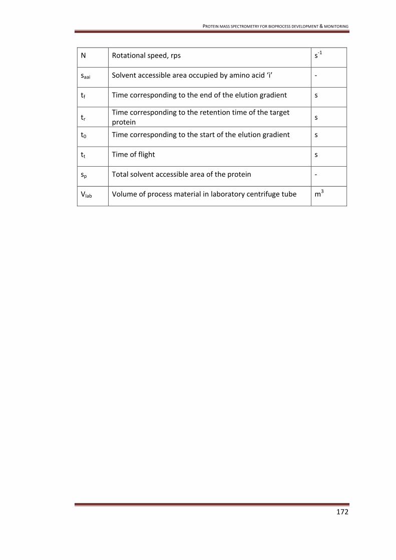
PROTEIN MASS SPECTROMETRY FOR BIOPROCESS DEVELOPMENT & MONITORING
172
N Rotational speed, rps s-1
saai Solvent accessible area occupied by amino acid ‘i’ -
tf Time corresponding to the end of the elution gradient s
tr Time corresponding to the retention time of the target protein
s
t0 Time corresponding to the start of the elution gradient s
tt Time of flight s
sp Total solvent accessible area of the protein -
Vlab Volume of process material in laboratory centrifuge tube m3

PROTEIN MASS SPECTROMETRY FOR BIOPROCESS DEVELOPMENT & MONITORING
173
APPENDIX - MATLAB CODE
Reading spectra data
%Enter directory where the data is located clear all path=cd; directory=find(path=='\'); currentfile=path(directory(end-1)+1:end); currentfile(currentfile=='.')='_'; currentfile(currentfile=='\')='_'; %Read Data cd([cd '\' 'Processed']); [MZ SELDIProcessed,Metapropnames,Metapropvalues,filenames]=readProcessed; cd .. cd([cd '\' 'Raw']); [Raw]=readRaw(filenames); %Remove redundant data from Processed & Raw [u numcols]=size(MZ); for i=1:numcols; b(:,i)=min(find(isnan(MZ(:,i))==1)); end clearInd=max(b); MZ(clearInd:end,:)=[]; SELDIProcessed(clearInd:end,:)=[]; Raw(clearInd:end,:)=[]; % Process Raw Data [Raw,Smoothed]=basecorr2(Raw, MZ, 6, 10, 6);

PROTEIN MASS SPECTROMETRY FOR BIOPROCESS DEVELOPMENT & MONITORING
174
%Mass=MZ; clear b AverageMass1=[]; AverageMass2=[]; clear e for i=1:numel(filenames); %remove csv a=strmatch(filenames{i}(:,end-3:end),'.csv'); if a==1; filenames{i}=filenames{i}(:,1:end-4); end %remove _1 b=strmatch(filenames{i}(:,end-1:end),'_1'); if b==1; filenames{i}=filenames{i}(:,1:end-2); end end for i=1:numel(filenames); f=numel(strmatch(filenames{i},filenames)); e(i,1:f)=strmatch(filenames{i},filenames)'; end [l m n]=unique(e,'rows'); variablenames=[]; variables=[]; variablelevels=repmat(NaN,numel(filenames),1); %Sorting Variables if numel(filenames(Karpievitch et al. 2007))==10 & numel(filenames{end})==10; [variablenames variables variablelevels]=datasorting2(filenames,MZ); end

PROTEIN MASS SPECTROMETRY FOR BIOPROCESS DEVELOPMENT & MONITORING
175
MinCutOff=5000; MidCutOff=22500; MaxCutOff=90000; lowStepSize=200; highStepSize=200; lowWindowSize=900; highWindowSize=1800; %if numel(f)>=2; newMass=repmat(NaN,25000,numel(m)); newSELDIProcessed=repmat(NaN,25000,numel(m)); newRaw=repmat(NaN,25000,numel(m)); newBaselineCorrected=repmat(NaN,25000,numel(m)); newSmoothed=repmat(NaN,25000,numel(m)); %if numel(m)<numel(l); %Merging of low & high mass spectra if m(1)==2; for i=1:numel(m); o=min(MZ(:,l(i,1))); p=max(MZ(:,l(i,1))); AverageMass1=(o+p)/2; x=min(MZ(:,l(i,2))); y=max(MZ(:,l(i,2))); AverageMass2=(x+y)/2; if AverageMass1<AverageMass2; %Process low and high mass data lowMass=MZ(:,l(i,1)); highMass=MZ(:,l(i,2)); lowSmoothed=Smoothed(:,l(i,1)); highSmoothed=Smoothed(:,l(i,2)); lowMass(isnan(lowMass))=[]; highMass(isnan(highMass))=[]; lowSmoothed(isnan(lowSmoothed))=[]; highSmoothed(isnan(highSmoothed))=[];

PROTEIN MASS SPECTROMETRY FOR BIOPROCESS DEVELOPMENT & MONITORING
176
baselineCorrectedlow=msbackadj(lowMass,lowSmoothed,'WindowSize',lowWindowSize,'StepSize',lowStepSize,'QuantileValue',0.1); baselineCorrectedhigh=msbackadj(highMass,highSmoothed,'WindowSize',highWindowSize,'StepSize',highStepSize,'QuantileValue',0.1);
%Splice treated data together a=max(find(MZ(:,l(i,1))<MinCutOff)); b=max(find(MZ(:,l(i,1))<MidCutOff)); c=max(find(MZ(:,l(i,2))<MidCutOff)); d=max(find(MZ(:,l(i,2))<MaxCutOff)); if isempty(a); [u a]=min(MZ(:,l(i,1))); end if isempty(b)||isempty(c) ; [u b]=max(MZ(:,l(i,1))); %some NaN so can not have numel, need max c=max(find(MZ(:,l(i,2))<=max(MZ(:,l(i,1))))); end if isempty(d); [u d]=max(MZ(:,l(i,2))); end k=length([MZ(a:b,l(i,1));MZ(c:d,l(i,2))]); newMass(1:k,i)=[MZ(a:b,l(i,1));MZ(c:d,l(i,2))];
newSELDIProcessed(1:k,i)=[SELDIProcessed(a:b,l(i,1));SELDIProcessed(c:d,l(i,2))];
newMetapropvalues(:,i)=[Metapropvalues(:,l(i,1));Metapropvalues(:,l(i,2))]; newRaw(1:k,i)=[Raw(a:b,l(i,1));Raw(c:d,l(i,2))];
newBaselineCorrected(1:k,i)=[baselineCorrectedlow(a:b,:);baselineCorrectedhigh(c:d,:)];
newSmoothed(1:k,i)=[Smoothed(a:b,l(i,1));Smoothed(c:d,l(i,2))]; V(i,:)=variablelevels(l(i,1),:); end if AverageMass1>AverageMass2; % Process low and high mass data lowMass=MZ(:,l(i,2));

PROTEIN MASS SPECTROMETRY FOR BIOPROCESS DEVELOPMENT & MONITORING
177
highMass=MZ(:,l(i,1)); lowSmoothed=Smoothed(:,l(i,2)); highSmoothed=Smoothed(:,l(i,1)); lowMass(isnan(lowMass))=[]; highMass(isnan(highMass))=[]; lowSmoothed(isnan(lowSmoothed))=[]; highSmoothed(isnan(highSmoothed))=[];
baselineCorrectedlow=msbackadj(lowMass,lowSmoothed,'WindowSize',lowWindowSize,'StepSize',lowStepSize,'QuantileValue',0.1); baselineCorrectedhigh=msbackadj(highMass,highSmoothed,'WindowSize',highWindowSize,'StepSize',highStepSize,'QuantileValue',0.1);
%Splice treated data together a=max(find(MZ(:,l(i,2))<=MinCutOff)); b=max(find(MZ(:,l(i,2))<=MidCutOff)); c=max(find(MZ(:,l(i,1))<=MidCutOff)); d=max(find(MZ(:,l(i,1))<=MaxCutOff)); if isempty(a); [u a]=min(MZ(:,l(i,2))); end if isempty(b)||isempty(c) ; [u b]=max(MZ(:,l(i,2))); %some NaN so can not have numel, need max c=max(find(MZ(:,l(i,1))<=max(MZ(:,l(i,2))))); end if isempty(d); [u d]=max(MZ(:,l(i,1))); end k=length([MZ(a:b,l(i,2));MZ(c:d,l(i,1))]); newMass(1:k,i)=[MZ(a:b,l(i,2));MZ(c:d,l(i,1))];
newSELDIProcessed(1:k,i)=[SELDIProcessed(a:b,l(i,2));SELDIProcessed(c:d,l(i,1))];
newMetapropvalues(:,i)=[Metapropvalues(:,l(i,2));Metapropvalues(:,l(i,1))]; newRaw(1:k,i)=[Raw(a:b,l(i,2));Raw(c:d,l(i,1))];
newBaselineCorrected(1:k,i)=[baselineCorrectedlow(a:b,:);baselineCorrectedhigh(c:d,:)];

PROTEIN MASS SPECTROMETRY FOR BIOPROCESS DEVELOPMENT & MONITORING
178
newSmoothed(1:k,i)=[Smoothed(a:b,l(i,2));Smoothed(c:d,l(i,1))]; V(i,:)=variablelevels(l(i,1),:); end end % if one spectrum per condition no splicing necessary else for i=1:numel(variablelevels(:,1)); a=max(find(MZ(:,i)<=MinCutOff)); b=max(find(MZ(:,i)<=MidCutOff)); d=max(find(MZ(:,i)<=MaxCutOff)); if isempty(a); [u a]=min(MZ(:,i)); end if isempty(b); b=(a+d)/2; end if isempty(d); [u d]=max(MZ(:,i)); end k=length([MZ(a:d,i)]); newSELDIProcessed(1:k,i)=[SELDIProcessed(a:d,i)]; newMetapropvalues(:,i)=[Metapropvalues(:,i)]; lowMass=MZ(a:b,i); highMass=MZ(b+1:d,i); lowSmoothed=Smoothed(a:b,i); highSmoothed=Smoothed(b+1:d,i); lowMass(isnan(lowMass))=[]; highMass(isnan(highMass))=[]; lowSmoothed(isnan(lowSmoothed))=[]; highSmoothed(isnan(highSmoothed))=[];
baselineCorrectedlow=msbackadj(lowMass,lowSmoothed,'WindowSize',lowWindowSize,'StepSize',lowStepSize,'QuantileValue',0.1);

PROTEIN MASS SPECTROMETRY FOR BIOPROCESS DEVELOPMENT & MONITORING
179
baselineCorrectedhigh=msbackadj(highMass,highSmoothed,'WindowSize',highWindowSize,'StepSize',highStepSize,'QuantileValue',0.1);
newMass(1:k,i)=[lowMass;highMass]; newRaw(1:k,i)=[Raw(a:b,i);Raw(b+1:d,i)]; newBaselineCorrected(1:k,i)=[baselineCorrectedlow;baselineCorrectedhigh]; newSmoothed(1:k,i)=[lowSmoothed;highSmoothed]; V(i,:)=variablelevels(i,:); end end %End of Sorting Masses function %clear a clear variablevels; clear Metapropvalues; clear Mass; clear SELDIProcessed; clear Raw; clear BaselineCorrected; clear Smoothed; variablelevels=V; Metapropvalues=newMetapropvalues; Mass=newMass; SELDIProcessed=newSELDIProcessed; Raw=newRaw; BaselineCorrected=newBaselineCorrected; Smoothed=newSmoothed; %crop data at max mass [z clearInd]=max(Mass(:,1)); Mass(clearInd+1:end,:)=[]; SELDIProcessed(clearInd+1:end,:)=[]; Raw(clearInd+1:end,:)=[]; BaselineCorrected(clearInd+1:end,:)=[]; Smoothed(clearInd+1:end,:)=[]; Mass=Mass/1000; %Mass(Mass==0)=NaN; Noise=Raw-Smoothed; Baseline=Smoothed-BaselineCorrected; cd ..

PROTEIN MASS SPECTROMETRY FOR BIOPROCESS DEVELOPMENT & MONITORING
180
save([currentfile 'Spectra'],'Mass','SELDIProcessed','Raw','Noise','Smoothed','Baseline','BaselineCorrected')%,'AUCMass','AUCSELDIProcessed','AUCRaw','AUCSmoothed','AUCBaselineCorrected'); save([currentfile 'MetaData'],'Metapropnames','Metapropvalues','filenames','variablenames','variables','variablelevels'); [nummasses numcols]=size(Mass); %stop it crashing from too much data if numcols<=32; load([currentfile 'Spectra']); xlswrite([currentfile 'Raw'],[Mass Raw]); xlswrite([currentfile 'SELDIProcessed'],[Mass SELDIProcessed]); xlswrite([currentfile 'Noise'],[Mass Noise]); xlswrite([currentfile 'Smoothed'],[Mass Smoothed]); xlswrite([currentfile 'Baseline'],[Mass Baseline]); xlswrite([currentfile 'BaselineCorrected'],[Mass BaselineCorrected]); load([currentfile 'MetaData']); if AverageMass1~=AverageMass2; xlswrite([currentfile 'MetaData'],[repmat(Metapropnames,2,1) Metapropvalues]); else xlswrite([currentfile 'MetaData'],[Metapropnames Metapropvalues]); end end
Reading processed data
%Read data %Enter directory files are in and run function [MZ,Processed,propertyname,propval,csvfiles]=readProcessed %Upload data and get conditions %function [operatingparameters,MZ,Int,datafilenames]=uploadSELDInp20data(fullpath);

PROTEIN MASS SPECTROMETRY FOR BIOPROCESS DEVELOPMENT & MONITORING
181
%fullpath=['c:\My Documents\Matlab\Functions & Data\Data\' sample '\' 'SELDI' '\' experiment '\' intensity '\' datatreatment '\' 'Raw' '\']; Int=[]; %cd(fullpath); [stat,mess]=fileattrib('*'); [r numfiles]=size(mess); for i=1:numfiles; datafullpath{i}=mess(i).Name; end [pathrows pathcols]=size(cd); for i=1:numfiles; datafilenames{i}=datafullpath{i}(:,pathcols+2:end); end datafilenames=datafilenames'; chardatafilenames=char(datafilenames); for i=1:numfiles; a=char(datafilenames(i,:)); [numletters]=length(deblank(a)); x=strmatch(a(:,numletters-2:numletters),'csv','exact'); if x==1; j(i)=i; end end csvfiles=datafilenames(nonzeros(j),:)'; [numfiles]=length(csvfiles); %csvfiles [MZ, Processed, propertyname, propval]=GetSpectraData(csvfiles); Reading raw data
%Read data %Enter directory files are in and run

PROTEIN MASS SPECTROMETRY FOR BIOPROCESS DEVELOPMENT & MONITORING
182
function [Raw]=readRaw(csvfiles) %path=['c:\My Documents\Matlab\Data\' sample '\' samplenumber '\' equipment '\' experiment '\' intensity '\' datatreatment '\']; %Upload data and get conditions %function [operatingparameters,MZ,Int,datafilenames]=uploadSELDInp20data(fullpath); [numfiles]=length(csvfiles); Raw=repmat(NaN,150000,numfiles); for i=1:numfiles; [propertyname propertyvalue]=textread(csvfiles{i},'%s %s','delimiter','=','emptyvalue',NaN); %propertyname %Take mass spec data from propertyname [a]=size(str2num(char(propertyvalue))); TOFmultiplier=str2num(char(propertyvalue(a(1)-1))); TOFdata=str2num(char(propertyname(a(1)+2:end,:))); size(TOFmultiplier); lenTOFdata=length(TOFdata); Raw(1:lenTOFdata,i)=TOFdata*TOFmultiplier; end Retrieve spectra data
function [lowcatMZ, lowcatInt, propertyname, propval]=GetSpectraData(csvfiles); numcsvfiles=numel(csvfiles); lowcatMZ=repmat(NaN,150000,numcsvfiles); lowcatInt=repmat(NaN,150000,numcsvfiles); propertyname=[]; propval=cell(130,numcsvfiles); for i=1:numcsvfiles; [propertyname propertyvalue]=textread(csvfiles{i},'%s %s','delimiter','=');

PROTEIN MASS SPECTROMETRY FOR BIOPROCESS DEVELOPMENT & MONITORING
183
a=strmatch(char('M/Z, Intensity'),char(propertyname)); %a %propertyname %Take mass spec data from propertyname [massspecdata]=str2num(char(propertyname(a+1:end))); MZ=massspecdata(:,1); Int=massspecdata(:,2); lowcatMZ(1:numel(MZ),i)=MZ; lowcatInt(1:numel(Int),i)=Int; %Clear mass spec data at end propertyvalue(a:end,:)=[]; %size(propval) t(:,i)=a; propval(1:a-1,i)=propertyvalue; end maxt=max(t); propval(maxt:end,:)=[]; propertyname(maxt:end,:)=[]; Sort data
function [variablenames variables variablelevels]=datasorting2(filenames,Mass); variablelevels=[]; variables=[]; charcsvfiles=char(filenames); [Chip]=charcsvfiles(:,1); ChipInd=repmat(NaN,numel(Chip),1); pHInd=repmat(NaN,numel(Chip),1); SaltInd=repmat(NaN,numel(Chip),1); SaltConcInd=repmat(NaN,numel(Chip),1); %ChipType cm=strmatch('c',Chip);

PROTEIN MASS SPECTROMETRY FOR BIOPROCESS DEVELOPMENT & MONITORING
184
qu=strmatch('q',Chip); hyd=strmatch('h',Chip); nor=strmatch('n',Chip); ima=strmatch('i',Chip); dea=strmatch('d',Chip); ChipInd(cm,:)=1; ChipInd(qu,:)=2; ChipInd(hyd,:)=3; ChipInd(nor,:)=4; ChipInd(ima,:)=5; ChipInd(dea,:)=6; %Mass [Mass b MassInd]=unique(min(Mass)); %pH [pH b pHInd]=unique(str2num(charcsvfiles(:,2:5))); pH=pH/100; %SaltType [Salt]=charcsvfiles(:,6); sod=strmatch('s',Salt); amm=strmatch('a',Salt); imi=strmatch('i',Salt); SaltInd(sod,:)=1; SaltInd(amm,:)=2; SaltInd(imi,:)=3; %Salt Conc [SaltConc b SaltConcInd]=unique(str2num(charcsvfiles(:,7:10))); variables(Karpievitch et al. 2007)={'c';'q';'h';'n';'i';'d'}; variables(Karpievitch et al. 2007;Merchant and Weinberger,2000)=Mass; variables(Coombes et al. 2005;Karpievitch et al. 2007)=pH; variables(Karpievitch et al. 2007;Xu and Lenhoff,2008)={'s';'a';'i'}; variables(Chen et al. 2008;Karpievitch et al. 2007)=SaltConc; size(ChipInd) size(MassInd) variablelevels(:,1)=ChipInd; variablelevels(:,2)=MassInd'; variablelevels(:,3)=pHInd; variablelevels(:,4)=SaltInd;

PROTEIN MASS SPECTROMETRY FOR BIOPROCESS DEVELOPMENT & MONITORING
185
variablelevels(:,5)=SaltConcInd; %FractionInd %variablelevels(:,6)=FractionInd; %variablelevels(:,7)=SpotInd; %variablelevels(:,8)=PartitionInd; Raw data treatment
The code in this section is from the UT M.D. Anderson Cancer Center and follows
these conditions:
CONDITIONS FOR USE:
Copyright (c) 2003, 2004 UT M.D. Anderson Cancer Center. All rights reserved.
This software is distributed and licensed to you on a non-exclusive
basis, free-of-charge. Redistribution and use in source and binary forms,
with or without modification, are permitted provided that the following
conditions are met:
1. Redistribution of source code must retain the above copyright notice,
this list of conditions and the following disclaimer.
2. Redistribution in binary form must reproduce the above copyright notice,
this list of conditions and the following disclaimer in the
documentation and/or other materials provided with the distribution.
3. All advertising materials mentioning features or use of this software
must display the following acknowledgment: This product includes
software developed by the University of Texas M.D. Anderson Cancer
Center, Houston, Texas and its contributors.
4. Neither the name of the University nor the names of its contributors
may be used to endorse or promote products derived from this software
without specific prior written permission.
THIS SOFTWARE IS PROVIDED BY UNIVERSITY OF TEXAS M.D. ANDERSON CANCER

PROTEIN MASS SPECTROMETRY FOR BIOPROCESS DEVELOPMENT & MONITORING
186
CENTER, HOUSTON, TEXAS, AND CONTRIBUTORS AS IS AND ANY EXPRESS OR
IMPLIED WARRANTIES, INCLUDING, BUT NOT LIMITED TO, THE IMPLIED
WARRANTIES OF MERCHANTABILITY AND FITNESS FOR A PARTICULAR PURPOSE
ARE
DISCLAIMED. IN NO EVENT SHALL M.D. ANDERSON OR CONTRIBUTORS BE
LIABLE FOR ANY DIRECT, INDIRECT, INCIDENTAL, SPECIAL, EXEMPLARY, OR
CONSEQUENTIAL DAMAGES (INCLUDING, BUT NOT LIMITED TO, PROCUREMENT OF
SUBSTITUTE GOODS OR SERVICES; LOSS OF USE, DATA, OR PROFITS; OR
BUSINESS INTERRUPTIONS) HOWEVER CAUSED AND ON ANY THEORY OF LIABILITY,
WHETHER IN CONTRACT, STRICT LIABILITY, OR TORT (INCLUDING NEGLIGENCE
OR OTHERWISE), PRODUCT LIABILITY, OR OTHERWISE ARISING IN ANY WAY OUT
OF THE USE OF THIS SOFTWARE, EVEN IF ADVISED OF THE POSSIBILITY OF
SUCH DAMAGE.
Applicable code:
function [RawEnd, sm]= basecorr2(raw, rawMass, M, L, thld) w = size(raw); % how much data are we handling? n = floor(w(1)/2^L); % number of length 2^L pieces available echo on status = 'start baseline correction' tic echo off midmass=22500; bc = repmat(NaN, [w(1) w(2)]); sm = repmat(NaN, [w(1) w(2)]); RawEnd = repmat(NaN, [w(1) w(2)]); for i = 1:w(2), Mass=rawMass(:,i); RawInt=raw(:,i); Mass(isnan(Mass))=[]; RawInt(isnan(RawInt))=[]; [s] = waveletSmoothAndBaselineCorrect(Mass,RawInt, thld, M, L, midmass);

PROTEIN MASS SPECTROMETRY FOR BIOPROCESS DEVELOPMENT & MONITORING
187
c=length(Mass); RawEnd(1:c,i)=RawInt; sm(1:c,i)=s; end echo on toc status = 'end baseline correction' echo off Peak selection %Enter directory where the data is located clear all path=cd; directory=find(path=='\'); currentfile=path(directory(end-1)+1:end); currentfile(currentfile=='.')='_'; currentfile(currentfile=='\')='_'; load([currentfile 'Spectra']); load([currentfile 'MetaData']); PeakMass=[6.855;9.277;11.16;12.22;12.41;12.66;13.06;13.28;14.03;14.6;26.2;27.18;28.15;28.62;29.05;31.75;35.19;36.88;38.09;40.13;41.18;42.06;43.18;46.57;48.59;52.18;54.62;55.95;57.48;84.23]; [a b]=size(PeakMass); [peakindexrange peakindexmid]=mspeakfindman(Mass(:,1),PeakMass); RawPeakHeight=Raw(peakindexmid,:); BcPeakHeight=BaselineCorrected(peakindexmid,:); CiphergenPeakHeight=SELDIProcessed(peakindexmid,:); for i=1:a; %SELDIInt(i,:)=sum(NormSELDIAUC(peakindexrange(i,1):peakindexrange(i,2),:),1); BcPeakArea(i,:)=sum(BaselineCorrected(peakindexrange(i,1):peakindexrange(i,2),:),1);

PROTEIN MASS SPECTROMETRY FOR BIOPROCESS DEVELOPMENT & MONITORING
188
CiphergenPeakArea(i,:)=sum(SELDIProcessed(peakindexrange(i,1):peakindexrange(i,2),:),1); [a b]=size(BcPeakArea); maxBcPeakArea=max(BcPeakArea,[],2); maxBcPeakArea=repmat(maxBcPeakArea,[1 b]); normBcPeakArea=BcPeakArea./maxBcPeakArea; xlswrite([currentfile 'RawPeakHeight'],[PeakMass RawPeakHeight]); xlswrite([currentfile 'CiphergenPeakHeight'],[PeakMass CiphergenPeakHeight]); xlswrite([currentfile 'CiphergenPeakArea'],[PeakMass CiphergenPeakArea]); xlswrite([currentfile 'BcPeakHeight'],[PeakMass BcPeakHeight]); xlswrite([currentfile 'BcPeakArea'],[PeakMass BcPeakArea]); xlswrite([currentfile 'normBcPeakArea'],[PeakMass normBcPeakArea]); save([currentfile 'Peaks'],'Mass','PeakMass','RawPeakHeight','CiphergenPeakHeight','BcPeakHeight','CiphergenPeakArea','BcPeakArea','normBcPeakArea'); Nearest neighbour predictor %Enter directory where the data is located path=cd; directory=find(path=='\'); currentfile=path(directory(end-1)+1:end); currentfile(currentfile=='.')='_'; currentfile(currentfile=='\')='_'; load([currentfile 'Peaks']); load([currentfile 'MetaData']); MassString=char(num2str(PeakMass,'%.1f')); figure h=glyphplot(normBcPeakArea,'Obslabels',MassString); set(h(:,3),'FontSize',16); Y = pdist(normBcPeakArea); Z = linkage(Y); figure [H,T] = dendrogram(Z,'colorthreshold', 0,'labels',MassString,'orientation','right');

PROTEIN MASS SPECTROMETRY FOR BIOPROCESS DEVELOPMENT & MONITORING
189
set(gca,'FontSize',14); set(H,'LineWidth',2); xlabel('Relationship (\muA)','FontSize',16) ylabel('Protein ID (kDa/e)','FontSize',16) TargetInd=13; SquareDist=squareform(Y); TargetDist=SquareDist(TargetInd,:); TargetDist(TargetDist==0)=NaN; [a NearestNeighbourInd]=min(TargetDist); NearestNeighbour=PeakMass(NearestNeighbourInd) ph=variables(Coombes et al. 2005)(variablelevels(:,3)); conc=variables(Chen et al. 2008)(variablelevels(:,5))/1000; [a]=find(variablelevels(:,1)==1); [b]=find(variablelevels(:,1)==2); [c]=find(variablelevels(:,1)==3); %Cation Exchange if size(a)>0; figure subplot(1,2,1); [XI,YI] = meshgrid(unique(ph(a)),unique(conc(a))) [catph,catconc,catpeakarea] = griddata(ph(a),conc(a),normBcPeakArea(TargetInd,a),XI,YI,'v4'); contourf(catph,catconc,catpeakarea,500,'EdgeColor','none') view(0,90) axis tight subplot(1,2,2); [XI,YI] = meshgrid(unique(ph(a)),unique(conc(a))) [catph,catconc,catpeakarea] = griddata(ph(a),conc(a),normBcPeakArea(NearestNeighbourInd,a),XI,YI,'v4'); contourf(catph,catconc,catpeakarea,500,'EdgeColor','none') view(0,90) axis tight end

PROTEIN MASS SPECTROMETRY FOR BIOPROCESS DEVELOPMENT & MONITORING
190
if size(b)>0; figure subplot(1,2,1); [XI,YI] = meshgrid(unique(ph(b)),unique(conc(b))) [catph,catconc,catpeakarea] = griddata(ph(b),conc(b),normBcPeakArea(TargetInd,b),XI,YI,'v4'); contourf(catph,catconc,catpeakarea,500,'EdgeColor','none') view(0,90) axis tight subplot(1,2,2); [XI,YI] = meshgrid(unique(ph(b)),unique(conc(b))) [catph,catconc,catpeakarea] = griddata(ph(b),conc(b),normBcPeakArea(NearestNeighbourInd,b),XI,YI,'v4'); %colormap('default') contourf(catph,catconc,catpeakarea,500,'EdgeColor','none') view(0,90) axis tight end if size(c)>0; figure subplot(1,2,1); [XI,YI] = meshgrid(unique(ph(c)),unique(conc(c))) [catph,catconc,catpeakarea] = griddata(ph(c),conc(c),normBcPeakArea(TargetInd,c),XI,YI,'v4'); contourf(catph,catconc,catpeakarea,500,'EdgeColor','none') view(0,90) axis tight subplot(1,2,2); [XI,YI] = meshgrid(unique(ph(c)),unique(conc(c))) [catph,catconc,catpeakarea] = griddata(ph(c),conc(c),normBcPeakArea(NearestNeighbourInd,c),XI,YI,'v4'); contourf(catph,catconc,catpeakarea,500,'EdgeColor','none') view(0,90)

PROTEIN MASS SPECTROMETRY FOR BIOPROCESS DEVELOPMENT & MONITORING
191
axis tight end

PROTEIN MASS SPECTROMETRY FOR BIOPROCESS DEVELOPMENT & MONITORING
192
REFERENCES
Ambler CM. 1961. Theory - the Fundamentals of Separation, Including Sharples
Sigma Value for Predicting Equipment Performance. Industrial and Engineering
Chemistry 53:430-433.
Annesley TM. 2003. Ion suppression in mass spectrometry. Clin Chem 49:1041-
1044.
Arakawa T, Timasheff SN. 1984. Mechanism of Protein Salting in and Salting Out by
Divalent-Cation Salts - Balance Between Hydration and Salt Binding. Biochemistry
23:5912-5923.
Asenjo JA, Andrews BA. 2004. Is there a rational method to purify proteins? From
expert systems to proteomics. J Mol Recognit 17:236-247.
Barzaghi D, Isbister JD, Lauer KP, Born TL. 2004. Use of surface-enhanced laser
desorption/ionization -time of flight to explore bacterial proteomes. Proteomics
4:2624-2628.
Berggren K, Wolf A, Asenjo JA, Andrews BA, Tjerneld F. 2002. The surface exposed
amino acid residues of monomeric proteins determine the partitioning in aqueous
two-phase systems. Biochim Biophys Acta 1596:253-268.
Berrill A, Ho SV, Bracewell DG. 2008. Ultra scale-down to define and improve the
relationship between Flocculation and disc-stack centrifugation. Biotechnology
Progress 24:426-431.

PROTEIN MASS SPECTROMETRY FOR BIOPROCESS DEVELOPMENT & MONITORING
193
Berrill A, Ho SV, Bracewell DG. 2009. Product and contaminant measurement in
bioprocess development by SELDI-MS. Biotechnol Prog.
Biddlecombe JG, Craig AV, Zhang H, Uddin S, Mulot S, Fish BC, Bracewell DG. 2007.
Determining antibody stability: creation of solid-liquid interfacial effects within a
high shear environment. Biotechnol Prog 23:1218-1222.
Bonet I, Rodríguez A, Grau R, García MM, Saez Y, Nowé A. 2008. Comparing
Distance Measures with Visual Methods. Lecture Notes in Computer Science.
Springer Berlin / Heidelberg. p 90-99.
Bonnerjea J, Oh S, Hoare M, Dunnill P. 1986. Protein purification: The right step at
the right time. Biotechnology 4:954-958.
Borhani DW, Rogers DP, Engler JA, Brouillette CG. 1997. Crystal structure of
truncated human apolipoprotein A-I suggests a lipid-bound conformation. Proc Natl
Acad Sci U S A 94:12291-12296.
Bowering LC, Bracewell DG, Kesharvarz-Moore E, Hoare M, Weir AN. 2002.
Comparison of techniques for monitoring antibody fragment production in E. coli
fermentation cultures. Biotechnol Prog 18:1431-1438.
Boychyn M, Doyle W, Bulmer M, More J, Hoare M. 2000. Laboratory scaledown of
protein purification processes involving fractional precipitation and centrifugal
recovery. Biotechnol Bioeng 69:1-10.

PROTEIN MASS SPECTROMETRY FOR BIOPROCESS DEVELOPMENT & MONITORING
194
Boychyn M, Yim SS, Bulmer M, More J, Bracewell DG, Hoare M. 2004. Performance
prediction of industrial centrifuges using scale-down models. Bioprocess Biosyst Eng
26:385-391.
Boychyn M, Yim SSS, Shamlou PA, Bulmer M, More J, Hoare A. 2001.
Characterization of flow intensity in continuous centrifuges for the development of
laboratory mimics. Chemical Engineering Science 56:4759-4770.
Brenac B, V, Schapman A, Santambien P, Britsch L. 2008. Fast purification process
optimization using mixed-mode chromatography sorbents in pre-packed mini-
columns. J Chromatogr A 1177:226-233.
Brenac V, Mouz N, Schapman A, Ravault V. 2006. Expression optimization and
purification process development of an engineered soluble recombinant mouse
linker of activation of T cells using surface enhanced laser desorption/ionization-
mass spectrometry. Protein Expr Purif 47:533-541.
Brozkova K, Budinska E, Bouchal P, Hernychova L, Knoflickova D, Valik D, Vyzula R,
Vojtesek B, Nenutil R. 2008. Surface-enhanced laser desorption/ionization time-of-
flight proteomic profiling of breast carcinomas identifies clinicopathologically
relevant groups of patients similar to previously defined clusters from cDNA
expression. Breast Cancer Res 10:R48.
Caputo E, Moharram R, Martin BM. 2003. Methods for on-chip protein analysis.
Anal Biochem 321:116-124.
Chen J, Cramer SM. 2007. Protein adsorption isotherm behavior in hydrophobic
interaction chromatography. J Chromatogr A 1165:67-77.

PROTEIN MASS SPECTROMETRY FOR BIOPROCESS DEVELOPMENT & MONITORING
195
Chen J, Luo Q, Breneman CM, Cramer SM. 2007. Classification of protein adsorption
and recovery at low salt conditions in hydrophobic interaction chromatographic
systems. J Chromatogr A 1139:236-246.
Chen J, Yang T, Cramer SM. 2008. Prediction of protein retention times in gradient
hydrophobic interaction chromatographic systems. J Chromatogr A 1177:207-214.
Coombes KR, Tsavachidis S, Morris JS, Baggerly KA, Hung MC, Kuerer HM. 2005.
Improved peak detection and quantification of mass spectrometry data acquired
from surface-enhanced laser desorption and ionization by denoising spectra with
the undecimated discrete wavelet transform. Proteomics 5:4107-4117.
Cordingley HC, Roberts SL, Tooke P, Armitage JR, Lane PW, Wu W, Wildsmith SE.
2003. Multifactorial screening design and analysis of SELDI-TOF ProteinChip array
optimization experiments. Biotechniques 34:364-373.
Cramer SM, Jayaraman G. 1993. Preparative chromatography in biotechnology. Curr
Opin Biotechnol 4:217-225.
Cromwell ME, Hilario E, Jacobson F. 2006. Protein aggregation and bioprocessing.
AAPS J 8:E572-E579.
Dijkstra M, Roelofsen H, Vonk RJ, Jansen RC. 2006. Peak quantification in surface-
enhanced laser desorption/ionization by using mixture models. Proteomics 6:5106-
5116.
Dijkstra M, Vonk RJ, Jansen RC. 2007. SELDI-TOF mass spectra: a view on sources of
variation. J Chromatogr B Analyt Technol Biomed Life Sci 847:12-23.

PROTEIN MASS SPECTROMETRY FOR BIOPROCESS DEVELOPMENT & MONITORING
196
Dyr JE, Suttnar J. 1997. Separation used for purification of recombinant proteins. J
Chromatogr 699:383-401.
Favre-Kontula L, Sattonnet-Roche P, Magnenat E, Proudfoot AEI, Boschert U,
Xenarios L, Vilbois F, Antonsson B. 2008. Detection and identification of plasma
proteins that bind GlialCAM using ProteinChip (TM) arrays, SELDI-TOF MS, and
nano-LC MS/MS. Proteomics 8:378-388.
Fortis F, Guerrier L, Righetti PG, Antonioli P, Boschetti E. 2006. A new approach for
the removal of protein impurities from purified biologicals using combinatorial
solid-phase ligand libraries. Electrophoresis 27:3018-3027.
Garcia T, Cook G, Nosal R. 2008. PQLI key topics - Criticality, design space, and
control strategy. J Pharm Innov 3:60-68.
Gasner LL, Wang DIC. 1970. Microbial Cell Recovery Enhancement Through
Flocculation. Biotechnology and Bioengineering 12:873-&.
GE gel filtration principles and methods. 2003. GE gel filtration principles and
methods.
GE HIC principle and methods. 2003. GE HIC principle and methods.
GE protein purification. 2003. GE protein purification.
Groep ME, Gregory ME, Kershenbaum LS, Bogle IDL. 1997. Performance Modelling
and Simulation of Biochemical Process Sequences with Interacting Unit Operations.
Biotechnol Bioeng 67:300-311.

PROTEIN MASS SPECTROMETRY FOR BIOPROCESS DEVELOPMENT & MONITORING
197
Gu Z, Glatz CE. 2007. A method for three-dimensional protein characterization and
its application to a complex plant (corn) extract. Biotechnol Bioeng 97:1158-1169.
Guerrier L, Lomas L, Boschetti E. 2005. A simplified monobuffer multidimensional
chromatography for high-throughput proteome fractionation. J Chromatogr A
1073:25-33.
Guerrier L, Lomas L, Boschetti E. 2007. A new general approach to purify proteins
from complex mixtures. J Chromatogr A 1156:188-195.
Humphreys DP, Heywood SP, King LM, Bowering LC, Turner JP, Lane SE. 2004.
Engineering of Escherichia coli to improve the purification of periplasmic Fab'
fragments: changing the pI of the chromosomally encoded PhoS/PstS protein.
Protein Expr Purif 37:109-118.
Hunter AK, Wang X, Suda EJ, Herberg JT, Shell RE, Thomas KE, Dufield RL, Gustafson
ME, Mozier NM, Ho SV. 2009. Separation of Product Associating E. coli Host Cell
Proteins OppA and DppA from Recombinant Apolipoprotein A-I-milano in an
Industrial HIC Unit Operation. Biotechnology Progress 25:446-453.
Hutchens TW, Yip TT. 1993. New Desorption Strategies for the Mass-Spectrometric
Analysis of Macromolecules. Rapid Communications in Mass Spectrometry 7:576-
580.
Hutchinson N, Bingham N, Murrell N, Farid S, Hoare M. 2006. Shear stress analysis
of mammalian cell suspensions for prediction of industrial centrifugation and its
verification. Biotechnology and Bioengineering 95:483-491.

PROTEIN MASS SPECTROMETRY FOR BIOPROCESS DEVELOPMENT & MONITORING
198
Jeffries N. 2005. Algorithms for alignment of mass spectrometry proteomic data.
Bioinformatics 21:3066-3073.
Jenkins RE, Pennington SR. 2001. Arrays for protein expression profiling: towards a
viable alternative to two-dimensional gel electrophoresis? Proteomics 1:13-29.
Karpievitch YV, Hill EG, Smolka AJ, Morris JS, Coombes KR, Baggerly KA, Almeida JS.
2007. PrepMS: TOF MS data graphical preprocessing tool. Bioinformatics 23:264-
265.
Kim JS, Akeprathumchai S, Wickramasinghe SR. 2001. Flocculation to enhance
microfiltration. Journal of Membrane Science 182:161-172.
Kumar N, Maurya P, Gammell P, Dowling P, Clynes M, Meleady P. 2008. Proteomic
profiling of secreted proteins from CHO cells using Surface-Enhanced Laser
desorption ionization time-of-flight mass spectrometry. Biotechnol Prog 24:273-
278.
Ladiwala A, Rege K, Breneman CM, Cramer SM. 2005. A priori prediction of
adsorption isotherm parameters and chromatographic behavior in ion-exchange
systems. Proc Natl Acad Sci U S A 102:11710-11715.
Ladiwala A, Xia F, Luo Q, Breneman CM, Cramer SM. 2006. Investigation of protein
retention and selectivity in HIC systems using quantitative structure retention
relationship models. Biotechnol Bioeng 93:836-850.
Lienqueo ME, Asenjo JA. 2000. Use of expert systems for the synthesis of
downstream protein processes. Computers & Chemical Engineering 24:2339-2350.

PROTEIN MASS SPECTROMETRY FOR BIOPROCESS DEVELOPMENT & MONITORING
199
Lienqueo ME, Mahn A, Asenjo JA. 2002. Mathematical correlations for predicting
protein retention times in hydrophobic interaction chromatography. J Chromatogr
A 978:71-79.
Lienqueo ME, Mahn A, Navarro G, Salgado JC, Perez-Acle T, Rapaport I, Asenjo JA.
2006. New approaches for predicting protein retention time in hydrophobic
interaction chromatography. J Mol Recognit 19:260-269.
Lienqueo ME, Mahn A, Salgado JC, Asenjo JA. 2007. Current insights on protein
behaviour in hydrophobic interaction chromatography. J Chromatogr B Analyt
Technol Biomed Life Sci 849:53-68.
Lienqueo ME, Mahn A, Vasquez L, Asenjo JA. 2003. Methodology for predicting the
separation of proteins by hydrophobic interaction chromatography and its
application to a cell extract. J Chromatogr A 1009:189-196.
Lodish H, Berk A, Zipursky ZL, Matsudaira P, Balimore D, Darnell JE. 1999.Molecular
Cell Biology. W.H. Freeman & Company.
Mahn A, Asenjo JA. 2005. Prediction of protein retention in hydrophobic interaction
chromatography. Biotechnol Adv 23:359-368.
Mahn A, Lienqueo ME, Asenjo JA. 2007. Optimal operation conditions for protein
separation in hydrophobic interaction chromatography. J Chromatogr B Analyt
Technol Biomed Life Sci 849:236-242.

PROTEIN MASS SPECTROMETRY FOR BIOPROCESS DEVELOPMENT & MONITORING
200
Mahn A, Lienqueo ME, Asenjo JA. 2004. Effect of surface hydrophobicity
distribution on retention of ribonucleases in hydrophobic interaction
chromatography. J Chromatogr A 1043:47-55.
Mahn A, Zapata-Torres G, Asenjo JA. 2005. A theory of protein-resin interaction in
hydrophobic interaction chromatography. J Chromatogr A 1066:81-88.
Malmquist G, Nilsson UH, Norrman M, Skarp U, Stromgren M, Carredano E. 2006.
Electrostatic calculations and quantitative protein retention models for ion
exchange chromatography. J Chromatogr A 1115:164-186.
Maybury JP, Hoare M, Dunnill P. 2000. The use of laboratory centrifugation studies
to predict performance of industrial machines: Studies of shear-insensitive and
shear-sensitive materials. Biotechnology and Bioengineering 67:265-273.
Maybury JP, Mannweiler K, Titchener-Hooker NJ, Hoare M, Dunnill P. 1998. The
performance of a scaled down industrial disc stack centrifuge with a reduced feed
material requirement. Bioprocess Engineering 18:191-199.
McGregor WC, Finn RK. 1969. Factors affecting the flocculation of bacteria by
chemical additives. Biotechnol Bioeng 11:127-138.
Merchant M, Weinberger SR. 2000. Recent advancements in surface-enhanced
laser desorption/ionization-time of flight-mass spectrometry. Electrophoresis
21:1164-1177.
Mine Y. 1995. Recent Advances in the Understanding of Egg-White Protein
Functionality. Trends in Food Science & Technology 6:225-232.

PROTEIN MASS SPECTROMETRY FOR BIOPROCESS DEVELOPMENT & MONITORING
201
Neal G, Christie J, Keshavarz-Moore E, Shamlou PA. 2003. Ultra scale-down
approach for the prediction of full-scale recovery of ovine polycolonal
immunoglobulins used in the manufacture of snake venom-specific Fab fragment.
Biotechnol Bioeng 81:149-157.
Ou K, Seow TK, Liang RC, Ong SE, Chung MC. 2001. Proteome analysis of a human
heptocellular carcinoma cell line, HCC-M: an update. Electrophoresis 22:2804-2811.
Panicker G, Lee DR, Unger ER. 2009. Optimization of SELDI-TOF protein profiling for
analysis of cervical mucous. J Proteomics 71:637-646.
Park JT, Bradbury L, Kragl FJ, Lukens DC, Valdes JJ. 2006. Rapid optimization of
antibotulinum toxin antibody fragment production by an integral approach utilizing
RC-SELDI mass spectrometry and statistical design. Biotechnol Prog 22:233-240.
Parris N, Baginski MA. 1991. A Rapid Method for the Determination of Whey-
Protein Denaturation. Journal of Dairy Science 74:58-64.
Rege K, Pepsin M, Falcon B, Steele L, Heng M. 2006. High-throughput process
development for recombinant protein purification. Biotechnol Bioeng 93:618-630.
Reynolds T, Boychyn M, Sanderson T, Bulmer M, More J, Hoare M. 2003. Scale-
down of continuous filtration for rapid bioprocess design: Recovery and dewatering
of protein precipitate suspensions. Biotechnol Bioeng 83:454-464.
Richardson JF, Zaki WN. 1997. Sedimentation and fluidisation: part 1. Chemical
Engineering Research & Design 75:S82-S99.

PROTEIN MASS SPECTROMETRY FOR BIOPROCESS DEVELOPMENT & MONITORING
202
Richardson P, Hoare M, Dunnill P. 1990. A new biochemical engineering approach
to the fractional precipitation of proteins. Biotechnol Bioeng 36:354-366.
Salgado JC, Andrews BA, Ortuzar MF, Asenjo JA. 2008. Prediction of the partitioning
behaviour of proteins in aqueous two-phase systems using only their amino acid
composition. J Chromatogr A 1178:134-144.
Salgado JC, Rapaport I, Asenjo JA. 2005a. Is it possible to predict the average
surface hydrophobicity of a protein using only its amino acid composition? J
Chromatogr A 1075:133-143.
Salgado JC, Rapaport I, Asenjo JA. 2005b. Prediction of retention times of proteins
in hydrophobic interaction chromatography using only their amino acid
composition. J Chromatogr A 1098:44-54.
Salgado JC, Rapaport I, Asenjo JA. 2006a. Predicting the behaviour of proteins in
hydrophobic interaction chromatography. 1: Using the hydrophobic imbalance (HI)
to describe their surface amino acid distribution. J Chromatogr A 1107:110-119.
Salgado JC, Rapaport I, Asenjo JA. 2006b. Predicting the behaviour of proteins in
hydrophobic interaction chromatography. 2. Using a statistical description of their
surface amino acid distribution. J Chromatogr A 1107:120-129.
Salte H, King JP, Titchener-Hooker N. 2005. Centrifuge selection for recovery of high
solids density cell broths and visualization of separation performance via windows
of operation. Abstracts of Papers of the American Chemical Society 229:U179.

PROTEIN MASS SPECTROMETRY FOR BIOPROCESS DEVELOPMENT & MONITORING
203
Semmes OJ, Feng Z, Adam BL, Banez LL, Bigbee WL, Campos D, Cazares LH, Chan
DW, Grizzle WE, Izbicka E, Kagan J, Malik G, McLerran D, Moul JW, Partin A,
Prasanna P, Rosenzweig J, Sokoll LJ, Srivastava S, Srivastava S, Thompson I, Welsh
MJ, White N, Winget M, Yasui Y, Zhang Z, Zhu L. 2005. Evaluation of serum protein
profiling by surface-enhanced laser desorption/ionization time-of-flight mass
spectrometry for the detection of prostate cancer: I. Assessment of platform
reproducibility. Clin Chem 51:102-112.
Shan JG, Xia J, Guo YX, Zhang XQ. 1996. Flocculation of cell, cell debris and soluble
protein with methacryloyloxyethyl trimethylammonium chloride-acrylonitrile
copolymer. Journal of Biotechnology 49:173-178.
Suurkuusk M, Hallen D. 1999. Apolipoprotein A-I(Milano) unfolds via an
intermediate state as studied by differential scanning calorimetry and circular
dichroism. Eur J Biochem 264:183-190.
Tang N, Tornatore P, Weinberger SR. 2004. Current developments in SELDI affinity
technology. Mass Spectrom Rev 23:34-44.
Thommes J, Etzel M. 2007. Alternatives to chromatographic separations. Biotechnol
Prog 23:42-45.
Tustian AD, Salte H, Willoughby NA, Hassan I, Rose MH, Baganz F, Hoare M,
Titchener-Hooker NJ. 2007. Adapted ultra scale-down approach for predicting the
centrifugal separation behavior of high cell density cultures. Biotechnology Progress
23:1404-1410.

PROTEIN MASS SPECTROMETRY FOR BIOPROCESS DEVELOPMENT & MONITORING
204
Vachier MC, Piot M, Awade AC. 1995. Isolation of hen egg white lysozyme,
ovotransferrin and ovalbumin, using a quaternary ammonium bound to a highly
crosslinked agarose matrix. J Chromatogr B Biomed Appl 664:201-210.
Vitello LB, Scanu AM. 1976. Studies on human serum high-density lipoproteins. Self-
association of human serum apolipoprotein A-II in aqueous solutions. Biochemistry
15:1161-1165.
Wall DB, Parus SJ, Lubman DM. 2002. Three-dimensional protein map according to
pI, hydrophobicity and molecular mass. J Chromatogr B Analyt Technol Biomed Life
Sci 774:53-58.
Weinberger SR, Boschetti E, Santambien P, Brenac V. 2002a. Surface-enhanced
laser desorption-ionization retentate chromatography mass spectrometry (SELDI-
RC-MS): a new method for rapid development of process chromatography
conditions. J Chromatogr B Analyt Technol Biomed Life Sci 782:307-316.
Weinberger SR, Dalmasso EA, Fung ET. 2002b. Current achievements using
ProteinChip Array technology. Curr Opin Chem Biol 6:86-91.
Weinberger SR, Viner RI, Ho P. 2002c. Tagless extraction-retentate
chromatography: a new global protein digestion strategy for monitoring differential
protein expression. Electrophoresis 23:3182-3192.
Wenger MD, Dephillips P, Price CE, Bracewell DG. 2007. An automated microscale
chromatographic purification of virus-like particles as a strategy for process
development. Biotechnol Appl Biochem 47:131-139.

PROTEIN MASS SPECTROMETRY FOR BIOPROCESS DEVELOPMENT & MONITORING
205
Wierling PS, Bogumil R, Knieps-Grunhagen E, Hubbuch J. 2007. High-throughput
screening of packed-bed chromatography coupled with SELDI-TOF MS analysis:
monoclonal antibodies versus host cell protein. Biotechnol Bioeng 98:440-450.
Willoughby N, Martin P, Titchener-Hooker N. 2004. Extreme scale-down of
expanded bed adsorption: Purification of an antibody fragment directly from
recombinant E. coli culture. Biotechnol Bioeng 87:641-647.
Woolley JF, Al Rubeai M. 2009. The application of SELDI-TOF mass spectrometry to
mammalian cell culture. Biotechnol Adv 27:177-184.
Xu L, Glatz CE. 2009. Predicting protein retention time in ion-exchange
chromatography based on three-dimensional protein characterization. J
Chromatogr A 1216:274-280.
Xu X, Lenhoff AM. 2008. A predictive approach to correlating protein adsorption
isotherms on ion-exchange media. J Phys Chem B 112:1028-1040.
Yang T, Breneman CM, Cramer SM. 2007a. Investigation of multi-modal high-salt
binding ion-exchange chromatography using quantitative structure-property
relationship modeling. J Chromatogr A 1175:96-105.
Yang T, Sundling MC, Freed AS, Breneman CM, Cramer SM. 2007b. Prediction of pH-
dependent chromatographic behavior in ion-exchange systems. Anal Chem
79:8927-8939.