PROTECTIVE ROLE OF DIETARY BIOACTIVE COMPOUNDS: … · glucosinolati, precursori di isotiocianati...
162
I GAIA BONACINA 2009/2010 Graduate School in Molecular Sciences and Plant, Food and Environmental Biotechnology PROTECTIVE ROLE OF DIETARY BIOACTIVE COMPOUNDS: MECHANISMS AND HYPOTHESIS
Transcript of PROTECTIVE ROLE OF DIETARY BIOACTIVE COMPOUNDS: … · glucosinolati, precursori di isotiocianati...

I
GAIA BONACINA
2009/2010
Graduate School in Molecular Sciences and Plant, Food
and Environmental Biotechnology
PROTECTIVE ROLE OF DIETARY
BIOACTIVE COMPOUNDS:
MECHANISMS AND HYPOTHESIS

II
Università degli Studi di Milano – Facoltà di Agraria
Graduate School in Molecular Sciences and Plant, Food and Environmental
Biotechnology
PhD in Food Technology and Biotechnology (XXII cycle)
Coordinator: Prof. Luciano Piergiovanni
Title: PROTECTIVE ROLE OF DIETARY BIOACTIVE COMPOUNDS:
MECHANISMS AND HYPOTHESIS
PhD Student: Dr. Gaia Bonacina, matr. R07142
Tutor: Prof. Marisa Porrini
Co-tutor: Dr. Patrizia Riso
Supervisors (Bergamo): Dr. Anna Falanga and Dr. Marina Marchetti
2009/2010

III
Abstract
Scientific evidences support the protective role of diets rich in fruit and vegetables
against chronic diseases, like cancer and cardiovascular disease. In particular two
classes of chemopreventive phytochemicals, i.e. isothiocyanates presence in Cruciferae
and anthocyanins (ACNs) presence in berries and other colored fruits and vegetables,
are considered in this thesis.
Cruciferae and especially Brassica genus contain high concentration of
constituents with antioxidant properties (e.g. carotenoids, vitamin C, folate) as well as
glucosinolate precursors of isothiocyanates (ITCs) and indoles that modulate
xenobiotic biotransformation enzymes, such as Glutathione S-Transferase (GST). The
protective effect of broccoli (Brassica oleracea L. var. italica) was tested through two
different study protocols: a regular-intake intervention study with daily consumption
of one serving of steamed broccoli for 10 days and a single-meal study (i.e. single
consumption of broccoli, 250g). Analyzed broccoli resulted to be a good source of
antioxidant compounds (carotenoids, vitamin C) and glucosinolates as ITCs.
Subjects involved in the study were young male smokers recruited on the basis of
anthropometric characteristics, food habits and their GSTM1 genotype.
Thirty subjects were enrolled for the regular-intake study. A single blind
randomized cross-over experimental design was scheduled. Fasting blood samples
were collected at the beginning and at the end of each treatment period (0, 10, 30, 40
days). Concentration of carotenoids, lutein, β-carotene and folate was assessed in
plasma and serum samples. Lymphocytes were used for the determination of
biomarkers of oxidative stress: cell resistance against oxidative stress, endogenous
DNA damage (i.e. oxidized purines), in vitro DNA repair activity and mRNA
expression of OGG1, NUDT1 and HO-1 levels. Cell protection against H2O2-induced
DNA damage was higher after broccoli diet with respect to control diet in the whole
group of subjects. Folate and lutein concentrations increased significantly after broccoli
diet. Broccoli intake caused a more consistent and significant effect of protection
against DNA damage in GSTM1 null (-27.6%) subjects compared to GSTM1 positive (-
13.1%) individuals (p<0.05). Oxidized purines decreased significantly (p<0.05) after
broccoli intake (-22.6%) while no effect of polymorphism was observed. DNA repair
activity and OGG1, HO-1 and NUDT1 mRNA expression levels did not differ
throughout the intervention study.
Twelve volunteers were selected for the single-meal study. Fasting blood
samples were collected before broccoli consumption and after 3, 6, 8 and 24 h from
broccoli intake in order to evaluate: cell resistance to oxidative DNA damage,
bioavailability of bioactive compounds (folate, lutein, β-carotene and vitamin C) and
total GST activity. In particular, serum folate, plasma vitamin C and β-carotene
concentrations significantly increased. Furthermore, plasma GST activity increased
significantly in individuals with GSTM1 positive genotype at 6 h with respect to 3 h
and 24 h but not compared to baseline. Interestingly, ex-vivo induced DNA damage
was significantly reduced after 24 h from broccoli consumption.

IV
In conclusion, these two studies demonstrated that 10 days of consumption of steamed
broccoli could improve defence against DNA damage, without affecting repair activity
in young healthy smokers. Moreover, a single portion of steamed broccoli was able to
decrease DNA damage and to modulate GST activity. Even if preliminary, our data
suggest a “diet/genetic” interaction.
Delphinidin-3-glucoside (Dp-3-glc) and cyanidin-3-glucoside (Cy-3-glc) are two
ACNs able to inhibit tumor cell proliferation.
An in vitro study was designed to investigate the effect of Dp-3-glc and Cy-3-glc on the
angiogenic and Dp-3-glc on the procoagulant activities of human microvascular
endothelial cells (HMEC-1).
HMEC-1 were incubated for up to 24h with culture media ± ACNs (0.1, 1, 10, 100 µM)
alone or in combination with purified proangiogenic factor (VEGF), or bacterial
endotoxin (LPS). Angiogenesis was evaluated by the capillary-like tube formation in
Matrigel and the wound healing assay; while the pro-coagulant activity was tested by
the thrombin generation (TG) assay and Tissue Factor (TF) expression as antigen, one-
stage recalcification assay and mRNA quantitation.
Results showed that Dp-3-glc was able to inhibit angiogenesis in resting cells and in
VEGF-stimulated conditions at 100 µM; interestingly, the inhibition of the migratory
VEGF-dependent stimulus started at the Dp-3-glc concentration of 1 µM. In the TG
assay Dp-3-glc significantly contrasted with the pro-thrombotic stimulus of LPS
starting from 10 µM on HMEC-1 intact monolayer and TF expression at 100 µM
concentration. Differently, Cy-3-glc did not show any effect on angiogenesis.
In conclusion, this study, in terms of anti-angiogenic and anti-coagulant properties of
Dp-3-glc make this compound a potential cancer chemopreventive agent.
Together our results, from both in vivo studies on humans and in vitro cell
culture models, support potential health benefits derived from the intake of fruit
(coloured berries) and vegetable (broccoli).

V
Riassunto
Evidenze scientifiche dimostrano il ruolo protettivo frutta e verdura nei confronti delle
malattie croniche, come cancro e patologie cardiovascolari. In particolare due classi di
composti chemopreventivi sono stati presi in considerazione in questa tesi:
isotiocianati (Cruciferae) ed antocianine (ACN) (bacche e vegetali colorati).
Le Cruciferae, specialmente il genere Brassica contengono un’elevata
concentrazione di composti ad attività antiossidante (carotenoidi, vitamina C e folati) e
glucosinolati, precursori di isotiocianati (ITCs) e indoli che modulano enzimi di
biotrasformazione degli xenobiotici, come la Glutatione-S-Trasferasi (GST).
L’effetto protettivo dei broccoli (Brassica oleracea L. var. italica) è stato valutato tramite
due protocolli: uno studio d’intervento con consumo regolare di una porzione di
broccoli cotti a vapore al giorno per 10 giorni, e uno studio singola porzione (250g). I
broccoli analizzati sono una buona fonte di composti ad attività antiossidante
(carotenoidi, vitamina C) e glucosinolati come ITCs. I soggetti coinvolti nello studio
erano giovani maschi fumatori reclutati sulla base di caratteristiche antropometriche,
abitudini alimentari e genotipo GSTM1.
Trenta soggetti sono stati arruolati per lo studio sul consumo regolare (singolo
cieco randomizzato cross-over), raccogliendo i campioni di sangue all’inizio e alla fine
di ogni periodo di trattamento (0, 10, 30, 40 giorni). La concentrazione di carotenoidi,
luteina, β-carotene e folati è stata determinata in plasma e siero. I linfociti sono stati
usati per determinare biomarker di stress ossidativo: resistenza del DNA contro stress
ossidativo, danno endogeno al DNA (purine ossidate), attività di riparazione del DNA
ed espressione dell’mRNA di OGG1, NUDT1 e HO-1. La protezione contro danno al
DNA indotto da H2O2 era più elevata dopo la dieta broccoli rispetto alla dieta controllo
in tutto il gruppo di soggetti, con concomitante aumento delle concentrazioni di folati
e luteina, e diminuzione delle purine ossidate (-22,6%). Si è visto un effetto più
consistente di tutela contro i danni del DNA in individui con genotipo GSTM1 nulli (-
27,6%) rispetto ai soggetti GSTM1 positivo (-13,1%) (p <0,05). L’attività di riparazione
del DNA e i livelli di espressione di mRNA di OGG1, HO-1 e NUDT1 non differivano
per tutto lo studio di intervento.
Dodici volontari sono stati selezionati per lo studio a singolo pasto,
raccogliendo i campioni di sangue prima del consumo di broccoli e dopo 3, 6, 8 e 24
ore, valutando: resistenza del DNA contro stress ossidativo, biodisponibilità di folati,
luteina, β-carotene e vitamina C e attività totale della GST. Il consumo del pasto di
broccoli ha determinato un aumento dei valori plasmatici di folati, vitamina C e β-
carotene e attività della GST, e una riduzione del danno al DNA indotto ex-vivo in
modo maggiore nei soggetti con genotipo GSTM1 positivo.
In conclusione, questi due studi dimostrano che 10 giorni di consumo di broccoli cotti
al vapore potrebbero migliorare la difesa contro i danni del DNA, ma non influenzano
l'attività di riparazione del DNA in giovani fumatori sani. Inoltre, una singola porzione
di broccoli al vapore è in grado di ridurre il danno al DNA e di modulare l'attività di
GST. Anche se preliminari, i nostri dati suggeriscono una interazione "dieta/profilo
genetico".

VI
Delfinidina-3-glucoside (Dp-3-glc) e cianidina-3-glucoside (Cy-3-glc) sono due
ACN in grado di inibire la proliferazione delle cellule tumorali.
Uno studio in vitro è stato progettato per studiare l'effetto di Dp-3-glc e Cy-3-glc
sull’angiogenesi e Dp-3-glc sull’attività procoagulante di cellule endoteliali
microvascolari (HMEC-1). Le HMEC-1 sono state incubate per un massimo di 24 ore
con terreni di coltura ± ACN (0,1, 1, 10, 100 µM) da sole o in combinazione con il
VEGF, o endotossina batterica (LPS). L'angiogenesi è stata valutata mediante i saggi di
formazione di strutture capillaro-simili in Matrigel e “wound healing”, mentre
l'attività pro-coagulante è stata testata mediante il test di generazione di trombina (TG)
e l’espressione del fattore tissutale (TF), come antigene, saggio di ricalcificazione e
quantificazione dell’mRNA. I risultati mostrano che la Dp-3-glc ha inibito l'angiogenesi
in condizioni basali e stimolate da VEGF a 100 µM; l'inibizione dello stimolo
migratorio del VEGF è iniziato alla concentrazione di 1 µM. Nel test di TG, la Dp-3-glc
ha significativamente contrastato lo stimolo pro-trombotico di LPS a partire dalla
concentrazione 10 µM su monostrato intatto di HMEC-1 e l’espressione di TF a 100
µM. Diversamente, la Cy-3-glc non ha mostrato alcun effetto sulla angiogenesi.
In conclusione, la Dp-3-glc ha dimostrato proprietà anti-angiogeniche e anti-coagulanti
che rendono questo composto un potenziale agente chemiopreventivo del cancro.
Nel complesso i nostri risultati degli studi in vivo sull'uomo e in modelli in vitro
in coltura cellulare sostengono benefici potenziali per la salute provenienti dal
consumo di frutti (bacche colorate) e ortaggi (broccoli).

VII
INDEX
0. PREFACE 1
0.1 References 2
1. STATE OF THE ART 3
1.1 References 31
2. AIMS OF THE STUDY 37
2.1 References 38
3. RESULTS AND DISCUSSION 40
3.1 TOPIC I: Role of broccoli intake, a glucosinolate-rich source, on biomarkers of
oxidative stress in young smokers 40
3.2 Materials and methods 50
3.2.1 Characteristics and analyses of the product 50
3.2.2 Regular-intake dietary intervention study 52
3.2.3 Single-meal study 57
3.3 Results and discussion TOPIC I 59
3.3.1 Characteristics and analyses of the product 59
3.3.2 Regular-intake dietary intervention study 59
3.3.2.1 Discussion regular-intake study 62
3.3.3 Single-meal study 67
3.3.3.1 Discussion of single-meal study 69
3.4 Conclusions TOPIC I 73
3.5 References 74
3.6 TOPIC II: Effect of anthocyanins on angiogenic and pro-thrombotic properties of
endothelial cells in vitro 84
3.7 Materials and methods 105
3.7.1 Angiogenesis study 106
3.7.2 HMEC-1 pro-coagulant properties 107
3.8 Results and discussion TOPIC II: 111
3.8.1 Angiogenesis study 111
3.8.2 HMEC-1 pro-coagulant properties 115
3.8.3 Discussion 118
3.9 Conclusions TOPIC II 121
3.10 References 122
4. GLOSSARY 130
APPENDIX 1- PUBLICATION AND TRAINING ACTIVITIES 136
APPENDIX 2- COPIES OF PAPER 138
APPENDIX 3- FOOD FREQUENCY QUESTIONNAIRES 148

1
0. PREFACE
Chronic diseases are one of the main causes of death world-wide. In particular, the
increase of degenerative diseases, such as metabolic syndrome, cancer and
cardiovascular disease, in our country seems to be related to a modification of lifestyle
and dietary habits.
The rise in chronic diseases reflects a significant change in diet habits, physical activity
levels, and tobacco consumption world-wide as a result of industrialization,
urbanization, economic development and food market globalization. People are
consuming a more energy-dense, nutrient-poor diet and are less physically active.
These are no longer only diseases of the developed world: about 80% of all
cardiovascular disease (CVD) deaths world-wide took place in developing, low and
middle-income countries, while these countries also accounted for 86% of the global
CVD disease burden. In developing countries people are being exposed to these risk
factors for longer periods and a high proportion of disease takes place in people of
working age. As reported by the World Health Organization (WHO, 2003) chronic
conditions, including cardiovascular diseases (CVD), diabetes, obesity, cancers and
respiratory diseases, account for 59% of the 57 million deaths annually and 46% of the
global burden of disease. Five out of the 10 leading global disease burden risk factors
identified by World Health Report 2002 - high blood pressure, high cholesterol,
obesity, physical inactivity and insufficient consumption of fruits and vegetables - are
strongly related to diet and physical activity. Together with alcohol and tobacco use,
these preventable risks play a key role in the development of chronic diseases, which
frequently involve overlapping risk factors and chronic conditions. In particular, low
fruit and vegetable intake accounts for 2.7 million deaths.
There is good evidence that a change in dietary habits, physical activity and tobacco
control can produce rapid changes in population risk factors and disease burden
prevalence for these chronic diseases. Thus, the demonstration of functional effects of
foods is crucial for the development of health promoting strategies.
The healthy effect of fruit and vegetable seems to be related to their content in
vitamins, minerals and non-nutrient components, like fibre and bioactive compounds,
called phytochemicals. This is a collective term for a variety of plant components that
often perform important functions in the plant, such as providing colour, flavour, or
protection, but are not essential in the human diet. All of these compounds have been
shown either in humans or in laboratory experiments to have potential benefit for
health wellness when they are included in diets. However, the bioavailability of these
compounds is variable and their ultimate health effects uncertain.
The importance of diet and its relationship to disease prevention and maintenance of
health has been the topic of numerous studies. Recently, the concept of the ability of
some foods to carriers of bioactive compounds with possible effect on disease
prevention, disease arrest or therapy has been under world-wide investigation.

2
Multidisciplinary approaches could allow demonstration of the importance of either
whole diet or single food, starting from epidemiological relevance to the
understanding of cellular and molecular mechanisms.
In particular, attention has been given to study and development of functional foods.
Functional foods are any healthy food declared to have an health-promoting or
disease-preventing properties beyond the basic function of supplying nutrients (i.e.
fermented foods with live cultures with probiotic benefits; fruit and vegetable with
effects on cardiovascular disease or anti-tumor properties, etc.).
Functional food products need to include health claims on their label describing their
benefits. A nutrition claim states that a food has beneficial nutritional properties. An
health claim is any statement on labels, advertising or other marketing products
describing health benefits resulted from its consumption. Health claims must be
supported by credible scientific evidence regarding a relationship between a substance
(specific food or food component) and a disease or health-related condition. Both of
these elements - a substance and a disease - must be present in the health claim. An
example of an authorized health claim is: "Calcium may reduce the risk of
osteoporosis". The European Commission has requested the European Food Safety
Authority (EFSA) to provide relevant scientific advice for the setting of nutrient
profiles (conditions concerning the nutrient content of foods) that foods or certain
groups of foods must respect in order to bear nutrition and health claims.
The Nutrition EFSA Scientific Panel on dietetic products, nutrition and allergies (NDA)
of Europe’s food safety watchdog delivered in 2008 scientific advice to assist the
European Commission and Member States in defining nutrient profile for foods
bearing nutrition and health claims. The Panel has defined scientific criteria that could
be utilised by EU policy makers in assessing which foods may carry nutrition and
health claims. The Panel concluded that the main scientific consideration in
establishing nutrient profiles is the potential of a food to adversely affect overall
dietary balance, as defined by nutrient intake recommendations. The dietary role of
different food groups must also be taken into account and the nutrient profiles should
be consistent with food-based dietary guidelines established in EU Member States.
Since now, only few products obtained health claim, as most of results in literature
failed in the demonstration of a cause-effect relationship between the intake of food
and the benefit claimed.
Current research efforts are directed toward understanding the mechanisms and
identifying both individual bioactive plant components and whole foods that may
improve protection against chronic diseases.
0.1 References
World Health Organization (2003) Information sheet: Chronic disease - key risk factors
include high cholesterol, high blood pressure, low fruit and vegetable intake

3
1. STATE OF THE ART
Vegetables and fruits intake: an overview
Nutrition science investigates the metabolic and physiological responses of the body to
diet. With advances in the fields of molecular biology, biochemistry, and genetics, the
study of nutrition is increasingly concerned with metabolism and metabolic pathways:
the sequences of biochemical steps through which substances in living things change
from one form to another.
Vegetables and fruits are generally low in energy density (with a few exceptions) and,
when consumed in different assortment, are sources of many vitamins, minerals, and
other bioactive compounds (phytochemicals). Many non-starchy vegetables, including
salad vegetables and fruits, may be eaten raw and may also be cooked. Legumes are
high in protein. Traditional diets all over the world combine cereals (grains) with
pulses (legumes) and, in this way, ensure sufficient protein of adequate quality,
usually with small amounts of animal foods. Nuts and seeds are concentrated sources
of numerous micronutrients and of essential fatty acids. All these foods are sources of
dietary fibre. Many herbs and spices have potent pharmacological as well as culinary
properties. Consumption of vegetables and fruits is very variable: high around the
Mediterranean littoral and some tropical countries; low in many low-income countries,
including some in which fruits are abundant. Consumption of legumes is also very
variable: beans and chickpeas and their products are basic foods in a number of Latin
American, Middle Eastern, and Asian countries, but pulses are insignificant in typical
North American and most European diets. Consumption of nuts, seeds, herbs, and
spices also varies. Traditional Middle Eastern and Indian cuisines use a great variety of
herbs and spices; garlic, usually classified as a herb, is consumed in remarkable
quantities in some countries (WCRF/AICR 2007).
Vegetables and fruits (including berries), nuts and seeds, and herbs and spices, where
they grow and can be cultivated, have always been part of human diets. Gatherer–
hunters and pastoral peoples probably consumed more than relatively impoverished
urban dwellers: for them, vegetables were the main sources of many vitamins, and
fruits were a main source of energy, from sugar (also found in wild honey). They are
consumed abundantly as part of many long-established traditional cuisines, around
the Mediterranean littoral, the Middle East, in many Asian countries, and the Pacific
islands, where substantial amounts of meat, dairy products, and other animal foods
are traditionally consumed only occasionally. In contrast, monotonous ‘poverty’ diets
include few of these foods. Globally, consumption of these foods is lower than now
generally recommended. Vegetables and fruits are sometimes seen as relatively
expensive. Well stocked supermarkets usually now display a variety of local and
imported fresh vegetables and fruits, although supplies in smaller stores are more
variable. Consumption of fresh vegetables and fruits in many tropical countries in
Africa and Latin America is low: on average people in Brazil, for example, consume
roughly the same as people in Britain. The explanation may be that in Africa, many
rural communities are obliged to grow cash crops that displace gardens, and that in

4
Latin America knowledge of the value — and pleasure — of many indigenous
vegetables and fruits has been lost. Many programmes in tropical countries are now
dedicated to regaining this knowledge (Burkitt & Trowell, 1977).
Even before the discovery of vitamins as essential nutrients beginning in the early 20th
century, vegetables and fruits have been recommended as ‘protective foods’. Early
reports concerned with nutritional deficiencies paid less attention to pulses (legumes),
nuts, and seeds, even though these plant foods contain protein, and nuts and seeds are
nutrient- and also energy-dense, perhaps because they are not much consumed in the
countries where most such reports were compiled. Instead, as already mentioned,
priority was given to energy- and nutrient-dense foods of animal origin. By the 1980s,
most reports concerned with prevention of chronic diseases recommended relatively
high intakes of vegetables and fruits and sometimes also legumes, either because these
foods were seen as nourishing substitutes for energy-dense fatty or sugary foods, or
else because they were identified as positively protective against cardiovascular
disease (Trowell & Burkitt, 1986). Evidence that vegetables and fruits might be
protective against some cancers emerged in the 1990s (WHO 2003).
A common recommendation has been for at least five portions (or at least 400 g) of
vegetables and fruits a day (WHO 1990).
Nutritional characteristics of vegetables and fruits
There are six major classes of nutrients: carbohydrates, fats, minerals, protein,
vitamins, and water. These nutrient classes can be categorized as either macronutrients
(needed in relatively large amounts) or micronutrients (needed in smaller quantities).
The macronutrients include carbohydrates, fats, protein, and water. The
micronutrients are minerals and vitamins.
The macronutrients (excluding water) provide structural material (amino acids from
which proteins are built, and lipids from which cell membranes and some signaling
molecules are built), and energy. Vitamins, minerals, fiber, and water do not provide
energy, but are required for other reasons. A third class of dietary material, fiber (i.e.,
non-digestible material such as cellulose), is also required, for both mechanical and
biochemical reasons, although the exact reasons remain unclear.
Other micronutrients include bioactive compounds, which are said to influence (or
protect) some body systems. In particular plants contain a wide range of biologically
active compounds, some of which are known as phytochemicals. There may be as
many as 100,000 different compounds, which determine particular properties in plants,
and in the fruits and vegetables they produce, such as flavour and colour.
Phytochemicals are classified according to their chemical structure and functional
characteristics, and include salicylates, phytosterols, saponins, glucosinolates,
polyphenols, protease inhibitors, monoterpenes, phytoestrogens, sulphides, terpenes,
and lectins.
It is widely believed that the health benefits of diets high in fruits and vegetables are
likely to be due partly to the presence of phytochemicals. For instance, several act as
antioxidants, preventing oxidative damage to cells, proteins, and DNA. It is likely that

5
other bioactive compounds have yet to be identified, and those that are known may
have additional properties in the body that are not yet understood. But it is thought
that nutrients, phytochemicals, and other, as yet unknown, bioactive components act
together to influence physiological responses. Although many of these substances are
bioactive, they are not essential in the diet and there is no daily requirement, so they
are not classed as nutrients. Humans have developed tastes for some phytochemicals,
such as the hot flavours of mustard oil, bitter alkaloids, and irritating capsaicins. There
is genetically inherited variation in sensitivity to some tastes, for example, the bitter
taste of isothiocyanates in cruciferous vegetables such as cabbage.
The composition of fruits and vegetables depends both on species and on subtype, as
well as on the environmental, farming, production, and storage conditions. These
include factors such as sun exposure, soil quality, agricultural practices, harvesting
time, ripeness, length of time between harvest and consumption, and preservation and
preparation methods.
Functions of different classes of bioactive compounds and their food sources
Vegetables, fruits, legumes, nuts, and seeds are sources of a wide variety of
micronutrients and other bioactive compounds. Foods containing several of these
constituents have been identified in the systematic literature reviews as being inversely
associated with cancer risk. These are: carotenoids (including β-carotene and
lycopene), folate, vitamin C, vitamin D, vitamin E, polyphenols (e.g. quercetin),
pyridoxine, and selenium, explained below. However, it is not possible to ascribe the
association between these foods and lower cancer risk to a causal effect of specific
compounds with confidence, as each food contains a complex mixture of different
constituents, all of which might also contribute to any observed effect.
Carotenoids are found in varying concentrations in all vegetables, particularly those
that are red or orange. They are a family of more than 600 fat-soluble red/orange
pigments that comprise xanthophylls (such as lutein) and carotenes (such as α-and β-
carotene, and lycopene). Some carotenoids, most importantly β-carotene, can be
converted by the body to retinol and are sometimes called pro-vitamin A carotenoids.
These compounds tend to be the main dietary source of vitamin A in low-income
countries. Only about half of the 50 or so carotenoids in human diets can be absorbed.
They have antioxidant and other bioactivities. Sources of carotenoids include spinach,
kale, butternut squash, pumpkin, red (bell) peppers, carrots, tomatoes, cantaloupe
melon, and sweet potatoes. β-carotene is found in yellow, orange, and green fruits and
green, leafy vegetables including carrots, spinach, lettuce, tomatoes, sweet potatoes,
broccoli, cantaloupe melon, oranges, and winter squash (pumpkin). As a rule of
thumb, the greater the intensity of the colour of the fruit or vegetable the more β-
carotene it contains. The most concentrated source of lycopene is tomatoes, but it is
also present in watermelon, red (bell) peppers, pink or red grapefruit, pink-fleshed
guava, and persimmons (kaki).
The B-vitamin folate is a family of compounds essential for human health. Folic acid,
the synthetic form, is used to fortify manufactured cereal products, spreads, and, in

6
some countries, flour or grains. Folates are involved in a number of metabolic
pathways, especially in the synthesis of purines and pyrimidines, which are important
for DNA synthesis and cell replication. Sources of dietary folate include liver, beans,
spinach, broccoli, romaine lettuce, chicory, oranges, and papaya.
Vitamin C (ascorbic acid) is a water-soluble vitamin. Humans, like a small number of
other animals, cannot synthesise vitamin C, so it is an essential part of diets. Vitamin C
is essential for collagen synthesis and also has antioxidant activity. Severe deficiency
causes scurvy. It is added to many foods, including bread and soft drinks, in small
amounts as an antioxidant preservative. Natural dietary sources are vegetables,tubers,
and fruits, including red/yellow (bell) peppers, kiwi fruits, broccoli, papaya, citrus
fruits, strawberries, and potatoes, but it is destroyed by heat or contact with the air (for
instance, when vegetables are chopped), or lost into cooking water.
Vitamin E is a fat-soluble vitamin and a potent antioxidant that occurs as eight
different forms: α- and γ-tocopherol are the most common. The most important dietary
sources of vitamin E are vegetable oils such as palm, sunflower, corn, soya bean, and
olive oils. Nuts, sunflower seeds, and wheat-germ are also sources. Whole-grains, fish,
peanut butter, green, leafy vegetables, and fortified breakfast cereals also contain this
vitamin.
Pyridoxine is one of a group of water-soluble compounds collectively known as
vitamin B6. This vitamin is involved in neurotransmitter synthesis, red blood cell
formation and function, niacin (vitamin B3) formation, steroid hormone function, and
nucleic acid synthesis (Leklem, 1999). Food sources include bananas, fish, poultry,
liver, potatoes eaten with the skin, green, leafy vegetables, beans, legumes, nuts,
wholegrains, and fortified breakfast cereals .
Selenium is a mineral element that occurs in different chemical forms. It is toxic in
large amounts, but is essential in the diet at trace levels. It is present at varying
concentrations in different soils; and since plants take up selenium from the soil, these
levels determine the amount present in vegetables. Thus selenium deficiency is more
prevalent in regions where the soil selenium content is low. Selenium is a component
of the amino acids selenocysteine and selenomethionine, which are integrated into
proteins to form selenoproteins. Selenoproteins include antioxidant enzymes such as
glutathione peroxidases, thioredoxin reductase, which is important for DNA synthesis,
and iodothyronine deiodinase, which is important for the synthesis of thyroid
hormones (Geissler & Powers, 2005). Dietary sources of selenium include brazil nuts,
fish, wholegrains, wheatgerm, and sunflower seeds.
The polyphenols is a group of chemical substances found in plants, characterized by
the presence of more than one phenol unit or building block per molecule, they are not
essential dietary components. Polyphenols are generally divided into hydrolyzable
tannins (gallic acid esters of glucose and other sugars) and phenylpropanoids, such as
lignins, flavonoids, and condensed tannins. They are derived from secondary plant
metabolism of the shikimate pathway, where they exert different functions. They
possess antioxidant activity and are widely studied for the mechanisms involved in
their health benefit effects. One of the most studied is quercetin, a flavonoid. Many

7
studies in cultured cells and animals suggest that quercetin has antioxidant activity,
which could give rise to a range of biological activities, including reducing
inflammation. Quercetin is found in apples, green and black tea, onions, raspberries,
red wine, red grapes, citrus fruits, leafy, green vegetables, cherries, elderberries,
broccoli, blueberries, cranberries, and bilberries.
Different levels of scientific evidence
Three different and consequent levels of scientific evidence occur to demonstrate the
(protective) effect of a food on human health. Through epidemiological evidence nwe
can study the relationship between the consumption of a certain food and its
protective effect; by means of experimental study it is possible to investigate molecular
and cellular mechanisms and bioavailability of most interesting compounds by in vitro
and in vivo models. Finally intervention trials on humans will be necessary to confirm
the relationship between the intake of foods and their protective effect and to help in
the study of the mechanisms involved.
A brief explanation of each one of these steps is given below.
1. Epidemiological evidence: relationship food intake-protective effect.
Epidemiological research describes and seeks to explain the distribution of health and
disease within human populations. The methods used are based mainly on
comparative observations made at the level of whole populations, special groups (such
as migrants), or individuals within populations. This type of investigation is known as
observational. By relating differences in circumstances and behavior to differences in
the incidence of disease, associations are identified that may be causal. In
epidemiological studies, an ‘exposure’ is a factor or condition that may increase or
decrease the risk of disease.
Descriptive studies give information about statistics on disease incidence or mortality.
Descriptive epidemiology informs cancer surveillance programmes, and is a basic tool
for determining patterns of cancer, relative rates of cancer and other diseases, and
changes in patterns and trends over time. Remarkable changes in the incidence of
cancers provide first lines of evidence pointing to causation due to corresponding
changes in environmental circumstances. Like all types of study, descriptive
epidemiology has limitations. Apparent trends in cancer incidence and mortality may
be due in part to changes and developments in screening, diagnosis, or treatment.
Ecological studies are designed to explore relationships between environmental
factors and disease amongst populations rather than individuals. While ecological
studies, like other observational studies, may suggest a relationship between a specific
environmental factor (such as an aspect of food and nutrition) and disease, the actual
causal relationship may be with a different ‘confounding’ factor, which may or may
not be associated with the environmental factor being investigated. Ecological studies
are often used to identify associations or trends that warrant further investigation.
They have special strengths, particularly when conducted between populations, either
internationally, or cross-culturally among different populations within a country.

8
Thus, the contrast in dietary intake between countries is often much larger than the
contrast within countries. In addition, average national diets are likely to be more
stable over time than the diets of communities, families, or individual people. For most
countries, the changes in overall national dietary intakes over a decade or two are
relatively small. Migrant studies compare cancer rates for migrants, and for their
offspring, in their current country of residence, with rates in their country of origin.
(Last, 2001). These studies show that populations migrating between areas with
different cancer incidence rates acquire the rates characteristic of their new location for
some cancers, often after only one or two generations. This shows that environmental,
rather than inherited, factors are primarily responsible for the large differences in
cancer rates in different regions and countries. Those diseases for which incidence
shifts with migration, such as cancer, are diseases with evidently important
environmental causes.
In case-control studies, individuals diagnosed with a specific type of cancer (‘cases’)
are compared with otherwise similar individuals who have not been diagnosed with
cancer (‘controls’). The control group is a sample of the population from which the
cases arose, and provides an estimate of how the exposures being studied are
distributed in that population. Identifying and enrolling appropriate controls is a
major challenge in case-control studies.
In prospective cohort studies (usually simply called cohort studies), the diets, body
compositions, and/or physical activity levels of a large group (cohort) of people who
are assumed to be healthy are assessed, and the group is followed over a period of
time. During the follow-up period, some members of the cohort will develop and be
diagnosed with cancer, while others will not, and comparisons are then made between
these two groups. Because measurements are made before any cancer diagnosis, cohort
studies are not subject to recall bias. A single cohort study allows examination of the
effects of diet and physical activity on multiple types of cancer and other diseases.
Also, in cohort studies, blood and tissue samples are often collected and stored for
future analysis. Finally, cohort studies provide the opportunity to obtain repeated
assessments of participants’ diets at regular intervals, which may improve the dietary
assessment. Cohort studies may need to be very large (up to tens or even hundreds of
thousands of participants) to have sufficient statistical power to identify factors that
may increase cancer risk by as little as 20 or 30 per cent. Also, meaningful comparisons
between cases and non-cases can be made only for factors that vary sufficiently within
the cohort. Cohort studies are expensive, so they have been conducted mostly in high-
income countries.
Meta-analysis is a method used to combine the results of several studies addressing
similar questions. Unless an epidemiological study is sufficiently large, modest but
potentially important associations can be missed, simply because of the inadequate
statistical power of the individual study. Meta-analysis is used to provide summaries
of selected collections of studies. Study-level meta-analysis provides single estimates
of effect using information from multiple studies of the same design. These summary

9
estimates can provide evidence regarding the presence or absence of an association, as
well as examining possible dose-response relationships (WCRF/AICR 2007).
Epidemiological evidence and relationship cancer-lifestyle
The first evidence suggesting that cancer is a largely preventable disease has come
from studies noting variations in cancer incidence across time and place. The most
impressive initial evidence showing that patterns of cancer are altered by
environmental factors, and are not mainly genetically determined, comes from studies
describing changes in the rates of different cancers in genetically identical populations
that migrate from their native countries to other countries. Such studies consistently
show that changes in the rates of some of the most common cancers, including those of
the stomach, colorectum, breast, and prostate, can be remarkable, even over one or two
generations. Patterns of food and drink, of physical activity, and of body composition
have changed remarkably throughout human history. With industrialization and
urbanisation, food supplies usually become more secure, and more food is available
for consumption. In general, diets become more energy dense, containing fewer
starchy foods, more fats and oils, sugars, and additives, and often more alcoholic
drinks. At the same time, patterns of physical activity change: populations become
increasingly sedentary, their need for energy from food drops, and rates of overweight
and obesity increase. These changes correlate with changes in the patterns of cancer
throughout the world. Middleand low-income regions and countries within Africa,
Asia, and Latin America have generally experienced comparatively high rates of
cancers of the upper aerodigestive tract (of the mouth, pharynx, larynx, nasopharynx,
and oesophagus), and of the stomach, liver (primary), and cervix. Rates of some
cancers, especially stomach cancer, are now generally decreasing. In contrast, high-
income countries, and urbanized and industrialised areas of middle- and low-income
regions and countries, have higher rates of colorectal cancer and of hormone-related
cancers (of the breast, ovary, endometrium, and prostate). Lung cancer is now the most
common type in the world because of the increase in tobacco smoking and exposure to
environmental tobacco smoke. Rates of these cancers, some of which may have been
historically rare, are increasing. Globally, the number of people with cancer is
projected to double by the year 2030, with most of this increase likely to occur in
middle- and lowincome countries. Such an increase would only partly be accounted
for by the projected rise in the size and average age of the global population. This
makes the task of cancer prevention all the more urgent and important.
2. Experimental evidence: studies to investigate mechanisms of action
Epidemiological studies all have strengths and limitations. So do laboratory and
mechanistic studies; their main strength is control. The environment of these research
studies is defined by chosen experimental conditions: precise manipulations can be
made and relatively exact measures taken. Occasionally the test participant is a human
volunteer, but usually these studies are conducted in animals (in vivo) or using human
or animal cells grown in the laboratory (in vitro). Rodents (usually rats or mice) are the

10
most commonly used animals in laboratory experiments. Their relatively short lifespan
provides comparatively fast results in cancer studies, and they offer a ‘whole body
system’ suited to a wide variety of tests. Rodent studies can show how nutrients and
other compounds might affect the cancer process. But it is known that some
interventions that affect rodents do not affect humans, or do not affect them in the
same ways or to the same degrees, and vice versa. Also, experiments on animals may
be highly artificial, using special breeds of rodents initially given massive doses of
carcinogenic agents, and then fed nutrients or other substances at levels far higher than
humans would normally consume, or could ethically be given. Human or animal cells,
sometimes derived from particular cancers, can be grown in vitro in the laboratory and
used in experiments to help researchers understand mechanisms that may lead to the
development of cancer. In vitro studies are conducted using cells or other test systems.
Human cells, animal cells, mechanistic test systems, and bacterial systems can be used.
Cell cultures can be primary, where tissue (such as a tumor biopsy) is taken directly
from humans or animals and then cultured; or secondary, where the original cells are
cultured a number of times. Such cell lines are commonly used in laboratory research,
and can become immortal — cultured again and again. The cells or tissues are
subjected to potential carcinogens, and then markers of damage are measured.
Conducting studies in vitro has two main advantages. First, specific, well defined
interventions can be tested; and second, intracellular mechanisms can be examined.
However, these studies do not allow the study of integrated systems, such as how
organs or the whole body responds to the interventions. Therefore extrapolation of
results to humans is limited.
When we consume a food or drink, the nutrients contained are released from the
matrix, absorbed into the bloodstream and transported to their respective target tissues
(EUFIC, 2010). However, not all nutrients can be utilized to the same extent. In other
words, they differ in their bioavailability. Several definitions exist for nutrient
bioavailability, but broadly it refers to the proportion of a nutrient that is absorbed
from the diet and used for normal body functions (Aggett, 2010). The following
components describe the different steps of the metabolic pathway where changes in
nutrient bioavailability may occur (Aggett, 2010): • release of the nutrient from the physicochemical dietary matrix;
• effects of digestive enzymes in the intestine;
• binding and uptake by the intestinal mucosa;
• transfer across the gut wall (passing through the cells, in-between them or
both) to the blood or lymphatic circulation;
• systemic distribution;
• systemic deposition (stores);
• metabolic and functional use;
• excretion (via urine or faeces).
As it is evident from this list, the bioavailability of a nutrient is governed by external
and internal factors. The bioavailability of macronutrients – carbohydrates, proteins,
fats – is usually very high at more than 90% of the amount ingested. On the other

11
hand, micronutrients, i.e. vitamins and minerals, and bioactive phytochemicals can
vary widely in the extent they are absorbed and utilised. The first step in making a
nutrient bioavailable is to liberate it from the food matrix and turn it into a chemical
form that can bind to and enter the gut cells or pass between them. Collectively this is
referred to as bioaccessibility (Holst & Williamson, 2008). Nutrients are rendered
bioaccessible by the processes of chewing (mastication) and initial enzymatic digestion
of the food in the mouth, mixing with acid and further enzymes in the gastric juice
upon swallowing, and finally release into the small intestine, the major site of nutrient
absorption. Here, yet more enzymes, supplied by the pancreatic juice, continue
breaking down the food matrix. In addition to the bodily means of mastication and
enzyme action, the digestibility of food matrices, especially of plant foods, is aided by
cooking or pureeing the food. Minerals and other nutrients exist in different chemical
forms in the food and this can influence their bioavailability. Nutrients can interact
with one another or with other dietary components at the site of absorption, resulting
in either a change in bioavailability or – if enhancers and inhibitors cancel each other
out – a nil effect. Enhancers can act in different ways such as keeping a nutrient soluble
or protecting it from interaction with inhibitors. For example, since carotenoids are fat-
soluble, adding small quantities of fat or oil to the meal (3-5 g per meal) improves their
bioavailability. Inhibitors may reduce nutrient bioavailability by: binding the nutrient
in question in a form that is not recognized by the uptake systems on the surface of
intestinal cells, rendering the nutrient insoluble and thus unavailable for absorption, or
competing for the same uptake system. Systemic factors include deficiency of a certain
nutrient or changes in physiologic state, e.g. pregnancy. In both cases, the body may
respond by increasing the respective nutrient absorptive pathway or utilisation to meet
the increased demand. Internal or host-related factors can be subdivided into
gastrointestinal and systemic factors. Thus, the study of bioavailability of whole food
or phytochemicals needs a scientific approach. Bioavailability studies can be assessed
both on human and animals models. Compounds of interest or their metabolite are
monitored during a certain time in blood, urine or faeces and target tissues (in
particular for animal models), in order to obtain a quantification over the time of the
presence of the constituent of interest within the body. Parameters of kinetics of a
compound are: Cmax (the maximal concentration achieved in blood); T1/2 (halftime for
reaching the half of Cmax in blood); AUC (area under the curve) in a curve
concentration (y assis) and time (x assis) (Figure 1.1).

12
Figure 1.1 Bioavalability curve.
3. Human intervention studies: the most important in vivo evidence
A randomized controlled trial (RCT) is an experiment of design in which participants
are randomly assigned to groups, often called intervention and control groups, to
receive or not receive an experimental intervention. The main use of RCTs has
generally been to test the efficacy of drugs and other medical treatments. In a ‘double
blind’ RCT, neither the participants nor the investigators know to which group
(intervention or control) the participant has been assigned. Blinding is used because
the knowledge of group assignment might influence study results, but it is usually
impossible to achieve with trials involving physical activity, or those investigating
foods and drinks in their usual form. An effective use of RCTs is to test the effects of
supplementation with specified doses of dietary micronutrients (as pills or by other
means). However, pharmacological doses of supplements are often studied — doses
much higher than can be derived from diets — and results may not be directly relevant
to dietary intakes of that micronutrient. Such trials may yield powerful evidence of the
effect of a specific dietary constituent. However, they are often conducted as a result of
promising epidemiological studies that have shown protective effects of a particular
group of foods, and there is always a possibility that the actual active agent or
combination of agents in the foods has not been used in the trial. Dietary constituents
that are or may be protective when contained within foods may have unexpected
effects in isolation, especially at doses higher than those found in normal diets. RCTs
are also used to test interventions designed to change behavior, including dietary
intakes and physical activity. Such trials require a high level of commitment by
participants, and learning how to conduct them well is a topic of active investigation.
A unique and important strength of sufficiently large RCTs is that confounding
variables, both known and unknown, will on average be distributed equally between
the treatment and control groups, and will therefore not bias the study results.
The research approaches in the evaluation of the protective role of diet
Many scientists are studying dietary cancer prevention, yet often data are missing on
how to translate much of the research generated into clear guidelines for the consumer.

13
For example, “broccoli may decrease risk for prostate cancer” sounds clear, but leaves
both the consumer and the clinician interested in designing a robust clinical trial
unsure of dose, frequency of inclusion into the diet, or whether variety or preparation
method is important for gaining the health benefit, or even if a sulforaphane (SF)
supplement could replace whole broccoli. As a result of this lack of information on
foods that have health benefits, neither the clinician nor the consumer has any
knowledge of an effective dose. This is of particular concern when considering cancer
prevention, since there is no easy, short-term endpoint/health outcome in order to
judge effectiveness, such as plasma cholesterol levels for cardiovascular health.
Clinical trials carried out prior to filling these knowledge gaps may not be optimized
for these parameters and may provide confusing, disappointing, and maybe even
harmful results. Substantial data gaps must be filled, to provide the detailed, evidence-
based information necessary for the optimized design of these clinical trials:
epidemiological data alone cannot provide the information necessary to design a
robust clinical trial; in vitro data, even based on epidemiological studies, cannot
provide the necessary detail or justification for designing a robust clinical trial; animal
modeling of efficacy, bioavailability, and kinetics are essential for designing a robust
clinical trial (Jeffery & Keck, 2008).
Whereas epidemiological studies are an excellent source of material for hypothesis
generation, basing marketing, or lifestyle guidelines on epidemiological data alone
may not always prove useful. A recent report even suggested that epidemiological
studies are considerably less than 50% reproducible (Tuma, 2007). This low
reproducibility might be due to our lack of knowledge of different aspects of the food
under study, such as changes in the content of bioactive components with plant variety
or cooking method. Mechanistic evidence for bioactivity is often derived from in vitro
studies of purified components isolated from foods, and has gone far to persuade
scientists of the potential benefit of plant foods, even though cell culture studies do not
address disposition. The effects of bioactive components on cellular physiology may
change with both dose and cell type. It is well known that all compounds are toxic and
that a safe and tolerable upper level needs to be determined, particularly when
bioactive components are isolated from whole foods and provided as dietary
supplements. Potential interactions with drugs, dietary supplements and other
bioactive food components, positive or negative, may also be of concern. Many foods
contain more than one bioactive component, and yet scientists have a tendency to
identify a major bioactive and then equate effects of that individual component with
the effect of the whole food. Cell culture studies can be very informative about
mechanism, but it is not enough to know that a component has bioactivity in cell
culture. Cell culture studies may use doses that cannot be achieved physiologically,
and they cannot provide information on bioavailability. Additionally, they may miss
interactions with additional components in the whole food. For these reasons, in vitro
studies do not extrapolate directly to dietary effects. The greatest gap in our
understanding of health effects of bioactive food components may be details on
disposition: bioavailability from different products, distribution, and metabolism, the

14
effective dose and the tolerable upper level. Animal studies can provide information
on many of these questions to permit moving forward to small clinical studies. Animal
studies can compare the purified component(s) used in cell culture with the complex
foods in our diet, confirming (or refuting) the mechanisms identified in cell culture.
Although animal studies do not always reflect in vitro findings, it is imperative to
compare similar doses, forms, and extent of exposure before rejecting in vitro findings.
Animal studies can highlight or dismiss concerns over bioavailability, efficacy, and
kinetics. Frequently only a fraction of a dietary dose is absorbed. Animal studies can
provide information about digestion, kinetics, and metabolism that may, for example,
suggest specific processing methods to optimize bioavailability. Once a metabolic
pathway is identified in animals, detecting and confirming it in a small clinical study
can be relatively straightforward. Once efficacy has been established in animal models,
translation to humans requires measurement of exposure and disposition, as well as
measurement of efficacy. Whereas animal studies do not always appear to translate
successfully to clinical findings, this is often because of differences in dose, exposure
route, duration and frequency of exposure, or genetic diversity within the human
population under study (Jeffery & Keck, 2008).
Availability of robust biomarkers of efficacy in healthy individuals is still extremely
sparse. Whereas multiple biomarkers for effective maintenance of health have been
developed in association with cardiac health, there remains a lack of biomarkers for
determining successful prevention of cancers. As these are identified, it will be
necessary to determine how new biomarkers can best be utilized to help the general
public choose a diet that prevents cancer. Methods for the evaluation of safety and
efficacy of drugs undergoing development typically start with in vitro screening
assays, and all steps are well established, as outlined in Figure 1.2-A. For an optimal
scientific approach to study the health benefits of foods it is essential to conduct
detailed preclinical studies and small human studies before taking the step to fully
randomized, double blind placebo-controlled human trials (Figure 1.2-B).

15
A: Drug development
Pre-clinical Clinical
B: An optimal scientific approach to the study of foods with health benefits
Pre-clinical Clinical
Figure 1.2 A: A drug development; B: An optimal scientific approach to the study of foods with
health benefits. A solid line represents common practice and dotted line represents less
frequent occurrence (Jeffery & Keck, 2008).
Biomarkers
A biomarker is a characteristic that can be objectively measured and evaluated as an
indicator of normal and disease processes or pharmacological responses (Biomarkers
Definitions Working Group (2001)).
A biomarker has been described as “a biological molecule that can be modified by an
environmental or endogenous factor; the variation in the molecule can be quantified”.
In case of oxidative processes that often occur in the organism as the result of a high
production of reactive species (ROS and RNS), a biomarker can be considered as “a
biological molecule that has arisen from attack by reactive oxygen, nitrogen or halide
In vitro Animal Screening Toxicity and Efficacy
Pharmakokinetics
Phase
I II III Market
2nd Effect
Epidemiology
Study
In vitro Animal Mechanisms Efficacy
Bioavailability/Kinetics
Small studies Large studies Adequate Intake
Tolerable Upper Limit
Epidemiology
Study
Market
2nd Effect
Epidemiology
Study

16
species.” Reactive species originate from a range of cellular processes, external factors
and/or disease states (Ferguson et al., 2006). Reactive species can cause damage to
lipids, proteins and DNA. Therefore, biomarkers used in intervention studies regard
lipids, proteins and DNA, as well as plasma antioxidant status as several molecules
(such as phytochemicals) could act as antioxidants counteracting oxidant species.
The cancer process
Carcinogenesis is characterized by a complex process that involves a series of
individual steps. Tumor development has been generally considered to consist in three
distinct steps: initiation, promotion and progression (Figure 1.3).
Figure 1.3 Carcinogenic process. Multistage process, which simplified, comprises initiation (attack by
ROS, carcinogen), accumulation of carcinogenic mutations, progresses trough preneoplastic
stages by the acquisition of more mutations, promotion by a tumor promoter, progression
and development of angiogenic potential leading to expression of tumor (Trueba et al., 2004)
Initiation is an irreversible event that begins when cells in normal tissues are exposed
to a carcinogen and their genomic DNA undergoes damage that remains unrepaired or
misrepaired. In case of chemically-induced carcinogenesi, initiation involves uptake of
a given carcinogenic agent with subsequent distribution and transport to organs and
tissues where metabolism occurs, the interaction of a reactive metabolite with cellular
DNA with subsequent structural alterations in the DNA molecule, and final fixation of

17
the genotoxic damage to cause mutation. The resulting somatic mutation in a damaged
cell can be reproduced during mitosis, which gives rise to a clone of mutated cells.
Promotion is the expansion of the damaged cells to form an actively proliferating
multi-cellular premalignant tumor cell population. Progression is the irreversible
process which produces a new clone of tumor cells with increased proliferative
capacity, invasiveness and metastatic potential (Fimognari et al., 2008).
Food and nutrition modify the risk of cancers at a large number of sites. This means
that some foods and drinks, dietary constituents (or their balance in diets), and
methods of food production, processing, preservation, and preparation influence the
development of some cancers. More recently, evidence has accumulated about the
effects of physical activity and body composition on the risk of a number of cancers,
suggesting that bioenergetics is another factor determining cancer risk and tumor
behavior (WCRF/AICR 2007). Since the mid-1990s, great progress has been made in
understanding the cancer process, and which internal and external factors modify
cancer risk. Mapping of the human genome has enabled the establishment and
development of new disciplines devoted to understanding biological processes at the
most basic level, including those that prevent cancer, those that cause cancer, and those
that modify its behavior. Evolution in living organisms depends on the accumulation
of adaptations as a result of changes in the expression of the genetic information
carried in DNA. Even with no changes in the DNA, alterations in how the message in
the genetic code is translated can lead to functional changes. More importantly, the
DNA itself is susceptible to mutation — changes in the genetic code itself — as a result
of damage from external causes such as radiation or simply due to the process of
metabolism. Such mutations are the essential basis for human evolution, by producing
adaptations that are beneficial in particular environmental circumstances. At the same
time, some mutations can contribute to the harmful changes in cells that eventually
lead to cancer. The integrity of the genetic information is protected by many systems
that prevent DNA damage, or remove or repair damaged DNA if it occurs.
Imperfections in these systems limit the ability to block all damage and allow both
helpful and harmful mutations to occur. Cancers result when sufficient mutations have
accumulated, most presenting at an age that was rarely reached in the evolutionary
past of human beings. The development of cancer may be seen as a corollary of the
ability of humans to evolve and adapt. Ultimately it is both the genetic information
(genotype) and its expression that control the characteristics (or phenotype) of an
individual. Any exposure during the life course that affects the genotype or its
expression may also have an effect on the phenotype. At any point in time, the
phenotype is related not only to the genotype but also to a host of environmental
factors, including nutritional exposures. This accumulated metabolic experience may
begin during maternal and early life, and proceed throughout a person’s lifetime
(WCRF/AICR 2007).
In the introduction of each topic of this PhD thesis detailed information will be given
regard the mechanisms studied in the different research models.

18
Cancer
Cancer is a group of more than 100 diseases characterized by uncontrolled cellular
growth as a result of changes in the genetic information of cells. Cells and tissues are
complex systems with critical stages and checkpoints to ensure normal growth,
development, and function. Normally the division, differentiation, and death of cells
are carefully regulated. All cancers start as a single cell that has lost control of its
normal growth and replication processes. Human adults are made up of around 1013
(or 10 000 000 000 000) cells, which are renewed and replaced constantly. About 5–10
per cent of cancers result directly from inheriting genes associated with cancer, but the
majority involve alterations or damage accumulated over time to the genetic material
within cells. The causes of damage are both endogenous (internal) and exogenous
(environmental). Food, nutrition, and physical activity are important environmental
factors in the development of cancer. Each type of cancer has different characteristics,
but one feature of all these diseases is unregulated cell growth and/or cell death. Apart
from haematological cancers such as leukaemias, this results in a tumour or mass, and
cancerous cells often invade the surrounding tissue. Spread of cancer cells from the
primary site to other parts of the body is called metastasis. Benign tumours do not
invade or metastasise. Malignant tumours do not remain localised but can invade
and/or metastasise.
Genetic material
The genetic material of mammalian cells is composed of double-stranded DNA made
from four organic bases — cytosine, guanine, adenine, and thymine — within a helical
spine comprising deoxyribose (a sugar) and phosphate. The combination of a base
with phosphate and deoxyribose is called a nucleotide. Humans have 3 billion base
pairs in the DNA code that encode approximately 30 000 different genes. The nucleus
of a cell contains DNA, and the information in the code is ‘read’ to generate proteins in
the cytoplasm of the cell. This is achieved by transcribing the DNA into RNA, and then
translating the information in RNA to synthesise protein. For transcription, the two
DNA strands separate and an intermediary, complementary copy of the DNA is made
from mRNA (which differs slightly in structure from DNA and is single stranded). For
translation, the RNA leaves the nucleus and binds to an organelle in the cytoplasm
called the ribosome. The RNA nucleotides encode for 21 different amino acids, with
the ribosome moving along the RNA molecule and translating the genetic code into a
sequence of amino acids that build into a protein. The normal metabolic processes in
cells are controlled by proteins, each of which is a product of a single gene from the
DNA in the nucleus. Although each cell in the body contains exactly the same genes,
cells from different organs have different structures and functions because there is a
process of regulation that determines which genes are expressed; that is, which genes
are turned on and which are not. This differential gene expression varies not only from
tissue to tissue but also from time to time over the course of a person’s life, from
embryonic and fetal stages onwards.

19
Gene expression is regulated by promoter regions of genes in the DNA, as well as by
epigenetic factors — those that alter gene expression without changing the nucleotide
sequence. The availability of nutrients within the immediate environment influences
these processes (Figure 1.4).
Figure 1.4 The basis for the study of food, nutrition, bioactive food components and the cancer
process. The genetic message in the DNA code is translated into RNA, and then into
protein synthesis, and so determines the metabolic process. Research methods called “-
omics” address these different stages.
An integrated framework that simultaneously examines genetics and associated
polymorphisms with diet-related diseases (nutrigenetics), nutrient induced changes in
DNA methylation and chromatin alterations (nutritional epigenomics), nutrient
induced changes in gene expression (nutritional transcriptomics), and altered
formation and/or bioactivation of proteins (proteomics) will allow for a greater
understanding of the interrelationships between diet and cancer risk and tumor
behavior. Since the response to a bioactive food component may be subtle, careful
attention will need to be given to characterizing how the quantity and timing of
exposure influence small molecular weight cellular constituents (metabolomics).
Nutrigenomics and cancer
Unraveling links between diet and cancer is complex, as thousands of dietary
components are consumed each day; a typical diet may provide more than 25000
bioactive food constituents (Craig, 1997). Assessing intakes of some constituents is
difficult due to wide variations in the amounts of bioactive components within a
particular food (McNaughton & Marks, 2003; Finley, 2006). Dietary constituents
modify a multitude of processes in both normal and cancer cells (Milner, 2004; Finley,
2005). The response is further complicated since a single, bioactive food constituent can

20
modify multiple steps in the cancer process. Likewise, many of these processes can be
influenced by several food components. Normal and cancer cells also differ in their
responses to bioactive food components in terms of the dose (quantity), timing, and
duration of exposure required to bring about effects. To unravel the contribution of
nutrition to cancer, the biological processes underpinning cancer development need to
be understood. Extensive evidence exists for nutritional factors in several processes
related to cancer development (Figure 1.5).
Figure 1.5 Bioactive food components may modify simultaneously more than one cancer process
including such diverse events as carcinogen metabolism, hormonal balance, cell
signalling, cell-cycle control, apoptosis, and angiogenesis. (Trujillo et al., 2006)
However, because of the complexity of the process, it is not possible to conclude that
modifying any one, or more, of these processes influences cancer risk. The recent
expansion of knowledge in molecular biology has allowed new techniques to be
developed to explain these mechanisms. Nutrigenomics is a new field with profound
implications in cancer prevention and therapy, since it seeks to clarify the impact of
nutrition in the maintenance of genome stability, and to dissect out the influence of
genotype in determining our response to diet. Nutrigenomics is the study of
nutritional influences on the phenotypic variability of individuals based on genomic
diversity (Figure 1.4). This determines the sequence and functions of genes, and
studies single nucleotide polymorphisms (SNPs), and amplifications and deletions
within the DNA sequence as modifiers of the response to foods and beverages and
their constituents. Nutritional epigenomics is another key determinant of gene

21
expression patterns. It includes non-coding modification of genes (such as methylation,
changes in histone homeostasis, miRNA, and DNA stability) in response to nutrition.
Nutritional transcriptomics is the study of gene expression patterns at the RNA level,
and it can identify common nutritional response elements in gene promoters that can
be modulated by diet. Proteomics studies the proteins that can be expressed by a cell,
many of which can be influenced by nutrition. Metabolomics studies the range of
metabolic processes in a cell and metabolic regulation in cells or tissues, which again
are heavily influenced by food, nutrition, and physical activity.
Cellular processes
The role of nutrition in cancer depends on how it impacts on fundamental cellular
processes including the cell cycle. To understand cancer biology, it is important first to
understand normal cellular processes. The integrity of tissues and organs depends on a
regulated balance between cell proliferation and death, and appropriate cell
differentiation. This regulation is controlled by several types of genes including
oncogenes and tumor suppressor genes, and factors in the cellular environment that
influence their expression. Maintenance of the DNA sequence and structure as cells
divide is essential: several cellular mechanisms exist to ensure this is achieved.
Cell signalling
Cells detect and respond to external stimuli and send messages to other cells through a
molecular mechanism known as cell signalling. Cells within a tissue normally
communicate with each other through a network of locally produced chemicals called
cytokines (including some growth factors). Cell proliferation is a tightly controlled and
coordinated process, and is stimulated by growth factors. These soluble proteins can
be produced locally, either from the same cell (autocrine), or from different cells
(paracrine), or as hormones (endocrine) produced by a distant tissue and transported
in the blood. Growth factors bind to specific receptors on the cell surface and transmit
a signal into the cell, which is relayed to the nucleus. In the nucleus, genes are
switched on to produce the proteins necessary for cell division. Getting the growth
signal from the outside of the cell to the nucleus requires a series of steps. The shape of
the receptor changes when the growth factor binds to it, which causes part of the
receptor to become activated, usually by a process called phosphorylation. A regulated
process of phosphorylation and dephosphorylation is necessary for the appropriate
initiation, transmission, and cessation of signals.
Gene expression
Gene expression is the process by which the information within a gene is ‘turned on’ or
‘turned off’. The information is used to create the associated proteins and modify the
amounts produced. Also see Figure 1.3. Transcription factors are proteins involved in
the regulation of gene expression and carry the signal from the cytoplasm to the
nucleus. They bind to the promoter regions of genes and have the effect of either
switching gene expression on or off. There are also nuclear receptors, such as retinoic
acid receptors, that function as transcription factors by binding directly to specific
DNA sequences. Some so-called ‘housekeeping’ genes are expressed by almost all cell
types. These genes generally encode proteins that participate in basic cell functions

22
such as metabolic pathways and synthesis, and processing of DNA, RNA, or proteins.
Other genes have more restricted expression, and are expressed only in specific cell
types, and/or stages of development. Gene expression can also be influenced by
changes outside the DNA of genes. DNA is closely organised and tightly packaged in
the nucleus of cells. To achieve this, DNA is spooled around proteins called histones.
Histone structure can be modified either, like DNA itself, by methylation, or more
commonly by acetylation (addition of an acetyl group). Acetylation and deacetylation
(removal) are mediated by the enzymes histone acetyl transferase (HAT) and histone
deacetylase (HDAC), respectively. HATs relax the packaged DNA structure, which is
associated with enhanced transcription, whereas HDACs stabilise the structure with
higher levels of packaging, and so suppress transcription. Butyrate, produced in the
colon by bacterial fermentation of non-starch polysaccharide (dietary fibre), diallyl
disulphide from garlic and other allium vegetables, and sulphoraphane, a
glucosinolate from cruciferous vegetables, can behave as histone deacetylase inhibitors
(Dashwood et al., 2006), and act to maintain DNA stability or enhance transcription.
Micro RNAs (miRNAs) are RNA molecules that do not encode proteins; instead they
function as negative regulators of gene expression. Some mutations in miRNAs have
been associated with human cancers, and they can function as oncogenes or tumour
suppressor genes. miRNAs are short, single-stranded RNA molecules of
approximately 22 nucleotides. For instance, to silence (turn off) genes, they may bind
to complementary mRNA sequences and degrade them before they have been
translated. Profiling miRNA signatures within cancer cells may aid the diagnosis,
classification, and treatment of cancer. For example, a certain miRNA that is down-
regulated in lung cancer is associated with decreased survival (Esquela-Kerscher &
Slack, 2006). Research on the interactions between nutrition and non-coding RNA
molecules is at an early stage but is potentially relevant to cancer.
DNA damage and repair
Each time a cell divides into two new daughter cells, there is potential for an error in
replication of the DNA. These mutations result in non-functioning genes or in proteins
with altered amino acid sequences that can change cell function. DNA is continuously
exposed to damage from products of normal intracellular metabolism, including
reactive oxygen species, hydroxyl radicals, and hydrogen peroxide; and also to
damage from external factors such as ultra-violet (UV) light, as well as other
environmental factors including food, nutrition, and physical activity. There are
several mechanisms for DNA repair, a vital defense in maintaining cellular integrity
and preventing a cell being transformed from normal to cancerous (see also chapter
3.1). Various studies suggest that nutritional status and/or certain food constituents
may influence DNA repair. Data from observational studies suggest that severe
malnutrition can impair DNA repair (Gonzalez et al., 2002). Although the processes of
cell development, signalling, and DNA repair are tightly controlled, errors will occur
during the trillions of cell divisions that occur over a lifetime. This may result in
inappropriate proliferation or failure of damaged cells to die. These changes could
provide the altered cell with a growth advantage over the normal cells in the tissue. If

23
additional alterations occur, this can result in a cell with the potential to become
cancerous.
Causes of cancer
A number of different types of exogenous (environmental) factors are known causes of
cancer. These include some aspects of food and nutrition that are established as
carcinogenic by the International Agency for Research on Cancer, although it is
difficult to estimate the proportion of cancers directly attributable to these. Known
causes of cancer include: tobacco smoking and its use, infectious agents, medication,
radiation, and industrial chemicals, and also carcinogenic agents in food and drink.
1. Endogenous causes
Inherited germ line mutations
As mentioned, only a minority (5–10 per cent) of cancers are linked to single inherited
genes. Such inherited alterations are termed germ line mutations, and are passed on
from egg or sperm DNA. Individuals with inherited germ line mutations will not
definitely get cancer but have an increased risk of developing cancer compared with
the general population. Often mutations in tumor suppressor genes increase the
chance of developing cancer at a young age.
The other type of genetic mutation — somatic gene changes — develops during the life
course. Such somatic mutations are not passed on to offspring. This DNA damage is
caused by exposure to external factors such as radiation or carcinogens, or harmful
products of normal aerobic metabolism.
Oxidative stress
Reactive oxygen species generated through normal oxidative metabolism have the
potential to cause extensive DNA damage. The body has several mechanisms, which
can scavenge reactive oxygen species to prevent such damage occurring, or block the
effects. Reactive oxygen species cause oxidative damage to DNA. During repair, the
damaged, oxidized bases are excreted in the urine. Levels of urinary 8-hydroxy-2'-
deoxyguanosine, an oxidative DNA damage adduct, can be used as an indicator of
oxidative DNA damage in humans and rodents. Antioxidants can scavenge reactive
oxygen species. Vitamins C and E can donate electrons to free radicals and block their
damaging activity. Dietary constituents such as ITCs and polyphenols can also activate
the signaling pathways that lead to activation of the antioxidant response element, and
up-regulation of the expression of detoxifying enzymes.
Inflammation
Inflammation is a physiological response to infection, foreign bodies, trauma, or
chemical or other irritation, and in the acute phase can be helpful. However, chronic
inflammation can result in DNA damage and cancer promotion. Chronically inflamed
tissue is infiltrated with a variety of inflammatory cells that produce a wide variety of
bioactive chemicals. These include cytokines, growth factors, reactive oxygen and
nitrogen species, cyclooxygenase, and lipoxygenase products. A chronic inflammatory
environment can increase proliferation and differentiation, inhibit apoptosis
(programmed cell death), and induce angiogenesis (generation of new blood vessels).

24
Epidemiological and experimental evidence has demonstrated that long-term use of
non-steroidal anti-inflammatory drugs can inhibit cancer development in a number of
tissues including colon, oesophagus, and breast (Coussens & Werb, 2002; Hawkes,
2005). Cancer induced by inflammation may be susceptible to nutritional influences.
Thus, dietary constituents could be involved in generation of reactive oxygen species,
could influence antioxidant defenses, or could suppress the inflammatory process. For
example, the glucocorticoid receptor pathway and the vitamin D receptor are capable
of suppressing inflammation.
The immune system can be divided into innate and adaptive responses. Innate
immunity provides initial defenses. Adaptive immunity develops later and involves
activation of lymphocytes and their differentiation into effector and memory cells. The
‘immune surveillance’ hypothesis proposes that both the innate and adaptive immune
systems constantly survey for and eliminate newly formed cancer cells, and that onset
and progression of cancer are kept under control by the immune system (Smyth et al.,
2006). Immuno-surveillance requires that the immune system recognizes something
different about cancer cells compared with normal cells within the same tissue – often
different proteins (termed tumor antigens) that are expressed on the surface of a cancer
cell. This recognition of ‘altered self’ allows the immune system to generate a response
to these tumor antigens. They can be proteins that are only expressed by cancer cells,
and newly expressed during cancer development; or proteins that have become
mutated during the cancer process and so are different from the non-mutated protein;
or proteins expressed due to differentiation of cancer cells (termed differentiation
antigens); or proteins that are normally expressed by cells but that are expressed at
much higher levels by cancer cells. Evasion of immune-surveillance is sometimes
referred to as a further hallmark of cancer(Zitvogel et al., 2006), although the evidence
remains speculative. Specialized mucosal cells form the interface between the inside
and outside of the body (Miura et al., 1998). These are normally an efficient barrier
against pathogens. The gut barrier consists of gut-associated lymphoid cells that can
sense pathogens, and participate in innate and adaptive responses (Tlaskalova-
Hogenova et al., 2002). The function of these cells is dependent on nutrition. For
example, n- 3 PUFAs can enhance immunity, whereas high concentrations of n-6
unsaturated fatty acids can have a suppressive effect (Gleeson et al., 2004). Various
factors have been shown to modulate both inflammation and immunity, including
vitamins A and E, copper, selenium, zinc, PUFAs, and epigallocatechin-3-gallate
(EGCG) from green tea (Kubena & McMurray, 1996).
The cytokine IL-6 can act as a pro- or anti-inflammatory cytokine. In cancer, IL-6 can
either stimulate proliferation or exert anti-tumor effects by enhancing both innate and
adaptive immunity (Trikha et al., 2003). Dietary phytoestrogens, such as soy
isoflavones, downregulate IL-6 gene expression and thus potentially influence the
development of hormone-related cancers (Dijsselbloem et al., 2004).
25
2. Exogenous causes
Tobacco use
Tobacco causes an estimated 20 per cent of all cancer deaths, and an estimated total of
1.2 million in 2002 World Health Organization and International Agency for Research
on Cancer (World Cancer Report 2003). Smokers have increased risk of a number of
different cancers. Worldwide, around 80 per cent of lung cancer cases in men and 50
per cent in women are caused by tobacco smoking (Mackay et al., 2006). In 2002, out of
all new cases of cancer in low-income countries, over 1 in 5 in men and almost 4 per
cent in women were attributable to tobacco. In high-income countries, one third of all
new cancer cases in men and just over 1 in 8 in women were attributed to tobacco
smoking. Cigarette smoke contains at least 80 known mutagenic carcinogens,
including arsenic, cadmium, ammonia, formaldehyde, and benzopyrene. Each will
have a separate mechanism for causing cancer. For example, following metabolic
activation, the activated derivative of benzopyrene, benzo(a)pyrenediol epoxide, can
form DNA adducts in lung epithelial cells (Nishikawa et al., 2004). Cigarette smoke is a
powerful carcinogen and also a source of oxidative stress. Compared with non-
smokers, active smokers have lower circulating concentrations of several antioxidant
micronutrients including α-carotene, β-carotene, cryptoxanthin, and ascorbic acid
(Alberg, 2002).
Carcinogenic agents in food
Food may be contaminated with natural or man-made carcinogenic toxicants. Moulds
and the toxins produced by some moulds cause DNA adducts and are carcinogenic.
Aflatoxin B, a product of the Aspergillus fungus and a common contaminant of cereals
(grains) and peanuts, is an established cause of liver cancer. Fumonisin B, a toxin
produced by the fungus Fusarium verticillioides, may be found on maize and may be
carcinogenic, although epidemiological studies are lacking. Some carcinogenic
compounds are formed during food preparation. Heterocyclic amines are formed by
cooking meat at high temperatures, and polycyclic aromatic hydrocarbons can be
produced in meat and fish that has been grilled (broiled) or barbecued (charbroiled)
over a direct flame. High environmental concentrations of polycyclic aromatic
hydrocarbons, which also come from pollution caused by traffic and industry, can
contaminate other foods such as cereals, vegetables, and fruits. Some N-nitroso
compounds are carcinogens, and are formed in foods containing added nitrates or
nitrites; examples include fish and meat preserved with salting or preservatives, and
smoking or drying. These carcinogens can also be generated from ingested foods
containing nitrate or nitrite. N-nitroso compounds are also produced endogenously in
the stomach and colon of people who eat large amounts of red meat or take nitrite
supplements.
Nutrition and cancer
The majority of cancers are not inherited. Cancer is, however, a disease of altered gene
expression that originates in changes to DNA, the carrier of genetic information. For a

26
cell to be transformed from normal to cancerous, it has to acquire different phenotypic
characteristics that result from alterations to the genotype. Most cancers develop to the
stage of being clinically identifiable only years or decades after the initial DNA
damage. Cancer development, or carcinogenesis, requires a series of cellular changes.
No single gene causes cancer. It is a multistep process caused by accumulated errors in
the genes that control cellular processes. One genetic mutation may allow a single trait
(such as increased survival) to be acquired by a lineage of cells, and descendants of
these cells may then acquire additional genetic mutations. However, cancer only
develops when several genes are altered that confer growth and survival advantages
over neighbouring normal cells. The capacity of a cell to achieve effective cancer
prevention or repair is dependent on the extracellular microenvironment, including
the availability of energy and the presence of appropriate macro- and micronutrients.
Tumors are not simply masses of cancer cells. Rather, they are heterogeneous
collections of cancer cells with many other cell types — so-called stromal cells; cancer
cells communicate with stromal cells within the tumor. The tumor microenvironment
comprises many cell types including infiltrating immune cells such as lymphocytes
and macrophages, endothelial cells, nerve cells, and fibroblasts. All these cell types can
produce growth factors, inflammatory mediators, and cytokines, which can support
malignant transformation and tumor growth, and attenuate host responses. In
addition, factors produced by the cancer cells themselves modulate the activity and
behavior of the tumor stroma. Initiation is the exposure of a cell or tissue to an agent
that results in the first genetic mutation. This can be an inherited mutation or an
exogenous or endogenous (produced through oxidative metabolism) factor. Even
without external oxidative stress, hundreds of sites within DNA are damaged each day
but are normally repaired or eliminated. Exposure to the carcinogen initiates DNA
damage, usually via the formation of DNA adducts. If left uncorrected, these adducts
can be transferred to daughter cells during division and confer the potential for
neoplastic (new and abnormal) growth. Initiation alone is insufficient for cancer to
develop. An initiated cell must go through a process of clonal expansion during
promotion to become neoplastic; the larger the number of initiated cells, the greater the
risk of progressing to cancer. Promotion involves exposure of the initiated cell to a
promoting agent. This may allow alterations in the rate of proliferation or additional
DNA damage to occur, leading to further mutations within the same cell, which alter
gene expression and cellular proliferation. Finally, these initiated and promoted cells
grow and expand to form a tumor mass. DNA damage continues at this stage and
cancer cells often contain multiple copies of chromosomes. This clear, sequential
process is typical of experimentally induced cancers but may be less clear in sporadic
cancers in humans. At the end of the multistage process of carcinogenesis, the cell will
bear some or all of the hallmarks of cancer (Hanahan & Weinberg, 2000). Several genes
can contribute to each hallmark and one gene (for example p53) can contribute to
several of the hallmarks. These hallmarks or traits are shared by most, if not all, cancer
cells. The six hallmarks of cancer cells are (Hanahan & Weinberg, 2000):
1) self-sufficiency in growth signals;

27
2) insensitivity to antigrowth signals;
3) limitless replicative potential;
4) evasion of apoptosis;
5) sustained angiogenesis;
6) tissue evasion and metastasis.
Food, nutrition, and physical activity-related factors influence cellular processes and
lead to cells accumulating these traits.
1) Growth signal autonomy
Unlike normal cells, cancer cells are not dependent on external growth factors to
stimulate their division. Instead, they can generate their own signals or respond to
lower concentrations of external signals. This frees cancer cells from the growth
constraints of normal cells.
2) Insensitivity to antigrowth signals
Normal cells also receive growth inhibitory signals. Indeed, most cells of the body are
quiescent and not actively dividing. Cells respond to negative environmental signals
such as contact with other cells. Cancer cells have acquired mutations that interfere
with these pathways and so do not respond to growth inhibitory signals.
3) Limitless replicative potential
Normal cells can divide a finite number of times. Once they have replicated 60 or 70
times they stop — a process termed senescence, which is thought to constitute a
protective mechanism against unlimited proliferation. This preordained number of cell
doublings is controlled by telomeres. Telomeres are segments of DNA on the ends of
chromosomes, which are shortened during each round of DNA replication. Eventually
when the telomeres are too short, the cell can no longer divide and it undergoes
apoptosis. By contrast, cancer cells have acquired the ability to maintain the length of
their telomeres, which means they can replicate endlessly. Recent work has suggested
that senescence can be induced prematurely, particularly in premalignant cells, by
activation of the normal, non-mutated forms of genes such as p53 and Rb (Hanahan &
Weinberg, 2000). This senescence is a normal active process involving genetic and
phenotypic changes that may protect against cancer development; for example, it may
be one mechanism preventing benign moles from progressing to malignant melanoma.
However, in malignant melanoma, cell markers of senescence are lost (Sharpless &
DePinho, 2005). In experimental conditions, many constituents of food such as retinol,
calcium, allyl sulphide, n-3 fatty acids, and genistein are known to influence
progression of cells through the cell cycle. These studies, when conducted in cells in
culture, need to be assessed cautiously because they may not always adequately reflect
events in vivo. However, they can and do provide evidence additional to that gained
from epidemiological studies.
Specific dietary components have effects on cell cycle progression and proliferation in
experimental settings. Some known or hypothesized benefits of some dietary
constituents are summarized here.
Vitamin A (in the form of retinol) can lead to cell cycle arrest (Bohnsack & Hirschi,
2004). Retinoids and carotenoids inhibit proliferation by binding retinoid receptors on

28
the cell surface. Reduced expression of retinoid receptors occurs during development
of lung cancer (Lotan, 1999); retinoic acid receptor silencing is also common in other
malignancies. Retinoic acid, a metabolite of vitamin A, has been used as a
chemopreventive and therapeutic agent in cervical cancer (Palan et al., 1998). Retinoids
can inhibit proliferation of initiated cells by inducing apoptosis or inducing
differentiation of abnormal cells back to normal (Butterworth et al., 1982). Retinoids
may also cause regression of precancerous lesions in the cervix (Abu et al., 2005).
Butyrate and diallyl disulphide can act as histone deacetylase inhibitors (Dashwood et
al., 2006) , and arrest the cell cycle. Folate is a necessary cofactor for DNA synthesis,
and deficiency can reduce cell proliferation due to decreased DNA synthesis. Phenolic
compounds, including genistein and EGCG, can inhibit some cyclins and cyclin-
dependent kinases (Chen & Kong, 2005). Specifically, in people with oral leukoplakia,
green tea (which contains EGCG) has been associated with significant decreases in the
size of cancers and of micronuclei formation in exfoliated oral cells (Li et al., 1999).
Phytoestrogens are found in high concentrations in soya beans, and have been shown
in vitro to exhibit a plethora of different anti-cancer effects, including inhibiting
proliferation (Strauss et al., 1998; Adlercreutz, 2002). Only about one third of people
have the types of microbial flora in their gut that are capable of metabolising the
dietary isoflavone daidzein to equol. Compared with Western populations, Asian
populations are more likely to produce equol, and this affects the expression of genes
involved in cell signalling and differentiation, and cell division. Equol can also
modulate oestrogenresponsive oestrogenresponsive genes (Niculescu et al., 2007).
Glucosinolates from cruciferous vegetables are converted in the liver to ITCs, which
can arrest cell cycle progression, as well as induce phase II enzymes, which can
promote carcinogen excretion. In a variety of animal studies, certain dietary
components have shown reductions in experimentally induced cancers. Allyl
sulphides in garlic inhibit experimentally induced colon tumor formation. Although
this is not completely understood, experiments with diallyl disulphide suggest a block
in the G2/M phase in the progression of the cell cycle, and induction of apoptosis
(Knowles & Milner, 2001). Fish oil supplements decrease the number of tumors in
experimental models of colorectal cancer (Rao et al., 2001). Long-chain n-3 PUFAs in
fish oils can limit tumor cell proliferation (Nkondjock et al., 2003; Roynette et al., 2004)
by modifying signaling pathways, (Gupta et al., 2000; Price et al., 2000; Novak et al.,
2003) for example, by decreasing signaling of activated oncogenes (Collett et al., 2001).
Animals that receive a diet supplemented with n-3 fatty acids have fewer colonic
tumors than those fed a diet supplemented with corn oil (Chapkin et al., 1998), due to
dietary fibre-altering, fatty acid-binding, protein expression in colonocytes during
tumor development.
Various growth factors and hormones involved in normal cell processes can be used or
produced by cancer cells to maintain or augment uncontrolled cell proliferation. The
receptor for IGF-1 is over-expressed on many cancer cells. IGF-1 can enhance the
growth of a variety of cancer cell lines (LeRoith et al., 1995) by stimulating progression
of the cell cycle from G1 to S phase (Calle & Kaaks, 2004). Insulin itself can also act as a

29
growth factor for tumor cell proliferation, both by binding to the insulin receptor on
cancer cells and by stimulating increased IGF-1 production by the liver (Calle & Kaaks,
2004; Yakar et al., 2005). Insulin resistance increases with body fatness, in particular
abdominal fatness, and the pancreas compensates by increasing insulin production.
This hyperinsulinaemia is associated with a risk of cancers of the colon and
endometrium, and possibly of the pancreas and kidney(Calle & Kaaks, 2004). Leptin, a
hormone produced by fat cells, can also stimulate proliferation of many premalignant
and malignant cell types (Fenton et al., 2005), as can a number of sex steroid hormones.
Physical activity improves insulin sensitivity and decreases levels of insulin (Grimm,
1999). However, exercise has little or no long-term effects on circulating IGF-1 levels
(Schmitz et al., 2002; McTiernan et al., 2005). IGF binding activity may increase with
physical activity, and thus overall IGF-1 bioavailability and activity may decrease.
Physical activity decreases serum estrogen and androgens in postmenopausal women.
In premenopausal women, it decreases circulating estrogens, increases cycle length,
and decreases ovulation, all of which provide a protective effect for breast and
endometrial cancers.
In experimental animals, energy restriction leads to a reduction in cell proliferation
(Hursting et al., 2003) . At the molecular level, dietary energy restriction affects levels
of cell cycle control proteins (decreased cyclins, increased levels of cyclindependent
kinase inhibitors, and decreased cyclin-dependent kinases), leading to reduced Rb
phosphorylation and inhibited cell cycle progression (Thompson et al., 2003). This, in
turn, may directly inhibit tumor growth and/or indirectly reduce cancer development
by reducing the number of cell divisions, thus reducing the chances for incorrect DNA
replication or preventing damaged DNA from being replicated.
4) Evasion of apoptosis
Apoptosis is the tightly regulated process of cell death that controls cell numbers,
removes damaged cells, and prevents damaged cells being replicated, thereby
maintaining tissue integrity and protecting against cancer. Ultimately, cells break into
small membrane-surrounded fragments (apoptotic bodies) that are phagocytosed
without inducing inflammation. Triggers for apoptosis in normal cells include DNA
damage, disruption of the cell cycle, hypoxia, reactive oxygen species, and physical or
chemical insult. Two nonexclusive pathways, the intrinsic (mitochondrial) pathway or
the extrinsic (death-receptor) pathway, can be activated. Both involve activation of
caspases, a family of protease enzymes that cleave intracellular proteins (Fischer &
Schulze-Osthoff, 2005). In apoptosis, p53 functions as a transcriptional activator of
genes encoding apoptosis effectors. p53 can also exert a direct apoptotic effect by
damaging mitochondria (Mihara et al., 2003). Cancer cells have acquired mutations in
genes regulating apoptosis and therefore can evade apoptotic signals. Defects in
apoptosis are often observed in established cancers. In cancer cells, many signals that
normally induce apoptosis, such as damaged DNA or expression of activated
oncogenes, are present but apoptosis is not induced. This avoidance of apoptosis
allows further opportunity for additional mutations to develop. In cancer cells with
mutations in p53 or other members of this family, apoptosis may not occur.
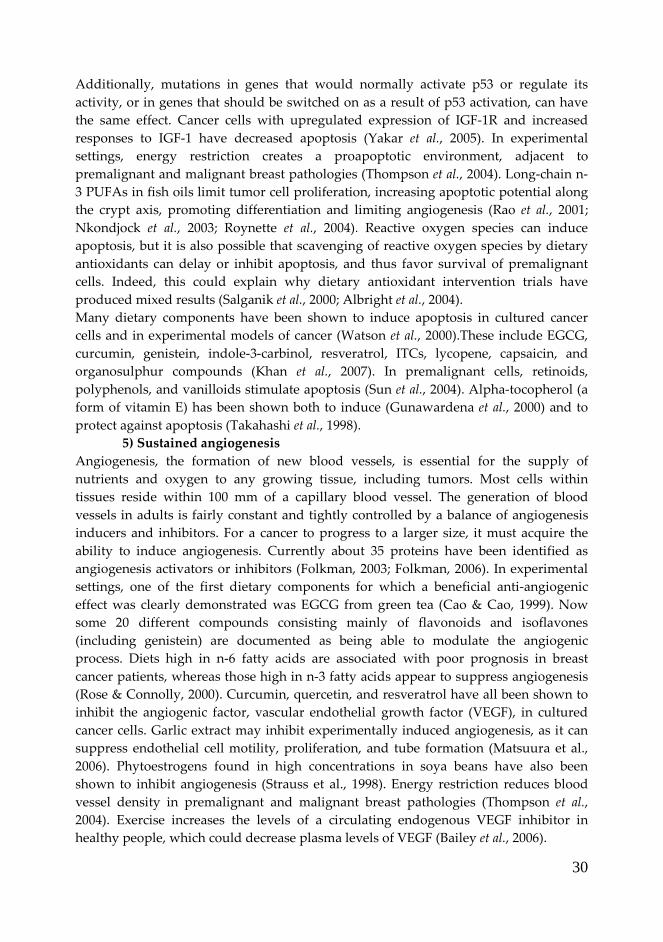
30
Additionally, mutations in genes that would normally activate p53 or regulate its
activity, or in genes that should be switched on as a result of p53 activation, can have
the same effect. Cancer cells with upregulated expression of IGF-1R and increased
responses to IGF-1 have decreased apoptosis (Yakar et al., 2005). In experimental
settings, energy restriction creates a proapoptotic environment, adjacent to
premalignant and malignant breast pathologies (Thompson et al., 2004). Long-chain n-
3 PUFAs in fish oils limit tumor cell proliferation, increasing apoptotic potential along
the crypt axis, promoting differentiation and limiting angiogenesis (Rao et al., 2001;
Nkondjock et al., 2003; Roynette et al., 2004). Reactive oxygen species can induce
apoptosis, but it is also possible that scavenging of reactive oxygen species by dietary
antioxidants can delay or inhibit apoptosis, and thus favor survival of premalignant
cells. Indeed, this could explain why dietary antioxidant intervention trials have
produced mixed results (Salganik et al., 2000; Albright et al., 2004).
Many dietary components have been shown to induce apoptosis in cultured cancer
cells and in experimental models of cancer (Watson et al., 2000).These include EGCG,
curcumin, genistein, indole-3-carbinol, resveratrol, ITCs, lycopene, capsaicin, and
organosulphur compounds (Khan et al., 2007). In premalignant cells, retinoids,
polyphenols, and vanilloids stimulate apoptosis (Sun et al., 2004). Alpha-tocopherol (a
form of vitamin E) has been shown both to induce (Gunawardena et al., 2000) and to
protect against apoptosis (Takahashi et al., 1998).
5) Sustained angiogenesis
Angiogenesis, the formation of new blood vessels, is essential for the supply of
nutrients and oxygen to any growing tissue, including tumors. Most cells within
tissues reside within 100 mm of a capillary blood vessel. The generation of blood
vessels in adults is fairly constant and tightly controlled by a balance of angiogenesis
inducers and inhibitors. For a cancer to progress to a larger size, it must acquire the
ability to induce angiogenesis. Currently about 35 proteins have been identified as
angiogenesis activators or inhibitors (Folkman, 2003; Folkman, 2006). In experimental
settings, one of the first dietary components for which a beneficial anti-angiogenic
effect was clearly demonstrated was EGCG from green tea (Cao & Cao, 1999). Now
some 20 different compounds consisting mainly of flavonoids and isoflavones
(including genistein) are documented as being able to modulate the angiogenic
process. Diets high in n-6 fatty acids are associated with poor prognosis in breast
cancer patients, whereas those high in n-3 fatty acids appear to suppress angiogenesis
(Rose & Connolly, 2000). Curcumin, quercetin, and resveratrol have all been shown to
inhibit the angiogenic factor, vascular endothelial growth factor (VEGF), in cultured
cancer cells. Garlic extract may inhibit experimentally induced angiogenesis, as it can
suppress endothelial cell motility, proliferation, and tube formation (Matsuura et al.,
2006). Phytoestrogens found in high concentrations in soya beans have also been
shown to inhibit angiogenesis (Strauss et al., 1998). Energy restriction reduces blood
vessel density in premalignant and malignant breast pathologies (Thompson et al.,
2004). Exercise increases the levels of a circulating endogenous VEGF inhibitor in
healthy people, which could decrease plasma levels of VEGF (Bailey et al., 2006).

31
6) Tissue invasion and metastasis
Normal cells in solid tissues maintain their position in the body and generally do not
migrate. As a cancer increases, it eventually reaches the membrane encapsulating the
organ. Tumor cells secrete enzymes such as matrix metalloproteases (MMPs), which
digest the membrane and allow the cancer to invade adjacent tissue. Once through the
membrane, cancer cells can access other sites via the blood and lymphatic systems.
This migration of cancer cells, or metastasis, is a common characteristic of most cancer
deaths. There is limited evidence for dietary components to influence these late stages
of cancer, although in vitro, EGCG, resveratrol, quercetin, curcumin, and genistein can
inhibit one or more MMPs. Vitamin C can inhibit MMP production by a number of
human cancer cell lines and prevent invasion of these lines in vitro (Roomi et al., 2006).
Vitamin E can inhibit metastasis of pre-established tumors in mouse models of breast
cancer (Hahn et al., 2006).
1.1 References
Biomarkers Definitions Working Group (2001) Biomarkers and surrogate endpoints:
preferred definitions and conceptual framework. Clin Pharmacol Ther 69, 89-95.
Abu J, Batuwangala M, Herbert K & Symonds P (2005) Retinoic acid and retinoid
receptors: potential chemopreventive and therapeutic role in cervical cancer.
Lancet Oncol 6, 712-720.
Adlercreutz H (2002) Phyto-oestrogens and cancer. Lancet Oncol 3, 364-373.
Aggett PJ (2010) Population reference intakes and micronutrient bioavailability: a
European perspective. Am J Clin Nutr 91, 1433S-1437S.
Alberg A (2002) The influence of cigarette smoking on circulating concentrations of
antioxidant micronutrients. Toxicology 180, 121-137.
Albright CD, Salganik RI & Van Dyke T (2004) Dietary depletion of vitamin E and
vitamin A inhibits mammary tumor growth and metastasis in transgenic mice.
J Nutr 134, 1139-1144.
Bailey AP, Shparago M & Gu JW (2006) Exercise increases soluble vascular endothelial
growth factor receptor-1 (sFlt-1) in circulation of healthy volunteers. Med Sci
Monit 12, CR45-50.
Bohnsack BL & Hirschi KK (2004) Nutrient regulation of cell cycle progression. Annu
Rev Nutr 24, 433-453.
Burkitt DP & Trowell HC (1977) Dietary fibre and western diseases. Ir Med J 70, 272-
277.
Butterworth CE, Jr., Hatch KD, Gore H, Mueller H & Krumdieck CL (1982)
Improvement in cervical dysplasia associated with folic acid therapy in users
of oral contraceptives. Am J Clin Nutr 35, 73-82.
Calle EE & Kaaks R (2004) Overweight, obesity and cancer: epidemiological evidence
and proposed mechanisms. Nat Rev Cancer 4, 579-591.
Cao Y & Cao R (1999) Angiogenesis inhibited by drinking tea. Nature 398, 381.

32
Chapkin RS, Clark AE, Davidson LA, Schroeder F, Zoran DL & Lupton JR (1998)
Dietary fiber differentially alters cellular fatty acid-binding protein expression
in exfoliated colonocytes during tumor development. Nutr Cancer 32, 107-112.
Chen C & Kong AN (2005) Dietary cancer-chemopreventive compounds: from
signaling and gene expression to pharmacological effects. Trends Pharmacol Sci
26, 318-326.
Collett ED, Davidson LA, Fan YY, Lupton JR & Chapkin RS (2001) n-6 and n-3
polyunsaturated fatty acids differentially modulate oncogenic Ras activation
in colonocytes. Am J Physiol Cell Physiol 280, C1066-1075.
Coussens LM & Werb Z (2002) Inflammation and cancer. Nature 420, 860-867.
Craig WJ (1997) Phytochemicals: guardians of our health. J Am Diet Assoc 97, S199-204.
Dashwood RH, Myzak MC & Ho E (2006) Dietary HDAC inhibitors: time to rethink
weak ligands in cancer chemoprevention? Carcinogenesis 27, 344-349.
Dijsselbloem N, Vanden Berghe W, De Naeyer A & Haegeman G (2004) Soy isoflavone
phyto-pharmaceuticals in interleukin-6 affections. Multi-purpose
nutraceuticals at the crossroad of hormone replacement, anti-cancer and anti-
inflammatory therapy. Biochem Pharmacol 68, 1171-1185.
Esquela-Kerscher A & Slack FJ (2006) Oncomirs - microRNAs with a role in cancer. Nat
Rev Cancer 6, 259-269.
EUFIC, Food Today 05/2010, Nutrient bioavailability - getting the most out of food
(http://www.eufic.org/article/en/artid/Nutrient-bioavailability-food/)
Fenton JI, Hord NG, Lavigne JA, Perkins SN & Hursting SD (2005) Leptin, insulin-like
growth factor-1, and insulin-like growth factor-2 are mitogens in ApcMin/+
but not Apc+/+ colonic epithelial cell lines. Cancer Epidemiol Biomarkers Prev 14,
1646-1652.
Ferguson LR, Philpott M & Karunasinghe N (2006) Oxidative DNA damage and
repair: significance and biomarkers. J Nutr 136, 2687S-2689S.
Fimognari C, Lenzi M & Hrelia P (2008) Chemoprevention of cancer by isothiocyanates
and anthocyanins: mechanisms of action and structure-activity relationship.
Curr Med Chem 15, 440-447.
Finley JW (2005) Proposed criteria for assessing the efficacy of cancer reduction by
plant foods enriched in carotenoids, glucosinolates, polyphenols and
selenocompounds. Ann Bot 95, 1075-1096.
Finley JW (2006) Bioavailability of selenium from foods. Nutr Rev 64, 146-151.
Fischer U & Schulze-Osthoff K (2005) Apoptosis-based therapies and drug targets. Cell
Death Differ 12 Suppl 1, 942-961.
Folkman J (2003) Fundamental concepts of the angiogenic process. Curr Mol Med 3,
643-651.
Folkman J (2006) Angiogenesis. Annu Rev Med 57, 1-18.
Geissler C, Powers H, editors. (2005) Human Nutrition. 11th ed. London: Elsevier
Churchill Livingstone.
Gleeson M, Nieman DC & Pedersen BK (2004) Exercise, nutrition and immune
function. J Sports Sci 22, 115-125.

33
Gonzalez C, Najera O, Cortes E, Toledo G, Lopez L, Betancourt M & Ortiz R (2002)
Hydrogen peroxide-induced DNA damage and DNA repair in lymphocytes
from malnourished children. Environ Mol Mutagen 39, 33-42.
Grimm JJ (1999) Interaction of physical activity and diet: implications for insulin-
glucose dynamics. Public Health Nutr 2, 363-368.
Gunawardena K, Murray DK & Meikle AW (2000) Vitamin E and other antioxidants
inhibit human prostate cancer cells through apoptosis. Prostate 44, 287-295.
Gupta RA, Tan J, Krause WF, Geraci MW, Willson TM, Dey SK & DuBois RN (2000)
Prostacyclin-mediated activation of peroxisome proliferator-activated receptor
delta in colorectal cancer. Proc Natl Acad Sci U S A 97, 13275-13280.
Hahn T, Szabo L, Gold M, Ramanathapuram L, Hurley LH & Akporiaye ET (2006)
Dietary administration of the proapoptotic vitamin E analogue alpha-
tocopheryloxyacetic acid inhibits metastatic murine breast cancer. Cancer Res
66, 9374-9378.
Hanahan D & Weinberg RA (2000) The hallmarks of cancer. Cell 100, 57-70.
Hawkes C (2005) The role of foreign direct investment in the nutrition transition. Public
Health Nutr 8, 357-365.
Holst B & Williamson G (2008) Nutrients and phytochemicals: from bioavailability to
bioefficacy beyond antioxidants. Curr Opin Biotechnol 19, 73-82.
Hursting SD, Lavigne JA, Berrigan D, Perkins SN & Barrett JC (2003) Calorie
restriction, aging, and cancer prevention: mechanisms of action and
applicability to humans. Annu Rev Med 54, 131-152.
Jeffery E & Keck A (2008) Translating knowledge generated by epidemiological and in
vitro studies into dietary cancer prevention. Mol. Nutr. Food Res. 52, s7-s17.
Khan N, Afaq F & Mukhtar H (2007) Apoptosis by dietary factors: the suicide solution
for delaying cancer growth. Carcinogenesis 28, 233-239.
Knowles LM & Milner JA (2001) Possible mechanism by which allyl sulfides suppress
neoplastic cell proliferation. J Nutr 131, 1061S-1066S.
Kubena KS & McMurray DN (1996) Nutrition and the immune system: a review of
nutrient-nutrient interactions. J Am Diet Assoc 96, 1156-1164; quiz 1165-1156.
Last JM, editor. (2001) A Dictionary of Epidemiology. 4th ed. New York: Oxford
University Press.
Leklem JE. (1999) Vitamin B-6. In: Shils M, Olson JA, Shike M, et al., editors. Nutrition
in Health and Disease. 9th ed. Baltimore: Williams & Wilkins.
LeRoith D, Baserga R, Helman L & Roberts CT, Jr. (1995) Insulin-like growth factors
and cancer. Ann Intern Med 122, 54-59.
Li N, Sun Z, Han C & Chen J (1999) The chemopreventive effects of tea on human oral
precancerous mucosa lesions. Proc Soc Exp Biol Med 220, 218-224.
Lotan R (1999) Aberrant expression of retinoid receptors and lung carcinogenesis. J
Natl Cancer Inst 91, 989-991.
Mackay J, Eriksen M, Shafey O. (2006) The Tobacco Atlas. 2nd ed. Atlanta: American
Cancer Society.

34
Mackay J, Jemal A, Lee N, et al. (2006) The Cancer Atlas. Atlanta: American Cancer
Society.
Matsuura N, Miyamae Y, Yamane K, Nagao Y, Hamada Y, Kawaguchi N, Katsuki T,
Hirata K, Sumi S & Ishikawa H (2006) Aged garlic extract inhibits angiogenesis
and proliferation of colorectal carcinoma cells. J Nutr 136, 842S-846S.
McNaughton SA & Marks GC (2003) Development of a food composition database for
the estimation of dietary intakes of glucosinolates, the biologically active
constituents of cruciferous vegetables. Br J Nutr 90, 687-697.
McTiernan A, Sorensen B, Yasui Y, Tworoger SS, Ulrich CM, Irwin ML, Rudolph RE,
Stanczyk FZ, Schwartz RS & Potter JD (2005) No effect of exercise on insulin-
like growth factor 1 and insulin-like growth factor binding protein 3 in
postmenopausal women: a 12-month randomized clinical trial. Cancer
Epidemiol Biomarkers Prev 14, 1020-1021.
Mihara M, Erster S, Zaika A, Petrenko O, Chittenden T, Pancoska P & Moll UM (2003)
p53 has a direct apoptogenic role at the mitochondria. Mol Cell 11, 577-590.
Milner JA (2004) Molecular targets for bioactive food components. J Nutr 134, 2492S-
2498S.
Miura S, Tsuzuki Y, Hokari R & Ishii H (1998) Modulation of intestinal immune system
by dietary fat intake: relevance to Crohn's disease. J Gastroenterol Hepatol 13,
1183-1190.
Niculescu MD, Pop EA, Fischer LM & Zeisel SH (2007) Dietary isoflavones
differentially induce gene expression changes in lymphocytes from
postmenopausal women who form equol as compared with those who do not.
J Nutr Biochem 18, 380-390.
Nishikawa A, Mori Y, Lee IS, Tanaka T & Hirose M (2004) Cigarette smoking,
metabolic activation and carcinogenesis. Curr Drug Metab 5, 363-373.
Nkondjock A, Shatenstein B, Maisonneuve P & Ghadirian P (2003) Specific fatty acids
and human colorectal cancer: an overview. Cancer Detect Prev 27, 55-66.
Novak TE, Babcock TA, Jho DH, Helton WS & Espat NJ (2003) NF-kappa B inhibition
by omega -3 fatty acids modulates LPS-stimulated macrophage TNF-alpha
transcription. Am J Physiol Lung Cell Mol Physiol 284, L84-89.
Palan PR, Chang CJ, Mikhail MS, Ho GY, Basu J & Romney SL (1998) Plasma
concentrations of micronutrients during a nine-month clinical trial of beta-
carotene in women with precursor cervical cancer lesions. Nutr Cancer 30, 46-
52.
Price PT, Nelson CM & Clarke SD (2000) Omega-3 polyunsaturated fatty acid
regulation of gene expression. Curr Opin Lipidol 11, 3-7.
Rao CV, Hirose Y, Indranie C & Reddy BS (2001) Modulation of experimental colon
tumorigenesis by types and amounts of dietary fatty acids. Cancer Res 61, 1927-
1933.
Roomi MW, Ivanov V, Kalinovsky T, Niedzwiecki A & Rath M (2006) Inhibition of
matrix metalloproteinase-2 secretion and invasion by human ovarian cancer

35
cell line SK-OV-3 with lysine, proline, arginine, ascorbic acid and green tea
extract. J Obstet Gynaecol Res 32, 148-154.
Rose DP & Connolly JM (2000) Regulation of tumor angiogenesis by dietary fatty acids
and eicosanoids. Nutr Cancer 37, 119-127.
Roynette CE, Calder PC, Dupertuis YM & Pichard C (2004) n-3 polyunsaturated fatty
acids and colon cancer prevention. Clin Nutr 23, 139-151.
Salganik RI, Albright CD, Rodgers J, Kim J, Zeisel SH, Sivashinskiy MS & Van Dyke
TA (2000) Dietary antioxidant depletion: enhancement of tumor apoptosis and
inhibition of brain tumor growth in transgenic mice. Carcinogenesis 21, 909-914.
Schmitz KH, Ahmed RL & Yee D (2002) Effects of a 9-month strength training
intervention on insulin, insulin-like growth factor (IGF)-I, IGF-binding protein
(IGFBP)-1, and IGFBP-3 in 30-50-year-old women. Cancer Epidemiol Biomarkers
Prev 11, 1597-1604.
Sharpless NE & DePinho RA (2005) Cancer: crime and punishment. Nature 436, 636-
637.
Smyth MJ, Dunn GP & Schreiber RD (2006) Cancer immunosurveillance and
immunoediting: the roles of immunity in suppressing tumor development and
shaping tumor immunogenicity. Adv Immunol 90, 1-50.
Strauss L, Santti R, Saarinen N, Streng T, Joshi S & Makela S (1998) Dietary
phytoestrogens and their role in hormonally dependent disease. Toxicol Lett
102-103, 349-354.
Sun SY, Hail N, Jr. & Lotan R (2004) Apoptosis as a novel target for cancer
chemoprevention. J Natl Cancer Inst 96, 662-672.
Takahashi H, Kosaka N & Nakagawa S (1998) alpha-Tocopherol protects PC12 cells
from hyperoxia-induced apoptosis. J Neurosci Res 52, 184-191.
Thompson HJ, Zhu Z & Jiang W (2003) Dietary energy restriction in breast cancer
prevention. J Mammary Gland Biol Neoplasia 8, 133-142.
Thompson HJ, Zhu Z & Jiang W (2004) Weight control and breast cancer prevention:
are the effects of reduced energy intake equivalent to those of increased energy
expenditure? J Nutr 134, 3407S-3411S.
Tlaskalova-Hogenova H, Tuckova L, Lodinova-Zadnikova R, Stepankova R,
Cukrowska B, Funda DP, Striz I, Kozakova H, Trebichavsky I, Sokol D,
Rehakova Z, Sinkora J, Fundova P, Horakova D, Jelinkova L & Sanchez D
(2002) Mucosal immunity: its role in defense and allergy. Int Arch Allergy
Immunol 128, 77-89.
Trikha M, Corringham R, Klein B & Rossi JF (2003) Targeted anti-interleukin-6
monoclonal antibody therapy for cancer: a review of the rationale and clinical
evidence. Clin Cancer Res 9, 4653-4665.
Trowell H & Burkitt D (1986) Physiological role of dietary fiber: a ten-year review.
ASDC J Dent Child 53, 444-447.
Trueba GP, Sanchez GM & Giuliani A (2004) Oxygen free radical and antioxidant
defense mechanism in cancer. Front Biosci 9, 2029-2044.

36
Trujillo E, Davis C & Milner J (2006) Nutrigenomics, proteomics, metabolomics, and
the practice of dietetics. J Am Diet Assoc 106, 403-413.
Tuma RS (2007) Statisticians set sights on observational studies. J Natl Cancer Inst 99,
664-665, 668.
Watson WH, Cai J & Jones DP (2000) Diet and apoptosis. Annu Rev Nutr 20, 485-505.
World Cancer Report. (2003) Lyon: IARC Press.
World Health Organization. (2003) Diet, Nutrition and the Prevention of Chronic
Diseases: Report of a Joint WHO/FAO Expert Consultation. In: WHO Technical
Report Series no 916.
World Health Organization. (1990) Diet, Nutrition, and the Prevention of Chronic
Diseases. Report of a WHO Study Group. In: WHO Technical Report Series no
797

37
2. AIMS OF THE STUDY
The intake of several foods rich in bioactive constituents has been identified in the
literature reviews as being associated with lower chronic-degenerative disease risk
(Liu, 2004). The bioactivity of these compounds is in part related to their antioxidant
properties which are important to decrease the risk of the onset development of many
of the chronic degenerative diseases (Espin et al., 2007).
Despite of this consideration, more studies are needed to demonstrate the protective
role of specific food and the mechanisms of action of bioactive compounds.
This PhD research project will be divided in two parts.
1) The first part will concern to the evaluation of the impact of bioactive
compounds in Cruciferae, on humans with different genetic polymorphisms of
enzymes, involved in endogenous biotransformation systems. Particularly, two types
of study will be designed analyzing: the bioavailability of protective compounds from
broccoli and their role in the protection against biomarkers of oxidative stress, taking
into account individual genetic susceptibility. The response of different biomarkers to
both single meal and regular consumption of broccoli will be investigated. DNA
damage will be evaluated as resistance to exogenous damage, endogenous DNA
damage and OGG1 repair ability; while OGG1, HO-1 and NUDT1 mRNA levels will
be considered as markers of repair and defence systems.
Recently, the inverse relationship found between cruciferous vegetable intake and
cancer risk increased interest among scientists involved in cancer prevention studies.
Dietary or environmental carcinogens must be activated to induce cancer. The ability
of dietary constituents in cruciferous vegetables to inhibit phase I activation and to
induce phase II enzymes are of primary importance to their cancer chemopreventive
effects (Fimognari et al., 2008). In particular, phase II enzymes decrease carcinogenicity
by blocking carcinogen metabolic activation and enhancing carcinogen detoxification.
Glutathione-S-Transferase (GST), is included in the phase II family enzymes activated
by Cruciferae consumption (Jeffery & Keck, 2008). Moreover its genetic polymorphism
is related to cancer risk (Qiu et al., 2010; Wan et al., 2010). Thus, we will evaluate the
different susceptibility to risk of different GST polymorphisms in relation to broccoli
consumption. Controversial data are found in literature, and human intervention
studies have not clearly demonstrated the relationship between GST polymorphisms
and protective effect of broccoli. It is hypothesized that GST genotypes associated with
lower protection against carcinogens benefit most from cruciferous vegetable
consumption.
The first part of this PhD research project will be carried out at the Dipartimento di
Scienze e Tecnologie Alimentari e Microbiologiche (DiSTAM)-sezione Nutrizione
(Università degli Studi di Milano), under the direction of Prof. Marisa Porrini and Dr.
Patrizia Riso.
2) The second part will be focused on the study of mechanisms of action
involved at cellular level induced by single compounds. Since most cancers are
dependent on the growth of tumor blood vessels, angiogenesis may thus provide an

38
efficient strategy to delay or block tumor growth. Migration and proliferation of
endothelial cells are critical steps in angiogenesis since these events allow cells to
disseminate from pre-existing vessels to form new capillary structures (Folkman,
2006). Moreover, blood coagulation and cancer biology are strictly related (ten Cate &
Falanga, 2008).
Capillary-like formation assay is a reliable approach able to test pro- or anti-angiogenic
properties of a chemical compound. After a review of the literature two anthocyanins,
i.e. Delphinidin-3-glucoside (Dp-3-glc) and Cyanidin-3-glucoside (Cy-3-glc), will be
selected for this type of studies. These two compounds are able to inhibit tumor cell
proliferation (Katsube et al., 2003; Fimognari et al., 2004; Olsson et al., 2004), but it is not
known whether they can also act on pro-angiogenic and the pro-thrombotic properties
of endothelial cells. Models of angiogenesis on human microvascular endothelial cells
will be used to test the role of Dp-3-glc and Cy-3-glc in capillary network formation
and wound healing assay in resting and VEGF-stimulated conditions. The same cell
line will be used to test the protective role of Dp-3-glc in pro-coagulant activity both in
resting and LPS-stimulated conditions. The results will enhance our knowledge of the
intricate roles and functions that ACNs act at both cellular and molecular level.
This part of research will be performed at the Hemostasis and Thrombosis Center,
Ospedali Riuniti di Bergamo, Italy, supervised by Dr. Anna Falanga and Dr. Marina
Marchetti.
2.1 References
Espin JC, Garcia-Conesa MT & Tomas-Barberan FA (2007) Nutraceuticals: facts and
fiction. Phytochemistry 68, 2986-3008.
Fimognari C, Berti F, Nusse M, Cantelli-Forti G & Hrelia P (2004) Induction of
apoptosis in two human leukemia cell lines as well as differentiation in human
promyelocytic cells by cyanidin-3-O-beta-glucopyranoside. Biochem Pharmacol
67, 2047-2056.
Fimognari C, Lenzi M & Hrelia P (2008) Chemoprevention of cancer by isothiocyanates
and anthocyanins: mechanisms of action and structure-activity relationship.
Curr Med Chem 15, 440-447.
Folkman J (2006) Angiogenesis. Annu Rev Med 57, 1-18.
Jeffery E & Keck A (2008) Translating knowledge generated by epidemiological and in
vitro studies into dietary cancer prevention. Mol. Nutr. Food Res. 52, s7-s17.
Katsube N, Iwashita K, Tsushida T, Yamaki K & Kobori M (2003) Induction of
apoptosis in cancer cells by Bilberry (Vaccinium myrtillus) and the
anthocyanins. J Agric Food Chem 51, 68-75.
Liu RH (2004) Potential synergy of phytochemicals in cancer prevention: mechanism of
action. J Nutr 134, 3479S-3485S.
Olsson ME, Gustavsson KE, Andersson S, Nilsson A & Duan RD (2004) Inhibition of
cancer cell proliferation in vitro by fruit and berry extracts and correlations
with antioxidant levels. J Agric Food Chem 52, 7264-7271.

39
Qiu LX, Yuan H, Yu KD, Mao C, Chen B, Zhan P, Xue K, Zhang J & Hu XC (2010)
Glutathione S-transferase M1 polymorphism and breast cancer susceptibility: a
meta-analysis involving 46,281 subjects. Breast Cancer Res Treat 121, 703-708.
ten Cate H & Falanga A (2008) Overview of the postulated mechanisms linking cancer
and thrombosis. Pathophysiol Haemost Thromb 36, 122-130.
Wan H, Zhou Y, Yang P, Chen B, Jia G & Wu X (2010) Genetic polymorphism of
glutathione S-transferase T1 and the risk of colorectal cancer: a meta-analysis.
Cancer Epidemiol 34, 66-72.

40
3. RESULTS AND DISCUSSION
3.1 TOPIC I: Role of broccoli intake, a glucosinolate-rich source, on biomarkers
of oxidative stress in young smokers
Abstract
Scientific evidences support the protective role of diets rich in fruit and vegetables
against chronic diseases, like cancer and cardiovascular disease (WCRF/AICR 2007).
Fruits and vegetables compounds, able to reduce the risk of chronic diseases, include
vitamins, minerals and numerous phytochemicals. The latter are bioactive compounds
that have been shown to exert a wide range of biological activities. Since they are
ingested regularly and in significant amounts as part of the diet, they may have a
noticeable long-term beneficial effect.
Cruciferae and especially Brassica genus contain high concentration of constituents
with antioxidant properties (e.g. carotenoids, vitamin C, folate) as well as glucosinolate
precursors of isothiocyanates (ITCs) and indoles that modulate xenobiotic
biotransformation enzymes, such as Glutathione S-Transferase (GST). Different GST
polymorphisms seem to be related to altered protective effect against xenobiotics. The
protective effect of broccoli (Brassica oleracea L. var. italica) was tested through two
different study protocols performed at DiSTAM-sezione Nutrizione (Università degli
Studi di Milano): a regular-intake intervention study with daily consumption of one
serving of steamed broccoli for 10 days and a single-meal study (i.e. with one serving
of broccoli). Broccoli were chemically characterized for the content in antioxidant
compounds (carotenoids, vitamin C) and glucosinolates as isothiocyanates (ITCs). One
portion of 250g of steamed broccoli provided: 2.75 mg of lutein, 1.50 mg of β-carotene,
172 mg of vitamin C and 160 µmol of phenethyl- isothiocyanate (PEITC) equivalents of
ITCs.
Subjects involved in the study were young male smokers recruited on the basis of
anthropometric characteristics, food habits and their GSTM1 genotype.
Thirty subjects were enrolled for the regular-intake study. A single blind randomized
cross-over experimental design was scheduled: 15 volunteers followed the sequence
broccoli diet/wash-out/control diet; while the others followed the opposite sequence.
Broccoli and control diet were 10 days long and they were spaced out from 20 days of
free diet. One serving consisted of 250 g of steamed broccoli. Fasting blood samples
were collected at the beginning and at the end of each treatment period (0, 10, 30, 40
days). Plasma, serum and lymphocytes were separated and stored for specific
analyses. Concentration of carotenoids, lutein, β-carotene and folate was assessed in
plasma and serum samples. Lymphocytes were separated and used immediately for
the determination of cell resistance against oxidative stress (H2O2-induced DNA
strands breaks) through the COMET assay. Additionally, lymphocytes were isolated
and appropriately stored at -80°C for the subsequent determination of biomarkers of
oxidative stress: endogenous DNA damage (i.e. oxidized purines), in vitro DNA repair
activity and mRNA expression of OGG1, NUDT1 and HO-1 levels. Twenty-seven male

41
smokers of the 30 enrolled completed the regular-intake study (age 22.1±2.5 y; BMI
23.0±2.7 kg/m2). Cell protection against H2O2-induced DNA damage was higher after
broccoli diet with respect to control diet in the whole group of subjects. Folate and
lutein concentrations increased significantly after broccoli diet. By analyzing data
considering genotype as independent factor, broccoli intake caused a more consistent
and significant effect of protection against DNA damage in GSTM1 null (-27.6%)
compared to GSTM1 positive (-13.1%) subjects (p<0.05). Oxidatively damaged purine
bases decreased significantly (p<0.05) after broccoli intake (-22.6%), while no effect of
polymorphism was observed. DNA repair activity and OGG1, HO-1 and NUDT1
mRNA expression levels did not differ throughout the intervention study.
Twelve volunteers were selected for the single-meal study. One serving of 250 g of
steamed broccoli with 70 g of pasta was consumed as first meal. Fasting blood samples
were collected before broccoli consumption and at 3, 6, 8 and 24 h after broccoli intake.
Plasma, serum and lymphocytes were separated and stored for specific analyses.
Cell resistance to oxidative stress (H2O2-induced DNA strands breaks) was determined
through the COMET assay. Additionally lymphocytes were isolated and appropriately
stored at -80°C for the subsequent determination of concentration of bioactive
compounds (folate, lutein, β-carotene and vitamin C) and total GST activity. Total GST
activity in plasma and lymphocytes was calculated by measuring the conjugation of 1-
chloro-2,4-dinitrobenzene (CDNB) with reduced glutathione, and normalized by the
amount of proteins found in each sample. All the 12 selected volunteers (age 20.8±1.6
y; BMI 22.5±2.1 kg/m2) completed the single meal study. Serum folate, plasma vitamin
C and β-carotene concentrations increased significantly after 3h for the first two, and at
6h for β-carotene; while antioxidant content in lymphocytes did not show any increase
after broccoli meal. In general, individuals with positive GSTM1 genotype showed a
greater plasma and lymphocyte GST activity compared to individuals with GSTM1
null genotype. After broccoli consumption, plasma GST activity increased significantly
(p<0.05) in individuals with GSTM1 positive genotype at 6 h with respect to 3 h and 24
h but not compared to baseline. No significant difference was observed in lymphocyte
GST activity. A significant reduction of ex-vivo induced DNA damage (p<0.05) after 24
h from broccoli consumption intake with respect to time 0, 3 and 6 h was observed.
Broccoli under controlled dietary conditions modulated GST activity even if it seems to
be more evident in individuals with GSTM1 positive genotype, who showed also a
greater protection against DNA damage.
In conclusion, these two studies demonstrated that 10 days of consumption of steamed
broccoli could improve defence against DNA damage but did not affect repair activity
in young healthy smokers. Our data are in accordance with shown protective effects of
cruciferous vegetables, including Brussel sprouts, broccoli and watercress, on
biomarkers of oxidative damage to DNA in human intervention studies. Also a single
portion of steamed broccoli was able to decrease DNA damage and to modulate GST
activity. Even if preliminary, our data suggest a “diet/genetic” interaction. In particular
we supported the hypothesis that GSTM1 null genotype subjects might have particular
benefit from regular brassica vegetable intake.

42
Introduction
As reported in detail in the State of the Art (chapter 1) the onset of several chronic
diseases is linked to oxidative stress. In fact, oxidative DNA damage formed during
oxidative stress and DNA repair may mediate several cellular processes, like
replication and transcription, mutagenesis and apoptosis and thus may be for the
organism development as well as its pathogenesis, including cancer. Diet seems to
play a significant role in these mechanisms and the aetiology of several cancers. The
evidence of the protective role for diets rich in fruit and vegetable is convincing and,
associated with an healthy lifestyle, provides low incidence of cancer (WCRF/AICR
2007). In this regard, a class of specific vegetable food with hypothesized health
benefits is Cruciferae vegetable. Despite many in vitro and animal studies, only little
evidence is present in dietary controlled human intervention studies.
Intake and health benefits of Cruciferae
Specific groups of vegetables seem to offer enhanced sources of protective
phytochemicals, notably Cruciferae and especially Brassica genus (Moreno et al.,
2006), which contain many putative protective agents. Cruciferae include 20 species in
the Brassica genus (Table 3.1.1).
Table 3.1.1 Cruciferous vegatables (adapted from Thomson and Green 2003): species and common
names of different plants
Genus Species Plant
Brassica oleracea Broccoli, brussel sprouts, cabbage, savoy cabbage, red cabbage,
cauliflower, collards, kale, Chinese kale, Italian broccoli, asparagous
broccoli, sea kale, wild cabbage, kohlrabi
Brassica rapa Chinese cabbage, celery ,cabbage, spinaci mustard, turnip, turnip tops,
turnip broccoli, toria
Brassica napus Rape, colza, Siberian kale, rutabaga
Brassica japonica wasabi
Brassica juncea Chinese mustards, mustard greens, curled mustard, pak choi, bok
celery, brown mustard
Brassica alba White mustard
Brassica nigra Black mustard oil, black mustard seeds
Sisymbrium officinale Hedge mustard
Nasturtium amoracea Horseradish
Nasturtium officinale Watercress
Bioactive compounds contained in Brassica include glucosinolates (GLS), phenolic
compounds and antioxidants such as carotenoids (lutein and β-carotene) and vitamins
(C, K1 and folate). The single active compounds responsible for chemopreventive
properties probably do not exist since the protective activity results from the
synergistic action of many different compounds, but glucosinolates are indeed the

43
most distinguishing compounds of these vegetables, and more than 120 GLS have been
characterized so far (Halkier & Gershenzon, 2006; Sonderby et al., 2010).
Table 3.1.2 shows a list of epidemiological studies which pointed out an effect on
cancer prevention, related to cruciferous vegetables consumption.
Table 3.1.2 Epidemiological studies on cruciferous vegetables consumption
Cruciferous vegetable Study population References
Mustard green and Kale Singapore (MacLennan et al., 1977)
Cabbage, Cauliflower, Rutabaga Norway (Kvale et al., 1983)
Broccoli Massachusset (Colditz et al., 1985)
Cruciferous vegetables Greece (Katsouyanni et al., 1986)
Broccoli Texas (Bond et al., 1987)
Broccoli Louisiana (Fontham et al., 1988)
Brassica vegetables Hawaii (Le Marchand et al., 1989)
Broccoli Hawaii (Goodman et al., 1992)
Brassica vegetables Shanghai (Zhang et al., 1992)
Cabbage Japan (Gao et al., 1993)
Cabbage and Cauliflower India (Sankaranarayanan et al., 1994)
Brassica vegetables The Netherlands (Voorrips et al., 2000)
Brassica vegetables USA (Slattery et al., 2000)
Brassica vegetables Sweden (Terry et al., 2001)
The chemical structure of glucosinolate consists of a β-D-glucopyranose residue linked
via a sulfur atom to a N-hydroximinosulfate ester, plus a variable R group derived
from one of eight amino acids (Halkier & Gershenzon, 2006).
Biosynthesis of glucosinolates in vegetables can be conveniently divided into three
separate phases (Figure 3.1.1):
1. certain aliphatic and aromatic amino acids (e.g. methionine and
phenylalanine for aromatic glucosinolates and tryptophan for indoles) are elongated
by inserting methylene groups into their side chains;
2. the amino acid moiety itself, whether elongated or not, is metabolically
reconfigured to give the core structure of glucosinolates;
3. the initially formed glucosinolates are modified by various secondary
transformations which consist in the de-saturation and hydroxylation of the chain
(Halkier & Gershenzon, 2006).

44
Figure 3.1.1 Biosynthesis of glucosinolates (Fahey et al., 2001)
Figure 3.1.2 Formation of isothiocyanates, nitriles and thiocyanates from glucosinolates by
myrosinase-catalyzed degradation (Thornalley, 2002)
Glucosinolates are sulfur-rich, anionic natural products that following hydrolysis by
myrosinases (endogenous thioglucosidases) produce several different products (for
example isothiocyanates, thiocyanates, and nitriles) (Figure 3.1.2).
Isothiocyanates are absorbed in the intestinal cells by passive diffusion and
transported to liver by serum albumine. They are electrophilic and thus react with
nucleophilic agents including thiols. The main reactive group in the cell is glutathione.
The reaction between isothiocyanate and glutathione is spontaneous but can also be
catalyzed by glutathione S-transferase (GST). The first product of this reaction is a
dithiocarbamate, which can be further metabolized to the cysteine conjugate and then
N-acetylated to form the corresponding mercapturic acid which is readily excreted in
urine (Ye et al., 2002).
Sulforaphane (1-isothiocyanato-4-methylsulfinyl butane) is the major isothiocyanate
derived from the 4-methylsulfinylbutyl glucosinolate that is accumulated in broccoli
florets.

45
Table 3.1.3 Mechanisms of chemoprevention associated to isothiocyanates adapted from
(Fimognari et al., 2008) and (Thomson and Green 2003)
Chemo-preventive
mechanism
Cruciferous/constituent References
Apoptosis and regulation of
cell cycle
Indole-3-carbinol
Sulforaphane
(Chung et al., 2000)
(Chiao et al., 2000)
Inhibition of phase I enzymes Dithiolthione (Steinkellner et al., 2001)
Ascorbigen
Phenethyliothiocyanate
(Hecht, 2000)
Induction of phase II enzymes
Dithiolthione
Indole-3-carbinol
Isothiocyanates
Sulforaphane
Indole
(Brooks et al., 2001)
(Morimitsu et al., 2000)
(Hecht, 2000)
(Steinkellner et al., 2001)
Inhibition of alpha
hydroxilation of nitrosamine
Indoles
Isothiocyanates
(Steinkellner et al., 2001)
(Hecht, 2000)
Radical and electrophil
scavenging
Indole-3-carbinol
Ascorbigen
(Shertzer & Senft, 2000)
It is a potent inducer of phase II detoxification enzymes, inhibits phase I enzymes and
can induce cell cycle arrest and apoptosis (Traka et al., 2005). However, there are many
other natural bioactive constituents which may be responsible of chemopreventive
mechanisms: indole-3-carbinol, benzyl isothiocyanate and phentyl isothiocyanate
(Visanji et al., 2004). They have been extensively studied in experimental in vitro and in
vivo carcinogenesis models. Numerous studies have consistently shown that
chemoprotective agents influence carcinogenesis during the initiation and promotion
phases of cancer development. In Table 3.1.3 the mechanisms for chemoprevention by
constituents of cruciferous vegetables are reported.
Cancer protective mechanisms of sulforaphane have been widely studied with several
in vitro studies as reported in Table 3.1.4.
In recent years, the inverse relationship found between cruciferous vegetable intake
and cancer risk increased interest among scientists involved in cancer prevention
studies.
Dietary or environmental carcinogens must be activated to induce cancer. The ability
of dietary constituents in cruciferous vegetables to inhibit phase I enzymes activation
are of primary importance to their cancer chemopreventive effects. The ability of
cruciferous vegetables to activate phase II drug-metabolizing enzymes was
demonstrated in 1979. Phase II enzymes decrease carcinogenicity by blocking
carcinogen metabolic activation and enhancing carcinogen detoxification.

46
Table 3.1.4 Mechanisms of chemoprevention associated to sulforaphane adapted from (Fimognari
et al., 2008; Jeffery & Keck, 2008) Endpoints SF dose (µM) Cancer cell type References
Induction of phase II
detoxification
enzyme
0.4-0.8 Liver (mouse) (Zhang et al., 1992)
2-5 Liver (human) (Bacon et al., 2007)
5-10 Colon (human) (Bacon et al., 2007)
0.5-5 Prostate (human) (Brooks et al., 2001)
0.2 Liver (mouse) (Posner et al., 1994)
Inhibition of histone
deacetylation
15 Prostate (human) (Myzak et al., 2006)
5-25 Breast (human) (Pledgie-Tracy et al., 2007)
Cell cycle arrest 15 Colon (human) (Gamet-Payrastre et al., 2000)
25-50 Colon (human) (Jakubikova et al., 2005)
10-40 Prostate (human) (Herman-Antosiewicz et al., 2007)
15 Breast (human) (Jackson & Singletary, 2004)
30 T-cells (human) (Fimognari et al., 2002)
Apoptosis 15 Colon (human) (Gamet-Payrastre et al., 2000)
10-20 Prostate (human) (Choi et al., 2007; Herman-
Antosiewicz et al., 2007)
30 T-cells (human) (Fimognari et al., 2002)
Inhibit cell growth 27 Bladder (human) (Shan et al., 2006)
40 Ovarian (human) (Chaudhuri et al., 2007)
8-10 Breast (human) (Pledgie-Tracy et al., 2007)
15 Breast (human) (Jackson & Singletary, 2004)
10-30 T-cells (human) (Fimognari et al., 2002)
12 Bladder (human) (Tang et al., 2006)
In Phase I reactions, a small polar group (containing both positive and negative
charges) is either exposed on the toxicant or added to the toxicant. The three main
Phase I reactions are oxidation, reduction, and hydrolysis. The most common
enzymatic process is through the addition of oxygen catalyzed by cytochrome P450
enzymes. Some of the intermediates formed in this process may be elecrophiles, which
can react with nucleophilic sites in critical macromolecules such as DNA, RNA and
protein and form a covalent binding product called adducts. DNA adducts that persist
unrepaired can cause miscoding and thus produce mutations in critical genes.
A xenobiotic that has undergone a Phase I reaction is now a new intermediate
metabolite that contains a reactive chemical group, e.g., hydroxyl (-OH), amino (-NH2),
and carboxyl (-COOH). Many of these intermediate metabolites do not possess
sufficient hydrophilicity to permit elimination from the body. These metabolites must
undergo additional biotransformation as a Phase II reaction.
Phase II reactions are conjugation reactions, that is, a molecule normally present in the
body is added to the reactive site of the Phase I metabolite. The result is a conjugated
metabolite that is more water-soluble than the original xenobiotic or Phase I
metabolite. Usually the Phase II metabolite is quite hydrophilic and can be readily
eliminated from the body. Some of the Phase II ezymes are quinone reductase,
glutathione-S-transferase, UDP-glucurunosyl tranferase and sulfotransferase.

47
Blocking carcinogen metabolic activation by Phase I enzymes and enhancing
carcinogen detoxification by Phase II enzymes are two ways to decrease carcinogenity
(Fimognari et al., 2008).
Figure 3.1.3 The glutathione-S-transferase supergene family, (adapted from (Strange et al., 2001)
On the basis of sequence homology and immunologic cross-reactivity, human cytosolic
GSTs have been grouped into seven families, designated GST-α, -μ, π, σ, ω, θ, and ζ.
The GST-μ family is encoded by a 100-kb gene cluster at 1p13.3 arranged as 5′-
GSTM4-GSTM2-GSTM1-GSTM5-GSTM3–3’. Deletion of the GSTM1 gene,
GSTM1*0, frequently affects both alleles, resulting in the so-called null genotype,
GSTM1−/− (Roodi et al., 2004).
In recent years, evidence has accumulated to support hypothesis that genetic
polymorphisms in carcinogen-metabolizing enzymes may be of importance in
determining individual susceptibility to cancer. The Glutathione S-transferases (GSTs),
a family of multifunctional enzymes (Figure 3.1.3), metabolize a variety of xenobiotics
with a large overlap in substrate specificity. Individuals who are homozygous for the
null GSTM1 or null GSTT1 alleles lack the respective enzyme functions. The null
GSTM1 genotype appears to be common in both Asians and Caucasians, whereas the
null GSTT1 genotype exhibits population frequencies that depend on ethnicity. The
GSTM1 and GSTT1 defects seem to be associated with increased risk of certain cancers;
however, conflicting data have been observed (Chen et al., 1996; Qiu et al., 2010; Wan et
al., 2010). This may reflect the fact that certain GSTs may be more efficient in
detoxifying certain carcinogens, but others may result in a higher risk for some types
of exposure, which has been especially demonstrated with GSTT1 (Huang et al., 2000;
Agalliu et al., 2006; Lampe, 2007).
DNA repair and defence systems
Mammals have five types of DNA repair system (Hoeijmakers, 2001; Larsen et al.,
2005):
1. Direct reversal (DR) corrects rather than removes damaged DNA bases;

48
2. Base-excision repair (BER) corrects DNA damage caused by reactive oxygen
species, deamination, and hydroxylation arising from cellular metabolism and
spontaneous depurination.
3. Nucleotide-excision repair (NER) removes lesions that distort the structure of
the DNA helix, such as pyrimidine dimers and DNA adducts.
4. Recombinational repair (RER). Homologous recombination and non-
homologous endjoining repair double-strand breaks. Homologous
recombination is used when a second identical DNA copy is available, for
example after replication; non-homologous end-joining re-links the ‘broken’
ends of a double-strand break.
5. DNA mismatch repair (MMR) detects and repairs copying errors made during
replication.
One protective mechanism of action of Brassica vegetables is thought to involve
reduction of free radical-related molecular damage and this could be particularly
important in subjects with high exposure to oxidative stress. Cigarette smoke contains
a large amount of reactive oxygen species (ROS) as well as other substances which
generate ROS (Frei et al., 1991). ROS can cause oxidative damage to DNA such as
oxidized bases and strand breaks. These DNA lesions, including the promutagenic 8-
oxo-7,8-dihydro-2’-deoxyguaninosine (8-oxodG) could be implicated in early steps of
cancer development (Evans et al., 2004; Cooke et al., 2006). Fortunately, mammalian
cells have a large array of repair systems for the removal of oxidized DNA lesions; the
most important pathway for oxidized bases is considered to be base excision repair
(BER), whereas the nucleotide excision repair enzymes most likely functions as a back-
up system. The repair of 8-oxodG by the BER system is initiated by the enzyme 8-
oxoguanine DNA glycosylase (OGG1), which cleaves the N-glycosidic bond leaving
behind a free base and an AP-site (apurinic/apyrimidinic site), as shown in Figure 3.1.4
(Moller et al., 2003; Loft & Moller, 2006; Tudek et al., 2006).

49
Figure 3.1.4 OGG1 cleaves the glycosylic bond between 8-oxoG and the deoxyribose, thereby
releasing the damaged base from DNA. The remaining abasic site is then cleaved 5’ to
the lesion by an AP endonuclease, APE, also called HAP1, or by the lyase activity of
OGG1. The dRPase (deoxyribose phosphate lyase) activity of DNA polymerase b
cleaves 3’ to the lesion, and the missing nucleotide is inserted by DNA polymerase β,
followed by ligation. Additional repair proteins are involved in this process.
Nucleotide pool modification is an important source of nucleic acids damage.
Intracellular free nucleotides play essential roles as precursors in the synthesis of DNA
and RNA, and as molecules for energy storage, cofactors of metabolic pathways and
regulators of signal transduction. Free nucleotides can, however, undergo various
chemical modifications by endogenous and exogenous reactive molecules, some of
which are inevitably produced in living cells (Iyama et al., 2010). In the nucleotide pool, dGTP can be oxidized to 8-oxo-2’-deoxyguanosine 5’-
triphosphate (8-oxo-dGTP), which may give rise to 8-oxodG in DNA if the oxidized
nucleotide is incorporated during replication or repair. 8-OxodGTP is removed from
the nucleotide pool by the nucleoside diphosphate linked moiety X-type motif 1
(NUDT1) enzyme (Loft & Moller, 2006), as described in Figure 3.1.5. Misincorporation
of oxidized nucleoside triphosphates into DNA/RNA during replication and
transcription can cause mutations that may result in carcinogenesis or
50
neurodegeneration. The protein encoded by this gene is an enzyme that hydrolyzes
oxidized purine nucleoside triphosphates, such as 8-oxo-dGTP, 8-oxo-dATP, 2-
hydroxy-dATP, and 2-hydroxy rATP, to monophosphates, thereby preventing
misincorporation. The encoded protein is localized mainly in the cytoplasm, with some
in the mitochondria, suggesting that it is involved in the sanitization of nucleotide
pools both for nuclear and mitochondrial genomes.
Figure 3.1.5 Oxidation of guanine in DNA and the nucleotide pool with consequences in terms of
potential mutations as well as the involved repair pathways. NER, nucleotide excision
repair; TCR, transcription coupled repair. Bold G indicates oxidized guanine (Loft &
Moller, 2006).
Heme oxigenase-1 (HO-1) is responsible of the degradation of heme to biliverdin and
is a well known stress response protein inducible by various agents able to generate
ROS and thereby causing oxidative stress (e.g. U.V., X-ray radiation, oxidants such as
H2O2, heavy metals, pro-inflammatory mediators). OH-1 is found over-expressed in
various tissues affected by diseases such as atherosclerosis, hypertension, lung injury
and cancer. Thus the level of HO-1 mRNA can be considered an important oxidative
stress response and defence enzyme (Risom et al., 2003).
3.2 Materials and methods
3.2.1 Characteristics and analyses of the product
“Marathon” broccoli (Brassica oleracea L. Var italica) was used for this intervention
study. It was a kind gift from Di Stasi Company, Italy. The company guaranteed that
the whole amount of vegetables used were from a single batch of production (same
condition of growth). The vegetables arrived at the DiSTAM laboratory one day after
the harvest and were immediately processed at the Experimental Institute of
Agricultural Products and Technologies (IVTPA, Milan). Broccoli florets were handled

51
gently, excised from the main stem, immediately blanched (high temperature steaming
of vegetables to deactivate enzymes) and, after cooling at room temperature for some
minutes, frozen at -25°C. Aliquots of broccoli (raw and steamed) were lyophilized and
finely ground before their analytical characterization.
Analysis of Carotenoids
Lyophilized broccoli (50 mg) were extracted at least four times (until colourless) in an
ultrasonic bath with 5 mL tetrahydrofuran (THF), vortexed for 3 min and centrifuged
at 1300 x g for 10 minutes.
The supernatants were combined, dried under nitrogen and stored at -80°C until
HPLC analysis. The residue was dissolved in 100 µL eluent (MetOH:THF, 95:5) and
put into dark vials for HPLC analysis. The HPLC system (Waters- Alliance and DAD)
was equipped with a Vydac 201TP54 (250 x 4.6 mm) column. The elution was carried
out by linear gradient using MetOT and THF as eluents, the gradient was: MetOH
100% for 5 min; MetOH:THF (95:5) for 15 min and MetOH:THF (85:15) for 10 min. The
flow rate was 1.2 mL/min. Lutein, zeaxanthin, β-cryptoxanthin, α-carotene and β-
carotene were acquired at 445 nm wavelenght. Lycopene was acquired at 472 nm. The
standars were from SIGMA (Taufkirchen, Germany) and Roche (Basel, Switzerland).
Analysis of Vitamin C
About 5 g of freshly steamed broccoli ready to eat were gently chopped with a fork,
and the vitamin quickly stabilized with metaphosphoric acid (MPA 10%) and vortexed
for about 5 min. The final volume was 50 mL; the supernatant was centrifuged for 5
min at 2000 x g and immediately injected into HPLC system. Samples were compared
with an external calibration curve (5-20 µg/mL; 14-114 µmol/L) of ascorbic acid (Sigma)
freshly prepared with MPA 10%.
HPLC quantification was performed by injecting 50 µL into the HPLC system: pump
Waters 510; detector UV-Vis Waters mod 486MS set at 245 nm; column C18 Atlantis 5
µm 250 x 4.6 mm Waters; eluent 0.1% formic acid flow 1.4 mL/min.
Analysis of Isothiocyanates (ITCs) derived glucosinolates
Broccoli samples were analyzed for content in non-indolyl glucosinolates measuring
related isothiocyanates through the cyclocondensation reaction according to (Conaway
et al., 2000). Indole glucosinalates were not measured as, with this method, their
hydrolysis products decompose spontaneously (Shapiro et al., 1998).
Samples of lyophilized powder (0.2 g) were weighed in 10 mL glass tubes and
extracted with 3.5 mL MetOH:water (70:30, v/v). Samples were heated in a heating
bath at 70°C for 10 min. The mixture was centrifuged (1500 x g; 10 min, 4°C),
supernatant was removed and pellets were re-extracted twice: the first one with
MetOH:water 70:30 and the last one with only water. All supernatants were combined
and made up to a final volume of 10 mL. This extract underwent the
cyclocondensation reaction and ITCs were quantified after myrosinase treatment. One
mL of extract was incubated with 1 mL myrosinase (Sigma T4528), 0.35 U/mL in 0.1
mol/L potassium phosphate buffer pH 6.6 at 37°C for 2 h, 100 µL of mixture were used
for the cyclocondensation reaction. The cyclocondensation reaction was performed in 5
mL tubes, 0.1 mL of supernatant was added to 0.4 mL distilled water, 0.5 mL of 100

52
mmol/L potassium phosphate buffer pH 8.5 and 1 mL of 20 nmol/L 1,2 benzenedithiole
(SIGMA 27086) in acetonitrile; the mixture was incubated for 2 h at 65°C. Once
derivatized, samples were immediately loaded into an auto-sampler and injected, after
filtration with 0.45 µm PTFE filters. 1,3 benzenedithiole-2-thione peak was eluted
isocratically with 80% MetOH and 20% water (flow rate 1.8 mL/min) on a Whatman
Partisil 10 ODS-2 reverse-phase (4.6 x 250 mm) column equipped with Whatman guard
cartridges RP. The HPLC system was an Alliance Waters 2996 coupled with DAD
photodiode array detector 2996; the acquisition was performed at 365 nm wavelenght.
The peak of 1,3 benzenedithiole-2-thione was identified on the basis of retention time
and UV spectra. Samples were compared with an external standard curve by using
concentrations of 100 ng/mL (0.61 µmol/L), 250 ng/mL (1.53 µmol/L), 500 ng/mL (3.06
µmol/L), 1000 ng/mL (6.13 µmol/L) of PEITC (phenethyl isothiocyanate, Sigma 253731)
in acetonitrile that was treated with cyclocondensation reaction as the samples.
Results were expressed as µmol/L PEITC equivalents.
3.2.2 Regular-intake dietary intervention study
Subjects
Healthy males were recruited from the student population of the Università degli
Studi di Milano and enrolled on the basis of their food habits, evaluated by means of a
food frequency questionnaire (APPENDIX 3), and GSTM1 genotype. Subjects with
high (>5 portions/day) or low (<2 portions/day) intake of fruit vegetables and those
who followed macrobiotic or other alternative diets and who took supplements were
excluded. Smoker males were included in the study if they smoked more than 10
cigarettes/day. The study was proved by the Local Ethical Committee and was in
accordance with the Declaration of Helsinki. Informed consent was signed by each
participant, who received a fee for the participation to the intervention study.
Polymorphisms analysis/GSTM1 genotyping
A PCR method was used to detect the presence or absence of the GSTM1 gene in
genomic DNA samples obtained from blood samples through DNeasy® Blood and
Tissue kit (QIAGEN) as reported on handbook.
Then genomic DNA was amplified in a total reaction volume of 25 µl containing 4 µl of
DNA, 1,5 mM dNTPs, 1.5 mM MgCl2, 5 pmol of each oligonucleotide primer and 0.12
µl (5U/µl) TaqGOLD. DNA samples were amplified using the following primers: for
GSTM1 forward 5’-GGG CTCA AAT ATA CGG TGG A and reverse 5’- GGA GGA
ACT CCC TGA AAA GC; for albumine forward 5’- AAA GCC AGA GCT GGA AGT
CA and reverse 5’- CAG CTT TGG GAA ATC TCT GG; with a PCR product of 233 and
349 bp respectively.
PCR was performed with: TaqGOLD activation for 5 min at 95°C, the temperature
profile repeated for the first two cycles was: denaturing 30 sec at 95°C, annealing 15 sec
at 60°C and extension 30 sec at 72°C; for other 2 cycles: denaturing 30 sec at 95°C,
annealing 15 sec at 58°C and extension 30 sec at 72°C; for other 2 cycles: denaturing 30
sec at 95°C, annealing 15 sec at 56°C and extension 30 sec at 72°C; and 29 cycles with

53
denaturing 30 sec at 95°C, annealing 15 sec at 54°C and extension 30 sec at 72°C; final
extension for 5 min at 72°C and storage at 4°C. The amplified products were
electrophoresed through 2% agarose gel and visualized by ethidium bromide staining.
Experimental design
A single blind randomized cross-over dietary intervention study was scheduled as
represented in Figure 3.2.2.1. Thirty subjects were randomly divided into two groups
of 15 subjects each: group 1 was assigned to the sequence broccoli diet/wash-
out/control diet, whereas group 2 followed the sequence control diet/wash-out/broccoli
diet. In this way each participant was his own control, which excluded confounding by
factors that are stable within an individual over time but vary among subjects. Broccoli
and control diet were 10 d long and they were spaced out from a 20 d wash-out period
between dietary treatments. During the control diet subjects could consume their
habitual diet avoiding the intake of cruciferous vegetables. A list of not allowed
vegetables was given and a dietary daily diary was demanded during both the broccoli
and control diet periods (APPENDIX 3).
day 0 day 10 day 30 day 40
Group 1
(n=15)
broccoli diet wash-out control diet
day 0 day 10 day 30 day 40
Group 2
(n=15)
control diet wash-out broccoli diet
Figure 3.2.2.1 Scheme of cross-over dietary intervention trial
Diet
During the period of supplementation with broccoli, subjects received a daily portion
(250 g) of “Marathon” broccoli (Brassica oleracea L. var. italica) frozen as previously
described. During the trial, every day a defined amount of broccoli was steam-cooked
for 15 min and portioned into appropriate food containers. On Friday subjects were
given with two portions to eat during the weekend.
Blood samples
Blood samples were collected at the beginning and at the end of each treatment period
(0, 10, 30, 40 d), early in the morning after overnight fasting. Samples were drawn into
microtubes with heparin as the anticoagulant. Lymphocytes were separated by density
gradient centrifugation of 100 µL whole blood with Histopaque 1077 (Sigma Chemicals
Co, St Louis, MO, USA). The lymphocyte layer was removed from the gradient,
washed with Phosphate Buffer Saline (PBS, Sigma), centrifuged and used immediately
for the determination of cell resistance to an oxidative stress (H2O2-induced DNA

54
strand breaks). Additional lymphocytes were isolated, diluted into a solution
containing 50% fetal bovine serum (FBS, Gibco, Gaithersburg, MD, U.S.A.), 40%
culture medium (RPMI 1640, Gibco) and 10% dimethylsulfoxide (DMSO, Sigma) and
stored at -80°C for the subsequent determination of: oxidized bases (i.e.
formamidopyrimidine DNA glycosilase (FPG) sensitive sites), OGG1 DNA repair
activity and mRNA expression levels of OGG1, HO-1 and NUDT1.
Detection of Biomarkers of Oxidative Stress
Hydrogen peroxide sensitivity estimation by the Comet assay
The resistance of lymphocytes to DNA oxidative stress was evaluated by means of the
Comet assay, already applied in previous studies (Riso et al., 1999; Riso et al., 2004).
The measurement of H2O2-induced DNA strand breaks is considered to be a biomarker
of the cellular resistance to oxidative stress because H2O2 generates DNA damage
through oxidative attack.
Separated lymphocytes were fixed with agarose on fully frosted slides (Richardson
Supply Co, London, U.K.). Two slides for each subject were prepared. One slide was
subjected to a H2O2 treatment (submerging in a solution of H2O2 500 µmol/l in PBS for
5 min in the dark) while the other one acted as a control (submerging in PBS for 5 min).
Slides were put in lysis buffer (0.1 M Na2EDTA; 10 mM TRIS; 2.5 M NaCl; 1 %
sarcosine; pH 10 with NaOH; 1% triton X-100 and 1% DMSO added just before use) for
1 h at 4°C and then were left for 40 min in electrophoresis buffer alkaline solution (10
M NaOH, 0.2 M Na2EDTA, pH>13) in a horizontal electrophoresis tank, prior to
electrophoresis in the same solution at 25 V, 300 mA for 20 min in the dark. Slides were
successively neutralized to pH 7, stained with ethidium bromide (2 µg/ml), washed in
PBS, drained, and lastly covered with coverslips. One hundred cells for each slide were
electronically captured using an epifluorescence microscope (Olympus CX 41,
Olympus Italia) attached to a high-sensitivity CCD video-camera (CFW 1808M, Scion
Corporation, Germany) and to a computer provided with an image analysis system
(Cometa 1.5, Comet Program developed and improved on Image Pro Plus 5.1;
Immagini e Computer, Bareggio, Milan, Italy). Levels of strand breaks were calculated
as percentage DNA in tail. For each subject, the percentage DNA in tail of control cells
(not treated with H2O2, i.e. background strand breaks, SBs in PBS) was subtracted from
the percentage DNA in tail of treated cells.
FPG-sensitive sites estimation by the Comet assay
Detection of oxidative endogenous DNA base damage was performed by means of the
enzyme FPG, which introduces breaks at sites of oxidized purines such as 8-oxo-2'-
deoxyguanosine (8-oxodG, the major purine oxidation product), as well as other
altered purines. The European Comet assay Validation Group demonstrated that the
intra-assay variation of the comet assay is very low and we could detect dose-
dependent increase in FPG sites in coded samples (Forchhammer et al., 2010).
The concentration of FPG enzyme was selected after a dose-finding test through the
Comet assay. Cryo-preserved lymphocytes were thawed and washed with fresh RPMI
and cold PBS and then fixed with agarose on fully frosted slides to perform the Comet

55
assay as above described. After the lysis phase, slides were immersed in Endo Buffer
(EB, 40 mM HEPES; 0.1 M KCl; 0.5 mM EDTA; 0.2 mg/ml bovine serum albumine
(BSA); pH 8.0 with KOH, used also for the enzyme dilution) for 5 min for 3 times and
then added with 50 µL of a diluted solution of FPG (100 ng/mL) or with buffer alone
(control) and incubated at 37°C for 45 minutes. The FPG enzyme was a gift from Prof
Andrew Collins (University of Oslo, Norway). FPG added to the DNA in gel converts
altered purines into DNA breaks (Collins et al., 1996). Alkaline treatment and
electrophoresis then followed. Net enzyme-sensitive sites, calculated by subtracting %
DNA in the tail after incubation with buffer alone (EB, background SBs) from the %
DNA in the tail with enzyme, indicated the extent of base oxidation.
DNA repair activity
The DNA repair activity was determined in cryo-preserved lymphocytes. Assessment
of DNA repair incisions was analysed by a new version of theComet assay as
described by (Guarnieri et al., 2008).
The repair activity was measured as the incision activity of substrate DNA treated with
a photosentisiser/white light, which generates 8-oxoG (Collins et al., 2001a; Collins &
Horvathova, 2001; Collins, 2004; Guarnieri et al., 2008). Oxidized bases were
introduced into A549 lung epithelial cells substrate nuclei by irradiating cells with
white light in PBS solution with 1 µM Ro 19-8022 at 0°C (the photosentisiser was a gift
from Hoffman-LaRoche, Basel, Switzerland). The cells were washed and re-suspended
in freezing medium (50% FBS, 40% RPMI 1640 and 10 % DMSO) to a concentration of
3x10⁶ cells/mL and frozen at –80°C.
For the preparation of human lymphocytes extracts, the cryo-preserved cells were
centrifuged (300 g, 5 min, 4°C), and the pellet was re-suspended in buffer A (45 mM
HEPES, 0.4 M KCl, 1 mM EDTA, 0.1 mM dithiothreitol, 10% glycerol, pH 7.8) at a
volume of 20 µL per 106 cells. The re-suspended cells were divided in aliquots of 50 µL
to which 12 µL 1% Triton X-100 was added. The lysate was centrifuged (700 g, 5 min,
4°C) and the supernatant was mixed with 200 µL buffer B (40 mM HEPES, 0.1 M KCl,
0.5 mM Na2EDTA, 0.2 mM BSA, pH 8). Approximately 3x10⁴ substrate cells were
embedded in agarose and applied on 85×100 mm GelBond® films and lysed as
described for the Comet assay. After 3 times washing (5 min each) in buffer B, repair
incisions were detected by incubation of the agarose-embedded nuclei with 60 µL
lymphocytes extract or buffer B for 20 min at 37°C. The subsequent alkaline treatment
and electrophoresis were identical to the conditions used to determine DNA damage
using the Comet assay. An assay control (a sample for FPG sentitive-sites evaluation)
was included in each electrophoresis run.
After neutralisation with 0.4 M Tris-HCl (pH 7.5) cells were placed in 96% ethanol for
1.5h or overnight. Nuclei were visualized as previously described for strand breaks
and FPG-sensitive sites estimation after staining with 40 µl ethidium bromide (4
µg/ml) in PBS solution. The repair activity of the lymphocytes extract was determined
as the difference in %DNA in the tail between parallel gels incubated with extract and
control solution.

56
Quantification of OGG1, HO-1 and NUDT1 mRNA expression in lymphocytes by real
time RT-PCR
Lymphocytes samples were stored in freezing medium as above described.
Approximately 0.4 µg RNA was used for cDNA synthesis in a reaction volume of 20
µL using the TaqMan GeneAmp RT-PCR kit as recommended by Applied Biosystems
(Nærum, Denmark). Quantative PCR reactions were carried out in ABI PRISM 7900HT
(Applied Biosystems), using primers and cDNA-specific probes purchased from
Applied Biosystems. We used as the reference gene 18S rRNA, which is commercially
available as a probe and primer solution (Eukaryotic 18S rRNA Endogenous Control,
4352930E; Applied Biosystems). Below are probes and primers for the genes. Sequence
accession ID numbers are from GenBank (http://www.ncbi.nlm.nih. gov/Genbank/;
accessed 12 February 2007): hHO-1: forward primer: 5´-CAT GAG GAA CTT TCA
GAA GGG C-3´; reverse primer: GAT GTG GTA CAG GGA GGC CAT-3; TaqMan
probe: 5´-6-FAM-TGA CCC GAG ACG GCT TCA AGC AGC TG-TAMRA-3´
(NM_002133). hOGG1: forward primer: 5´-AAA TTC CAA GGT GTG CGA CTG-3´;
reverse primer: 5´-GCG ATG TTG TTG TTG GAG GA-3´; TaqMan probe: 5´-6-FAM-
CAA GAC CCC ATC GAA TGC CTT TTC TCT TTTAMRA- 3´ (U96710). hNUDT1:
forward primer: 5´-CAT CGAGGA TGG GGC TAG -3´; reverse primer: CAG AAG
ACA TGC ACG TCC ATG A-3´; TaqMan probe: 5´-6-FAM-TCG CCC ACG AAC TCA
AAC ACG ATC T-TAMRA-3´ (D16581). We performed the PCR reactions in triplicate
using TaqMan Fast Universal PCR Master Mix (Applied Biosystems) according to the
manufacturer’s protocol. For the PCR reaction the following protocol was used:
activation of TAQ polymerase for 20 sec at 95°C, followed by a total of 45 temperature
cycles for 0.01 sec at 95°C and 20 sec at 60°C. In each run a standard was included and
verified on the efficiency plot, and the variation coefficients of the repeated
measurements were 2.98% (Brauner et al., 2007).
Determination of carotenoids (lutein and β-carotene) in plasma
Fasting blood samples were drawn into Vacuntainers™ containing heparine as
anticoagulant. Plasma was obtained by centrifugation at 250 g for 15 min at 4°C. After
centrifugation the samples were stored at -80°C in aliquots for no longer than 2
months. Carotenoids were extracted from plasma using the method developed by
(Porrini et al., 1998). Briefly, the compounds were extracted from 100 µL of plasma
with hexane after a first step of deproteinization with absolute ethanol containing
echineone as internal standard. After extraction, the solvent was evaporated under N2
and samples stored at -80°C until HPLC analysis. HPLC conditions were the same as
used for the determination of carotenoids in broccoli samples. Recoveries were
between 80 and 100%; coefficients of variation were lower than 8% for both the
carotenoids.
Determination of folate in serum
Fasting blood samples were drawn into Vacuntainers TM containing silicone. Samples
were allowed to clot for 30 minutes before centrifugation for 15 min at 250 g at 4°C.
Serum was immediately moved, aliquoted and stored at -80°C.Analysis of folate in

57
serum was performed using AIA-PACK FOLATE enzyme immunoassay kit (Tosoh
Bioscience; Inc; Grova City U.S.A.).
Statistical analysis
An analysis of variance (ANOVA) with sequence (broccoli intake vs control diet or vice
versa) as independent factor was used in order to evaluate the presence of a carry-over
effect. As it was not detected (always p>0.05), data from the two groups of subjects
were consider independently from the sequence of treatment and analyzed together
using ANOVA for repeated-measure design. This analysis with type of treatment and
time as independent factors was used to investigate the effect of broccoli consumption
on: cell resistance against induced DNA oxidative stress, levels of FPG-sensitive sites,
OGG1 DNA repair activity, OGG1, HO-1 and NUDT1 mRNA expressions.
Data were also stratified by considering polymorphisms in GSTM1 gene as
independent factor to evaluate the genetic susceptibility of these variables.
Post-hoc analyses of differences between mean values of treatments or genotypes were
further assessed by Fisher Least Significant Difference (LSD) test. Differences were
considered significant at p<0.05. Regression analysis was applied to verify correlation
between % changes in plasma carotenoids and serum folate concentrations registered
in the four points of experimentation and the % changes in the end point of DNA
damage considered. Statistical analyses were performed by means of STATISTICA 5.0
software (Statsoft Inc, Tulsa, OK).
3.2.3 Single-meal study
Experimental design
Twelve volunteers were selected for the “single-meal” study. Volunteers were divided
into 4 groups of 3 subjects. Each group was analyzed the same day, as reported in
Figure 3.2.1.1.
t=0 broccoli 3h 6h 8h 24h
meal
Figure 3.2.3.1 Scheme of single-meal intervention study for each group of 3 subjects.
Diet
For the experimentation “Marathon” broccoli (Brassica oleracea L. var. italica) were
steamed under standardized condition (10 min), portioned and served to the
volunteers, together with 70 g pasta (“Penne Rigate”, Barilla, Foggia, Italy). One
serving was consumed as first meal at 9.30 in the morning.
Throughout the experimental period, subjects were instructed to observe some rules
and to avoid smoking 30 min before blood collection.

58
Blood samples
Fasting blood samples were collected before broccoli and pasta consumption and then
at 3, 6, 8 and 24h after broccoli intake in VacuntainersTM containing heparine as
anticoagulant. Lymphocytes were isolated as described in paragraph 3.2.2 for detection
of Oxidative DNA damage with the Comet assay. Additional lymphocytes samples
were stored at -80°C in PBS and used for GST activity quantification after three cycles
of freezing/thawing. Plasma was obtained by centrifugation at 250 x g for 15 minutes at
4°C of whole blood samples. After centrifugation the samples were stored at -80°C in
aliquots for no longer than 2 months.
Determination of carotenoids (lutein and β-carotene) in plasma and folate in serum
As reported for “regular-intake” intervention trial.
Determination of vitamin C in plasma and lymphocytes
One hundred microliter MPA (10%) solution were added to 100 µL plasma in
duplicate; the samples were vortexed, centrifuged at 2200 x g for 2 minutes in order to
separate the supernatant and injected. Lymphocyte pellets were suspended in 150 µL
PBS. From each sample four aliquots were obtained. Two aliquots of 40 µL were added
with 40 µL MPA (10%) solution and vortexed, while two aliquots of 35 µL were used
for protein quantification. All the samples were kept at -80°C until HPLC analysis.
Vitamin C concentrations in lymphocytes were expressed adjusting the results for
protein concentrations. Then the prepared samples were injected as reported for the
analysis of vitamin C in broccoli (paragraph 3.2.1).
Detection of Oxidative DNA damage in lymphocytes with the Comet assay
Hydrogen peroxide sensitivity estimation was performed on freshly lymphocytes as
described in paragraph 3.2.2 for “regular-intake” study.
GST activity in plasma and lymphocytes
Plasma and lymphocytes were separated as previously described for the “regular-
intake” study.
GST analysis was performed by TECAN spectrophotometer Infinite F200 a microplate
reader and a 96-well plate (Greiner 96 wells, crystal clear, 500x346x176mm).
Total GST activity in plasma and lymphocyte was calculated by measuring the
conjugation of 1-chloro-2.4-dinitrobenzene (CDNB) with reduced glutathione. The
reaction is accompanied by an increase of absorbance at 340nm. CDNB solution (20
mM CDNB in EtOH 95%) and GSH solution (20 mM GSH distilled water)were diluited
1:16 with buffer solution (0.1 M KH2PO4, 1mM EDTA in distilled water; pH 6.5). In
each well 88 µL CDNB and 88 µL GSH solutions were gently mixed and added with 50
µL of plasma or lymphocytes samples or buffer solution. Reaction was monitored for 5
min at 340 nm. GST activity obtained was standardized to mg protein plasma or
lymphocytes content.
Protein quantification
Proteins were analyzed by BCA™ Protein Assay Kit (Pierce, rockford, IL). The assay is
a detergent-compatible formulation based on bicinchoninic acid (BCA) for the
colorimetric detection and quantification of total protein. This method combined the
well-known reduction of Cu+2 to Cu+1 by protein in alkaline medium with the highly

59
sensitive and selective colorimetric detection of the cuprous cation (Cu+1) using a
reagent containing BCA.
Statistical analysis
An analysis of variance (ANOVA) was performed with time as independent factor was
used to investigate the effect of broccoli consumption on: cell resistance against
induced DNA oxidative stress, GST activity, antioxidant compound concentrations.
Data were also stratified by considering polymorphisms in GSTM1 gene as
independent factor to evaluate the genetic susceptibility of these variables.
Post-hoc analyses of differences between mean values of treatments or genotypes were
further assessed by Fisher Least Significant Difference (LSD) test. Differences were
considered significant at p<0.05. Statistical analyses were performed by means of
STATISTICA 5.0 software (Statsoft Inc, Tulsa, OK).
3.3 Results and discussion TOPIC I
3.3.1 Characteristic and analysis of the product
In table 3.3.1.1 the content of vitamins and protective compounds in steamed broccoli
are summarized. We chose the portion of 250 g of broccoli after the quantification of
glucosinolates as non-indolyl ITCs to provide 160 µmol of ITCs. One serving of
steamed broccoli provided very low amount of carotenoids in particular lutein and
beta-carotene, high amount of vitamin C (three time the italian raccomanded daily
intake) and a discreet amount of isothiocyanates by considering data in literature (200
µmol). Table 3.3.1.1 Lutein, β-carotene, vitamin C and isothyocianates content in one portion of steamed
broccoli (mean ± SD). (*) Data are expressed as μmol PEITC (phenethyl
isothiocyanate) equivalents.
Steamed broccoli mg/portion
Lutein 2.75±0.10
β-Carotene 1.50±0.03
Vitamin C 172.00±3.25
Isothiocyanates 160.00±0.05*
3.3.2 Regular-intake dietary intervention study
Characteristics of subjects
Twenty-seven male smokers of the 30 enrolled completed the “regular-intake” study
(age 22.1±2.5 y; BMI 23.0±2.7 kg/m2); while 3 volunteers dropped out during the
experimental period. Fourteen were GSTM1 positive and 13 were GSTM1 null
genotype (Figure 3.3.2.1).

60
Figure 3.3.2.1 Example of image of gel electrophoresis of PCR for GSTM1 determination.
M: DNA molecular marker
Number of subjects
b: blank, no template sample
No side effects were observed in the whole group of subjects.
In table 3.3.2.2 characteristic of subjects at recruitment are summarized.
Table 3.3.2.2 Characteristics of subjects at recruitment (mean value ± SD)
Characteristics Number of subjects Results
Age (years) 27 22.1±2.5
BMI (kg*m-2) 27 23.0±2.7
H2O2- induced DNA strand breaks (% DNA in tail) 27 62.9±8.8
Oxidized purines (% DNA in tail) 19 14.0±6.3
OGG1 repair activity 11 1.9±2.3
NUDT1/18S mRNA (*10-6) 17 0.5±1.3
OGG1/18S mRNA (*10-6) 17 4.6±5.8
HO-1/18S mRNA (*10-5) 17 1.6±3.0 BMI: body max index;
NUDT1: human nucleoside diphosphate linked moiety X-type motif 1;
OGG1: human 7,8-dihydro-8-oxoguanine-DNA glycosilase;
HO-1: human heme oxigenase 1;
18S: reference gene 18S rRNA.
Oxidative lymphocyte DNA damage
The level of H2O2-induced DNA strand breaks in lymphocytes decreased by 23% [95%
CI: +13%, +34%] in the period of broccoli intake as compared to the control diet. This
difference was driven by a reduction in the level of H2O2-induced DNA strand breaks
after the broccoli intake period (-22%, [95% CI: -13%, -31%]), whereas no difference
was detected after the control diet. (-0.4%, [95% CI: -10%, +9.4%]). The levels of FPG-
sensitive sites and H2O2-induced strand breaks are reported in Table 3.2.4.3. The
subjects had 41% [95% CI: +10%, +72%] lower level of FPG-sensitive sites after 10 days
of broccoli consumption with respect to the control diet. The difference was driven by
decreased level of FPG sites in the period of broccoli intake (-34%, [95% CI: -53%,
ALBUMIN
349 bp
GSTM1
233 bp
M 32 37 38 39 40 41 42 43 44 45 46 b

61
+15%]); whereas it was unaltered during the control diet period (+4%, [95% CI: -20%,
+28%]). Analysis of effect-modification by the GSTM1 genotype revealed significant
single-factor effects of the genotype (p<0.05) and diet (p<0.01) on H2O2-induced DNA
strand breaks, whereas the interaction was not statistically significant (p>0.05). The
subjects with GSTM1 null genotype had higher baseline levels of H2O2-induced strand
breaks (Table 3.3.2.3), however at the end of the 10 days broccoli diet there was no
difference in the resistance to ex vivo generation of strand breaks depending on the
genotype. Consequently, the effect on resistance towards ex vivo generation of strand
breaks following broccoli intake (% changes) was most pronounced in GSTM1 null
subjects (-27%, [95% CI: -37.9%, -17.4%]), whereas the subjects with GSTM1 positive
genotype had lower effect on the level of resistance (-13%, [95% CI: -27.3%, +1.1%]).
Table 3.3.2.3 Results of “regular intake” study (Mean value±SD).
Bbd Abd Bcd Acd
Background SBs in PBS (% DNA in tail)
All subjects (n=27)
7.5±2.6
7.7±2.3
7.4±2.8
7.4±2.6
H2O2-induced DNA SBs (% DNA in tail)
All subjects (n=27) 61.4±11.1 47.9±11.4c 55.4±13.3 55.2±9.6
GSTM1 positive (n=14) 57.6±11.8 48.8±12.1a 52.4±12.7 54.0±9.4
GSTM1null (n=13) 65.4±9.1 46.9±11.1c 58.7±13.7 56.5±10.1
Background SBs in EB (% DNA in tail)
All subjects (n=19)
23.0±8.0
21.3±6.5
21.8±8.8
19.8±8.0
Oxidized purines (%DNA in tail)
All subjects (n=19) 16.5±7.7 11.0±5.4b 13.2±9.8 13.8±7.5
GSTM1 positive (n=9) 17.3±7.2 11.1±5.2a 17.1±12.0 16.1±9.4
GSTM1 null (n=10) 15.9±8.4 10.9±5.9a 9.8±6.0 11.6±4.8
DNA repair activity (%DNA in tail)
All subjects (n=11) 3.7±3.0 3.6±2.5 3.0±3.0 4.4±3.1
OGG1/18S mRNA (*10-6)
All subjects (n=17) 6.4±12.4 5.4±6.3 4.2±3.9 6.2±5.5
HO-1/18S mRNA (*10-5)
All subjects (n=17) 3.3±9.0 2.2±4.5 1.5±2.7 1.8±1.6
NUDT1/18S mRNA (*10-6)
All subjects (n=17) 1.6±5.5 0.8±2.0 0.6±1.3 0.5±0.7 Bbd: before broccoli diet, Abd: after broccoli diet, Bcd: before control diet, Acd: after control diet.
SBs: DNA strand breaks. NUDT1: human nucleoside diphosphate linked moiety X-type motif 1; OGG1: human 7,8-
dihydro-8-oxoguanine-DNA glycosilase; HO-1: human heme oxigenase 1; 18S: reference gene 18S rRNA.
Significant difference between “before” and “after” each treatment: a: p<0.05; b: p<0.005; c: p<0.001.
In contrast, the effect of broccoli consumption on the level of FPG sensitive sites was
not statistically related to GSTM1 polymorphism. DNA repair activity was analyzed
on lymphocyte samples of 11 subjects (with 7 GSTM1 positive and 4 GSTM1 null
genotypes): it did not differ throughout the intervention study. OGG1, HO-1 and
NUDT1 mRNA expression evaluated on 17 samples (with 8 GSTM1 positive and 9
GSTM1null genotypes) were not significantly modulated (Table 3.3.2.3).

62
Carotenoids and folate concentrations in blood
The circulating levels of folate and lutein significantly increased after broccoli
consumption (+17.1% and +39.3% respectively), while no significant effect on β-
carotene concentration was observed (Table 3.3.2.4).
Regression analysis showed an inverse and significant correlation between changes in
serum folate concentrations and changes in H2O2-induced strand breaks (R2=0.23;
p<0.001).
Table 3.3.2.4 Concentrations of folate (in serum), lutein and β -carotene (in plasma) before and after
broccoli and control diet. Data are expressed as mean value ±SD.
Bbd: before broccoli diet, Abd: after broccoli diet, Bcd: before control diet, Acd: after control diet.
Significant difference between “before” and “after” each treatment: * p <0.001; ** p <0.01.
3.3.2.1 Discussion of regular-intake study
The present dietary intervention study showed that 10 days of consumption of
steamed broccoli (200g/day) by young smokers decreased the level of endogenous
oxidized DNA bases and H2O2-induced DNA strand breaks in lymphocytes. Whereas,
OGG1 repair activity and mRNA expression levels of OGG1, NUDT1 and HO-1 in the
same cells were unaltered, at least in the sub-sample of data analyzed. Moreover
broccoli diet induced also an increase in folate and lutein concentrations in blood.
These data are in accordance with the fairly consistently shown protective effects of
cruciferous vegetables, including Brussels sprouts, broccoli and watercress, on
biomarkers of oxidative damage to DNA in human intervention studies (Verhagen et
al., 1995; Verhagen et al., 1997; Gill et al., 2004; Murashima et al., 2004; Gill et al., 2007;
Hoelzl et al., 2008; Riso et al., 2009b).
Verhagen and colleagues investigated on the effect of consumption of 300g/day of
Brussel sprouts in non-smokers healthy volunteers measuring 8-oxodG in 24h urine
after 3 weeks in the first study and 1 week in the second one of intake. They found a
decrease of 28% in urine 8-oxodG in the first study performed on male subjects
(Verhagen et al., 1995); and in the following trial the same effect was determined only
in the male volunteers and not in females (Verhagen et al., 1997). Gill et al (2004)
designed a dietary intervention trial where 18 subjects (male and female, non-smokers,
subdivided into the control and treatment groups) consumed 113g of Crouciferous and
Leguminous Sprouts (CLSs, composed of equal mix of sprouts of broccoli, radish,
alphaaplha and clover) daily for 14 days. A significant anti-genotoxic effect against
H2O2-induced DNA damage was shown in peripheral blood lymphocytes of the
supplemented diet group respect to the control group; whereas no significant
Bbd Abd Bcd Acd
Folate (nmol/L) (n=27) 11.3±3.9 13.2±3.3** 12.0±5.9 12.1±4.7
Lutein (µmol/L) (n=27) 0.31±0.13 0.43±0.21* 0.30±0.14 0.29±0.13
β-carotene (µmol/L) (n=27) 0.28±0.25 0.31±0.22 0.28±0.24 0.29±0.29

63
induction in detoxifying enzymes (GST, GPX and SOD) was observed during the
study, neither plasma antioxidant levels or activity were altered (Gill et al., 2004).
Murashima and colleagues the same year investigated the effect of broccoli sprouts on
the induction of various biochemical oxidative stress markers. Twelve healthy subjects
(6 males and 6 females) consumed fresh broccoli sprouts (100 g/day) for 1 week. With
treatment, total cholesterol and LDL cholesterol decreased, and HDL cholesterol
increased significantly. Plasma cystine decreased significantly. All subjects showed
reduced serum phosphatidylcholine hydroperoxide, urinary 8-isoprostane and 8-
oxodG, and increased serum coenzyme CoQ(10)H(2)/CoQ(10) ratio (Murashima et al.,
2004). In a more recent intervention trial, Hoezl and colleagues (2008) found a
protective effect of Brussel sprouts consumption (300g/day for 6 days) on peripheral
human lymphocytes against 2-amino-1-methyl-6-phenylimidazo[4,5-b]pyridine (PhIP)
and oxidative DNA-damage (measured as H(2)O(2)-induced DNA strand breaks and
FPG and EndoIII sensitive sites) (Hoelzl et al., 2008).
Such effects may be particularly important in subjects exposed to high levels of ROS,
e.g. from cigarette smoke. Moreover smoking is strongly associated with lung cancer
risk and smokers tend to consume fewer vegetables and fruits than non-smokers
(Subar et al., 1990; McPhillips et al., 1994; Cranganu & Camporeale, 2009). Tang and
colleagues (2010) observed significant linear inverse associations between intake of
fruits, total vegetables, and cruciferous vegetables and risk of lung cancer in a case-
control study conducted in the US. Increasing cruciferous vegetable intake was
significantly associated with reduction of lung cancer risk among smokers. The
authors concluded that a diet high in cruciferous vegetables, particularly when
consumed as raw, may reduce the risk of lung cancer among smokers (Tang et al.,
2010). Similar results were found recently by Lam and colleagues (2010) with a nested
case-control study matched on cigarette smoking. The authors found an inverse
association between cruciferous vegetables intake and lung cancer risk more
significant for former and current smokers (Lam et al., 2010), confirming results of a
previous systematic review (Lam et al., 2009).
Indeed, moving to dietary intervention trials, Gill et al (2007) showed that smokers had
larger reduction in basal and H2O2-induced DNA strand breaks in lymphocytes than
non-smokers following an intervention with 85 g of raw watercress daily for 8 weeks
(30 smokers and 30 non-smokers) (Gill et al., 2007). Similarly, Riso and colleagues
(2009) found that FPG sites in lymphocytes decreased only in smokers, whereas the
level of H2O2-induced DNA strand breaks decreased significantly both in smokers
(n=10) and non-smokers (n=10) consuming 200 g of broccoli daily for 10 days (Riso et
al., 2009b).
A lowered level of FPG-sensitive sites in lymphocytes can in principle be caused by a
decreased rate of guanine oxidation in DNA or in the nucleotide pool or an increased
rate of repair (Loft & Moller, 2006). It has previously been observed that consumption
of kiwi fruits increased the DNA repair activity and lowered the level of FPG sites in
lymphocytes (Collins et al., 2003). In addition, Guarnieri et al (2008) investigated the
repair activity towards oxidized DNA in human mononuclear blood cells in two

64
placebo-controlled antioxidant intervention studies, one with well-nourished subjects
who consumed 600g/day of fruits and vegetables or tablets containing the same
amount of vitamins and minerals for 24 days and another one with poorly nourished
male smokers who consumed 500 mg/day vitamin C as slow- or plain- release
formulations together with 182 mg/day vitamin E for 4 weeks (Guarnieri et al., 2008).
Only male smokers supplemented with slow-release vitamin C tablets had increased
DNA repair activity by about 27%, while there was no significant change in subjects
supplemented with tablets with plain-release formulation of vitamin C and in subjects
who consumed fruits and vegetables. In the present study, with a reduced level of FPG
sites after broccoli intake no change in repair activity towards oxidatively damaged
guanine in DNA in extracts from the same pool of lymphocytes was found. Similarly,
there was no change in the mRNA expression of OGG1 and NUDT1 levels, the
protein of which is responsible for repair of oxidised guanine in DNA and the
nucleotide pool, respectively. A possible limitation of the present study is the number
of missing data (due to samples storage problems) in the analysis of DNA repair
activity and the mRNA expression. Still, it should be emphasized that neither the
OGG1 expression nor the repair activity pointed in the direction of increased level after
broccoli intake, indicating that we would not find beneficial effects with even a
substantially larger number of subjects in the study.
An increased resistance toward H2O2–induced DNA strand breaks was detected in
lymphocytes. Thus, these data point mainly to increased resistance to oxidative stress
in lymphocytes after a broccoli rich diet. This phenomena induced by cruciferous
vegetables could be related to up-regulation of antioxidant, phase II and other
defence enzymes and/or increased levels of scavengers of ROS. No change in the
expression of HO-1 was found; HO-1 is highly susceptible to oxidative stress and also
a part of the nrf2- dependent gene battery of phase II enzymes frequently ascribed
protective effects of cruciferous vegetables (AICR 2004). Moreover, in a previous study
with a similar broccoli intervention no modulation of GST activity in plasma was
detected (Riso et al., 2009a). Similarly, in the watercress consumption study described
above by Gill et al (2007) no change in mRNA levels of CAT, GPX1, GSTA4, GSTP1,
SOD2 or UGT1A1 was found in lymphocytes despite considerable decreases in basal
and oxidatively induced DNA damage (Hofmann et al., 2009). In the same study the
activity of SOD and GPX in red cells was not significantly changed overall, although
there seemed to be an increase among subjects with the GSTM1 null genotype
(Hofmann et al., 2009). In another study, the activity of SOD and GPX were unchanged
after Brussels sprouts intake despite reduction of oxidatively induced DNA damage in
lymphocytes as previously reported (Hoelzl et al., 2008). Moreover, in a 3-phase
crossover dietary intervention trial (standard broccoli, high glucosinolate broccoli, and
water), the intake of standard broccoli changed the expression of only one gene tested
in a very large battery of xenobiotic metabolism genes, through microarray and real-
time RT-PCR analyses, in gastric mucosal tissue 6h after the consumption. On the
contrary, high glucosinolate broccoli up-regulated the expression of more and different

65
genes, including thioredoxin reductase, aldoketoreductases, and glutamate cysteine
ligase modifier subunit (Gasper et al., 2007).
In the present study differences in the level of background DNA strand breaks after
the broccoli supplementation were not detected. This is in keeping with the conclusion
from a literature review showing that only very few publications have reported effect
on background DNA strand breaks in leukocytes (or subsets of leukocytes such as
lymphocytes or mononuclear blood cells), whereas there is a larger proportion of
publications that have reported an effect of antioxidant supplementation in terms of
FPG sensitive sites or 8-oxodG in leukocytes (Moller & Loft, 2006). The null effect in
terms of background DNA strand breaks in this study is further strengthened by the
fact that it was determined in both the analyses of H2O2-sensitivity (with PBS) and FPG
sensitive sites (with Endo Buffer, EB). The level of background DNA strand breaks
analyzed was higher in the estimation of oxidized purines than the analysis of H2O2-
sensitivity. The difference in the level of DNA strand breaks between the two
measurements may be due to methodological differences because the determination of
H2O2-induced strand breaks was carried out on fresh lymphocytes separated from
blood samples, whereas the level of FPG-sensitive sites was determined on
cryopreserved samples. It should be noted that the higher level of strand breaks in the
determination of FPG sensitive sites does not affect the validity of the analysis of
oxidatively damaged DNA. In fact the level of FPG-sensitive sites measured is similar
to the levels reported in other studies (Moller, 2006). However, inter-individual and
intra-individual variation in the levels of oxidized purines and sensitivity to H2O2-
induced DNA strand breaks were observed. In addition, there is large effect of broccoli
intake in some subjects, whereas other subjects appear to have no benefit. We cannot
determine whether this heterogeneity is because some subjects are non-responders,
since this trait would only be revealed if the person participated in several
independent intervention trials. Nevertheless, it should be emphasized that this cross-
over study was not completely controlled in regard to habits of the subjects.
Environmental and occupational exposures could affect the level of DNA damage and
sensitivity to H2O2-induced DNA strand breaks, such as sunlight, air pollution,
exhaustive exercise or therapeutics (Moller et al., 2000). In addition, it is also possible
that some subjects entering a trial on beneficial effects of dietary products change
toward a healthier lifestyle. In the present study this might be displayed as decreased
levels of FPG-sites and increased resistance toward H2O2-induced DNA strand breaks.
Still, the reduction of DNA damage, as found in this study, may be due to direct or
indirect antioxidant scavenger functions of compounds in cruciferous vegetables,
including vitamin C, carotenoids, polyphenols, folates and/or some ICTs. Indeed, the
previous intervention trial (Riso et al., 2009a) showed that the broccoli consumption
regime used in the present study increased the plasma concentrations of lutein and β-
carotene in smokers respectively by about 45% and 33% as well as folate (about 16%)
and non-indolyl ITCs (about 86%). The present study confirmed an increase of folate
and lutein concentrations after broccoli diet, while, differently from the previous
study, β-carotene levels were not significantly increased after the intervention

66
(probably because of the higher individual variability). A significant inverse
correlation between the % changes in folate levels and those of H2O2-induced DNA
damage was detected; this finding supported the contribution of the vitamin to the
DNA protection against ex-vivo induced damage and/or repair. Despite the lack of
correlation for single carotenoids a role of carotenoids in the DNA protection was not
excluded because the effect of these antioxidants might depend on the synergy of
multiple bioactive substances contained in whole food rather than single compounds.
The broccoli used in the present study had lower concentration of ITCs compared to
the broccoli used the previous study (about 160 µmol vs. 200 µmol ITCs per portion
provided), whereas there was the same level of protection against ex vivo generation
of DNA strand breaks and endogenous levels of FPG-sites in lymphocytes from
smokers (Riso et al., 2009b). Thus the mechanisms for reduction of oxidative damage to
DNA do not seem to involve specifically the ITCs with enzyme inducing properties.
This further supports the notion that whole foods can exert a protective effect in virtue
of the numerous compounds present able to act in synergy.
Data from epidemiological studies show that selected sub-groups of population have
more evident protective effect by the consumption of Brassica gender vegetables. In
particular GST genetic polymorphysms seem to play an important role at this regard.
Some cross-sectional studies on air pollution exposure and diet do suggest that
subjects with the GSTM1 null and positive genotype differ in respect to levels and
response of FPG-sensitive sites and H2O2-induced strand breaks in leukocytes
(Dusinska et al., 2001; Avogbe et al., 2005; Dusinska & Collins, 2008).
However, in intervention trials, the relationship between GST genetic polymorphysms
and the protective role of Brassica gender vegetables is more difficult to evaluate.
A review by Steck & Herbert (2009), summarizes the results of feeding studies in
humans that examine effects of polymorphisms in GSTs on ITC metabolite excretion,
evaluates the evidence for modulation of Heterocyclic aromatic amines (HAA)
mutagenicity by ITCs, and discusses the need for feeding studies examining potential
interactions among polymorphic genes encoding phase I and phase II metabolizing
enzymes, meat intake, and Brassica intake to elucidate their role in cancer etiology
(Steck & Hebert, 2009).
We registered a higher baseline level of DNA SBs in GSTM1 null genotype subjects
respect to the positive ones. The GSTM1 null genotype individuals may have a lower
cell protection against oxidative stress and a lower ability to detoxify xenobiotics. Since
GST is involved in the biotrasformation of tobacco smoke carcinogens, such as
polycyclic aromatic hydrocarbons, individuals that lack some of the enzyme isoforms
and are subjected to smoke oxidative stress may have an increased risk of developing
cancer. In this study broccoli consumption induced an increased resistance to H2O2–
induced DNA strand breaks in GSTM1 null subjects as compared to the positive ones.

67
3.3.3 Single-meal study
Characteristics of subjects
All the 12 selected volunteers completed the study, in Table 3.3.3.1 the characteristics
of subjects at recruitment were summarized. Six were GSTM1 positive and 6 were
GSTM1 null genotype. Results were analyzed in 9 subjects (4 GSTM1 positive
genotype and 5 GSTM1 null genotype) because data of 3 subjects were outliers
possibly due to analytical problems.
Table 3.3.3.1 Characteristics of subjects at recruitment (mean ± SD)
Characteristics Results
Age (years) 20.8±1.6
BMI (kg*m-2) 22.5±2.1
H2O2-induced strand breaks (% DNA in tail) 53.3±9.3
GST activity in lymphocytes (nmol/min/mg*pr) 39.7±11.4
GST activity in plasma (pmol/min/mg*pr) 65.7±19.3
BMI: body max index
Carotenoids, folate and vitamin C levels
Plasma carotenoid concentrations measured at baseline and up to 24 h after
consumption of broccoli (Table 3.3.3.2). Plasma lutein did not change along the time
after broccoli consumption. Only slight modification for β-carotene was detected. In
particular despite the low concentration provided by the portion of broccoli, β-
carotene increased significantly (p<0.05) at 6h by about 0.04 µmol/L (+11%) respect to
baseline, after its intake. Lymphocyte carotenoid concentrations measured from
baseline and up to 24 h after consumption of broccoli meal, are represented in table
3.3.3.2: no significant difference was found at any time considered.
Plasma and lymphocyte vitamin C concentrations measured at baseline and up to 24 h
after the consumption of broccoli, are represented in table 3.2.4.6. Plasma vitamin C
concentrations increased significantly (p<0.0001) 3h after broccoli consumption by
about 21.6 µmol/L (+35%). The increase was also present at 6 and 24 h with respect to
baseline (p<0.001). The average increase corresponded to about 17.7 µmol/L (+28.6%)
and 5.2 µmol/L (+8.5%) respectively. Vitamin C concentrations did not increase in
lymphocytes following the broccoli intake.
Serum folate concentrations measured at baseline and up to 24 h after consumption of
broccoli, are represented in table 3.2.4.6. Folate concentrations increased significantly
(p<0.0001) 3 h after broccoli intake by about 8.3 nmol/L (+70%). The increase was also
significantly different (p<0.05) after 6 h and 24h respect to baseline 4.8 nmol/L (+41%)
and 1.1 nmol/L (+9.6%) respectively.

68
Table 3.3.3.2 Carotenoids, vitamin C, folate levels in plasma, lymphocytes and serum samples (mean
± SD). Data with different letters are significantly different (p<0.05) on the same row.
Time 0 3 h 6 h 24 h
β-carotene in plasma (µmol/L) 0.38 ± 0.20ab 0.39 ± 0.22ac 0.41 ± 028bc 0.40 ± 0.22ac
Lutein in plasma (µmol/L) 0.40 ± 0.16 0.39 ± 0.13 0.38 ± 0.13 0.41 ± 0.11
β-carotene in lymphocytes (nmol*mg p) 0.08 ± 0.05 0.06 ± 0.04 0.07 ± 0.04 0.07 ± 0.04
Lutein in lymphocytes (nmol*mg p) 0.14 ± 0.04 0.12 ± 0.05 0.12 ± 0.05 0.12 ± 0.05
Vitamin C in plasma (µmol/L) 61.7± 19.5a 83.3 ± 20.6b 79.4 ± 21.5c 66.9 ± 18.1d
Vitamin C in lymphocytes (nmol*mg p) 17.5 ± 2.2 15.2 ± 2.9 16.6 ± 3.8 17.5 ± 3.8
Folate in serum (nmol/L) 11.8 ± 3.6a 20.1 ± 4.2b 16.7 ± 4.2c 13.0 ± 4.3d
Oxidative lymphocyte DNA damage
A significant reduction of ex-vivo induced DNA damage (p<0.05) after 24 h from
broccoli consumption intake with respect to time 0, 3 and 6 h was observed.
One-way ANOVA showed a significant reduction of ex-vivo induced DNA damage
(p<0.05) after 24 h from broccoli consumption intake with respect to 0 h, 3 h and 6 h.
A further analysis by considering the different GST polymorphism as independent
factor, showed that individuals GSTM1 positive polymorphism had lower DNA
damage (p<0.05) at 24h with respect to 0, 3, and 6h; while no significant difference was
registered in individuals with GSTM1 null genotype. Individuals with GSTM1 positive
genotype showed a greater protection against DNA damage (Table 3.3.3.3).
Table 3.3.3.3 Results of H2O2-induced DNA strand breaks single-dose study (Mean±SD). Data with
different letters are significantly different (p<0.05) on the same row.
Time 0 3 h 6 h 24 h
H2O2-induced DNA strand breaks (% DNA in tail)
All subjects (n=9) 53.3±9.3 54.7±7.8 58.1±12.5 43.4±16.4a
GSTM1 positive (n=4) 54.6±10.3 56.0±5.6 66.0±7.3 33.2±13.1a
GSTM1 null (n=5) 52.2±9.5 53.6±9.8 51.8±12.7 51.5±15.0
GST activity in plasma and lymphocytes
GST activity in plasma and lymphocytes analyzed in 9 subjects is reported in Table
3.3.3.4. In general, individuals with positive GSTM1 genotype showed a greater
plasma and lymphocyte GST activity compared to individuals with GSTM1 null
genotype. However lymphocyte GST activity resulted significantly different (p<0.05)
between individuals with GSTM1 positive and GSTM1 null genotype. After broccoli
consumption, plasma GST activity significantly increased (p<0.05) in individuals with
GSTM1 positive genotype at 6 h with respect to 3 h and 24 h but not with respect to
baseline. No significant difference was observed in lymphocytes GST activity (Table
3.3.3.4).

69
Table 3.3.3.4 Results of GST activity in plasma and lymphocytes of single-dose study (Mean±SD).
Data with different letters are significantly different (p<0.05) on the same row.
Time 0 3 h 6 h 24 h
GST activity (n=9)
Plasma (pmol/min/mg*pr)
All subjects 65.7±19.3 58.1±24.5 74.5±30.0 58.9±13.3
GSTM1 positive (n=5) 76.2±17.3a 68.6±29.4ab 87.5±35.2b 64.5±15.3a
GSTM1 null (n=4) 52.6±13.4 45.0±5.9 58.1±10.8 51.9±6.3
Lymphocytes(nmol/min/mg*pr)
All subjects 39.7±11.4 36.6±21.3 35.1±27.3 35.8±12.9
GSTM1 positive (n=5) 45.9±10.8 49.2±20.9 50.4±24.6 41.5±13.7
GSTM1 null (n=4) 31.8±6.9 21.0±6.8 16.1±17.8 28.7±8.2
3.3.3.1 Discussion of single-meal study
A single portion of steamed broccoli was able to decrease DNA damage and to
modulate plasma GST activity. Broccoli protective effect has been related to multiple
substances such as carotenoids, vitamin C, isothiocyanates and polyphenols. They
display complementary and overlapping mechanisms of action, including the
induction of detoxification enzymes and antioxidant effects.
In the present study, we have monitored the plasma concentrations of carotenoids,
vitamin C and folate along the time after the intake of one portion of broccoli. The
portion of broccoli (250 g) provided about 2.8 mg lutein, 1.5 mg β-carotene, 172 mg
vitamin C and 160 µmol ITCs.
The consumption of broccoli significantly increased some of the compounds such as
vitamin C, folate and β-carotene in plasma. A significant increase of vitamin C plasma
concentration was found 3 h after the consumption of broccoli and the levels were
maintained significantly high until 24 h with respect to baseline. Few studies have
shown similar effect after the consumption of vegetables and fruit. In fact in a recent
study, in which seven healthy, non-smoking female volunteers assumed a single
portion (300 ml) of orange juice providing 150 mg vitamin C, the concentration of
vitamin C increased significantly 1 h after the drink reaching a peak at 2-4 h, however
at 24 h plasma concentrations were comparable to baseline values (Guarnieri et al.,
2007). One possible reason to explain the differences between studies could be related
to differences among food matrices. Orange juice is a simple food matrix, thus vitamin
C could be more quickly absorbed with respect to broccoli. Also in a recent
intervention trial, where smokers and no smokers consumed broccoli for 10 days, no
differences in vitamin C concentration in plasma after intake was found (Riso et al.,
2009a), even if the amount of vitamin C provided was high (from 60 to 150 mg per
portion).
Concerning carotenoids, no significant differences were observed for their
concentration in plasma, except for β-carotene. The levels of β-carotene increased
significantly following consumption of broccoli (+ 13% respect to baseline). However,
limited studies have shown similar increase. In the study of Riso and colleagues
(2009a), with 200 g of broccoli for 10 days, plasma lutein and β-carotene increased by

70
about 45% and 33% respectively. Similar results were found after 4 days of broccoli
intake with an increase of 133% and about 28% for lutein and β-carotene plasma levels
respectively (van het Hof et al., 1999). Also Bohn and co-authors (2010), in a 8-week
randomized controlled clinical trial of male smokers, found a significant increase in
carotenoid concentrations in plasma in the two intervention groups (diet with various
antioxidant-rich foods and kiwifruit diet) compared to the control group; in particular
lutein and β-carotene increased by about 37% and 50% respectively after the
antioxidant-rich diet; while after kiwifruit diet the increase of lutein plasma
concentration was of 37% (Bohn et al., 2010). Brown and colleagues (1989) did not find
significant differences in plasma β-carotene concentrations after one portion of broccoli
(600 g providing 6 mg β-carotene) (Brown et al., 1989). Similarly, the intake for one
week of 200 g of broccoli induced a significant increase in lutein and not in β-carotene
concentrations in plasma (Granado et al., 2006).
We have found similar results for lutein concentration, in fact no significant difference
was observed after broccoli intake. Three possible reasons can be hypothesized to
explain the lack of increase of lutein. The first one could be related to the low amount
present in broccoli. The second one could be due to the fact that lutein peak in plasma
could have been occurred between 8 and 24 h after broccoli intake. In fact, in
accordance with data reported in literature, each carotenoid shows its own pattern of
absorption, clearance and metabolism. In a human study in which single equimolar
doses (0.5 µmol/kg body weight) of lutein and β-carotene in oil solution were given to
eight adults subjects, the mean serum concentration of lutein had a single maximum at
16 h, β-carotene had a first peak at 6 h and then a second at 32 h (Brown et al., 1989).
The third hypothesis could be related to an effect of competition between β-carotene
and lutein for absorption. Kostic and colleagues showed that simultaneous ingestion of
purified lutein and β-carotene decreased the bioavailability of lutein and consequently
of the absorption (Kostic et al., 1995).
From our results no significant difference in the concentration of the carotenoids and
vitamin C in lymphocytes was observed. Probably, one portion of broccoli was not
sufficient to increase the antioxidant content of such cells.
Broccoli is also a good source of folate (generally the concentration vary from 90-100
µg per 100 g of broccoli). Folate play an important role in nucleotide biosynthesis,
DNA methylation and methionine biosynthesis (Blakley & Cocco, 1984). A sub-optimal
status of folate is often associated to increased risk of cardiovascular disease (Boushey
et al., 1995) and cancer (Kim, 1999). A high intake of folate, on the contrary, may hide a
vitamin B12 deficiency, thus fortification may be at risk for some population groups
(e.g. elderly people), while the intake of naturally rich vegetables should be advised.
In a short intervention study, no significant increase was found after 4 day
consumption of 300 g broccoli providing 260 µg folate (van het Hof et al., 1999). In the
study by Riso et al (2009a), a significant increase of serum folate (about 15%) was
registered after 10 days of broccoli consumption (200g), afterwards values decreased to
basal levels after crouciferous free diet (Riso et al., 2009a). Even if in the present study
it was not possible to determine folate in broccoli, a significant increase in serum

71
concentration was shown. In particular, the concentration of folate increased by 70%
after 3 h the broccoli intake. The levels were maintained significantly high until 24 h
with respect to baseline.
Apart from antioxidants and vitamins, broccoli are a very important source of
glucosinolates whose bioactive derivate are isothiocyanates (ITCs). Studies in
literature demonstrated that ITCs are rapidly absorbed, cleared from blood and
eliminated almost exclusively in urine (Shapiro et al., 1998). In fact after absorption,
ITCs react with glutathione; the reaction is spontaneous but can also be catalyzed by
glutathione-S-transferase (GST), a family of iso-enzyme involved in the detoxification
of carcinogens. In the present study we evaluated whether one portion of broccoli
could modulate GST activity; moreover we further try to verify whether differences
between subjects with different GST polymorphisms could be revealed. After broccoli
intake, plasma GST activity significantly increased at 6 h. This increment was partly
due a fluctuation at 3 h but it may be also due to an effect of the absorption of ITCs. An
increase of ITCs at 6 h was in fact demonstrated by Conaway after 200 g steamed
broccoli intake and was found in a pilot study performed in our laboratory (Conaway
et al., 2000). It was recently demonstrated that a regular intake of broccoli can increase
the basal circulating levels of ITCs, in fact after 10 days of broccoli consumption (200g),
ITCs levels doubled (Riso et al., 2009a).
In general, individuals with positive GSTM1 genotype showed a greater plasma and
lymphocyte GST activity with respect to individuals with GSTM1 null genotype
(Lampe & Peterson, 2002). This suggests that individuals with GSTM1 null genotype
may had, in the whole, a low GST activity and consequently a lower ability to excrete
ITCs with respect to GSTM1 positive subjects. In lymphocytes of GSTM1 null genotype
subjects we found GST activity significantly lower than GSTM1 positive individuals.
After broccoli intake the increase of GST activity at 6 h was present only in individuals
with GSTM1 positive genotype. This suggests that GSTM1 could be involved in the
metabolism of ITCs, which seem able to increase the activity in individuals with
GSTM1 positive genotype. However considering the small number of individuals and
the heterogeneous results obtained, more studies are necessary to support the role of
ITCs in the modulation of GST activity. Data in literature illustrated that cruciferous
vegetable diets could modulate GST iso-enzymes level or activity, and the modulation
effects were differently affected by GSTM1 genotype. Individuals with GSTM1 null
genotype showed a reduction of lymphocyte activity between response to basal and
vegetable-supplemented diets (Lampe et al., 2000); instead Pool-Zobel showed that the
induction of GSTM1 activity was more pronounced in individuals with positive
genotype (Pool-Zobel et al., 2005). Gasper et al (2005) suggest that GSTM1 null subjects
can excrete essentially all SFN metabolites via the mercapturic acid pathway after
broccoli consumption; on the contrary only 70% of the ingested SFN could be excreted
by GSTM1 positive subjects consuming the same amount of broccoli. The authors
speculated that, in contrast to GSTM1 null genotype subjects, in GSTM1 positive ones
a proportion of SFN could be retained within the body and this may mediate the
anticarcinogenic activity of broccoli (Gasper et al., 2005). This hypothesis was sustained

72
by recent results obtained by Steck et al (2007) (Steck et al., 2007). However the regular
consumption of broccoli (200g) for 10 days did not affect total GST activity, evaluated
using CDNB as substrate (Riso et al., 2009a). The same results were found in another
study for GST(CDNB) activity, while GSTM1 null genotypes had an increased serum
GST-α level and GST(NBD-CI) activity (Lampe & Peterson, 2002). Considering these
findings, it may be that the effect on GST activity could be examined with other
substrates than CDNB or with specific GST evaluation.
Another aim of the study was also to evaluate the effect of the portion of broccoli on
lymphocyte protection from an ex vivo induced DNA oxidative damage. As a
consequence of broccoli intake the cell resistance to DNA damage increased
significantly at 24 h. A further analysis showed that only GSTM1 positive individuals
increased significantly their protection ability at 24 h. This result, together with the
observation that GST activity increased at 6 h from broccoli intake in GSTM1 positive
individuals, could support the hypothesis of a lower disease risk of such subjects and a
higher response to broccoli intake with respect to GSTM1 null individuals. As
previously reported, Gasper et al., (2005) found a lower concentration of sulforaphane
metabolites excreted in the urine of GSTM1-positive persons than in null ones (Gasper
et al., 2005). Researchers speculated that SFN could be stored from tissues or have
different metabolic pathway. This could explain why GSTM1-positive genotype had
more protection from a small amount of broccoli. However researchers suggest that
GSTM1 null individuals may compensate their genotype increasing the frequency of
broccoli consumption in order to obtain effects similar to that observed in GSTM1
positive subjects (Gasper et al., 2005).
One of the hypothesis tested in the present study is that an increase of plasma and cell
levels of antioxidants could determine a modulation of biomarkers such as
lymphocytes resistance to oxidative stress and cell enzyme activity. For example
studies showed an increase of the resistance to DNA oxidatively-induced damage, but
no effect on endogenous DNA damage, in concomitance with an increase of plasma
vitamin C concentration after the consumption of 0.5 L of a kiwifruit drink (Collins et
al., 2001b). Similar results were also shown by Panayotidis & Collins (1997) in a human
study, in which a single large dose of antioxidants (vitamin C, vitamin E and β-
carotene) was introduced (Panayiotidis & Collins, 1997). Also Guarnieri and colleagues
(2007) found a protective effect of a single dose of Blood Orange Juice with a decrease
of DNA damage after 3h (18%), maintained constant until 24 h (16%) (Guarnieri et al.,
2007). However Moller et al (2003) did not find an effect on DNA damage biomarkers
after the intake of a 600 g fruit and vegetable diet or a supplementation containing the
same amount of minerals and vitamins for 24 days (Moller et al., 2003). In a very recent
dietary intervention trial 10 servings/day for 2 weeks of botanically defined fruit and
vegetable did not affect endogenous DNA damage and repair measures in
lymphocytes (Chang et al., 2010). In a similar study, where two groups of individuals
consumed antagonistic diets, a protective effect of the diet rich in organic products,
integral grains, fruit and vegetable and poor in industrialized products was found
against oxidative DNA damage and DNA damage induced by H2O2 (Prado et al., 2010).

73
However in the present study we have demonstrated that the protection against DNA
damage was not correlated with plasma concentration of vitamin C, folate or
carotenoids at 24 h. This result supports some hypothesis reported in literature by
which the antioxidant protection is not exerted by single compounds but by their
synergy. This may require long time or, as in this case, be apparent already after 24 h
of the intake of a food portion.
3.4 Conclusions TOPIC I
Many epidemiological studies suggest that a diet rich in fruits and vegetables, in
particular vegetables of the Brassicaceae family, is associated with a decreased risk and
incidence of many degenerative diseases such as cancer.
Up to now, “regular-intake” study is the first published work reporting the possible
relationship between GSTM1 polymorphism and modulation of DNA damage
following broccoli intake (Riso et al., 2010). We found that individuals with GSTM1
null genotype could benefit more than subjects with GSTM1 positive genotype from
Brassica vegetables intake for 10 days. Differently, in the “single-meal” study, one
portion of broccoli was able to affect more GSTM1 positive genotype subjects respect
to the GSTM1 null genotypes, compared to the protection against DNA damage and
modulation of GST activity. Broccoli seems to be a good bioavailable source of vitamin
C, folate and carotenoids in both studies.
Is the protective effect due to the action of ITCs? It seems that ITCs have a slower
excretion in those individuals that lack GST enzyme. Moreover ITCs induce the
expression of the same enzyme, increasing their own metabolism. So the amount of
broccoli ingested, the accumulating exposure given by a regular consumption can
make the difference. If these results will be confirmed in future studies where a larger
group of subjects is recruited, it will be possible to verify whether higher amounts of
broccoli or longer time of consumption could increase GST activity and protection
against DNA damage also in null genotype subjects.
It has been demonstrated that altered metabolism leads to a higher bioavailability of
sulphoraphane or total ITCs, as shown in studies with administration of a single
broccoli meal, although differences were small and the interpretation is debated
(Gasper et al., 2005; Steck et al., 2007; Lampe, 2009).
The impact of GSTM1 on possible cancer protective effects of cruciferous vegetables is
also debated with effect mainly shown with the GSTM1 and GSTT1 double null
genotype in Chinese and European populations (Kim & Park, 2009), whereas the
GSTM1 positive genotype might convey beneficial effects in subjects from North
America (Gasper et al., 2005). Intake of cruciferous vegetables, including broccoli, is
most popular in the latter region, whereas other Brassica vegetables in the former
region as well as multiple other dietary, life style and genetic differences can play roles
in these apparent discrepancies. Even within a single family of vegetables, such as the
crucifers and a specific genus, Brassica, effects may differ between different vegetables,
and there may be alternative routes of metabolism for the different ITC (namely
sulforaphane in the broccoli studies). There is speculation that there may be

74
differential effects of different Brassica vegetables on cancer risk because of differences
in their phytochemical constitution (Gasper et al., 2005). Other factors that may explain
the differential excretion of ITCs between individuals include the amount of chewing,
which affects the release of myrosinase from the vegetable. In the case of cooked
vegetables, where myrosinase has been inactivated, conversion of glucosinolates to
ITCs still occurs via the gastrointestinal microflora, so differences in ITC excretion
between individuals also may be related to differences in the gastrointestinal
microflora. Finally, there it is supposed that the GSTs affect the rate of ITC excretion,
rather than the absolute amount excreted; in which case, a single urinary measurement
of ITC excretion would have little value within either a feeding study or epidemiologic
study designed to account for this difference in rate (Steck & Hebert, 2009).
In conclusion, this PhD research revealed a protective effect exerted by both regular
intake and a single portion of broccoli, as far as DNA damage is concerned, in young
male smokers who are exposed to oxidative stress. This outcome may be due to
improved antioxidant status and synergic action of all the bioactive compounds
present in broccoli.
Acknowledgements
I want to thank: Prof. Marisa Porrini, my tutor in this PhD, director of DiSTAM and of
the laboratory where I performed this part of the research, Dr. Patrizia Riso, my co-
tutor and more close in practice. And all the people of the lab who participate to the
studies: Dr. Serena Guarnieri, Dr. Daniela Martini, Dr. Cristian Del Bo, Dr. Stefano
Vendrame and Dr. Cristiana Berti, within the “students”; Dr. Claudio Gardana, Dr.
Antonella Brusamolino and Daniele Contino, the “essential technicians”.
I greatly acknowledge Cariplo Foundation for its support [grant number 2007.5810], in
the project “Use of biosensors within a multidisciplinary approach for the study of
degenerative disease prevention through diet”, under the supervision of Prof. Dorothy
Klimis-Zacas (University of Maine, U.S.).
3.5 References
Agalliu I, Langeberg WJ, Lampe JW, Salinas CA & Stanford JL (2006) Glutathione S-
transferase M1, T1, and P1 polymorphisms and prostate cancer risk in middle-
aged men. Prostate 66, 146-156.
AICR, The International Agency for Research on Cancer (2004) Cruciferous vegetables,
isothiocyanates and indoles. Vol.9. IARC press, Lyon, France.
Avogbe PH, Ayi-Fanou L, Autrup H, Loft S, Fayomi B, Sanni A, Vinzents P & Moller P
(2005) Ultrafine particulate matter and high-level benzene urban air pollution
in relation to oxidative DNA damage. Carcinogenesis 26, 613-620.
Bacon JR, Plumb GW, Howie AF, Beckett GJ, Wang W & Bao Y (2007) Dual action of
sulforaphane in the regulation of thioredoxin reductase and thioredoxin in
human HepG2 and Caco-2 cells. J Agric Food Chem 55, 1170-1176.
Blakley RL & Cocco L (1984) Dismutation of dihydrofolate by dihydrofolate reductase.
Biochemistry 23, 2377-2383.

75
Bohn SK, Myhrstad MC, Thoresen M, Holden M, Karlsen A, Tunheim SH, Erlund I,
Svendsen M, Seljeflot I, Moskaug JO, Duttaroy AK, Laake P, Arnesen H,
Tonstad S, Collins A, Drevon CA & Blomhoff R (2010) Blood cell gene
expression associated with cellular stress defense is modulated by antioxidant-
rich food in a randomised controlled clinical trial of male smokers. BMC Med 8,
54.
Bond GG, Thompson FE & Cook RR (1987) Dietary vitamin A and lung cancer: results
of a case-control study among chemical workers. Nutr Cancer 9, 109-121.
Boushey CJ, Beresford SA, Omenn GS & Motulsky AG (1995) A quantitative
assessment of plasma homocysteine as a risk factor for vascular disease.
Probable benefits of increasing folic acid intakes. JAMA 274, 1049-1057.
Brauner EV, Forchhammer L, Moller P, Simonsen J, Glasius M, Wahlin P, Raaschou-
Nielsen O & Loft S (2007) Exposure to ultrafine particles from ambient air and
oxidative stress-induced DNA damage. Environ Health Perspect 115, 1177-1182.
Brooks JD, Paton VG & Vidanes G (2001) Potent induction of phase 2 enzymes in
human prostate cells by sulforaphane. Cancer Epidemiol Biomarkers Prev 10, 949-
954.
Brown ED, Micozzi MS, Craft NE, Bieri JG, Beecher G, Edwards BK, Rose A, Taylor PR
& Smith JC, Jr. (1989) Plasma carotenoids in normal men after a single
ingestion of vegetables or purified beta-carotene. Am J Clin Nutr 49, 1258-1265.
Chang JL, Chen G, Ulrich CM, Bigler J, King IB, Schwarz Y, Li S, Li L, Potter JD &
Lampe JW (2010) DNA damage and repair: fruit and vegetable effects in a
feeding trial. Nutr Cancer 62, 329-335.
Chaudhuri D, Orsulic S & Ashok BT (2007) Antiproliferative activity of sulforaphane
in Akt-overexpressing ovarian cancer cells. Mol Cancer Ther 6, 334-345.
Chen C, Madeleine MM, Lubinski C, Weiss NS, Tickman EW & Daling JR (1996)
Glutathione S-transferase M1 genotypes and the risk of anal cancer: a
population-based case-control study. Cancer Epidemiol Biomarkers Prev 5, 985-
991.
Chiao JW, Chung F, Krzeminski J, Amin S, Arshad R, Ahmed T & Conaway CC (2000)
Modulation of growth of human prostate cancer cells by the N-acetylcysteine
conjugate of phenethyl isothiocyanate. Int J Oncol 16, 1215-1219.
Choi S, Lew KL, Xiao H, Herman-Antosiewicz A, Xiao D, Brown CK & Singh SV (2007)
D,L-Sulforaphane-induced cell death in human prostate cancer cells is
regulated by inhibitor of apoptosis family proteins and Apaf-1. Carcinogenesis
28, 151-162.
Chung FL, Conaway CC, Rao CV & Reddy BS (2000) Chemoprevention of colonic
aberrant crypt foci in Fischer rats by sulforaphane and phenethyl
isothiocyanate. Carcinogenesis 21, 2287-2291.
Colditz GA, Branch LG, Lipnick RJ, Willett WC, Rosner B, Posner BM & Hennekens
CH (1985) Increased green and yellow vegetable intake and lowered cancer
deaths in an elderly population. Am J Clin Nutr 41, 32-36.

76
Collins AR (2004) The comet assay for DNA damage and repair: principles,
applications, and limitations. Mol Biotechnol 26, 249-261.
Collins AR, Dusinska M, Gedik CM & Stetina R (1996) Oxidative damage to DNA: do
we have a reliable biomarker? Environ Health Perspect 104 Suppl 3, 465-469.
Collins AR, Dusinska M & Horska A (2001a) Detection of alkylation damage in human
lymphocyte DNA with the comet assay. Acta Biochim Pol 48, 611-614.
Collins AR, Harrington V, Drew J & Melvin R (2003) Nutritional modulation of DNA
repair in a human intervention study. Carcinogenesis 24, 511-515.
Collins AR & Horvathova E (2001) Oxidative DNA damage, antioxidants and DNA
repair: applications of the comet assay. Biochem Soc Trans 29, 337-341.
Collins BH, Horska A, Hotten PM, Riddoch C & Collins AR (2001b) Kiwifruit protects
against oxidative DNA damage in human cells and in vitro. Nutr Cancer 39,
148-153.
Conaway CC, Getahun SM, Liebes LL, Pusateri DJ, Topham DK, Botero-Omary M &
Chung FL (2000) Disposition of glucosinolates and sulforaphane in humans
after ingestion of steamed and fresh broccoli. Nutr Cancer 38, 168-178.
Cooke MS, Olinski R & Evans MD (2006) Does measurement of oxidative damage to
DNA have clinical significance? Clin Chim Acta 365, 30-49.
Cranganu A & Camporeale J (2009) Nutrition aspects of lung cancer. Nutr Clin Pract 24,
688-700.
Dusinska M & Collins AR (2008) The comet assay in human biomonitoring: gene-
environment interactions. Mutagenesis 23, 191-205.
Dusinska M, Ficek A, Horska A, Raslova K, Petrovska H, Vallova B, Drlickova M,
Wood SG, Stupakova A, Gasparovic J, Bobek P, Nagyova A, Kovacikova Z,
Blazicek P, Liegebel U & Collins AR (2001) Glutathione S-transferase
polymorphisms influence the level of oxidative DNA damage and antioxidant
protection in humans. Mutat Res 482, 47-55.
Evans MD, Dizdaroglu M & Cooke MS (2004) Oxidative DNA damage and disease:
induction, repair and significance. Mutat Res 567, 1-61.
Fahey JW, Zalcmann AT & Talalay P (2001) The chemical diversity and distribution of
glucosinolates and isothiocyanates among plants. Phytochemistry 56, 5-51.
Fimognari C, Lenzi M & Hrelia P (2008) Chemoprevention of cancer by isothiocyanates
and anthocyanins: mechanisms of action and structure-activity relationship.
Curr Med Chem 15, 440-447.
Fimognari C, Nusse M, Cesari R, Iori R, Cantelli-Forti G & Hrelia P (2002) Growth
inhibition, cell-cycle arrest and apoptosis in human T-cell leukemia by the
isothiocyanate sulforaphane. Carcinogenesis 23, 581-586.
Fontham ET, Pickle LW, Haenszel W, Correa P, Lin YP & Falk RT (1988) Dietary
vitamins A and C and lung cancer risk in Louisiana. Cancer 62, 2267-2273.
Forchhammer L, Johansson C, Loft S, Moller L, Godschalk RW, Langie SA, Jones GD,
Kwok RW, Collins AR, Azqueta A, Phillips DH, Sozeri O, Stepnik M, Palus J,
Vogel U, Wallin H, Routledge MN, Handforth C, Allione A, Matullo G,
Teixeira JP, Costa S, Riso P, Porrini M & Moller P (2010) Variation in the

77
measurement of DNA damage by comet assay measured by the ECVAG inter-
laboratory validation trial. Mutagenesis 25, 113-123.
Frei B, Forte TM, Ames BN & Cross CE (1991) Gas phase oxidants of cigarette smoke
induce lipid peroxidation and changes in lipoprotein properties in human
blood plasma. Protective effects of ascorbic acid. Biochem J 277 ( Pt 1), 133-138.
Gamet-Payrastre L, Li P, Lumeau S, Cassar G, Dupont MA, Chevolleau S, Gasc N,
Tulliez J & Terce F (2000) Sulforaphane, a naturally occurring isothiocyanate,
induces cell cycle arrest and apoptosis in HT29 human colon cancer cells.
Cancer Res 60, 1426-1433.
Gao CM, Tajima K, Kuroishi T, Hirose K & Inoue M (1993) Protective effects of raw
vegetables and fruit against lung cancer among smokers and ex-smokers: a
case-control study in the Tokai area of Japan. Jpn J Cancer Res 84, 594-600.
Gasper AV, Al-Janobi A, Smith JA, Bacon JR, Fortun P, Atherton C, Taylor MA,
Hawkey CJ, Barrett DA & Mithen RF (2005) Glutathione S-transferase M1
polymorphism and metabolism of sulforaphane from standard and high-
glucosinolate broccoli. Am J Clin Nutr 82, 1283-1291.
Gasper AV, Traka M, Bacon JR, Smith JA, Taylor MA, Hawkey CJ, Barrett DA &
Mithen RF (2007) Consuming broccoli does not induce genes associated with
xenobiotic metabolism and cell cycle control in human gastric mucosa. J Nutr
137, 1718-1724.
Gill CI, Haldar S, Boyd LA, Bennett R, Whiteford J, Butler M, Pearson JR, Bradbury I &
Rowland IR (2007) Watercress supplementation in diet reduces lymphocyte
DNA damage and alters blood antioxidant status in healthy adults. Am J Clin
Nutr 85, 504-510.
Gill CI, Haldar S, Porter S, Matthews S, Sullivan S, Coulter J, McGlynn H & Rowland I
(2004) The effect of cruciferous and leguminous sprouts on genotoxicity, in
vitro and in vivo. Cancer Epidemiol Biomarkers Prev 13, 1199-1205.
Goodman MT, Kolonel LN, Wilkens LR, Yoshizawa CN, Le Marchand L & Hankin JH
(1992) Dietary factors in lung cancer prognosis. Eur J Cancer 28, 495-501.
Granado F, Olmedilla B, Herrero C, Perez-Sacristan B, Blanco I & Blazquez S (2006)
Bioavailability of carotenoids and tocopherols from broccoli: in vivo and in
vitro assessment. Exp Biol Med (Maywood) 231, 1733-1738.
Guarnieri S, Loft S, Riso P, Porrini M, Risom L, Poulsen HE, Dragsted LO & Moller P
(2008) DNA repair phenotype and dietary antioxidant supplementation. Br J
Nutr 99, 1018-1024.
Guarnieri S, Riso P & Porrini M (2007) Orange juice vs vitamin C: effect on hydrogen
peroxide-induced DNA damage in mononuclear blood cells. Br J Nutr 97, 639-
643.
Halkier BA & Gershenzon J (2006) Biology and biochemistry of glucosinolates. Annu
Rev Plant Biol 57, 303-333.
Hecht SS (2000) Inhibition of carcinogenesis by isothiocyanates. Drug Metab Rev 32,
395-411.

78
Herman-Antosiewicz A, Xiao H, Lew KL & Singh SV (2007) Induction of p21 protein
protects against sulforaphane-induced mitotic arrest in LNCaP human
prostate cancer cell line. Mol Cancer Ther 6, 1673-1681.
Hoeijmakers JH (2001) Genome maintenance mechanisms for preventing cancer.
Nature 411, 366-374.
Hoelzl C, Glatt H, Meinl W, Sontag G, Haidinger G, Kundi M, Simic T, Chakraborty A,
Bichler J, Ferk F, Angelis K, Nersesyan A & Knasmuller S (2008) Consumption
of Brussels sprouts protects peripheral human lymphocytes against 2-amino-1-
methyl-6-phenylimidazo[4,5-b]pyridine (PhIP) and oxidative DNA-damage:
results of a controlled human intervention trial. Mol Nutr Food Res 52, 330-341.
Hofmann T, Kuhnert A, Schubert A, Gill C, Rowland IR, Pool-Zobel BL & Glei M
(2009) Modulation of detoxification enzymes by watercress: in vitro and in vivo
investigations in human peripheral blood cells. Eur J Nutr 48, 483-491.
Huang J, Bay BH & Tan PH (2000) Nuclear morphometry and glutathione S-transferase
pi expression in breast cancer. Oncol Rep 7, 609-613.
Iyama T, Abolhassani N, Tsuchimoto D, Nonaka M & Nakabeppu Y (2010) NUDT16 is
a (deoxy)inosine diphosphatase, and its deficiency induces accumulation of
single-strand breaks in nuclear DNA and growth arrest. Nucleic Acids Res 38,
4834-4843.
Jackson SJ & Singletary KW (2004) Sulforaphane inhibits human MCF-7 mammary
cancer cell mitotic progression and tubulin polymerization. J Nutr 134, 2229-
2236.
Jakubikova J, Sedlak J, Mithen R & Bao Y (2005) Role of PI3K/Akt and MEK/ERK
signaling pathways in sulforaphane- and erucin-induced phase II enzymes
and MRP2 transcription, G2/M arrest and cell death in Caco-2 cells. Biochem
Pharmacol 69, 1543-1552.
Jeffery E & Keck A (2008) Translating knowledge generated by epidemiological and in
vitro studies into dietary cancer prevention. Mol. Nutr. Food Res. 52, s7-s17.
Katsouyanni K, Trichopoulos D, Boyle P, Xirouchaki E, Trichopoulou A, Lisseos B,
Vasilaros S & MacMahon B (1986) Diet and breast cancer: a case-control study
in Greece. Int J Cancer 38, 815-820.
Kim MK & Park JH (2009) Conference on "Multidisciplinary approaches to nutritional
problems". Symposium on "Nutrition and health". Cruciferous vegetable
intake and the risk of human cancer: epidemiological evidence. Proc Nutr Soc
68, 103-110.
Kim YI (1999) Folate and carcinogenesis: evidence, mechanisms, and implications. J
Nutr Biochem 10, 66-88.
Kostic D, White WS & Olson JA (1995) Intestinal absorption, serum clearance, and
interactions between lutein and beta-carotene when administered to human
adults in separate or combined oral doses. Am J Clin Nutr 62, 604-610.
Kvale G, Bjelke E & Gart JJ (1983) Dietary habits and lung cancer risk. Int J Cancer 31,
397-405.

79
Lam TK, Gallicchio L, Lindsley K, Shiels M, Hammond E, Tao XG, Chen L, Robinson
KA, Caulfield LE, Herman JG, Guallar E & Alberg AJ (2009) Cruciferous
vegetable consumption and lung cancer risk: a systematic review. Cancer
Epidemiol Biomarkers Prev 18, 184-195.
Lam TK, Ruczinski I, Helzlsouer KJ, Shugart YY, Caulfield LE & Alberg AJ (2010)
Cruciferous vegetable intake and lung cancer risk: a nested case-control study
matched on cigarette smoking. Cancer Epidemiol Biomarkers Prev 19, 2534-2540.
Lampe JW (2007) Diet, genetic polymorphisms, detoxification, and health risks. Altern
Ther Health Med 13, S108-111.
Lampe JW (2009) Interindividual differences in response to plant-based diets:
implications for cancer risk. Am J Clin Nutr 89, 1553S-1557S.
Lampe JW, Chen C, Li S, Prunty J, Grate MT, Meehan DE, Barale KV, Dightman DA,
Feng Z & Potter JD (2000) Modulation of human glutathione S-transferases by
botanically defined vegetable diets. Cancer Epidemiol Biomarkers Prev 9, 787-793.
Lampe JW & Peterson S (2002) Brassica, biotransformation and cancer risk: genetic
polymorphisms alter the preventive effects of cruciferous vegetables. J Nutr
132, 2991-2994.
Larsen NB, Rasmussen M & Rasmussen LJ (2005) Nuclear and mitochondrial DNA
repair: similar pathways? Mitochondrion 5, 89-108.
Le Marchand L, Yoshizawa CN, Kolonel LN, Hankin JH & Goodman MT (1989)
Vegetable consumption and lung cancer risk: a population-based case-control
study in Hawaii. J Natl Cancer Inst 81, 1158-1164.
Loft S & Moller P (2006) Oxidative DNA damage and human cancer: need for cohort
studies. Antioxid Redox Signal 8, 1021-1031.
MacLennan R, Da Costa J, Day NE, Law CH, Ng YK & Shanmugaratnam K (1977) Risk
factors for lung cancer in Singapore Chinese, a population with high female
incidence rates. Int J Cancer 20, 854-860.
McPhillips JB, Eaton CB, Gans KM, Derby CA, Lasater TM, McKenney JL & Carleton
RA (1994) Dietary differences in smokers and nonsmokers from two
southeastern New England communities. J Am Diet Assoc 94, 287-292.
Moller P (2006) Assessment of reference values for DNA damage detected by the
comet assay in human blood cell DNA. Mutat Res 612, 84-104.
Moller P, Knudsen LE, Loft S & Wallin H (2000) The comet assay as a rapid test in
biomonitoring occupational exposure to DNA-damaging agents and effect of
confounding factors. Cancer Epidemiol Biomarkers Prev 9, 1005-1015.
Moller P & Loft S (2006) Dietary antioxidants and beneficial effect on oxidatively
damaged DNA. Free Radic Biol Med 41, 388-415.
Moller P, Vogel U, Pedersen A, Dragsted LO, Sandstrom B & Loft S (2003) No effect of
600 grams fruit and vegetables per day on oxidative DNA damage and repair
in healthy nonsmokers. Cancer Epidemiol Biomarkers Prev 12, 1016-1022.
Moreno DA, Carvajal M, Lopez-Berenguer C & Garcia-Viguera C (2006) Chemical and
biological characterisation of nutraceutical compounds of broccoli. J Pharm
Biomed Anal 41, 1508-1522.

80
Morimitsu Y, Hayashi K, Nakagawa Y, Horio F, Uchida K & Osawa T (2000)
Antiplatelet and anticancer isothiocyanates in Japanese domestic horseradish,
wasabi. Biofactors 13, 271-276.
Murashima M, Watanabe S, Zhuo XG, Uehara M & Kurashige A (2004) Phase 1 study
of multiple biomarkers for metabolism and oxidative stress after one-week
intake of broccoli sprouts. Biofactors 22, 271-275.
Myzak MC, Hardin K, Wang R, Dashwood RH & Ho E (2006) Sulforaphane inhibits
histone deacetylase activity in BPH-1, LnCaP and PC-3 prostate epithelial cells.
Carcinogenesis 27, 811-819.
Panayiotidis M & Collins AR (1997) Ex vivo assessment of lymphocyte antioxidant
status using the comet assay. Free Radic Res 27, 533-537.
Pledgie-Tracy A, Sobolewski MD & Davidson NE (2007) Sulforaphane induces cell
type-specific apoptosis in human breast cancer cell lines. Mol Cancer Ther 6,
1013-1021.
Pool-Zobel B, Veeriah S & Bohmer FD (2005) Modulation of xenobiotic metabolising
enzymes by anticarcinogens -- focus on glutathione S-transferases and their
role as targets of dietary chemoprevention in colorectal carcinogenesis. Mutat
Res 591, 74-92.
Porrini M, Riso P & Testolin G (1998) Absorption of lycopene from single or daily
portions of raw and processed tomato. Br J Nutr 80, 353-361.
Posner GH, Cho CG, Green JV, Zhang Y & Talalay P (1994) Design and synthesis of
bifunctional isothiocyanate analogs of sulforaphane: correlation between
structure and potency as inducers of anticarcinogenic detoxication enzymes. J
Med Chem 37, 170-176.
Prado RP, dos Santos BF, Pinto CL, de Assis KR, Salvadori DM & Ladeira MS (2010)
Influence of diet on oxidative DNA damage, uracil misincorporation and DNA
repair capability. Mutagenesis 25, 483-487.
Qiu LX, Yuan H, Yu KD, Mao C, Chen B, Zhan P, Xue K, Zhang J & Hu XC (2010)
Glutathione S-transferase M1 polymorphism and breast cancer susceptibility: a
meta-analysis involving 46,281 subjects. Breast Cancer Res Treat 121, 703-708.
Riso P, Brusamolino A, Moro M & Porrini M (2009a) Absorption of bioactive
compounds from steamed broccoli and their effect on plasma glutathione S-
transferase activity. Int J Food Sci Nutr 60 Suppl 1, 56-71.
Riso P, Martini D, Moller P, Loft S, Bonacina G, Moro M & Porrini M (2010) DNA
damage and repair activity after broccoli intake in young healthy smokers.
Mutagenesis.
Riso P, Martini D, Visioli F, Martinetti A & Porrini M (2009b) Effect of broccoli intake
on markers related to oxidative stress and cancer risk in healthy smokers and
nonsmokers. Nutr Cancer 61, 232-237.
Riso P, Pinder A, Santangelo A & Porrini M (1999) Does tomato consumption
effectively increase the resistance of lymphocyte DNA to oxidative damage?
Am J Clin Nutr 69, 712-718.

81
Riso P, Visioli F, Erba D, Testolin G & Porrini M (2004) Lycopene and vitamin C
concentrations increase in plasma and lymphocytes after tomato intake. Effects
on cellular antioxidant protection. Eur J Clin Nutr 58, 1350-1358.
Risom L, Moller P, Vogel U, Kristjansen PE & Loft S (2003) X-ray-induced oxidative
stress: DNA damage and gene expression of HO-1, ERCC1 and OGG1 in
mouse lung. Free Radic Res 37, 957-966.
Roodi N, Dupont WD, Moore JH & Parl FF (2004) Association of homozygous wild-
type glutathione S-transferase M1 genotype with increased breast cancer risk.
Cancer Res 64, 1233-1236.
Sankaranarayanan R, Varghese C, Duffy SW, Padmakumary G, Day NE & Nair MK
(1994) A case-control study of diet and lung cancer in Kerala, south India. Int J
Cancer 58, 644-649.
Shan Y, Sun C, Zhao X, Wu K, Cassidy A & Bao Y (2006) Effect of sulforaphane on cell
growth, G(0)/G(1) phase cell progression and apoptosis in human bladder
cancer T24 cells. Int J Oncol 29, 883-888.
Shapiro TA, Fahey JW, Wade KL, Stephenson KK & Talalay P (1998) Human
metabolism and excretion of cancer chemoprotective glucosinolates and
isothiocyanates of cruciferous vegetables. Cancer Epidemiol Biomarkers Prev 7,
1091-1100.
Shertzer HG & Senft AP (2000) The micronutrient indole-3-carbinol: implications for
disease and chemoprevention. Drug Metabol Drug Interact 17, 159-188.
Slattery ML, Kampman E, Samowitz W, Caan BJ & Potter JD (2000) Interplay between
dietary inducers of GST and the GSTM-1 genotype in colon cancer. Int J Cancer
87, 728-733.
Sonderby IE, Geu-Flores F & Halkier BA (2010) Biosynthesis of glucosinolates--gene
discovery and beyond. Trends Plant Sci 15, 283-290.
Steck SE, Gammon MD, Hebert JR, Wall DE & Zeisel SH (2007) GSTM1, GSTT1,
GSTP1, and GSTA1 polymorphisms and urinary isothiocyanate metabolites
following broccoli consumption in humans. J Nutr 137, 904-909.
Steck SE & Hebert JR (2009) GST polymorphism and excretion of heterocyclic aromatic
amine and isothiocyanate metabolites after Brassica consumption. Environ Mol
Mutagen 50, 238-246.
Steinkellner H, Rabot S, Freywald C, Nobis E, Scharf G, Chabicovsky M, Knasmuller S
& Kassie F (2001) Effects of cruciferous vegetables and their constituents on
drug metabolizing enzymes involved in the bioactivation of DNA-reactive
dietary carcinogens. Mutat Res 480-481, 285-297.
Strange RC, Spiteri MA, Ramachandran S & Fryer AA (2001) Glutathione-S-transferase
family of enzymes. Mutat Res 482, 21-26.
Subar AF, Harlan LC & Mattson ME (1990) Food and nutrient intake differences
between smokers and non-smokers in the US. Am J Public Health 80, 1323-1329.
Tang L, Li G, Song L & Zhang Y (2006) The principal urinary metabolites of dietary
isothiocyanates, N-acetylcysteine conjugates, elicit the same anti-proliferative

82
response as their parent compounds in human bladder cancer cells. Anticancer
Drugs 17, 297-305.
Tang L, Zirpoli GR, Jayaprakash V, Reid ME, McCann SE, Nwogu CE, Zhang Y,
Ambrosone CB & Moysich KB (2010) Cruciferous vegetable intake is inversely
associated with lung cancer risk among smokers: a case-control study. BMC
Cancer 10, 162.
Terry P, Terry JB & Wolk A (2001) Fruit and vegetable consumption in the prevention
of cancer: an update. J Intern Med 250, 280-290.
Thomson CA, Green TL (2003) “Cruciferous vegetables and cancer prevention.” In
Watson, R.R. “Functional foods and Nutraceuticals in cancer prevention.”
Iowa State Press, pp. 263-289.
Thornalley PJ (2002) Isothiocyanates: mechanism of cancer chemopreventive action.
Anticancer Drugs 13, 331-338.
Traka M, Gasper AV, Smith JA, Hawkey CJ, Bao Y & Mithen RF (2005) Transcriptome
analysis of human colon Caco-2 cells exposed to sulforaphane. J Nutr 135,
1865-1872.
Tudek B, Swoboda M, Kowalczyk P & Olinski R (2006) Modulation of oxidative DNA
damage repair by the diet, inflammation and neoplastic transformation. J
Physiol Pharmacol 57 Suppl 7, 33-49.
van het Hof KH, Tijburg LB, Pietrzik K & Weststrate JA (1999) Influence of feeding
different vegetables on plasma levels of carotenoids, folate and vitamin C.
Effect of disruption of the vegetable matrix. Br J Nutr 82, 203-212.
Verhagen H, de Vries A, Nijhoff WA, Schouten A, van Poppel G, Peters WH & van den
Berg H (1997) Effect of Brussels sprouts on oxidative DNA-damage in man.
Cancer Lett 114, 127-130.
Verhagen H, Poulsen HE, Loft S, van Poppel G, Willems MI & van Bladeren PJ (1995)
Reduction of oxidative DNA-damage in humans by brussels sprouts.
Carcinogenesis 16, 969-970.
Visanji JM, Duthie SJ, Pirie L, Thompson DG & Padfield PJ (2004) Dietary
isothiocyanates inhibit Caco-2 cell proliferation and induce G2/M phase cell
cycle arrest, DNA damage, and G2/M checkpoint activation. J Nutr 134, 3121-
3126.
Voorrips LE, Goldbohm RA, Verhoeven DT, van Poppel GA, Sturmans F, Hermus RJ
& van den Brandt PA (2000) Vegetable and fruit consumption and lung cancer
risk in the Netherlands Cohort Study on diet and cancer. Cancer Causes Control
11, 101-115.
Wan H, Zhou Y, Yang P, Chen B, Jia G & Wu X (2010) Genetic polymorphism of
glutathione S-transferase T1 and the risk of colorectal cancer: a meta-analysis.
Cancer Epidemiol 34, 66-72.
World Cancer Research Fund / American Institute for Cancer Research. Food, (2007)
Nutrition, Physical Activity, and the Prevention of Cancer: a Global
Perspective. Washington DC: AICR.

83
Ye L, Dinkova-Kostova AT, Wade KL, Zhang Y, Shapiro TA & Talalay P (2002)
Quantitative determination of dithiocarbamates in human plasma, serum,
erythrocytes and urine: pharmacokinetics of broccoli sprout isothiocyanates in
humans. Clin Chim Acta 316, 43-53.
Zhang Y, Talalay P, Cho CG & Posner GH (1992) A major inducer of anticarcinogenic
protective enzymes from broccoli: isolation and elucidation of structure. Proc
Natl Acad Sci U S A 89, 2399-2403.

84
3.6 TOPIC II: Effect of anthocyanins on angiogenic and pro-thrombotic properties of
endothelial cells in vitro
Abstract
Anthocyanins (ACNs), compounds of the flavonoid class, are natural pigments that
provide colours from dark blue to purple to fruits and vegetables, including edible
berries. Studies that investigated the effects of ACNs on health showed that these
flavonoids possess anti-inflammatory and anti-carcinogenic properties. Delphinidin-3-
glucoside (Dp-3-glc) and cyanidin-3-glucoside (Cy-3-glc) are two ACNs contained in
berries. These two compounds are able to inhibit tumor cell proliferation, but it is not
known whether they can also act on pro-angiogenic and the pro-thrombotic properties
of endothelial cells.
An in vitro study was designed to investigate the effect of Dp-3-glc on the angiogenic
and the pro-coagulant activities of human microvascular endothelial cells (HMEC-1).
HMEC-1 were incubated for up to 24h with culture media ± Dp-3-glc (0.1, 1, 10, 100
µM) alone (i.e. resting conditions) or in combination with purified proangiogenic factor
(VEGF, 50 ng/mL), or bacterial endotoxin (LPS, 10 µg/mL), stimulated conditions. After
incubation, angiogenesis was evaluated by the capillary-like tube formation in
Matrigel and the wound healing assay, while procoagulant activity was tested by the
thrombin generation assay (TG) and Tissue Factor (TF) expression by antigen, one-
stage recalcification assay on normal plasma and mRNA quantitation.
HMEC-1 were also incubated for 24h with culture media ± Cy-3-glc (0.1, 1, 10, 100 µM)
alone or in combination with purified proangiogenic factor (VEGF) to evaluate if Cy-3-
glc was able to affect HMEC-1 angiogenesis, by the capillary-like tube formation in
Matrigel and the wound healing assay.
The results show that Dp-3-glc inhibited capillary tube formation both in resting and
in VEGF-stimulated conditions, reaching statistically significant reduction and
inhibition of stimulus at 100 µM (p<0.05). At the same concentration, Dp-3-glc
significantly reduced the migration of resting HMEC-1; interestingly, the inhibition of
the migratory VEGF-dependent stimulus started at the Dp-3-glc concentration of 1 µM.
In the TG assay Dp-3-glc significantly counteracted the pro-thrombotic stimulus of LPS
starting from 10 µM on HMEC-1 intact monolayers; while the ACN inhibited LPS-
induced TF expression at 100 µM concentration. Differently, Cy-3-glc did not show any
effect on capillary-like tube formation and migration of HMEC-1 both alone and in
combination with VEGF.
In conclusion, the data show that Dp-3-glc can affect angiogenic, migratory and
procoagulant properties of endothelial cells. These findings, together with the known
capacity of Dp-3-glc to affect tumor cell proliferation, make this compound a potential
cancer chemopreventive agent.
Introduction
Anthocyanins: chemical structure, sources and bioavailability
Berries such as cranberries, blueberries, strawberries, blackcurrant and raspberries
contain significant amounts of non-nutritive phytochemicals including the

85
polyphenols and other antioxidants. Polyphenols are defined chemically as substances
that have an aromatic ring with an hydroxyl substituents, including esters and
glycosides. Because of the diverse range of their chemical structures, polyphenols are
grouped into different classes, depending on the number of phenol rings they possess
and the type and number of structural elements binding the phenolic rings together
(Manach et al., 2004). The classes include: simple phenolic acids (e.g. ferulic acid, gallic
acid, Figure 3.6.1); stilbenes (e.g. resveratrol, Figure 3.6.1); more complex chalcones
(e.g. phloridzin, Figure 3.6.1); flavonoids, split into seven subclasses that include
flavonols (e.g. quercetin, Figure 3.6.1), flavanols (e.g. epigallocatechingallate, Figure
3.6.1), flavones, flavanones, flavanonols, isoflavones and anthocyanins (Nicholson et
al., 2008).
Figure 3.6.1 Chemical structures of selected polyphenols (Nicholson et al., 2008)
Anthocyanins (ACNs) are flavonoids, which are the largest group of water-soluble
pigments, occur ubiquitously in the plant kingdom and confer bright red or blue

86
coloration on many fruits (particularly berries), vegetables, grains, flowers and leaves.
They can be find in nature in different structural arrangements (Figure 3.6.2),
depending on the number of glycosilating sugars, their position in the aglycone
(anthocyanidin) and the degree and nature of esterification of the sugar with aliphatic
or aromatic acids (Giordano et al., 2007).
Depending on the nutrition habits, ACNs daily intake has been estimated to be from 3-
15 mg up to 200 mg in US (Wu et al., 2006; Duthie, 2007; Crozier et al., 2009; Fernandes
et al., 2010); the anthocyanidins intake in the US has been estimated to be 180-255
mg/day (Cooke et al., 2005).
Figure 3.6.2 Chemical structures of selected anthocyanidins (Nicholson et al., 2008)
Bioavailability studies are considered a fundamental issue, in order to understand the
biological effects of anthocyanins. This implies the development of sensitive and
validated analytical methods to quantify anthocyanins in biological fluids such as
plasma and urine.
Following the ingestion of dietary flavonoids which exist in planta predominantly as
glycoside conjugates, absorption of some but not all components into the circulatory
system occurs in the small intestine (Silberberg et al., 2006). Typically, this is associated
with hydrolysis, releasing the aglycone, as a result of the action of lactase phloridizin
hydrolase (LPH) in the brush-border of the small intestine epithelial cells. LPH exhibits
broad substrate specificity for flavonoid-O-β-D-glucosides, and the released aglycone
may then enter the epithelial cells by passive diffusion as a result of its increased
lipophilicity and its proximity to the cellular membrane (Day et al., 2000). An
alternative site of hydrolysis is a cytosolic β-glucosidase (CBG) within the epithelial
cells. In order for CBG-mediated hydrolysis to occur the polar glucosides must be
transported into the epithelial cells, possibly with the involvement of the active
sodium-dependent glucose transporter SGLT1 (Gee et al., 2000). Thus, it has been
accepted that there are two possible routes by which the glucoside conjugates are
hydrolysed and the resultant aglycones appear in the epithelial cells, namely
‘LPH/diffusion’ and ‘transport/CBG’. However, a recent investigation, in which SGLT1

87
was expressed in Xenopus laevis oocytes, indicated that SLGT1 does not transport
flavonoids and that glycosylated flavonoids, and some aglycones, have the capability
to inhibit the glucose transporter (Kottra & Daniel, 2007). Prior to passage into the
blood stream the aglycones undergo metabolism, forming sulfate, glucuronide and/or
methylated metabolites through the respective action of sulfotransferases (SULT),
uridine-5′-diphosphate glucuronosyltransferases (UGTs) and catechol-O-
methyltransferases (COMT). There is also efflux of at least some of the metabolites
back into the lumen of the small intestine and this is thought to involve members of
the adenosine triphosphate (ATP)-binding cassette (ABC) family of transporters
including multidrug resistance protein (MRP) and P-glycoprotein (P-gp). Once in the
bloodstream, metabolites can be subjected to phase II metabolism with further
conversions occurring in the liver, where enterohepatic transport in the bile may result
in some recycling back to the small intestine (Donovan et al., 2006).
Flavonoids and their metabolites not absorbed in the small intestine can be absorbed in
the large intestine but will be subjected to the action of the colonic microflora, which
will cleave conjugating moieties and subject the resultant aglycones to ring fission,
leading to the production of phenolic acids and hydroxycinnamates. These can be
absorbed and ultimately excreted in urine in substantial quantities that, in most
instances, are well in excess of the flavonoid metabolites that entered the circulatory
system via the small intestine.
A detailed review on the bioavailability of polyphenols in humans was published in
2005 by Manach and colleagues (Manach et al., 2005). Much of the research covered
involved feeding volunteers a single supplement and monitoring the levels of
flavonoids in plasma and urine over a 24 h period. As flavonoid metabolites were
rarely available, analysis almost invariably involved treatment of samples with
mollusc glucuronidase/sulfatase preparations and subsequent quantification of the
released aglycones by HPLC using either absorbance, fluorescence or electrochemical
detection. While at the time such studies provided valuable insights, it is important to
appreciate their potential shortcomings. Namely, information yielded on the
metabolites produced is very indirect, and quantitative estimates, although precise, are
not necessarily accurate as there are few data on the efficiency with which the enzymes
hydrolyse individual metabolites and release the aglycone (Mullen et al., 2008a).
Indeed, the only study on the subject to date reports that the use of enzyme hydrolysis
results in an underestimation of isoflavone metabolites (Gu et al., 2005). Anthocyanins,
for people who eat berries and drink red wine on a routine basis, are major dietary
components. Although there are exceptions, unlike other flavonoids that are absorbed
and excreted, most anthocyanins do not appear to undergo extensive metabolism of
the parent glycosides to glucurono, sulfo or methyl derivatives (McGhie et al., 2003;
Ichiyanagi et al., 2005). In feeding studies with animals and humans, typically ca. 0.1%
of the quantities ingested, and sometimes much less, has been detected in urine. The
available data imply that the determinants of absorption and excretion are influenced
not only by the nature of the sugar moiety but also by the structure of the
anthocyanidin aglycone (McGhie et al., 2003; Wu et al., 2005). The complex array of

88
information on anthocyanin bioavailability obtained with human and animal test
systems has been reviewed by Prior and Wu (Prior & Wu, 2006) . One of the reasons
for the complicated picture that has emerged is that many feeds have involved berry or
fruit supplements containing several structurally diverse anthocyanins. For instance,
black raspberries contain five cyanidin-3-O-sugar conjugates ranging from mono to
trisaccharides while blueberries contain a total of 12 anthocyanins, principally 3-O-
glucosides, galactosides and arabinosides of cyanidin, delphinidin, petunidin and
malvidin (McGhie et al., 2003). This makes the complex anthocyanin content of plasma
and urine exceedingly difficult, if not impossible, to assess in terms of absorption,
excretion and potential phase I and phase II metabolism, especially when 3′-O-
methylation can convert cyanidin to peonidin, and delphinidin to petunidin, and 5′-O-
methylation converts petunidin to malvidin. Much simpler anthocyanin profiles are
found in strawberries and blackberries, both of which contain one predominant
anthocyanin, pelargonidin-3-O-glucoside in the former and cyanidin-3-O-glucoside in
the latter (Wu et al., 2006). As a consequence, data on anthocyanin bioavailability after
ingestion of these berries by humans are potentially more straightforward to interpret.
In a recent human study, 200 g of strawberries containing 222 µmol of pelargonidin-3-
O-glucoside and trace quantities of pelargonidin-3-O-rutinoside (13 µmol) and
cyanidin-3-O-glucoside (6 µmol) were consumed by six subjects, after which plasma
and urine were collected over a 24 h period (Mullen et al., 2008b). The plasma
contained a pelargonidin-O-glucuronide in substantial quantities along with non-
quantifiable amounts of three other pelargonidin-O-glucuronides and pelargonidin-3-
O-glucoside, the latter perhaps derived from removal of the 6″-rhamnose moiety from
pelargonidin-3-O-rutinoside. The main pelargonidin-O-glucuronide had a Cmax of 274
± 24 nmol/L, a Tmax of 1.1 ± 0.4 h, in keeping with small intestine absorption, and T1/2
of 2.1 ± 0.7 h. All the plasma anthocyanins also appeared in urine along with small
quantities of pelargonidin aglycone and a pelargonidin-O-sulfate. The pelargonidin-O-
glucuronide, that was the main metabolite in plasma, was by far the predominant
component in urine, accounting over 0–24 h for 1498 nmol of a total of 1672 nmol of
anthocyanins excreted. This corresponds to 0.75% of pelargonidin-3-O-glucoside
intake. There is, therefore, no evidence of substantive post-absorption metabolism
prior to excretion. In an earlier feeding study with strawberries, Felgines and co-
workers (Felgines et al., 2003) reported a urinary excretion equivalent to 1.8% of the 179
µmol of ingested pelargonidin-3-O-glucoside and this is also similar to values obtained
in a 15–60 µmol dose study with strawberries (Carkeet et al., 2008). These urinary
recoveries are high for anthocyanins, and suggest that pelargonidin-3-O-glucoside is
absorbed more readily that other anthocyanins. In a separate human feeding study
with 200 g of blackberries containing 960 µmol of cyanidin-3-O-glucoside, 12
anthocyanins were excreted including unmetabolised cyanidin-3-O-glucoside, a
cyanidin-O-glucuronide and a peonidin-O-glucuronide in quantities equivalent to
0.16% of intake (Felgines et al., 2005). This suggests that pelargonidin-3-O-glucoside,
while it is metabolized to fewer products, may be absorbed more readily than
cyanidin-3-O-glucoside. However, the high cyanidin-3-O-glucoside content of the

89
blackberry supplement may have had an impact on absorption and/or excretion. In the
circumstances, it would be of interest to carry out a feeding study and to determine not
only the urine but also the plasma anthocyanin profile after ingestion of blackberries
and strawberries containing similar quantities of anthocyanins. After a single dose of
black currant (87.9 µmol, 58.8 mg of ACNs), the pharmacokinetics of delphinidin-3-
glucoside (Dp-3-glc), delphinidin-3-rutinoside (Dp-3-rut), cyaniding-3-glucoside (Cy-3-
glc) and cyaniding-3-rutinoside (Cy-3-rut) were analyzed in human plasma: Tmax
were equal for Dp-3-rut and Cy-3-rut (1.25h), which were greater than the values of
Dp-3-glc and Cy-3-glc (0.75h). These suggest that absorption is influenced by the
difference in the sugars moiety. The Cmax and AUC values varied in the order: Dp-3-
rut>Cy-3-glc>Dp-3-glc>Cy-3-glc, which was the same order as the quantity of the four
components in the black currant ACNs ingested (Nakamura et al., 2010). These data
confirmed the previous study of the same authors (Matsumoto et al., 2001). In another
study with the ingestion of a single dose of black currant juice (330 mL, providing 98.2
mg of Dp-3-glc, 493.3 mg of Dp-3-rut, 60.2 mg of Cy-3-glc, 358.2 mg of Cy-3-rut and
29.3 mg of 4 unidentified ACNs) in the morning, blood samples were collected in the
following 6h and urine until the following day at 4 pm. The four major native
anthocyanidin glycosides of blackcurrant juice, delphinidin-3-glucoside, delphinidin-3-
rutinoside, cyanidin-3-glucoside and cyanidin-3-rutinoside, were detected and
identified in low amounts by HPLC and LC-MS in plasma and urine post-ingestion.
Elimination of the anthocyanins was fast (maximum excretion after 1 h) and plasma
levels (0–128.6 nmol/l) and total urinary excretion (0.07– 1.35 mg; 0.007–0.133% of the
dose ingested) were low (Rechner et al., 2002). The quantitative results derived in this
study indicate that the majority of the ingested polyphenols from the blackcurrant
juice are subjected to metabolism in the colon. The ingestion of high amounts of
polyphenols have also been linked with increased urinary excretion of hippuric acids,
such as hippuric acid, 3-hydroxyhippuric acid, and 4-hydroxyhippuric acid (Rechner et
al., 2002).
Another source of ACNs is blood orange juice, which contains 72 mg/L of ACNs
(mainly represented by Cy-3-glc, 25.8 mg/L, Dp-3-glc, 4 mg/L and cyanidin-3-(6-
malonylglucoside), Cy-M-glc, 17.9 mg/L). The analysis of samples obtained from
human plasma and urine after the ingestion of a single dose of two-fold concentrated
blood orange juice (500 mL), permitted to detect the three compounds in biological
samples. Plasma levels after 1h of the ingestion were: Cy-3-glc 1.98±0.55 ng/mL, Dp-3-
glc 0.20±0.04 ng/mL, Cy-M-glc 0.38±0.06 ng/mL; urinary excretion whitin 4 h after the
ingestion was respectively: 1.51±0.55 µg, 14.89±3.28 µg and 3.07±0.50 µg for Dp-3-glc,
Cy-3-glc and Cy-M-glc (Giordano et al., 2007). A regular consumption of blood orange
juice (600 mL/day for 21 days) provided 3.5 mg/100 mL of Cy-3-glc, and 1.2 mg/100 mL
of cyanidin-3-glucoside-6’’-malonyl (Cy-3-glc-6-M) in a dietary intervention study by
Riso and colleagues (2005). The Cy-3-glc plasma concentration was very low and close
to the detection limit of the instrument at baseline and after the washout period (about
0.6 nmol/L on average) and increased to 8 nmol/L after 3 weeks of blood orange juice

90
intake, i.e., an increase of more than 10-fold. Both the aglycon and the Cy-3-glc-6-M
were not detected in plasma (Riso et al., 2005).
Wild blueberry (WB, Vaccinium augustifolium) is composed of different anthocyanidins
such as delphinidin, malvidin, petunidin, cyanidin and peonidin. Del Bo’ and
colleagues (2010) investigated ACNs absorption, metabolism, and distribution in the
plasma, liver, brain, and their excretion in urine and feces in the Sprague-Dawley (SD)
rats fed a wild blueberry-enriched diet (8%) over time (4 and 8 weeks). Anthocyanins
were not detected in the plasma, liver, and brain samples after dietary treatments.
Since they are rapidly metabolized and the animals were sacrificed 3-4 h after the last
meal, it was not possible to detect their native forms in the plasma, liver, or brain
tissues. ACNs content in the urine significantly increased at 8 weeks compared to that
at 4 weeks (i.e., Cy-gal, Cy-glc, Pt-gal, Pn-gal, Pt-ara, Mv-gal, and Mv-glc-ac). ACNs
that were not absorbed or excreted by bile, were detected in the feces. Pn-gal, Pt-ara,
and Pn-glc-ac were the dominant excretory products after WB consumption (Del Bo et
al., 2010a). Phenolic acids and in particular hippuric acid were the principal
metabolites detected in all the analyzed tissues. The amount of metabolites increased
with time of exposure to the ACNs, suggesting a modulatory effect on metabolic
pathways or an increased efficiency in absorption/excretion in the older animals (8
weeks vs 4 weeks) (Del Bo et al., 2010a).
A point of note is that anthocyanins are readily distinguished from other flavonoids as
they undergo re-arrangements in response to pH. The red flavylium cation
predominates at pH 1–3 but as the pH increases to 4 and above the colourless carbinol
pseudobase is the major component along with smaller amounts of the colourless
chalcone pseudobase and the blue quinoidal base (Clifford et al., 2000). Anthocyanins
are traditionally extracted and analyzed in acidic medium as the red flavylium cation
is the most stable form. However, it is not known what forms predominate in vivo. The
limited available experimental evidence indicates that, in the acidic conditions that
prevail in the stomach, anthocyanins are in the red flavylium form, but once they enter
more basic conditions in the small intestine the carbinol pseudobase is likely to
predominate. It could be that the colourless carbinol pseudobase is the main form in
the small intestine where it undergoes limited absorption, possibly being metabolised
to conjugates that are overlooked because they cannot be converted to red flavylium
forms prior to the eventual analysis. It is also possible that significant amounts of the
carbinol pseudobase might pass into the large intestine where degradation, to as-yet
undetermined products, occurs due to the action of colonic bacteria. More subtle
scenarios may exist, and detailed information is unlikely to be forthcoming until ring-
labelled 14C-anthocyanins become available (Crozier et al., 2009).
The percentage of intake that is absorbed varies with structure (for example being
significantly modulated by which sugars are attached to a flavonoid aglycone), and the
food matrix. The exact yields and proportions of metabolites from any substrate will
vary not only with the individual's genetic profile but also with the composition and
competence of that individual's intestinal microflora. Any biological effects produced
by these metabolites will be a function of the concentration achieved at the relevant site

91
and the susceptibility of the organelle (receptor, enzyme, transporter, etc.) that again
might vary with the individual's genetic profile. It is not possible to quantify the
magnitude of the variation produced by these factors, but it would not be
unreasonable to assume an order of magnitude overall (Crozier et al., 2009).
There is now emerging evidence that some phytochemicals, at concentrations that
might be achieved under normal dietary circumstances, can exert modulatory effects in
cells through selective actions on multiple intracellular signalling cascades, which are
vital for cellular functions such as growth, proliferation and death (apoptosis). The
identification of these molecular targets is an important first step that must be attained
before the molecular mode(s) of action can be elucidated and understood, and this
understanding is essential in order to formulate dietary strategies that might manage
or even prevent certain non-communicable diseases. Emphasis on the mode of action
in subsequent sections will cover investigations that either deal with clinical and
preclinical studies or in vitro investigations that use either phenolic compounds and/or
their main in vivo metabolites at concentrations that, at least, approach what might be
achieved through diet (Crozier et al., 2009).
Biological activities of ACNs
Anthocyanins possess a variety of pharmacological properties rendering them
interesting as potential chemo-preventive agents. Antioxidant activity (capacity to
scavenge free-radicals) shown by ACNs in vitro studies has been thought to be the
most important property, because it is involved in the onset development of many of
the chronic degenerative diseases (LDL oxidation in atheroma plaque development,
DNA oxidation and cancer, oxidation and ageing, inflammation, etc.) (Espin et al.,
2007). However many recent studies reported the effects of ACNs on cellular events
related to tumor progression or promotion (Fimognari et al., 2008). Promising research
has shown that ACNs may serve an important role in promoting health by reducing
the risk of atherosclerosis and cancer, and ameliorating inflammation (Neto, 2007).
Chemoprevention, which is referred to as the use of nontoxic natural or synthetic
chemicals to intervene in multistage carcinogenesis, has emerged as promising
approach to reduce the risk cancer and to control cancer, alternative therapy that has
some limitation and drawbreaks in treating patients. Rational and successful
implementation of chemopreventive strategies rely on the precise understanding of
underlying molecular mechanisms. Alteration of carcinogen metabolism and
subsequent DNA adduction may represent an important mechanism by which a given
agent modulates chemically-induced carcinogenesis in the initiation stage. Modulation
of carcinogen metabolism, cell-cycle regulation and/or induction if apoptosis are two
of the main mechanisms considered for recent chemopreventive strategies (Fimognari
et al., 2008).
Antioxidant activity
The phenolic structure of anthocyanins conveys marked antioxidant activity in model
systems via donation of electrons or transfer of hydrogen atoms from hydroxyl
moieties to free radicals. Several studies confirmed antioxidant properties of ACNs.

92
Accumulation of oxidative DNA damage over the human lifespan may be a significant
factor in cancer development (Halliwell, 2002). Reactive oxygen species generated
endogenously through aerobic metabolism are potent genotoxins, causing mutations,
DNA strand breakage and oxidative DNA base damage in vitro and in vivo. The
anticarcinogenic value of a diet high in fruits and vegetables may be due to the
relatively high abundance of antioxidants in these foods, which act to inhibit oxidative
DNA damage through a variety of mechanisms including free radical scavenging and
metal chelation. Berry extracts and individual berry components, including the
anthocyanins, are strong antioxidants in vitro and exhibit a broad spectrum of
antioxidant activity in chemical and cellular systems (Duthie, 2007). Flavonoids are
strong metal chelators and are able to suppress peroxyl and hydroxyl radical-induced
supercoiled DNA strand scission and generation of reactive oxygen species from
activated human granulocytes (Gasiorowski et al., 1997; Duthie et al., 2000). Quercetin,
the anthocyanidin cyanidin and its glycoside cyanidin 3-glycoside are powerful
inhibitors of oxidant-induced DNA damage in normal human colon mucosal cells in
vitro (Duthie et al., 2005), while 3-glucopyranosides of delphinidin, cyanidin,
petunidin, peonidin and malvidin ameliorate the effects of vitamin E depletion on
oxidative DNA damage in rat liver (Ramirez-Tortosa et al., 2001). Black raspberry
extract significantly inhibits O-8oxo-dG formation and tumorigenesis in animals
treated with the carcinogen AZM (Harris et al., 2001). Regular blood orange juice
consumption (600 mL/day for 21 days) determined a significant increase in
lymphocyte DNA resistance to oxidative stress in a human intervention trial (Riso et
al., 2005). The effect of the intake of a single portion of blood orange juice (BOJ, 300 ml,
providing 150 mg vitamin C) on mononuclear blood cell (MNBC) DNA damage,
compared with a drink supplemented with the same amount of vitamin C (C-drink) or
sugars (S-drink). DNA damage significantly decreased 3 h after BOJ intake (about 18
%; P<0.01) and remained constant at 24 h (about 16 %; P<0.01). No effect of the C-drink
and S-drink was observed (Guarnieri et al., 2007). An in vitro study was assessed to
determine if the organic red oranges have a higher phytochemical content (i. e.,
phenolics, anthocyanins and ascorbic acid), total antioxidant activity and in vitro
bioactivity, in terms of protective effect against oxidative damage at cellular level, than
nonorganic red oranges. Results demonstrated that organic oranges had significantly
higher total phenolics, total anthocyanins and ascorbic acid levels than the
corresponding non-organic oranges (all p<0.05). Moreover, the organic orange extracts
had a higher total antioxidant activity capacity of inhibiting the production of
conjugated diene containing lipids and free radicals in rat cardiomyocytes and
differentiated Caco-2 cells, respectively (Tarozzi et al., 2006). In another in vitro study,
HUVEC were treated with ferulic acid, quercetin and resveratrol (Nicholson et al.,
2008). Quantitative RT–PCR studies indicated that resveratrol (0.1 µM) significantly
increased the expression of the gene encoding endothelial NO synthase (eNOS), which
synthesises the vasodilator molecule NO, and both resveratrol and quercetin decreased
expression of the potent vasoconstrictor, endothelin-1 (ET-1), while ferulic acid had no
effect. The effects of resveratrol (0.1 µM) were also investigated when HUVEC were

93
under oxidative stress following treatment with H2O2 (0–50 µM), which dose-
dependently increased expression of eNOS and ET-1. Resveratrol stimulated eNOS
mRNA in the absence of H2O2 and still allowed the increase with H2O2, but the effects
were not additive. In contrast, resveratrol blocked the stimulatory effect of H2O2 on ET-
1 expression. Hence, resveratrol has potent effects at a physiological concentration (0.1
µM) that would be expected to result in vasodilation and therefore help reduce blood
pressure and the risk of CVD (Nicholson et al., 2008).
Anthocyanins, including cyanidin and several glycosides are antimutagens in both the
Ames and sister chromatid exchange (SCE) genotoxicity tests (Gasiorowski et al., 1997)
and effectively inhibit oxidative DNA damage in rat smooth muscle cells (Lazze et al.,
2003). Anthocyanins are bioactive in different in vivo model systems. An effect on the
resistance of H2O2-induced DNA damage was detected in the Sprague-Dawley (SD) rat
fed a wild blueberry-enriched diet (8%) over time (8 weeks), but not after four weeks.
No significant effect was detected in plasma antioxidant capacity at four and eight
weeks (Del Bo et al., 2010b). Hepatic O-8oxo-dG was significantly decreased in vitamin
E-deficient rats fed a complex anthocyanin extract containing glucosides of
delphinidin, cyanidin, petunidin, peonidin and malvidin (1 g/kg diet) for 2 wk
(Ramirez-Tortosa et al., 2001). However, in a study using only cyanidin-3-glycoside at a
more nutritionally relevant concentration, no effect was seen on endogenous DNA
strand breakage and oxidative base damage in isolated lymphocytes and O-8oxo-dG
levels in liver and colon from vitamin E-deficient rats. Moreover, lymphocytes isolated
from vitamin E-deficient rats fed cyanidin- 3-glycoside were as susceptible to induced
oxidative stress as rats fed control diet (Duthie et al., 2005). These data suggest that
certain polyphenols can modulate oxidative DNA damage in vivo, but others cannot.
Moreover, in those studies where complex anthocyanin extracts or single anthocyanin
aglycones or glycosides have been shown to be affective against oxidative damage, the
phytochemicals have generally been fed at supra-nutritional or pharmacological doses.
Modulation of phase I and II drug metabolizing enzyme activities and
inhibition of DNA damage
Berry phytochemicals may exert their anticarcinogenic effect by modulating the
enzyme systems that metabolise carcinogens or procarcinogens to genotoxins.
Activation of the procarcinogen may be inhibited or it may be converted to a less
reactive compound before it is able to bind to DNA and initiate mutagenesis and
carcinogenesis.
Berry extracts inhibit mutagenesis by metabolically activated carcinogens in the Ames
test, but are not as effective against carcinogens acting directly on DNA (Ono et al.,
2003). Several freeze-dried berry extracts inhibit NMBA-induced esophageal cancer in
vivo, with strawberry, black raspberry and blackberry preparations decreasing
esophageal papilloma numbers by 24–56% relative to control animals. Inhibition of
tumorigenesis was associated with a concomitant decrease in the level of the DNA
adduct, O-6-methylguanine, suggesting that the berry extract was acting to inhibit
formation of the mutagenic agent from the procarcinogen (Kresty et al., 2001).
Moreover, tumor burden was reduced up to 64% when berry extract was fed after

94
cancer initiation by NMBA indicating that berry extracts function as potent
chemoprotectants at a number of sites along the cancer cascade. The cytochrome P450
superfamily of enzymes metabolise a large number of procarcinogens to reactive
intermediates that covalently bind to DNA and induce malignant transformation P450
activity can be induced or inhibited by flavonoids. Ellagic acid inhibits mutagenesis
and carcinogenesis by acting on both P450 xenobiotic metabolism and several phase 2
detoxifying enzymes. Rats fed ellagic acid have significantly fewer carcinogen-induced
hepatic and esophageal tumours and this reduction in tumour burden is associated
with changes in the activity of several enzymes involved in carcinogen activation and
detoxification. Liver P450 reductase activity was decreased significantly (25%) in rats
fed ellagic acid for 23 days, while glutathione transferase (GST), NADPH quinone
reductase and UDP glucuronyltransferase (UDPGT) activities increased. In vitro
studies indicate that specific isozymes of rat liver P450s are inhibited by ellagic acid,
with those isozymes inhibited the most, generally the most important in the
bioactivation of procarcinogens in rats and humans (Ahn et al., 1996). In addition to
altering activation or binding of the chemical carcinogen with DNA, black raspberry
extract also alters expression of genes associated with inflammation and
carcinogenesis. COX-2 gene expression and subsequently prostaglandin production
and nitric oxide synthase activity are inhibited in premalignant rat esophageal cells
following feeding with berry extract (Stoner et al., 2006). A study on human colorectal
adenocarcinoma HT-29 cancer cells was carried out to evaluate if anthocyanin fractions
from high bush blueberry cultivars affect the activity of the detoxifying enzymes GST
and QR. The QR activity was lower in all cells treated with an anthocyanin fraction
from Tifblue, Powderblue, Brightblue, and Brightwell cultivars than in control cells (P
< 0.05). The activity decreased gradually when treated with increased concentrations of
anthocyanin fractions (50-150 µg/mL) in the Tifblue and Powderblue cultivars. The
GST activity was lower (P < 0.05) in cells treated with anthocyanin fractions from all of
the cultivars and at all concentrations (Srivastava et al., 2007)
Inhibition of cell growth and induction of apoptosis
Anthocyanidins, including delphinidin, malvidin, peonidin and petunidin inhibit
proliferation of cancer cells derived from various tissues including colon, breast, blood
and lung at high micromolar concentrations (Cooke et al., 2005), as shown in Table
3.6.1. Similarly, cyanidin and a mixture of several of its glycosides dose-dependently
inhibit HCT116 and HT29 colon cancer cell growth, with an IC50 of 63 and 780µM,
respectively (Serraino et al., 2003). In general, potency reflects the chemical structure of
the polyphenol with the anthocyanin glycoside less effective than the parent aglycone
(Cooke et al., 2005). Cyanidin and its glucoside cyanidin-3-glycoside, reduce oxidant-
induced DNA strand breakage in normal human lymphocytes ex vivo and are as
potent chemoprotectants as the flavonols quercetin and myricetin. Moreover, the
anthocyanins were more effective than several other antioxidants including
kaempferol, silymarin, vitamin C, vitamin E and its water-soluble analogue Trolox
suppress cancer cell metastasis by inhibiting the motility, adhesiveness and
invasiveness of the metastatic human lung cancer cell line A549 (Chen et al., 2006).

95
Cultured lung cells pretreated with anthocyanin metabolites (0–100 µM) for 24 h have
a reduced capacity to migrate through a matrigel layer. Both cyaniding-3-glucoside
and cyaniding-2-rutinoside inhibited cell invasion by approximately 40% at the highest
concentration tested (Chen et al., 2006). Cyanidin 3-glucoside was also a potent
inhibitor of cell adhesion (Chen et al., 2006). A study on human colorectal
adenocarcinoma HT-29 cancer cells was carried out to evaluate if anthocyanin fractions
from high bush blueberry cultivars increase apoptosis using two different methods:
DNA fragmentation and caspase-3 activity. The results indicated that apoptosis was
confirmed in HT-29 cells when treated with anthocyanins from blueberry cultivars at
50-150 µg/mL concentrations (Srivastava et al., 2007).
Table 3.6.1 Summary of in vitro growth inhibitory effects of anthocyandins, anthocyanins and
anthocyanin-rich extracts on different cell lines adapted from (Cooke et al., 2005).
Compounds References
Delphinidin (Meiers et al., 2001; Katsube et al., 2003; Lazze et al., 2004; Marko et al.,
2004; Zhang et al., 2005)
Cyanidin (Meiers et al., 2001; Kang et al., 2003; Katsube et al., 2003; Hyun &
Chung, 2004; Marko et al., 2004; Zhang et al., 2005)
Malvidin (Meiers et al., 2001; Hyun & Chung, 2004; Zhang et al., 2005)
Peonidin (Marko et al., 2004)
Pelargonidin (Marko et al., 2004; Zhang et al., 2005)
Petunidin (Zhang et al., 2005)
Delphinidin-3-galactoside (Katsube et al., 2003)
Delphinidin-3-glucoside (Katsube et al., 2003; Olsson et al., 2004)
Cyanidin-3-galactoside (Meiers et al., 2001)
Cyanidin-3-glucoside (Fimognari et al., 2004; Olsson et al., 2004)
Malvidin-3-glucoside (Meiers et al., 2001; Olsson et al., 2004)
Chokeberry extract (Malik et al., 2003; Olsson et al., 2004; Zhao et al., 2004)
Grape extract (Singletary et al., 2003; Olsson et al., 2004; Zhao et al., 2004)
Cherry extract (Kang et al., 2003; Zhao et al., 2004)
Bilberry extract (Katsube et al., 2003)
Black currant extract (Olsson et al., 2004)
Cranberry anthocianins (Seeram et al., 2004)
Several studies report that anthocyanins inhibit tumor formation in vivo. Mammary
tumor formation, multiplicity and tumor volume is decreased in DMBA-treated female
rats fed 15 different anthocyanins from grape juice (Singletary et al., 2003), while
anthocyanins, given either as a mixture (estimated as 6.4 mg/day), a single pure
cyanidin solution (estimated as 1.6 mg/day) or as freeze-dried tart cherries (600
mg/day) inhibit dysplasia in cancer-susceptible mice (Apcmin). Mice consuming
anthocyanins, cyanidin or tart cherries had significantly fewer and smaller caecal
adenomas than mice consuming the control diet. The authors calculated that the
comparative human doses required to modulate tumor formation would be
approximately 2.4 or 0.6 g of anthocyanins or cyanidin, respectively, which is
obviously supraphysiological and unachievable by normal dietary intervention
(Serraino et al., 2003). Disturbingly, tart cherry consumption was associated with a
significantly increased tumor volume in the small intestine. Conversely, cyanidin 3-

96
glucoside decreased the number of malignant and benign skin tumors in mice treated
with DMBA as an initiator and TPA as promotor (Ding et al., 2006)
Some anthocyanins suppress cancer cell growth in vitro by modifying cell signalling
pathways. Benzo[a]pyrene diolepoxide (BPDE) treatment of mouse epidermal JB6 CI
41 cells induces activated protein 1 (AP-1) and nuclear factor kappaB (NFkB). Changes
in expression of these proteins, which function normally to regulate cell proliferation
and cell cycle control, are believed to be involved in human cancer development.
Pretreatment of JB6 CI 41 mouse cells with methanol-extracted black raspberry
fractions (1–100 µg/mL) dose-dependently inhibits carcinogen-induced AP-1 or NFkB
protein expression in a luciferase reporter assay without affecting carcinogen/DNA
binding (Huang et al., 2002). Inhibition of the AP-1 pathway or NFkB activation was
mediated by a decrease in the phosphorylation of members of the MAPK protein
family and inhibition of IkBα phosphorylation and degradation, respectively (Huang
et al., 2002). Berry extracts were inhibitory only when given prior to or during
carcinogen exposure. Similarly, methanol-extracted blackberry extract dramatically
inhibited BPDE-induced activation of AP-1 and NFkB and subsequent expression of
vascular endothelial growth factor (VEGF) and COX-2, proteins believed to have a key
role in tumor promotion and progression. Further studies indicated that the blackberry
extract was altering the cell signalling pathways responsible for activating AP-1 and
NFkB by inhibiting MAPKs activity and IkBα phosphorylation (Lu et al., 2006).
Blackberry and strawberry extracts and cyaniding-3-glucoside produced similar effects
in the AP-1 and MAPK pathways in JB6 mouse cell treated with UVB or TPA (Feng et
al., 2004; Wang et al., 2005; Ding et al., 2006). Whether down-regulation of this gene
cascade alters tumorigenesis in vivo remains to be established. The process of
metastasis, the spread of malignant cancer cells from their primary location to
secondary sites in the body, is dependent on specific changes within the tumor. Cancer
cells must attain increased motility, surface adhesion properties and increased
extracellular protease activity to facilitate cell movement and invasiveness.
Degradation and rupture of the extracellular matrix by proteases such as
metalloproteinases (MMPs), serine proteinases and cathepsins, allow cancer cell
migration and metastasis. Two glucosides of the anthocyanidin cyanidin (cyanidin 3-
rutinoside and cyanidin 3-glucoside) reduce the invasive ability of the highly
metastatic human lung cancer cell line A549 by differentially altering transcription of
several proteases involved in carcinogenesis (Chen et al., 2006). Expression of matrix
metalloproteinase-2 (MMP-2) and urokinase-plasminogen activator (u-PA) was
inhibited, while conversely, expression of tissue inhibitor of matrix metalloprotein-2
(TIMP-2) and plasminogen activator inhibitor (PAI), endogenous inhibitors of MMP-2
and u-PA, respectively, was increased (Chen et al., 2006). Extracts from blueberry
down-regulate MMP activity and increase TIMP-1 and TIMP-2 activity in human
prostate cancer cells in vitro (Manach et al., 2004). In a study of Katsube et al. (2003),
among ethanol extracts of 10 edible berries, bilberry extract was found to be the most
effective at inhibiting the growth of HL60 human leukemia cells and HCT116 human
colon carcinoma cells in vitro. Bilberry extract induced apoptotic cell bodies and

97
nucleosomal DNA fragmentation in HL60 cells. The proportion of apoptotic cells
induced by bilberry extract in HCT116 was much lower than that in HL60 cells, and
DNA fragmentation was not induced in the former. Of the extracts tested, that from
bilberry contained the largest amounts of phenolic compounds, including
anthocyanins, and showed the greatest 1,1-diphenyl-2-picrylhydrazyl (DPPH) radical
scavenging activity. Pure delphinidin and malvidin, like the glycosides isolated from
the bilberry extract, induced apoptosis in HL60 cells. These results indicate that the
bilberry extract and the anthocyanins, bearing delphinidin or malvidin as the aglycon,
inhibit the growth of HL60 cells through the induction of apoptosis. Only pure
delphinidin and the glycoside isolated from the bilberry extract, but not malvidin and
the glycoside, inhibited the growth of HCT116 cells (Katsube et al., 2003). Another
study evaluated the bioactivities of phenolic compounds in rabbiteye blueberries and
assessed their potential antiproliferation and apoptosis induction effects using two
colon cancer cell lines, HT-29 and Caco-2. Polyphenols in three blueberry cultivars,
Briteblue, Tifblue, and Powderblue, were extracted, freeze-dried and further separated
into phenolic acids, tannins, flavonols, and anthocyanins. The dried extracts and
fractions were added to the cell culture medium to test for antiproliferation activities
and induction of apoptosis. Flavonol and tannin fractions resulted in 50% inhibition of
cell proliferation at concentrations of 70-100 and 50-100 µg/mL in HT-29 and Caco-2
cells, respectively. The phenolic acid fraction showed relatively lower bioactivities
with 50% inhibition at ca. 1000 µg/mL. The greatest antiproliferation effect among all
four fractions was from the anthocyanin fractions. Both HT-29 and Caco-2 cell growth
was significantly inhibited by >50% by the anthocyanin fractions at concentrations of
15-50 µg/mL. Anthocyanin fractions also resulted in 2-7 times increases in DNA
fragmentation, indicating the induction of apoptosis (Yi et al., 2005). In the study of
Shin et al (2009), the authors investigated if the anthocyanins isolated from meoru
(Vitis coignetiae Pulliat) exerted antiproliferative and anti-invasive and apoptotic
effects on human hepatoma Hep3B cells. It was found that the anthocyanins could
inhibit cell growth by 75% at the concentration of 400 µg/mL for 48 h. Flow cytometric
analysis showed that the anthocyanins increased the amount of DNA fragments (sub-
G1 fraction) in a dose-dependent manner, which is closely related to mitochondrial
dysfunction and reduction in antiapoptotic proteins (Bcl-2, xIAP, cIAP-1, and cIAP-2).
The anthocyanins also significantly inhibited the migration and invasion of Hep3B
cells through a matrigel-coated chamber (Shin et al., 2009). Anthocyanins, cyanidin-3-
glucoside (Cy-3-glc) and delphinidin-3-glucoside (Dp-3-glc) and their respective
vinylpyranoanthocyanin-catechins (portisins) inhibited, in a dose-dependent manner,
the growth of the breast cancer cell line (ER-α) MCF-7. The cytotoxicity effect was
higher when cells were treated with Dp-3-glc and its respective portisin, however this
did not correspond to modification on mRNA ER expression (Fernandes et al., 2010).
Inhibition of angiogenesis
Angiogenesis is the process by which new blood vessels are formed from pre-existing
vessels. Angiogenesis, a normal physiological process in growth and development and
in wound healing, is also a fundamental step in the transition of tumors from a benign

98
to a malignant or invasive state. Tumors induce angiogenesis by secreting growth
factors such as vascular endothelial growth factor (VEGF) to induce capillary growth
into the tumor allowing tumor expansion and spread. VEGF is a chemical signal
produced by cells that stimulates the growth of new blood vessels. It is part of the
system that restores the oxygen supply to tissues when blood circulation is inadequate.
When VEGF is over-expressed, it can contribute to disease. Solid cancers cannot grow
beyond a limited size without an adequate blood supply; cancers that can express
VEGF are able to grow and metastasize (Folkman, 2006).
Figure 3.6.3 Interaction of key-players of the angiogenic process. Ang 1/2, angiopoetin 1 and 2;
bFGF, basic fibroblast growth factor; CEC, circulating endothelial cell; EC, endothelial
cell; ECM, extracellular matrix; EPC, endothelial progenitor cell; HIF, hypoxia-
inducible factor; MMP, matrix metalloproteinase; NO, nitric oxide; PDGF,
plateletderived growth factor; SMC, smooth muscle cell; TGF-b, tumor growth factor-
b; TNF-a, tumour necrosis factor-a; TSP-1, thrombospondin-1; and VEGF, vascular
endothelial growth factor. Adapted from (de Vos et al., 2004).
Cancer cells can dissociate from the main tumor and be carried in the circulation to a
site distant from the primary tumor where they implant and begin formation of a
secondary tumor (Figure 3.6.3).
Several berry derivatives have potent anti-angiogenesis properties in vitro. In a series
of elegant experiments, berry extracts (wild blueberry, bilberry, cranberry, elderberry,
raspberry seed and strawberry) were tested for their ability to inhibit angiogenesis via
altered VEGF expression and invasiveness. Human HaCaT keratinocytes, which are
normally quiescent, retain the ability to initiate angiogenesis in response to certain
stimuli. Each of the berry extracts inhibited hydrogen peroxide and TNF-α induced
VEGF expression in these cells. Inducible VEGF expression was related to the
flavonoid component of the berry extracts, independently of their antioxidant potential
(Roy et al., 2002). Moreover, in the same study, berry extracts were able to inhibit
angiogenesis in human dermal microvascular endothelial cells (Roy et al., 2002).

99
Similarly, crude whole black raspberry extract (at a concentration of 0.1% w/v) was
antiangiogenic in a human placental tissue-based fibrin clot angiogenesis assay (Liu et
al., 2005). Fractionation of crude extract revealed a highly potent anti-angiogenic
component that accounted for only 1% of the fresh weight of the whole berries. This
highly active fraction completely inhibited angiogenic initiation and vessel sprouting.
Several active compounds, including gallic acid, were subsequently identified,
although none was as effective individually as the whole fraction, indicating that these
compounds act synergistically (Liu et al., 2005). Berry extracts can also inhibit
angiogenesis in animals. Hemangiomas are abnormally dense collections of dilated
capillaries that can occur in the skin or internal organs and have been used to model
angiogenesis in vivo. Mouse endothelioma cells (EOMA) injected into compatible host
animals proliferate to form blood vessel conduits that fuse with the systemic
circulation, drawing blood into the hemangioma. EOMA cells pretreated in culture
either with wild blueberry or a proprietary berry mix (OptiBerry) were unable to form
hemangioma tumors as effectively as placebo-treated cells after injection into host
mice. In the wild blueberry treatment group, less that 50% of the animals tested
positive for the presence of a hemangioma and in those animals that tested positively,
tumor mass was below 50% of that observed in the untreated group. Moreover,
macrophage infiltration was significantly lower in these animals (Atalay et al., 2003).
Macrophages, recruited to sites of inflammation or infection, produce growth factors
and cytokines that regulate angiogenesis. Berry extracts may work to inhibit
angiogenesis by inhibiting inducible expression of NFkB and basal expression of
monocyte chemotactic protein 1 (MCP-1) (Atalay et al., 2003).
Basal and VEGF-stimulated HUVEC migration and proliferation are potently inhibited
by delphinidin (10-20 µg/mL). Flow cytometric analysis demonstrated that delphinidin
inhibition of proliferation is correlated with the blockade of cell cycle in G(0)/G(1)
phase. Western blot analysis showed that delphinidin reverses the VEGF-induced
decrease in expression of cyclin-dependent kinase inhibitor p27(kip1) and the VEGF-
induced increase of cyclin D1 and cyclin A, both being necessary to achieve the G(1)-
to-S transition. Furthermore, delphinidin inhibits neovascularisation in vivo in
chorioallantoic membrane model (Favot et al., 2003).
Among the tested anthocyanidins (cyanidin, delphinidin, malvidin, pelargonidin,
peonidin and petunidin), Lamy and colleagues (2006) found that delphinidin was the
most potent angiogenic inhibitor. In vitro, low concentrations of delphinidin (25 µM)
inhibited VEGF-induced tyrosine phosphorylation of VEGF receptor (VEGFR)-2,
leading to the inhibition of downstream signaling triggered by VEGFR-2. Inhibition of
VEGFR-2 by delphinidin repressed the VEGF-induced activation of ERK-1/2 signaling
and the chemotactic motility of HUVEC as well as their differentiation into capillary-
like tubular structures in Matrigel and within fibrin gels. In vivo, delphinidin (300 µM)
was able to suppress basic fibroblast growth factor-induced vessel formation in the
mouse Matrigel plug assay (Lamy et al., 2006).
Recently, tumor vascular targeting has expanded to include not only endothelial cells
(EC) but also smooth muscle cells (SMC), which contribute to a mature and functional

100
vasculature. In a more recent study, Lamy showed that delphinidin (25 µM) also
inhibits activation of the platelet-derived growth factor (PDGF)-BB receptor-beta
[platelet-derived growth factor receptor-beta (PDGFR-beta)] in SMC and that this
inhibition may contribute to its antitumor effect. The inhibitory effect of delphinidin on
PDGFR-beta was very rapid and led to the inhibition of PDGF-BB-induced activation
of extracellular signal-regulated kinase (ERK)-1/2 signaling, in a dose-dependent
manner, with a complete inhibition at 20 µM, and of the chemotactic motility of SMC,
as well as the differentiation and stabilization of EC and SMC into capillary-like
tubular structures in a three-dimensional co-culture system. Using an anthocyan-rich
extract of berries, the authors showed that berry extracts were able to suppress the
synergistic induction of vessel formation by basic fibroblast growth factor-2 and
PDGF-BB in the mouse Matrigel plug assay. Oral administration of the berry extract
also significantly retarded tumor growth in a lung carcinoma xenograft model (Lamy
et al., 2008).
A very recent study (Matsunaga et al., 2010) was designed to examine the
antiangiogenic properties and antioxidant activities of the main anthocyanidins
(delphinidin, cyanidin and malvidin) found as constituents in Vaccinium myrtillus
(bilberry) anthocyanosides (VMA) and of N-acetyl-L-cysteine (NAC). Each of these
anthocyanidins concentration-dependently inhibited VEGF-induced tube formation in
a co-culture of human umbilical vein endothelial cells (HUVECs) and fibroblasts, the
effect of each anthocyanidin being significant at 3 and/or 10 µM, while NAC
significantly inhibited such tube formation at 1 µM (the only concentration tested).
Moreover, each anthocyanidin (0.3-10 µM) and NAC (1-1000 µM) concentration-
dependently scavenged the 2,2-diphenyl-1-picrylhydrazyl (DPPH) radical. The
inhibitory effects against angiogenesis were similar among the anthocyanidins, as were
those against the DPPH radical. Moreover, their radical-scavenging effects were
induced by concentrations that were at or below those that induced their
antiangiogenic effects. These findings indicate that the inhibitory effect of VMA on
angiogenesis may depend on those of its main constituent anthocyanidins
(delphinidin, cyanidin and malvidin), presumably via antioxidant effects (Matsunaga
et al., 2010).
Antinflammatory activity
Other effects of ACNs associated with protection from oxidative stress involve their
role in reducing the inflammatory response as demonstrated by a decline of markers
such IL-1-β, IL-6, TNF-α in rats (Tsuda et al., 2002) and in humans (Bagchi et al., 2004;
Neto, 2007).
In a review on anti-inflammatory compounds of plant origin, Calixto and colleagues
(2004) found that many phenolic compounds exert their effects through the
modulation of the cytokine system. In particular the effects described for ACNs and
phorogucinol derivates seem to be dependent on the blockade of NFkB activation, but
might also include the inhibition of other pro-inflammatory pathways (Calixto et al.,
2004). Tsuda and colleagues (2002) reported that oral administration of Cy-3-glc
suppressed the zymosan-induced response in rats. Treatments with Cy-3-glc also

101
reduced the elevation of NOx, TNF-α, IL-1β, IL-6 and CINC-1 concentrations.
Furthermore, Cy-3-glc normalized several acute phase proteins in rats treated with
zymosan. Kim and co-authors (2006) examined the inhibition of the expression of some
inflammatory genes associated with ischemia-reperfusion (I/R) injury by anthocyanins
isolated from black soybean seed coat in (TNF-α)-treated bovine aortic endothelial
cells. In addition, its potential use on I/R-injury was investigated using rats subjected
to 30-min occlusion of left descending coronary artery followed by 24-h reperfusion.
Western blot analysis and luciferase activity assay showed that anthocyanins inhibited
TNF-α-induced VCAM-1, ICAM-1, and COX-2 levels, which is through NFkB-
dependent pathway. Further, anthocyanins protected myocardiac injury from I/R in
rats (Kim et al., 2006). CD40 is a member of TNF receptor superfamily that provides
activation signals not only in antigen-presenting cells but also in a variety of non-
immune cells, including vascular cells like endothelial cells (ECs). Intracellular tumor
necrosis factor receptor-associated factors (TRAFs) translocation to lipid rafts is a key
element in CD40-induced signaling. Treatment of ECs with anthocyanin (Cy-3-glc and
Pn-3-glc, 100µM) prevented from CD40-induced proinflammatory status, measured by
production of IL-6, IL-8, and monocyte chemoattractant protein-1 through inhibiting
CD40-induced NFkB activation. TRAF-2 played pivotal role in CD40–NFkB pathway
as TRAF-2 small interference RNA (siRNA) diminished CD40-induced NFkB
activation and inflammation. TRAF-2 overexpression increased CD40-mediated NFkB
activation. Moreover, TRAF-2 almost totally recruited to lipid rafts after stimulation by
CD40 ligand and depletion of cholesterol diminished CD40-mediated NFkB activation.
Exposure to anthocyanin not only interrupted TRAF-2 recruitment to lipid rafts but
also decreased cholesterol content in Triton X-100 insoluble lipid rafts. However,
anthocyanin did not influence the interaction between CD40 ligand and CD40
receptor. So the authors conclude that their findings suggest that anthocyanin protects
from CD40-induced proinflammatory signaling by preventing TRAF-2 translocation to
lipid rafts through regulation of cholesterol distribution, which thereby may represent
a mechanism that would explain the anti-inflammatory response of anthocyanin (Xia et
al., 2007).
Sustained microglial activation in the central nervous system (CNS) has been
extensively investigated in age-related neurodegenerative diseases and has been
postulated to lead to neuronal cell loss in these conditions. Lau and co- authors (2007)
investigated the effect of blueberry extract on preventing inflammation-induced
activation of microglia. Results indicated that treatments with blueberry extract
inhibited the production of the inflammatory mediator nitric oxide (NO) as well as the
cytokines IL-1β and TNFα, in cell conditioned media from lipopolysaccharide (LPS)-
activated BV2 microglia. Also, mRNA and protein levels of inducible nitric oxide
synthase and COX-2 in LPS-activated BV2 cells were significantly reduced by
treatments with blueberry extract (Lau et al., 2007)
Relationship thrombosis-tumor: the role of endothelium
Endothelial cells are involved in many aspects of vascular biology, including:
vasoconstriction and vasodilatation, and hence the control of blood pressure; blood

102
clotting (thrombosis & fibrinolysis); atherosclerosis; formation of new blood vessels
(angiogenesis); inflammation and swelling (oedema) (Grandel & Grimminger, 2003).
Endothelial cells also play an important role in the passage of various materials and of
white blood cells from bloodstream to tissues and vice versa (Figure 3.6.4). In some
organs, endothelial cells are highly differentiated to perform specialized 'filtering'
functions. Examples of such unique endothelial structures include the renal
glomerulus and the blood-brain barrier (Grandel & Grimminger, 2003).
Under physiological conditions, endothelial cells generate an active antithrombotic
surface that inhibits clotting activation and reduce the adhesion of platelets and other
circulating cells. When exposed to physical or biochemical stimuli, endothelium
undertakes a series of phenotypic changes that leads to a shift towards a
prothrombotic condition. In particular, bacterial products (e.g. lipopolysaccharide,
LPS), as well as inflammatory cytokines, stimulate endothelial cells to express
procoagulant tissue factor (TF) and cell adhesion molecules (Dauphinee & Karsan,
2006). It seems that LPS directly induce the prothrombotic state by upregulating the
endothelial expression of TF through an NFkB-dependent mechanism (Parry &
Mackman, 1995).
Figure 3.6.4 Endothelium is the layer of thin specialized epithelium, comprised of a simple
squamous layer of cells that line the interior surface of blood vessels, forming an
interface between circulating blood in the lumen and the rest of the vessel wall (simple
squamous epithelium). Endothelial cells line the entire circulatory system, from the
heart to the smallest capillary (Jaffe, 1987).

103
These modifications can lead to the activation of the blood clotting cascade (Figure
3.6.5), which culminates in fibrin generation, in addition to increasing cellular adhesion
processes, which can further stimulate localized coagulation. These phenomena are
also involved in the progression of chronic inflammatory diseases and are of particular
relevance at microcirculation level, where the interactions between the vascular wall
and circulating cells are enhanced. TF, also known as FIII, is a cell-bound
transmembrane glycoprotein that is constitutively expressed by fibroblasts and smooth
muscle cells of the vessel wall (Furie & Furie, 2008). It is also constitutively expressed
in many tissues such as the brain, heart, lungs, kidneys, testes, and placenta,
highlighting the importance of this glycoprotein (Semeraro & Colucci, 1997).
Inflammatory stimuli (eg, LPS in sepsis), adhesion molecules (eg, P-selectin expressed
on activated platelets and CD40 ligand expressed on activated white blood cells),
inflammatory cytokines (eg, interleukin-6 and tumor necrosis factor), and oxidized
low-density lipoprotein (LDL) can induce TF expression in monocytes and endothelial
cells (Grignani & Maiolo, 2000). Circulating microparticles derived from monocytes
and other cell types have also been shown to express TF on their surface (Panes et al.,
2007).
Figure 3.6.5 Overview of the coagulation cascade
TF not only plays an important role in coagulation but also mediates intracellular
signaling events in inflammation, apoptosis, embryonic development, and cell
migration (Rao & Pendurthi, 2005). In veins, endothelial cell activation results in the
expression of adhesion molecules that then tether TF-bearing monocytes or
microparticles onto their surface. It is thought that these tethered cells may trigger
venous thrombosis (Furie & Furie, 2008). TF-bearing cells or microparticles provide the
key cellular surface for the initiation of coagulation (Furie & Furie, 2008). Coagulation
is initiated when TF binds to circulating FVIIa, forming a catalytic complex, TF-FVIIa,
the so-called extrinsic tenase complex, on the phospholipid surface of the TF-bearing
cell membrane (Hoffman & Monroe, 2007; Furie & Furie, 2008). See also Figure 3.6.5.

104
The extrinsic tenase complex then activates FIX and FX, and the resultant FXa converts
a small amount of prothrombin (FII) to thrombin. This small amount of thrombin
triggers the amplification phase by activating platelets as well as FV, FVIII, and FXI.
Activated platelets provide a surface on which the coagulation factors assemble
together. FXIa generates additional FIXa, which then binds to FVIIIa on the surface of
activated platelets to form the intrinsic tenase complex. This complex propagates
coagulation by efficiently activating FX. FXa binds to FVa on the surface of activated
platelets to form the prothrombinase complex. A potent activator of prothrombin, the
prothrombinase complex triggers a burst of thrombin generation (Hoffman & Monroe,
2007; Furie & Furie, 2008).
Blood coagulation and cancer biology are strictly related. In patients with cancer, blood
coagulation is activated in the direction of a prothrombotic state. Moreover, a pro-
coagulant environment may promote cancer progression in different ways. Blood
coagulation proteins interact with cells in the vasculature to maintain hemostasis.
However, many proteins that are involved in coagulation and anticoagulation, as well
as fibrinolysis, are also found in extravascular tissues. In different organs, these
proteins may be involved in cell-signaling mechanisms, through interaction with cell
receptors like protease-activated receptors (PARs). Such interactions may drive
inflammation, angiogenesis and cell proliferation. The potential procarcinogenic
actions of proteases like thrombin may be counteracted by the anticoagulant and anti-
inflammatory actions of the protein C-thrombomodulin mechanism (ten Cate &
Falanga, 2008). Patients with hematologic malignancies are at high risk of thrombotic
or hemorrhagic complications. The incidence of these events is greatly variable and is
influenced by many factors, including the type of disease, the type of chemotherapy,
and the use of a central venous device. As in solid tumors, a number of clinical risk
factors have been identified and contribute to the increasing thrombotic rate in
hematologic malignancies. Biologic properties of the tumor cells can influence the
hypercoagulable state of patients with these malignancies by several mechanisms. Of
interest, oncogenes responsible for neoplastic transformation in leukemia also may be
involved in clotting activation (Falanga & Marchetti, 2009). In the blood of cancer
patients, the balance is usually shifted towards a procoagulant direction. The resulting
excess thrombin- and fibrin-forming activity promotes venous thrombosis and may in
the extravascular compartment stimulate cancer progression. The activation of
platelets and their interaction with leukocytes may propagate this process. In addition
to the therapeutic modulation of the prothrombotic environment, the induction of
specific anticoagulant proteins including thrombomodulin may have effects on tumor
growth or dissemination, but the nature of these effects still remains hard to predict
(ten Cate & Falanga, 2008). Recently, emphasis has been given to the potential risk of
cancer therapy (both surgery and chemotherapy) in enhancing the risk for
thromboembolic disease. Possible contributory causes for thromboembolic disease in
cancer include the capacity of tumor cells and their products to interact with platelets,
clotting and fibrinolytic systems, as well as their interactions with endothelial cells and
tumor-associated macrophages. Cytokine release, acute phase reaction and

105
neovascularization may contribute, in cancer patient, to in vivo clotting activation,
which is well documented by several plasmatic markers of an hypercoagulable state.
Last but not least, a direct pathogenetic role of clotting activation in the progression of
malignancy has been repeatedly proposed on the basis of pharmacological studies
with anticoagulant/fibrinolytic drugs in experimental animals and selected clinical
malignancies, as well as, lately, in genetically modified animal models (e.g. mice
transgenic for PAI-1) (Donati, 1994).
Given this background and the important role of vegetables and fruits intake in the
protection and prevention against tumor development, it is of interest to determine
bioactive compounds action at the microvascular level. In particular little is known
about the effects of phytochemicals on angiogenesis and procoagulant activity of
endothelial cells.
We designed an in vitro study to investigate the effect of selected fruit bioactive
compounds, delphinidin-3-glucoside (Dp-3-glc) and cyanidin-3-glucoside (Cy-3-glc)
(Figure 3.6.6), on different endothelial cell functions involved in both angiogenesis and
hemostasis.
Figure 3.6.6 Chemical structures of Cyanidin-3-glucoside (Cy-3-glc)
and Delphinidin-3-glucoside (Dp-3-glc)
3.7 Materials and methods
Endothelial cells
The immortalized human microvascular endothelial cell line-1 (HMEC-1) was kindly
supplied by Dr. F.J. Candal (CDC, Centers for Disease Control and Prevention,
Atlanta, GA, USA) (Ribeiro et al., 1995). HMEC-1 were grown in RPMI 1640 medium
(Gibco, Gaithersburg, MD, U.S.A.) supplemented with 5% fetal bovine serum (FBS,

106
Gibco), 2 mM L-glutamine (Gibco), 10 ng/ml epidermal growth factor (ICN, Costa
Mesa, CA, U.S.A.) and 1 µg/ml hydrocortisone (ICN), 100 µg/ml streptomycin (Bristol-
Myers Squibb, Princeton, NJ, USA), 5 µg/ml amphotericin B (Bristol-Myers Squibb).
HMEC-1 were kept in a 5% CO2/95% air atmosphere in a humidified incubator
(Heraeus, Milan, Italy) at 37°C and serially passaged in T25 flasks (Falcon, Becton
Dickinson, Mountain View, CA, USA) by trypsin/EDTA treatment (0.25%
trypsin/EDTA solution, Life Technologies, Inc., Rockville, MD, U.S.A.) at 1:3 split ratio,
twice a week. At the end of each experiment, endothelial cells were counted under
microscopy and the Trypan Blue exclusion test was used to determine cell viability.
More than 95% of cells resulted viable in all experiments.
Preparation of Pool of platelet-poor plasma (PPP)
Pool of platelet-poor plasma (PPP) was isolated from citrated blood of 40 healthy
volunteers by double sequential centrifugation at 3,000 g for 15 min at room
temperature, aliquoted, snap-frozen in liquid nitrogen, and stored at -80°C until
testing.
3.7.1 Angiogenesis study
Since most cancers are dependent on the growth of tumor blood vessels, angiogenesis
may thus provide an efficient strategy to delay or block tumor growth. Capillary-like
formation assay is a reliable approach able to test pro- or or anti-angiogenic properties
of a chemical compound (Folkman, 2003). The method is based on the differentiation
of endothelial cells to form capillary-like structures on a basement membrane matrix,
Matrigel. It is a matrix of a mouse basement membrane neoplasm. Matrigel is
composed of a complex mixture of basement proteins, including laminin, type IV
collagen, entactin/nitrogen and proteoheparan sulfate, and also contains growth
factors. Migration and proliferation of endothelial cells are critical steps in
angiogenesis since these events allow cells to disseminate from pre-existing vessels to
form new capillary structures.
Capillary-like tube formation assay in Matrigel
A Matrigel-based capillary-genesis assay was performed on HMEC-1 to assess the
ability of these cells to form an organized tubular network. Standard Delphinidin-3-O-
glucoside (Dp-3-glc) and Cyanidin-3-glucoside (Cy-3-glc) were purchased from
Polyphenols Laboratory (Sadnes, Norway), their chemical structures are represented in
Figure 3.6.6. They were stored dried at -20°C until the execution of each experiment.
Appropriate amounts of Dp-3-glc and Cy-3-glc were dissolved in RPMI 1640 medium
at the appropriate concentrations just before each experiment. The day before the
experiment confluent HMEC-1 were starved overnight with fresh RPMI 1640 medium
supplemented with 1% FBS. Growth factor-reduced Matrigel® (Becton Dickinson) was
thawed overnight on ice at +4°C. The day of the assay Matrigel was placed on the
bottom of 96-well plate, and left at 37°C for 1h to gelificate. Starved HMEC-1 were
collected by enzymatic detachment (0.25% trypsin-EDTA), counted and re-suspended

107
in RPMI 1640 medium supplemented with 0.5% FBS. Then, 10,000 HMEC-1 cells/well
were seeded on Matrigel and incubated at 37°C in a 5% CO2-humidified atmosphere
with 50 ng/mL of purified recombinant human (rHu) VEGF165 (R&D System,
Minneapolis, MN), in absence or presence of increasing doses of Dp-3-glc and Cy-3-glc
(0.1-1-10-100 µM). In resting condition experiments, the effect of ACN was evaluated
in the absence of VEGF pro-angiogenic stimulus. After 24h of incubation tube
formation was examined under inverted light microscopy (LEICA DM IRB, Germany).
Digital images were acquired using a microcamera system (Optikam PRO 3 Digital
Camera, Ponteranica, Italy) at 5X magnification. Three images per well were randomly
taken in different areas of the well, selecting those areas that were distinct and distant
enough to not overlap each other. Total tube length was determined by an image
analysis software (Image J, National Institutes of Health, Bethesda, MD, U.S.A.). Photo
capture and tube length quantification were performed by two observers blinded to
treatment group. Results are expressed as mm tube length/cm2 area.
Wound healing assay
A wound healing assay was set-up to determine the capacity of HMEC-1 to
migrate/proliferate in both resting and rHu VEGF165-stimulated condition. The same
method was recently used in our laboratory to determine the ability of heparins to
affect HMEC-1 migration (Vignoli et al., in press 2010).
Particularly, HMEC-1 were collected by enzymatic detachment (0.25% trypsin-EDTA
solution), counted and re-suspended in RPMI 1640 medium with 5% FBS. Then 10,000
cell/well (100 µL) were seeded in a 96-well culture plate and incubated at 37°C in a 5%
CO2-humidified atmosphere until they reached the confluence (i.e. 3-4 days). In the
middle of the well, a wound was created by aspiration of cells with vacuum system
using a p10 µl tip. After 2 washes with RPMI 1640 medium, pre-treatment images were
acquired using a microcamera system (5X objective), as explained for capillary-like
tube formation assay. Then washing medium was removed and HMEC-1 were
incubated at 37°C in the incubator with 50 ng/mL of purified VEGF165, in absence or
presence of increasing doses of Dp-3-glc and Cy-3-glc (0.1-1-10-100 µM). In resting
condition experiments, the effect of ACN was evaluated in the absence of VEGF pro-
angiogenic stimulus. After 18h incubation post-treatment images were acquired. For
each well pre- and post-treatment images were compared, and the area occupied by
migrating/proliferating cells (“re-growth area”) was calculated using the ImageJ 1.42q
software. Results are expressed as percentage of re-growth, assuming the area
occupied by control cells as 100% of re-growth.
3.7.2 HMEC-1 pro-coagulant properties
Activation of the coagulation system and ensuing thrombin generation is dependent
on expression of TF and the simultaneous down-regulation of endothelial-bound
anticoagulant mechanisms and endogenous fibrinolysis. Thrombin generation (TG),
converting fibrinogen to fibrin and leading to clot formation, is the endpoint of a

108
complex series of proteolytic reactions, as reported in detail in the introduction of this
topic (Gailani & Broze, 1991; Hemker & Beguin, 1995).
In previous works conducted in the Hemostasis & Thrombosis Center in Bergamo,
treatment of HMEC-1 with LPS induced a time-dependent increase in TF expression
evaluated as activity, antigen and mRNA expression with a maximum effect at 4-6h
and then a progressive decline towards basal values at 24-48h (Vignoli et al., 2006). In
order to evaluate the activity of Dp-3-glc on LPS-induced TF-expression, we chose the
4h time point, i.e. when the effect of the bacterial endotoxin stimulus was maximum.
Thrombin generation assay (TG) on HMEC-1 intact monolayer
HMEC-1 were collected by enzymatic detachment (0.25% trypsin-EDTA solution),
counted and re-suspended in RPMI 1640 medium containing 5% FBS. Then 10,000
cell/well (100 µL) were seeded in a 96-well plate and incubated at 37°C and 5% CO2 in
a humidified atmosphere. At confluence (i.e. 3-4 days), HMEC-1 were incubated for 4h
in complete culture medium with increasing concentrations of Dp-3-glc (1, 10, 100 µM)
both in the absence or presence of 10 µg/ml LPS (Lipopolysaccharide, bacterial
endotoxin from Escherichia coli, Sigma) or complete culture medium (control cells,
CTR). Additional experiments were performed with Dp-3-glc alone with 24h of
incubation. At the end of incubation, HMEC-1 were washed 3 times with phosphate-
buffered saline (PBS, pH 7.4), and immediately tested for their capacity to induce
thrombin generation (TG) in normal pool plasma (PPP) by the Calibrated Automated
Thrombogram (CAT) assay (Hemker et al., 2006). This method uses a low-affinity
fluorogenic substrate for thrombin to monitor the formation and inhibition of
thrombin in a plasma sample triggered by TF, phospholipids, and CaCl2. Briefly, 80 µl
PFP was added to each culture well plate together with 20 µl of HEPES, or rHuTF (1
pM and 5 pM plus 4 µM phospholipids) or calibration factor (CAL). After 10 minutes
at 37°C, the reaction was started by the addition of 20 µl of a mixture containing the
fluorogenic substrate (Z-Gly-Gly-Arg-AMC; Bachem, Bubendorf, Switzerland) and
CaCl2. Fluorescence was read in a Fluoroskan Ascent reader (Thermo Labsystems,
Helsinki, Finland), and thrombin generation curves (Figure 3.7.2.1) were calculated
using Thrombinoscope software (Thrombinoscope, Maastricht, The Netherlands). TG
curves were described in terms of lag-time, time to Peak (ttPeak), and area under the
curve (endogenous thrombin potential, ETP).
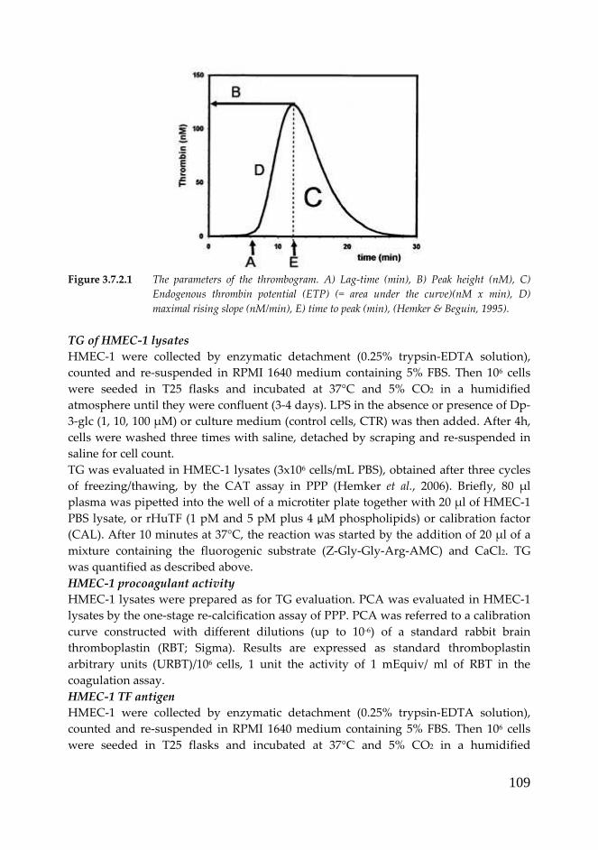
109
Figure 3.7.2.1 The parameters of the thrombogram. A) Lag-time (min), B) Peak height (nM), C)
Endogenous thrombin potential (ETP) (= area under the curve)(nM x min), D)
maximal rising slope (nM/min), E) time to peak (min), (Hemker & Beguin, 1995).
TG of HMEC-1 lysates
HMEC-1 were collected by enzymatic detachment (0.25% trypsin-EDTA solution),
counted and re-suspended in RPMI 1640 medium containing 5% FBS. Then 106 cells
were seeded in T25 flasks and incubated at 37°C and 5% CO2 in a humidified
atmosphere until they were confluent (3-4 days). LPS in the absence or presence of Dp-
3-glc (1, 10, 100 µM) or culture medium (control cells, CTR) was then added. After 4h,
cells were washed three times with saline, detached by scraping and re-suspended in
saline for cell count.
TG was evaluated in HMEC-1 lysates (3x10⁶ cells/mL PBS), obtained after three cycles
of freezing/thawing, by the CAT assay in PPP (Hemker et al., 2006). Briefly, 80 µl
plasma was pipetted into the well of a microtiter plate together with 20 µl of HMEC-1
PBS lysate, or rHuTF (1 pM and 5 pM plus 4 µM phospholipids) or calibration factor
(CAL). After 10 minutes at 37°C, the reaction was started by the addition of 20 µl of a
mixture containing the fluorogenic substrate (Z-Gly-Gly-Arg-AMC) and CaCl2. TG
was quantified as described above.
HMEC-1 procoagulant activity
HMEC-1 lysates were prepared as for TG evaluation. PCA was evaluated in HMEC-1
lysates by the one-stage re-calcification assay of PPP. PCA was referred to a calibration
curve constructed with different dilutions (up to 10-6) of a standard rabbit brain
thromboplastin (RBT; Sigma). Results are expressed as standard thromboplastin
arbitrary units (URBT)/106 cells, 1 unit the activity of 1 mEquiv/ ml of RBT in the
coagulation assay.
HMEC-1 TF antigen
HMEC-1 were collected by enzymatic detachment (0.25% trypsin-EDTA solution),
counted and re-suspended in RPMI 1640 medium containing 5% FBS. Then 106 cells
were seeded in T25 flasks and incubated at 37°C and 5% CO2 in a humidified

110
atmosphere until they were confluent (3-4 days). LPS in the absence or presence of Dp-
3-glc (1, 10, 100 µM) or culture medium (CTR) was then added. After 4h, cells were
washed three times with saline, detached by scraping and re-suspended in saline for
cell count.
TF antigen levels were quantified in HMEC-1 extracts (0.5x10⁶ cell/mL) in TRIS buffer
(50 mmol/l Tris, 100 mmol/l NaCl and 1% Triton-X 100, pH 7.5) by IMUBIND® Tissue
factor ELISA (American Diagnostica Inc., Stamford, CT, USA) according to the
manufacturer’s instructions. Results are expressed as pg/106 cells.
HMEC-1 TF mRNA expression
Evaluation of mRNA expression of TF in HMEC-1 was determined with Real Time RT-
PCR. HMEC-1 were cultured and treated in T25 flasks as for the previous experiments.
Then 1.5x10⁶ washed cells were re-suspended in 350 µL guanidinium thiocyanate and
stored at -80°C until RNA extraction.
Total cellular RNA was extracted using RNeasy® Mini kit (Qiagen). Retro-
transcription of cDNA was performed with RT-kit plus (Nanogen Advanced
Diagnostics S.r.L., Italy) according to manufacturer’s instructions. cDNA was used for
amplification of Tissue Factor (TF) in Real Time PCR (Rotor Gene™ 6000, Explera a
Qiagen Company, Italy). Simultaneous amplification of the invariant housekeeping
gene glyceraldheyde-3-phosphate dehydrogenase (GAPDH) was performed. The
following oligonucleotides were used : TF forward: 5’- AGG TGG GAA CAA AAG
TGA ATG TG-3’, and TF reverse exon 4-5: 5’-TTT TGG CTG TTT TCT TTC CTG AAC-
3’; GAPDH forward: 5’-GAA GGT GAA GGT CGG AGT C-3’ and GAPDH reverse: 5’-
GAA GAT GGT GAT GGG ATT TC-3’, (Laboratoires Eurobio, Les Uils, France).
Thermocycling consisted of: 2 min at 95°C for Go Taq® Hot Start Polymerase
activation, 50 cycles of 15 sec at 95°C followed by of 60 sec at 56°C (acquiring step), and
60 sec at 72°C and stopped with a melting curve (ramp from 50°C to 99°C rising by 1°C
each step, waiting for 90 sec of pre-melt conditioning on first step and 5 sec for each
step afterwards).
PCR amplification was performed with Go Taq qPCR Master Mix (Promega
Corporation, Madison, USA): 12.5 µL of Go Taq qPCR Master Mix 2X, 0.5 µL of each
oligonucleotide (0.08 µM), 2 µL of cDNA (100 ng) and 9.5 µL of nuclease-free water in
a final volume of 25 µL. Data analysis was performed with comparative quantitation of
Ct sample values. The internal calibrator selected was CTR (not treated cells) with
reported concentration of 1 fold. Then the ratio of TF on GAPDH values used as
control was calculated for each sample.
Statistical analysis
Student’s paired and unpaired t-test was used for the determination of significance
levels between the different treatments. Differences were considered significant when
p<0.05.

111
3.8 Results and discussion TOPIC II
3.8.1 Angiogenesis study
Capillary-like tube formation assay in Matrigel
To assess whether Dp-3-glc and Cy-3-glc might affect the formation of capillary-like
tubes, HMEC-1 were seeded on Matrigel and treated for 24h with 0.1-1-10-100 µM Dp-
3-glc or Cy-3-glc in the absence of exogenous pro-angiogenic stimuli (i.e. resting
condition). A
B
Figure 3.8.1.1 Effect of Dp-3-glc on capillary-like tube formation. Dp-3-glc showed a significant
inhibitory effect in tube formation at the 100 μM concentration in resting (panel A)
and VEGF-stimulated (panel B) conditions. Data are expressed as the mean of three
independent experiments ± SD. * p<0.05 vs CTR; § p<0.05 vs VEGF.
As shown in Figure 3.8.1.1-A, only at 100 µM concentration Dp-3-glc affected in a
statistically significant manner (p<0.005) the capacity of HMEC-1 to form the capillary-
like tubes, with a reduction of 50% compared to control cells (CTR). Similar

112
experiments were performed with rHuVEGF165-stimulated HMEC-1. Treatment with
50 ng/ml rHuVEGF165 induced a statistically significant (p<0.005) increase in capillary
tube length compared to control cells. The addition of Dp-3-glc was able to dose-
dependently counteract VEGF stimulus, reaching a complete inhibition at the
concentration of 100 µM (p<0.05) (Figure 3.8.1.1-B).
A
B
Figure 3.8.1.2 Effect of Cy-3-glc on capillary-like tube formation. Cy-3-glc did not show a significant
inhibitory effect in tube formation in resting (panel A) and VEGF-stimulated (panel B)
conditions. Data are expressed as the mean of three independent experiments ± SD.
* p<0.05 vs CTR
Differently, Cy-3-glc in the same experimental conditions did not show any statistically
significant inhibitory effect on the formation of capillary structures, in either resting or
VEGF-stimulated HMEC-1 (Figure 3.8.1.2).

113
Wound healing assay
The effect of Dp-3-glc and Cy-3-glc on HMEC-1 migration/proliferation was tested in
both resting and VEGF-stimulated conditions by the wound healing assay.
In resting conditions (Figure 3.8.1.3-A), 100 µM Dp-3-glc significantly decreased by
90% the migration of HMEC-1 (p<0.0001). At lower Dp-3-glc concentrations no
statistically significant effects on HMEC-1 migration were observed. VEGF induced a
significant (p<0.001) increase in HMEC-1 re-growth: this effect that was significantly
counteracted by Dp-3-glc starting from the concentration of 1 µM, reaching a complete
(100%) inhibition at 100 µM (p<0.01), (Figure 3.8.1.3-B).
A
B
Figure 3.8.1.3 Effect of Dp-3-glc on migration in the wound healing assay. Dp-3-glc showed a
significant inhibitory effect of re-growth area at the maximum concentration in resting
conditions (panel A) and at 1 μM in combination with VEGF (panel B). Data are
expressed as the mean of three independent experiments ± SD. * p<0.05 vs CTR; §
p<0.05 vs VEGF.

114
As observed with the capillary-like tube formation assay, Cy-3-glc did not show any
statistically significant effect on HMEC-1 migration in both resting and VEGF-
stimulated condition (Figure 3.8.1.4).
A
B
Figure 3.8.1.4 Effect of Cy-3-glc on migration in the wound healing assay. Cy-3-glc did not show a
significant inhibitory effect in re-growth area in resting (A, on the left) and VEGF-
stimulated (B, on the right) conditions. Data are expressed as the mean of three
independent experiments ± SD. *p<0.05 vs CTR

115
3.8.2 HMEC-1 pro-coagulant properties
TG on HMEC-1 intact monolayer
The study of the effect of Dp-3-glc on endothelial cell-induced TG was performed in 2
different experimental conditions: resting and LPS-stimulated HMEC-1.
In the absence of any procoagulant stimulus, the treatment with Dp-3-glc (from 4h to
24h) did not significantly affect TG potential of HMEC-1 monolayer (data not shown).
Differently, as shown in Figure 3.8.2.1, the pro-thrombotic stimulus of LPS induced an
increase in TG potential of HMEC-1, expressed by a significant (p<0.05) reduction in
lag-time (1.9 vs 5.1 min) and ttPeak (6.8 vs 10.0 min), and an increase of ETP (1021 vs
749 nM*min) compared to control cells. Dp-3-glc significantly counteracted the
prothrombotic stimulus of LPS starting from 10 µM: at this concentration, the values of
lag-time, ttPeak and ETP were similar to that of unstimulated HMEC-1 cells.
Figure 3.8.2.1 Dp-3-glc anti-thrombotic effect on TG potential of LPS-stimulated HMEC-1
monolayer. Dp-3-glc showed a significant effect (* p<0.05 vs CTR; § p<0.05 vs LPS)
on thrombogram parameters when added to LPS. Data are expressed as mean ± SD of
three independent experiments. Red square: Lag-time (min); green triangle: ETP
(nM*min); blue diamond: ttPeak (min).
TG of HMEC-1 lysates
TG of PBS lysates of HMEC-1 treated with LPS demonstrated a significant (p<0.05)
reduction in lag-time (4.1 vs 8.5 min) and ttPeak (10.3 vs 14.3 min), and an increase of
ETP (1118 vs 975 nM*min) compared to control cells. The addition of Dp-3-glc did not
significantly affect any TG parameter (Figure 3.8.2.2).

116
Figure 3.8.2.2 Dp-3-glc effect on TG potential of HMEC-1 lysates. LPS induced a statistically
significant effect on the three parameters considered (* p<0.05 vs CTR): the addition of
Dp-3-glc to LPS did not determine any statistically significant effect compared to LPS
alone. Data are expressed as mean ± SD of three independent experiments. Red square:
Lag-time (min); green triangle: ETP (nM*min); blue diamond: ttPeak (min).
HMEC-1 procoagulant activity
Figure 3.8.2.3 shows the results of PCA determination in cell lysates after 4 h
incubation of HMEC-1 with LPS in absence or presence of increasing concentrations of
Dp-3-glc. LPS induced a significant (p<0.05) increase of TF-PCA; while Dp-3-glc at hte
concentration of 100 µM produced a statistically relevant inhibition of 59% in URBT as
compared to LPS-treated lysates (p<0.05).
Figure 3.8.2.3 Effect of Dp-3-glc on PCA of HMEC-1 stimulated with LPS. Dp-3-glc showed a
significant effect (* p<0.05 vs CTR, § p<0.05 vs LPS) at the maximum concentration
tested. Data are expressed as mean ± SD of three independent experiments.

117
HMEC-1 TF antigen
Figure 3.8.2.4 shows the results of TF antigen determination in extracts of HMEC-1
after 4 h incubation of these cells with LPS, in absence or presence of increasing
concentrations of Dp-3-glc. In particular, TF antigen was dose-dependently decreased
by Dp-3-glc, reaching a 25% inhibition at the concentration of 100 µM (p<0.05).
Figure 3.8.2.4 Effect of Dp-3-glc on TF antigen expression by HMEC-1 stimulated with LPS.
Data are expressed as mean ± SD of three independent experiments.
* p<0.05 vs CTR, § p<0.05 vs LPS
HMEC-1 TF mRNA expression
The expression of TF was determined by quantitative Real Time RT-PCR on HMEC-1
treated with LPS in combination with Dp-3-glc for 4h. A significant increase (85%) of
TF mRNA expression was induced by LPS treatment (p<0.05). The combination with
the anthocyanin Dp-3-glc determined a significant effect at the maximum
concentration tested (100 µM), with a inhibition of 28% versus LPS alone, as shown in
Figure 3.8.2.5.
Figure 3.8.2.5 Dp-3-glc effect on TF mRNA expression of HMEC-1. Data are expressed as mean ±
SD of three independent experiments.* p<0.05 vs CTR; § p<0.05 vs LPS.

118
3.8.3 Discussion
In the present study we evaluated whether two anthocyanins (i.e. Dp-3-glc and Cy-3-
glc, mainly represented in black currant, wild blueberry, blood orange, and other
colored fruits and vegetables) can affect angiogenic properties of endothelium;
moreover, we investigated on the capacity of Dp-3-glc to affect thrombotic properties
of microvascular endothelial cells.
The anti-proliferative activity of these natural compounds against cancer cell lines
have been already studied and available in the literature.
Katsube and colleagues (2003) demonstrated that, among ethanol extracts of 10 edible
berries, bilberry extract was found to be the most effective at inhibiting the growth of
HL60 human leukemia cells and HCT116 human colon carcinoma cells in vitro. In
particular, the anthocyanin Dp-3-glc was able to inhibit the growth of HCT116 cells of
85% and 80% with the concentrations 863 µM and 431 µM respectively; while for HL60
cells of 75% with 216 µM (Katsube et al., 2003).
Olsson and colleagues (2004) investigated on the effects of 10 different extracts of fruits
and berries on cell proliferation of colon cancer cells HT29 and breast cancer cells
MCF-7. The fruits and berries used were rosehips, blueberries, black currant, black
chokeberries, apple, sea buckthorn, plum, lingonberries, cherries, and raspberries. The
extracts decreased the proliferation of both colon cancer cells HT29 and breast cancer
cells MCF-7, and the effect was concentration-dependent. In particular, standards Dp-
3-glc and Cy-3-glc decreased significantly (p<0.001) the proliferation of both cell lines
to a high extent (87 and 82% respectively) at the concentration of 200 µg/mL (431 µM)
in the medium. At a lower concentration of the anthocyanin standards, i.e. 20 µg/mL,
no effects was found on the proliferation of HT29 and MCF-7 cells (Olsson et al., 2004).
Fimognari et al (2004) investigated the in vitro capacity of the anthocyanin cyanidin-3-
O-beta-glucopyranoside (Cy-3-glc) to induce apoptosis in Jurkat T-lymphoblastoid, as
well as apoptosis and differentiation in HL-60 promyelocytic cells. Although Cy-3-glc
induced apoptosis (as well as necrosis) in the two systems, HL-60 cells were much less
sensitive than T-lymphoblastoid cells. In particular, IC50 for Jurkat cells was 391 µM,
while in HL-60 cells the growth inhibition of 37% was reached with the concentration
of 446 µM. Moreover, treatment of HL-60 cells with Cy-3-glc caused differentiation
into macrophage-like cells and granulocytes. The authors conclude that these
interesting biological properties should encourage further investigation into the
chemopreventive and/or chemotherapeutic potential of Cy-3-glc (Fimognari et al.,
2004).
Recently, Fernandes and colleagues (2010) published a work on the influence of
anthocyanins and other polyphenols on breast cancer cell (MCF-7) proliferation. The
antiproliferative effect was higher when cells were treated with Dp-3-glc, even if it was
not followed by modification of ER expression. Differently, Cy-3-glc showed no
significant antiproliferative effect on MCF-7 proliferation (Fernandes et al., 2010).
However, no studies are available on the effects of anthocyanins on tumor-indiced
neoangiogenesis. Angiogenesis, the formation of new vessels, has a major role in

119
tumor growth, dissemination and metastasis in both solid and hematological tumors
(Folkman, 2003). Angiogenesis is driven by microvascular endothelial cells, which
upon activation by angiogenic growth factors degrade their basement membrane,
migrate into the interstitial matrix, proliferate and form new capillary structures
(Marchetti et al., 2003). The angiogenic switch in cancer is a highly complex event and
involves several tumor-derived factors. Inhibition of angiogenesis is one of the
proposed strategies to control cancer.
The results of our angiogenesis study indicate that Dp-3-glc can interfere with both
resting and activated endothelium, by a standard stimulus like VEGF. In fact, we
obtained an increase in capillary tube formation and cell migration induced by VEGF
respect to basal HMEC-1, while treatment Dp-3-glc was able to inhibit VEGF stimulus
at 1 µM concentration. Differently, Cy-3-glc did not show the same effect. The different
behaviour of the two anthocyanins could be due to their different chemical structure
given that the only structural feature which differs in their structures is the
hydroxylation pattern in ring B (Figure 3.6.6), as hypothesized also by Fernandes et al
(2010). No in vitro studies are available on the effects of Dp-3-glc and Cy-3-glc on
microvascular endothelial cells, in fact the attention has been more directed to the anti-
angiogenic activity exerted by anthocyanidins. Delphinidin (Dp), aglycone form of
Dp-3-glc, was recently demonstrated to inhibit proliferation migration and
angiogenesis of human umbilical vein endothelial cells (HUVECs) (Favot et al., 2003;
Lamy et al., 2006; Lamy et al., 2008; Matsunaga et al., 2010). Briefly, Matsunaga and
colleagues (2010) examined the anti-angiogenic properties and antioxidant activities of
the main anthocyanidins (delphinidin, cyanidin and malvidin) found as constituents
in bilberry (Vaccinium myrtillus). Each of these anthocyanidins dose-dependently
inhibited VEGF-induced tube formation in a co-culture of HUVECs and fibroblasts,
with the effect of each anthocyanidin being significant at 3 and/or 10 µM. Moreover,
their radical-scavenging effects were induced by concentrations that were at or below
those that induced their anti-angiogenic effects (Matsunaga et al., 2010). Lamy and
colleagues in 2006 conducted a study where, among the anthocyanidins tested
(cyanidin, delphinidin, malvidin, pelargonidin, peonidin and petunidin), delphinidin
was the most potent angiogenic inhibitor. In vitro, low concentrations of Dp inhibited
VEGF-induced tyrosine phosphorylation of VEGF receptor (VEGFR)-2, leading to the
inhibition of downstream signaling triggered by VEGFR-2. Inhibition of VEGFR-2 by
Dp reserved the VEGF-induced activation of ERK-1/2 signaling and the chemotactic
motility of HUVECs as well as their differentiation into capillary-like tubular
structures in Matrigel and within fibrin gels. In vivo, Dp was able to suppress basic
fibroblast growth factor-induced vessel formation in the mouse Matrigel plug assay
(Lamy et al., 2006). In a subsequent study (2008), Lamy and colleagues showed that Dp
also inhibits activation of the platelet-derived growth factor (PDGF)-BB receptor-β
[platelet-derived growth factor receptor-beta (PDGFR-β)] in pulmonary aortic smooth
muscle cells (PASMC) and that this inhibition may contribute to its antitumor effect.
The inhibitory effect of delphinidin on PDGFR-β was very rapid and led to the
inhibition of PDGF-BB-induced activation of extracellular signal-regulated kinase

120
(ERK)-1/2 signaling and of the chemotactic motility of PASMC, as well as the
differentiation and stabilization of HUVEC and PASMC into capillary-like tubular
structures in a three-dimensional co-culture system. Using an anthocyan-rich extract of
berries, the authors showed that berry extracts were able to suppress the synergistic
induction of vessel formation by basic fibroblast growth factor-2 and PDGF-BB in the
mouse Matrigel plug assay. Oral administration of the berry extract also significantly
retarded tumor growth in a lung carcinoma xenograft model. Taken together, these
results provide new insight into the molecular mechanisms underlying the
antiangiogenic activity of delphinidin that will be helpful for the development of
dietary-based chemopreventive strategies (Lamy et al., 2008). Favot and colleagues
(2003) verified that VEGF-stimulated HUVEC cell migration and proliferation were
potently inhibited by Dp in concentration-dependent manner (with maximum effect of
50% inhibition for migration and proliferation at 64 µM). Flow cytometric analysis
demonstrated that Dp inhibition of proliferation is correlated with the blockade of cell
cycle in G(0)/G(1) phase. Western blot analysis showed that Dp reversed the VEGF-
induced decrease in expression of cyclin-dependent kinase inhibitor p27(kip1) and the
VEGF-induced increase of cyclin D1 and cyclin A, both being necessary to achieve the
G(1)-to-S transition. Furthermore, Dp inhibited neovascularisation in vivo in
chorioallantoic membrane model (Favot et al., 2003). In a series of experiments, berry
extracts were tested for their ability to inhibit angiogenensis via altered VEGF
expression and invasiveness (Roy et al., 2002). Anti-angiogenic properties were
investigated in two cell lines: immortalized HaCaT human keratinocytes and human
microvascular dermal endothelial (HMVE) cells. First the effect of selected berry
samples on TNFα and H2O2-induced VEGF expression by HaCaT cells was
investigated. The authors speculate that the flavonoid component of berry samples
may be responsible for the observed effect on inducible VEGF expression and release.
Next, an in vitro angiogenesis assay was performed with HMVE cells; two of the berry
mix samples (50µg/mL) impaired in vitro angiogenesis (Roy et al., 2002).
Our study of anti-coagulant effects of Dp-3-glc was carried out on HMEC-1
stimulated by LPS, a known activator of endothelial pro-coagulant response. In these
experiments, the dose of LPS was used according to a standard procedure established
on the basis of recent studies (Falanga et al., 2003). TF is the major activator of blood
coagulation. The reduction in TF expression on microvascular endothelial cells may be
important for maintaining physiological endothelial function and preventing
thrombotic microangiopathies. The results of our study indicate that Dp-3-glc
significantly counteract LPS-induced TF expression at microvascular site starting at 10
µM concentration when tested with TG assay on intact monolayer of HMEC-1.
Determination of TG on HMEC-1 lysates did not reveal any anti-coagulant effect, even
at the highest dose tested. On the contrary, in the subsequent experiments performed,
Dp-3-glc treatment (100 µM) induced a significant reduction in total PCA on HMEC-1
lysates, determined with the one-stage recalcification assay. The further quantitation of
TF, as antigen and as mRNA expression, showed a significant inhibition induced of
LPS activation of HMEC-1 procoagulant conditions by 100 µM Dp-3-glc.

121
To our knowledge, there are no other studies in the literature investigating the anti-
thrombotic effect of anthocyanins on microvascular cell lines models.
Yamamoto and colleagues (2006) found that mulberry (Morus alba) varieties could be
grouped into subclasses with either anti- or prothrombotic activities. A shear-induced
in vitro platelet reactivity/thrombolysis test (Gorog Thrombosis Test) was used to
screen for antiplatelet and thrombolytic activities. In case of effectiveness, it was
followed by an in vivo test of laser-induced thrombosis in mice. Antioxidant activities
and polyphenolic contents were also evaluated and they did not affect platelets but
might enhance endogenous thrombolysis, thus causing an overall antithrombotic effect
(Yamamoto et al., 2006).
Berry phytochemicals have been widely studied with in vitro and animal models and
different protective effects were demonstrated, as reported in detail in the introduction
of this topic (paragraph 3.7).
However, which single berry constituent(s) induce this protective response remains
uncertain, and in the vast majority of studies the concentration of extract or
phytochemical employed is substantially higher than could ever be achieved
nutritionally (Duthie, 2007). We have also to consider that berry compounds are
extensively metabolized and further converted by colonic microflora into related
molecules. These compounds may persist in vivo, accumulate in target tissues and
contribute significantly to the biological effects. Moreover, the levels of this type of
phytochemicals may be underestimated due to limitations in laboratory extraction
procedures because these compounds may bind to proteins, etc., causing their
extraction for chemical analyses to be difficult (Seeram, 2008). However, the
anthocyanins delphinidin-3-glucoside and cyanidin-3-glucoside, selected for this
study, have been reported to be incorporated into human plasma with their structures
maintained or converted from other structures (see also paragraph 3.6 for details).
3.9 Conclusions TOPIC II
Our results, together with the known capacity of Dp-3-glc to affect tumor cell
proliferation, make this compound a potential cancer chemopreventive agent.
Although considerable progress has been made in understanding the role that
bioactive compounds, like anthocyanins, play in affecting human health and disease,
there are still important gaps in our knowledge concerning the biology and chemistry
of these compounds. Future studies should be designed to enhance our knowledge of
the intricate roles and functions that ACNs can act at both cellular and molecular level.
Moreover there is little evidence from human intervention studies, which provide the
confirmation of the protective role of bioactive compounds. This in vitro study
supports a protective effect on human endothelium. However, the concentration tested
in vitro hardly could be achieved in vivo. Thus, the next step might be organization of a
human feeding study where biomarkers of angiogenesis, such as VEGF, or marker of
coagulation status, such as TF, TG and PCA could be easily analyzed in plasma
samples.

122
In conclusion, our results support health promotion strategies that favour fruit and
vegetable consumption for prevention and protection against chronic diseases.
Acknowledgements
I want to greatly thank Dr. Anna Falanga (director of Division of Immunohematology
and Transfusion Medicine & Hemostasis and Thrombosis Center, Ospedali Riuniti,
Bergamo, Italy), who gave me the opportunity to realize this part of my research. I
thank all the people of the Hemostasis and Thrombosis Center: Dr. Marina Marchetti,
Dr. Alfonso Vignoli, Dr. Erika Diani, Dr. Marina Panova-Noeva, Dr. Carmen Julia
Tartari, Dr. Laura Russo, Dr. Anna Leuzzi, Dr. Elena Cantalino and Dr. Rosanna
Mancini.
3.10 References
Ahn D, Putt D, Kresty L, Stoner GD, Fromm D & Hollenberg PF (1996) The effects of
dietary ellagic acid on rat hepatic and esophageal mucosal cytochromes P450
and phase II enzymes. Carcinogenesis 17, 821-828.
Atalay M, Gordillo G, Roy S, Rovin B, Bagchi D, Bagchi M & Sen CK (2003) Anti-
angiogenic property of edible berry in a model of hemangioma. FEBS Lett 544,
252-257.
Bagchi D, Sen CK, Bacghi M & Atalay M (2004) Anti-angiogenic, antioxidant, anti-
carcinogenic properties of a novel anthocyanin-rich berry extract formula.
Biochemistry (Moscow) 69, 75-80.
Calixto JB, Campos MM, Otuki MF & Santos AR (2004) Anti-inflammatory compounds
of plant origin. Part II. modulation of pro-inflammatory cytokines, chemokines
and adhesion molecules. Planta Med 70, 93-103.
Carkeet C, Clevidence BA & Novotny JA (2008) Anthocyanin excretion by humans
increases linearly with increasing strawberry dose. J Nutr 138, 897-902.
Chen PN, Chu SC, Chiou HL, Kuo WH, Chiang CL & Hsieh YS (2006) Mulberry
anthocyanins, cyanidin 3-rutinoside and cyanidin 3-glucoside, exhibited an
inhibitory effect on the migration and invasion of a human lung cancer cell
line. Cancer Lett 235, 248-259.
Clifford MN, Copeland EL, Bloxsidge JP & Mitchell LA (2000) Hippuric acid as a major
excretion product associated with black tea consumption. Xenobiotica 30, 317-
326.
Cooke D, Steward WP, Gescher AJ & Marczylo T (2005) Anthocyans from fruits and
vegetables--does bright colour signal cancer chemopreventive activity? Eur J
Cancer 41, 1931-1940.
Crozier A, Jaganath IB & Clifford MN (2009) Dietary phenolics: chemistry,
bioavailability and effects on health. Nat Prod Rep 26, 1001-1043.
Dauphinee SM & Karsan A (2006) Lipopolysaccharide signaling in endothelial cells.
Lab Invest 86, 9-22.
Day AJ, Canada FJ, Diaz JC, Kroon PA, McLauchlan R, Faulds CB, Plumb GW, Morgan
MR & Williamson G (2000) Dietary flavonoid and isoflavone glycosides are

123
hydrolysed by the lactase site of lactase phlorizin hydrolase. FEBS Lett 468,
166-170.
de Vos FY, Willemse PH, de Vries EG & Gietema JA (2004) Endothelial cell effects of
cytotoxics: balance between desired and unwanted effects. Cancer Treat Rev 30,
495-513.
Del Bo C, Kristo AS, Kalea AZ, Ciappellano S, Riso P, Porrini M & Klimis-Zacas D
(2010a) The temporal effect of a wild blueberry (Vaccinium angustifolium)-
enriched diet on vasomotor tone in the Sprague-Dawley rat. Nutr Metab
Cardiovasc Dis.
Del Bo C, Martini D, Vendrame S, Riso P, Ciappellano S, Klimis-Zacas D & Porrini M
(2010b) Improvement of lymphocyte resistance against H2O2-induced DNA
damage in Sprague-Dawley rats after eight weeks of a wild blueberry
(Vaccinium angustifolium)-enriched diet. Mutat Res.
Ding M, Feng R, Wang SY, Bowman L, Lu Y, Qian Y, Castranova V, Jiang BH & Shi X
(2006) Cyanidin-3-glucoside, a natural product derived from blackberry,
exhibits chemopreventive and chemotherapeutic activity. J Biol Chem 281,
17359-17368.
Donati MB (1994) Cancer and thrombosis. Haemostasis 24, 128-131.
Donovan JL, Manach C, Faulks RM & Kroon P in Plant Secondary Metabolites.
Occurrence, Structure and Role in the Human Diet, ed. Crozier A, Clifford MN
and Ashihara H, Blackwell Publishing, Oxford, 2006, pp. 303–351. Duthie GG, Duthie SJ & Kyle JA (2000) Plant polyphenols in cancer and heart disease:
implications as nutritional antioxidants. Nutr Res Rev 13, 79-106.
Duthie SJ (2007) Berry phytochemicals, genomic stability and cancer: evidence for
chemoprotection at several stages in the carcinogenic process. Mol Nutr Food
Res 51, 665-674.
Duthie SJ, Gardner PT, Morrice PC, Wood SG, Pirie L, Bestwick CC, Milne L & Duthie
GG (2005) DNA stability and lipid peroxidation in vitamin E-deficient rats in
vivo and colon cells in vitro--modulation by the dietary anthocyanin, cyanidin-
3-glycoside. Eur J Nutr 44, 195-203.
Espin JC, Garcia-Conesa MT & Tomas-Barberan FA (2007) Nutraceuticals: facts and
fiction. Phytochemistry 68, 2986-3008.
Falanga A & Marchetti M (2009) Venous thromboembolism in the hematologic
malignancies. J Clin Oncol 27, 4848-4857.
Falanga A, Vignoli A, Marchetti M & Barbui T (2003) Defibrotide reduces procoagulant
activity and increases fibrinolytic properties of endothelial cells. Leukemia 17,
1636-1642.
Favot L, Martin S, Keravis T, Andriantsitohaina R & Lugnier C (2003) Involvement of
cyclin-dependent pathway in the inhibitory effect of delphinidin on
angiogenesis. Cardiovasc Res 59, 479-487.
Felgines C, Talavera S, Gonthier MP, Texier O, Scalbert A, Lamaison JL & Remesy C
(2003) Strawberry anthocyanins are recovered in urine as glucuro- and
sulfoconjugates in humans. J Nutr 133, 1296-1301.

124
Felgines C, Talavera S, Texier O, Gil-Izquierdo A, Lamaison JL & Remesy C (2005)
Blackberry anthocyanins are mainly recovered from urine as methylated and
glucuronidated conjugates in humans. J Agric Food Chem 53, 7721-7727.
Feng R, Bowman LL, Lu Y, Leonard SS, Shi X, Jiang BH, Castranova V, Vallyathan V &
Ding M (2004) Blackberry extracts inhibit activating protein 1 activation and
cell transformation by perturbing the mitogenic signaling pathway. Nutr
Cancer 50, 80-89.
Fernandes I, Faria A, Azevedo J, Soares S, Calhau C, De Freitas V & Mateus N (2010)
Influence of anthocyanins, derivative pigments and other catechol and
pyrogallol-type phenolics on breast cancer cell proliferation. J Agric Food Chem
58, 3785-3792.
Fimognari C, Berti F, Nusse M, Cantelli-Forti G & Hrelia P (2004) Induction of
apoptosis in two human leukemia cell lines as well as differentiation in human
promyelocytic cells by cyanidin-3-O-beta-glucopyranoside. Biochem Pharmacol
67, 2047-2056.
Fimognari C, Lenzi M & Hrelia P (2008) Chemoprevention of cancer by isothiocyanates
and anthocyanins: mechanisms of action and structure-activity relationship.
Curr Med Chem 15, 440-447.
Folkman J (2003) Fundamental concepts of the angiogenic process. Curr Mol Med 3,
643-651.
Folkman J (2006) Angiogenesis. Annu Rev Med 57, 1-18.
Furie B & Furie BC (2008) Mechanisms of thrombus formation. N Engl J Med 359, 938-
949.
Gailani D & Broze GJ, Jr. (1991) Factor XI activation in a revised model of blood
coagulation. Science 253, 909-912.
Gasiorowski K, Szyba K, Brokos B, Kolaczynska B, Jankowiak-Wlodarczyk M &
Oszmianski J (1997) Antimutagenic activity of anthocyanins isolated from
Aronia melanocarpa fruits. Cancer Lett 119, 37-46.
Gee JM, DuPont MS, Day AJ, Plumb GW, Williamson G & Johnson IT (2000) Intestinal
transport of quercetin glycosides in rats involves both deglycosylation and
interaction with the hexose transport pathway. J Nutr 130, 2765-2771.
Giordano L, Coletta W, Rapisarda P, Donati MB & Rotilio D (2007) Development and
validation of an LC-MS/MS analysis for simultaneous determination of
delphinidin-3-glucoside, cyanidin-3-glucoside and cyanidin-3-(6-
malonylglucoside) in human plasma and urine after blood orange juice
administration. J Sep Sci 30, 3127-3136.
Grandel U & Grimminger F (2003) Endothelial responses to bacterial toxins in sepsis.
Crit Rev Immunol 23, 267-299.
Grignani G & Maiolo A (2000) Cytokines and hemostasis. Haematologica 85, 967-972.
Gu L, Laly M, Chang HC, Prior RL, Fang N, Ronis MJ & Badger TM (2005) Isoflavone
conjugates are underestimated in tissues using enzymatic hydrolysis. J Agric
Food Chem 53, 6858-6863.

125
Guarnieri S, Riso P & Porrini M (2007) Orange juice vs vitamin C: effect on hydrogen
peroxide-induced DNA damage in mononuclear blood cells. Br J Nutr 97, 639-
643.
Halliwell B (2002) Effect of diet on cancer development: is oxidative DNA damage a
biomarker? Free Radic Biol Med 32, 968-974.
Harris GK, Gupta A, Nines RG, Kresty LA, Habib SG, Frankel WL, LaPerle K, Gallaher
DD, Schwartz SJ & Stoner GD (2001) Effects of lyophilized black raspberries on
azoxymethane-induced colon cancer and 8-hydroxy-2'-deoxyguanosine levels
in the Fischer 344 rat. Nutr Cancer 40, 125-133.
Hemker HC, Al Dieri R, De Smedt E & Beguin S (2006) Thrombin generation, a
function test of the haemostatic-thrombotic system. Thromb Haemost 96, 553-
561.
Hemker HC & Beguin S (1995) Thrombin generation in plasma: its assessment via the
endogenous thrombin potential. Thromb Haemost 74, 134-138.
Hoffman M & Monroe DM (2007) Coagulation 2006: a modern view of hemostasis.
Hematol Oncol Clin North Am 21, 1-11.
Huang C, Huang Y, Li J, Hu W, Aziz R, Tang MS, Sun N, Cassady J & Stoner GD
(2002) Inhibition of benzo(a)pyrene diol-epoxide-induced transactivation of
activated protein 1 and nuclear factor kappaB by black raspberry extracts.
Cancer Res 62, 6857-6863.
Hyun JW & Chung HS (2004) Cyanidin and Malvidin from Oryza sativa cv.
Heugjinjubyeo mediate cytotoxicity against human monocytic leukemia cells
by arrest of G(2)/M phase and induction of apoptosis. J Agric Food Chem 52,
2213-2217.
Ichiyanagi T, Shida Y, Rahman MM, Hatano Y, Matsumoto H, Hirayama M & Konishi
T (2005) Metabolic pathway of cyanidin 3-O-beta-D-glucopyranoside in rats. J
Agric Food Chem 53, 145-150.
Jaffe EA (1987) Cell biology of endothelial cells. Hum Pathol 18, 234-239.
Kang SY, Seeram NP, Nair MG & Bourquin LD (2003) Tart cherry anthocyanins inhibit
tumor development in Apc(Min) mice and reduce proliferation of human
colon cancer cells. Cancer Lett 194, 13-19.
Katsube N, Iwashita K, Tsushida T, Yamaki K & Kobori M (2003) Induction of
apoptosis in cancer cells by Bilberry (Vaccinium myrtillus) and the
anthocyanins. J Agric Food Chem 51, 68-75.
Kim HJ, Tsoy I, Park JM, Chung JI, Shin SC & Chang KC (2006) Anthocyanins from
soybean seed coat inhibit the expression of TNF-alpha-induced genes
associated with ischemia/reperfusion in endothelial cell by NF-kappaB-
dependent pathway and reduce rat myocardial damages incurred by ischemia
and reperfusion in vivo. FEBS Lett 580, 1391-1397.
Kottra G & Daniel H (2007) Flavonoid glycosides are not transported by the human
Na+/glucose transporter when expressed in Xenopus laevis oocytes, but
effectively inhibit electrogenic glucose uptake. J Pharmacol Exp Ther 322, 829-
835.

126
Kresty LA, Morse MA, Morgan C, Carlton PS, Lu J, Gupta A, Blackwood M & Stoner
GD (2001) Chemoprevention of esophageal tumorigenesis by dietary
administration of lyophilized black raspberries. Cancer Res 61, 6112-6119.
Lamy S, Beaulieu E, Labbe D, Bedard V, Moghrabi A, Barrette S, Gingras D & Beliveau
R (2008) Delphinidin, a dietary anthocyanidin, inhibits platelet-derived growth
factor ligand/receptor (PDGF/PDGFR) signaling. Carcinogenesis 29, 1033-1041.
Lamy S, Blanchette M, Michaud-Levesque J, Lafleur R, Durocher Y, Moghrabi A,
Barrette S, Gingras D & Beliveau R (2006) Delphinidin, a dietary
anthocyanidin, inhibits vascular endothelial growth factor receptor-2
phosphorylation. Carcinogenesis 27, 989-996.
Lau FC, Bielinski DF & Joseph JA (2007) Inhibitory effects of blueberry extract on the
production of inflammatory mediators in lipopolysaccharide-activated BV2
microglia. J Neurosci Res 85, 1010-1017.
Lazze MC, Pizzala R, Savio M, Stivala LA, Prosperi E & Bianchi L (2003) Anthocyanins
protect against DNA damage induced by tert-butyl-hydroperoxide in rat
smooth muscle and hepatoma cells. Mutat Res 535, 103-115.
Lazze MC, Savio M, Pizzala R, Cazzalini O, Perucca P, Scovassi AI, Stivala LA &
Bianchi L (2004) Anthocyanins induce cell cycle perturbations and apoptosis in
different human cell lines. Carcinogenesis 25, 1427-1433.
Liu Z, Schwimer J, Liu D, Greenway FL, Anthony CT & Woltering EA (2005) Black
raspberry extract and fractions contain angiogenesis inhibitors. J Agric Food
Chem 53, 3909-3915.
Lu H, Li J, Zhang D, Stoner GD & Huang C (2006) Molecular mechanisms involved in
chemoprevention of black raspberry extracts: from transcription factors to
their target genes. Nutr Cancer 54, 69-78.
Malik M, Zhao C, Schoene N, Guisti MM, Moyer MP & Magnuson BA (2003)
Anthocyanin-rich extract from Aronia meloncarpa E induces a cell cycle block
in colon cancer but not normal colonic cells. Nutr Cancer 46, 186-196.
Manach C, Scalbert A, Morand C, Remesy C & Jimenez L (2004) Polyphenols: food
sources and bioavailability. Am J Clin Nutr 79, 727-747.
Manach C, Williamson G, Morand C, Scalbert A & Remesy C (2005) Bioavailability and
bioefficacy of polyphenols in humans. I. Review of 97 bioavailability studies.
Am J Clin Nutr 81, 230S-242S.
Marchetti M, Vignoli A, Bani MR, Balducci D, Barbui T & Falanga A (2003) All-trans
retinoic acid modulates microvascular endothelial cell hemostatic properties.
Haematologica 88, 895-905.
Marko D, Puppel N, Tjaden Z, Jakobs S & Pahlke G (2004) The substitution pattern of
anthocyanidins affects different cellular signaling cascades regulating cell
proliferation. Mol Nutr Food Res 48, 318-325.
Matsumoto H, Inaba H, Kishi M, Tominaga S, Hirayama M & Tsuda T (2001) Orally
administered delphinidin 3-rutinoside and cyanidin 3-rutinoside are directly
absorbed in rats and humans and appear in the blood as the intact forms. J
Agric Food Chem 49, 1546-1551.

127
Matsunaga N, Tsuruma K, Shimazawa M, Yokota S & Hara H (2010) Inhibitory actions
of bilberry anthocyanidins on angiogenesis. Phytother Res 24 Suppl 1, S42-47.
McGhie TK, Ainge GD, Barnett LE, Cooney JM & Jensen DJ (2003) Anthocyanin
glycosides from berry fruit are absorbed and excreted unmetabolized by both
humans and rats. J Agric Food Chem 51, 4539-4548.
Meiers S, Kemeny M, Weyand U, Gastpar R, von Angerer E & Marko D (2001) The
anthocyanidins cyanidin and delphinidin are potent inhibitors of the
epidermal growth-factor receptor. J Agric Food Chem 49, 958-962.
Mullen W, Archeveque MA, Edwards CA, Matsumoto H & Crozier A (2008a)
Bioavailability and metabolism of orange juice flavanones in humans: impact
of a full-fat yogurt. J Agric Food Chem 56, 11157-11164.
Mullen W, Edwards CA, Serafini M & Crozier A (2008b) Bioavailability of
pelargonidin-3-O-glucoside and its metabolites in humans following the
ingestion of strawberries with and without cream. J Agric Food Chem 56, 713-
719.
Nakamura Y, Matsumoto H, Morifuji M, Iida H & Takeuchi Y (2010) Development and
validation of a liquid chromatography tandem mass spectrometry method for
simultaneous determination of four anthocyanins in human plasma after black
currant anthocyanins ingestion. J Agric Food Chem 58, 1174-1179.
Neto CC (2007) Cranberry and blueberry: evidence for protective effects against cancer
and vascular diseases. Mol Nutr Food Res 51, 652-664.
Nicholson SK, Tucker GA & Brameld JM (2008) Effects of dietary polyphenols on gene
expression in human vascular endothelial cells. Proc Nutr Soc 67, 42-47.
Olsson ME, Gustavsson KE, Andersson S, Nilsson A & Duan RD (2004) Inhibition of
cancer cell proliferation in vitro by fruit and berry extracts and correlations
with antioxidant levels. J Agric Food Chem 52, 7264-7271.
Ono M, Tateishi M, Masuoka C, Kobayashi H, Igoshi K, Komatsu H, Ito Y, Okawa M &
Nohara T (2003) A new triterpene glucosyl ester from the fruit of the
blackberry (Rubus allegheniensis). Chem Pharm Bull (Tokyo) 51, 200-202.
Panes O, Matus V, Saez CG, Quiroga T, Pereira J & Mezzano D (2007) Human platelets
synthesize and express functional tissue factor. Blood 109, 5242-5250.
Parry GC & Mackman N (1995) Transcriptional regulation of tissue factor expression in
human endothelial cells. Arterioscler Thromb Vasc Biol 15, 612-621.
Prior RL & Wu X (2006) Anthocyanins: structural characteristics that result in unique
metabolic patterns and biological activities. Free Radic Res 40, 1014-1028.
Ramirez-Tortosa C, Andersen OM, Gardner PT, Morrice PC, Wood SG, Duthie SJ,
Collins AR & Duthie GG (2001) Anthocyanin-rich extract decreases indices of
lipid peroxidation and DNA damage in vitamin E-depleted rats. Free Radic Biol
Med 31, 1033-1037.
Rao LV & Pendurthi UR (2005) Tissue factor-factor VIIa signaling. Arterioscler Thromb
Vasc Biol 25, 47-56.
Rechner AR, Kuhnle G, Hu H, Roedig-Penman A, van den Braak MH, Moore KP &
Rice-Evans CA (2002) The metabolism of dietary polyphenols and the

128
relevance to circulating levels of conjugated metabolites. Free Radic Res 36,
1229-1241.
Ribeiro MJ, Phillips DJ, Benson JM, Evatt BL, Ades EW & Hooper WC (1995)
Hemostatic properties of the SV-40 transfected human microvascular
endothelial cell line (HMEC-1). A representative in vitro model for
microvascular endothelium. Thromb Res 79, 153-161.
Riso P, Visioli F, Gardana C, Grande S, Brusamolino A, Galvano F, Galvano G &
Porrini M (2005) Effects of blood orange juice intake on antioxidant
bioavailability and on different markers related to oxidative stress. J Agric Food
Chem 53, 941-947.
Roy S, Khanna S, Alessio HM, Vider J, Bagchi D, Bagchi M & Sen CK (2002) Anti-
angiogenic property of edible berries. Free Radic Res 36, 1023-1031.
Seeram NP (2008) Berry fruits: compositional elements, biochemical activities, and the
impact of their intake on human health, performance, and disease. J Agric Food
Chem 56, 627-629.
Seeram NP, Adams LS, Hardy ML & Heber D (2004) Total cranberry extract versus its
phytochemical constituents: antiproliferative and synergistic effects against
human tumor cell lines. J Agric Food Chem 52, 2512-2517.
Semeraro N & Colucci M (1997) Tissue factor in health and disease. Thromb Haemost 78,
759-764.
Serraino I, Dugo L, Dugo P, Mondello L, Mazzon E, Dugo G, Caputi AP & Cuzzocrea S
(2003) Protective effects of cyanidin-3-O-glucoside from blackberry extract
against peroxynitrite-induced endothelial dysfunction and vascular failure.
Life Sci 73, 1097-1114.
Shin DY, Ryu CH, Lee WS, Kim DC, Kim SH, Hah YS, Lee SJ, Shin SC, Kang HS &
Choi YH (2009) Induction of apoptosis and inhibition of invasion in human
hepatoma cells by anthocyanins from meoru. Ann N Y Acad Sci 1171, 137-148.
Silberberg M, Morand C, Mathevon T, Besson C, Manach C, Scalbert A & Remesy C
(2006) The bioavailability of polyphenols is highly governed by the capacity of
the intestine and of the liver to secrete conjugated metabolites. Eur J Nutr 45,
88-96.
Singletary KW, Stansbury MJ, Giusti M, Van Breemen RB, Wallig M & Rimando A
(2003) Inhibition of rat mammary tumorigenesis by concord grape juice
constituents. J Agric Food Chem 51, 7280-7286.
Srivastava A, Akoh CC, Fischer J & Krewer G (2007) Effect of anthocyanin fractions
from selected cultivars of Georgia-grown blueberries on apoptosis and phase
II enzymes. J Agric Food Chem 55, 3180-3185.
Stoner GD, Chen T, Kresty LA, Aziz RM, Reinemann T & Nines R (2006) Protection
against esophageal cancer in rodents with lyophilized berries: potential
mechanisms. Nutr Cancer 54, 33-46.
Tarozzi A, Hrelia S, Angeloni C, Morroni F, Biagi P, Guardigli M, Cantelli-Forti G &
Hrelia P (2006) Antioxidant effectiveness of organically and non-organically
grown red oranges in cell culture systems. Eur J Nutr 45, 152-158.

129
ten Cate H & Falanga A (2008) Overview of the postulated mechanisms linking cancer
and thrombosis. Pathophysiol Haemost Thromb 36, 122-130.
Tsuda T, Horio F & Osawa T (2002) Cyanidin 3-O-beta-D-glucoside suppresses nitric
oxide production during a zymosan treatment in rats. J Nutr Sci Vitaminol
(Tokyo) 48, 305-310.
Vignoli A, Marchetti M, Balducci D, Barbui T & Falanga A (2006) Differential effect of
the low-molecular-weight heparin, dalteparin, and unfractionated heparin on
microvascular endothelial cell hemostatic properties. Haematologica 91, 207-214.
Vignoli A, Marchetti M, Russo L, Cantalino E, Bonacina G & Falanga A. (2010) LMWH
bemiparin and ULMWH RO-14 reduce the endothelial angiogenic features
elicited by leukemia, lung cancer, or breast cancer cells. Cancer Investigations in
press
Wang SY, Feng R, Lu Y, Bowman L & Ding M (2005) Inhibitory effect on activator
protein-1, nuclear factor-kappaB, and cell transformation by extracts of
strawberries (Fragaria x ananassa Duch.). J Agric Food Chem 53, 4187-4193.
Wu X, Beecher GR, Holden JM, Haytowitz DB, Gebhardt SE & Prior RL (2006)
Concentrations of anthocyanins in common foods in the United States and
estimation of normal consumption. J Agric Food Chem 54, 4069-4075.
Wu X, Pittman HE, 3rd, McKay S & Prior RL (2005) Aglycones and sugar moieties alter
anthocyanin absorption and metabolism after berry consumption in weanling
pigs. J Nutr 135, 2417-2424.
Xia M, Ling W, Zhu H, Wang Q, Ma J, Hou M, Tang Z, Li L & Ye Q (2007)
Anthocyanin prevents CD40-activated proinflammatory signaling in
endothelial cells by regulating cholesterol distribution. Arterioscler Thromb Vasc
Biol 27, 519-524.
Yamamoto J, Naemura A, Ura M, Ijiri Y, Yamashita T, Kurioka A & Koyama A (2006)
Testing various fruits for anti-thrombotic effect: i. Mulberries. Platelets 17, 555-
564.
Yi W, Fischer J, Krewer G & Akoh CC (2005) Phenolic compounds from blueberries can
inhibit colon cancer cell proliferation and induce apoptosis. J Agric Food Chem
53, 7320-7329.
Zhang Y, Vareed SK & Nair MG (2005) Human tumor cell growth inhibition by
nontoxic anthocyanidins, the pigments in fruits and vegetables. Life Sci 76,
1465-1472.
Zhao C, Giusti MM, Malik M, Moyer MP & Magnuson BA (2004) Effects of commercial
anthocyanin-rich extracts on colonic cancer and nontumorigenic colonic cell
growth. J Agric Food Chem 52, 6122-6128.

130
4. GLOSSARY
Angiogenensis: the physiological process involving the growth of new blood vessels
from pre-existing vessels, essential for the supply of nutrients and oxygen to any
growing tissue, including tumors.
Anthocyanidin: polyphenol, flavonoid, aglycone form of anthocyanidin.
Anthocyanin: polyphenol, flavonoid, which are the largest group of water-soluble
pigments; they occur ubiquitously in the plant kingdom and confer bright red or
blue coloration on many fruits (particularly berries), vegetables, grains, flowers
and leaves. They can be found in different structural arrangements depending
on the number of glycosilating sugars, their position in the aglycone
(anthocyanidin) and the degree and nature of esterification of the sugar with
aliphatic or aromatic acids.
Antioxidant: a molecule capable of inhibiting the oxidation of other molecules.
Oxidation is a chemical reaction that transfers electrons from a substance to an
oxidizing agent. Oxidation reactions can produce free radicals. In turn, these
radicals can start chain reactions that damage cells. Antioxidants terminate these
chain reactions by removing free radical intermediates, and inhibit other
oxidation reactions. They do this by being oxidized themselves, so antioxidants
are often reducing agents such as thiols, ascorbic acid or polyphenols.
Bioactive compound: a chemical compound or substance produced by a living
organism, found in nature that usually has a pharmacological or biological
activity.
Bioavailability: in pharmacology, used to describe the fraction of an administered
dose of unchanged drug that reaches the systemic circulation, one of the
principal pharmacokinetic properties of drugs. Nutrient bioavailability is the
proportion of a nutrient that is absorbed from the diet and used for normal body
functions.
Biomarker: a characteristic that is objectively measured and evaluated as an indicator
of normal biological processes, pathogenic processes, or pharmacologic
responses to a therapeutic intervention.
Biotransformation: the chemical modification (or modifications) made by an organism
on a chemical compound. It means chemical alteration of chemicals such as (but
not limited to) nutrients, amino acids, toxins, or drugs in the body. It is also
needed to render nonpolar compounds polar so that they are not reabsorbed in
renal tubules and are excreted.
Blood: a specialized bodily fluid that delivers necessary substances to the body's cells –
such as nutrients and oxygen – and transports waste products away from those
same cells.
BMI: body mass index, is a heuristic measure of body weight based on a person's
weight and height (weight in kg/(height in m)2).
Brassica: a genus of plants in the mustard family (Brassicaceae or Cruciferae).
131
Broccoli: is a plant of the family Brassicaceae (formerly Cruciferae). It is classified in
the Italica cultivar group of the species Brassica oleracea. Broccoli has large flower
heads, usually green in color, arranged in a tree-like fashion on branches
sprouting from a thick, edible stalk. The mass of flower heads is surrounded by
leaves.
Cancer: a class of diseases in which a cell, or a group of cells display uncontrolled
growth (division beyond the normal limits), invasion (intrusion on and
destruction of adjacent tissues), and sometimes metastasis (spread to other
locations in the body via lymph or blood). These three malignant properties of
cancers differentiate them from benign tumors, which are self-limited, and do
not invade or metastasize.
Carcinogen: any substance, radionuclide or radiation, that is an agent directly
involved in causing cancer. This may be due to the ability to damage the
genome or to the disruption of cellular metabolic processes. Several radioactive
substances are considered carcinogens, but their carcinogenic activity is
attributed to the radiation, for example gamma rays and alpha particles, which
they emit. Common examples of carcinogens are inhaled asbestos, certain
dioxins, and tobacco smoke.
Chronic disease: a disease that is long-lasting or recurrent. The term chronic describes
the course of the disease, or its rate of onset and development. Examples of
chronic diseases include: asthma, chronic osteoarticular diseases, chronic
respiratory diseases, diabetes mellitus, cardiovascular diseases, osteoporosis,
cancer. Clotting: a complex process by which blood forms clots. It is an important part of
hemostasis (the cessation of blood loss from a damaged vessel), wherein a
damaged blood vessel wall is covered by a platelet and fibrin-containing clot to
stop bleeding and begin repair of the damaged vessel. Disorders of coagulation
can lead to an increased risk of bleeding (hemorrhage) or obstructive clotting
(thrombosis).
COMET assay: Single Cell Gel Elecrophoresis, a sensitive technique for the detection
of DNA damage at the level of the individual eukaryotic cell. It involves the
encapsulation of cells in a low-melting-point agarose suspension, lysis of the
cells in neutral or alkaline (pH>13) conditions, and electrophoresis of the
suspended lysed cells. This is followed by visual analysis with staining of DNA
and calculating fluorescence to determine the extent of DNA damage.
Cruciferae: an older name of Brassicaceae, meaning "cross-bearing", because the
four petals of their flowers are reminiscent of a cross. Diet: the sum of food consumed by a person or other organism.
DNA: DeoxyriboNucleic Acid, a nucleic acid that contains the genetic instructions
used in the development and functioning of all known living organisms.
DNA damage: it may be due to environmental factors and normal metabolic processes.
Affects the primary structure of the double helix; that is, the bases themselves

132
are chemically modified. These modifications can in turn disrupt the molecules'
regular helical structure by introducing non-native chemical bonds or bulky
adducts that do not fit in the standard double helix. There are five main types of
damage to DNA due to endogenous cellular processes. oxidation of bases [e.g. 8-
oxo-7,8-dihydroguanine (8-oxoG)] and generation of DNA strand interruptions
from reactive oxygen species. alkylation of bases (usually methylation), such as
formation of 7-methylguanine, 1-methyladenine, 6-O-Methylguanine; hydrolysis
of bases, such as deamination, depurination and depyrimidination; "bulky
adduct formation" (i.e. benzo[a]pyrene diol epoxide-dG adduct); mismatch of
bases, due to errors in DNA replication, in which the wrong DNA base is
stitched into place in a newly forming DNA strand, or a DNA base is skipped
over or mistakenly inserted.
Endothelium: the thin layer of cells that lines the interior surface of blood vessels,
forming an interface between circulating blood in the lumen and the rest of the
vessel wall. These cells are called endothelial cells. Endothelial cells line the
entire circulatory system, from the heart to the smallest capillary. Endothelial
tissue is a specialized type of epithelium tissue (one of the four types of
biological tissue in animals). More specifically, it is simple squamous
epithelium. Epidemiology: the study of patterns of health and illness and associated factors at the
population level. It is the cornerstone method of public health research, and
helps inform evidence-based medicine for identifying risk factors for disease
and determining optimal treatment approaches to clinical practice and for
preventative medicine. In the study of communicable and non-communicable
diseases, epidemiologists are involved in outbreak investigation to study design,
data collection, statistical analysis, documentation of results and submission for
publication.
Flavonoid: a class of plant secondary metabolites, belonging to the class of
polyphenols. Polyphenol: a chemical compound belonging to a group of chemical substances found
in plants, characterized by the presence of more than one phenol unit or
building block per molecule. Derived from secondary plant metabolism of the
shikimate pathway.
Functional food: any healthy food claimed to have a health-promoting or disease-
preventing property beyond the basic function of supplying nutrients.
Genotype: the genetic constitution of a cell, an organism, or an individual (i.e. the
specific allele make-up of the individual) usually with reference to a specific
character under consideration.
Glucosinolate: a class of organic compounds that contain sulfur and nitrogen and are
derived from glucose and an amino acid. They occur as secondary metabolites of
almost all plants of the order Brassicales (including the family Brassicaceae,
Capparidaceae and Caricaceae), but also in the genus Drypetes (family
Euphorbiaceae).

133
GST: Glutathione-S-Transferase family enzymes, which catalyse the conjugation of
reduced glutathione — via a sulfhydryl group — to electrophilic centers on a
wide variety of substrates.This activity detoxifies endogenous compounds such
as peroxidised lipids, as well as breakdown of xenobiotics. GSTs may also bind
toxins and function as transport proteins. The mammalian GST super-family
consists of cytosolic dimeric isoenzymes of 45–55 kDa size that have been
assigned to at least six classes: Alpha, Mu, Pi, Theta, Zeta and Omega.
Health claims: a statement usually put on food labels, they are claims by
manufacturers of food products that their food has been scientifically
demonstrated to reduce the risk of developing a disease or condition.
HMEC-1: Human microvascular endothelial cell line 1. They are an immortalized cell
line that retains microvascular endothelial cell characteristics. HMEC-1 cells
exhibit typical cobblestone morphology when grown in monolayer culture,
rapidly form tubules when cultured on Martigel. HMEC-1 cells express cell
surface molecules typically associated with endothelial cells and cell adhesion
molecules.
Human intervention study: used to test interventions designed to change behavior,
including dietary intakes and physical activity. Such trials require a high level of
commitment by participants.
Inflammation: a protective attempt by the organism to remove the injurious stimuli
and to initiate the healing process. Inflammation can be classified as either acute
or chronic. Acute inflammation is the initial response of the body to harmful
stimuli and is achieved by the increased movement of plasma and leukocytes
(especially granulocytes ) from the blood into the injured tissues. A cascade of
biochemical events propagates and matures the inflammatory response,
involving the local vascular system, the immune system, and various cells
within the injured tissue. Prolonged inflammation, known as chronic
inflammation, leads to a progressive shift in the type of cells present at the site
of inflammation and is characterized by simultaneous destruction and healing of
the tissue from the inflammatory process.
Isothiocyanate: the chemical group –N=C=S, formed by substituting sulfur for oxygen
in the isocyanate group. Many natural isothiocyanates from plants are produced
by enzymatic conversion of metabolites called glucosinolates.
LPS: lipopolysaccharides, large molecules consisting of a lipid and a polysaccharide
joined by a covalent bond; they are found in the outer membrane of Gram-
negative bacteria, act as endotoxins and elicit strong immune responses in
animals. Lymphocyte: a type of white blood cell in the vertebrate immune system.
Metabolic syndrome: a combination of medical disorders that increase the risk of
developing cardiovascular disease and diabetes.
Metabolism: the set of chemical reactions that happen in living organisms to maintain
life.

134
mRNA: messenger RiboNucleic Acid, it is a molecule of RNA encoding a chemical
"blueprint" for a protein product. mRNA is transcribed from a DNA template,
and carries coding information to the sites of protein synthesis: the ribosomes.
Here, the nucleic acid polymer is translated into a polymer of amino acids: a
protein. In mRNA as in DNA, genetic information is encoded in the sequence of
nucleotides arranged into codons consisting of three bases each. Each codon
encodes for a specific amino acid, except the stop codons that terminate protein
synthesis. This process requires two other types of RNA: transfer RNA (tRNA)
mediates recognition of the codon and provides the corresponding amino acid,
while ribosomal RNA (rRNA) is the central component of the ribosome's protein
manufacturing machinery.
Nutrigenomics: the study of the effects of foods and food constituents on gene
expression. It is about how our DNA is transcribed into mRNA and then to
proteins and provides a basis for understanding the biological activity of food
components. Nutrigenomics has also been described by the influence of genetic
variation on nutrition by correlating gene expression or single-nucleotide
polymorphisms with a nutrient's absorption, metabolism, elimination or
biological effects. By doing so, nutrigenomics aims to develop rational means to
optimise nutrition, with respect to the subject's genotype.
Oxidative stress: an imbalance between the production and manifestation of reactive
oxygen species and a biological system's ability to readily detoxify the reactive
intermediates or to repair the resulting damage. Disturbances in the normal
redox state of tissues can cause toxic effects through the production of peroxides
and free radicals that damage all components of the cell, including proteins,
lipids, and DNA.
Phytochemical: chemical compounds that occur naturally in plants. The term is
generally used to refer to those chemicals that may affect health, but are not yet
established as essential nutrients. Plasma: the yellow liquid component of blood in which the blood cells in whole blood
are normally suspended. It makes up about 55% of the total blood volume. It is
the intravascular fluid part of extracellular fluid (all body fluid outside of cells).
It is mostly water (90% by volume) and contains dissolved proteins, glucose,
clotting factors, mineral ions, hormones and carbon dioxide (plasma being the
main medium for excretory product transportation). Blood plasma is prepared
by spinning a tube of fresh blood containing an anti-coagulant in a centrifuge
until the blood cells fall to the bottom of the tube.
Polymorphism: genetic polymorphism is the simultaneous occurrence in the same
locality of two or more discontinuous forms in such proportions that the rarest
of them cannot be maintained just by recurrent mutation. Genetic
polymorphism is actively and steadily maintained in populations by natural
selection.

135
Prevention: preventive medicine or preventive care refers to measures taken to
prevent diseases, (or injuries) rather than curing them or treating their
symptoms.
Serum: the component that is neither a blood cell (serum does not contain white or red
blood cells) nor a clotting factor; it is the blood plasma with the fibrinogens
removed. Serum includes all proteins not used in blood clotting (coagulation)
and all the electrolytes, antibodies, antigens, hormones, and any exogenous
substances (e.g., drugs and microorganisms).
TF: Tissue Factor, also called platelet tissue factor, factor III, thrombokinase, or CD142.
It is a protein present in subendothelial tissue, platelets, and leukocytes
necessary for the initiation of thrombin formation from the zymogen
prothrombin.
TG: Thrombin Generation, converting fibrinogen to fibrin and leading to clot
formation. It is the endpoint of a complex series of proteolytic reactions of the
clotting cascade.
Thrombosis: the formation of a blood clot (thrombus; Greek: θρόµβος) inside a blood
vessel, obstructing the flow of blood through the circulatory system. When a
blood vessel is injured, the body uses platelets and fibrin to form a blood clot to
prevent blood loss. Alternatively, even when a blood vessel is not injured, blood
clots may form in the body if the proper conditions present themselves.
VEGF: Vascular Endothelial Growth Factor, a signal protein produced by cells that
stimulates the growth of new blood vessels. It is part of the system that restores
the oxygen supply to tissues when blood circulation is inadequate. VEGF's
normal function is to create new blood vessels during embryonic development,
new blood vessels after injury, muscle following exercise, and new vessels
(collateral circulation) to bypass blocked vessels. When VEGF is over-expressed,
it can contribute to disease. Solid cancers cannot grow beyond a limited size
without an adequate blood supply; cancers that can express VEGF are able to
grow and metastasize. Wound healing: an intricate process in which an organ repairs itself after injury. The
classic model of wound healing is divided into three or four sequential, yet
overlapping, phases: (1) hemostasis (not considered a phase by some authors),
(2) inflammatory, (3) proliferative and (4) remodeling. The proliferative phase is
characterized by angiogenesis, collagen deposition, granulation tissue
formation, epithelialization, and wound contraction.

136
APPENDIX 1- PUBLICATION AND TRAINING ACTIVITIES
Picozzi C., Bonacina G., Vigentini I., Foschino R. (2010) Genetic diversity in
Italian Lactobacillus sanfranciscensis strains assessed by Multilocus Sequence Typing and
Pulsed Field Gel Electrophoresis analyses. Microbiology. 2010 Jul; 156(Pt 7):2035-45.
Epub 2010 Apr 1
Riso P., Martini D., Moller P., Loft S., Bonacina G., Moro M., Porrini M. DNA
damage and repair activity after broccoli intake in young healthy smokers. Mutagenesis
2010 Aug 16. [Epub ahead of print]
Vignoli A., Marchetti M., Russo L., Cantatalino E., Diani E., Bonacina G., Falanga
A. “LMWH bemiparin and ULMWH RO-14 reduce the endothelial angiogenic features
elicited by leukemia, lung cancer, or breast cancer cells”, Cancer Investigations, in press.
Bonacina G., poster communication 1st year PhD “Protective role of vegetable
food consumption: mechanisms and hypothesis”; 12th Workshop on the
Developments in the Italian PhD Research on Food Science Technology and
Biotechnology, University of Reggio Calabria, Reggio Calabria, September 12-14, 2007,
pp 501-502
Bonacina G., poster communication 2nd year PhD “Protective role of vegetable
food consumption: mechanisms and hypothesis”; 14th Workshop on the
Developments in the Italian PhD Research on Food Science Technology and
Biotechnology, Università degli Studi di Sassari, Oristano, September 16-18, 2009, pp
97-99
Bonacina G., oral communication 3rd year PhD “Protective role of bioactive
dietary compounds: mechanisms and hypothesis”; 15th Workshop on the
Developments in the Italian PhD Research on Food Science Technology and
Biotechnology, Università degli Studi di Napoli Federico II, Portici, September 15-17
2010, pp 13-17
Marchetti M., Vignoli A., Cantalino E., Diani E., Bonacina G., Falanga A. Poster
communication 3047 “Very low molecular weight heparins retain the capacity to
inhibit endothelial cell migration and capillary-like tube formation induced by tumor
cells” 51st American Society of Hematology (ASH) Annual Meeting and Exposition
(December 5-8, 2009), New Orleans, LA, USA
Bonacina G., Marchetti M., Vignoli A., Diani E., Porrini M. and Falanga A.;
poster selected for oral presentation: “Effects of an anthocyanin from wild blueberries
(delphinidin-3-glucoside) on the proangiogenic and prothrombotic properties of
endothelial cells” 5th International Conference on Thrombosis and Hemostasis Issues

137
in Cancer (ICTHIC) (April 23-25, 2010), Stresa, Italy, published on Thrombosis
Research Volume 125, Supplement 2, (April 2010), page S189
Vignoli A., Marchetti M., Cantatalino E., Diani E., Bonacina G., Falanga A. Poster
communication: “Very low molecular weight heparins (LMWH) retain the capacity to
inhibit endothelial cell migration and capillary-like tube formation induced by tumor
cells” 5th International Conference on Thrombosis and Hemostasis Issues in Cancer
(ICTHIC) (April 23-25, 2010), Stresa, Italy, published on Thrombosis Research Volume
125, Supplement 2, (April 2010), page S191
Bonacina G., Marchetti M., Vignoli A., Diani E., Panova-Noeva M., Riso P.,
Porrini M. and Falanga A. Oral communication “Delphinidin-3-glucoside, an
anthocyanin from wild blueberries, protects endothelial cells by proangiogenic and
prothrombotic stimuli” XXI Congresso Nazionale SISET 2010 (October 28-31 2010,
Bologna, Italy)
RESEARCH PROJECTS
Consumo di Brassicaceae e protezione dallo stress ossidativo: studio di intervento
dietetico nell'uomo- Ministry of Education, University and Research (MIUR)-PRIN
prot. 2005058197
Environmental Cancer Risk, Nutrition and Individual Susceptibility (ECNIS) Network
of Excellence (operating in the context of the 6th EU Framework Programme for
Research and Development) entitled the ‘ECNIS Comet Assay Validation Group’
(ECVAG) for the purpose of validation of the comet assay with respect to measures of
DNA damage formation and its repair, with special focus on its application in
biomonitoring studies.
Use of biosensors within a multidisciplinary approach for the study of degenerative
disease prevention through diet
Cariplo Foundation grant number 2007.5810
Progetto di Ricerca Finalizzata Oncologica 2006 - Caratterizzazione delle molecole
coinvolte nella regolazione delle angiogenesi tumorali
Fondazione Monzino Centro Cardiologico S.p.A., Milano & Hemostasis and
Thrombosis Center, Ospedali Riuniti di Bergamo.

138
APPENDIX 2- COPIES OF PAPERS

139

140

141

142

143

144

145

146
XXI Congresso Nazionale SISET 2010 (October 28-31 2010, Bologna, Italy)
Oral communication

147
5th International Conference on Thrombosis and Hemostasis Issues in Cancer
(ICTHIC) (April 23-25, 2010)
Poster selected for oral presentation.

148
APPENDIX 3- FOOD FREQUENCY QUESTIONNAIRES

149
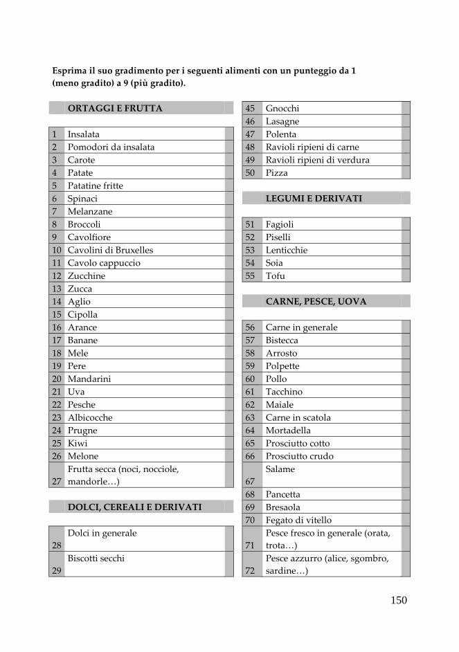
150
Esprima il suo gradimento per i seguenti alimenti con un punteggio da 1
(meno gradito) a 9 (più gradito).
ORTAGGI E FRUTTA 45 Gnocchi
46 Lasagne
1 Insalata 47 Polenta
2 Pomodori da insalata 48 Ravioli ripieni di carne
3 Carote 49 Ravioli ripieni di verdura
4 Patate 50 Pizza
5 Patatine fritte
6 Spinaci LEGUMI E DERIVATI
7 Melanzane
8 Broccoli 51 Fagioli
9 Cavolfiore 52 Piselli
10 Cavolini di Bruxelles 53 Lenticchie
11 Cavolo cappuccio 54 Soia
12 Zucchine 55 Tofu
13 Zucca
14 Aglio CARNE, PESCE, UOVA
15 Cipolla
16 Arance 56 Carne in generale
17 Banane 57 Bistecca
18 Mele 58 Arrosto
19 Pere 59 Polpette
20 Mandarini 60 Pollo
21 Uva 61 Tacchino
22 Pesche 62 Maiale
23 Albicocche 63 Carne in scatola
24 Prugne 64 Mortadella
25 Kiwi 65 Prosciutto cotto
26 Melone 66 Prosciutto crudo
27
Frutta secca (noci, nocciole,
mandorle…) 67
Salame
68 Pancetta
DOLCI, CEREALI E DERIVATI 69 Bresaola
70 Fegato di vitello
28
Dolci in generale
71
Pesce fresco in generale (orata,
trota…)
29
Biscotti secchi
72
Pesce azzurro (alice, sgombro,
sardine…)

151
30 Biscotti farciti 73 Tonno in scatola
31 Biscotti al cacao 74 Gamberetti
32 Merendine ripiene al cioccolato 75 Vongole
33 Merendine ripiene marmellata 76 Uova in generale
34 Merendine ripiene crema 77 Frittata
35 Croissants
36 Crackers normali LATTE E DERIVATI
37 Crackers integrali
38 Pane comune 78 Latte intero
39 Pane integrale 79 Latte scremato
40 Panino burro e marmellata 80 Yogurt bianco
41 Pasta 81 Yogurt alla frutta
42
Pasta integrale
82
Formaggi freschi (mozzarella,
crescenza...)
43
Riso
83
Formaggi stagionati
(parmigiano, fontina...)
44
Riso integrale
84
Formaggi molli (certosino,
asiago...)
BEVANDE E ALCOLICI PRODOTTI VARI
85 Acqua minerale naturale 95 Burro
86 Acqua minerale gassata 96 Olio d’oliva
87 Birra 97 Olio di semi
88 Vino bianco 98 Aceto
89
Vino rosso
99
Sugo a base di pomodoro e
cipolla
90
Superalcolici
100
Sugo a base di pomodoro e
aglio
91 Bibite analcoliche 101 Ragù
92 The 102 Pesto alla genovese
93 Caffè 103 Gelato alla frutta
94 Infusi e tisane 104 Gelato alla crema
105 Cioccolato
106 Marmellata di frutta
107 Miele

152
SEMPRE N° VOLTE A
SETTIMANA
N°
VOLTE
AL MESE
MAI NOTE
PRIMI
Pasta/riso al pomodoro
Pasta/riso al ragù
Pasta con verdure
Minestrone, passato…
Pasta/minestra di
legumi
SECONDI
Spezzatino
Carne alla pizzaiola
CONTORNI
Pomodori
Peperoni
Carote
Insalata
Spinaci, erbette
Broccoli
Cavolini di Bruxelles
Cavolfiori
Verze
Cime di rapa
Melanzane
Zucca
FRUTTA
Arance
Mandarini
Pompelmi
Kiwi
ALTRO
Pizza margherita
Pizza con verdure
Torte salate con
verdure

153
BEVANDE
Tè
Tè verde
Vino bianco
Vino rosso
Caffè
Succhi di frutta
Spremuta d'arance
CONDIMENTI
Olio d'oliva
Olio di semi
Burro
NOTE

154
DIARIO ALIMENTARE
NOME
COGNOME
DIETA STANDARD
DATA
COLAZIONE
alimento / piatto quantità
SPUNTINO
PRANZO
SPUNTINO
CENA
SPUNTINO

155
DIARIO ALIMENTARE
NOME
COGNOME
DIETA BROCCOLI
DATA
COLAZIONE
alimento / piatto quantità
SPUNTINO
PRANZO
SPUNTINO
CENA
SPUNTINO