Protease expression by microorganisms and its relevance to ... · this scenario, very few...
11
Protease expression by microorganisms and its relevance to crucial physiological/pathological events André Luis Souza dos Santos André Luis Souza dos Santos, Laboratory of Multidisciplinary Studies on Microbial Biochemistry, Department of General Mi- crobiology, Institute of Microbiology Prof. Paulo de Góes, Fed- eral University of Rio de Janeiro, Rio de Janeiro, RJ 21941-902, Brazil Author contributions: dos Santos ALS solely contributed to this paper. Supported by Grants from the Brazilian Agencies: Conselho Nacional de Desenvolvimento Científico e Tecnológico (CNPq), Coordenação de Aperfeiçoamento Pessoal de Nível Superior and Fundação Carlos Chagas Filho de Amparo à Pesquisa do Estado do Rio de Janeiro (FAPERJ); the author was supported by CNPq and FAPERJ fellowships Correspondence to: André Luis Souza dos Santos, As- sociate Professor, Laboratory of Multidisciplinary Studies on Microbial Biochemistry, Department of General Microbiology, Institute of Microbiology Prof. Paulo de Góes, Federal University of Rio de Janeiro, Rio de Janeiro, RJ 21941-902, Brazil. [email protected] Telephone: +55-21-25983035 Fax: +55-21-25608344 Received: January 8, 2011 Revised: February 21, 2011 Accepted: February 28, 2011 Published online: March 26, 2011 Abstract The treatment of infections caused by fungi and try- panosomatids is difficult due to the eukaryotic nature of these microbial cells, which are similar in several bio- chemical and genetic aspects to host cells. Aggravating this scenario, very few antifungal and anti-trypanoso- matidal agents are in clinical use and, therefore, ther- apy is limited by drug safety considerations and their narrow spectrum of activity, efficacy and resistance. The search for new bioactive agents against fungi and trypanosomatids has been expanded because progress in biochemistry and molecular biology has led to a bet- ter understanding of important and essential pathways in these microorganisms including nutrition, growth, proliferation, signaling, differentiation and death. In this context, proteolytic enzymes produced by these eukaryotic microorganisms are appointed and, in some cases, proven to be excellent targets for searching novel natural and/or synthetic pharmacological compounds, in order to cure or prevent invasive fungal/trypanosomatid diseases. With this task in mind, our research group and others have focused on aspartic-type proteases, since the activity of this class of hydrolytic enzymes is directly implicated in several facets of basic biological processes of both fungal and trypanosomatid cells as well as due to the participation in numerous events of interaction between these microorganisms and host structures. In the present paper, a concise revision of the beneficial effects of aspartic protease inhibitors, with emphasis on the aspartic protease inhibitors used in the anti-human immunodeficiency virus therapy, will be presented and discussed using our experience with the following micro- bial models: the yeast Candida albicans , the filamentous fungus Fonsecaea pedrosoi and the protozoan trypano- somatid Leishmania amazonensis . © 2011 Baishideng. All rights reserved. Key words: Protease; Aspartic protease inhibitors; Try- panosomatids; Fungi; Cell biology; Virulence; Chemo- therapy AUTOBIOGRAPHY OF EDITORIAL BOARD MEMBERS World J Biol Chem 2011 March 26; 2(3): 48-58 ISSN 1949-8454 (online) © 2011 Baishideng. All rights reserved. Online Submissions: http://www.wjgnet.com/1949-8454office [email protected] doi:10.4331/wjbc.v2.i3.48 World Journal of Biological Chemistry WJB C 48 March 26, 2011|Volume 2|Issue 3| WJBC|www.wjgnet.com Figure 1 André Luis Souza dos Santos, Associate Professor, Laboratory of Multi- disciplinary Studies on Microbial Biochem- istry, Department of General Microbiology, Institute of Microbiology Prof. Paulo de Góes, Federal University of Rio de Janeiro, Rio de Janeiro, RJ 21941-902, Brazil.
Transcript of Protease expression by microorganisms and its relevance to ... · this scenario, very few...

Protease expression by microorganisms and its relevance to crucial physiological/pathological events
André Luis Souza dos Santos
André Luis Souza dos Santos, Laboratory of Multidisciplinary Studies on Microbial Biochemistry, Department of General Mi-crobiology, Institute of Microbiology Prof. Paulo de Góes, Fed-eral University of Rio de Janeiro, Rio de Janeiro, RJ 21941-902, BrazilAuthor contributions: dos Santos ALS solely contributed to this paper.Supported by Grants from the Brazilian Agencies: Conselho Nacional de Desenvolvimento Científico e Tecnológico (CNPq), Coordenação de Aperfeiçoamento Pessoal de Nível Superior and Fundação Carlos Chagas Filho de Amparo à Pesquisa do Estado do Rio de Janeiro (FAPERJ); the author was supported by CNPq and FAPERJ fellowshipsCorrespondence to: André Luis Souza dos Santos, As-sociate Professor, Laboratory of Multidisciplinary Studies on Microbial Biochemistry, Department of General Microbiology, Institute of Microbiology Prof. Paulo de Góes, Federal University of Rio de Janeiro, Rio de Janeiro, RJ 21941-902, Brazil. [email protected]: +55-21-25983035 Fax: +55-21-25608344Received: January 8, 2011 Revised: February 21, 2011Accepted: February 28, 2011Published online: March 26, 2011
AbstractThe treatment of infections caused by fungi and try-panosomatids is difficult due to the eukaryotic nature of these microbial cells, which are similar in several bio-chemical and genetic aspects to host cells. Aggravating this scenario, very few antifungal and anti-trypanoso-matidal agents are in clinical use and, therefore, ther-apy is limited by drug safety considerations and their narrow spectrum of activity, efficacy and resistance. The search for new bioactive agents against fungi and trypanosomatids has been expanded because progress in biochemistry and molecular biology has led to a bet-ter understanding of important and essential pathways in these microorganisms including nutrition, growth, proliferation, signaling, differentiation and death. In this context, proteolytic enzymes produced by these
eukaryotic microorganisms are appointed and, in some cases, proven to be excellent targets for searching novel natural and/or synthetic pharmacological compounds, in order to cure or prevent invasive fungal/trypanosomatid diseases. With this task in mind, our research group and others have focused on aspartic-type proteases, since the activity of this class of hydrolytic enzymes is directly implicated in several facets of basic biological processes of both fungal and trypanosomatid cells as well as due to the participation in numerous events of interaction between these microorganisms and host structures. In the present paper, a concise revision of the beneficial effects of aspartic protease inhibitors, with emphasis on the aspartic protease inhibitors used in the anti-human immunodeficiency virus therapy, will be presented and discussed using our experience with the following micro-bial models: the yeast Candida albicans , the filamentous fungus Fonsecaea pedrosoi and the protozoan trypano-somatid Leishmania amazonensis .
© 2011 Baishideng. All rights reserved.
Key words: Protease; Aspartic protease inhibitors; Try-panosomatids; Fungi; Cell biology; Virulence; Chemo-therapy
AutobiogrAphy of EditoriAl boArd MEMbErs
World J Biol Chem 2011 March 26; 2(3): 48-58 ISSN 1949-8454 (online)
© 2011 Baishideng. All rights reserved.
Online Submissions: http://www.wjgnet.com/[email protected]:10.4331/wjbc.v2.i3.48
World Journal ofBiological ChemistryW J B C
48 March 26, 2011|Volume 2|Issue 3|WJBC|www.wjgnet.com
Figure 1 André Luis Souza dos Santos, Associate Professor, Laboratory of Multi-disciplinary Studies on Microbial Biochem-istry, Department of General Microbiology, Institute of Microbiology Prof. Paulo de Góes, Federal University of Rio de Janeiro, Rio de Janeiro, RJ 21941-902, Brazil.

dos Santos ALS. Relevance of proteases to trypanosomatids and fungi
Peer reviewers: Chen-Yong Lin, PhD, Associate Professor, Bio-chemistry and Molecular Biology, University of MarylandSchool of Medicine, 655 w Baltimore Street, 10-027 BRB, Baltimore, MD 21201, United States; Michiaki Yamashita, PhD, Chief, Food Biotechnology Section, National Research Institute of Fisheries Science, 2-12-4 Fukuura, Yokohama 236-8648, Japan
dos Santos ALS. Protease expression by microorganisms and its relevance to crucial physiological/pathological events. World J Biol Chem 2011; 2(3): 48-58 Available from: URL: http://www.wjgnet.com/1949-8454/full/v2/i3/48.htm DOI: http://dx.doi.org/10.4331/wjbc.v2.i3.48
INTRODUCTION AND EDUCATIONAL EXPERIENCESince I was young, I (Figure 1) have been interested in being a teacher, and that feeling grew and consolidated along with my professional journey. The scientific world was introduced to me during high school. From 1990 to 1994, I studied the Biotechnology course in a reputable Federal Institution from Rio de Janeiro State, Brazil, called Escola Técnica Federal de Química - ETFQ (cur-rently CEFETEQ), an excellent technical school. Over those years, the disciplines related to the Microbiology area (Bacteriology, Mycology, Virology, Protozoology and Immunology) and the laboratory classes produced a great curiosity, motivation and stimulation of scientific thought, which ignited my desire to be a scientist. With this proposal in mind, in 1994, I started my bachelor de-gree in the Microbiology and Immunology course at the Federal University of Rio de Janeiro (UFRJ), being one of the 35 students approved to constitute the first class of that novel graduation course. In parallel, I worked as a Biotechnology technician at the Biochemistry Depart-ment of the State University of Rio de Janeiro (UERJ) under the supervision of Dr. Claudia Vitória de Moura Gallo, an exemplar professional and an excellent person, who contributed notably to turn my dream into reality. In early 1999, I finished the undergraduate program and started a Master’s degree at the Institute of Microbiology Prof. Paulo de Góes (IMPPG)-UFRJ. During the period from mid 2000 until early 2002, I developed my doctoral thesis at the IMPPG-UFRJ under the supervision of Dr. Rosangela Maria de Araújo Soares. Since August 2002, I have been Professor at the Department of General Mi-crobiology of the IMPPG-UFRJ and, since then, I have been teaching lessons to several undergraduate courses including Microbiology and Immunology, Nursing, Biol-ogy and Pharmacy. Still, I effectively participate in two postgraduate courses at UFRJ: Microbiology from IMP-PG and Biochemistry from Chemistry Institute.
Indubitably, my professional work has only been fully developed because I have a research group consisting of competent professionals, including technicians and un-dergraduate, masters, doctoral and postdoctoral students, who are extremely dedicated and committed to scientific
thinking. I would like take this opportunity to express and reiterate my full admiration and gratitude to all my stu-dents. I would also like to thank to the several Brazilian researchers who have contributed immensely to my work, in particular Dr. Marta Helena Branquinha (IMPPG-UFRJ), Dr. Eliana Barreto-Bergter (IMPPG-UFRJ), Dr. Lucy Seldin (IMPPG-UFRJ), Dr. Celuta Sales Alviano (IMPPG-UFRJ), Dr. Claudia Masini d’Avila-Levy (Funda-ção Oswaldo Cruz-FIOCRUZ) and Dr. Lucimar Ferreira Kneipp (FIOCRUZ). My research has been supported by the Brazilian agencies: Conselho Nacional de Desen-volvimento Científico e Tecnológico (CNPq), Conselho de Ensino para Graduados e Pesquisas (CEPG/UFRJ), Fundação de Amparo à Pesquisa do Estado do Rio de Janeiro (FAPERJ), Fundação Universitária José Bonifácio (FUJB) and Coordenação de Aperfeiçoamento de Pessoal de Nível Superior (CAPES). I have also been supported by a CNPq fellowship since 2005 and by a FAPERJ fel-lowship since 2007.
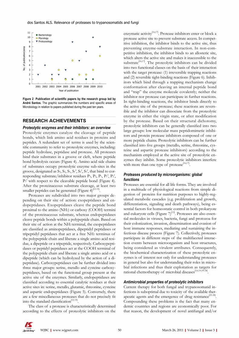
Over the past 10 years: (1) I supervised 16 monographs of graduate students, 10 master theses and 4 doctoral the-ses; (2) I published 79 papers in the field of Bacteriology (n = 5), Mycology (n = 22) and Protozoology (n = 52) (Figure 2); and (3) I was invited to participate as a speaker at national and international meetings. I am a peer review-er for international scientific journals, as well as career and research grant committees. In addition, I have accepted invitations to write reviews and book chapters on the themes: (1) relevance of proteolytic enzymes produced by microorganisms; and (2) antimicrobial properties of pro-tease inhibitors[1-11].
ACADEMIC STRATEGIES AND GOALSOur work group is distinguished by its multidisciplinary nature, with direct involvement of different research in-stitutions from Brazil (other Departments and Institutes from UFRJ, UERJ, FIOCRUZ, Universidade Federal Flu-minense (UFF), Universidade Federal do Estado do Rio de Janeiro (UNIRIO), Universidade do Estado de São Paulo (USP), Universidade Federal de São Paulo (UNIFESP), Universidade Federal do Espírito Santo (UFES)) and from other countries, generating productive and effective collab-orations. Several publications in high-ranked journals, e.g. FEMS Microbiology Reviews, PLoS One, Archives of Biochemis-try and Biophysics, Journal of Antimicrobial Chemotherapy, Journal of Clinical Microbiology, International Journal of Antimicrobial Agents, Microbes and Infection, International Journal for Parasitol-ogy, Protist, Parasitology and Medical Mycology, were produced in collaboration with these partners.
Over the last years, my group has focused on the identification, biochemical characterization and discovery of biological functions of proteases produced by micro-organisms, with emphasis in trypanosomatids and fungi (Figure 3). More recently, we have started to study prote-ase inhibitors in an attempt to use these bioactive com-pounds as a new therapeutic proposal against eukaryotic pathogenic microorganisms (Figure 3).
49 March 26, 2011|Volume 2|Issue 3|WJBC|www.wjgnet.com
RESEARCH ACHIEVEMENTSProteolytic enzymes and their inhibitors: an overviewProteolytic enzymes catalyze the cleavage of peptide bonds, which link amino acid residues in proteins and peptides. A redundant set of terms is used by the scien-tific community to refer to proteolytic enzymes, including: peptide hydrolase, peptidase and protease. All proteases bind their substrates in a groove or cleft, where peptide bond hydrolysis occurs (Figure 4). Amino acid side chains of substrates occupy proteolytic enzyme sub-sites in the groove, designated as S3, S2, S1, S1’, S2’, S3’, that bind to cor-responding substrate/inhibitor residues P3, P2, P1, P1’, P2’, P3’ with respect to the cleavable peptide bond (Figure 4). After the proteinaceous substrate cleavage, at least two smaller peptides can be generated (Figure 4)[12-15].
Proteases are subdivided into two major groups de-pending on their site of action: exopeptidases and en-dopeptidases. Exopeptidases cleave the peptide bond proximal to the amino (NH2) or carboxy (COOH) termini of the proteinaceous substrate, whereas endopeptidases cleave peptide bonds within a polypeptide chain. Based on their site of action at the NH2 terminal, the exopeptidases are classified as aminopeptidases, dipeptidyl peptidases or tripeptidyl peptidases that act at a free NH2 terminus of the polypeptide chain and liberate a single amino acid resi-due, a dipeptide or a tripeptide, respectively. Carboxypepti-dases or peptidyl peptidases act at the COOH terminal of the polypeptide chain and liberate a single amino acid or a dipeptide (which can be hydrolyzed by the action of a di-peptidase). Carboxypeptidases can be further divided into three major groups: serine, metallo and cysteine carboxy-peptidases, based on the functional group present at the active site of the enzymes. Similarly, endopeptidases are classified according to essential catalytic residues at their active sites in: serine, metallo, glutamic, threonine, cysteine and aspartic endopeptidases (Figure 5). Conversely, there are a few miscellaneous proteases that do not precisely fit into the standard classification[12-15].
The class of a protease is characteristically determined according to the effects of proteolytic inhibitors on the
enzymatic activity[16,17]. Protease inhibitors enter or block a protease active site to prevent substrate access. In compet-itive inhibition, the inhibitor binds to the active site, thus preventing enzyme-substrate interaction. In non-com-petitive inhibition, the inhibitor binds to an allosteric site, which alters the active site and makes it inaccessible to the substrate[16,17]. The proteolytic inhibitors can be divided into two functional classes on the basis of their interaction with the target protease: (1) irreversible trapping reactions and (2) reversible tight-binding reactions (Figure 6). Inhib-itors which bind through a trapping mechanism change conformation after cleaving an internal peptide bond and “trap” the enzyme molecule covalently; neither the inhibitor nor protease can participate in further reactions. In tight-binding reactions, the inhibitor binds directly to the active site of the protease; these reactions are revers-ible and the inhibitor can dissociate from the proteolytic enzyme in either the virgin state, or after modification by the protease. Based on their structural dichotomy, proteolytic inhibitors can be generally classified into two large groups: low molecular mass peptidomimetic inhibi-tors and protein protease inhibitors composed of one or more peptide chains. Proteolytic inhibitors can be further classified into five groups (metallo, serine, threonine, cys-teine and aspartic protease inhibitors) according to the mechanism employed at the active site of proteolytic en-zymes they inhibit. Some proteolytic inhibitors interfere with more than one type of protease[16,17].
Proteases produced by microorganisms: global functionsProteases are essential for all life forms. They are involved in a multitude of physiological reactions from simple di-gestion of proteins for nutrition purposes to highly-reg-ulated metabolic cascades (e.g. proliferation and growth, differentiation, signaling and death pathways), being es-sential factors for homeostatic control in both prokaryote and eukaryote cells (Figure 7)[12]. Proteases are also essen-tial molecules in viruses, bacteria, fungi and protozoa for their colonization, invasion, dissemination and evasion of host immune responses, mediating and sustaining the in-fectious disease process (Figure 7). Collectively, proteases participate in different steps of the multifaceted interac-tion events between microorganism and host structures, being considered as virulent attributes. Consequently, the biochemical characterization of these proteolytic en-zymes is of interest not only for understanding proteases in general but also for understanding their roles in micro-bial infections and thus their exploitation as targets for rational chemotherapy of microbial diseases[3,6,10,18-24].
Antimicrobial properties of proteolytic inhibitorsCurrent therapy for both fungal and trypanosomatid in-fections is suboptimal due to toxicity of the available ther-apeutic agents and the emergence of drug resistance[25-28]. Compounding these problems is the fact that many en-demic countries and regions are economically poor. For that reason, the development of novel antifungal and/or
50 March 26, 2011|Volume 2|Issue 3|WJBC|www.wjgnet.com
11
10
9
8
7
6
5
4
3
2
1
0
No.
of
publ
icat
ion/
area
BacteriologyMycologyProtozoology
2001 2002 2003 2004 2005 2006 2007 2008 2009 2010
Year of publication
Figure 2 Publication of scientific papers by the research group led by André Santos. The graphic summarizes the numbers and specific areas of Microbiology in relation to papers published during the past ten years.
dos Santos ALS. Relevance of proteases to trypanosomatids and fungi

anti-trypanosomatidal drugs is an imperative requirement. A number of new strategies to obstruct fungal/trypano-somatid biological processes have emerged; one of them
51 March 26, 2011|Volume 2|Issue 3|WJBC|www.wjgnet.com
Trypanosomatids Fungi
Proteolytic enzymes
Biochemical characterization
Identification
Functional implications
Physiological roles Pathological roles
Effects of protease inhibitors
Figure 3 Rationale of the research works developed in the André Santos’ laboratory. The main purpose of our study focuses on the identification and biochemi-cal characterization of cellular and/or extracellular proteases produced by eukaryotic microorganisms, especially trypanosomatids and fungi. Subsequently, we have focused on the discovery of possible biological functions for these hydrolytic enzymes in both the social context of the microbial cell and the participation in interaction events with biotic and abiotic substrates. Finally, we have used the protease inhibitors in an attempt to block vital processes in microbial cells, thus preventing a suc-cessful infection.
...
Tripeptidyl peptidasesDipeptidyl peptidases
Aminopeptidases
H2N
Threonine
Glutamic
Aspartic
Cysteine
Serine
Metallo
COOH
Dipeptides
COOH
Peptidyl peptidases
Carboxypeptidases
Endopeptidases
Figure 5 Classification of proteases. Gray circles represent amino acids and red circles indicate the amino acid sequence that is bound to the proteolytic en-zyme. Yellow arrows point to the site of cleavage. The blue arrows indicate the classification of carboxypeptidases and the red arrow shows the box containing all the classes of endopeptidases, according to the chemical group present in their catalytic sites.
Active site
S3 S2 S1 S’1 S’2S’3
P3P2 P1 P’1 P’2
P’3
COOHNH2
Protease
P3P2 P1
P’1 P’2P’3
COOHNH2 Substrate
Products
Scissile bond
Figure 4 Schematic representation of binding region and catalytic site of a protease. This hypothetical protease possesses six subsites (S1-S3 and S1’-S3’) in its catalytic site and, consequently, is able to recognize and bind to a sequence of six amino acids (P1-P3 and P1’-P3’) in the proteinaceous substrate. After proteolysis, at least two smaller peptides are generated as the reaction products.
dos Santos ALS. Relevance of proteases to trypanosomatids and fungi

is focused on protease inhibition. Currently, the main ap-proach has been to obtain good inhibitors of the target protease, in the belief that inhibition of the activity will be therapeutic. In this context, our research group has pub-lished some works that corroborate this premise[1-6,10,29-39].
Aspartic protease inhibitors used in anti-human immunodeficiency virus therapy present anti-microbial propertiesLessons from the yeast Candida albicans (C. albicans), the fila-mentous fungus Fonsecaea pedrosoi (F. pedrosoi) and the pro-tozoan Leishmania amazonensis (L. amazonensis) are illustrated as follows.
C. albicans : C. albicans is both a successful commensal and pathogen of humans that can infect a broad range of body sites[40]. The transition from commensalism to parasitism requires a susceptible host, which includes individuals with humoral and/or cellular deficiencies as well as persons submitted to different immunosuppres-sive procedures. Candidiasis is the most common fungal infection diagnosed in humans[41-43]. Due to the emergence of pathogens resistant to conventional antifungals and the toxicity of some antimycotics, intense efforts have
been made to develop more effective antifungal agents for clinical use[44-48]. The pathogenesis of C. albicans is multi-factorial and different virulence attributes are important during the various stages of infection[20,21,49-55]. Secreted aspartic proteases (Saps) play a role in several infection stages of C. albicans, being the most important virulence factors expressed by this opportunistic fungus. Actu-ally, C. albicans possesses ten different SAP genes (SAP1 to SAP10), which are expressed according to distinct environments and host conditions[56-60]. Therefore, Saps are potential targets for the development of novel anti- C. albicans drugs[1,2,34,35]. In this context, several groups have demonstrated that aspartic protease inhibitors, including pepstatin A and the first generation of protease inhibitors used in anti-human immunodeficiency virus (HIV) thera-py (nelfinavir, saquinavir, ritonavir and indinavir), are able to restrain Sap activity (especially Sap1, Sap2 and Sap3) as well as arrest crucial events of C. albicans yeast cells such as proliferation and adhesion to both abiotic (e.g. plastic and acrylic substrates) and biotic structures (e.g. surface of different epithelial cell lineages)[61-72]. Our results showed that amprenavir[72] (unpublished data) and lopinavir (un-published data), two HIV aspartic protease inhibitors of the second generation, significantly inhibited the hydrolyt-ic activity of Sap2 and also blocked the yeasts into mycelia transformation, an essential step during the candidiasis pathogenesis. In addition, scanning electron microscopy revealed prominent ultrastructural alterations of yeast
52 March 26, 2011|Volume 2|Issue 3|WJBC|www.wjgnet.com
Microbialproteases
Physiological roles Pathological roles
Protease inhibitors
Nutrition
Growth/proliferation
Differentiation
Signaling
Death
Adhesion
Intracellular survival
Escape
Dissemination
Immunomodulation
Figure 7 Possible functions played by microbial proteases. Surface and/or secreted proteases are able to cleave different host components such as serum proteins, antimicrobial peptides, surface molecules and structural proteinaceous compounds. The degradation of host proteins can help the microorganisms in several steps of their life cycle and pathogenesis including dissemination, adhe-sion, escape, nutrition and immunomodulation of the host immune response. These proteases can also contribute to maintaining basic metabolic processes in a microbial cell, which govern crucial events like proliferation, differentiation, signaling and death pathways. Proteolytic inhibitors are able to block one or several of these fundamental events.
Inhibitor competes with substrate
Enzyme binds inhibitor
Enzyme binds substrate
Enzyme releases products
InhibitorSubstrate
ProteaseActive site
Reversibleinhibition
Irreversibleinhibition
Figure 6 Mechanisms of protease inhibition. The protease inhibitor com-petes with the substrate to bind to the active site of a protease and two distinct possibilities arise: (1) substrate binds to the catalytic site and then is cleaved by the protease, which releases the products or (2) inhibitor binds to the active site and by steric hindrance blocks the substrate attachment. In this last case, the inhibitor can promote an irreversible (the conformational structure of the prote-ase is completely lost) or reversible inhibition (when the inhibitor disconnects from the enzyme, the substrate can bind to it).
dos Santos ALS. Relevance of proteases to trypanosomatids and fungi

cells, which corroborated the inhibition of cellular division by these protease inhibitors. Several surface and/or secret-ed molecules have had their expression/production signif-icantly diminished including (1) mannose- and sialic acid-rich surface glycoconjugates, which are directly involved in adhesive properties and biofilm formation; (2) sterol content, which controls the membrane fluidity; (3) secre-tion of lipases (e.g. esterases and phospholipases), which are related to the host membrane disruption; and (4) cata-lase activity, which reduces the ability of yeasts to escape from oxidative stress generated by hydrogen peroxide, for example, released by host phagocytes[72] (unpublished data). However, it is also important to note that the inhibi-tory effects of HIV protease inhibitors both in in vitro and in vivo experimental models were observed at concentra-tions (μmol/L range) much higher than those needed for HIV protease inhibition (nmol/L range). This probably reflects a much lower affinity of these drugs for Sap than that for HIV protease[31,34]. Another explanation is that, in contrast to the very small and structurally simplified HIV protease, Saps are larger and more complex[60,73]. They possess a relatively large active site which might be responsible for the broader substrate specificity and also their susceptibilities to distinct aspartic protease inhibi-tors[60]. Nevertheless, the above concentrations may be achieved under current highly active antiretroviral therapy (HAART) regimens both in the blood[31], in human saliva (at least for indinavir)[74] and in lungs (at least for lopina-vir)[75]. In this sense, our group has showed that lopinavir at 10 mg/kg promoted a therapeutic effect in an experi-mental murine model of disseminated candidiasis, with an efficacy comparable to that of fluconazole, a recognized anti-candidal drug (unpublished results).
F. pedrosoi : Fonsecaea is a genus containing pigmented filamentous fungus isolated from soil, rotten wood and decomposing plant material. F. pedrosoi is one of the major causative agents of chromoblastomycosis, a post-traumatic and chronic infection of subcutaneous tissues in humid tropical areas specially South America and Japan[8,76-79]. F. pedrosoi is a valuable model in cell biology, since its life cycle comprises different morphological states that include reproduction structures (conidia) and fungal forms usually found in the saprophytic (mycelia) and parasitic stage (sclerotic bodies)[8]. The first report on protease production by F. pedrosoi was described by our group[80], which demonstrated that the pattern of prote-ase production and secretion by F. pedrosoi conidial cells was closely dependent on the culture medium composi-tion: metalloproteases were induced after cultivation in complex culture medium, while aspartic proteases were detected under chemically defined growth conditions. Mycelia[81] and sclerotic cells (unpublished results) of F. pedrosoi were also able to secrete aspartic-type prote-ases. The aspartic proteases produced by conidia and mycelia were capable of degrading relevant host serum proteins (e.g. IgG and albumin) as well as extracellular matrix components (e.g. laminin, fibronectin and col-lagen)[80,81]. For that reason, the extracellular hydrolytic
enzymes produced by F. pedrosoi cells, such as proteases and lipases[82], could support the initial development of this fungus inside the host, and the existence of two biochemically distinct secreted proteases makes it pos-sible to cover a wide range of host conditions. The effect of saquinavir, ritonavir, indinavir and nelfinavir on the secreted proteases of F. pedrosoi was evaluated[81,83]. These compounds inhibited the extracellular aspartic proteolytic activity produced by both conidial and mycelial forms in a dose-dependent manner. Nelfinavir was the best inhibi-tor of the aspartic protease activity secreted by conidia and mycelia, restraining the hydrolytic activities around 80% at 50 μmol/L. Interestingly, recent isolated strains of F. pedrosoi produced higher levels of extracellular pro-tease activity when compared with a laboratory-adapted strain[81,83], suggesting that the production of secreted aspartic-type proteases may be stimulated by interac-tion with the host. HIV aspartic protease inhibitors and pepstatin A also arrested the growth of conidial forms as well as transformation into mycelia[83], an essential step during the F. pedrosoi life cycle and virulence[8]. Pepstatin A showed a significant inhibition of conidial viability even at low concentration (0.1 μmol/L); however, the HIV protease inhibitors were toxic only at high concentrations (ranging from 50 to 200 μmol/L). The synergistic action on proliferation behavior between nelfinavir (25 μmol/L) and amphotericin B (3 μg/mL), when both were used at sub-inhibitory concentrations, was also observed[83]. Interestingly, HIV protease inhibitors-treated conidial cells presented irreversible ultrastructural alterations, as shown by transmission electron microscopy images such as invaginations in the cytoplasmic membrane and with-drawal of the cytoplasmic membrane from within the cell wall, disorder and detachment of the cell wall, rupture of internal organelles, detection of large and irregular cytoplasmic vacuoles, some of them containing small vesicles, abnormal cellular division and breakage of cell wall. Furthermore, the aspartic protease inhibitors drasti-cally reduced the adhesion and endocytic indexes during the interaction between F. pedrosoi conidia and epithelial cells of the Chinese hamster ovary lineage, fibroblasts or macrophages. Aspartic protease inhibitors also promoted a significant increase in the susceptibility killing by mac-rophage cells, promoting a significant reduction in the number of viable intracellular conidia after the treatment of infected macrophage monolayers with indinavir, nel-finavir and ritonavir at 6.25 μmol/L for 24 h[83].
L. amazonensis : Leishmania are digenetic protozoan parasites that live as promastigotes in the digestive tract of sand flies and as amastigotes in the phagolysosomes of mammalian macrophages. They cause a wide spectrum of clinical manifestations (generically known as leishmani-asis), and its clinical manifestations are dependent on both parasite species and immune response of the host[84-89]. The increase in the incidence of the disease, associated with higher morbidity rates, the spread of some forms of leishmaniasis to new geographical areas and Leishmania-HIV co-infection, has become an important public health
53 March 26, 2011|Volume 2|Issue 3|WJBC|www.wjgnet.com
dos Santos ALS. Relevance of proteases to trypanosomatids and fungi
54 March 26, 2011|Volume 2|Issue 3|WJBC|www.wjgnet.com
problem in the world[90-93]. However, the incidence of HIV-Leishmania co-infections has been decreasing since the introduction of HAART, in which aspartic-type protease inhibitors were included[94,95]. These findings in-stigated the research to confirm the possible connection between aspartic protease expression and basic molecular processes in Leishmania[96-103]. Our group showed that HIV protease inhibitors were able to impair in vitro proliferation of L. amazonensis promastigotes in a dose-dependent man-ner and in different extensions, in which nelfinavir (IC50 = 15.1 ± 1.1 μmol/L), lopinavir (IC50 = 16.5 ± 0.8 μmol/L) and amprenavir (IC50 = 62.0 ± 2.1 μmol/L) were the most potent compounds[103]. These three protease inhibi-tors (at the IC50 value) caused profound changes in the leishmania ultrastructure, including cytoplasm shrinking, increase in the number of lipid inclusions and some cells with the nucleus closely wrapped by endoplasmic reticu-lum, resembling an autophagic process, as well as chro-matin condensation that is suggestive of apoptotic death. The treatment with HIV protease inhibitors of either the promastigote forms preceding the interaction with macrophage cells or the amastigote forms inside macro-phages drastically reduced the association indexes (when inhibitors were used at 50 μmol/L) and the number of intracellular amastigotes (when inhibitors were used at 3.12 μmol/L)[103]. The hydrolysis of HIV protease sub-strate by L. amazonensis extract was fully inhibited by pep-statin A and HIV protease inhibitors at 10 μmol/L, sug-gesting that an aspartic protease may be the parasite target of the inhibitors. Despite all these beneficial effects, the HIV protease inhibitors induced an increase in the expres-sion of cysteine protease b (cpb)[19] and the metalloprote-ase gp63[24], two well-known virulence factors expressed by Leishmania spp., probably in an attempt to compensate the parasite aspartic protease inhibition[103].
Proposals of the molecular mechanisms of the aspartic protease inhibitors on the aspartic protease produced by microorganismsDirect actions - inhibition of aspartic proteases: The binding of the aspartic protease inhibitor to the active site of an aspartic protease blocks the binding of substrate to the enzyme. Therefore, the substrate remains intact and no peptides and/or amino acids are generated. Ob-viously aspartic protease inhibition will be more or less drastic depending on several parameters like the inhibitor affinity constant for the active site, its ability to revers-ibly or irreversibly bind to the enzyme, and the ratio of inhibitor in relation to the available substrate and enzyme. (1) The inhibition of secreted and/or surface aspartic proteases can result in an inability of the microorgan-ism to obtain peptides and amino acids to its nutrition, leading to a reduction or a complete interruption in the proliferation rate. This phenomenon is clearly observed in C. albicans yeast cells when cultured under chemically defined medium containing large proteins (e.g. albumin and hemoglobin) as a unique nitrogenous source, but not when Candida cells are cultured in a medium containing an
unlimited nitrogenous source[104-108]; (2) Some intracellular aspartic proteases produced by microorganisms also con-trol the cleavage of important own proteins in order to promote protein activation and/or perfect functioning of a biosynthetic route; their inhibition can arrest signaling events and/or metabolic pathways, as a result inhibiting some crucial biological processes for microbial cells such as morphogenesis or expression of surface molecules re-sponsible for adhesion or fungal protection. For example, some of these aspartic protease inhibitors alter the lipid biosynthesis, including ergosterol, resulting in altered membrane permeability[68,72,83]. These inhibitory actions will depend on the ability of the aspartic protease inhibi-tors to (a) enter in the microbial cells and (b) accumulate inside them; and (3) Some surface aspartic proteases par-ticipate in the assembly and organization of the microbial surface. For instance, in contrast to all other members of the Sap family, the proteases Sap9 and Sap10 are bound to the C. albicans cell surface by a glycosylphosphatidylinosi-tol anchor motif. Sap9 seems to be predominantly located in the cell membrane, and Sap10 is located in the cell wall and membrane[109]. Recently, Schild et al[110] demonstrated that Sap9 and Sap10 cleave covalently linked cell wall pro-teins, including chitinase Cht2 and the glucan-cross-link-ing protein Pir1. Deletion of the SAP9 and SAP10 genes resulted in a reduction of cell-associated chitinase activity similar to that upon deletion of CHT2, suggesting a direct influence of Sap9 and Sap10 on Cht2 function. The treat-ment with amprenavir[72] and lopinavir (data not shown) promoted the removal of the amorphous layer that covers the entire surface of C. albicans, turning the rough surface into a smooth one. Moreover, surface aspartic proteases can promote microorganism adhesion (by functioning as an adhesive molecule or by destroying some receptors at the host surface, exposing and/or facilitating the adhesion event); therefore, their inhibition can diminish the ability of a microorganism to interact with host structures.
Indirect actions - binding to unrelated molecules: The possibility of aspartic protease inhibitors binding to or interfere with other molecules than aspartic proteases can not be excluded[35]. In this context, these compounds can generate irreversible toxic effects by perturbing the ho-meostasis of the microbial cells, culminating in death of microorganisms.
CONCLUSIONMicrobial pathogenesis is a multifactorial process and dif-ferent virulence factors are important during the various phases of infection. Some virulence attributes, such as the aspartic proteases, play a role in several infection stag-es and the inhibition of one of the many stages probably will contribute to the containment of the pathogen and thus should help in the treatment of disease. Therefore, aspartic proteases synthesized by pathogenic fungi and trypanosomatids are prospective targets for the develop-ment of new chemotherapeutic compounds. Both in vitro
dos Santos ALS. Relevance of proteases to trypanosomatids and fungi

55 March 26, 2011|Volume 2|Issue 3|WJBC|www.wjgnet.com
and in vivo studies demonstrated that the use of HIV pro-tease inhibitors promoted a drastic reduction in the pres-ence of both fungal and trypanosomatid opportunistic diseases as well as clearly revealing that these inhibitors are able to arrest vital events in microbial cells presenting eukaryotic architecture, including proliferation, differen-tiation and nutrition. These inhibitors also impair the de-velopment of infection in culture or animal models due to their capability of blocking adhesion, internalization, evasion and escape of host responses. Together, all these beneficial effects culminate in death of the microorgan-ism and/or its inadequate ability to develop an efficient and successful infection. Future studies must investigate combination drug therapy, which may reduce the inci-dence of toxicity due to individual drugs and may also delay the emergence of drug resistance. In addition, the purification of aspartic proteases produced by fungi and trypanosomatids, the knowledge of its biochemical prop-erties and the crystallization of the tertiary structure will contribute to better understanding of the functioning of these proteolytic enzymes as well as allowing the design of more specific inhibitors. At least for C. albicans, the crystal structure of Sap2 complexed with pepstatin A has been known since 1993[111], whereas the crystal structure of Sap3 and its complex with pepstatin A was first pre-sented in 2007[112]. The secondary structures of Sap2 and Sap3 as well as Sap1 and Sap5 were recently described[113]. These data could help in the development of novel and more effective anti-C. albicans compounds.
I really hope that all these findings together arouse the curiosity and the enthusiasm of other researchers in order to look for novel compounds with the ability to in-hibit aspartic proteases produced by fungi and trypanoso-matids. These novel compounds must be more specific, powerful and with reduced side effects, in an attempt to increase our armamentarium to treat fungal and trypano-somatid diseases.
ACKNOWLEDGMENTSIt is so difficult to put in words my scientific trajectory; firstly, because it is difficult to write about myself and, secondly, because some of the professional moments that I came through were intimately associated with my personal life, which has been difficult. But all of these difficult times were compensated by satisfactory and unforgettable moments. In this sense, I am grateful to the past and present members of my laboratory for their contributions to our studies. I also wish to express my gratitude to several colleagues and investigators for their collaborations, in especial Marta Helena Branquinha and Claudia Masini d’Avila-Levy, with whom I share all my scientific achievements and victories and to whom I dedi-cate this publication.
REFERENCES1 Santos ALS. Aspartic proteases of human pathogenic fungi
are prospective targets for the generation of novel and effec-
tive antifungal inhibitors. Curr Enz Inhib 2011; In press2 dos Santos ALS. HIV aspartyl protease inhibitors as promis-
ing compounds against Candida albicans. World J Biol Chem 2010; 1: 21-30
3 Vermelho AB, Branquinha MH, D’Ávila-Levy CM, dos Santos ALS, Paraguai de Souza Dias E, Nogueira de Melo AC. Biological roles of peptidases in trypanosomatids. Open Parasitol J 2010; 4: 5-23
4 Santos ALS, d’Avila-Levy CM, Branquinha MH. Calpain-like proteins in trypanosomatids: effects of calpain inhibitors on the parasites’ physiology and motivations for their pos-sible application as chemotherapeutic agents. In: Cohen JB, Ryseck LP, editors. Cystatins: protease inhibitors, biomark-ers and immunomodulators. New York: Nova Science Pub-lishers, 2011: In press
5 Santos ALS, d’Avila-Levy CM, Branquinha MH. Anti-try-panosomatid properties of cystatin superfamily: implications on parasite development and virulence. In: Cohen JB, Ryseck LP, editors. Cystatins: protease inhibitors, biomarkers and immunomodulators. New York: Nova Science Publishers, 2011: In press
6 Santos ALS. Aspartic peptidase inhibitors as potential bio-active pharmacological compounds against human fungal pathogens. Chapter 13. In: Ahmad I, Owais M, Shahid M, Aqil F, editors. Combating fungal infections: problems and remedy. Berlin: Springer-Verlag, 2010: 289-326
7 Santos AL, Bittencourt VC, Pinto MR, Silva BA, Barreto-Bergter E. Biochemical characterization of potential virulence markers in the human fungal pathogen Pseudallescheria boydii. Med Mycol 2009; 47: 375-386
8 Santos AL, Palmeira VF, Rozental S, Kneipp LF, Nimrich-ter L, Alviano DS, Rodrigues ML, Alviano CS. Biology and pathogenesis of Fonsecaea pedrosoi, the major etiologic agent of chromoblastomycosis. FEMS Microbiol Rev 2007; 31: 570-591
9 Santos AL, d'Avila-Levy CM, Elias CG, Vermelho AB, Bran-quinha MH. Phytomonas serpens: immunological similari-ties with the human trypanosomatid pathogens. Microbes Infect 2007; 9: 915-921
10 Vermelho AB, Giovanni De Simone S, d'Avila-Levy CM, Santos ALS, Nogueira de Melo AC, Silva-Junior FP, Bom EP, Branquinha MH. Trypanosomatidae peptidases: a target for drugs development. Curr Enz Inhib 2007; 3: 19-48
11 Santos AL, Branquinha MH, D'Avila-Levy CM. The ubiqui-tous gp63-like metalloprotease from lower trypanosomatids: in the search for a function. An Acad Bras Cienc 2006; 78: 687-714
12 Rao MB, Tanksale AM, Ghatge MS, Deshpande VV. Mo-lecular and biotechnological aspects of microbial proteases. Microbiol Mol Biol Rev 1998; 62: 597-635
13 Barrett AJ, Rawlings ND, O'Brien EA. The MEROPS data-base as a protease information system. J Struct Biol 2001; 134: 95-102
14 Barrett AJ, Tolle DP, Rawlings ND. Managing peptidases in the genomic era. Biol Chem 2003; 384: 873-882
15 Rawlings ND, Morton FR, Barrett AJ. MEROPS: the pepti-dase database. Nucleic Acids Res 2006; 34: D270-D272
16 Rawlings ND, Tolle DP, Barrett AJ. Evolutionary families of peptidase inhibitors. Biochem J 2004; 378: 705-716
17 Bode W, Huber R. Structural basis of the endoproteinase-protein inhibitor interaction. Biochim Biophys Acta 2000; 1477: 241-252
18 McKerrow JH, Sun E, Rosenthal PJ, Bouvier J. The proteases and pathogenicity of parasitic protozoa. Annu Rev Microbiol 1993; 47: 821-853
19 Mottram JC, Brooks DR, Coombs GH. Roles of cysteine pro-teinases of trypanosomes and Leishmania in host-parasite interactions. Curr Opin Microbiol 1998; 1: 455-460
20 Hube B. Extracellular proteinases of human pathogenic fungi. Contrib Microbiol 2000; 5: 126-137
dos Santos ALS. Relevance of proteases to trypanosomatids and fungi

56 March 26, 2011|Volume 2|Issue 3|WJBC|www.wjgnet.com
21 Monod M, Capoccia S, Léchenne B, Zaugg C, Holdom M, Jousson O. Secreted proteases from pathogenic fungi. Int J Med Microbiol 2002; 292: 405-419
22 Klemba M, Goldberg DE. Biological roles of proteases in parasitic protozoa. Annu Rev Biochem 2002; 71: 275-305
23 Atkinson HJ, Babbitt PC, Sajid M. The global cysteine pepti-dase landscape in parasites. Trends Parasitol 2009; 25: 573-581
24 Yao C. Major surface protease of trypanosomatids: one size fits all? Infect Immun 2010; 78: 22-31
25 Rodriques Coura J, de Castro SL. A critical review on Cha-gas disease chemotherapy. Mem Inst Oswaldo Cruz 2002; 97: 3-24
26 Croft SL, Yardley V. Chemotherapy of leishmaniasis. Curr Pharm Des 2002; 8: 319-342
27 Juang P. Update on new antifungal therapy. AACN Adv Crit Care 2007; 18: 253-260; quiz 261-262
28 Lai CC, Tan CK, Huang YT, Shao PL, Hsueh PR. Current challenges in the management of invasive fungal infections. J Infect Chemother 2008; 14: 77-85
29 Leung D, Abbenante G, Fairlie DP. Protease inhibitors: current status and future prospects. J Med Chem 2000; 43: 305-341
30 Abbenante G, Fairlie DP. Protease inhibitors in the clinic. Med Chem 2005; 1: 71-104
31 Flexner C. HIV-protease inhibitors. N Engl J Med 1998; 338: 1281-1292
32 McKerrow JH, Engel JC, Caffrey CR. Cysteine protease in-hibitors as chemotherapy for parasitic infections. Bioorg Med Chem 1999; 7: 639-644
33 Coombs GH, Goldberg DE, Klemba M, Berry C, Kay J, Mot-tram JC. Aspartic proteases of Plasmodium falciparum and other parasitic protozoa as drug targets. Trends Parasitol 2001; 17: 532-537
34 Stewart K, Abad-Zapatero C. Candida proteases and their inhibition: prospects for antifungal therapy. Curr Med Chem 2001; 8: 941-948
35 Dash C, Kulkarni A, Dunn B, Rao M. Aspartic peptidase in-hibitors: implications in drug development. Crit Rev Biochem Mol Biol 2003; 38: 89-119
36 Pozio E, Morales MA. The impact of HIV-protease inhibitors on opportunistic parasites. Trends Parasitol 2005; 21: 58-63
37 Mastrolorenzo A, Rusconi S, Scozzafava A, Barbaro G, Su-puran CT. Inhibitors of HIV-1 protease: current state of the art 10 years after their introduction. From antiretroviral drugs to antifungal, antibacterial and antitumor agents based on as-partic protease inhibitors. Curr Med Chem 2007; 14: 2734-2748
38 Nguyen JT, Hamada Y, Kimura T, Kiso Y. Design of potent aspartic protease inhibitors to treat various diseases. Arch Pharm (Weinheim) 2008; 341: 523-535
39 McKerrow JH, Rosenthal PJ, Swenerton R, Doyle P. Devel-opment of protease inhibitors for protozoan infections. Curr Opin Infect Dis 2008; 21: 668-672
40 Calderone R, Odds FC, Boekhout T. Candida albicans: fun-damental research on an opportunistic human pathogen. FEMS Yeast Res 2009; 9: 971-972
41 Pfaller MA, Diekema DJ. Epidemiology of invasive candi-diasis: a persistent public health problem. Clin Microbiol Rev 2007; 20: 133-163
42 López-Martínez R. Candidosis, a new challenge. Clin Der-matol 2010; 28: 178-184
43 van de Veerdonk FL, Kullberg BJ, Netea MG. Pathogenesis of invasive candidiasis. Curr Opin Crit Care 2010; 16: 453-459
44 Wilson LS, Reyes CM, Stolpman M, Speckman J, Allen K, Beney J. The direct cost and incidence of systemic fungal in-fections. Value Health 2002; 5: 26-34
45 Pfaller MA, Diekema DJ. Role of sentinel surveillance of candidemia: trends in species distribution and antifungal susceptibility. J Clin Microbiol 2002; 40: 3551-3557
46 Montravers P, Jabbour K. Clinical consequences of resistant Candida infections in intensive care. Int J Antimicrob Agents
2006; 27: 1-647 Hsueh PR, Graybill JR, Playford EG, Watcharananan SP, Oh
MD, Ja'alam K, Huang S, Nangia V, Kurup A, Padiglione AA. Consensus statement on the management of invasive candidiasis in Intensive Care Units in the Asia-Pacific Re-gion. Int J Antimicrob Agents 2009; 34: 205-209
48 Niimi M, Firth NA, Cannon RD. Antifungal drug resistance of oral fungi. Odontology 2010; 98: 15-25
49 Ghannoum MA. Potential role of phospholipases in viru-lence and fungal pathogenesis. Clin Microbiol Rev 2000; 13: 122-143, table of contents
50 Calderone RA, Ronald LC. Host recognition. In: Calderone RA, Ronald LC, editors. Fungal pathogenesis: principles and clinical applications. New York: Marcel Dekker, 2002: 1-24
51 Yang YL. Virulence factors of Candida species. J Microbiol Immunol Infect 2003; 36: 223-228
52 Whiteway M, Oberholzer U. Candida morphogenesis and host-pathogen interactions. Curr Opin Microbiol 2004; 7: 350-357
53 Brown AJ, Odds FC, Gow NA. Infection-related gene ex-pression in Candida albicans. Curr Opin Microbiol 2007; 10: 307-313
54 Seider K, Heyken A, Lüttich A, Miramón P, Hube B. Interac-tion of pathogenic yeasts with phagocytes: survival, persis-tence and escape. Curr Opin Microbiol 2010; 13: 392-400
55 Schaller M, Borelli C, Korting HC, Hube B. Hydrolytic en-zymes as virulence factors of Candida albicans. Mycoses 2005; 48: 365-377
56 De Bernardis F, Sullivan PA, Cassone A. Aspartyl protein-ases of Candida albicans and their role in pathogenicity. Med Mycol 2001; 39: 303-313
57 Hube B, Naglik J. Candida albicans proteinases: resolving the mystery of a gene family. Microbiology 2001; 147: 1997-2005
58 Naglik JR, Challacombe SJ, Hube B. Candida albicans se-creted aspartyl proteinases in virulence and pathogenesis. Microbiol Mol Biol Rev 2003; 67: 400-428, table of contents
59 Naglik J, Albrecht A, Bader O, Hube B. Candida albicans proteinases and host/pathogen interactions. Cell Microbiol 2004; 6: 915-926
60 Abad-Zapatero C, Goldman R, Muchmore SW, Hutchins C, Stewart K, Navaza J, Payne CD, Ray TL. Structure of a secret-ed aspartic protease from C. albicans complexed with a potent inhibitor: implications for the design of antifungal agents. Pro-tein Sci 1996; 5: 640-652
61 Korting HC, Schaller M, Eder G, Hamm G, Böhmer U, Hube B. Effects of the human immunodeficiency virus (HIV) pro-teinase inhibitors saquinavir and indinavir on in vitro activi-ties of secreted aspartyl proteinases of Candida albicans iso-lates from HIV-infected patients. Antimicrob Agents Chemother 1999; 43: 2038-2042
62 Cassone A, De Bernardis F, Torosantucci A, Tacconelli E, Tumbarello M, Cauda R. In vitro and in vivo anticandidal ac-tivity of human immunodeficiency virus protease inhibitors. J Infect Dis 1999; 180: 448-453
63 Borg-von Zepelin M, Meyer I, Thomssen R, Würzner R, San-glard D, Telenti A, Monod M. HIV-Protease inhibitors reduce cell adherence of Candida albicans strains by inhibition of yeast secreted aspartic proteases. J Invest Dermatol 1999; 113: 747-751
64 Gruber A, Speth C, Lukasser-Vogl E, Zangerle R, Borg-von Zepelin M, Dierich MP, Würzner R. Human immunodeficien-cy virus type 1 protease inhibitor attenuates Candida albicans virulence properties in vitro. Immunopharmacology 1999; 41: 227-234
65 Gruber A, Berlit J, Speth C, Lass-Flörl C, Kofler G, Nagl M, Borg-von Zepelin M, Dierich MP, Würzner R. Dissimilar attenuation of Candida albicans virulence properties by hu-man immunodeficiency virus type 1 protease inhibitors. Im-munobiology 1999; 201: 133-144
66 Bektić J, Lell CP, Fuchs A, Stoiber H, Speth C, Lass-Flörl C, Borg-von Zepelin M, Dierich MP, Würzner R. HIV protease
dos Santos ALS. Relevance of proteases to trypanosomatids and fungi
57 March 26, 2011|Volume 2|Issue 3|WJBC|www.wjgnet.com
inhibitors attenuate adherence of Candida albicans to epithe-lial cells in vitro. FEMS Immunol Med Microbiol 2001; 31: 65-71
67 Schaller M, Krnjaic N, Niewerth M, Hamm G, Hube B, Kort-ing HC. Effect of antimycotic agents on the activity of aspar-tyl proteinases secreted by Candida albicans. J Med Microbiol 2003; 52: 247-249
68 Casolari C, Rossi T, Baggio G, Coppi A, Zandomeneghi G, Ruberto AI, Farina C, Fabio G, Zanca A, Castelli M. Interac-tion between saquinavir and antimycotic drugs on C. albicans and C. neoformans strains. Pharmacol Res 2004; 50: 605-610
69 Falkensammer B, Pilz G, Bektić J, Imwidthaya P, Jöhrer K, Speth C, Lass-Flörl C, Dierich MP, Würzner R. Absent reduc-tion by HIV protease inhibitors of Candida albicans adhesion to endothelial cells. Mycoses 2007; 50: 172-177
70 Cenci E, Francisci D, Belfiori B, Pierucci S, Baldelli F, Bistoni F, Vecchiarelli A. Tipranavir exhibits different effects on op-portunistic pathogenic fungi. J Infect 2008; 56: 58-64
71 Tsang CS, Hong I. HIV protease inhibitors differentially inhibit adhesion of Candida albicans to acrylic surfaces. My-coses 2010; 53: 488-494
72 Braga-Silva LA, Mogami SS, Valle RS, Silva-Neto ID, Santos AL. Multiple effects of amprenavir against Candida albicans. FEMS Yeast Res 2010; 10: 221-224
73 Katoh I, Yasunaga T, Ikawa Y, Yoshinaka Y. Inhibition of ret-roviral protease activity by an aspartyl proteinase inhibitor. Nature 1987; 329: 654-656
74 Hugen PW, Burger DM, de Graaff M, ter Hofstede HJ, Hoetelmans RM, Brinkman K, Meenhorst PL, Mulder JW, Koopmans PP, Hekster YA. Saliva as a specimen for monitor-ing compliance but not for predicting plasma concentrations in patients with HIV treated with indinavir. Ther Drug Monit 2000; 22: 437-445
75 Atzori C, Villani P, Regazzi M, Maruzzi M, Cargnel A. De-tection of intrapulmonary concentration of lopinavir in an HIV-infected patient. AIDS 2003; 17: 1710-1711
76 Bonifaz A, Carrasco-Gerard E, Saúl A. Chromoblastomy-cosis: clinical and mycologic experience of 51 cases. Mycoses 2001; 44: 1-7
77 Esterre P, Queiroz-Telles F. Management of chromoblasto-mycosis: novel perspectives. Curr Opin Infect Dis 2006; 19: 148-152
78 López Martínez R, Méndez Tovar LJ. Chromoblastomycosis. Clin Dermatol 2007; 25: 188-194
79 Ameen M. Chromoblastomycosis: clinical presentation and management. Clin Exp Dermatol 2009; 34: 849-854
80 Palmeira VF, Kneipp LF, Alviano CS, dos Santos AL. The major chromoblastomycosis fungal pathogen, Fonsecaea pedrosoi, extracellularly releases proteolytic enzymes whose expression is modulated by culture medium composition: implications on the fungal development and cleavage of key's host structures. FEMS Immunol Med Microbiol 2006; 46: 21-29
81 Palmeira VF, Kneipp LF, Alviano CS, dos Santos AL. Secre-tory aspartyl peptidase activity from mycelia of the human fungal pathogen Fonsecaea pedrosoi: effect of HIV aspartyl proteolytic inhibitors. Res Microbiol 2006; 157: 819-826
82 Palmeira VF, Kneipp LF, Alviano CS, dos Santos AL. Phos-pholipase and esterase production by clinical strains of Fon-secaea pedrosoi and their interactions with epithelial cells. Mycopathologia 2010; 170: 31-37
83 Palmeira VF, Kneipp LF, Rozental S, Alviano CS, Santos AL. Beneficial effects of HIV peptidase inhibitors on Fonsecaea pedrosoi: promising compounds to arrest key fungal biologi-cal processes and virulence. PLoS One 2008; 3: e3382
84 Matlashewski G. Leishmania infection and virulence. Med Microbiol Immunol 2001; 190: 37-42
85 De Souza W. From the cell biology to the development of new chemotherapeutic approaches against trypanosomatids: dreams and reality. Kinetoplastid Biol Dis 2002; 1: 3
86 Castillo E, Dea-Ayuela MA, Bolás-Fernández F, Rangel M, González-Rosende ME. The kinetoplastid chemotherapy
revisited: current drugs, recent advances and future perspec-tives. Curr Med Chem 2010; 17: 4027-4051
87 Bates PA, Rogers ME. New insights into the developmental biology and transmission mechanisms of Leishmania. Curr Mol Med 2004; 4: 601-609
88 Bañuls AL, Hide M, Prugnolle F. Leishmania and the leish-maniases: a parasite genetic update and advances in taxono-my, epidemiology and pathogenicity in humans. Adv Parasitol 2007; 64: 1-109
89 Sereno D, Cordeiro da Silva A, Mathieu-Daude F, Ouaissi A. Advances and perspectives in Leishmania cell based drug-screening procedures. Parasitol Int 2007; 56: 3-7
90 Desjeux P, Alvar J. Leishmania/HIV co-infections: epidemi-ology in Europe. Ann Trop Med Parasitol 2003; 97 Suppl 1: 3-15
91 Rabello A, Orsini M, Disch J. Leishmania/HIV co-infection in Brazil: an appraisal. Ann Trop Med Parasitol 2003; 97 Suppl 1: 17-28
92 Alvar J, Aparicio P, Aseffa A, Den Boer M, Cañavate C, De-det JP, Gradoni L, Ter Horst R, López-Vélez R, Moreno J. The relationship between leishmaniasis and AIDS: the second 10 years. Clin Microbiol Rev 2008; 21: 334-359, table of contents
93 Ezra N, Ochoa MT, Craft N. Human immunodeficiency virus and leishmaniasis. J Glob Infect Dis 2010; 2: 248-257
94 del Giudice P, Mary-Krause M, Pradier C, Grabar S, Del-lamonica P, Marty P, Gastaut JA, Costagliola D, Rosenthal E. Impact of highly active antiretroviral therapy on the inci-dence of visceral leishmaniasis in a French cohort of patients infected with human immunodeficiency virus. J Infect Dis 2002; 186: 1366-1370
95 de La Rosa R, Pineda JA, Delgado J, Macías J, Morillas F, Mira JA, Sánchez-Quijano A, Leal M, Lissen E. Incidence of and risk factors for symptomatic visceral leishmaniasis among human immunodeficiency virus type 1-infected patients from Spain in the era of highly active antiretroviral therapy. J Clin Microbiol 2002; 40: 762-767
96 Alves CR, Corte-Real S, Bourguignon SC, Chaves CS, Sarai-va EM. Leishmania amazonensis: early proteinase activities during promastigote-amastigote differentiation in vitro. Exp Parasitol 2005; 109: 38-48
97 Valdivieso E, Dagger F, Rascón A. Leishmania mexicana: identification and characterization of an aspartyl proteinase activity. Exp Parasitol 2007; 116: 77-82
98 Zhang T, Maekawa Y, Yasutomo K, Ishikawa H, Fawzy Nashed B, Dainichi T, Hisaeda H, Sakai T, Kasai M, Mizuochi T, Asao T, Katunuma N, Himeno K. Pepstatin A-sensitive aspartic proteases in lysosome are involved in degradation of the invariant chain and antigen-processing in antigen pre-senting cells of mice infected with Leishmania major. Biochem Biophys Res Commun 2000; 276: 693-701
99 Savoia D, Allice T, Tovo PA. Antileishmanial activity of HIV protease inhibitors. Int J Antimicrob Agents 2005; 26: 92-94
100 Trudel N, Garg R, Messier N, Sundar S, Ouellette M, Trem-blay MJ. Intracellular survival of Leishmania species that cause visceral leishmaniasis is significantly reduced by HIV-1 protease inhibitors. J Infect Dis 2008; 198: 1292-1299
101 Kumar P, Lodge R, Trudel N, Ouellet M, Ouellette M, Trem-blay MJ. Nelfinavir, an HIV-1 protease inhibitor, induces oxidative stress-mediated, caspase-independent apoptosis in Leishmania amastigotes. PLoS Negl Trop Dis 2010; 4: e642
102 Valdivieso E, Rangel A, Moreno J, Saugar JM, Cañavate C, Alvar J, Dagger F. Effects of HIV aspartyl-proteinase inhibi-tors on Leishmania sp. Exp Parasitol 2010; 126: 557-563
103 Santos LO, Marinho FA, Altoé EF, Vitório BS, Alves CR, Britto C, Motta MC, Branquinha MH, Santos AL, d'Avila-Levy CM. HIV aspartyl peptidase inhibitors interfere with cellular pro-liferation, ultrastructure and macrophage infection of Leish-mania amazonensis. PLoS One 2009; 4: e4918
104 White TC, Miyasaki SH, Agabian N. Three distinct secreted aspartyl proteinases in Candida albicans. J Bacteriol 1993; 175: 6126-6133
dos Santos ALS. Relevance of proteases to trypanosomatids and fungi

58 March 26, 2011|Volume 2|Issue 3|WJBC|www.wjgnet.com
105 de Brito Costa EM, dos Santos AL, Cardoso AS, Portela MB, Abreu CM, Alviano CS, Hagler AN, de Araújo Soares RM. Heterogeneity of metallo and serine extracellular proteinases in oral clinical isolates of Candida albicans in HIV-positive and healthy children from Rio de Janeiro, Brazil. FEMS Im-munol Med Microbiol 2003; 38: 173-180
106 dos Santos AL, de Carvalho IM, da Silva BA, Portela MB, Al-viano CS, de Araújo Soares RM. Secretion of serine peptidase by a clinical strain of Candida albicans: influence of growth conditions and cleavage of human serum proteins and ex-tracellular matrix components. FEMS Immunol Med Microbiol 2006; 46: 209-220
107 Braga-Silva LA, Mesquita DG, Ribeiro MD, Carvalho SM, Fracalanzza SE, Santos AL. Trailing end-point phenotype antibiotic-sensitive strains of Candida albicans produce dif-ferent amounts of aspartyl peptidases. Braz J Med Biol Res 2009; 42: 765-770
108 Dos Santos AL, Soares RM. Candida guilliermondii isolated from HIV-infected human secretes a 50 kDa serine protein-ase that cleaves a broad spectrum of proteinaceous sub-strates. FEMS Immunol Med Microbiol 2005; 43: 13-20
109 Albrecht A, Felk A, Pichova I, Naglik JR, Schaller M, de Groot P, Maccallum D, Odds FC, Schäfer W, Klis F, Monod M, Hube B. Glycosylphosphatidylinositol-anchored proteas-es of Candida albicans target proteins necessary for both cel-lular processes and host-pathogen interactions. J Biol Chem 2006; 281: 688-694
110 Schild L, Heyken A, de Groot PW, Hiller E, Mock M, de Koster C, Horn U, Rupp S, Hube B. Proteolytic cleavage of covalently linked cell wall proteins by Candida albicans Sap9 and Sap10. Eukaryot Cell 2011; 10: 98-109
111 Cutfield S, Marshall C, Moody P, Sullivan P, Cutfield J. Crystallization of inhibited aspartic proteinase from Candida albicans. J Mol Biol 1993; 234: 1266-1269
112 Borelli C, Ruge E, Schaller M, Monod M, Korting HC, Huber R, Maskos K. The crystal structure of the secreted aspartic proteinase 3 from Candida albicans and its complex with pepstatin A. Proteins 2007; 68: 738-748
113 Borelli C, Ruge E, Lee JH, Schaller M, Vogelsang A, Monod M, Korting HC, Huber R, Maskos K. X-ray structures of Sap1 and Sap5: structural comparison of the secreted aspartic pro-teinases from Candida albicans. Proteins 2008; 72: 1308-1319
S- Editor Cheng JX L- Editor O’Neill M E- Editor Zheng XM
dos Santos ALS. Relevance of proteases to trypanosomatids and fungi