Prospects in x-ray science emerging from quantum optics ...
14
Appl. Phys. Lett. 119, 130502 (2021); https://doi.org/10.1063/5.0060552 119, 130502 © 2021 Author(s). Prospects in x-ray science emerging from quantum optics and nanomaterials Cite as: Appl. Phys. Lett. 119, 130502 (2021); https://doi.org/10.1063/5.0060552 Submitted: 20 June 2021 . Accepted: 04 September 2021 . Published Online: 30 September 2021 Liang Jie Wong and Ido Kaminer COLLECTIONS This paper was selected as Featured ARTICLES YOU MAY BE INTERESTED IN Effect field controlled magnetization in NiFe 2 O 4 /SrRuO 3 /PMN-PT heterostructures for nonvolatile memory applications: XMCD study Applied Physics Letters 119, 112902 (2021); https://doi.org/10.1063/5.0061470 Perspective on traveling wave microwave parametric amplifiers Applied Physics Letters 119, 120501 (2021); https://doi.org/10.1063/5.0064892 Femtosecond laser direct writing continuous phase vortex gratings with proportionally distributed diffraction energy Applied Physics Letters 119, 131101 (2021); https://doi.org/10.1063/5.0061590
Transcript of Prospects in x-ray science emerging from quantum optics ...
Appl. Phys. Lett. 119, 130502 (2021); https://doi.org/10.1063/5.0060552 119, 130502
© 2021 Author(s).
Prospects in x-ray science emerging fromquantum optics and nanomaterials Cite as: Appl. Phys. Lett. 119, 130502 (2021); https://doi.org/10.1063/5.0060552Submitted: 20 June 2021 . Accepted: 04 September 2021 . Published Online: 30 September 2021
Liang Jie Wong and Ido Kaminer
COLLECTIONS
This paper was selected as Featured
ARTICLES YOU MAY BE INTERESTED IN
Effect field controlled magnetization in NiFe2O4/SrRuO3/PMN-PT heterostructures for
nonvolatile memory applications: XMCD studyApplied Physics Letters 119, 112902 (2021); https://doi.org/10.1063/5.0061470
Perspective on traveling wave microwave parametric amplifiersApplied Physics Letters 119, 120501 (2021); https://doi.org/10.1063/5.0064892
Femtosecond laser direct writing continuous phase vortex gratings with proportionallydistributed diffraction energyApplied Physics Letters 119, 131101 (2021); https://doi.org/10.1063/5.0061590

Prospects in x-ray science emerging fromquantum optics and nanomaterials
Cite as: Appl. Phys. Lett. 119, 130502 (2021); doi: 10.1063/5.0060552Submitted: 20 June 2021 . Accepted: 4 September 2021 .Published Online: 30 September 2021
Liang Jie Wong1,a) and Ido Kaminer2,a)
AFFILIATIONS1School of Electrical and Electronic Engineering, Nanyang Technological University, 50 Nanyang Ave, Singapore 639798, Singapore2Department of Electrical and Computer Engineering, Technion, Haifa 32000, Israel
a)Authors to whom correspondence should be addressed: [email protected] and [email protected]
ABSTRACT
The science of x-rays is by now over 125 years old, starting with Wilhelm R€ontgen’s discovery of x-rays in 1895, for which R€ontgen wasawarded the first Nobel Prize in Physics. X-rays have fundamentally changed the world in areas, including medical imaging, security scan-ners, industrial inspection, materials development, and drugs spectroscopy. X-ray science has been so far responsible for over 25 NobelPrizes in Physics, Chemistry, and Medicine/Physiology. With x-ray generation being a highly commercialized, widely adopted technology, itmay appear that there is little left to discover regarding the fundamentals of x-ray science. Contrary to this notion, recent years have shownrenewed interest in the research and development of innovative x-ray concepts. We highlight, in this Perspective, promising directions forfuture research in x-ray science that result from advances in quantum science and in nanomaterials. Specifically, we describe three key oppor-tunities for advancing x-ray science in the near future: (1) emerging material platforms for x-ray generation, especially 2D materials and theirheterostructures; (2) free-electron-driven emission of entangled photon–photon and electron–photon pairs for x-ray quantum optics;and (3) shaping free-electron wavepackets for controllable x-ray emission. These research directions could lead to improvements in x-rayresonance fluoroscopy, high-contrast x-ray imaging, stimulated coherent x rays, x-ray superradiance, and other prospects for x-ray quantumoptics.
Published under an exclusive license by AIP Publishing. https://doi.org/10.1063/5.0060552
I. INTRODUCTION
X-rays are indispensable in modern society, with widespread usesin medical imaging, industrial quality inspection, security scanning,and the pursuit of fundamental research.1,2 While x-ray tubes areubiquitous in medical, industrial, and scientific applications, recentdecades have also witnessed the rise of intense, tunable, and directionalx-ray sources in the form of enormous, expensive synchrotron, andfree-electron laser facilities.3–5 By producing ultrashort x-ray pulses,these facilities open the doors to spectroscopy of material dynamicsand biological processes.6 Moreover, the coherence of such x-ray sour-ces enables higher resolution imaging through phase contrast techni-ques,7 safer medical imaging by reducing the needed dosage,8,9 andnext-generation security inspection of microchips.10 The size andexpense of synchrotrons and free-electron lasers, however, have beenan obstacle to their widespread adoption in commercial and medicalapplications. Soft x rays have proven useful in biological imaging, espe-cially in the water window where water is transparent to x rays, facili-tating the study of organic compounds and biological specimens intheir natural aqueous environment.11 Hard x rays are especially
important for medical imaging and security scanners, with even harderx rays bordering on gamma-rays being used in large-scale industrialapplications.
Free electrons are central to x-ray generation for a variety of rea-sons. They are readily produced and accelerated to relativistic veloci-ties at relatively low kinetic energies, enabling them to serve effectivelyas highly nonlinear optical media. This fact is perhaps most readilyseen in inverse Compton scattering, whereby light scattering off amoving electron can be Doppler up-shifted to a much higher fre-quency.12 Based on similar principles, x-ray synchrotrons and free-electron laser facilities generate x rays by accelerating electrons toultrarealistic kinetic energies (e.g., 100’s of MeV to few GeV), beforesending these electrons through magnetic undulators.
The drive for ever more compact, efficient, and high-qualityx-ray sources has led to substantial innovation. The prospect ofshrinking the electron accelerator stage in free-electron x-ray sources(such as x-ray free-electron lasers) has motivated the study of high-gradient acceleration mechanisms, including laser-driven plasmaacceleration13–15 and dielectric laser acceleration16–18 that use on-chip
Appl. Phys. Lett. 119, 130502 (2021); doi: 10.1063/5.0060552 119, 130502-1
Published under an exclusive license by AIP Publishing
Applied Physics Letters PERSPECTIVE scitation.org/journal/apl

silicon photonics19 and terahertz acceleration.20,21 In laser-driven free-electron radiation mechanisms like inverse Compton scattering, theeffective undulator period can be 100–1000 times smaller than that ofconventional magnetic wigglers and undulators, allowing for x-rayemission with relatively low energy electrons, and thus more compactfootprints.22–26 In x-ray tubes, carbon nanotube emitters have beenshown to possess advantages over conventional cathodes, such asimproved electron beam profiling, higher beam current, and enhancedtemporal stability.31
X-ray generation mechanisms that do not involve an externalsource of free electrons have also attracted much interest. The interac-tion of intense lasers with plasmas in high harmonic generation pro-duces soft x-ray attosecond pulses and frequency combs.10,27,28
Compact, high-flux hard x-ray sources have been demonstrated vialaser-driven characteristic x-ray emission from solid targets.29,30
Triboluminescence has emerged as a niche method of generating x-raypulses on the order of 10 ns in duration.32 It should be noted thatalthough these techniques do not involve an external source of freeelectrons, they all rely on acceleration of at-least-partly free electronsby electromagnetic fields to reach relatively high kinetic energies, usu-ally followed by a recombination event that emits x rays.
With myriad x-ray generation techniques being well-known andrefined for many years, x-ray science is by now a mature research area,and it may seem that everything that is to know about it is alreadyknown. Yet, renewed interest in the field in recent years has resulted inimportant technical advances and proposals of innovative concepts forx-ray sources.
In this Perspective, we emphasize emerging research directions inx-ray science that open up unique opportunities for future x-ray tech-nology and are only now accessible owing to advances in nanophoton-ics, 2D materials, and quantum optics. We describe how the highlynonlinear and spectrally broadband nature of the free electron allowsus to leverage the versatility of nanophotonics for the manipulationand enhancement of x-ray emission. This concept relies on the alreadywell-known abilities of nanophotonics to mold the flow of light atoptical, infrared, terahertz, and microwave wavelengths. We identifythree main themes of immediate interest to x-ray science: the study ofemerging materials to serve as nanophotonic platforms for compact,tunable, and high-brightness x-ray generation; free-electron-drivenemission of entangled photon–photon and electron–photon pairs forquantum optics; and the quantum shaping of electron wavefunctionsto control the spatiotemporal profile of emitted x-rays. The overallprocess and prospects of free-electron x-ray nanophotonics is illus-trated in Fig. 1. We also discuss prospects for future research along theway.
II. EMERGING MATERIAL PLATFORMS FOR X-RAYGENERATION
Rapid strides in nanofabrication methods have led to the dis-covery of an ever-widening range of materials to manipulate lighton a subwavelength scale. These materials—which include plas-monic materials, 2D materials, metamaterials, metasurfaces, andtopological materials (both electronic and photonic)33–41—havebeen used in the design and enhancement of classical and quantumlight sources.42–44 However, these light sources had been restrictedto frequencies far below the x-ray regime, due to atomic transitionspredominantly being in the visible and near-ultraviolet range,
i.e., corresponding to low energy photons (�1–10 eV). Even incases where extreme ultraviolet (EUV) and x-ray transitions exist(e.g., core–shell transitions), the transition rates are too fast andhad been impractical for extending the concepts of lasers to theEUV and x ray.
Free electrons can bridge the gap between nanophotonics and xrays as a result of one (or both) of two unique properties: the spectrallybroadband nature of the charged particle’s Coulomb field and the abil-ity of a moving free electron to behave as a highly nonlinear opticalmedium.45 Equivalently, nanophotonics can be understood as a proxythat converts energy from the free electrons to x-ray radiation. Theoptical nonlinearity of electrons has already been leveraged for x-raygeneration in synchrotrons and free-electron lasers, where relativisticelectron velocities Doppler-shift the centimeter-scale periodicity of theundulator into sub-nanometer x-ray wavelengths. The prospect ofreducing the effective undulator periodicity to the micrometer scale,allowing for more compact setups with lower energy electrons, hasmotivated inverse Compton scattering designs based on infrared oroptical sources.46,47 In inverse Compton scattering, the use of Braggstructures has been proposed to guide the counter-propagating laserpulse, thereby overcoming diffraction issues in free space laser pulsesand enhancing the output intensity by orders of magnitude48
[Fig. 2(a)]. All-dielectric undulators that use dielectric structures toshape the profile of high-repetition-rate and moderate-power laserfields for electron deflection have been proposed as a means to realizetable-top x-ray free-electron lasers49 [Fig. 2(b)].
Smith–Purcell radiation, which leverages the spectrally broad-band electron near-field, has also been studied as a compact source ofx-ray radiation.50 The x-ray component of an electron’s Coulomb fieldis diffracted off a periodic nanostructure into propagating x-rayradiation.
Rapid advances in emerging materials like plasmonic platformsand van der Waals (vdW) structures have broadened the possibilitiesfor compact x-ray free-electron sources. Two-dimensional (2D) mate-rials like graphene can support surface plasmon polaritons with fieldconfinements of over two orders of magnitude.51–53 This results ineffective undulator periodicities of 10–100nm or less. The highmomentum of these strongly confined graphene plasmons enables thegeneration of exceptionally high-energy output photons when elec-trons scatter off these plasmons [Fig. 2(c)]. Considering graphene plas-mons of field confinement factor of 180, highly directional (<10 mradangular spread), tunable, and monochromatic (0.25% photon energyspread) 20 keV x rays can be generated using modestly relativistic elec-trons of kinetic energy 3.7MeV, in a micrometer-scale footprint.54
This compact x-ray generation scheme bypasses the need for lengthyelectron acceleration stages or extreme laser intensities. Such a devicewould also feature exceptional tunability—spanning the electromag-netic spectrum from infrared to hard x-ray frequencies—that is con-trollable in three ways: by varying the electron energy, the frequency ofthe surface plasmon, and the graphene doping (for example, by electri-cal gating).54
Even greater versatility can be achieved in the polariton-basedfree-electron x-ray source by using plasmonic metasurfaces. In partic-ular, it has been shown that the output x-ray spectral profile can bearbitrary shaped by controlling the metasurface geometry, the electronenergy, and the incidence angle of the ultrashort laser pulse responsi-ble for inducing polaritons55 [Fig. 2(d)]. A variant of the above
Applied Physics Letters PERSPECTIVE scitation.org/journal/apl
Appl. Phys. Lett. 119, 130502 (2021); doi: 10.1063/5.0060552 119, 130502-2
Published under an exclusive license by AIP Publishing
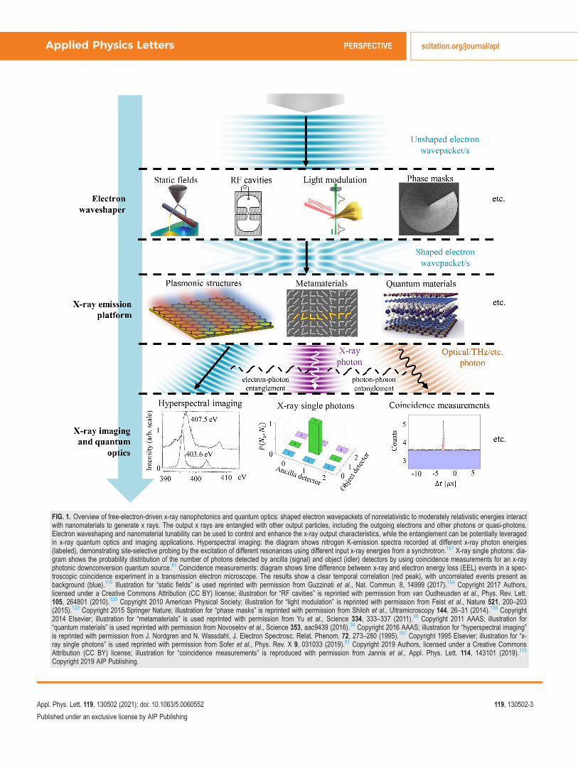
FIG. 1. Overview of free-electron-driven x-ray nanophotonics and quantum optics: shaped electron wavepackets of nonrelativistic to moderately relativistic energies interactwith nanomaterials to generate x rays. The output x rays are entangled with other output particles, including the outgoing electrons and other photons or quasi-photons.Electron waveshaping and nanomaterial tunability can be used to control and enhance the x-ray output characteristics, while the entanglement can be potentially leveragedin x-ray quantum optics and imaging applications. Hyperspectral imaging: the diagram shows nitrogen K-emission spectra recorded at different x-ray photon energies(labeled), demonstrating site-selective probing by the excitation of different resonances using different input x-ray energies from a synchrotron.157 X-ray single photons: dia-gram shows the probability distribution of the number of photons detected by ancilla (signal) and object (idler) detectors by using coincidence measurements for an x-rayphotonic downconversion quantum source.81 Coincidence measurements: diagram shows time difference between x-ray and electron energy loss (EEL) events in a spec-troscopic coincidence experiment in a transmission electron microscope. The results show a clear temporal correlation (red peak), with uncorrelated events present asbackground (blue).115 Illustration for “static fields” is used reprinted with permission from Guzzinati et al., Nat. Commun. 8, 14999 (2017).154 Copyright 2017 Authors,licensed under a Creative Commons Attribution (CC BY) license; illustration for “RF cavities” is reprinted with permission from van Oudheusden et al., Phys. Rev. Lett.105, 264801 (2010).120 Copyright 2010 American Physical Society; illustration for “light modulation” is reprinted with permission from Feist et al., Nature 521, 200–203(2015).122 Copyright 2015 Springer Nature; illustration for “phase masks” is reprinted with permission from Shiloh et al., Ultramicroscopy 144, 26–31 (2014).136 Copyright2014 Elsevier; illustration for “metamaterials” is used reprinted with permission from Yu et al., Science 334, 333–337 (2011).36 Copyright 2011 AAAS; illustration for“quantum materials” is used reprinted with permission from Novoselov et al., Science 353, aac9439 (2016).34 Copyright 2016 AAAS; illustration for “hyperspectral imaging”is reprinted with permission from J. Nordgren and N. Wassdahl, J. Electron Spectrosc. Relat. Phenom. 72, 273–280 (1995).157 Copyright 1995 Elsevier; illustration for “x-ray single photons” is used reprinted with permission from Sofer et al., Phys. Rev. X 9, 031033 (2019).81 Copyright 2019 Authors, licensed under a Creative CommonsAttribution (CC BY) license; illustration for “coincidence measurements” is reproduced with permission from Jannis et al., Appl. Phys. Lett. 114, 143101 (2019).115
Copyright 2019 AIP Publishing.
Applied Physics Letters PERSPECTIVE scitation.org/journal/apl
Appl. Phys. Lett. 119, 130502 (2021); doi: 10.1063/5.0060552 119, 130502-3
Published under an exclusive license by AIP Publishing

FIG. 2. Nanophotonic x-ray emission platforms. (a) Inverse Compton scattering in a Bragg waveguide, which increases the interaction length and leads to x-ray intensityenhancements of over two orders of magnitude compared to a free space laser beam; (b) dielectric laser undulator in which an incident laser beam wiggles the input electrons,causing them to emit x rays; (c) graphene plasmon-based x-ray source (left), which leverages the high confinement factor of graphene plasmons to generate tunable, highlydirectional hard x rays from moderately relativistic electrons (the right diagram shows the output x-ray spectral intensity for 3.7 MeV electrons interacting with graphene plas-mons of free space wavelength 1.5l m and confinement factor 180, over an interaction length of 1.5l m); (d) plasmonic metasurfaces (graphene nanoribbons in this specificexample) as a versatile source of multi-harmonic x rays, whose spectral content can be controlled by tailoring the near-field through the metasurface design; (e) plasmonicmetamaterials as a means of increasing the interaction volume of electron–plasmon interaction, allowing x-ray intensity enhancements of over 100 times; (f) van der Waalsmaterials as a platform for tunable, high-brightness soft x-ray generation (left), demonstrating the concept of x-ray tunability via material design on the atomic scale. The rightdiagram shows the output x-ray spectral intensity for different incident electron energies (labeled) on WSe2, with insets showing the sample image (top right first), diffractionpattern (top right second), and a 3D model for WSe2 (bottom right). (a) Figure reprinted with permission from Karagodsky et al., Phys. Rev. Lett. 104, 024801 (2010).48
Copyright 2010 American Physical Society; (b) used reprinted with permission from T. Plettner and R. L. Byer, Phys. Rev. Spec. Top.-Accel. Beams 11, 030704 (2008).49
Copyright 2008 Authors, licensed under a Creative Commons Attribution (CC BY) license; (c) reprinted with permission from Wong et al., Nat. Photonics 10, 46–52 (2016).54
Copyright 2016 Springer Nature; (d) used reprinted with permission from Rosolen et al., Light 7, 64 (2018).55 Copyright 2018 Authors, licensed under a Creative CommonsAttribution (CC BY) license; (e) used reprinted with permission from Pizzi et al., Adv. Sci. 7, 1901609 (2020).57 Copyright 2020 Authors, licensed under a Creative CommonsAttribution (CC BY) license; (f) reprinted with permission from Shentcis et al., Nat. Photonics 14, 686–692 (2020).62 Copyright 2020 Springer Nature.
Applied Physics Letters PERSPECTIVE scitation.org/journal/apl
Appl. Phys. Lett. 119, 130502 (2021); doi: 10.1063/5.0060552 119, 130502-4
Published under an exclusive license by AIP Publishing

polariton-based x-ray generation schemes, using terahertz-driven sur-face plasmons, has been proposed.56
The use of plasmonic metamaterials, such as graphene metama-terials [Fig. 2(e)], provides a means of scaling up the x-ray outputintensity of polariton-based undulators by over two orders of magni-tude, by supporting a larger interaction volume with the electronbeam.57 The graphene-metamaterial-based x-ray source also introdu-ces a unique paradigm for free-electron light sources where the elec-tron mean free path is longer than the device length, relaxing theconventional requirement that electrons must travel in vacuum toavoid beam degradation through scattering events.57
X rays can also be generated in nanocrystals, such as carbonnanotubes, through free-electron-based processes like channeling radi-ation, coherent bremsstrahlung (CB), parametric x-ray radiation(PXR), and nanotube undulator radiation.58 Nanocrystals like carbonnanotube ropes have advantages as x-ray sources through their rela-tively low x-ray absorption—allowing for more intense x-ray output—and more stable channeling, as compared to ordinary crystals.58
The concept of tunable x-ray generation by controlling materialresponse takes on an even more exciting aspect when applied to vdWmaterials, whose isolated atomic planes can be reassembled intodesigner heterostructures made layer by layer in a precisely chosensequence59 [Fig. 2(f)]. Emerging types of polaritons and innovativemethods of controlling them in vdW structures60,61 imply additionalways to design versatile, compact free-electron polariton-based x-raysources. The versatility of vdW structure design also extends to x-raygeneration processes based directly on the crystal lattice itself, such asparametric x-ray radiation and coherent bremsstrahlung. Recentexperiments62 have demonstrated the ability of van der Waals materi-als to serve as a platform for tunable x-ray generation when irradiatedby moderately relativistic electrons from a transmission electronmicroscope. The radiation spectrum can be precisely controlled bytuning the acceleration voltage of the incident electrons as well asadjusting the lattice structure of the van der Waals material. Thisexperiment, thus, demonstrated the concept of material design at theatomic level, where the wealth of possibilities in designing vdW mate-rials and other atomic superlattices opens up further areas of explora-tion in x-ray physics and technologies.62 The appeal of using vdWmaterials for x-ray generation is enhanced by the fact that many vdWmaterials possess high in-plane thermal conductivities,63 and somehave higher melting temperatures compared to conventional materi-als. Moreover, radiation damage can be further reduced by using het-erostructures combining different kinds of vdW materials.64,65 It hasalso been shown that the PXR mechanism in vdW materials can beparticularly designed to generate ultrashort pulses and delta-pulsetrains with a controllable period, angular distribution, andpolarizability.66
With many of these x-ray radiation stages occupying a chip-scalefootprint, the application of micro-electro-mechanical-system(MEMS) is an intriguing prospect for creating highly versatile, com-pact x-ray sources for fluoroscopy and imaging. MEMS has alreadybeen used to realize dynamic x-ray optics that can manipulate hard x-ray pulses on time scales down to 300 ps,67 within an order-of-magni-tude of the x-ray pulse duration from many standard synchrotronsources.
The burgeoning potential of x-ray free-electron nanophotonicscontinues to grow with the emerging materials and methods of
controlling electromagnetic fields in materials. The ability to realizehighly confined magnetic fields at the surface of nanopatterned ferro-magnets,68 for instance, has motivated the investigation of ferromag-netic nanograting x-ray undulators.69 Further possibilities remain tobe discovered, for instance, in the magnetic and topological propertiesof natural vdW crystals70 and their heterostructures, and in the exoticphysics of one-dimensional (1D) vdW heterostructures.71 The freeelectron is unique as a robust and powerful means of bridging the gapbetween control of physics at DC-to-optical wavelengths and controlof physics at extreme frequencies like the x-ray regime.
III. FREE-ELECTRON-DRIVEN EMISSIONOF ENTANGLED PHOTON–PHOTON ANDELECTRON–PHOTON PAIRS FOR X-RAYQUANTUM OPTICS
Quantum optics has been applied to improve the quality of mea-surements over what is possible with classical illumination, throughmethods like quantum imaging and quantum metrology.72–75
However, these methods have largely been limited to parts of the elec-tromagnetic spectrum far below the x-ray regime.
Extending quantum optics into the x-ray range has been arguedto be beneficial from both a fundamental and practical point of view.From the viewpoint of fundamental quantum optics, detectors in thex-ray range are capable of resolving the number of detected photonswith nominally zero background noise and a broadband unity quan-tum efficiency. Such capabilities are beyond the reach of quantumoptics in the optical range and could, thus, enable basic tests of funda-mental concepts in quantum information such as Bell-type inequal-ities,76 ideas like ghost imaging,77 and the creation of multi-photonFock states using efficient post-selection.78,79 Quantum imaging withincoherently scattered x rays has been demonstrated by leveraginghigher-order degrees of coherence.80
From the viewpoint of x-ray science and applications, the intro-duction of quantum-optical concepts can help access undiscoveredatomic-scale phenomena using the extremely short photon x-raywavelengths.81 X rays have been shown to be an enabling tool fornuclear quantum optics as well as studies of collective and virtualeffects in the interaction of identical atoms with single photons.82,83
For instance, it has been shown that the dynamics of M€ossbauer nucleican be coherently controlled using an x-ray double-pulse,84 and thatone-photon superradiance and Dicke state superradiance can beachieved using an ensemble of nuclei.85–88 An ensemble of nuclei canalso be used to shape the waveform of gamma-ray photons coherently,providing a source of single-photon, ultrashort gamma ray pulses withcontrollable waveforms.89
In the field of x-ray quantum optics, correlated x-ray photonswere recently demonstrated using x-ray downconversion.77,81,90–94
The mechanism has been used as a source for ghost imaging77 andshown to generate heralded photons with sub-Poissonian statis-tics.81 Instead of two entangled x-ray photons, spontaneous para-metric downconversion can also be used to produce an entangledphoton pair consisting of an x-ray photon and an opticalphoton.95–99
Such x-ray sources have substantial improvements in visibilityand signal-to-noise ratio in coincidence detection schemes involvingthe two entangled x-ray photons.81 Efficient interaction of heralded x-ray photons with beam splitters was recently demonstrated, opening
Applied Physics Letters PERSPECTIVE scitation.org/journal/apl
Appl. Phys. Lett. 119, 130502 (2021); doi: 10.1063/5.0060552 119, 130502-5
Published under an exclusive license by AIP Publishing

the doors to innovative setups for x-ray quantum optics that involvebeam splitters and single photon interactions.100 Nevertheless, all thesestudies used another photon for the post-selection, which meant thatthe efficiency of the coincidence events was limited by the efficiency ofthe nonlinear process, and the efficiency of the detection of the otherphoton. Correlating the x-ray photon and the electron could lead to amore efficient process and benefit from the higher efficiency of elec-tron detection.
It is noteworthy that quantum vacuum fluctuations can be usedto produce x rays from free electrons through Casimir-type forces thatundulate the electrons. For example, polariton-based free-electronx-ray generation is possible even in the absence of externally inducedpolaritons. Instead, the electromagnetic vacuum fluctuations (e.g., ofpolariton modes) that exist in the nanoscale vicinity of materials101
can interact with the electron and lead to x-ray generation [Fig. 3(a)].The interaction of these fluctuations with free electrons is equivalentto a quantum optical two-photon process in which a free electronspontaneously emits an entangled pair of a low-energy polariton and ahigh-energy photon. The radiated power is comparable with that froman equal-energy electron in an external magnetic field of strength onthe order of 1T. The strength of this x-ray generation process is relatedto the strong Casimir–Polder forces that atoms experience in thenanometer vicinity of materials, with the essential difference beingthat the fluctuating force here acts on free electrons, instead of neutral,polarizable atoms. The resulting x rays can be potentially shaped bycontrolling the nanophotonic geometry or the underlying materialelectromagnetic response at optical or infrared frequencies.101 Thismeans that concepts from nanophotonics that are used to shape thelocal photonic density of states in the optical spectrum (using metasur-faces, etc.) can affect the physics in the x-ray spectrum as well. Forexample, photonic crystals can be used to manipulate the local densityof photonic states in the optical range, to indirectly control the more-elusive local density of photonic states in the x-ray range.102
Free-electron-driven methods of generating x rays have an inher-ent advantage for applications in quantum optics because they involvemore than one output particle: the emitted x-ray photon is emitted ina joint entangled state with the emitted electron. The output particlesare entangled to one another due to energy-momentum conservationlaws, restricting the possible energy and direction of one particle whenthose of the other particle are known. Free-electron x-ray generationis, thus, a potential means of realizing detection schemes based onelectron–photon entanglement at x-ray wavelengths. For example, itwould be possible to realize heralded single-photon x-ray sourcesbased on the electron measurement [Fig. 3(b)].
Let us discuss what are the most promising experimental plat-forms for exploring x-ray quantum optics. All existing studies of x-rayquantum optics have involved either x-ray tubes, which are neithertunable nor directional, or large-scale facilities like synchrotrons,which are less accessible than lab-scale setups. A free-electron-basednanophotonic x-ray source (Sec. II, Fig. 2)—such as the vdW-basedx-ray source62 [Fig. 2(e)]—could be highly complementary to bothtechnologies, as it is not only lab-scale but also tunable and directional.It is advantageous that the brightness achieved by the vdW-basedx-ray source can potentially surpass that from x-ray tubes.62 The useof pulsed electrons (and ultrafast plasmon pulses in the case ofplasmon-driven free-electron x-ray sources) opens up the possibilityof sub-picosecond x-ray pulses from these nanophotonic schemes.
The high-brightness, dynamically tunable and highly directionalnature of free-electron-based nanophotonic x-ray sources also makethem promising for generating x-ray single-photons that are heraldedby measuring the electron energy loss.
The transmission electron microscope (TEM) is an excellent plat-form to explore quantum optical processes with free electrons. This isbecause it is designed to generate electron waves of high spatial coher-ence and sometimes also temporal coherence. Furthermore, it can usu-ally be equipped with multiple detectors for coincidence detection ofoutput electrons and photons. TEMs have been mainstays of scientificprogress since their invention exists almost a hundred years ago. Anelectron incident on a material leads to inelastic excitation processesthat remove energy from the electron and excite a photonic quasiparti-cle or another collective mode in the material (e.g., phonons, plas-mons, excitons, etc.).103 Some of these excitations later emit a photonthrough recombination (e.g., inner-shell excitation and de-excitationemit x-ray photons, while outer-shell electron–hole recombinationemits optical photons). This process is generally known as cathodolu-minescence,104 with the cases of x-ray emission more commonly stud-ied as part of energy dispersive x-ray spectroscopy (EDS).105 Theelectron energy loss and photon emission can be quantitatively mea-sured in order to characterize a material, through techniques like elec-tron energy loss spectroscopy (EELS), detecting the energy loss ofaccelerated electrons,106–108 and EDS, detecting x rays.109–111. In mod-ern TEM instruments, both EELS and EDS can be used to analyze thesample simultaneously.112,113 Previous works,114 including importantrecent advances,115 show that exploiting the intrinsic coupling betweenthe EELS and EDS signals in coincidence measurements can providespectroscopic information with a significantly suppressed background.Such coincidence measurements are the first to use the entanglementbetween the free electron and the excitation it creates, which has manymore promising prospects that exploit quantum correlations in elec-tron microscopy.116 A coincidence measurement setup based onentangled electron–photon pairs emitted from a vdW x-ray source,and realized in a TEM, is envisioned in Fig. 3(c).
IV. SHAPING FREE-ELECTRON WAVEPACKETSFOR CONTROLLABLE X-RAY GENERATION
The growing wealth of techniques to shape the spatiotemporal pro-file of an electron pulse provides undiscovered opportunities to controlx-ray generation from these electrons. These techniques include bothclassical shaping of the electron charge and current distribution andquantum shaping of single electron wavepackets: using static fields,117,118
radio frequency cavities,119,120 laser pulses,121–124 and material struc-tures,125 achieving spatial (temporal) shaping down to the picometerlength- (attosecond time-) scale. The quantum wavepacket shaping ofsingle electrons enables control over properties such as orbital angularmomentum (OAM),126,127 spin angular momentum,128,129 and propaga-tion trajectory.130,131 Such capabilities have been achieved via break-throughs in manipulating the phase-front of electron wavepackets132,133
using amplitude and phase holograms,134–136 nanoscale magneticneedles,137 and electron–photon interactions.138
These advances in electron waveshaping techniques raise the fun-damental question of whether quantum electrodynamical (QED) inter-actions (e.g., light emission) can be controlled via electron waveshaping.Schr€odinger first interpreted the quantum wavefunction as the smoothcharge density of a smeared-out particle.139 Contradictions arising from
Applied Physics Letters PERSPECTIVE scitation.org/journal/apl
Appl. Phys. Lett. 119, 130502 (2021); doi: 10.1063/5.0060552 119, 130502-6
Published under an exclusive license by AIP Publishing

FIG. 3. Free-electron-driven x-ray quantum optics: emission of entangled photon–photon pairs (a) and electron–photon pairs (b) and (c). In (a), nanophotonic vacuum fluctua-tions give rise to a spontaneous two-photon emission process in which the outgoing infrared/optical plasmon and the outgoing x-ray photon are entangled (left). The diagramon the right shows the emitted photon power per unit photon frequency and plasmon frequency from an electron of velocity 0.99c traveling 5 nm above the surface of a gra-phene sheet, doped to a Fermi energy of 0.5 eV. In (b), collisional inner shell ionization (top left inset) is used to generate entangled electron–photon pairs used in coincidencemeasurements to significantly enhance the measured signal (left). The diagram on the right shows a histogram of the time difference between x-ray and electron energy loss(EEL) events showing a clear temporal correlation (red peak), with uncorrelated events present as background (blue). The width of the coincidence peak is shown in the inset.(c) A proposed setup for performing coincidence measurements using entangled electron–photon pairs, generated in this specific example via parametric x-ray radiation and/orcoherent bremsstrhalung in van der Waals materials as per the scheme in Fig. 2(f). (a) Reprinted with permission from Rivera et al., Nat. Phys. 15, 1284–1289 (2019).101
Copyright 2019 Springer Nature; (b) reproduced with permission from Jannis et al., Appl. Phys. Lett. 114, 143101 (2019).115 Copyright 2019 AIP Publishing.
Applied Physics Letters PERSPECTIVE scitation.org/journal/apl
Appl. Phys. Lett. 119, 130502 (2021); doi: 10.1063/5.0060552 119, 130502-7
Published under an exclusive license by AIP Publishing

this view eventually led to the wavefunction being interpreted as theprobability density of a point particle:140 as Feynman put it, “the elec-tron is either here, or there, or somewhere else, but wherever it is, it is apoint charge.” 141 On the one hand, this has led to an overall under-standing that only higher order correlation measurements (rather thanthe intensity) would be affected by the wavefunction of the emitter.142
On the other hand, it had been observed that an electron behavesexactly like a smooth charge density in stimulated emission processes,which have been shown to depend on the waveshape of the emittingelectron in both experiment143 and semiclassical theory.144–146 This hasled to much interest in the relation of emission intensity to the shape ofthe electron wavefunction.147–152
Through a fully quantum theory, we showed in a recent work thatelectron waveshaping can affect the emitted radiation153 [Fig. 4(a)].This surprising result occurs when different contributions to an emitted
photon state are entangled to the same outgoing electron state. Weapplied our concept to bremsstrahlung, showing that it is possible tocontrol this process of spontaneous emission from a free electronthrough the quantum interference resulting from electron waveshaping.Specifically, we show that free-electron waveshaping can be used to tai-lor both the spatial and the spectral distribution of the radiated photons,enhancing the directionality, monochromaticity, and versatility of pho-ton emission compared to the unshaped case for both atomic andundulator bremsstrahlung. This theoretical concept agrees with theobservations of an earlier work, where it was demonstrated theoreticallyand experimentally that using post-selection of the electron, spontane-ous emission into near-field modes (rather than radiation) can dependon the symmetry of the initial electron wavefunction.154
Our study also revealed why such possibilities have not been seenbefore. For example, a recent experiment showed no dependence on
FIG. 4. Shaping free-electron quantumwavepackets for controlling radiation. (a)Shaping the single-electron wavefunctioncan be used to enhance the directionality ofoutput x rays comparing the (i) unshapedand the (ii) shaped scenarios. (b) Multi-electron entangled states create radiationpatterns that cannot be created from classi-cal electrons. Illustration comparing the (i)classical (unentangled) and (ii) entangledscenarios. In particular, entangled electronstates can create quantum superradiance,and subradiance is possible, significantlyaffecting the (iii) transverse profile and (iv)spectrum of the output radiation. (a) Usedreproduced with permission from Wonget al., Nat. Commun. 12, 1700 (2021).153
Copyright 2021 Authors, licensed under aCreative Commons Attribution (CC BY)license; (b) Figure reprinted with permissionfrom Karnieli et al., Phys. Rev. Lett. 127,060403 (2021).149 Copyright 2021 AmericanPhysical Society.
Applied Physics Letters PERSPECTIVE scitation.org/journal/apl
Appl. Phys. Lett. 119, 130502 (2021); doi: 10.1063/5.0060552 119, 130502-8
Published under an exclusive license by AIP Publishing

wavefunction for Smith–Purcell radiation.155 The reason for nowavefunction-dependence was that the contributions to the emissionfrom different initial electron angles could not interfere because eachphoton state was entangled to a different outgoing electron state.Similarly, work that considered shaping Cherenkov radiation throughthe orbital angular momentum (OAM) of electrons127 found nochange to the power spectrum, unless the outgoing electron was post-selected. We attribute these spontaneous emission results to the elec-tron behaving ultimately as a point-like particle (as nicely put byFeynman141) regardless of its wavefunction.
Combining electron waveshaping with free-electron nanopho-tonics concepts (Sec. II) suggests intriguing possibilities like tailoringthe electron wavepacket periodicity to match with the atomic lattice ofthe nanomaterial. Significant modifications to the output radiation canalso be achieved by using multiple entangled input electrons149
[Fig. 4(b)]. The ability to tailor the spatiotemporal attributes of photonemission via quantum interference, thus, provides additional degreesof freedom for shaping x rays, creating exciting prospects for undis-covered x-ray physics. As a future prospect, electron beam shapingcan potentially be used to suppress bremsstrahlung processes, leadingto reduction in background noise and improving measurements ofcharacteristic peaks. This would, in turn, improve contrast in x-rayspectroscopy techniques like x-ray resonance fluoroscopy.
V. OUTLOOK
X-ray sources based on free-electron nanophotonics are promis-ing for applications that require high-brightness, dynamically tunable,monochromatic, and highly directional x rays. Using one of more ofthe directions above promises additional degrees of freedom for con-trolling different aspects of x-ray generation, even including the x-rayphoton polarization. Traditional x-ray tubes fall short in these aspectsdue to lack of tunability—the output x-ray spectral peaks depend onthe anode material instead of on electron energy, and the emission isisotropic and unpolarized.
In soft x-ray fluorescence spectroscopy, tunable energy excitationwould allow one to investigate resonant phenomena and identifymulti-electron excitations leading to x-ray satellite structures.156
Furthermore, tunable x-ray photons enable site selectivity with respectto identical atomic species in different chemical environments sincethe x-ray photon energy can be chosen, so that a core electron is pro-moted to an unoccupied state with a strong localization on a particularsite.157 By leveraging the well-defined polarization of nanophotonic x-ray sources and detecting the x-ray emission with angular selectivity,one can obtain further information on bonding geometry, etc., of thesample.158 These prospects are especially important in techniques suchas ultrafast x-ray spectroscopy and phase contrast imaging that com-monly take place in x-ray facilities and would benefit from a morecompact and accessible source. It should be noted that phase contrastimaging has been achieved using conventional x-ray tubes with theuse of spatially varying masks, for instance through Talbot-Lau inter-ferometry159,160 and edge illumination.161,162 However, the presence ofabsorbing masks reduces x-ray flux, demands careful alignment andstability throughout a clinical examination, and requires precise fabri-cation of the masks, which are usually made of high-Z materials.These last two conditions are supposedly the most critical issues thathave hampered the widespread use of these techniques in the clinicalcontext.163
In free-electron-based x-ray generation processes where morethan one electron is involved, coherence in the output x-ray radiationcan be achieved via the structuring of the electron distribution. Forinstance, in x-ray free-electron lasers, the electron bunch co-propagates and interacts with the emitted x rays over long distances,leading to self-structuring of the electron bunch into sub-wavelengthnanobunches. These subwavelength structures in the electron densityare directly responsible for x-ray intensities that scale with N2 insteadof N164 (N being the number of electrons). This process is known asself-amplified spontaneous emission (SASE).
In addition to self-structuring techniques, the electron wave-packet can also be structured by external processes117–124 before beingmade to radiate x rays. A coherent, high-brightness x-ray source ispotentially useful for many phase-dependent imaging techniques, suchas coherent diffraction imaging and ptychography.165 A recent workhas shown that nanophotonic structures can be used to realize lasersbased on stimulated emission by free electrons, in a process analogousto SASE, but on the nanoscale.166 The associated threshold beam cur-rents are in the nanoampere range and could be realized in electronmicroscopes. However, the work considers emission at infrared wave-lengths, raising the exciting question of whether the scheme can bescaled to x-ray photon energies.
In conclusion, we have pointed out innovative research directionsin x-ray science that open up unique opportunities for future x-raytechnology and recently became more accessible due to advances innanomaterials and quantum optics. We have described how the highlynonlinear and spectrally broadband nature of the free electron allowsus to leverage the versatility of nanophotonics for the manipulationand enhancement of x-ray emission. We have identified three mainthemes that we believe to be of special interest: the study of emergingmaterials to serve as nanophotonic platforms for compact, tunable,and high-brightness x-ray generation; the quantum shaping of elec-tron wavefunctions to control the spatiotemporal profile of emitted xrays; and the entanglement of output electrons and photons for coinci-dence measurement schemes. We hope that this Perspective hashelped highlight the prospects for future research and will help drivefuture discoveries in the field.
ACKNOWLEDGMENTS
This work was supported by the Israel Science Foundation(ISF, Grant No. 830/19), the Agency for Science, Technology andResearch (A�STAR) Science & Engineering Research Council(Grant No. A1984c0043), and the Binational USA-Israel ScienceFoundation (BSF, Grant No. 2018288). L.J.W. acknowledges thesupport of the Nanyang Assistant Professorship Start-up Grant.The authors thank Lee Wei Wesley Wong for his help on logisticsassociated with the manuscript preparation.
DATA AVAILABILITY
Data sharing is not applicable to this article as no new data werecreated or analyzed in this study.
REFERENCES1W. C. R€ontgen, “On a new kind of rays,” Science 3, 227–231 (1896).2S. Galli, “X-ray crystallography: One century of Nobel Prizes,” J. Chem. Educ.91, 2009–2012 (2014).
Applied Physics Letters PERSPECTIVE scitation.org/journal/apl
Appl. Phys. Lett. 119, 130502 (2021); doi: 10.1063/5.0060552 119, 130502-9
Published under an exclusive license by AIP Publishing

3C. Pellegrini, A. Marinelli, and S. Reiche, “The physics of x-ray free-electronlasers,” Rev. Mod. Phys. 88, 015006 (2016).
4B. W. J. McNeil and N. R. Thompson, “X-ray free-electron lasers,” Nat.Photonics 4, 814 (2010).
5Z. Huang and K.-J. Kim, “Review of x-ray free-electron laser theory,” Phys.Rev. Spec. Top.-Accel. Beams 10, 034801 (2007).
6L. Young, K. Ueda, M. G€uhr et al., “Roadmap of ultrafast x-ray atomic andmolecular physics,” J. Phys. B 51, 032003 (2018).
7M. Endrizzi, “X-ray phase-contrast imaging,” Nucl. Instrum. Methods Phys.Res., Sect. A 878, 88–98 (2018).
8A. Bravin, P. Coan, and P. Suortti, “X-ray phase-contrast imaging: Frompre-clinical applications towards clinics,” Phys. Med. Biol. 58, R1–R35(2013).
9H. Labriet, C. Nemoz, M. Renier et al., “Significant dose reduction using syn-chrotron radiation computed tomography: First clinical case and applicationto high resolution CT exams,” Sci. Rep. 8, 12491 (2018).
10M. Holler, M. Guizar-Sicairos, E. H. R. Tsai, R. Dinapoli, E. M€uller, O. Bunk,J. Raabe, and G. Aeppli, “High-resolution non-destructive three-dimensionalimaging of integrated circuits,” Nature 543, 402–406 (2017).
11M. Kordel, A. Dehlinger, C. Seim, U. Vogt, E. Fogelqvist, J. A. Sellberg, H.Stiel, and H. M. Hertz, “Laboratory water-window x-ray microscopy,” Optica7, 658 (2020).
12F. V. Hartemann, W. J. Brown, D. J. Gibson et al., “High-energy scaling ofCompton scattering light sources,” Phys. Rev. Spec. Top.-Accel. Beams 8,100702 (2005).
13A. J. Gonsalves, K. Nakamura, J. Daniels et al., “Petawatt laser guiding andelectron beam acceleration to 8 GeV in a laser-heated capillary dischargewaveguide,” Phys. Rev. Lett. 122, 084801 (2019).
14C. Caizergues, S. Smartsev, V. Malka, and C. Thaury, “Phase-locked laser-wakefield electron acceleration,” Nat. Photonics 14, 475–479 (2020).
15E. Esarey, C. B. Schroeder, and W. P. Leemans, “Physics of laser-drivenplasma-based electron accelerators,” Rev. Mod. Phys. 81, 1229 (2009).
16R. J. England, R. J. Noble, K. Bane et al., “Dielectric laser accelerators,” Rev.Mod. Phys. 86, 1337 (2014).
17J. Breuer and P. Hommelhoff, “Laser-based acceleration of nonrelativistic elec-trons at a dielectric structure,” Phys. Rev. Lett. 111, 134803 (2013).
18E. A. Peralta, K. Soong, R. J. England et al., “Demonstration of electron accel-eration in a laser-driven dielectric microstructure,” Nature 503, 91–94 (2013).
19N. V. Sapra, K. Y. Yang, D. Vercruysse et al., “On-chip integrated laser-drivenparticle accelerator,” Science 367, 79–83 (2020).
20D. Zhang, A. Fallahi, M. Hemmer et al., “Segmented terahertz electron accel-erator and manipulator (STEAM),” Nat. Photonics 12, 336–342 (2018).
21L. J. Wong, A. Fallahi, and F. X. K€artner, “Compact electron acceleration andbunch compression in THz waveguides,” Opt. Express 21, 9792–9806 (2013).
22F. Albert and A. G. R. Thomas, “Applications of laser wakefield accelerator-based light sources,” Plasma Phys. Controlled Fusion 58, 103001 (2016).
23S. Corde, K. T. Phuoc, G. Lambert, R. Fitour, V. Malka, and A. Rousse,“Femtosecond x rays from laser-plasma accelerators,” Rev. Mod. Phys. 85, 1(2013).
24I. Gadjev, N. Sudar, M. Babzien et al., “An inverse free electron laseracceleration-driven Compton scattering x-ray source,” Sci. Rep. 9, 532 (2019).
25W. S. Graves, J. Bessuille, P. Brown et al., “Compact x-ray source based onburst-mode inverse Compton scattering at 100 kHz,” Phys. Rev. Spec. Top.-Accel. Beams 17, 120701 (2014).
26W. S. Graves, F. X. K€artner, D. E. Moncton, and P. Piot, “Intense superradiantx rays from a compact source using a nanocathode array and emittanceexchange,” Phys. Rev. Lett. 108, 263904 (2012).
27J. Li, J. Lu, A. Chew et al., “Attosecond science based on high harmonic gener-ation from gases and solids,” Nat. Commun. 11, 2748 (2020).
28R. Geneaux, H. J. B. Marroux, A. Guggenmos, D. M. Neumark, and S. R.Leone, “Transient absorption spectroscopy using high harmonic generation:A review of ultrafast x-ray dynamics in molecules and solids,” Philos. Trans.R. Soc. A 377, 20170463 (2019).
29J. Weisshaupt, V. Juv�e, M. Holtz et al., “High-brightness table-top hard x-raysource driven by sub-100-femtosecond mid-infrared pulses,” Nat. Photonics8, 927–930 (2014).
30M. Gambari, R. Clady, A. Stolidi, O. Ut�eza, M. Sentis, and A. Ferr�e,“Exploring phase contrast imaging with a laser-based Ka x-ray source up torelativistic laser intensity,” Sci. Rep. 10, 6766 (2020).
31R. J. Parmee, C. M. Collins, W. I. Milne, and M. T. Cole, “X-ray generationusing carbon nanotubes,” Nano Convergence 2, 1 (2015).
32C. G. Camara, J. V. Escobar, J. R. Hird, and S. J. Putterman, “Correlationbetween nanosecond x-ray flashes and stick–slip friction in peeling tape,”Nature 455, 1089 (2008).
33M. Jablan, H. Buljan, and M. Soljacic, “Plasmonics in graphene at infraredfrequencies,” Phys. Rev. B 80, 245435 (2009).
34K. S. Novoselov, A. Mishchenko, A. Carvalho, and A. H. Castro Neto, “2Dmaterials and van der Waals heterostructures,” Science 353, aac9439 (2016).
35S. Jahani and Z. Jacob, “All-dielectric metamaterials,” Nat. Nanotechnol. 11,23 (2016).
36N. F. Yu, P. Genevet, M. A. Kats, F. Aieta, J. P. Tetienne, F. Capasso, and Z.Gaburro, “Light propagation with phase discontinuities: Generalized laws ofreflection and refraction,” Science 334, 333–337 (2011).
37M. C. Rechtsman, J. M. Zeuner, Y. Plotnik et al., “Photonic Floquet topologi-cal insulators,” Nature 496, 196–200 (2013).
38Z. Wang, Y. Chong, J. D. Joannopoulos, and M. Soljacic, “Observation of uni-directional backscattering-immune topological electromagnetic states,”Nature 461, 772–775 (2009).
39L. Lu, J. D. Joannopoulos, and M. Soljacic, “Topological photonics,” Nat.Photonics 8, 821–829 (2014).
40T. Ozawa, H. M. Price, A. Amo et al., “Toplogical photonics,” Rev. Mod.Phys. 91, 015006 (2019).
41N. P. Armitage, E. J. Mele, and A. Vishnawath, “Weyl and Dirac semimetalsin three-dimensional solids,” Rev. Mod. Phys. 90, 015001 (2018).
42M. Pelton, “Modified spontaneous emission in nanophotonic structures,”Nat. Photonics 9, 427 (2015).
43Y. Liang, C. Li, Y.-Z. Huang, and Q. Zhang, “Plasmonic nanolasers in on-chiplight sources: Prospects and challenges,” ACS Nano 14, 14375–�14390(2020).
44H. A. Hafez, S. Kovalev, J. C. Deinert et al., “Extremely efficient terahertzhigh-harmonic generation in graphene by hot Dirac fermions,” Nature 561,507 (2018).
45J. D. Jackson, Classical Electrodynamics, 3rd ed. (John Wiley & Sons, NewYork, 1999).
46H. Schwoerer, B. Liesfeld, H.-P. Schlenvoigt, K.-U. Amthor, and R. Sauerbrey,“Thomson-backscattered x rays from laser-accelerated electrons,” Phys. Rev.Lett. 96, 014802 (2006).
47K. T. Phuoc, S. Corde, C. Thaury et al., “All-optical Compton gamma-raysource,” Nat. Photonics 6, 308–311 (2012).
48V. Karagodsky, D. Schieber, and L. Sch€achter, “Enhancing x-ray generationby electron-beam–laser interaction in an optical Bragg structure,” Phys. Rev.Lett. 104, 024801 (2010).
49T. Plettner and R. L. Byer, “Proposed dielectric-based microstructure laser-driven undulator,” Phys. Rev. Spec. Top.-Accel. Beams 11, 030704 (2008).
50D. Yu. Sergeeva, A. A. Tishchenko, and M. N. Strikhanov, “Conical diffrac-tion effect in optical and x-ray Smith-Purcell radiation,” Phys. Rev. Spec.Top.-Accel. Beams 18, 052801 (2015).
51A. Woessner, M. B. Lundeberg, Y. Gao et al., “Highly confined low-loss plas-mons in graphene–boron nitride heterostructures,” Nat. Mater. 14, 421–425(2015).
52Z. Fei, A. S. Rodin, G. O. Andreev et al., “Gate-tuning of graphene plasmonsrevealed by infrared nanoimaging,” Nature 487, 82–85 (2012).
53J. Chen, M. Badioli, P. Alonso-Gonz�alez et al., “Optical nano-imaging of gate-tunable graphene plasmons,” Nature 487, 77–81 (2012).
54L. J. Wong, I. Kaminer, O. Ilic, J. D. Joannopoulos, and M. Soljacic, “Towardsgraphene plasmon-based free-electron infrared to x-ray sources,” Nat.Photonics 10, 46–52 (2016).
55G. Rosolen, L. J. Wong, N. Rivera, B. Maes, M. Soljacic, and I. Kaminer,“Metasurface-based multi-harmonic free-electron light source,” Light. 7, 64(2018).
56D. Rohrbach, “THz-driven surface plasmon undulator as a compact highlydirectional narrow band incoherent x-ray source,” Phys. Rev. Spec. Top.-Accel. Beams 22, 090702 (2019).
Applied Physics Letters PERSPECTIVE scitation.org/journal/apl
Appl. Phys. Lett. 119, 130502 (2021); doi: 10.1063/5.0060552 119, 130502-10
Published under an exclusive license by AIP Publishing

57A. Pizzi, G. Rosolen, L. J. Wong et al., “Graphene metamaterials for intense,tunable, and compact extreme ultraviolet and x-ray sources,” Adv. Sci. 7,1901609 (2020).
58X. Artru, S. P. Fomin, N. F. Shul’ga, K. A. Ispirian, and N. K. Zhevagod,“Carbon nanotubes and fullerites in high-energy and x-ray physics,” Phys.Rep. 412, 89–189 (2005).
59A. K. Geim and I. V. Grigorievam, “Van der Waals heterostructures,” Nature499, 419 (2013).
60D. N. Basov, M. M. Fogler, and F. J. Garc�ıa de Abajo, “Polaritons in van derWaals materials,” Science 354, aag1992-1 (2016).
61G. Hu, Q. Ou, G. Si et al., “Topological polaritons and photonic magic anglesin twisted a-MoO3 bilayers,” Nature 582, 209 (2020).
62M. Shentcis et al., “Tunable free-electron x-ray radiation from van der Waalsmaterials,” Nat. Photonics 14, 686–692 (2020).
63P. Jiang, X. Qian, X. Gu, and R. Yang, “Probing anisotropic thermal conduc-tivity of transition metal dichalcogenides MX2 (M¼Mo, W and X¼ S, Se)using time-domain thermoreflectance,” Adv. Mater. 29, 1701068 (2017).
64R. Zan, Q. M. Ramasse, R. Jalil, T. Georgiou, U. Bangert, and K. S. Novoselov,“Control of radiation damage in MoS2 by graphene encapsulation,” ACSNano 7, 10167–10174 (2013).
65T. Lehnert, O. Lehtinen, G. Algara-Siller, and U. Kaiser, “Electron radiationdamage mechanisms in 2D MoSe2,” Appl. Phys. Lett. 110, 033106 (2017).
66A. Balanov, A. Gorlach, and I. Kaminer, “Temporal and spatial design of x-ray pulses based on free-electron–crystal interaction,” APL Photonics 6,070803 (2021).
67P. Chen, “Ultrafast photonic micro-systems to manipulate hard x-rays at 300picoseconds,” Nat. Commun. 10, 1158 (2019).
68A. S. Salasyuk, A. V. Rudkovskaya, A. P. Danilov et al., “Generation of a local-ized microwave magnetic field by coherent phonons in a ferromagnetic nano-grating,” Phys. Rev. B 97, 060404(R) (2018).
69S. Fisher, C. Roques-Carmes, N. Rivera, L. J. Wong, I. Kaminer, and M.Soljacic�, “Monochromatic x-ray source based on scattering from a magneticnanoundulator,” ACS Photonics 7, 1096–1103 (2020).
70J. Wu, F. Liu, M. Sasase et al., “Natural van der Waals heterostructural singlecrystals with both magnetic and topological properties,” Sci. Adv. 5, eaax9989(2019).
71R. Xiang, T. Inoue, Y. Zheng et al., “One-dimensional van der Waals hetero-structures,” Science 367, 537–542 (2020).
72G. Brida, M. Genovese, and I. R. Berchera, “Experimental realization of sub-shot-noise quantum imaging,” Nat. Photonics 4, 227 (2010).
73P. A. Morris, R. S. Aspden, J. E. C. Bell, R. W. Boyd, and M. J. Padgett,“Imaging with a small number of photons,” Nat. Commun. 6, 5913 (2015).
74V. Giovannetti, S. Lloyd, and L. Maccone, “Quantum metrology,” Phys. Rev.Lett. 96, 010401 (2006).
75R. Demkowicz-Dobrza�nski, J. Kołody�nski, and M. Gut�a, “The elusiveHeisenberg limit in quantum-enhanced metrology,” Nat. Commun. 3, 1063(2012).
76A. Aspect, “Bell’s inequality test: More ideal than ever,” Nature 398, 189(1999).
77A. Schori, D. Borodin, K. Tamasaku, and S. Shwartz, “Ghost imaging withpaired x-ray photons,” Phys. Rev. A 97, 063804 (2018).
78M. Cooper, L. J. Wright, C. S€oller, and B. J. Smith, “Experimental generationof multi-photon Fock states,” Opt. Express 21, 5309 (2013).
79A. Ourjoumtsev, R. Tualle-Brouri, and P. Grangier, “Quantum homodynetomography of a two-photon Fock state,” Phys. Rev. Lett. 96, 213601 (2006).
80R. Schneider, T. Mehringer, G. Mercurio et al., “Quantum imaging with inco-herently scattered light from a free-electron laser,” Nat. Phys. 14, 126 (2018).
81S. Sofer, E. Strizhevsky, A. Schori, K. Tamasaku, and S. Shwartz, “Quantumenhanced x-ray detection,” Phys. Rev. X 9, 031033 (2019).
82B. W. Adams, C. Buth, S. M. Cavaletto et al., “X-ray quantum optics,” J. Mod.Opt. 60, 2–21 (2013).
83R. R€ohlsberger and J. Evers, “Quantum optical phenomena in nuclear reso-nant scattering,” in Modern M€ossbauer Spectroscopy, Topics in AppliedPhysics Vol. 137, edited by Y. Yoshida and G. Langouche (Springer,Singapore, 2021), p. 105.
84K. P. Heeg, A. Kaldun, C. Strohm et al., “Coherent x-ray-optical control ofnuclear excitons,” Nature 590, 401 (2021).
85R. R€ohlsberger, K. Schlage, and B. Sahoo, “Collective lamb shift in single-photon superradiance,” Science 328, 1248 (2010).
86A. I. Chumakov, A. Q. R. Baron, I. Sergueev et al., “Superradiance of anensemble of nuclei excited by a free electron laser,” Nat. Phys. 14, 261 (2018).
87M. O. Scully, “Collective Lamb shift in single photon Dicke superradiance,”Phys. Rev. Lett. 102, 143601 (2009).
88A. A. Svidzinsky, L. Yuan, and M. O. Scully, “Quantum amplification bysuperradiant emission of radiation,” Phys. Rev. X 3, 041001 (2013).
89F. Vagizov, V. Antonov, Y. V. Radeonychev, R. N. Shakhmuratov, and O.Kocharovskaya, “Coherent control of the waveforms of recoilless c-ray photo-ns,” Nature 508, 80–83 (2014).
90P. M. Eisenberger and S. L. McCall, “X-ray parametric conversion,” Phys.Rev. Lett. 26, 684 (1971).
91Y. Yoda, T. Suzuki, X. W. Zhang, K. Hirano, and S. Kikuta, “X-ray parametricscattering by a diamond crystal,” J. Synchrotron Radiat. 5, 980 (1998).
92Nonlinear Optics, Quantum Optics, and Ultrafast Phenomena with X-Rays,edited by B. W. Adams (Kluwer Academic Publisher, Norwell, MA, 2008).
93S. Shwartz, R. N. Coffee, J. M. Feldkamp, Y. Feng, J. B. Hastings, G. Y. Yin,and S. E. Harris, “X-ray parametric down-conversion in the Langevinregime,” Phys. Rev. Lett. 109, 013602 (2012).
94D. Borodin, A. Schori, F. Zontone, and S. Shwartz, “X-ray photon pairs withhighly suppressed background,” Phys. Rev. A 94, 013843 (2016).
95I. Freund and B. F. Levine, “Optically modulated x-ray diffraction,” Phys.Rev. Lett. 25, 1241–1245 (1970).
96P. M. Eisenberger and S. L. McCall, “Mixing of x-ray and optical photons,”Phys. Rev. A 3, 1145–1151 (1971).
97T. E. Glover, D. M. Fritz, M. Cammarata et al., “X-ray and optical wavemixing,” Nature 488, 603 (2012).
98A. Schori, C. B€omer, D. Borodin et al., “Parametric down-conversion of xrays into the optical regime,” Phys. Rev. Lett. 119, 253902 (2017).
99R. Cohen and S. Shwartz, “Theory of nonlinear interactions between x raysand optical radiation in crystals,” Phys. Rev. Res. 1, 033133 (2019).
100E. Strizhevsky, D. Borodin, A. Schori, S. Francoual, R. R€ohlsberger, and S.Shwartz, “Efficient interaction of heralded x-ray photons with a beamsplitter,” arXiv:2102.01370 (2021).
101N. Rivera, L. J. Wong, J. D. Joannopoulos, M. Soljacic, and I. Kaminer, “Lightemission based on nanophotonic vacuum forces,” Nat. Phys. 15, 1284–1289(2019).
102E. Sendonaris, J. Sloan, N. Rivera, and M. Soljacic, “Optical control of x-rayemission,” in OSA Technical Digest (Optical Society of America, 2020), p.FW4H.4.
103F. J. Garc�ıa de Abajo, “Optical excitations in electron microscopy,” Rev. Mod.Phys. 82, 209 (2010).
104A. Polman, M. Kociak, and F. J. G. de Abajo, “Electron-beam spectroscopy fornanophotonics,” Nat. Mater. 18, 1158–1171 (2019).
105J. Goldstein, Scanning Electron Microscopy and X-Ray Microanalysis(Springer, 2003).
106R. Egerton, Electron Energy-Loss Spectroscopy in the Electron Microscope(Springer US, 2011).
107H. Tan, S. Turner, E. Yucelen, J. Verbeeck, and G. Van Tendeloo, “2D atomicmapping of oxidation states in transition metal oxides by scanning transmis-sion electron microscopy and electron energy-loss spectroscopy,” Phys. Rev.Lett. 107, 107602 (2011).
108H. Tan, J. Verbeeck, A. Abakumov, and G. V. Tendeloo, “Oxidation state andchemical shift investigation in transition metal oxides by EELS,”Ultramicroscopy 116, 24–33 (2012).
109E. V. Cappellen and J. C. Doukhan, “Quantitative transmission x-ray micro-analysis of ionic compounds,” Ultramicroscopy 53, 343–349 (1994).
110D. B. Williams and C. B. Carter, in Transmission Electron Microscopy: ATextbook for Materials Science, edited by D. B. Williams and C. B. Carter(Springer, 2009).
111P. Schlossmacher, D. Klenov, B. Freitag, and H. von Harrach, “Enhanced detec-tion sensitivity with a new windowless XEDS system for AEM based on silicondrift detector technology,” Microsc. Today 18, 14–20 (2010).
112I. Ali, R. D€orner, O. Jagutzki et al., “Multi-hit detector system for completemomentum balance in spectroscopy in molecular fragmentation processes,”Nucl. Instrum. Methods Phys. Res., Sect. B 149, 490–500 (1999).
Applied Physics Letters PERSPECTIVE scitation.org/journal/apl
Appl. Phys. Lett. 119, 130502 (2021); doi: 10.1063/5.0060552 119, 130502-11
Published under an exclusive license by AIP Publishing

113J. Spiegelberg, S. Muto, M. Ohtsuka, K. Pelckmans, and J. Rusz, “Unmixinghyperspectral data by using signal subspace sampling,” Ultramicroscopy 182,205–211 (2017).
114P. Kruit, H. Shuman, and A. Somlyo, “Detection of x-rays and electron energyloss events in time coincidence,” Ultramicroscopy 13, 205–213 (1984).
115D. Jannis, K. Muller-Caspary, A. Beche, A. Oelsner, and J. Verbeeck,“Spectroscopic coincidence experiments in transmission electron microscopy,”Appl. Phys. Lett. 114, 143101 (2019).
116C. Mechel, Y. Kurman, A. Karnieli, N. Rivera, A. Arie, and I. Kaminer,“Quantum correlations in electron microscopy,” Optica 8, 70 (2021).
117D. Kreier, D. Sabonis, and P. Baum, “Alignment of magnetic solenoid lensesfor minimizing temporal distortions,” J. Opt. 16, 075201 (2014).
118J. Zhu, P. Piot, D. Mihalcea, and C. R. Prokop, “Formation of compressed flatelectron beams with high transverse-emittance ratios,” Phys. Rev. Spec. Top.-Accel. Beams 17, 084401 (2014).
119A. Gliserin, M. Walbran, F. Krausz, and P. Baum, “Sub-phonon-period com-pression of electron pulses for atomic diffraction,” Nat. Commun. 6, 8723(2015).
120T. van Oudheusden, P. L. E. M. Pasmans, S. B. van der Geer, M. J. de Loos, M.J. van der Wiel, and O. J. Luiten, “Compression of subrelativistic space-charge-dominated electron bunches for single-shot femtosecond electrondiffraction,” Phys. Rev. Lett. 105, 264801 (2010).
121L. J. Wong, B. Freelon, T. Rohwer, N. Gedik, and S. G. Johnson, “All-opticalthree-dimensional electron pulse compression,” New J. Phys. 17, 013051(2015).
122A. Feist, K. E. Echternkamp, J. Schauss, S. V. Yalunin, S. Sch€afer, and C.Ropers, “Quantum coherent optical phase modulation in an ultrafast trans-mission electron microscope,” Nature 521, 200–203 (2015).
123K. E. Priebe, C. Rathje, S. V. Yalunin, T. Hohage, A. Feist, S. Sch€afer, and C.Ropers, “Attosecond electron pulse trains and quantum state reconstruction inultrafast transmission electron microscopy,” Nat. Photonics 11, 793 (2017).
124M. Kozak, N. Sch€onenberger, and P. Hommelhoff, “Ponderomotive generationand detection of attosecond free-electron pulse trains,” Phys. Rev. Lett. 120,103203 (2018).
125E. A. Nanni, W. S. Graves, and D. E. Moncton, “Nanomodulated electronbeams via electron diffraction and emittance exchange for coherent x-ray gen-eration,” Phys. Rev. Spec. Top.-Accel. Beams 21, 014401 (2018).
126B. J. McMorran, A. Agrawal, I. M. Anderson, A. A. Herzing, H. J. Lezec, J. J.McClelland, and J. Unguris, “Electron vortex beams with high quanta oforbital angular momentum,” Science 331, 192–195 (2011).
127I. Kaminer, M. Mutzafi, A. Levy, G. Harari, H. H. Sheinfux, S. Skirlo, J.Nemirovsky, J. D. Joannopoulos, M. Segev, and M. Soljacic, “QuantumCerenkov radiation: Spectral cutoffs and the role of spin and orbital angularmomentum,” Phys. Rev. X 6, 011006 (2016).
128S. McGregor, R. Bach, and H. Batelaan, “Transverse quantum Stern-Gerlachmagnets for electrons,” New J. Phys. 13, 065018 (2011).
129E. Karimi, L. Marrucci, V. Grillo, and E. Santamato, “Spin-to-orbital angularmomentum conversion and spin-polarization filtering in electron beams,”Phys. Rev. Lett. 108, 044801 (2012).
130N. Voloch-Bloch, Y. Lereah, Y. Lilach, A. Gover, and A. Arie, “Generation ofelectron Airy beams,” Nature 494, 331–335 (2013).
131I. Kaminer, J. Nemirovsky, M. Rechtsman, R. Bekenstein, and M. Segev, “Self-accelerating Dirac particles and prolonging the lifetime of relativisticfermions,” Nat. Phys. 11, 261 (2015).
132M. Uchida and A. Tonomura, “Generation of electron beams carrying orbitalangular momentum,” Nature 464, 737–739 (2010).
133J. Harris, V. Grillo, E. Mafakheri, G. C. Gazzadi, S. Frabboni, R. W. Boyd, andE. Karimi, “Structured quantum waves,” Nat. Phys. 11, 629 (2015).
134J. Verbeeck, H. Tian, and P. Schattschneider, “Production and application ofelectron vortex beams,” Nature 467, 301–304 (2010).
135V. Grillo, G. C. Gazzadi, E. Karimi, E. Mafakheri, R. W. Boyd, and S.Frabboni, “Highly efficient electron vortex beams generated by nanofabricatedphase holograms,” Appl. Phys. Lett. 104, 043109 (2014).
136R. Shiloh, Y. Lereah, Y. Lilach, and A. Arie, “Sculpturing the electronwave function using nanoscale phase masks,” Ultramicroscopy 144, 26–31(2014).
137A. B�ech�e, R. Van Boxem, G. Van Tendeloo, and J. Verbeeck, “Magnetic mono-pole field exposed by electrons,” Nat. Phys. 10, 26–29 (2014).
138A. G. Hayrapetyan, O. Matula, A. Aiello, A. Surzhykov, and S. Fritzsche,“Interaction of relativistic electron-vortex beams with few-cycle laser pulses,”Phys. Rev. Lett. 112, 134801 (2014).
139E. Schr€odinger, “An undulatory theory of the mechanics of atoms and mole-cules,” Phys. Rev. 28, 1049 (1926).
140M. Born, Physics in My Generation, 2nd ed. (Springer, New York,1969).
141R. P. Feynman, R. B. Leighton, and M. Sands, The Feynman Lectures onPhysics, Vol. III, Quantum Mechanics (Addison-Wesley Publishing Company,Reading, MA, 1965), Chap. 21-4.
142A. Karnieli, N. Rivera, A. Arie, and I. Kaminer, “The coherence of light is fun-damentally tied to the quantum coherence of the emitting particle,” Sci. Adv.7, eabf8096 (2021).
143B. Barwick, D. J. Flannigan, and A. H. Zewail, “Photon-induced near-fieldelectron microscopy,” Nature 462, 902 (2009).
144A. Gover and Y. Pan, “Dimension-dependent stimulated radiative interactionof a single electron quantum wavepacket,” Phys. Lett. A 382, 1550–1555(2018).
145Y. Pan and A. Gover, “Spontaneous and stimulated radiative emission of mod-ulated free-electron quantum wavepackets—semiclassical analysis,” J. Phys.Commun. 2, 115026 (2018).
146N. Talebi, “Schr€odinger electrons interacting with optical gratings: Quantummechanical study of the inverse Smith-Purcell effect,” New J. Phys. 18, 123006(2016).
147F. J. Garc�ıa de Abajo and V. D. Giulio, “Optical excitations with elec-tron beams: Challenges and opportunities,” ACS Photonics 8, 945(2021).
148O. Kfir, V. D. Giulio, F. J. Garc�ıa de Abajo, and C. Ropers, “Optical coherencetransfer mediated by free electrons,” Sci. Adv. 7, eabf6380 (2021).
149A. Karnieli, N. Rivera, A. Arie, and I. Kaminer, “Super- and subradiance byentangled free particles,” Phys. Rev. Lett. 127, 060403 (2021).
150A. Gover, B. Zhang, D. Ran, R. Ianconescu, A. Friedman, J. Scheuer,and A. Yariv, “Resonant interaction of modulation-correlated quantumelectron wavepackets with bound electron states,” arXiv:2010.15756(2020).
151A. Gover and A. Yariv, “Free-electron–bound-electron resonant interaction,”Phys. Rev. Lett. 124, 064801 (2020).
152D. V. Karlovets and A. M. Pupasov-Maksimov, “Nonlinear quantum effects inelectromagnetic radiation of a vortex electron,” Phys. Rev. A 103, 012214(2021).
153L. J. Wong, N. Rivera, C. Murdia, T. Christensen, J. D. Joannopoulos,M. Soljacic, and I. Kaminer, “Control of quantum electrodynamicalprocesses by shaping electron wavepackets,” Nat. Commun. 12, 1700(2021).
154G. Guzzinati, A. B�ech�e, H. Lourenco-Martins, J. Martin, M. Kociak, and J.Verbeeck, “Probing the symmetry of the potential of localized surface plas-mon resonances with phase-shaped electron beams,” Nat. Commun. 8,14999 (2017).
155R. Remez, A. Karnieli, S. Trajtenberg-Mills, N. Shapira, I. Kaminer, Y. Lereah,and A. Arie, “Observing the quantum wave nature of free electrons throughspontaneous emission,” Phys. Rev. Lett. 123, 060401 (2019).
156N. Wassdahl, J.-E. Rubensson, G. Bray, P. Glans, P. Bleckert, R. Nyholm,S. Cramm, N. Martensson, and J. Nordgren, Phys. Rev. Lett. 64, 2807(1990).
157J. Nordgren and N. Wassdahl, “Soft x-ray fluorescence spectroscopy usingtunable synchrotron radiation,” J. Electron Spectrosc. Relat. Phenom. 72,273–280 (1995).
158P. Wernet, D. Nordlund, U. Bergmann et al., “The structure of the first coordi-nation shell in liquid water,” Science 304, 995 (2004).
159F. Pfeiffer, T. Weitkamp, O. Bunk O, and C. David, “Phase retrieval and differ-ential phase-contrast imaging with low-brilliance x-ray sources,” Nat. Phys. 2,258 (2006).
160C. Arboleda, Z. Wang, K. Jefimovs et al., “Towards clinical grating-interferometry mammography,” Eur. Radiol. 30, 1419 (2020).
Applied Physics Letters PERSPECTIVE scitation.org/journal/apl
Appl. Phys. Lett. 119, 130502 (2021); doi: 10.1063/5.0060552 119, 130502-12
Published under an exclusive license by AIP Publishing

161M. Endrizzi, P. C. Diemoz, T. P. Millard et al., “Hard x-ray dark-field imagingwith incoherent sample illumination,” Appl. Phys. Lett. 104, 024106 (2014).
162G. Havariyoun, F. A. Vittoria, C. K. Hagen et al., “A compact system for intra-operative specimen imaging based on edge illumination x-ray phase contrast,”Phys. Med. Biol. 64, 235005 (2019).
163L. Brombal and X.-R. Phase, Contrast Tomography: Underlying Physics andDevelopments for Breast Imaging (University of Trieste, Trieste, Italy, 2020).
164A. Gover, R. Ianconescu, A. Friedman, C. Emma, N. Sudar, P. Musumeci, andC. Pellegrini, “Superradiant and stimulated-superradiant emission of bunchedelectron beams,” Rev. Mod. Phys. 91, 035003 (2019).
165F. Pfeiffer, “X-ray ptychography,” Nat. Photonics 12, 9 (2018).166N. Rivera, C. Roques-Carmes, I. Kaminer, and M. Soljacic, “Toward nanopho-
tonic free-electron lasers,” in OSA Technical Digest (Optical Society ofAmerica, 2020), p. FM2Q.3.
Applied Physics Letters PERSPECTIVE scitation.org/journal/apl
Appl. Phys. Lett. 119, 130502 (2021); doi: 10.1063/5.0060552 119, 130502-13
Published under an exclusive license by AIP Publishing