Proprioceptive pathways of the spinal cord - jnnp.bmj.com · volar surface as it moves in space, is...
17
Journal of Neurology, Neurosurgery, and Psychiatry, 1977, 40, 417 -433 Proprioceptive pathways of the spinal cord RICHARD J. SCHNEIDER, A. T. KULICS, AND THOMAS B. DUCKER From the Division of Neurological Surgery, University of Maryland School of Medicine, Baltimore, Maryland, USA SUMMARY In the Macaque, surgical lesions were made in the dorsal funiculus, in the dorso- lateral funiculus, and through half of the spinal cord. The somatosensory and motor capacity of the animal were examined neurologically and electrophysiologically. The exact lesion was then confirmed pathologically in detail. The results of these experiments indicate that limb position information from the distal limb and proximal limb are relayed to the brain in two different fashions. Distal limb position information, especielly the cortical representation of the limbs' volar surface as it moves in space, is drastically impaired by dorsal funiculus or posterior white column lesions. Proximal limb position may or may not be impaired by similar lesions, for this information while initially in the dorsal or posterior white columns is sorted out (as it ascends in the spinal cord) to the dorsolateral funiculus or white columns. For example, in the lower thoracic spinal cord, both distal and proximal hind limb sensation are impaired -by posterior white column damage; in the cervical cord, only distal sensation is impaired by the&same lesion, and proximal information is spared. We refer to this neuroanatomic rearranging as 'fibre sorting', and we believe that it is clinically significant in spinal cord disease. 'rhe role of the dorsal funiculus or the posterior white columns of the spinal cord in somatic sensi- bility has for some time been an issue of debate. Dissatisfaction has arisen with the accepted and widely disseminated viewpoint, as stated by Mountcastle and Darian-Smith (1968): 'What re- mains in the mechanoreceptive sphere after large fiber or dorsal column lesions is the capacity to recognize that a mechanical stimulus has oc- curred; it is no longer possible to specify it as to intensity, location or shape.' This interpretation is supported by much of the clinical literature. The results of neurological examinations of humans with various spinal cord pathologies are often interpreted as Walshe (1970) does: 'The . . . sensory afferent path which subserves touch and pressure, postural sense and also vibration sense turns upwards in the dorsal columns and reaches the dorsal column nucleus of the medulla on the side of entry.' Though recognition has been given by Walshe to the fact that the dissociation of func- This work was initiated at the University of Pittsburgh with NIH grants MHI11682 and NB07712, and completed at the University of Maryland with funds from the Bressler Foundation. Address for reprint requests: Dr R. J. Schneider, Neurosurgery Division, 12th Floor, University of Maryland Hospital, Baltimore, Maryland 21201, USA. Accepted 15 November 1976 417 tion between the dorsal funiculus and the lateral funiculus is not so clear-cut as diagrams picture it, the general interpretation accepts this dissocia- tion as fundamental. Animal experiments have not given such clear results. Thus Ferraro and Barrera (1934) found that macaques with posterior or dorsal funicular lesions rostral to the cerwical plexus showed few deficits in the ability to u5e their hindlimbs. They attributed this to the preservation of propriocep- tive sensation fibres from the hindlimb ascending in the spinocerebellar tracts in the lateral funi- culus, and thus escaping their lesions. Similar results from a series of experiments on macaques done by Gilman and Denny-Brown (1966) led them to hypothesise that lesions of the posterior white columns produced a deficit in the ability to explore and orient to extrapersonal space, but spared manipulation like grooming which involved intrapersonal space. Moreover, electrophysio- logical and anatomical studies have shown that somatosensory information can, under certain conditions, be transmitted by other than lemniscal pathways (Albe-Fessard, 1967), and that pathways other than those in the dorsal funiculus may con- tribute more heavily to somatosensation in certain species such as carnivores.(Bowsher, 1965). Protected by copyright. on 3 April 2019 by guest. http://jnnp.bmj.com/ J Neurol Neurosurg Psychiatry: first published as 10.1136/jnnp.40.5.417 on 1 May 1977. Downloaded from
Transcript of Proprioceptive pathways of the spinal cord - jnnp.bmj.com · volar surface as it moves in space, is...

Journal ofNeurology, Neurosurgery, and Psychiatry, 1977, 40, 417-433
Proprioceptive pathways of the spinal cordRICHARD J. SCHNEIDER, A. T. KULICS, AND THOMAS B. DUCKERFrom the Division of Neurological Surgery, University of Maryland School of Medicine, Baltimore,Maryland, USA
SUMMARY In the Macaque, surgical lesions were made in the dorsal funiculus, in the dorso-lateral funiculus, and through half of the spinal cord. The somatosensory and motor capacity ofthe animal were examined neurologically and electrophysiologically. The exact lesion was thenconfirmed pathologically in detail. The results of these experiments indicate that limb positioninformation from the distal limb and proximal limb are relayed to the brain in two differentfashions. Distal limb position information, especielly the cortical representation of the limbs'volar surface as it moves in space, is drastically impaired by dorsal funiculus or posterior whitecolumn lesions. Proximal limb position may or may not be impaired by similar lesions, for thisinformation while initially in the dorsal or posterior white columns is sorted out (as it ascendsin the spinal cord) to the dorsolateral funiculus or white columns. For example, in the lowerthoracic spinal cord, both distal and proximal hind limb sensation are impaired -by posteriorwhite column damage; in the cervical cord, only distal sensation is impaired by the&same lesion,and proximal information is spared. We refer to this neuroanatomic rearranging as 'fibresorting', and we believe that it is clinically significant in spinal cord disease.
'rhe role of the dorsal funiculus or the posteriorwhite columns of the spinal cord in somatic sensi-bility has for some time been an issue of debate.Dissatisfaction has arisen with the accepted andwidely disseminated viewpoint, as stated byMountcastle and Darian-Smith (1968): 'What re-mains in the mechanoreceptive sphere after largefiber or dorsal column lesions is the capacity torecognize that a mechanical stimulus has oc-curred; it is no longer possible to specify it as tointensity, location or shape.' This interpretation issupported by much of the clinical literature. Theresults of neurological examinations of humanswith various spinal cord pathologies are ofteninterpreted as Walshe (1970) does: 'The . . .
sensory afferent path which subserves touch andpressure, postural sense and also vibration senseturns upwards in the dorsal columns and reachesthe dorsal column nucleus of the medulla on theside of entry.' Though recognition has been givenby Walshe to the fact that the dissociation of func-This work was initiated at the University of Pittsburgh with NIHgrants MHI11682 and NB07712, and completed at the University ofMaryland with funds from the Bressler Foundation.Address for reprint requests: Dr R. J. Schneider, NeurosurgeryDivision, 12th Floor, University of Maryland Hospital, Baltimore,Maryland 21201, USA.Accepted 15 November 1976
417
tion between the dorsal funiculus and the lateralfuniculus is not so clear-cut as diagrams pictureit, the general interpretation accepts this dissocia-tion as fundamental.
Animal experiments have not given such clearresults. Thus Ferraro and Barrera (1934) foundthat macaques with posterior or dorsal funicularlesions rostral to the cerwical plexus showed fewdeficits in the ability to u5e their hindlimbs. Theyattributed this to the preservation of propriocep-tive sensation fibres from the hindlimb ascendingin the spinocerebellar tracts in the lateral funi-culus, and thus escaping their lesions. Similarresults from a series of experiments on macaquesdone by Gilman and Denny-Brown (1966) ledthem to hypothesise that lesions of the posteriorwhite columns produced a deficit in the ability toexplore and orient to extrapersonal space, butspared manipulation like grooming which involvedintrapersonal space. Moreover, electrophysio-logical and anatomical studies have shown thatsomatosensory information can, under certainconditions, be transmitted by other than lemniscalpathways (Albe-Fessard, 1967), and that pathwaysother than those in the dorsal funiculus may con-tribute more heavily to somatosensation in certainspecies such as carnivores.(Bowsher, 1965).
Protected by copyright.
on 3 April 2019 by guest.
http://jnnp.bmj.com
/J N
eurol Neurosurg P
sychiatry: first published as 10.1136/jnnp.40.5.417 on 1 May 1977. D
ownloaded from
Richard J. Schneider, A. T. Kulics, and Thomas B. Ducker
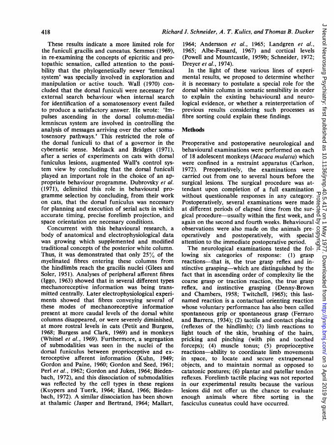
These results indicate a more limited role forthe funiculi gracilis and cuneatus. Semmes (1969),in re-examining the concepts of epicritic and pro-topathic sensation, called attention to the possi-bility that the phylogenetically newer 'lemniscalsystem' was specially involved in exploration andmanipulation or active touch. Wall (1970) con-cluded that the dorsal funiculi were necessary forexternal search behaviour when internal searchfor identification of a somatosensory event failedto produce a satisfactory answer. He wrote: 'Im-pulses ascending in the dorsal column-mediallemniscus system are involved in controlling theanalysis of messages arriving over the other soma-tosensory pathways.' This restricted the role ofthe dorsal funiculi to that of a governor in thecybernetic sense. Melzack and Bridges (1971),after a series of experiments on cats with dorsalfuniculus lesions, augmented Wall's control sys-tem view by concluding that the dorsal funiculiplayed an important role in the choice of an ap-propriate behaviour programme. Dubrovsky et al.(1971), delimited this role in behavioural pro-gramme selection by concluding, from their workon cats, that the dorsal funiculus was necessaryfor planning and execution of serial acts in whichaccurate timing, precise forelimb projection, andspace orientation are necessary conditions.
Concurrent with this behavioural research, abody of anatomical and electrophysiological datawas growing which supplemented and modifiedtraditional concepts of the posterior white column.Thus, it was demonstrated that only 25% of themyelinated fibres entering these columns fromthe hindlimbs reach the gracilis nuclei (Glees andSoler, 1951). Analyses of peripheral afferent fibres(Iggo, 1963) showed that in several different typesmechanoreceptive information was being trans-mitted centrally. Later electrophysiological experi-ments showed that fibres conveying several ofthese modes of mechanoreceptive informationpresent at more caudal levels of the dorsal whitecolumns disappeared, or were severely diminished,at more rostral levels in cats (Petit and Burgess,1968; Burgess and Clark, 1969) and in monkeys(Whitsel et al., 1969). Furthermore, a segregationof submodalities was seen in the nuclei of thedorsal funiculus between proprioceptive and ex-teroceptive afferent information (Kuhn, 1949;Gordon and Paine, 1960; Gordon and Seed, 1961;Perl et al., 1962; Gordon and Jukes, 1964; Bieden-bach, 1972), and this dissociation of submodalitieswas reflected by the cell types in these regions(Kuypers and Tuerk, 1964; Hand, 1966; Bieden-bach, 1972). A similar dissociation has been shownat thalamic (Jasper and Bertrand, 1964; Mallart,
1964; Andersson et al., 1965; Landgren et al.,1.965; Albe-Fessard, 1967) and cortical levels(Powell and Mountcastle, 1959b; Schneider, 1972;Dreyer et al., 1974).
In the light of these various lines of experi-mental results, we proposed to determine whetherit is necessary to postulate a special role for thedorsal white column in somatic sensibility in orderto explain the existing behavioural and neuro-logical evidence, or whether a reinterpretation ofprevious results considering such processes asfibre sorting could explain these findings.
Methods
Preoperative and postoperative neurological andbehavioural examinations were performed on eachof 18 adolescent monkeys (Macaca mulatta) whichwere confined in a restraint apparatus (Carlson,1972). Preoperatively, the examinations werecarried out from one to several hours before thesurgical lesions. The surgical procedure was at-tendant upon completion of a full examinationwithout questionable responses in any category.Postoperatively, several examinations were madeat different periods of elapsed time from the sur-gical procedure-usually within the first week, andagain on the second and fourth weeks. Behaviouralobservations were also made on the animals pre-operatively and postoperatively, with specialattention to the immediate postoperative period.The neurological examinations tested the fol-
lowing six categories of response: (1) graspreactions-that is, the true grasp reflex and in-stinctive grasping-which are distinguished by thefact that in ascending order of complexity lie thecoarse grasp or traction reaction, the true graspreflex, and instinctive grasping (Denny-Brownand Chambers, 1958; Twitchell, 1965); this last-named reaction is a contactual orienting reactionwhose voluntary performance has also been calledspontaneous grip or spontaneous grasp (Ferraroand Barrera, 1934); (2) tactile and contact placing(reflexes of the hindlimb); (3) limb reactions tolight touch of the skin, brushing of the hairs,pricking and pinching (with pin and toothedforceps); (4) muscle tonus; (5) proprioceptivereactions-ability to coordinate limb movementsin space, to locate and secure extrapersonalobjects, and to maintain normal as opposed tocatatonic postures; (6) plantar and patellar tendonreflexes. Forelimb tactile placing was not reportedin our experimental results because the variouslesions did not offer us the chance to evaluateenough animals where fibre sorting in thefasciculus cuneatus could have occurred.
418
Protected by copyright.
on 3 April 2019 by guest.
http://jnnp.bmj.com
/J N
eurol Neurosurg P
sychiatry: first published as 10.1136/jnnp.40.5.417 on 1 May 1977. D
ownloaded from

Proprioceptive pathways of the spinal cord
The standard neurological tests were undertakenespecially to evaluate any damage to motor path-ways in the lateral funiculus which might influenceour behavioural observations or electrophysio-logical recording. We felt that corticospinal tractdamage would have implied damage to the dorsalpart of the lateral funiculus which would alsohave interrupted somatosensory projections otherthan the dorsal white columns, such as the dorsalspino-cerebellar tract, and Morin's pathway (Haand Morin, 1964; Ha, 1971), the spino-cervico-lemniscal tract. Similarly, the integrity of theanterolateral projection was assessed by prickingand pinching stimuli, since damage here wouldimply ventral spinocerebellar tract involvement aswell as interruption of mechanoreceptive afferentfibres within the anterolateral pathway.The operative procedure was as follows.
Animals were anaesthetised with sodium pento-barbitone 30 mg/kg ip (five animals) or 25 mg/kgiv (13 animals). Intravenous catheters were in-serted for supplemental anaesthesia during sur-gery. A laminectomy was then performedaseptically. The dura mater was split and retractedwith suture ties under a microscope; the pia materwas also retracted. A Bard-Parker gauge 11 blade,cut off, marked in millimetre gradations, and heldin a haemostat was used to make the lesion. Thedura mater was then reapposed with 5-0 gut orsilk, gel-foam was placed over the cut spinousprocesses, and muscle, fascia, and skin rejoined.Procaine penicillin (one million units) was ad-ministered immediately after the operation, andfor several days thereafter. Animals were observedafter surgery, and any sign of infection wastreated promptly. Three control laminectomieswere performed using the same procedures;permanent neurological defects and electro-physiological changes were absent from thesepreparations.
Microelectrode recordings were obtained fromthe primary somato-sensory cortex of the monkeysapproximately one month after spinal surgery(Schneider, 1972; Dreyer et al., 1974). At the endof each electrophysiological experiment, theanimal was deeply anaesthetised with pentobarbi-tone, and sacrificed by intracardial infusion with0.9% saline followed by 10% neutral bufferedformalin. Appropriate portions of the brain wereimbedded in celloidin, and coronal or sagittalsections were taken at 30 um; the sections werestained with cresylecht violet. The spinal cord inthe area of the lesion, as well as selected sectionsrostral and caudal to it, were also imbedded incelloidin. The extent of the lesion was determinedby examining Mahon stained sections, while
anterograde and retrograde tract degenerationwere determined by the Marchi method. Recon-structions of spinal cord lesions were drawn fromslide projections of histological sections. These aredisplayed in Figs 1, 2, and 3. Cortical micro-electrode penetrations were reconstructed to per-mit analysis of the cytoarchitectural area traversedand laminar location of the neurones isolated ineach penetration (Schneider, 1972; Dreyer et al.,1974).
Results
Basically, there are four spinal cord lesions: thedorsal funiculus, the lateral funiculus (in thedorsal aspect only), the dorsal and dorsolateralfuniculus combination, and the combination lesionextended into the cord hemisection. The dorsalfunicular lesion cut the posterior white tracts,funiculus gracilis in the low thoracic area, andboth gracilis and cuneatus above. The lateralfunicular lesion spared the motor tracts and theanterolateral quadrant but attempted to cut theposterior spinocerebellar tracts and Morin's path-way. The dorsal and dorsolateral lesion (see Fig.4) included all those tracts to various extents.Finally, the hemisection definitely included themotor system.
LESIONS OF THE DORSAL FUNICULUSPerhaps the signal manifestation of an animalwith a complete dorsal white matter lesion is hispropensity to place the most distal aspect of theextremity (hand or foot) on the floor with itsdorsal rather than ventral surface down. We callthis dorsal placing. This observation has beenemphasised by others (Ferraro and Barrera, 1934;Gilman and Denny-Brown, 1966). It occurredeven when the limb was oriented appropriately,and it was most noticeable in animals with onlya unilateral lesion. but it was definitely present inbilateral lesions.
Lesions in the dorsal funiculus alone were eitherin the thoracic area or cervical area. When theanimal had a low thoracic lesion, both distal andproximal proprioceptive function were markedlyimpaired, and the animal had little use of thelower limb. In high thoracic lesions, the monkeydemonstrated normal posture and a steady gait.It would be active and climb the cage wall. Butin the feet, grasp was impaired, palpatory be-haviour was diminished, dorsal placing of thefoot did occur, and distal malalignment wasnoticeable. Thus, distal proprioceptive functionwas impaired but proximal proprioception ap-peared spared. In midcervical lesions, the same
419
Protected by copyright.
on 3 April 2019 by guest.
http://jnnp.bmj.com
/J N
eurol Neurosurg P
sychiatry: first published as 10.1136/jnnp.40.5.417 on 1 May 1977. D
ownloaded from

Richard J. Schneider, A. T. Kulics, and Thomas B. Ducker
Fig. 1
Figs. 1, 2, and 3 Drawings made from projections of stained tissue sections from each animal in thisexperimental series. Total extent of lesions was determined by overlapping consecutive sections. Stripedarea implies total damage, circles imply partial damage. Rostral and caudal drawings were made at levelsbeyond which lesion size diminished with no additional pathology. Rostral drawings in DCX:12 weredetermined by ascending degeneration pattern, only two drawings were done for DCX:8, and DCX:J5 wasdrawn from written account of total extent of lesion.
deficit was present in the feet and, in addition,the forelimbs were now markedly affected. Thearms flailed during ambulation, and were held ina sling-like posture at rest. The animal would usethe entire forearm to palpate the surroundings.There was some recovery with time but skilledmanipulation was dependent on visual cues. Inthe very high cervical lesions in one animal fore-limb, proximal proprioceptive function appearedto recover, but distal position and stereopsisremained disordered.
L.FSIONS OF THE LATERAL FUNICULUSIn lateral funicular lesions, in the dorsolateral(posterolateral) part of the cord, behind the
dentate ligaments, with sparing of the cortico-spinal tracts, the animals had surprising difficulty.In both low cervical lesions, the animals uponrecovery from surgery would climb their cageshelf and cage walls, but showed a preference forlying on the cage floor, with hindquarters slightlyelevated because of an increased tonus in the hipextensors. The limbs ipsilateral to the lesion hadno trouble localising objects in space, but uponreaching for such objects their grasp was markedlydiminished in vigour. The orientation of theirfeet at rest was often at unusual rotational anglesto the body-that is, at right angles to thedirection the body was facing. Forelimbs mostoften showed a posture of shoulder adduction,
420
Protected by copyright.
on 3 April 2019 by guest.
http://jnnp.bmj.com
/J N
eurol Neurosurg P
sychiatry: first published as 10.1136/jnnp.40.5.417 on 1 May 1977. D
ownloaded from

Proprioceptive pathways of the spinal cord
ROSTRAL
CAUL L
CAUDAL
Fig. 2
elbow and wrist flexion when the animals were ina sitting position, somewhat like the animals withhigh dorsal funiculus lesions. The affected hind-limbs manifested a slight resting tremor, andshowed plantar flexion of the toes at rest, whichwas greater at the interphalangeal than at themetatarso-phalangeal joints. During ambulationthe toes tended to be widely splayed providing agreater contactual circumference. With visiondiverted, distal limb contact with objects causedpeculiar reactions. Among these were staring inthe direction of the surface touched, repeatedcollision with the surface when vision remainedelsewhere directed, and what may have been astartle reaction to encountering the object.
MORE EXTENSIVE LESIONSCombining lesions of both dorsal and dorso-lateral funiculi from the sixth cervical to theseventh thoracic segment had a profound effecton the animal, for it made no attempt at move-ment in and around the cage. The monkeys
refused to stand and would lie in postural hyper-extension (Gilman and Denny-Brown, 1966).When the lesion was further extended to cordhemisection, there was additional flaccid paralysisof the ipsilateral extremity plus self-mutilation ofthe affected limb.
Tables 1 and 2 give the results of the neuro-logical examinations done on animals whosetractotomies included pathways as verified byhistology. The scheme of these tables includesonly residual changes at one month after surgerysince this was the time when electrophysiologicalmapping was commonly performed. Except withpinch (which was to study pain sensation), entriesrefer to the limb ipsilateral to the lesion.To make the tables more understandable, follow
a typical entry, DCX: 4. This was an animal witha lesion of the dorsal funiculus of the left side(see comment), at the level of the third thoracicsegment of the spinal cord. One month after sur-gery, his left forelimb pulled away from graspeliciting stimuli, and his left hindlimb failed to
421
Protected by copyright.
on 3 April 2019 by guest.
http://jnnp.bmj.com
/J N
eurol Neurosurg P
sychiatry: first published as 10.1136/jnnp.40.5.417 on 1 May 1977. D
ownloaded from

Richard J. Schneider, A. T. Kulics, and Thomas B. Ducker
Fig. 3
react to such stimuli. He would palpate and orientto things with his left forelimb but not with hisleft hindlimb. The left hindlimb did not placewhen stimulation was delivered to hairs or lightlyto the skin surface, but did place when deeperpressure to the foot was applied. The left forelimbbut not the hindlimb reacted to hair displacementand light tapping on the skin surface. There was anormal withdrawal to pinch by the right limbs,normal muscle tone in the left limbs, normal limbmovements on the left side when reaching forobjects or orienting in space, and a normalpatellar but no plantar reflex in the left hindlimb.The animal had a tractotomy which extended tothe right side where it included the dorsal anddorsolateral funiculi. It is thus listed in Table 2 aswell. (The comments column lists the side ex-amined when both sides were so used, or whenambiguity might exist.)
Several of these results should be emphasised.The hindlimb grasp reflex is almost uniformlyabsent after dorsal funicular transection and afterthe combination lesion, with or without motorinvolvement, when above the level of entry forafferent fibres from the affected limb. Instead, thehindlimbs usually display no reaction, and theforelimbs show tactile avoiding, a response whichappears in the early postoperative period in thehindlimbs, and diminishes thereafter. Partial dor-sal funicular lesions (DCX: 8, DCX: 9, DCX: 15)may or may not spare some grasp reflex. In pureseventh cervical dorsolateral transections, the fore-limbs were unaffected, while the hindlimbs couldshow a diminution which might be traceable tothe weakened grasp rather than an absence of thereflex. In order to abolish forelimb instinctivegrasping, most of the forelimb plexus should bebelow a dorsal funicular lesion or hemisection.
422
Protected by copyright.
on 3 April 2019 by guest.
http://jnnp.bmj.com
/J N
eurol Neurosurg P
sychiatry: first published as 10.1136/jnnp.40.5.417 on 1 May 1977. D
ownloaded from

Proprioceptive pathways of the spinal cord
A B
C
,- ".
11o. vI.,.,..w....:.
_'St-.AP
FEFig. 4 A. Complete right dorsal funiculus lesion at C5 segment with left dorsal funiculus involvement(DCX:16). B. Ascending pattern of axonal degeneration (DCX:16). C. Spinal cord h1emisection at C7segment, with partial sparing in medial regions of dorsal and ventral funiculi (DCX:2). D. Dorsal funiculuslesion at C3 segment with some dorsolateral funiculus involvement (DCX:6). E. Dorsolateral funiculuslesion at C7 segment involving mainly proposed area of projection of spino-cervico-lemniscal tract fibreswith some sparing of dorsal spinocerebellar tract fibres (DCX:17) (see Fig. 3). F. Dorsolateral funiculuslesion involving spino-cervico-lemniscal tract and presumably most dorsal spinocerebellar tract fibres(DCX: 18).
B
423
.s0
r.o
or
.ew -
Protected by copyright.
on 3 April 2019 by guest.
http://jnnp.bmj.com
/J N
eurol Neurosurg P
sychiatry: first published as 10.1136/jnnp.40.5.417 on 1 May 1977. D
ownloaded from

Richard J. Schneider, A. T. Kulics, and Thomas B. Ducker
C)V) C)7C)
o o Cd eVbe: o oX ; Y YX o Q
CUQ> CU
Cimd , o .: md m o m ,C, 0
0 ~~ ~~~>0CU CU
uC.6o
-+ 0 + -a' -a-t
O 0 + a- +
+ -i- -4- -i+ +
a+ + + + + +
±t + + a-
+ + + + + a-L
+ + + + ± +_
-a t- 0 + +
t- + 0 +
+ + + a- + a-
a- + 0 a- a+
o 0 0 0 0
-ai
f+t+ + t. + t + + +~~~~~~~~~~~~~~~~~~~~~~~~~~~~~~~~~~~~~~~~~~~~~~
0 0 O-- + +
O t-
0 0
++a + + 0
II + + + +
_ a a- + + +-a- +
-t- -+ a- °0 + a-
o 0 + +a-- 0
o 0 0 0 0 0
o 0 + + + 0
O 0 0 0 0
+
0 0
0 + 0
0 0 0 0
0 0 0O 0 0
0 0 + + + 0
0 0 0
+ a- -i- --a +--i+ + + !-a--
< 00 e 00 W0 C 't - C- o m~~~~~,It in) Uc O) ON 2
x x x x x x x x x x x x
0 0 0 0 0)
C) 0 ) C) Uz C
424
2 riOZ c
0_
LL
*2
-o0
.0
0
_0
._
.0
E,011
~C
+0
SIt0
Q0o
U)
z
Cs.2Co-
QZ)
L-
5Di;L
Protected by copyright.
on 3 April 2019 by guest.
http://jnnp.bmj.com
/J N
eurol Neurosurg P
sychiatry: first published as 10.1136/jnnp.40.5.417 on 1 May 1977. D
ownloaded from

Proprioceptive pathways of the spinal cord
5 4) --a0 )0
_ _ Q~~~~~~~~~E _0o to 0 to~
= .-
co .
-o0
+>4 + +* -f+t +
m v0 m
v 0 -
+ +
I I + t _4 >
+> 4
-+ 1- + t-"
+~~~~~~~~~4
0 '1- +
O 0 0 0 0C
o 0 4> 4
0 0 0 0
O 0 0 0 0
0, 0 0 0
,7 0 +
+- t 0 0
- I-~ -I> 2-4- -+ +
0
I- +1
U
"t _ _ _1 _2C) 0 c
U U U U U
-oo o
L.
0 c0
+Q+1 +
+-
+
1- -)
I
+
X XQa-4 -,,>I-L
0
Q-f.
_7to , -; E
o 0 0
-~~~~~~~~~~~~~~-
±1
o 0 4>
O +o 0 0
O
o 0 0CC
o 0 0
O + +
o 0 0
-t±- + + +
xo t
,Q Q Hu
xu0
x x
425
c
0
ot
s°0
s 1co <- .
.00 L2
-0{
cz
0w-4
0X -
-0
_ 4*tc
~0
0l
00Z
03In
0c
COH--
H0.0
Cd
Protected by copyright.
on 3 April 2019 by guest.
http://jnnp.bmj.com
/J N
eurol Neurosurg P
sychiatry: first published as 10.1136/jnnp.40.5.417 on 1 May 1977. D
ownloaded from

Richard J. Schneider, A. T. Kulics, and Thomas B. Ducker
Thus, a fourth cervical lesion (DCX: 11), but notan eighth cervical lesion (DCX: 5, DCX: 7),abolishes forelimb instinctive grasping, and a sixthcervical hemisection (DCX: 1) leads to abolition,while a partial lesion at the fifth or sixth cervicalsegment (DCX: 8, DCX: 9) may or may not spareinstinctive grasping in the forelimb. In the hind-limb, a pure dorsal funicular lesion at high thoracicto cervical levels will suffice to eliminate instinc-tive grasping, as does expanding the lesion toinclude additional pathology. But a seventh cer-vical dorsolateral lesion alone does not affectinstinctive grasp in any limb (DCX: 17, 18).
Tactile, but not contact, placing of the hind-limb is abolished by pure posterior white columnlesions, except in the cases of DCX: 13 which wasan unusually low lesion, and DCX: 15 which hadsome postoperative complications affecting thelegs. Both hindlimb tactile and contact placingdisappeared with combination lesions, but wereintact with pure dorsolateral lesions. Hair dis-placement reactions as well as light touch or tapreactions disappear pari passu with interruptionof posterior white column fibres related to recep-tive fields on the body surface in question. With-drawal to pinching is exhibited only in thecontralateral limb whose fibres enter caudal tohemisection. After a month, muscle tone is seldomaffected in pure posterior white column transectedpreparations, but hypotonia ensues with hemi-sections. Proprioception is completely absent inthe forelimb after either posterior white columnor a combination lesion above the brachial plexus.Proprioception is absent in the hindlimbs onlywhen the posterior white column lesion is justabove the lumbar enlargement, or when the lesionincludes the lateral funiculus at higher segmentallevels.
Discussion
We interpret our results to imply that the re-arrangement of sensory afferent fibres, which hasbeen shown to occur anatomically and electro-physiologically (Glees and Soler, 1951; Petit andBurgess, 1968; Burgess and Clark, 1969; Whitselet al., 1969), produces predictable behavioural andneurological changes in the effects of tractotomies.The main outlines of this rearrangement, whichwe call 'fibre sorting', are (1) the redistributionof proprioceptive information from the hindlimbto pathways other than the dorsal funiculus asthis information is transmitted rostrally, (2) theappearance of this proprioceptive information inthe lateral funiculus (dorsal aspect) at rostralspinal cord levels, and (3) the rostral reorganisa-
tion of exteroceptive information combined withthe proprioceptive redistribution which concen-trates, within the posterior white columns, afferentfibres related to hair displacement and those re-lated to light touch on the glabrous skin surfacesof the distal limbs. We believe our results supportthis scheme in the following ways.The contact (proprioceptive) placing reflex in
the hindlimb is not abolished in macaques whosedorsal funiculus is interrupted at levels aboveT5 segment (DCX: 4, 5, 6, 7, 8, 9, 10, 11, 16), butis absent in macaques with dorsal funiculus inter-ruptions at or below T5 (DCX: 13, 15), and inrnacaques with lateral funiculus or hemicord in-volvement above T4 segment (DCX: 1, 2, 3, 4,10). However, lateral funiculus tractotomies alone(DCX: 17, 18) do not abolish this reflex. Thecontact placing reflex depends upon proprioceptiveinformation being conveyed to the frontomotorcortex (Woolsey and Bard, 1963). Since transect-ing the dorsal funiculus does not necessarilyabolish this reflex, the implication is that thenecessary afferent proprioceptive information doesInot project in an uninterrupted manner exclu-sively through the posterior white column. Incontrast, hindlimb tactile (exteroceptive) placing,a reflex depending on exteroceptive informationbeing relayed to the parietal lobes (Woolsey andBard, 1963), is abolished by dorsal funiculuslesions regardless of the level, and is not abolishedby isolated lateral funiculus lesions. This impliesthat the necessary afferent exteroceptive informa-tion for the tactile placing reflex does project in anuninterrupted fashion exclusively through theposterior white column. The fact that focal lateralfuniculus lesions which are dorsally placed do notabolish proprioceptive placing may imply a sharedprojection of these afferent fibres between the twotracts. But a more parsimonious conclusion wouldbe that, since tactile placing is still intact inmacaques with isolated lateral tractotomnies, skincontact before the proprioceptive stimUlus thres-hold is reached elicits a tactile placing reflex whichobscures the fact that proprioceptive placing isabsent.
Placing reactions have been examined aftertransections of the dorsal funiculus of macaques(Ferraro and Barrera, 1934; Gilman and Denny-Brown, 1966; Liebman and Levitt, 1973), and indomestic cats (Lundberg and Norrsell, 1960;Dobry and Casey, 1972). In cats it was foundthat tactile placing disappeared after ipsilateraltransection of the medio-dorsal lateral funiculusrostral to the level of the hindlimb afferent entryof the spino-cervico-lemniscal tract (Morin'spathway), and caudal to the lateral cervical
426
Protected by copyright.
on 3 April 2019 by guest.
http://jnnp.bmj.com
/J N
eurol Neurosurg P
sychiatry: first published as 10.1136/jnnp.40.5.417 on 1 May 1977. D
ownloaded from
Proprioceptive pathways of the spinal cord
nucleus (Lundberg and Norrsell, 1960). However,lesions of Morin's pathway in its contralateralventral funicular region, cranial to the lateralcervical nucleus, do not abolish tactile placing incats (Norrsell and Voorhoeve, 1962). It was con-cluded that Morin's pathway is not the exclusiveafferent link in this cortical reflex for cats(Norrsell and Voorhoeve, 1962). Lesions of thedorsal funiculus at the first cervical level whichincluded more than 9000 of its afferent fibres,were also reported to abolish tactile placing incats (Dobry and Casey, 1972).
In macaques, Gilman and Denny-Brown (1966)reported that to demonstrate tactile placing inthe hindlimbs after fourth cervical dorsal funi-culus transection requires brisk cutaneous contactto proximal parts of the leg, and in four of theiranimals (DC2, DC4, DC15, and DC46) they con-sidered tactile placing to be absent. It was alsofound that the more extensive combination (dorsaland dorsolateral funiculus) lesion at the thirdcervical level was required to abolish contactplacing (since these authors did not use contactplacing, we make this assessment from theirstatement, 'no limb placed with even broad,heavy, prolonged contact') along with tactileplacing in the hindlimb, and that dorsolateralfunicular lesions alone left no residual defect intactile placing (Gilman and Denny-Brown, 1966).In support of the notion that both dorsal funiculusand dorsolateral funiculus share fibres involved inthe projection of proprioceptive information neces-sary to the contact placing reflex, other researchindicates that this is the case for position sense(Vierck, 1966). The same studies infer that theanterolateral funiculus also pays a role in positionsense.We believe that the proprioceptive disturbances
we observe are linked to afferent fibres whichtravel both in the dorsal funiculus and in pathwaysin the dorsolateral funiculus at more rostrallevels. This may account for the occurrence ofthese disturbances in the hindlimbs of animalswith low cervical and high thoracic, dorsal funi-culus plus lateral funiculus tractotomies (DCX: 1,2. 3, 4, 10, 14. 15); in the forelimbs of animalswith high cervical (DCX: 6, 10, 11) and the hind-limbs of animals with low thoracic (DCX: 13)dorsal funicular tractotomies (in these, proprio-ceptive fibres have not yet undergone fibre sorting,although, a decrease in hindlimb position sensefollowing a dorsal funiculus lesion as high as theseventh thoracic level has been reported (Ferraroand Barrera, 1934) and we observed one (DCX: 15at the fifth thoracic level) but not in the fore-limbs or hindlimbs of animals with dorsolateral
funicular tractotomies at low cervical levels(DCX: 17, 18). Animals occasionally showedproprioceptive dysfunctions which did not fitthis systematic pattern (DCX: 5, 11), but webelieve these are the exception rather than therule.
Electrophysiological results in the isolateddorsolateral funiculus tractotomy experimentslend support to the occurrence of proprioceptive(deep-submodality) afferent fibres in the rostraldorsolateral funiculus. These data show thatdamage involving Morin's pathway does not affectthe distal, deep-submodality, single cell representa-tion in Brodmann's area 2 of macaques as severelyas when the spinal cord position of the dorsalspinocerebellar tract is encroached upon(Schneider, 1972; Dreyer et al., 1974). This mayimply simply that there is a direct relationshipbetween the extent of a dorsolateral funiculuslesion, and the loss of afferent information pro-jecting to cytoarchitectural area 2 of the brain.This evidence tends to allocate a role, if not theexclusive role, in relaying deep afferent impulsesto the primate dorsal spinocerebellar tract. How-ever, the evidence is that the contact placingreflex is mediated through the frontomotor areasin macaques, so that change in occurrence ofproprioceptive units in Brodmann's area 2, whilesignificant for somatic sensation, would not ac-count for the abolition of this response. Recentresults (Asanuma and Rosen, 1972a and b) showthat, within the precentral motor area, there arecells which receive afferent proprioceptive input.When stimulated electrically these same cells pro-duce isolated peripheral muscle contractions.These cells may receive information identical tothat transmitted to the primary somatosensorycortex which may subserve positive feedback orprecentral cortical reflexes such as contact plac-ing. Elimination of such feedback has been shownto affect the output of cells in the motor cortex(Lewis et al., 1971).
Single cell studies of the cortex (Schneider,1972; Dreyer et al., 1974) reveal the presence ofdeep-submodality receiving cells from the hind-limb in Brodmann's areas 1 and 2 (postcentralcytoarchitectural areas known to receive such aproprioceptive population). However, there is areduction of the numbers of proprioceptiveneurones from the forelimbs of animals with highcervical dorsal funicular tractotomies, and fromthe hindlimbs of animals with damage laterally inthe dorsal spinocerebellar tract or with unilateralhemisections. These data support the interpreta-tion that the segregation of exteroceptive andproprioceptive afferent fibres within the spinal
427
Protected by copyright.
on 3 April 2019 by guest.
http://jnnp.bmj.com
/J N
eurol Neurosurg P
sychiatry: first published as 10.1136/jnnp.40.5.417 on 1 May 1977. D
ownloaded from

Richard J. Schneider, A. T. Kulics, and Thomas B. Ducker
cord (Whitsel et al., 1969, 1970) can account forthe selective diminution of proprioceptive orexteroceptive cells from the cortical populationwhen tractotomies transect these afferent fibresafter they have been separated. Thus, spinal cordfibres sorting can be shown to play an importantrole in the determination of the body representa-tion in the primary somatosensory cortex (Whitselet al., 1972). Such fibre sorting contributes to theevaluation of the rostro-caudal level and extent ofspinal cord lesions.
It should be noted that the assessment of pro-prioceptive losses may involve a number of re-lated, but different qualities. For example,Ferraro and Barrera (1934) used the failure ofslow movements of any limb to evoke eye move-ments in their fourth cervical preparation toattribute loss of proprioception to their dorsalfuniculus interruption. We find this phenomenonalso, but are not willing to say it implies completeabsence of proprioceptive sense. These authorsalso noted the prominent dorsal placing of thedistal limbs of such animals, and in one case usedit to assign proprioceptive loss to the hindlimb.Our electrophysiological results indicate that themarked impairment of cutaneous sensation on thevolar surface of both distal limbs, observed afterdorsal funiculus transection above the entry ofafferent fibres from those limbs, could probablyresult in this unusual posture, the feedbacknecessary for spatial position sense at rest beingobtainable only from the remaining innervation ofthe foot and hand. Thus, an exteroceptive ratherthan a proprioceptive loss may be the basis forthis observation. Schiff, in 1857, made a similarclaim (cf. Ferraro and Barrera, 1934). Gilmanand Denny-Brown (1966) cite catatonic posturesas evidence of loss of proprioception in animalswith a high cervical dorsal funiculus lesion. Whilewe have also observed this on occasion, theability to use the hindlimbs without visual guid-ance in such animals both in walking and climb-ing, and in directing movements in space, indicateto us at least some preservation of proprioceptivefunction. Agreement on this seems to come fromother dorsal funiculus tractotomy studies(Christiansen, 1966) in which neurological testingwas also used.We believe that the concentration of extero-
ceptive afferent fibres within the dorsal funiculusconfers upon it quantitatively the largest rolein conveying information useful in tactile explora-tion and manipulation through the macaque spinalcord. While we cannot draw the conclusion thatour hair movement and tap tests of animals withtransection of the dorsal funiculus demonstrated
lack of sensation, we do believe that inattentionor diminished sensitivity is indicated. This con-viction is supported by the radical diminution inhair and touch related single cells encounteredin microelectrode penetrations of the somato-sensory cortex (Schneider, 1972; Dreyer et al.,1974). This diminution is significantly more ex-tensive when the population of tactile receptorsof the glabrous skin of the fore and hindlimb isconsidered (Schneider, 1972; Dreyer et al., 1974).In the macaque, this refers to the palm and sole.The population of tactile receptors on the palmalso supplies the largest input to those cells of thehand represented in the motor cortex which pro-duce flexion of the fingers (Asanuma and Rosen,1972b). These data suggest critical roles for suchreceptors in tactile exploration, manipulation, andgrasping. This is not to say that these functionsmust be exclusively subserved by this receptorpopulation. In the macaque, there is evidencethat other pathways can be used in tasks involvingthese areas (Kuhn, 1949; Schwartz et al., 1972;Vierck, 1973). Even less can we conclude thatother body areas are the sole domain of oneafferent system (Mettler and Liss, 1959; Christian-sen, 1966; Norrsell, 1966; Kitai and Weinberg,1968; Schwartzman and Bogdonoff, 1968, 1969;Dobry and Casey, 1972; Eidelberg and Woodbury,1972; Mann et al., 1972; Schwartz et al., 1972;Liebman and Levitt, 1973). However, it has beendemonstrated (Ferraro and Barrera, 1934; Gilmanand Denny-Brown, 1966; Wall, 1970; Dubrovskyet al., 1971; Melzack and Bridges, 1971; Schneider,1972) that the quality of behaviour which usesthese receptors changes after the elimination ofdorsal funiculus pathways, and even when be-havioural testing is included the disturbances arenonetheless present.With regard to the possibility that inattention is
the result of a dorsal funiculus lesion, Wall (1970)presented such a hypothesis of dorsal funiculiisfunction, especially in relation to the lack oftouch deficits in human subjects with high cervicaland thoracic spinal cord lesions (Boshes and Pad-berg, 1953). In a retest of one of these patients,Wall was able to demonstrate a change in thequality of the remaining touch in the direction ofbeing less frequently felt during concomitantdistraction of the patient by vibratory sensation,or by having him read aloud (Wall, 1970). Electro-physiological results (Schneider, 1972; Dreyer etal., 1974) indicate that many of the single neuroneswhich are encountered in the somatosensorycortex after posterior white column transectionshow a rapid habituation, and require higherstimulus intensities to be excited. Such units, if
428
Protected by copyright.
on 3 April 2019 by guest.
http://jnnp.bmj.com
/J N
eurol Neurosurg P
sychiatry: first published as 10.1136/jnnp.40.5.417 on 1 May 1977. D
ownloaded from

Proprioceptive pathways of the spinal cord
they are characteristic of the remaining form ofsensation in the patients, could account for theirlack of sensitivity to one form of stimulationduring simultaneous application of vibratorystimulation. Oscillatory stimulation could be likewhite noise, masking sporadic transmission fromanother somatic source.
Another question is whether the size of the cagein which the animal is confined after surgerydetermines the amount of recovery of function.We believe that residual postural and motordeficiencies at one month after surgery may beaffected by cage size, but only to the extent thatfree roaming or colony caged animals have agreater opportunity to develop strategies to copewith their deficits. Such a strategy was reported(Goldberger, 1972) when tactile evasion was en-hanced to enable a macaque with a lesion ofBrodnunn's area 6 of the cerebral cortex to over-come the pathological grasp reflex. Thesestrategies may make the animal appear grosslyto be without deficits which subtler testing wouldreveal.A discussion of grasp responses must heed the
differences in type and in nomenclature whichhave been used by various authors. The importantdistinction is that in ascending order of com-plexity lie the coarse grasp or traction reaction,the true grasp reflex, and instinctive grasping(Denny-Brown and Chambers, 1958; Twitchell,1965). This last named reaction is a contactualorienting reaction whose voluntary performancewas called spontaneous grip or spontaneous graspby Ferraro and Barrera (1934).The concentration of distal afferent fibres re-
lated to grasping in the dorsal funiculus causesboth devastating loss of grasp behaviour withtotal tractotomies and resilience of it with partialsparing. Our complete dorsal funiculus lesionswere sufficient to abolish the hindlimb true graspreflex. Gilman and Denny-Brown (1966), whotested this reflex, found a brisk grasp reflex onthe hindlimb in only one of their experimentalanimals (DCI) which had a bilateral high cervicaldorsal funiculus section, but not on the other fouranimals with similar lesions (DC4, DC14, DC15,and DC46). Only coarse grasp reactions wereelicited in these latter animals. The occurrence ofthe true grasp reflex in the lone animal may haveresulted from the sparing of small bundles ofdorsal funiculus fibres by the lesion. Such sparingappears to have resulted in the retention of thisreflex even though diminished by our animalsDCX: 9 and DCX: 15. However, the lesion in theone animal mentioned above does seem com-plete; thus, we are at a loss to explain this result.
429
Nevertheless, we interpret our data to indicatethat the true grasp reflex depends on the dorsalfuniculus.
Dorsal funiculus lesions were also sufficientto replace forelimb grasp reflexes with activeavoidance responses. This occurred even whenthe lesions were below the brachial plexus. Hind-limb tactile avoidance was seen early in thepostoperative course with many animals, butgenerally disappeared or was greatly diminishedby the end of the first postoperative month.Gilman and Denny-Brown (1966) found suchavoiding on the hindlimb but not the forelimb inone animal (DCI) and found coarse avoiding inthe forelimbs of two (DC5 and DC15). A hyper-aesthesia following primary somatosensory cortexablations in macaques is reported (Schwartzmanand Semmes, 1971), which we believe is identicalto the phenomenon referred to by others (Denny-Brown and Chambers, 1958) as release of tactileavoiding, and which occurs with more extensivepostcentral cortical excisions in macaques. Thus,the limited cortical ablations will produce resultssimilar to those which dorsal funiculus lesionscreate in forelimbs and hindlimbs at different post-operative times. It would seem that a tonicinput from the dorsal funiculus to the postcentralcortex is necessary to maintain normal tactilereactivity.
Instinctive grasping, encompassing a higherlevel of complexity, is not preserved in the hind-limb by partial sparing of the dorsal funiculus(DCX: 8, 9, 15), but a greater amount of sparingcaused by the same lesion (DCX: 9) being justrostral to mid-plexus level, fails to abolish thisresponse in the forelimb. Lesions above thebrachial plexus do abolish this response in theforelimb (DCX: 10, 11). Ferraro and Barrera(1934) were only able to get crude spontaneousgrasping after one month in any limb of ananimal with a midcervical, dorsal funiculus lesion(CER80) or in the hindlimb of a midthoracic,dorsal funiculus lesioned animal (CER 135). Thedorsolateral funiculus lesions alone yielded noeffect on this response in either limb in our studies.Identical results were obtained by Gilman andDenny-Brown (1966) with a similar lesion. Ferraroand Barrera (1934) described the deficits in spon-taneous grasping common to animals with a dorsalfuniculus lesion. In an interesting observationthey relate that animals with such lesions try togrip things with their entire limb rather than justits distal part. We believe this perceptive observa-tion can be explained by the relative preservationof the proximal limb sensory input which we seein our electrophysiological work (Schneider, 1972;
Protected by copyright.
on 3 April 2019 by guest.
http://jnnp.bmj.com
/J N
eurol Neurosurg P
sychiatry: first published as 10.1136/jnnp.40.5.417 on 1 May 1977. D
ownloaded from

Richard J. Schneider, A. T. Kulics, and Thomas B. Ducker
Dreyer et al., 1974). These results may also ex-plain the necessity for using broad areas ofcutaneous contact with the limbs in producingtactile placing and traction responses as describedby Gilman and Denny-Brown (1966), and thefindings of Vierck (personal communication) thatdistal forelimb deficits in proprioception are moremarked than proximal deficits in macaques per-forming behavioural tasks after posterior whitccolumn tractotomies.The existence of a sorting process and its conse-
quences at levels rostral to the spinal cord intro-duce a new dimension into the analysis of dorsalfuniculus studies. For example, Melzack andBridges (1971) found that, with motor performancein cats, there was less drastic interference bylesions solely in the nuclei of the dorsal funiculusthan with those which were made only in thehigh cervical dorsal funiculus. They regarded thisas a paradoxical result. Since their lesions did notextend into an area of the nuclei which has beenshown to receive a large number of proprioceptiveafferent fibres (Kuhn, 1949; Gordon and Paine,1960; Gordon and Seed, 1961; Perl et al., 1962;Gordon and Jukes, 1964; Biedenbach, 1972), thatis, rostral to the obex, they may have spared an im-portant fraction of proprioceptive fibres. It wouldthus become extremely important to delimit theprecise caudal extent of each of their lesions sincethe less caudal a lesion is, the more fibres can bespared by sorting. Since dorsal funiculus nucleuslesions require a more rostral central nervoussystem exposure, the chances are that more spar-ing would have occurred in these animals than inthose with dorsal funiculus lesions alone, and thefibre sorting process could explain their paradoxicalresults.The effect of dorsal funiculus lesions on the
behaviour of the animal derives from the limita-tion of his ability to receive mechanoreceptiveinformation, especially from the distal, volar re-ceptors most involved in palpatory input. Tactualguidance is necessary in fine manipulative taskswhere vision is excluded (Brinkman and Kuypers,1972), and its sudden loss cannot be immediatelycompensated by visual input alone. Manipulationswhere spared sensory input may be used-forexample, vision, facial somatosensation, crudehand and foot dorsum somatosensation-like thoseinvolving feeding and grooming, tend to showquicker recovery, but a return to the status beforethe transection does not occur within a month.Recent behavioural data have shown that whenselective ablations are made of the anterior andposterior postcentral gyrus (Semmes and Porter,1972), or of Brodmann's cytoarchitectonic areas
3, 1, and 2 (Randolph and Semmes, 1974), theregion which is the primary central terminus ofimpulses travelling along dorsal funiculus fibres,there are differentiable, discriminative, behaviouraldeficits which last for months. Again, the receptorinformation which permits discrimination by pal-pation seems to be disturbed, and in addition, thedisturbance varies with the cytoarchitectural areainvolved.The picture that emerges is that submodalities
of mechanoreceptive somatosensation, and perhapstypes of receptor information within submodali-ties, are segregated as they ascend from caudalto cephalic levels of the nervous system so as toform somatosensory subunits in the primary corti-cal receiving area (Mountcastle and Powell, 1959a,b; Powell and Mountcastle, 1959a, b; Whitsel et al.,1972). The sorting process that takes place in thedorsal funiculus contributes to this segregation(Whitsel et al., 1969; Whitsel et al., 1970). Thissorting, and the existence of alternative somato-sensory pathways, however different in the mannerin which they handle the stimulus information,allow for the development of strategies to assistthe recovery of function assessed by many experi-ments after dorsal funiculus lesions in animals(Devito and Ruch, 1956; Mettler and Liss, 1959;Christiansen, 1966; Levitt and Schwartzman, 1966;Vierck, 1966; Kitai and Weinberg, 1968;Schwartzman and Bogdonoff, 1968, 1969; Dobryand Casey, 1972; Eidelberg and Woodbury, 1972;Mann et al., 1972; Schneider, 1972; Schwartz etal., 1972; Liebman and Levitt, 1973). Neurologicalobservations do not permit the same statement tobe made about humans, because those studieswhich report absence of deficits after dorsal funi-culus tractotomy lack the necessary histologicalconfirmation (Rabiner and Browder, 1948; Boshesand Padberg, 1953; Cook and Browder, 1965).Also, neurological examinations in humans con-tinue to implicate the dorsal funiculus in posturalsensation (Rose and Mountcastle, 1959; Poggioand Mountcastle, 1960). Whether differences re-lated to assessment procedures may account forthese results in humans is not yet known. How-ever attractive the new hypotheses of dorsal funi-culus function may be in offering explanations ofthe physiological role of the posterior whitecolumns, we suggest that further physiological andbehavioural evidence is required before conclusionscan be drawn about the function of the dorsalfuniculus. While we believe that our re-sortinghypothesis is correct in monkey, and may applyto man, we admit that it goes beyond existingevidence. We are currently recording the appro-priate clinical information in patients.
430
Protected by copyright.
on 3 April 2019 by guest.
http://jnnp.bmj.com
/J N
eurol Neurosurg P
sychiatry: first published as 10.1136/jnnp.40.5.417 on 1 May 1977. D
ownloaded from

Proprioceptive pathways of the spinal corcd
References
Albe-Fessard, Denise (1967). Organization of somaticcentral projections. In Contributions to SensoryPhysiology. Vol. 2. Edited by W. D. Neff. AcademicPress: New York.
Andersson, S. A., Landgren, S., and Wolsk, D. (1965).The thalamic relay and cortical projection of groupI muscle afferents from the forelimb of the cat.Journal of Physiology. 183, 576-591.
Asanuma, H., and Rosen, I. (1972a). Functional roleof afferent inputs to the monkey motor cortex.Brain Research. 40, 3-5.
Asanuma. H.. and Rosen. I. (1972b). Topographicalorganization of cortical efferent zones projectingto distal forelimb muscles in the monkey. Experi-mental Brain Research, 14, 243-256.
Biedenbach, Maria A. (1972). Cell density and regionaldistribution of cell types in the cuneate nucleus ofthe Rhesus monkey. Brain Research, 45, 1-14.
Boshes, B., and Padberg. F. (1953). Studies on thecervical spinal cord of man. Neurological Society ofMinneapolis. 3, 90-101.
Bowsher, D. (1965). The anatomophysiological basisof somatosensory discrimination. InternationalReview of Neutrobiology. 8, 35-75.
Brinkman. J.. and Kuypers, H. G. J. M. (1972). Split-brain monkeys: cerebral control of ipsilateral andcontralateral arm, hand, and finger movements.Science, 176, 536-539.
Burgess, P. R., and Clark. F. J. (1969). Dorsal columnprojection of fibres from the cat knee joint. Journalof Physiology. 203, 301-315.
Carlson, Kristen R. (1972). A temporary restraintchair for monkeys. Physiology and Behavior, 9,493-494.
Christiansen, J. (1966). Neurological observations ofmacaques with spinal cord lesions. AnatomicalRecord, 154, 330.
Cook. A. W.. and Browder, E. J. (1965). Functionsof posterior columns in man. Archives of Neuro-logy (Chicago), 12, 72-79.
Denny-Brown. D.. and Chambers, R. A. (1958). Theparietal lobe and behavior. In The Brain and HumanB&Aavior. Edited by H. C. Solomon, S. Cobb. andW. Penfield. Williams and Wilkins: Baltimore.
Devito, J. L., and Ruch. T. C. (1956). Central path-wa's subserving weight discrimination in monkeys.Federation Proceedings, 15, 48-49.
Dobrv. P. J. K.. and Casey, K. L. (1972). Roughnessdiscrimination in cats with dorsal column lesions.Brain Research, 44, 385-397.
Dreyer, D. A.. Schneider, R. J., Metz. C. B., andWhitsel, B. L. (1974). Differential contributions ofspinal pathways to body representation on thepostcentral gyrus. Journal of Neurophysiology, 37,119-145.
Dubrovsky. B.. Davelaar, E.. and Garcia-Rill, R.(1971). The role of dorsal columns in serial orderacts. Experimental Neurology, 33, 93-102.
Eidelberg, E.. and Woodbury, C. M. (1972). ApparentredUndancy in the somatosensory system in
monkeys. Experimental Neurology, 37, 573-581.Ferraro, A.. and Barrera, S. E. (1934). Effects of
experimental lesions of the posterior columns inMacacus rhesus monkeys. Brain, 57, 307-332.
Gilman, S., and Denny-Brown. D. (1966). Disordersof movement and behaviour following dorsalcolumn lesions. Brain. 89, 397-418.
Glees, P., and Soler, J. (1951). Fibre content of theposterior column and synaptic connections ofnucleus gracilis. Zeitschrift fur Zellforschung. 36,381-400.
Goldberger, M. E. (1972). Restitution of function inthe CNS: the pathologic grasp in Macaca mulatta.Experimental Brain Research. 15, 79-96.
Gordon. G., and Jukes, M. G. M. (1964). Dualorganization of the exteroceptive components of thecat's gracile nucleus. Journal of Physiology, 173,263-290.
Gordon, G., and Paine. C. H. (1960). Functionalorganization in the nucleus gracilis of the cat.Journal of Physiology, 15, 331-349.
Gordon, G.. and Seed. W. A. (1961). An investigationof nucleus gracilis of the cat by antidromicstimulation. Journal of Physiology, 155, 589-601.
Ha, H. (1971). Cervicothalamic tract in the Rhesusmonkey. Experimental Neurology, 33, 205-212.
Ha. H.. and Morin, F. (1964). Comparative anatomicalobservations of the cervical nucleus, N. cervicalislateralis. of some primates. A natomical Record,1489 374-375.
Hand, P. J. (1966). Lumbosacral dorsal root termina-tions in the nucleus gracilis of the cat. Some ob-servations on terminal degeneration in othermedullarv sensory nuclei. Journal of ComparativeNeurology, 126, 137-156.
Iggo. A. (1963). An electrophysiological analysis ofafferent fibres in primate skin. A cta Neurovegetativa,24. 226-240.
Jasper, H., and Bertrand, G. (1964). Stereotaxicmicroelectrede studies of single thalamic cells andfibres in patients with dyskinesia. Transactions ofthe A merican Neurological A ssociation. 89, 79-82.
Kitai, S. T., and Weinberg, J. (1968). Tactile discrimi-nation study of the dorsal column-mediallemniscal and spinocervico-thalamic tract in cats.Experimental Brain Research, 6, 234-246.
Kuhn, R. A. (1949). Topographical pattern of cutane-ous sensibility in the dorsal column nuclei of thecat. Transactions of the A merican NeurologicalA ssociation, 74, 227-230.
Kuypers, H. G. J. M.. and Tuerk, J. D. (1964). Thedistribution of the cortical fibres within the nucleicuneatus and gracilis in the cat. Journal ofAnatomy, 98, 143-162.
Landgren, A.. Nordwall, A.. and Wengstrom, C.(1965). The location of the thalamic relay in thespino-cervico-lemniscal path. A cta PhysiologicaScandinavica, 65, 164-175.
Levitt, M., and Schwartzman. R. (1966). Spinal sen-sory tracts and two-point tactile sensitivity. A na-tomical Record, 154, 476.
Lewis, M. McD., Porter, P.. and Horne, M. (1971).
431
Protected by copyright.
on 3 April 2019 by guest.
http://jnnp.bmj.com
/J N
eurol Neurosurg P
sychiatry: first published as 10.1136/jnnp.40.5.417 on 1 May 1977. D
ownloaded from

Richard J. Schneider, A. T. Kulics, and Thomas B. Ducker
The effects of impairment of afferent feedbackfrom the moving limb on the natural activities ofneurones in the precentral gyrus of consciousmonkeys: A preliminary investigation. BrainResearch, 32, 467-473.
Liebman, R. S., Levitt, M. (1973). Position sense
after combined spinal tractotomies and cere-bellectomies in macaques. Experimental Neurology,40, 170-182.
Lundberg, A., and Norrsell, U. (1960). Spinal afferentpathways of the tactile placing reaction.Experientia, 16, 123.
Mallart, A. (1964). Projection des afferences muscu-laires de la patte anterieure au niveau du thalamuschez le chat. Comptes Rendus Hebdomadaires desSe'ances (Paris), 259, 1215-1218.
Mann, M. D., Kasprzak, H., Hiltz, F. L., and Tapper,D. N. (1972). Activity in single cutaneous afferents:spinal pathways and cortical evoked potentials.Brain Research, 39, 61-70.
Melzack, R., and Bridges, J. A. (1971). Dorsal columncontributions to motor behavior. ExperimentalNeurology, 33, 53-68.
Mettler, R. A., and Liss, H. (1959). Functional re-
covery in primates after large subtotal spinal cordlesions. Journal of Neuropathology and Experi-mental Neurology, 18, 509-516.
Mountcastle, V. B., and Powell, T. P. S. (1 959a).Central nervous mechanisms subserving positionsense and kinesthesis. Bulletin of the Johns HopkinsHospital, 105, 163-200.
Mountcastle, V. B., and Powell, T. P. S. (1959b).Neural mechanisms subserving cutaneous sensi-bility with special reference to the role of afferentinhibition in sensory perception and discrimination.Bulletin of the Johns Hopkins Hospital, 105, 201-232.
Mountcastle, V. B., and Smith, I. D. (1968). Centralnervous mechanisms in sensation. In MedicalPhysiology. Vol.2. Edited by V. Mountcastle. C. V.Mosby: St. Louis.
Norrsell, U. (1966). The spinal afferent pathways ofconditional reflexes to cutaneous stimuli in thedog. Experimental Brain Research, 2, 269-282.
Norrsell, U., and Voorhoeve, P. (1962). Tactile path-ways from the hindlimb to the cerebral cortex incat. Acta Physiologica Scandinavica, 54, 9-17.
Perl, E. R., Whitlock, D. G., and Gentry, J. R.(1962). Cutaneous projection to second orderneurones of the dorsal column system. Journal ofNeurophysiology, 25, 337-358.
Petit, D., and Burgess, P. R. (1968). Dorsal columnprojection of receptors in cat hairy skin suppliedby myelinated fibers. Journal of Neurophysiology,31, 849-855.
Poggio, G. F., and Mountcastle, V. B. (1960). Astudy of the functional contributions of thelemniscal and spinothalamic systems to somaticsensibility. Central nervous mechanisms in pain.Bulletin of the Johns Hopkins Hospital, 106, 266-316.
Powell, T. P. S., and Mountcastle, V. B. (1959a).
The cytoarchitecture of the postcentral gyrus ofthe monkey Macaca mulatta. Bulletin of the JohnsHopkins Hospital, 105, 108-132.
Powell, T. P. S., and Mountcastle, V. B. (1959b).Some aspects of the functional organization of thecortex of the postcentral gyrus of the monkey: acorrelation of findings obtained in a single unitanalysis with cytoarchitecture. Bulletin of theJohns Hopkins Hospital, 105, 133-162.
Rabiner, A. M., and Browder, E. J. (1948). Concern-ing conduction of touch and deep sensibilitiesthrough the spinal cord. Transactions of theAmerican Neurological Association, 73, 137.
Randolph, M., and Semmes, J. (1974). Behavioralconsequences of selective subtotal ablations in thepostcentral gyrus of Macaca mulatta. BrainResearch, 70, 55-70.
Rose, J., and Mountcastle, V. B. (1959). Touch andkinesthesis. In Handbook of Physiology, Section 1,Neurophysiology, Vol. 1. Edited by J. Field, H. W.Magoun, and V. E. Hall. American PhysiologicalSociety: Washington, DC.
Schneider, R. J. (1972). The effects of lesions of theposterior funiculi of Macaca mulatta. PhD Disserta-tion, University of Pittsburgh.
Schwartz, A. S., Eidelberg, E., Marchok, P., andAzulay, A. (1972). Tactile discrimination in themonkey after section of the dorsal funiculus andlateral lemniscus. Experimental Neurology, 20, 43-51.
Schwartzman, R. J., and Bogdonoff, M. D. (1968).Behavioral and anatomical analysis of vibrationsensibility. Experimental Neurology, 20, 43-51.
Schwartzman, R. J., and Bogdonoff, M. D. (1969).Proprioception and vibration sensibility discrimina-tion in the absence of the posterior columns.Archives of Neurology (Chicago), 20, 349-353.
Schwartzman, R. J., and Semmes, J. (1971). Thesensory cortex and tactile sensitivity. ExperimentalNeurology, 33, 147-158.
Semmes, J. (1969). Protopathic and epicritic sensa-tion: a re-appraisal. In Contributions to ClinicalNeuropsychology. Edited by A. L. Benton. AldinePublishing: Chicago.
Semmes, J., and Porter, L. (1972). A comparison ofpre-central and postcentral cortical lesions onsomatosensory discrimination in the monkey. Cor-tex, 8, 249-264.
Twitchell, T. E. (1965). The automatic grasping re-sponses of infants. Neuropsychologia, 3, 247-259.
Vierck, C. J. (1966). Spinal pathways mediating limbposition sense. Anatomical Record, 154, 437.
Vierck, C. J. (1973). Alterations of spatio-tactile dis-crimination after lesions of primate spinal cord.Brain Research, 58, 69-79.
Wall. P. D. (1970). The sensory and motor role ofimpulses travelling in the dorsal columns towardscerebral cortex. Brain, 93, 505-524.
Walshe, Sir Francis. (1970). Diseases of the NervousSystem. Williams and Wilkins: Baltimore.
Whitsel, B. L., Petrucelli, L. M., and Sapiro, G.(1969). Modality representation in the lumbar and
432
Protected by copyright.
on 3 April 2019 by guest.
http://jnnp.bmj.com
/J N
eurol Neurosurg P
sychiatry: first published as 10.1136/jnnp.40.5.417 on 1 May 1977. D
ownloaded from

Proprioceptive pathways of the spinal cord
cervical fasciculus gracilis of squirrel monkeys.Brain Research, 15, 67-78.
Whitsel, B. L., Petrucelli, L. M., Sapiro, G., and Ha,H. (1970). Fiber sorting in the fasciculus gracilis ofsquirrel monkeys. Experimental Neurology, 29, 227-242.
Whitsel, B. L., Petrucelli, L. M., Ha. H., and Dreyer,
433
D. A. (1972). The resorting of spinal afferents asantecedent to the body representation in the post-central gyrus. Brain Behavior and Evolution, 5,303-341.
Woolsey, C. N., and Bard, P. (1963). Cortical controlof hopping and placing reactions in Macaca mulatta.American Journal of Physiology, 116, 165.
Protected by copyright.
on 3 April 2019 by guest.
http://jnnp.bmj.com
/J N
eurol Neurosurg P
sychiatry: first published as 10.1136/jnnp.40.5.417 on 1 May 1977. D
ownloaded from