ProOneDigitalX-rayImaging HardwareDiagramsand ... Lateral - PA TMJ Program L a te rl3 An gs TMJ P om...
8
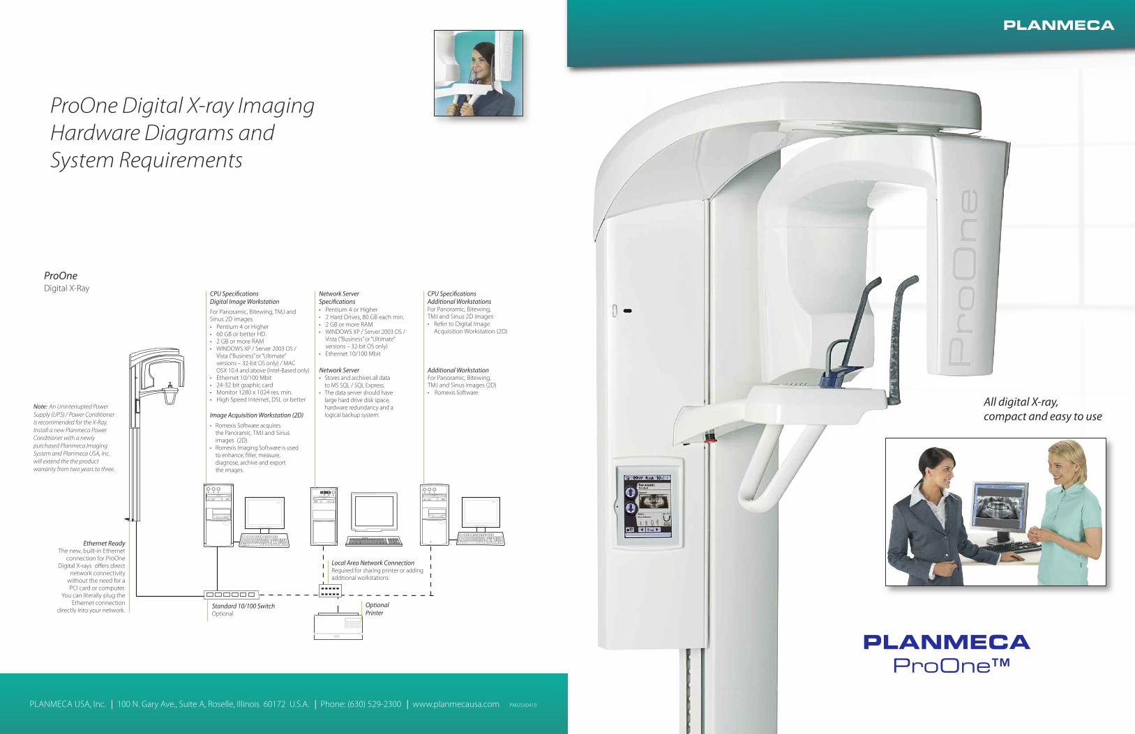
PLANMECA ProOne™ PLANMECA USA, Inc. 100 N. Gary Ave., Suite A, Roselle, Illinois 60172 U.S.A. Phone: (630) 529-2300 www.planmecausa.com PMUSA0410 All digital X-ray, compact and easy to use PLANMECA Ethernet Ready The new, built-in Ethernet connection for ProOne Digital X-rays offers direct network connectivity without the need for a PCI card or computer. You can literally plug the Ethernet connection directly into your network. Image Acquisition Workstation (2D) • Romexis Software acquires the Panoramic, TMJ and Sinus images (2D). • Romexis Imaging Software is used to enhance, filter, measure, diagnose, archive and export the images. Network Server • Stores and archives all data to MS SQL / SQL Express. • The data server should have large hard drive disk space, hardware redundancy and a logical backup system. Additional Workstation For Panoramic, Bitewing, TMJ and Sinus images (2D) • Romexis Software ProOne Digital X-Ray Local Area Network Connection Required for sharing printer or adding additional workstations. Standard 10/100 Switch Optional Optional Printer CPU Specifications Digital Image Workstation For Panoramic, Bitewing, TMJ and Sinus 2D images • Pentium 4 or Higher • 60 GB or better HD • 2 GB or more RAM • WINDOWS XP / Server 2003 OS / Vista (“Business” or “Ultimate” versions – 32-bit OS only) / MAC OSX 10.4 and above (Intel-Based only) • Ethernet 10/100 Mbit • 24-32 bit graphic card • Monitor 1280 x 1024 res. min. • High Speed Internet, DSL or better CPU Specifications Additional Workstations For Panoramic, Bitewing, TMJ and Sinus 2D images • Refer to Digital Image Acquisition Workstation (2D) Network Server Specifications • Pentium 4 or Higher • 2 Hard Drives, 80 GB each min. • 2 GB or more RAM • WINDOWS XP / Server 2003 OS / Vista (“Business” or “Ultimate” versions – 32-bit OS only) • Ethernet 10/100 Mbit Note: An Uninterrupted Power Supply (UPS) / Power Conditioner is recommended for the X-Ray. Install a new Planmeca Power Conditioner with a newly purchased Planmeca Imaging System and Planmeca USA, Inc. will extend the the product warranty from two years to three. ProOne Digital X-ray Imaging Hardware Diagrams and System Requirements
Transcript of ProOneDigitalX-rayImaging HardwareDiagramsand ... Lateral - PA TMJ Program L a te rl3 An gs TMJ P om...
PLANMECAProOne™
PLANMECA USA, Inc. 100 N. Gary Ave., Suite A, Roselle, Illinois 60172 U.S.A. Phone: (630) 529-2300 www.planmecausa.com PMUSA0410
All digital X-ray,compact and easy to use
PLANMECA
Ethernet ReadyThe new, built-in Ethernet
connection for ProOneDigital X-rays offers direct
network connectivitywithout the need for aPCI card or computer.
You can literally plug theEthernet connection
directly into your network.
Image Acquisition Workstation (2D)
• Romexis Software acquiresthe Panoramic, TMJ and Sinusimages (2D).
• Romexis Imaging Software is usedto enhance, filter, measure,diagnose, archive and exportthe images.
Network Server• Stores and archives all data
to MS SQL / SQL Express.• The data server should have
large hard drive disk space,hardware redundancy and alogical backup system.
Additional WorkstationFor Panoramic, Bitewing,TMJ and Sinus images (2D)• Romexis Software
ProOneDigital X-Ray
Local Area Network ConnectionRequired for sharing printer or addingadditional workstations.
Standard 10/100 SwitchOptional
OptionalPrinter
CPU SpecificationsDigital Image Workstation
For Panoramic, Bitewing, TMJ andSinus 2D images• Pentium 4 or Higher• 60 GB or better HD• 2 GB or more RAM• WINDOWS XP / Server 2003 OS /
Vista (“Business” or “Ultimate”versions – 32-bit OS only) / MACOSX 10.4 and above (Intel-Based only)
• Ethernet 10/100 Mbit• 24-32 bit graphic card• Monitor 1280 x 1024 res. min.• High Speed Internet, DSL or better
CPU SpecificationsAdditional WorkstationsFor Panoramic, Bitewing,TMJ and Sinus 2D images• Refer to Digital Image
Acquisition Workstation (2D)
Network ServerSpecifications• Pentium 4 or Higher• 2 Hard Drives, 80 GB each min.• 2 GB or more RAM• WINDOWS XP / Server 2003 OS /
Vista (“Business” or “Ultimate”versions – 32-bit OS only)
• Ethernet 10/100 Mbit
Note: An Uninterrupted PowerSupply (UPS) / Power Conditioneris recommended for the X-Ray.Install a new Planmeca PowerConditioner with a newlypurchased Planmeca ImagingSystem and Planmeca USA, Inc.will extend the the productwarranty from two years to three.
ProOne Digital X-ray ImagingHardware Diagrams andSystem Requirements

Simply Amazing
All digitalThe ProOne Digital Panoramic X-ray’s sleek yet functional design combinessimplicity with the latest state-of-the-art technology to provide ease of use, extensivediagnostic capabilities, and superior image quality in an affordable package.
The ProOne X-ray’s most notable feature is its compact footprint. Add to that its lightweight of 152 lbs., and the result is a panoramic X-ray that can be used in almost anydoctor’s office or imaging facility.
UpgradeableBest of all, the ProOne Digital X-ray’s panoramic and basic programming can beupgraded to the optional ProOne Professional Program Package which includesHorizontal and Vertical Segmenting and advanced programs like the ImprovedInterproximal Program, Improved Orthogonal (Perio) Program and Bitewing Program.The optional Dynamic Exposure Control (DEC) may also be added at any time.
The Panoramic Bitewing Program is also available separately (without the ProfessionalProgram Package).
PLANMECAProOne
2 3
ProOne’s compact and lightweight design brings the benefits of direct digitalimaging to almost any doctor’s office or imaging clinic.

Ease
ofop
erat
ion
For every dentist and any patient
PLANMECAProOne
4
Planmeca Romexis Digital ImagingSoftware helps with patient diagnosisRomexis Software used with digital imagingis very helpful when it comes to diagnostics.Romexis can help enhance critical areas,highlight, extract, change contrast, and evenenlarge an area of interest.
Digital imaging allows easy archiving andefficient record-keeping practicesA digital image requires much less storagespace, its easier to access, and can be foundin much less time than a film image.In addition, the Romexis imaging softwareis interfaced with or bridged with mostpractice management software programs,allowing easy access to all a patient’sinformation in one file.
Easy to operateThe ProOne X-ray unit provides absoluteease of use with cutting edge digitalimaging technology. Simply select thedesired image program, patient type, jawshape and size, and position the patient.The unit’s graphical user interface ensuresthat all types of radiographic examinationsare highly rapid and effortless to perform.
Quick and easy patient positioningProOne’s side entry and open patientpositioning features minimize radiographerrors caused by incorrect patientpositioning. Patient positioning is madequick, precise, and easy, as the user maymonitor the patient freely from the frontand side.
Triple laser beam alignmentPatient positioning is assisted by a triplelaser beam system which accuratelyindicates the correct anatomical positioningpoints. The midsagittal plane positioningbeam shows the correct sideways align-ment of the patient’s head. The Frankforthorizontal plane positioning beam allowsthe correct head tilt. The focal layerpositioning beam helps position the patientaccurately inside the focal layer for a sharpand clear image.
Extremely simple design - compact and lightweightThe PLANMECA ProOne is extremely compact and lightweightwith a simple, sleek design for quick and easy installation.ProOne is delivered fully assembled and ready to install. ProOne’sdesign consists of a simplified mechanical construction withoutmechanical buttons, minimal use of printed circuit boards,minimal cables and the latest universal components. Simply affixthe 152 lb. ProOne to a wall or a freestanding base and the unit isready for immediate use.
Accessible by all patientsThe PLANMECA ProOne easily handles any patient, from childrenand adults to those who are bedridden or wheelchair bound.
Side entryProOne’s side entry feature allows easy access for all patients;the exposure can be performed on a standing or a seatedpatient. If necessary, the patient can even remain seated in awheelchair or lie in a hospital bed with upright lifted backrest.
Open view patient positioningProOne does not require mirrors to position the patient.Instead, the patient has an open and comfortable view. (This isespecially useful when, for example, the patient is a small childwho needs reassurance that a parent is nearby.)
ProOne digital imaging benefitsNow the many benefits of directdigital radiography can be used in eventhe smallest of offices, offering manyadvantages both for the patient and forthe imaging workflow of the practice.
Direct digital imaging saves timeImages are seen on the computer screenwithin seconds after the exposure withoutwaiting for the processing of film in order tomake a proper diagnosis.
ProOne’s CCD Direct Digital TechnologyThe digital sensor technology of the ProOneeliminates chemical processing of films, aswell as the need for chemical developers,chemical disposal, and dark room. Digitalimaging puts an end to the retakes andoverhead associated with processing film.
Wheelchair and hospital bed patientsLaser beam-assisted head alignmentSide entry and open patient positioning
PLANMECAProOne
5
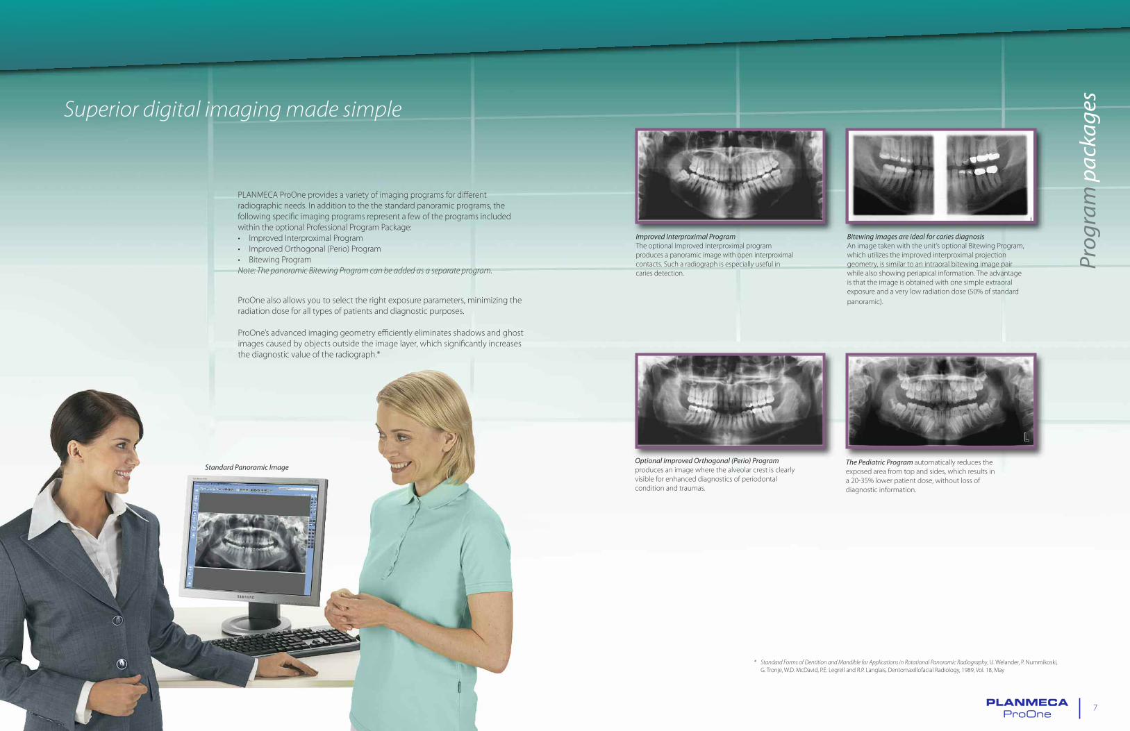
PLANMECA ProOne provides a variety of imaging programs for differentradiographic needs. In addition to the the standard panoramic programs, thefollowing specific imaging programs represent a few of the programs includedwithin the optional Professional Program Package:• Improved Interproximal Program• Improved Orthogonal (Perio) Program• Bitewing ProgramNote: The panoramic Bitewing Program can be added as a separate program.
ProOne also allows you to select the right exposure parameters, minimizing theradiation dose for all types of patients and diagnostic purposes.
ProOne’s advanced imaging geometry efficiently eliminates shadows and ghostimages caused by objects outside the image layer, which significantly increasesthe diagnostic value of the radiograph.*
Improved Interproximal ProgramThe optional Improved Interproximal programproduces a panoramic image with open interproximalcontacts. Such a radiograph is especially useful incaries detection.
Bitewing Images are ideal for caries diagnosisAn image taken with the unit’s optional Bitewing Program,which utilizes the improved interproximal projectiongeometry, is similar to an intraoral bitewing image pairwhile also showing periapical information. The advantageis that the image is obtained with one simple extraoralexposure and a very low radiation dose (50% of standardpanoramic).
Optional Improved Orthogonal (Perio) Programproduces an image where the alveolar crest is clearlyvisible for enhanced diagnostics of periodontalcondition and traumas.
The Pediatric Program automatically reduces theexposed area from top and sides, which results ina 20-35% lower patient dose, without loss ofdiagnostic information.
Standard Panoramic Image
Prog
ram
pack
agesSuperior digital imagingmade simple
* Standard Forms of Dentition andMandible for Applications in Rotational Panoramic Radiography, U. Welander, P. Nummikoski,G. Tronje, W.D. McDavid, P.E. Legrell and R.P. Langlais, Dentomaxillofacial Radiology, 1989, Vol. 18, May
PLANMECAProOne
7

Prog
ram
pack
age
The Automatic Double TMJ Programproduces a lateral view (above) or a posterior/anteriorview (above right) of open and closedtemporomandibular joints on one radiograph.
While the TMJ imaging procedure is straightforward, theradiograph allows easy diagnosis of the TMJ condition inone view.
Optional Segmenting Program limits the exposedarea only to the area of diagnostic interest. With asimple selection on the GUI, the patient dose can bereduced by up to 90% compared to a full areapanoramic exposure.**
The Optional Cross-sectional Program is intendedfor simple cross-sectional imaging of TMJs and jawsin the molar and premolar region. These imagesconvey highly valuable information on cross-sectionaldimensions and the structure of the jaw.
** Absorbed dose reduced by sliced exposure using sector selector systemwith rotational panoramic radiography, Y. Hayakawa,N. Kobayashi, Y. Kousuge, H. Fujimori and K. Kuroyanagi, Bulletin of Tokyo Dental College, Vol. 35, No. 3, pp. 127–131, August, 1994
Optional Sinus Programhas a specially designed image layer providing aradiograph with a clear view of the maxillary sinuses.
PLANMECAProOne
9
ProOne Digital Panoramic X-ray ProgramsAll 5 Basic Programs come standard with the unit. The Professional Program Package is optional andcontains over 12 advanced panoramic programs including the Bitewing Program. The optional BitewingProgram is also available separately.
Basic Programs
Standard Panoramic Program
Pediatric Program
Double Lateral TMJ Program
Double PA TMJ Program
PA Sinus Program
Professional Program Package
Horizontal and Vertical Segmenting
Improved Interproximal Program
Improved Orthogonal (Perio) Program
Bitewing Program
Double Lateral - PA TMJ Program
Lateral 3 Angles TMJ Program (left or right)
Lateral Sinus Program (left or right)
Lateral Midsagittal Sinus Program (left or right)
Cross-sections, Manual or Automatic
Available Separately
Bitewing Program

Rom
exis
imag
ing
soft
war
e
PLANMECAProOne
10
Romexis SoftwareThe Romexis Platform fully integrates digital imaging withthe patients other clinical data. The system provides directimage capture from Planmeca’s X-Ray Imaging Units aswell as interface with 3rd party devices and software. Ithas a pure Java based interface that runs in various operatingsystems and modern web environments. Romexis includes acopy of MS SQL Express for storing and archiving all data onserver hard drive..
Complete Digital ImagingPlanmeca Romexis includes all dental imaging modalities:intraoral, panoramic, cephalometric and 3D imaging,dental tomography as well as intraoral video and stillcamera images. With a complete set of tools for imageviewing, enhancement, measurement and annotation,Planmeca Romexis also improves the diagnostic valueof radiographs.
DICOM CompliantDICOM, short for Digital Imaging and Communications inMedicine, is a worldwide standard for image transfer inmedical information systems. Romexis is 100% DICOMcompatible and fully ADA compliant and provides, as anoption, the widest possible DICOM functionalities.
Romexis Software Tools• Customized templates• Image navigator tool• Image magnifier tool• Point-to-point measurements tool• Image filtering tool• Pseudo-color enhancement tool• Image inversion tool• Tooth range program• Image parameter tool• Gray level auto-adjustment program• Flashlight tool
TWAIN DriverThe TWAIN Driver allows direct digital X-rayimage acquisition into a third party imagingsoftware and for another vendor’s TWAINcompliant imaging software to directly acquiredigital X-ray images taken with the ProOne.
Easy to use Romexis Tools Romexis handles all image types
PLANMECAProOne
11

12
Graphic User InterfaceThe full-color TFT display has a graphical userinterface (GUI) that guides the operator withtext and clear graphic symbols.
Intuitive controlsThe GUI design is based on cognitiveergonomics: All settings are logically groupedand easy to understand. The imagingprocedure, program selection and exposureparameters are intuitive to the operator andallow full focus on patient positioning andcommunication. All necessary information isshown on the main display with a hygienicwipe-clean surface.
Focal layer adjustmentBy simply touching the GUI, the operator canadjust the shape of the focal layer accordingto the jaw size and shape characteristics ofthe patient.
Preview imageAfter the patient’s X-ray has been taken,a preview image is displayed across thegraphical user interface.
Preview magnificationFor improved image validation, the GUImagnifies a selected portion of the previewwhen it is touched by the operator.
Patient identificationIf the patient is not already identified onthe GUI through Romexis, simply touchingthe ID area on the screen will allow theoperator to enter patient information.
Optional Dynamic Exposure ControlThe unique digital Dynamic Exposure Control (DEC)optimizes the whole imaging chain individually for eachpatient. All components, from the X-ray generator tothe digital sensor, are tuned to produce the optimumimage quality.
Exposure Parameter Control• Adjusts the exposure parameters optimal for each
patient automatically• Prevents too low initial exposure parameters from
causing under-exposure and/or poor image quality• Prevents unnecessary high radiation levels
Automatic Gain Control• Adjusts the sensitivity of the sensor according to
the amount of incoming radiation• Adapts automatically to patient anatomy• Prevents pixel saturation even in soft tissue and
direct radiation areas• Works in all programs
A self-diagnostic control system continuously monitorsthe unit. The system displays “help” messages which guidethe operator and enable the correct use of the unit. Thecontrol system also displays error messages in case ofabnormal operation. These error messages are stored inan error log to help both the operator and assist withtechnical service.
Gra
phic
Use
rInt
erfa
cean
dop
tion
alD
EC
ProOne Digital Panoramic X-ray Programs1. Basic Programs:
• Standard Panoramic Program• Pediatric Program• Double Lateral TMJ Program• Double PA TMJ Program• PA Sinus Program
2. Optional Professional Program Package:• Horizontal and Vertical Segmenting• Improved Interproximal Program• Improved Orthogonal (Perio) Program• Bitewing Program• Double Lateral - PA TMJ Program• Lateral 3 Angles TMJ Program
(left or right)• Lateral Sinus Program (left or right)• Lateral Midsagittal Sinus Program
(left or right)• Cross-sections, Manual or automatic
Note: Bitewing Program can be added as aseparate programwithout purchasing the entireProfessional Program package.
Simple, easy-to-understand Graphic User Interfacemakes digital imaging quick, accurate and aseptic.
Optional Dynamic Exposure Control adjusts theexposure parameter automatically per eachpatient and controls the sensitivity of the digitalsensor for optimum image quality.
Exposure Control
Dynamic GainControl
Vertical and horizontal segmenting Select jaw size and shapePanoramic programs TMJ programs
Sinus programsTMJ selection Cross-Sections program Cross-Section manual angle
PLANMECAProOne
PLANMECAProOne
13

Tech
nica
lspe
cific
atio
ns
14 PLANMECAProOne
PLANMECAProOne
15
Actual Product DimensionsWidth: Depth: Height:30 inches 41 inches 88 inches
Minimum Operational DimensionsWidth: Depth: Height:38 inches 46.6 inches 89 inches
Optimal Operational DimensionsWidth: Depth: Height:54 inches 51 inches 89 inches
88”
(89”
Reco
mm
end
ed)
33.5
-69”
72.1
3”
Mounting BracketsWall Mount with Floor MountOptional 2nd Wall Mount
BracketHole Spacing
5.9”
7.9”
Appr
ox.5
9”
FloorMount
WallMount
Extension Plate16” Hole Spacing
Optional2ndWallMount
App
rox.
66.9
”
PortsUnder Back Cover
USBMemory
ExposureSwitch
MasterSwitch
Fuses
ExposureSwitch
Ethernet
30 in.(54 in. Recommended)
(51
in.R
ecom
men
ded
)
11.5” 19.5”
WALL
41in
.
31in
.10
”
� - Actual ProductDimensions
� - RecommendedInstallation Dimensions
ProOne Digital PanOverhead View
ProOne Digital PanFront & Side Views
Generator Constant potential,resonance modehigh frequency 60 - 80 kHz
X-ray tube D-058SBR
Digital Sensor CCD Technology
Sensor Pixel Size 33µm
Image Pixel Size 132µm
Focal spot size 0.5 x 0.5 mm (IEC 336)
SID 480 mm (19 in.)
Total filtration 2.5 mm Al
Anode voltage 60 - 70 kV
Anode current 2 - 7 mA DC
Exposure time 2 - 10 s
Magnification 1.22 - 1.29
Line voltage 100 - 132 V~ 50/60 Hz,180 - 240 V~ 50 Hz
Regulation ± 10 % (automatic)
Line current 8 - 16 A
Power uptake max: 850 W
Chin rest level 33.5 - 69 in. (85 - 175 cm)
Exterior color RAL 9016 (white)
Weight 69 kg (152 lbs.)