Project Completion Report - Amazon S3 · Project Completion Report PART I Project Code: ......
29
2 Project Completion Report PART I Project Code: 05-02 Subcontract/Account No. 557221 supported by 2003-38500-13505 Project Title: “Development of genetic markers to assess disease resistance in the eastern oyster” Reporting Period: February 2005 – January 2008 Funding Level: $128,486 Participants: Steven Roberts – adjunct scientist in MBL’s Scientific Aquaculture program and Assistant Professor at the University of Washington with background in molecular biology Roxanna Smolowitz – traditionally trained veterinary pathologist, has extensive experience studying bivalve disease Richard Karney – Director of Martha’s Vineyard Shellfish Group, Inc., holds culture experience with numerous bivalves Inke Sunila – invertebrate pathologist, has extensive experience in the diagnosis of bivalve diseases and working with aquaculturists Dale Leavitt – extensive experience in research and aquaculture of shellfish, hatchery management, and extension Bill Walton – aquaculture specialist with research experience in shellfish biology and outreach/extension activities Frederick Goetz – senior scientist with background in aquatic animal immunology and molecular biology Paul Bagnall – shellfish constable for Martha’s Vineyard with significant culture experience Reason for Termination: Project Completed Project Objectives: 1. To demonstrate seed originating from local wild oysters, that have experienced heavy disease (Dermo) pressure, could significantly contribute to the development of disease resistance in cultured oysters. 2. To genetically characterize regional oysters (C. virginica) that are putatively resistant (more tolerant) to Dermo, in order to development genetic markers and to better understand mechanism involved in immunity. 3. To communicate with northeastern hatchery operations and help them to identify local, potentially Dermo resistant broodstocks.
Transcript of Project Completion Report - Amazon S3 · Project Completion Report PART I Project Code: ......
2
Project Completion Report
PART I
Project Code: 05-02 Subcontract/Account No. 557221 supported by 2003-38500-13505 Project Title: “Development of genetic markers to assess disease resistance in the eastern oyster” Reporting Period: February 2005 – January 2008 Funding Level: $128,486 Participants: Steven Roberts – adjunct scientist in MBL’s Scientific Aquaculture program and Assistant
Professor at the University of Washington with background in molecular biology
Roxanna Smolowitz – traditionally trained veterinary pathologist, has extensive experience studying bivalve disease Richard Karney – Director of Martha’s Vineyard Shellfish Group, Inc., holds culture experience with numerous bivalves Inke Sunila – invertebrate pathologist, has extensive experience in the diagnosis of bivalve diseases and working with aquaculturists Dale Leavitt – extensive experience in research and aquaculture of shellfish, hatchery management, and extension Bill Walton – aquaculture specialist with research experience in shellfish biology and outreach/extension activities Frederick Goetz – senior scientist with background in aquatic animal immunology and molecular biology Paul Bagnall – shellfish constable for Martha’s Vineyard with significant culture experience Reason for Termination: Project Completed Project Objectives: 1. To demonstrate seed originating from local wild oysters, that have experienced heavy disease (Dermo) pressure, could significantly contribute to the development of disease resistance in cultured oysters. 2. To genetically characterize regional oysters (C. virginica) that are putatively resistant (more tolerant) to Dermo, in order to development genetic markers and to better understand mechanism involved in immunity. 3. To communicate with northeastern hatchery operations and help them to identify local, potentially Dermo resistant broodstocks.
3
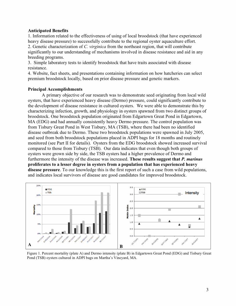
Anticipated Benefits 1. Information related to the effectiveness of using of local broodstock (that have experienced heavy disease pressure) to successfully contribute to the regional oyster aquaculture effort. 2. Genetic characterization of C. virginica from the northeast region, that will contribute significantly to our understanding of mechanisms involved in disease resistance and aid in any breeding programs. 3. Simple laboratory tests to identify broodstock that have traits associated with disease resistance. 4. Website, fact sheets, and presentations containing information on how hatcheries can select premium broodstock locally, based on prior disease pressure and genetic markers. Principal Accomplishments A primary objective of our research was to demonstrate seed originating from local wild oysters, that have experienced heavy disease (Dermo) pressure, could significantly contribute to the development of disease resistance in cultured oysters. We were able to demonstrate this by characterizing infection, growth, and physiology in oysters spawned from two distinct groups of broodstock. One broodstock population originated from Edgartown Great Pond in Edgartown, MA (EDG) and had annually consistently heavy Dermo pressure. The control population was from Tisbury Great Pond in West Tisbury, MA (TSB), where there had been no identified disease outbreak due to Dermo. These two broodstock populations were spawned in July 2005, and seed from both broodstock populations placed in ADPI bags for 18 months and routinely monitored (see Part II for details). Oysters from the EDG broodstock showed increased survival compared to those from Tisbury (TSB). Our data indicates that even though both groups of oysters were grown side by side, the TSB oysters had a higher prevalence of Dermo and furthermore the intensity of the disease was increased. These results suggest that P. marinus proliferates to a lesser degree in oysters from a population that has experienced heavy disease pressure. To our knowledge this is the first report of such a case from wild populations, and indicates local survivors of disease are good candidates for improved broodstock.
A B
Figure 1. Percent mortality (plate A) and Dermo intensity (plate B) in Edgartown Great Pond (EDG) and Tisbury Great Pond (TSB) oysters cultured in ADPI bags on Martha’s Vineyard, MA.
4
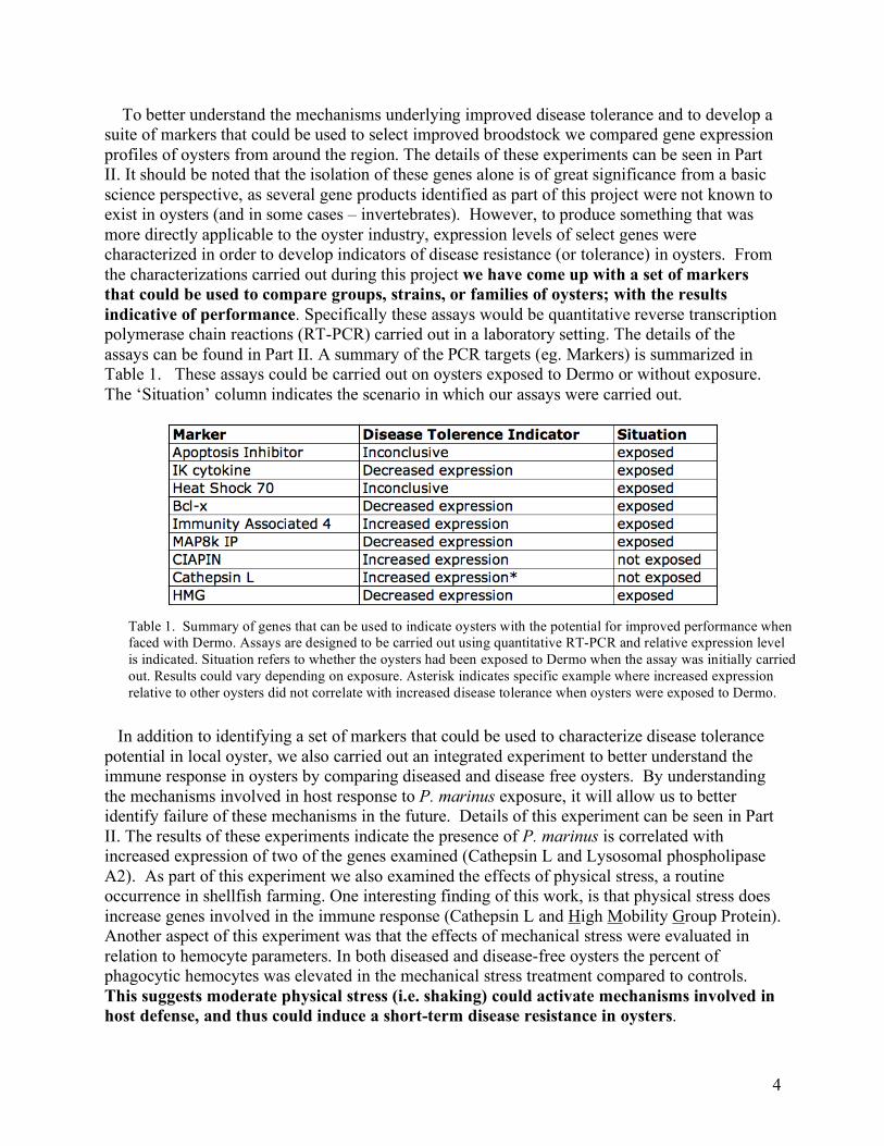
To better understand the mechanisms underlying improved disease tolerance and to develop a suite of markers that could be used to select improved broodstock we compared gene expression profiles of oysters from around the region. The details of these experiments can be seen in Part II. It should be noted that the isolation of these genes alone is of great significance from a basic science perspective, as several gene products identified as part of this project were not known to exist in oysters (and in some cases – invertebrates). However, to produce something that was more directly applicable to the oyster industry, expression levels of select genes were characterized in order to develop indicators of disease resistance (or tolerance) in oysters. From the characterizations carried out during this project we have come up with a set of markers that could be used to compare groups, strains, or families of oysters; with the results indicative of performance. Specifically these assays would be quantitative reverse transcription polymerase chain reactions (RT-PCR) carried out in a laboratory setting. The details of the assays can be found in Part II. A summary of the PCR targets (eg. Markers) is summarized in Table 1. These assays could be carried out on oysters exposed to Dermo or without exposure. The ‘Situation’ column indicates the scenario in which our assays were carried out.
In addition to identifying a set of markers that could be used to characterize disease tolerance potential in local oyster, we also carried out an integrated experiment to better understand the immune response in oysters by comparing diseased and disease free oysters. By understanding the mechanisms involved in host response to P. marinus exposure, it will allow us to better identify failure of these mechanisms in the future. Details of this experiment can be seen in Part II. The results of these experiments indicate the presence of P. marinus is correlated with increased expression of two of the genes examined (Cathepsin L and Lysosomal phospholipase A2). As part of this experiment we also examined the effects of physical stress, a routine occurrence in shellfish farming. One interesting finding of this work, is that physical stress does increase genes involved in the immune response (Cathepsin L and High Mobility Group Protein). Another aspect of this experiment was that the effects of mechanical stress were evaluated in relation to hemocyte parameters. In both diseased and disease-free oysters the percent of phagocytic hemocytes was elevated in the mechanical stress treatment compared to controls. This suggests moderate physical stress (i.e. shaking) could activate mechanisms involved in host defense, and thus could induce a short-term disease resistance in oysters.
Table 1. Summary of genes that can be used to indicate oysters with the potential for improved performance when faced with Dermo. Assays are designed to be carried out using quantitative RT-PCR and relative expression level is indicated. Situation refers to whether the oysters had been exposed to Dermo when the assay was initially carried out. Results could vary depending on exposure. Asterisk indicates specific example where increased expression relative to other oysters did not correlate with increased disease tolerance when oysters were exposed to Dermo.
5
An unintended product of this research that will also positively influence the shellfish aquaculture industry was the development of a quantitative polymerase chain reaction (qPCR) assay for detection of P. marinus. We developed this assay to more precisely correlate mortality and gene expression levels with the level of disease. The assay has been validated against traditional methods and will serve as a high-throughput, low cost alternative to current methods of diagnoses. Impacts Our data does suggest that shellfish farmers will be able to realize improved survival with local broodstock that has experienced persistent disease pressure. The superior broodstock identified in this project are now being grown in Maine as part of other NRAC research and will be used for local enhancement programs. In addition, our gene expression data can be used for marker-assisted selection activities. These results would not only be beneficial to the oyster industry but could likely be used in developing superior broodstock in other shellfish. Recommended Follow-up Activities: While our data is promising regarding the use of local broodstock, we acknowledge that this is based on one trial in Massachusetts. [At the most we have two examples, if you count the gene expression data from the Rhode Island oyster experiment.] The Edgartown Great Pond situation is somewhat unique in that there is geographic isolation and thereby limited opportunity of dilution of the gene pool by recruitment from other populations. This is what probably distinguishes what was observed during this trial compared to the Chesapeake Bay where, despite significant disease pressure, limited barriers facilitate population mixing. This could be related to the fact that a reliably resistant population has not been identified in the Chesapeake. These and other properties of the Edgartown Great Pond scenario raise several important questions. One is whether it takes 8 generations of selection to obtain improved disease resistance? Would the TSB oysters that have survived the trial perform similarly to the EDG oysters? Also, what factors are involved in “reversion” or loss of improved performance? Is consistent pressure needed? It is likely several complex factors influence this reversion including the mixing of populations described (i.e. seed and gametes from non-selected broodstock) and the underlying mechanism(s) responsible for the improved immune capability. Selection in different locations, or over varying number of generations, could select for different suites of genes. Depending on the physiological relevance or role these mechanisms play, the effects on other traits and loss of improved immune function could vary. Finally, another question worth examining is whether resistant stocks (e.g. Rutger’s lines) could be further selected for fitness by grow-outs in local waters. There are numerous “next steps” that could be taken to begin to address these questions. For instance, simply replicating the trial we carried out in Massachusetts in other areas, as well as focusing on other disease or other threatening stressors. To expand our knowledge of whether the improved performance of our specific local broodstock is observed across other stressors, the EDG oysters could be easily incorporated into other grow-out trials. This is, in fact, currently underway in Maine as part of another NRAC funded project carried out by Rawson (University of Maine) and Lindell (MBL). Just as basic performance (i.e. survivorship and growth) could be evaluated in other locals, it would be important to examine the consistency of marker accuracy across strains, locations, and stressors.
6
While the markers that were used as part of this project are all related to immune function in some manner, it is not clear from these experiments whether the differences in expression level are directly related to the active immune response or are merely indirectly correlated with these processes. To better understand the host immune response it would be advantageous to examine these interactions on a tissue and/or cellular level. This information would not necessarily be of direct use to growers, however this could provide important insight into new targets for selection. There are a couple of unforeseen results of our research that warrant further investigation. One is the limited evidence that suggest a link between mechanical stress and activation of immune response mechanisms. Specifically, we observed a correlation in gene expression levels and, more interestingly, an increase in percent phagocytic hemocytes following mechanical stress. While it is likely that excessive physical stress would have negative impacts on an oyster’s ability to defend against disease, directly examining the effects of light to moderate physical stress on immune function requires more research. The second product that deserves follow-up attention is the PCR-based, quantitative assay for P. marinus. In order to properly compare the reliability of such an assay it should be carried out (in conjunction with traditional methods) in a variety of locations under the auspices of different laboratories. Support NRAC-USDA Funding Matching Support Total Support Year 1 $56,962 $13,979 $70,941 Year 2 $71,542 $13,979 $85,503 TOTAL $128,486 $27,959 $156,445 Publications, manuscripts, or papers presented Roberts, SB. 2006. Genomic approaches in characterizing shellfish disease: interrelationships between animal, human and ecosystem health. Cummings School of Veterinary Medicine at Tufts University, Annual Symposium: Marine and Aquatic Medicine & Conservation. North Grafton, MA. April 22, 2006. Diner, E., Smolowitz, R., Gomez-Chiarri, M., Tammi, K., Leavitt, D., Roberts, S. 2006. Assessing disease tolerance in the eastern oyster using gene expression profiling. 26th Annual NOAA-NMFS Milford Aquaculture Symposium. Meridan, CT. February 28, 2006. Steven Roberts, Gary Wikfors, Inke Sunila, Christina Romano, Frederick Goetz, Mike Grzybowski. 2007. Gene expression profiling and cellular characteristics of Crassostrea virginica hemocytes: Evaluating interactions of physical stress and disease exposure. Aquaculture 2007 and NSA Conference. San Antonio, TX. March 1 2007.
7
Publications, manuscripts, or papers presented continued Smolowitz, R., Roberts, S., DeFaveri, J., Romano, C., Karney, R. 2006. Characterizing disease resistance in native oysters that have experienced disease pressure. NACE. Groton, CT. December 6, 2006. [http://fish.washington.edu/research/genefish/robertslab/dermo.html] DeFaveri, J., Roberts, S., Romano, C., Smolowitz, R. Real-time PCR for routine diagnosis of “Dermo” disease in Crassostrea virginica. NACE. Groton, CT. December 6, 2006. [http://fish.washington.edu/research/genefish/robertslab/dermo.html] Steven Roberts; Yannick Gueguen; Julien de Lorgeril; Frederick Goetz. 2008. Rapid accumulation of an interleukin 17 homolog transcript in Crassostrea gigas hemocytes following bacterial exposure. Submitted to Developmental and Comparative Immunology Roberts, S., Sunila, I., Wikfors, G., Goetz, F. 2008. Gene expression profiling and cellular characteristics of Crassostrea virginica hemocytes: Evaluating interactions of physical stress and disease exposure. Manuscript in preparation. Roberts, S. Smolowitz, R., Karney, R. 2008. Disease tolerance in native oysters that have experienced disease pressure. Manuscript in preparation. “In search of disease resistant oysters” Television Documentary. Martha’s Vineyard Community Television. Featuring Smolowitz, Karney, and Roberts. Produced by Gail Tipton. [http://fish.washington.edu/research/genefish/robertslab/dermo.html] Roberts, S. Smolowitz, R., Karney, R., Walton, W. 2008. Development of genetic markers to assess disease resistance in the eastern oyster. Factsheet. [http://fish.washington.edu/research/genefish/robertslab/dermo.html]
8
PART II Technical Analysis and Summary One of the major causes of decreased production for the oyster industry is disease. The two primary diseases that affect the adult eastern oyster are MSX and Dermo. Both diseases invade the oyster's soft body resulting in death of the individual. The disease MSX is caused by the protozoan parasite Haplosporidium nelsoni and is present along the entire east coast. The disease Dermo is caused by the parasite Perkinsus marinus. In the last decade, the disease has markedly affected oyster culture in the more northern portion of the parasite’s range (Connecticut, Rhode Island and Massachusetts) in addition to states already identified as problematic (New York to the Gulf of Mexico). Oyster disease is a particular concern of shellfish farmers in the northeast region not only due to periodic devastating oyster losses, but also because disease indirectly affects the industry by slowing financial investments. Realizing that oyster disease is a primary concern for the industry, the long-term goal of the proposed research is to assist in the development of disease resistant eastern oyster broodstocks. Previous research has demonstrated that genetic factors can be selected for that contribute to disease resistance in the eastern oyster. A majority of this prior work has involved hatchery-based selection practices with limited information on the performance of wild oyster populations that have survived heavy disease pressure. The first research objective of this project was to demonstrate seed originating from an isolated population of local wild oysters, that have experienced heavy disease (Dermo) pressure over several years, could significantly contribute to the development of disease resistance in cultured oysters. In order to carry out this objective, oyster seed originating from Martha’s Vineyard, MA were grown-out in ADPI bags. One broodstock population originated from Edgartown Great Pond in Edgartown, MA (EDG) and had annually consistently heavy disease pressure for greater than 8 years. The control population (eg. potentially susceptible to Dermo) was from Tisbury Great Pond in West Tisbury, MA (TSB), where there had been no identified disease outbreak due to Dermo and no positive Dermo samples for at least 3 years prior to broodstock selection from the pond. We had anticipated to also plant out seed from a proven disease resistant line (e.g. Rutgers), along with appropriate controls for generalized comparisons. However, when these seed were tested for disease at the Marine Biological Laboratory just prior to deployment, the Rutgers line tested positive. As a result, these groups were excluded from the trial. In July 2005, 8000 oyster seed (4000 from EDG broodstock and 4000 from TSB broodstock) were deployed in floating bags at two adjacent sites in Edgartown Great Pond. The average shell height of the oyster seed was 21 mm. In September 2005 the oysters were thinned out by adding additional ADPI bags at each site and evenly distributing the oysters. In September 2005 there were 12 bags at each site (6 with EGP oysters and 6 with TSB oysters. As the number of oysters decreased, the number of bags was reduced to maintain similar densities throughout the experiment. These sites in Edgartown Great Pond consistently have had evidence of Dermo. The bags remained on the surface except from October 2005 to May 2006, when the bags were submerged for overwintering
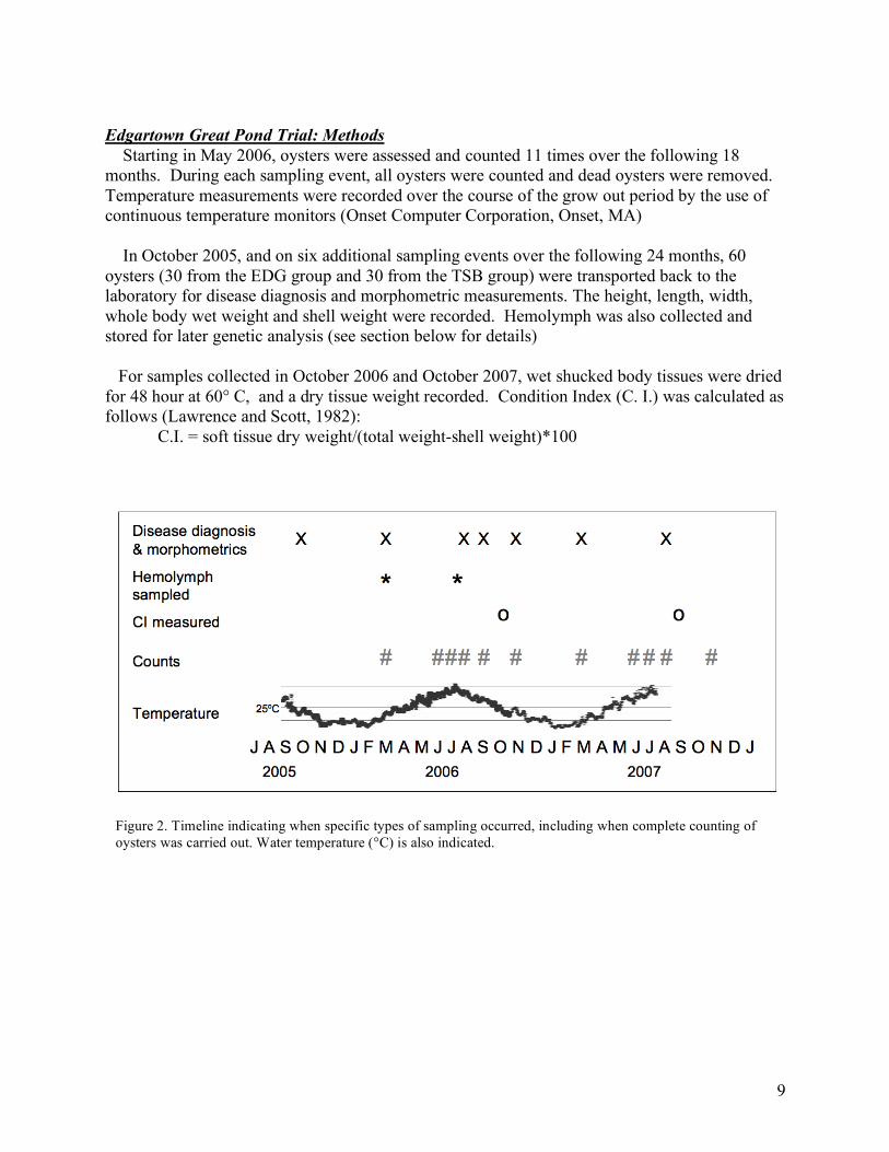
9
Edgartown Great Pond Trial: Methods Starting in May 2006, oysters were assessed and counted 11 times over the following 18 months. During each sampling event, all oysters were counted and dead oysters were removed. Temperature measurements were recorded over the course of the grow out period by the use of continuous temperature monitors (Onset Computer Corporation, Onset, MA) In October 2005, and on six additional sampling events over the following 24 months, 60 oysters (30 from the EDG group and 30 from the TSB group) were transported back to the laboratory for disease diagnosis and morphometric measurements. The height, length, width, whole body wet weight and shell weight were recorded. Hemolymph was also collected and stored for later genetic analysis (see section below for details) For samples collected in October 2006 and October 2007, wet shucked body tissues were dried for 48 hour at 60° C, and a dry tissue weight recorded. Condition Index (C. I.) was calculated as follows (Lawrence and Scott, 1982): C.I. = soft tissue dry weight/(total weight-shell weight)*100
Figure 2. Timeline indicating when specific types of sampling occurred, including when complete counting of oysters was carried out. Water temperature (°C) is also indicated.
10
Table 2. Makin Scale for rating Dermo infections. RANK INTENSITY DESCRIPTION 0.0 none no hypnospores present 0.5 very light 1-20 cells in entire tissue prep 1.0 light 20-100 cells in entire tissue prep 2.0 light to moderate Localized infections of 25-50 cells 3.0 moderate All fields at 100x magnification show several parasites 4.0 moderate to heavy Parasites present in large numbers 5.0 heavy Majority of tissue stained blue-black macroscopically; enormous numbers of parasites Source: Ray, S.M (1954) Biological Studies of Dermocystidium marinum. The Rice Institute Pamphlet, Spec. Iss., 114p Oysters transported back to the lab were tested for the presence and severity of infection by P. marinus using standard Thioglycollate culture methods (Ray 1954, Mackin 1962). Perkinsus marinus was diagnosed from anal-rectal tissues using Ray’s Fluid Thioglycollate Medium containing 2% NaCl and supplemented with a final concentration of 50 units of penicillin G and 50 µg of streptomycin sulfate per ml of medium (Bushek et al. 1994). The tissues were incubated for 8 days at room temperature. The intensity of the infection in the oyster was rated using the Makin Scale (0-5) (Table 2). Shucked animals were processed for histological examination using standard methods (Howard and Smith, 1983). Briefly, cross-sections of same oysters including digestive diverticulum, gills and mantle, were excised and fixed in Davidson’s fixative for 48 h at 4ºC. Samples were dehydrated and embedded in paraffin. Five-µm sections were stained using hematoxylin-eosin (Howard et al., 2004). Tissues/organs were evaluated for P. marinus occurrence and were rated for abundance of parasites (rare organisms seen = 1, moderate numbers seen = 2 and abundant numbers seen = 4), parasite forms identified in the sections (single cell inclusions only = 1, inclusions and signet rings = 2; all forms including rosettes = 3), location of the inflammatory response within the organ/tissue examined (focal = 1, multifocal = 2, diffuse = 3, focally extensive = 4) and severity of the inflammatory response (number of hemocytes in the tissue/organ) (mild = 1, moderate = 2, severe = 3). Scores for each organ/tissue in the sample group were averaged. Edgartown Great Pond Trial: Results and Discussion Temperature loggers were fastened to the ADPI bags throughout the experiment. A maximum temperature of 31.8 °C was recorded in August 2006 and a minimum temperature of -0.8 °C was recorded in January 2007. The average temperature over the course of the trial was 11.3 °C.
11
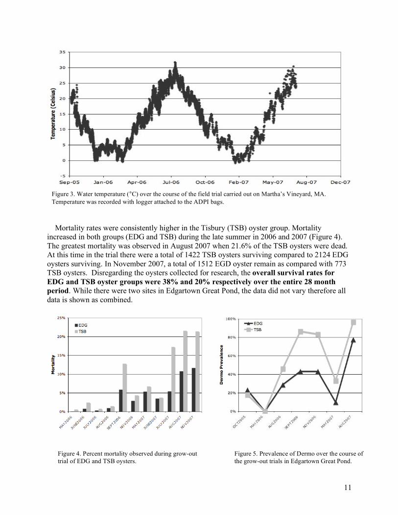
Mortality rates were consistently higher in the Tisbury (TSB) oyster group. Mortality increased in both groups (EDG and TSB) during the late summer in 2006 and 2007 (Figure 4). The greatest mortality was observed in August 2007 when 21.6% of the TSB oysters were dead. At this time in the trial there were a total of 1422 TSB oysters surviving compared to 2124 EDG oysters surviving. In November 2007, a total of 1512 EGD oyster remain as compared with 773 TSB oysters. Disregarding the oysters collected for research, the overall survival rates for EDG and TSB oyster groups were 38% and 20% respectively over the entire 28 month period. While there were two sites in Edgartown Great Pond, the data did not vary therefore all data is shown as combined.
Figure 3. Water temperature (°C) over the course of the field trial carried out on Martha’s Vineyard, MA. Temperature was recorded with logger attached to the ADPI bags.
Figure 4. Percent mortality observed during grow-out trial of EDG and TSB oysters.
Figure 5. Prevalence of Dermo over the course of the grow-out trials in Edgartown Great Pond.
12
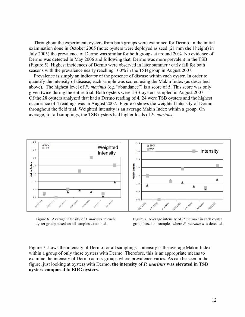
Throughout the experiment, oysters from both groups were examined for Dermo. In the initial examination done in October 2005 (note: oysters were deployed as seed (21 mm shell height) in July 2005) the prevalence of Dermo was similar for both groups at around 20%. No evidence of Dermo was detected in May 2006 and following that, Dermo was more prevalent in the TSB (Figure 5). Highest incidences of Dermo were observed in later summer / early fall for both seasons with the prevalence nearly reaching 100% in the TSB group in August 2007. Prevalence is simply an indicator of the presence of disease within each oyster. In order to quantify the intensity of disease, each sample was scored using the Makin Index (as described above). The highest level of P. marinus (eg. “abundance”) is a score of 5. This score was only given twice during the entire trial. Both oysters were TSB oysters sampled in August 2007. Of the 28 oysters analyzed that had a Dermo reading of 4, 24 were TSB oysters and the highest occurrence of 4 readings was in August 2007. Figure 6 shows the weighted intensity of Dermo throughout the field trial. Weighted intensity is an average Makin Index within a group. On average, for all samplings, the TSB oysters had higher loads of P. marinus.
Figure 7 shows the intensity of Dermo for all samplings. Intensity is the average Makin Index within a group of only those oysters with Dermo. Therefore, this is an appropriate means to examine the intensity of Dermo across groups where prevalence varies. As can be seen in the figure, just looking at oysters with Dermo, the intensity of P. marinus was elevated in TSB oysters compared to EDG oysters.
Weighted Intensity Intensity
Figure 6. Average intensity of P marinus in each oyster group based on all samples examined.
Figure 7. Average intensity of P marinus in each oyster group based on samples where P. marinus was detected.
13
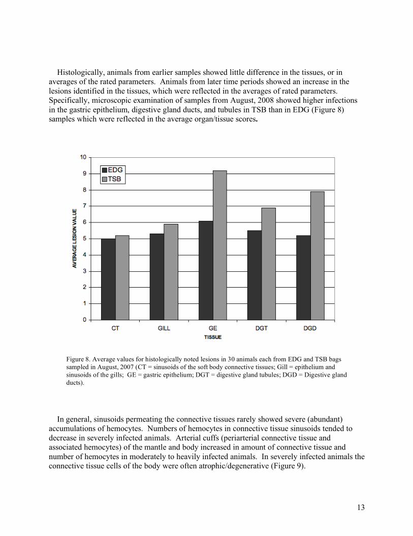
Histologically, animals from earlier samples showed little difference in the tissues, or in averages of the rated parameters. Animals from later time periods showed an increase in the lesions identified in the tissues, which were reflected in the averages of rated parameters. Specifically, microscopic examination of samples from August, 2008 showed higher infections in the gastric epithelium, digestive gland ducts, and tubules in TSB than in EDG (Figure 8) samples which were reflected in the average organ/tissue scores.
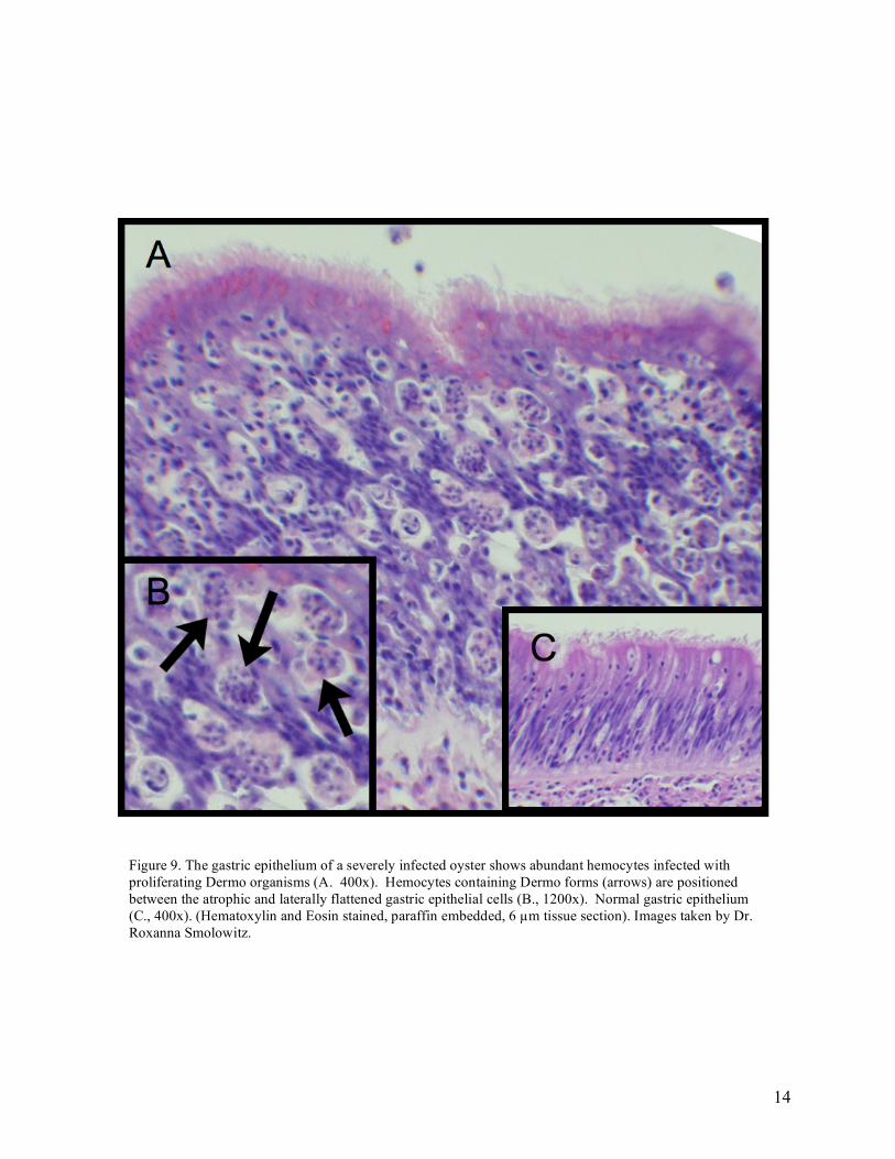
In general, sinusoids permeating the connective tissues rarely showed severe (abundant) accumulations of hemocytes. Numbers of hemocytes in connective tissue sinusoids tended to decrease in severely infected animals. Arterial cuffs (periarterial connective tissue and associated hemocytes) of the mantle and body increased in amount of connective tissue and number of hemocytes in moderately to heavily infected animals. In severely infected animals the connective tissue cells of the body were often atrophic/degenerative (Figure 9).
Figure 8. Average values for histologically noted lesions in 30 animals each from EDG and TSB bags sampled in August, 2007 (CT = sinusoids of the soft body connective tissues; Gill = epithelium and sinusoids of the gills; GE = gastric epithelium; DGT = digestive gland tubules; DGD = Digestive gland ducts).
14
Figure 9. The gastric epithelium of a severely infected oyster shows abundant hemocytes infected with proliferating Dermo organisms (A. 400x). Hemocytes containing Dermo forms (arrows) are positioned between the atrophic and laterally flattened gastric epithelial cells (B., 1200x). Normal gastric epithelium (C., 400x). (Hematoxylin and Eosin stained, paraffin embedded, 6 µm tissue section). Images taken by Dr. Roxanna Smolowitz.
15
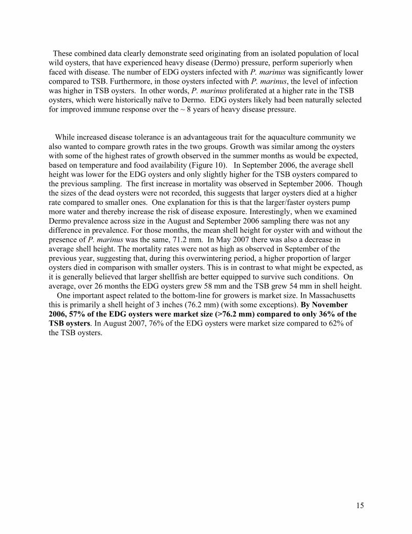
These combined data clearly demonstrate seed originating from an isolated population of local wild oysters, that have experienced heavy disease (Dermo) pressure, perform superiorly when faced with disease. The number of EDG oysters infected with P. marinus was significantly lower compared to TSB. Furthermore, in those oysters infected with P. marinus, the level of infection was higher in TSB oysters. In other words, P. marinus proliferated at a higher rate in the TSB oysters, which were historically naïve to Dermo. EDG oysters likely had been naturally selected for improved immune response over the ~ 8 years of heavy disease pressure. While increased disease tolerance is an advantageous trait for the aquaculture community we also wanted to compare growth rates in the two groups. Growth was similar among the oysters with some of the highest rates of growth observed in the summer months as would be expected, based on temperature and food availability (Figure 10). In September 2006, the average shell height was lower for the EDG oysters and only slightly higher for the TSB oysters compared to the previous sampling. The first increase in mortality was observed in September 2006. Though the sizes of the dead oysters were not recorded, this suggests that larger oysters died at a higher rate compared to smaller ones. One explanation for this is that the larger/faster oysters pump more water and thereby increase the risk of disease exposure. Interestingly, when we examined Dermo prevalence across size in the August and September 2006 sampling there was not any difference in prevalence. For those months, the mean shell height for oyster with and without the presence of P. marinus was the same, 71.2 mm. In May 2007 there was also a decrease in average shell height. The mortality rates were not as high as observed in September of the previous year, suggesting that, during this overwintering period, a higher proportion of larger oysters died in comparison with smaller oysters. This is in contrast to what might be expected, as it is generally believed that larger shellfish are better equipped to survive such conditions. On average, over 26 months the EDG oysters grew 58 mm and the TSB grew 54 mm in shell height. One important aspect related to the bottom-line for growers is market size. In Massachusetts this is primarily a shell height of 3 inches (76.2 mm) (with some exceptions). By November 2006, 57% of the EDG oysters were market size (>76.2 mm) compared to only 36% of the TSB oysters. In August 2007, 76% of the EDG oysters were market size compared to 62% of the TSB oysters.
16
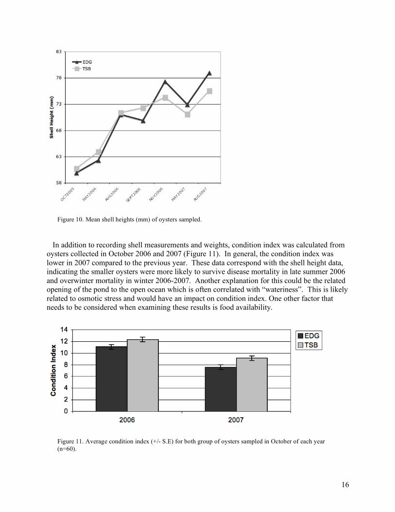
In addition to recording shell measurements and weights, condition index was calculated from oysters collected in October 2006 and 2007 (Figure 11). In general, the condition index was lower in 2007 compared to the previous year. These data correspond with the shell height data, indicating the smaller oysters were more likely to survive disease mortality in late summer 2006 and overwinter mortality in winter 2006-2007. Another explanation for this could be the related opening of the pond to the open ocean which is often correlated with “wateriness”. This is likely related to osmotic stress and would have an impact on condition index. One other factor that needs to be considered when examining these results is food availability.
Figure 10. Mean shell heights (mm) of oysters sampled.
Figure 11. Average condition index (+/- S.E) for both group of oysters sampled in October of each year (n=60).
17
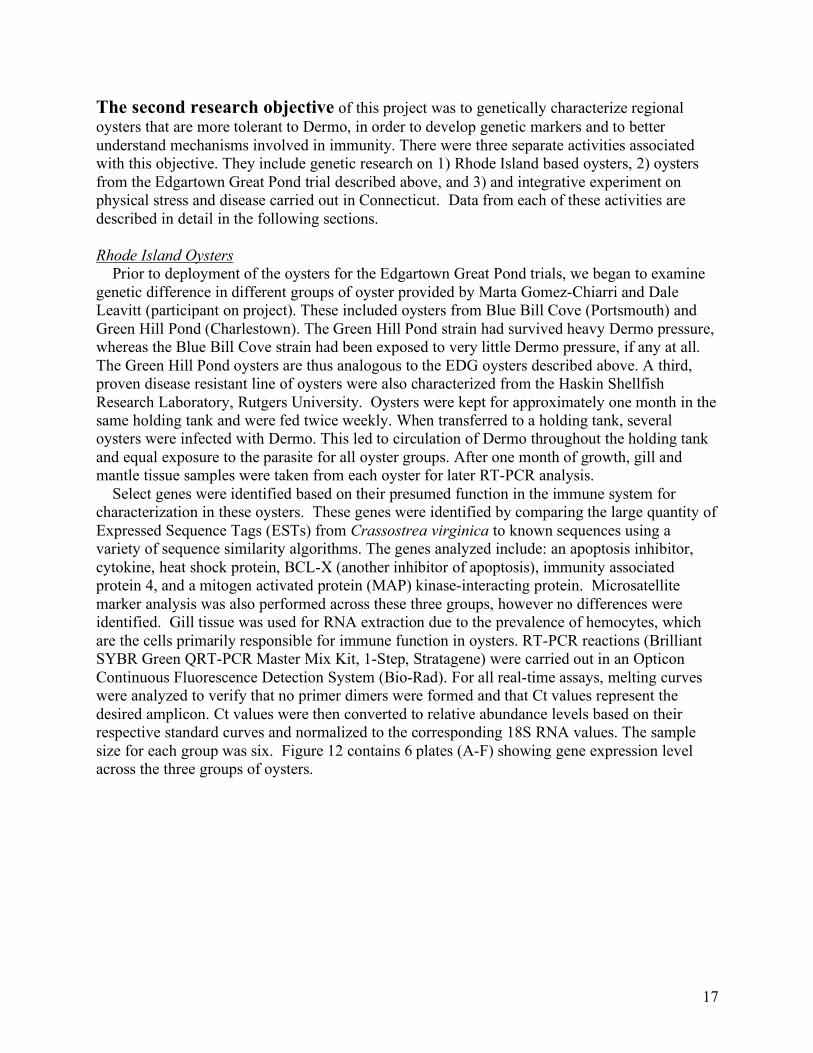
The second research objective of this project was to genetically characterize regional oysters that are more tolerant to Dermo, in order to develop genetic markers and to better understand mechanisms involved in immunity. There were three separate activities associated with this objective. They include genetic research on 1) Rhode Island based oysters, 2) oysters from the Edgartown Great Pond trial described above, and 3) and integrative experiment on physical stress and disease carried out in Connecticut. Data from each of these activities are described in detail in the following sections. Rhode Island Oysters Prior to deployment of the oysters for the Edgartown Great Pond trials, we began to examine genetic difference in different groups of oyster provided by Marta Gomez-Chiarri and Dale Leavitt (participant on project). These included oysters from Blue Bill Cove (Portsmouth) and Green Hill Pond (Charlestown). The Green Hill Pond strain had survived heavy Dermo pressure, whereas the Blue Bill Cove strain had been exposed to very little Dermo pressure, if any at all. The Green Hill Pond oysters are thus analogous to the EDG oysters described above. A third, proven disease resistant line of oysters were also characterized from the Haskin Shellfish Research Laboratory, Rutgers University. Oysters were kept for approximately one month in the same holding tank and were fed twice weekly. When transferred to a holding tank, several oysters were infected with Dermo. This led to circulation of Dermo throughout the holding tank and equal exposure to the parasite for all oyster groups. After one month of growth, gill and mantle tissue samples were taken from each oyster for later RT-PCR analysis. Select genes were identified based on their presumed function in the immune system for characterization in these oysters. These genes were identified by comparing the large quantity of Expressed Sequence Tags (ESTs) from Crassostrea virginica to known sequences using a variety of sequence similarity algorithms. The genes analyzed include: an apoptosis inhibitor, cytokine, heat shock protein, BCL-X (another inhibitor of apoptosis), immunity associated protein 4, and a mitogen activated protein (MAP) kinase-interacting protein. Microsatellite marker analysis was also performed across these three groups, however no differences were identified. Gill tissue was used for RNA extraction due to the prevalence of hemocytes, which are the cells primarily responsible for immune function in oysters. RT-PCR reactions (Brilliant SYBR Green QRT-PCR Master Mix Kit, 1-Step, Stratagene) were carried out in an Opticon Continuous Fluorescence Detection System (Bio-Rad). For all real-time assays, melting curves were analyzed to verify that no primer dimers were formed and that Ct values represent the desired amplicon. Ct values were then converted to relative abundance levels based on their respective standard curves and normalized to the corresponding 18S RNA values. The sample size for each group was six. Figure 12 contains 6 plates (A-F) showing gene expression level across the three groups of oysters.

18
Figu
re 1
2.
Rel
ativ
e ge
ne e
xpre
ssio
n le
vels
of s
ix im
mun
e-re
late
d ge
nes
in
oyst
ers (
n=6)
from
Gre
en H
ill P
ond
(his
toric
ally
exp
osed
to h
igh
Der
mo
pres
sure
), B
lue
Bill
Cov
e (h
isto
rical
ly e
xpos
ed to
lim
ited
dise
ase
pres
sure
), an
d R
utge
rs’ d
isea
se re
sist
ant l
ine.
All
oyst
ers w
ere
held
in a
com
mon
syst
em th
at
cont
aine
d P.
mar
inus
. Err
or b
ars
indi
cate
sta
ndar
d er
ror.
Put
ativ
e ge
ne n
ames
an
d ac
cess
ion
num
bers
are
indi
cate
d at
righ
t.
19
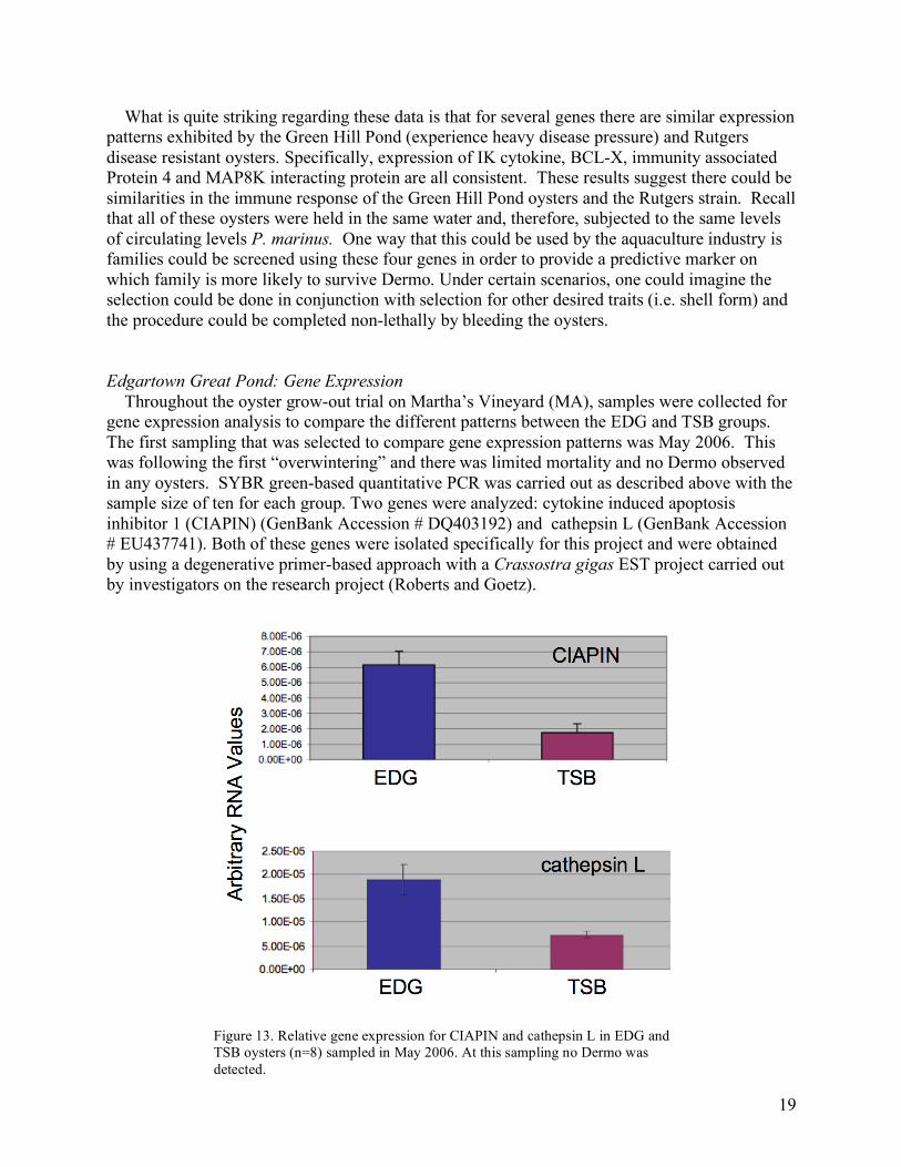
What is quite striking regarding these data is that for several genes there are similar expression patterns exhibited by the Green Hill Pond (experience heavy disease pressure) and Rutgers disease resistant oysters. Specifically, expression of IK cytokine, BCL-X, immunity associated Protein 4 and MAP8K interacting protein are all consistent. These results suggest there could be similarities in the immune response of the Green Hill Pond oysters and the Rutgers strain. Recall that all of these oysters were held in the same water and, therefore, subjected to the same levels of circulating levels P. marinus. One way that this could be used by the aquaculture industry is families could be screened using these four genes in order to provide a predictive marker on which family is more likely to survive Dermo. Under certain scenarios, one could imagine the selection could be done in conjunction with selection for other desired traits (i.e. shell form) and the procedure could be completed non-lethally by bleeding the oysters. Edgartown Great Pond: Gene Expression Throughout the oyster grow-out trial on Martha’s Vineyard (MA), samples were collected for gene expression analysis to compare the different patterns between the EDG and TSB groups. The first sampling that was selected to compare gene expression patterns was May 2006. This was following the first “overwintering” and there was limited mortality and no Dermo observed in any oysters. SYBR green-based quantitative PCR was carried out as described above with the sample size of ten for each group. Two genes were analyzed: cytokine induced apoptosis inhibitor 1 (CIAPIN) (GenBank Accession # DQ403192) and cathepsin L (GenBank Accession # EU437741). Both of these genes were isolated specifically for this project and were obtained by using a degenerative primer-based approach with a Crassostra gigas EST project carried out by investigators on the research project (Roberts and Goetz).
Figure 13. Relative gene expression for CIAPIN and cathepsin L in EDG and
TSB oysters (n=8) sampled in May 2006. At this sampling no Dermo was detected.
20
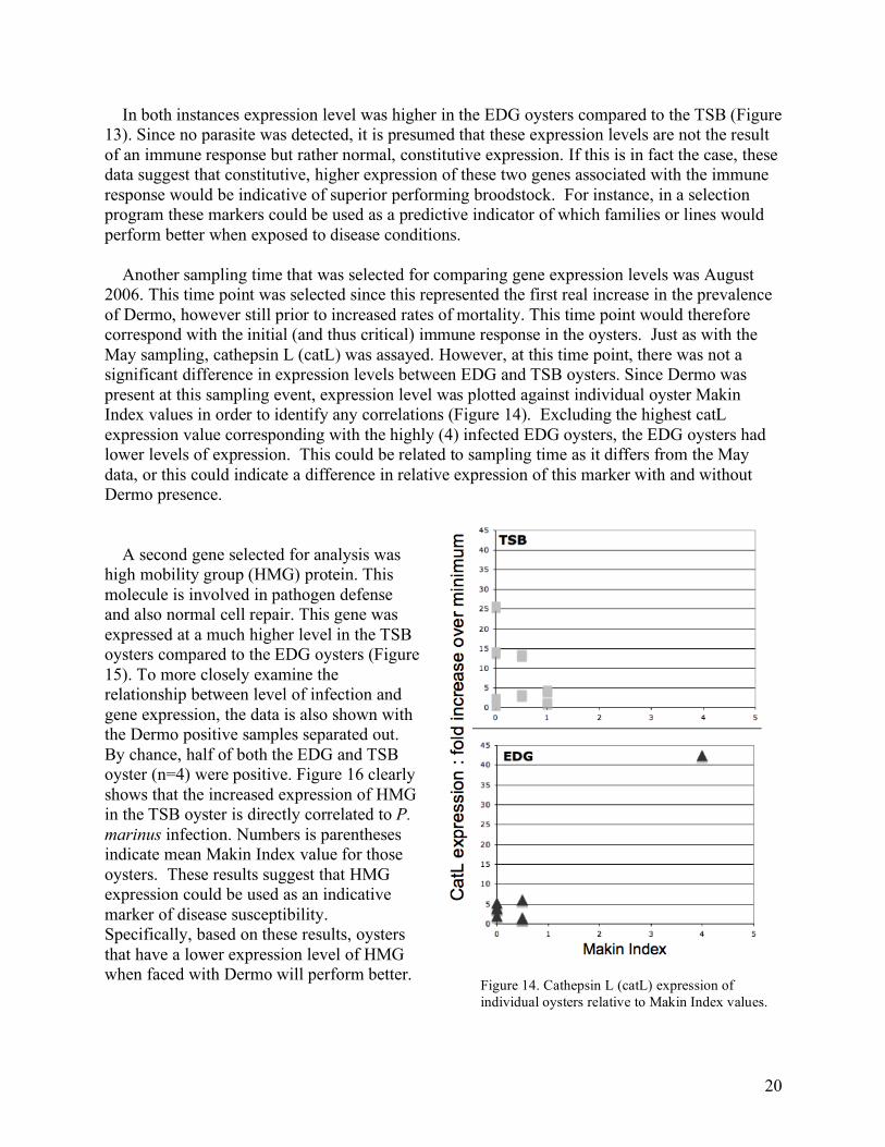
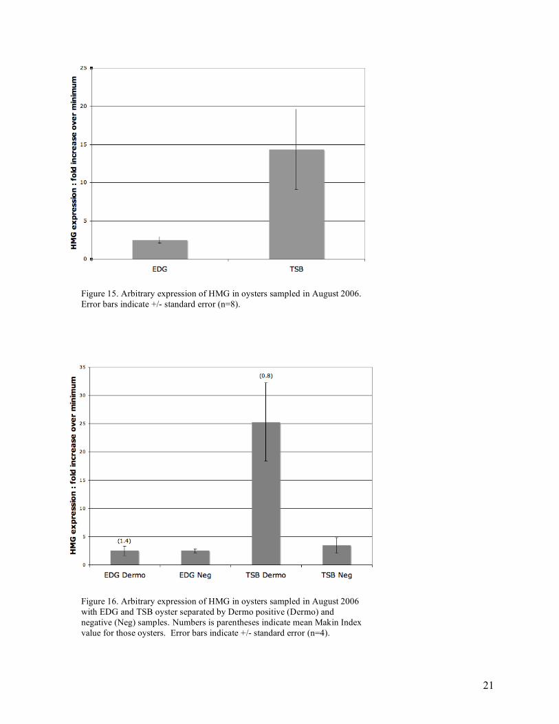
In both instances expression level was higher in the EDG oysters compared to the TSB (Figure 13). Since no parasite was detected, it is presumed that these expression levels are not the result of an immune response but rather normal, constitutive expression. If this is in fact the case, these data suggest that constitutive, higher expression of these two genes associated with the immune response would be indicative of superior performing broodstock. For instance, in a selection program these markers could be used as a predictive indicator of which families or lines would perform better when exposed to disease conditions. Another sampling time that was selected for comparing gene expression levels was August 2006. This time point was selected since this represented the first real increase in the prevalence of Dermo, however still prior to increased rates of mortality. This time point would therefore correspond with the initial (and thus critical) immune response in the oysters. Just as with the May sampling, cathepsin L (catL) was assayed. However, at this time point, there was not a significant difference in expression levels between EDG and TSB oysters. Since Dermo was present at this sampling event, expression level was plotted against individual oyster Makin Index values in order to identify any correlations (Figure 14). Excluding the highest catL expression value corresponding with the highly (4) infected EDG oysters, the EDG oysters had lower levels of expression. This could be related to sampling time as it differs from the May data, or this could indicate a difference in relative expression of this marker with and without Dermo presence. A second gene selected for analysis was high mobility group (HMG) protein. This molecule is involved in pathogen defense and also normal cell repair. This gene was expressed at a much higher level in the TSB oysters compared to the EDG oysters (Figure 15). To more closely examine the relationship between level of infection and gene expression, the data is also shown with the Dermo positive samples separated out. By chance, half of both the EDG and TSB oyster (n=4) were positive. Figure 16 clearly shows that the increased expression of HMG in the TSB oyster is directly correlated to P. marinus infection. Numbers is parentheses indicate mean Makin Index value for those oysters. These results suggest that HMG expression could be used as an indicative marker of disease susceptibility. Specifically, based on these results, oysters that have a lower expression level of HMG when faced with Dermo will perform better.
Figure 14. Cathepsin L (catL) expression of individual oysters relative to Makin Index values.
21
Figure 15. Arbitrary expression of HMG in oysters sampled in August 2006. Error bars indicate +/- standard error (n=8).
Figure 16. Arbitrary expression of HMG in oysters sampled in August 2006 with EDG and TSB oyster separated by Dermo positive (Dermo) and negative (Neg) samples. Numbers is parentheses indicate mean Makin Index value for those oysters. Error bars indicate +/- standard error (n=4).
22
Connecticut Oyster: Dermo and Mechanical Stress A third activity associated with identifying genes involved in the oyster immune response to Dermo was carried out in Milford, Connecticut by Gary Wikfors and Inke Sunila (participant on current project). Oysters from Connecticut with Dermo and oysters (C. virginica) from Washington State without Dermo, were subjected to mechanical stress. Hemocyte samples were harvested from each oyster and gene expression patterns were characterized to better understand the relationship of immune function, disease, and stress. Methods Potentially Perkinsus marinus –infected eastern oysters, Crassostrea virginica (108mm, SD 9.4), were collected from the east coast US from Milford Harbor in Connecticut, and potentially uninfected oysters C. virginica (98 mm, SD 6.7) were obtained from the west coast US from a commercial shellfish hatchery in Washington. 20 oysters from both sites were used. Oysters were maintained separately under the same conditions. Following this period, oysters originally from CT and WA were divided into two groups and half (n=10) were exposed to a physical stress event. Specifically, oysters were centrifuged for 5 minutes at 100 rpms. Hemolymph was harvested from the cardio sinus and placed at 4°C. Each hemolymph sample was divided for cellular characteristic analysis and quantitative PCR. Disease Diagnosis and Hemocyte Characterization Gross and histological examinations were performed to determine disease status in all individuals. Perkinsus marinus was diagnosed from anal-rectal tissues using Ray’s Fluid Thioglycollate Medium. The values for all animals in a sample were averaged to determine the weighted prevalence value. Histological slides (Methods described above; page 10) were examined to detect any infectious organisms (viruses, bacteria, or parasites) or any histopathological change such as inflammatory responses, degenerations, cell and tissue death, growth derangements, and hemodynamic and fluid derangements. Hemocyte function was analyzed by flow cytometry for viability, phagocytosis, and respiratory burst response. Measured morphological parameters included number and size of hemocytes. Details of techniques are described in Hégaret et al. 2003.
Gene Expression Analysis In order to characterize effects of Dermo and physical stress on immune physiology, quantitative RT-PCR was carried out on hemocytes from all oysters. RNA was extracted from hemocytes using Tri-Reagent (MRC) and subsequently treated with DNase (Ambion, Turbo DNAfree) to remove any possible genomic DNA carryover. For gene expression analysis, a set of genes involved in oyster immune response were selected that represent various mechanisms including apoptosis, inflammation, and protease activity (Table 3). RT-PCR reactions (Brilliant SYBR Green QRT-PCR Master Mix Kit, 1-Step, Stratagene) were carried out in an Opticon2 Continuous Fluorescence Detection System (Bio-Rd). For all real-time assays, melting curves were analyzed to verify that no primer dimers formed and that Ct values represent the desired amplicon. Control samples were analyzed without reverse transcriptase to ensure amplified products were not a result of genomic DNA. Analysis of PCR data was carried out based on the
23
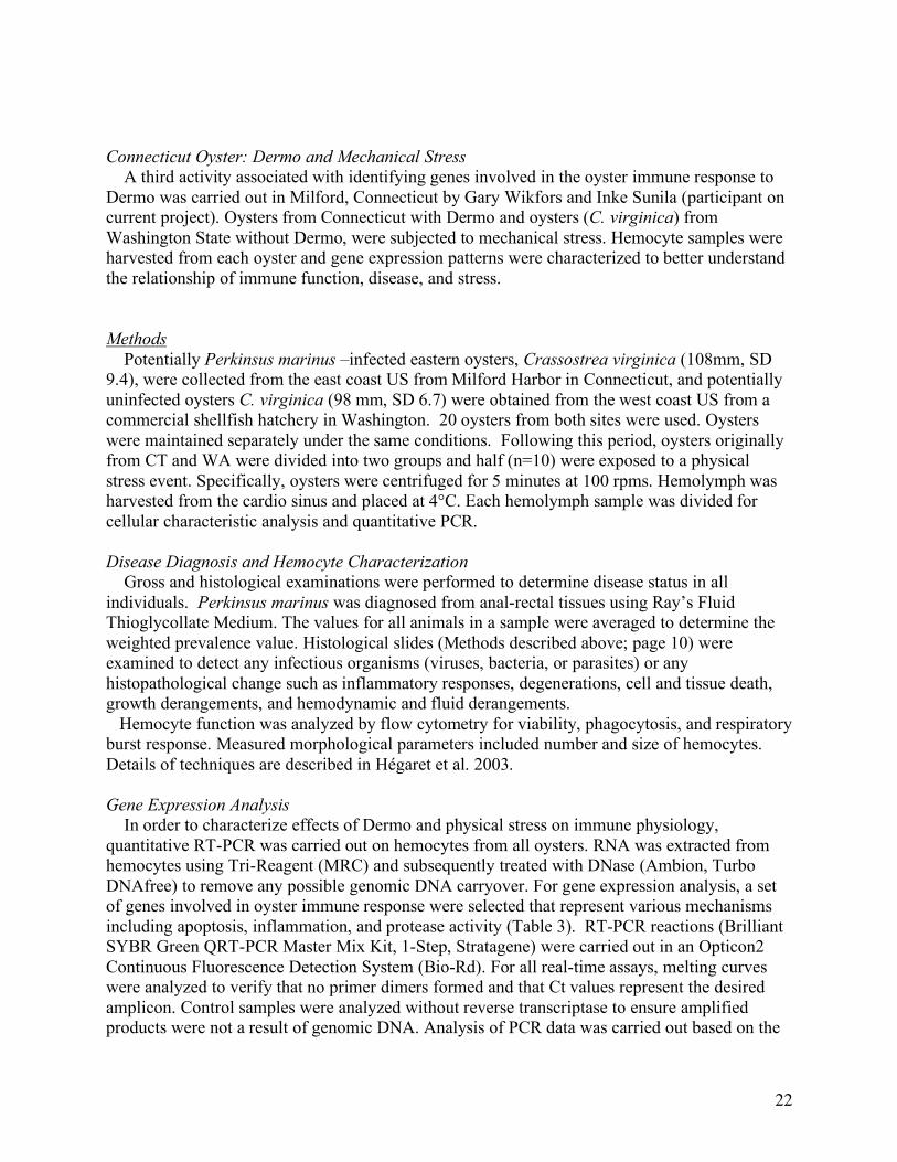
kinetics of individual PCR reactions using Real-time PCR Miner v2.1 (Zhao and Fernald, 2005). All data were normalized to corresponding 18S RNA values. Table 3. Genes assayed for differences in expression level in oysters from the Connecticut disease and mechanical stress trials. Gene Function CIAPIN | inhibitor of apoptosis Lysosomal phospholipase A2 | inflammation, proliferation Cathepsin L | cysteine endopeptidase Cathepsin Z | cysteine endopeptidase High Mobility Group Protein | cytokine mediating response to infection, injury Results Disease and Pathology Prevalence of Perkinsus marinus in Milford Harbor, CT, samples was 85%, and the weighted prevalence 1.3. Oyster samples from Washington contained one thioglycollate positive specimen with an intensity of 0.5 (very light infection). Thioglycollate assay is considered specific for Perkinsus family which contains several species, besides P. marinus, which infects oysters on the east coast US. Prevalence of Perkinsus sp. in Washington was 5%, and weighted prevalence was 0.025. Stressed specimens from Milford Harbor had unusual gill lesions that were not present in unstressed specimens from the same location or from Washington (chi-square 17.6, p<0.01). A normal gill of the eastern oyster is composed of two demibranchs, and each demibranch is composed of two lamellae. Lamellae consist of a series of folds, plicae, which contain several filaments. There is an intraplical sinus inside plicae that opens into hemolymph sinuses inside each filament (Fig. 17 A and B). Stressed specimens had dilated sinuses inside the plicae and filaments that gave the gills edematous appearance (Fig. 17 C and D). There was an apparent increase of hemocytes in the sinuses of affected specimens.
24
Milford Harbor sample presented characteristic parasite fauna for the area. There was a 100% prevalence of Polydora websteri, mud worms, inside the shells. There was a 55% prevalence of gregarines Nematopsis ostrearum (Porosporidae), a 30% prevalence of ciliates, Stegotricha sp., in the intestine or stomachs, and a 30% prevalence of trichodinid ciliates on the gills or the mantle. These parasites were not present in the oysters from Washington, but one specimen contained Stegotricha sp. in the digestive tubules. Five specimens from Milford Harbor had ceroidosis, a condition associated with Perkinsus marinus-infection. All oysters from Milford Harbor were sexually inactive with resting, indeterminate gonads. Sexes could not be separated. Oysters from Washington were in different stages of development. There was one resting, indeterminate specimen, three gametogenic males with apparent meiosis, but no mature gametes, four gametogenic females, two developing males showing progressive development from germinal cells to mature germ cells, seven developing females, and three ripe males with follicles full of sperm.
Fig. 17. Cross-sections of gills from the eastern oyster, Crassostrea virginica. Paraffin sections, hematoxylin-eosin. A. Unstressed oyster from Milford Harbor. B. Unstressed oyster from Milford Harbor. Higher magnification of A. C. Stressed oyster from Milford Harbor. D. Stressed oyster from Milford Harbor. Higher magnification of C. I=intraplical sinuses, F=filaments, H=, hemolymph sinus of filaments, P=plicae, W=water tube. Images taken by Dr. Inke Sunila
25
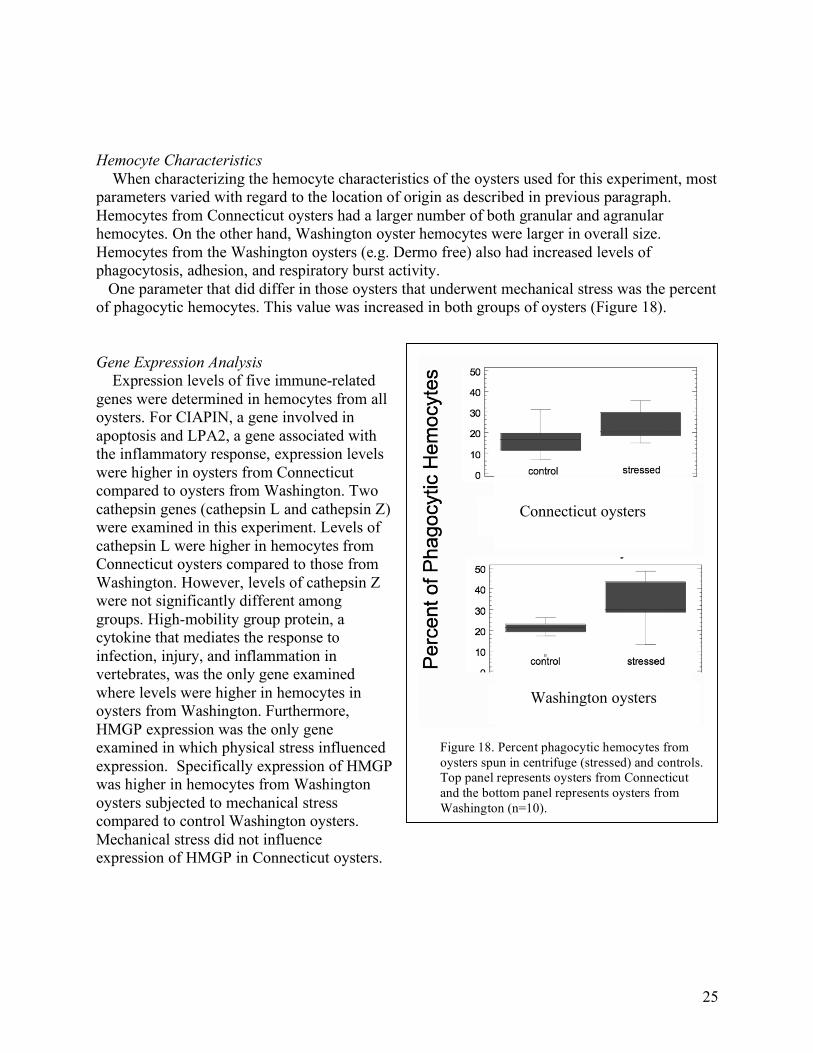
Hemocyte Characteristics When characterizing the hemocyte characteristics of the oysters used for this experiment, most parameters varied with regard to the location of origin as described in previous paragraph. Hemocytes from Connecticut oysters had a larger number of both granular and agranular hemocytes. On the other hand, Washington oyster hemocytes were larger in overall size. Hemocytes from the Washington oysters (e.g. Dermo free) also had increased levels of phagocytosis, adhesion, and respiratory burst activity. One parameter that did differ in those oysters that underwent mechanical stress was the percent of phagocytic hemocytes. This value was increased in both groups of oysters (Figure 18). Gene Expression Analysis Expression levels of five immune-related genes were determined in hemocytes from all oysters. For CIAPIN, a gene involved in apoptosis and LPA2, a gene associated with the inflammatory response, expression levels were higher in oysters from Connecticut compared to oysters from Washington. Two cathepsin genes (cathepsin L and cathepsin Z) were examined in this experiment. Levels of cathepsin L were higher in hemocytes from Connecticut oysters compared to those from Washington. However, levels of cathepsin Z were not significantly different among groups. High-mobility group protein, a cytokine that mediates the response to infection, injury, and inflammation in vertebrates, was the only gene examined where levels were higher in hemocytes in oysters from Washington. Furthermore, HMGP expression was the only gene examined in which physical stress influenced expression. Specifically expression of HMGP was higher in hemocytes from Washington oysters subjected to mechanical stress compared to control Washington oysters. Mechanical stress did not influence expression of HMGP in Connecticut oysters.
Figure 18. Percent phagocytic hemocytes from oysters spun in centrifuge (stressed) and controls. Top panel represents oysters from Connecticut and the bottom panel represents oysters from Washington (n=10).
Connecticut oysters
Washington oysters
26
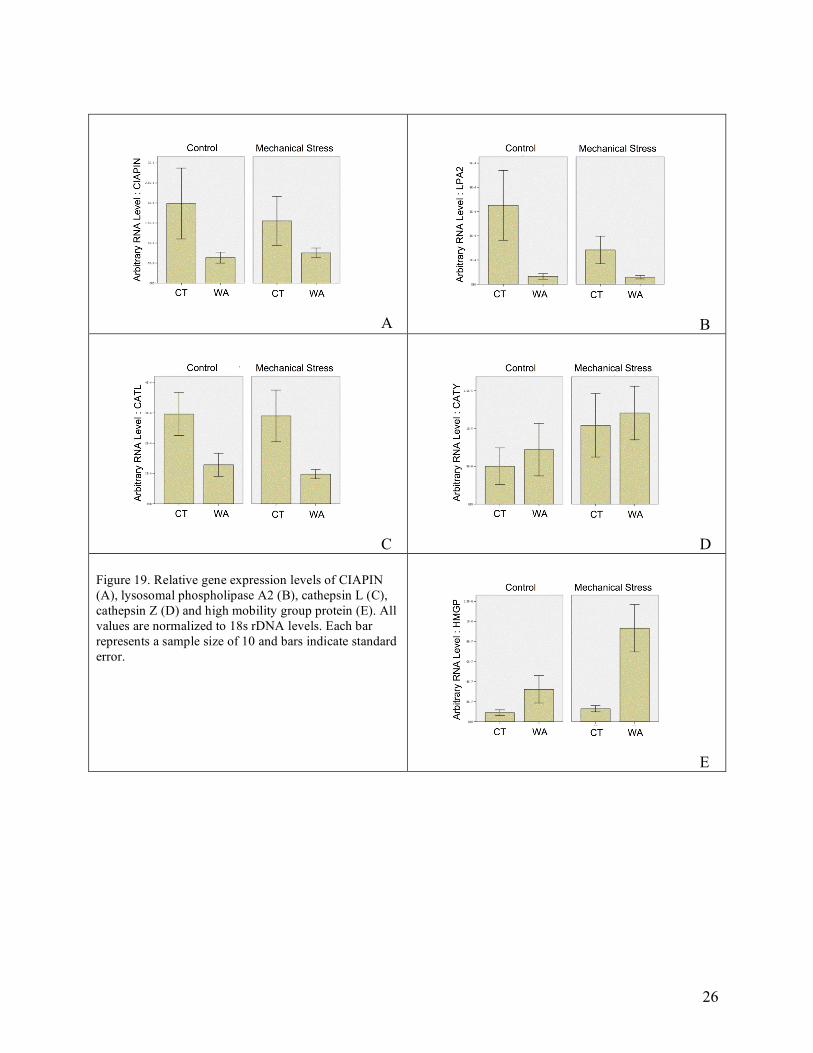
A B
C D Figure 19. Relative gene expression levels of CIAPIN (A), lysosomal phospholipase A2 (B), cathepsin L (C), cathepsin Z (D) and high mobility group protein (E). All values are normalized to 18s rDNA levels. Each bar represents a sample size of 10 and bars indicate standard error.
E
27
Discussion A primary goal of this research is to characterize the immune response in Crassostrea virginica, and to identify possible effects of acute mechanical stressors on this response. Oysters infected with P. marinus were collected from a disease prone location in Connecticut. In order to obtain Dermo-free oysters, samples were collected from Washington. Expression levels of an inhibitor of apoptosis, CIAPIN, were elevated in hemocytes from Connecticut oysters. One proposed mechanism for P. marinus pathogencity is apoptosis. Research has shown hemocyte populations are elevated in infected oysters. This could be due to an increased immune response or P. marinus could be influencing cell numbers indirectly via apoptosis. The parasites reside in the hemocytes, therefore this condition (increased number of hemocytes) would be favorable for P. marinus proliferation. In this study, levels of an inhibitor of apoptosis are elevated in infected hemocytes. This could indicate a pathway by which P. marinus alters normal apoptosis within its host that would ultimately lead to increased number of host cells. The mechanism by which P. marinus might interfere with normal oyster apoptosis is not known. Cathepsin L expression was induced in hemocytes of P marinus infected oysters (Connecticut). Cathepsin L is involved protein degradation in lysosomes. Higher levels of expression would therefore be expected to correlate with increased phagocytosis (and thereby pathogen killing). An additional Cathepsin gene was characterized which has sequence similarity to Cathepsin Z. In the hemocyte samples analyzed in this work, there was not a significant difference in expression across samples. High mobility group proteins are important molecules signaling both an immune response as well as cell damage. Of interest in the current experiment is that expression of HMG homologue was expressed at higher levels in non-diseased oysters. One possible explanation for this is that HMG is involved in pathogen response and the rate of protein translation is higher in the hemocytes of diseased oysters. Another explanation for this higher level of expression in the oysters from Washington could be the fact that, due to local rearing conditions, these oysters were reproductively mature. Exposure of the oysters to mechanical stress was associated with increased HMG expression in the Washington oysters. These results suggest that cell damage, without pathogen infection, does initiate a signaling pathway that includes HMG. This, along with the fact that mechanical stress increased the percentage of phagocytic hemocytes suggests (with obvious limitations) light to moderate physical stress might provide short-term defense stimulation. Limitations to this are likely that any stimulation would have to occur prior to disease exposure and excess stress would negatively impact immune function. The third objective of this research project is to communicate with northeastern hatchery operations and help them to identify local, potentially Dermo-resistant broodstocks. One way we have done this throughout the experiments is to present our findings at regional and national meetings. The first presentation was given in at the Milford Aquaculture Conference on the results from the Rhode Island oysters. More recently a presentation was given at the Northeastern Aquaculture Conference and Expo where we received good feedback from oyster growers. Other presentation have been given and the NSA, and while there is a larger proportion of growers at the regional meetings, this venue provided exposure to a diverse audience. Several of these presentations are available to the public via the website that follows the progress of our research: http://fish.washington.edu/research/genefish/robertslab/Dermo.html (also redirected from: http://tinyurl.com/22nhbs). This website is a important tool for getting
28
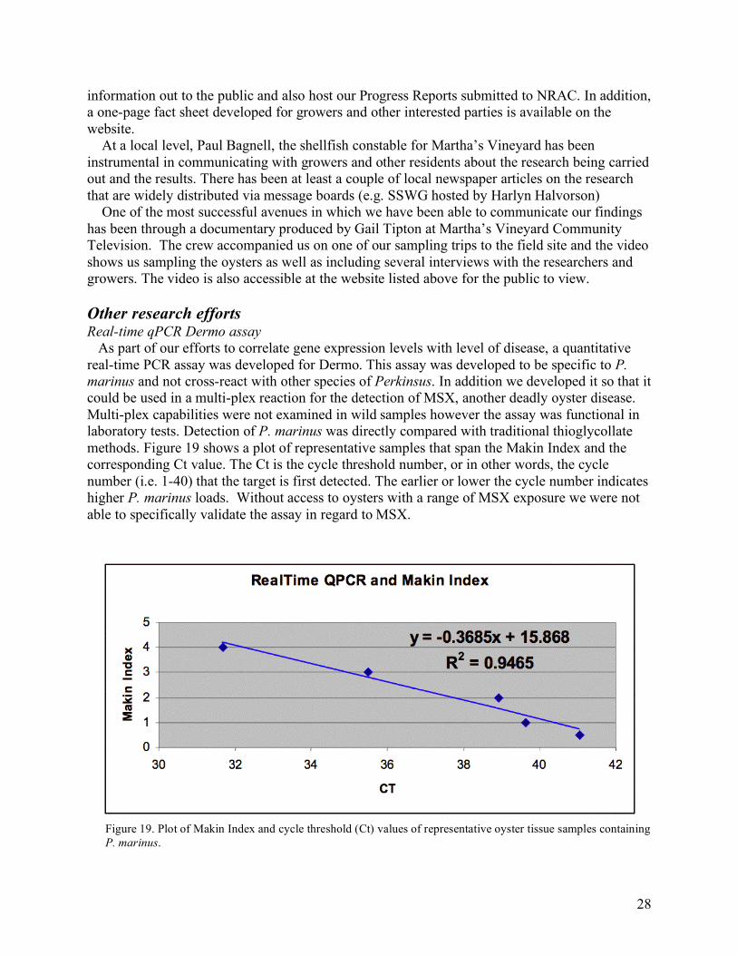
information out to the public and also host our Progress Reports submitted to NRAC. In addition, a one-page fact sheet developed for growers and other interested parties is available on the website. At a local level, Paul Bagnell, the shellfish constable for Martha’s Vineyard has been instrumental in communicating with growers and other residents about the research being carried out and the results. There has been at least a couple of local newspaper articles on the research that are widely distributed via message boards (e.g. SSWG hosted by Harlyn Halvorson) One of the most successful avenues in which we have been able to communicate our findings has been through a documentary produced by Gail Tipton at Martha’s Vineyard Community Television. The crew accompanied us on one of our sampling trips to the field site and the video shows us sampling the oysters as well as including several interviews with the researchers and growers. The video is also accessible at the website listed above for the public to view. Other research efforts Real-time qPCR Dermo assay As part of our efforts to correlate gene expression levels with level of disease, a quantitative real-time PCR assay was developed for Dermo. This assay was developed to be specific to P. marinus and not cross-react with other species of Perkinsus. In addition we developed it so that it could be used in a multi-plex reaction for the detection of MSX, another deadly oyster disease. Multi-plex capabilities were not examined in wild samples however the assay was functional in laboratory tests. Detection of P. marinus was directly compared with traditional thioglycollate methods. Figure 19 shows a plot of representative samples that span the Makin Index and the corresponding Ct value. The Ct is the cycle threshold number, or in other words, the cycle number (i.e. 1-40) that the target is first detected. The earlier or lower the cycle number indicates higher P. marinus loads. Without access to oysters with a range of MSX exposure we were not able to specifically validate the assay in regard to MSX.
Figure 19. Plot of Makin Index and cycle threshold (Ct) values of representative oyster tissue samples containing P. marinus.
29
The specific methodology used involved taking two equal tissue samples from oysters. One sample was evaluated using the RTFM method as described above, and ranked according to the Makin Index. DNA was extracted from the other half of the tissue sample by incubating the tissue sample in 10% Chelax-100 (Bio-Rad) at 90°C for 30 minutes.
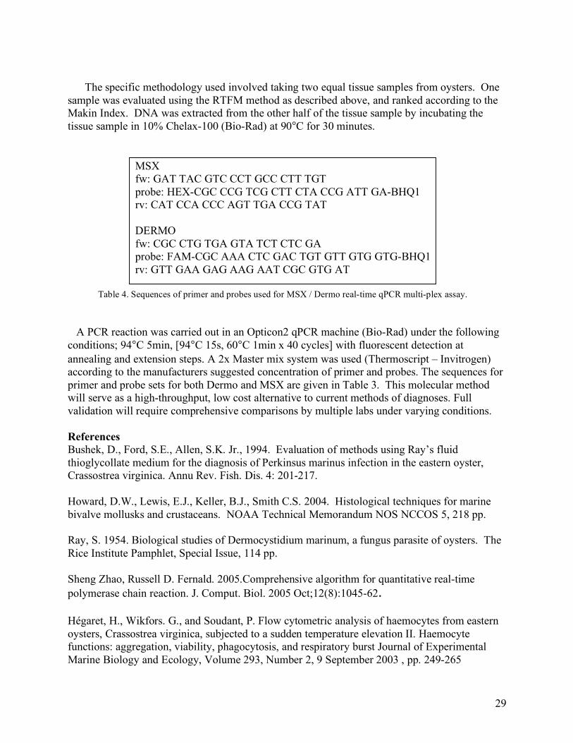
MSX fw: GAT TAC GTC CCT GCC CTT TGT probe: HEX-CGC CCG TCG CTT CTA CCG ATT GA-BHQ1 rv: CAT CCA CCC AGT TGA CCG TAT
DERMO fw: CGC CTG TGA GTA TCT CTC GA probe: FAM-CGC AAA CTC GAC TGT GTT GTG GTG-BHQ1 rv: GTT GAA GAG AAG AAT CGC GTG AT
Table 4. Sequences of primer and probes used for MSX / Dermo real-time qPCR multi-plex assay. A PCR reaction was carried out in an Opticon2 qPCR machine (Bio-Rad) under the following conditions; 94°C 5min, [94°C 15s, 60°C 1min x 40 cycles] with fluorescent detection at annealing and extension steps. A 2x Master mix system was used (Thermoscript – Invitrogen) according to the manufacturers suggested concentration of primer and probes. The sequences for primer and probe sets for both Dermo and MSX are given in Table 3. This molecular method will serve as a high-throughput, low cost alternative to current methods of diagnoses. Full validation will require comprehensive comparisons by multiple labs under varying conditions. References Bushek, D., Ford, S.E., Allen, S.K. Jr., 1994. Evaluation of methods using Ray’s fluid thioglycollate medium for the diagnosis of Perkinsus marinus infection in the eastern oyster, Crassostrea virginica. Annu Rev. Fish. Dis. 4: 201-217. Howard, D.W., Lewis, E.J., Keller, B.J., Smith C.S. 2004. Histological techniques for marine bivalve mollusks and crustaceans. NOAA Technical Memorandum NOS NCCOS 5, 218 pp. Ray, S. 1954. Biological studies of Dermocystidium marinum, a fungus parasite of oysters. The Rice Institute Pamphlet, Special Issue, 114 pp. Sheng Zhao, Russell D. Fernald. 2005.Comprehensive algorithm for quantitative real-time polymerase chain reaction. J. Comput. Biol. 2005 Oct;12(8):1045-62. Hégaret, H., Wikfors. G., and Soudant, P. Flow cytometric analysis of haemocytes from eastern oysters, Crassostrea virginica, subjected to a sudden temperature elevation II. Haemocyte functions: aggregation, viability, phagocytosis, and respiratory burst Journal of Experimental Marine Biology and Ecology, Volume 293, Number 2, 9 September 2003 , pp. 249-265
30
Project Completion Report
Signature Page
Project Code: 05-02 Subcontract/Account No. 557221 supported by 2003-38500-13505 Project Title: “Development of genetic markers to assess disease resistance in the eastern oyster” Prepared By:
Steven Roberts January 02, 2008