Progressive Fibrous Ankylosis of Temporomandibular Joint ... · or dysfunction. In this article,...
4
Remedy Publications LLC. Journal of Dentistry and Oral Biology 2016 | Volume 1 | Issue 1 | Article 1002 1 Introduction Psoriasis is a chronic, non-contagious skin disease that commonly leads to appearance of red scaly patches on the skin. Psoriatic arthritis (PsA) is a chronic, disabling inflammatory disease, associated with psoriasis. PsA causes joint destruction, associated with cartilage deterioration, bone damage and joint fusion. Prevalence of the disease is around 2-3% of the world population [1]. e distal interphalangeal joints are the most frequently affected site of PsA, followed by shoulders, knees, ankles, spine and sternal synchondrosis [2]. Although many cases have been reported since 1973 when PsA was first defined by Moll and Wright [3], Temporomandibular joints (TMJ) involvement is relatively rare. To the best of our knowledge, only 20 English-written articles with 47 cases of PsA of TMJ have been reported so far [4-23]. e etiology of PsA still remains unclear and the progressive procedure in TMJ is rarely mentioned. Herein, we reported a case with progressive fibrous Ankylosis of TMJ caused by PsA and its natural history within 4 months. Case Presentation In March 2014, a 22 year-old male was referred to the Department of Oral and Maxillofacial Surgery, Ninth People’s Hospital, Shanghai Jiao Tong University School of Medicine, with a chief complaint of limitation of mouth opening for 2 months. He had a history of psoriasis for 2 years with a main appearance of red patches on scalp and face. He had no complaint of clicking or pain in preauricular regions, and no toothache was mentioned. Physical examination revealed a symmetric face with some red patches on his nose (Figure 1A). e maximal mouth opening was 10 mm without deviation (Figure 1B). e occlusion was normal. Palpation was noticed in the right TMJ region. e patient had no history of trauma or rheumatoid arthrosis. His right little toe was swelling (Figure 1C), but no other joint was involved. Further physical examinations showed red scaly patches on the scalp. Laboratory test showed his full blood count was normal. e erythrocyte sedimentation was 10mm/1h (reference range was 0-15). Antistreptolysin O (ASO) was 301 IU/ml (reference range was 0-200). Rheumatoid factor was 21 IU/ml (reference range was 0-20). In consideration of the existence of temporomandibular disorders, a MRI scan for TMJ region was conducted. e results revealed normal disc-condyle relationship in the right TMJ, but increased effusion within the joint and the adjacent soſt tissues. e cortical bone of the condyle was obscure, and the density signal of the condylar marrow decreased. In the leſt TMJ, anterior disc displacement was found, and the cortical bone of the top of the condyle disappeared (Figure 1D and E). Furthermore, a CT scan for TMJ showed resorption of the right condyle and osteophytes in the joint space, together with blurred condylar surface and joint space narrowing in the leſt joint (Figure 1F). Based on his history of psoriasis, these findings suggested the diagnosis of right TMJ fibrous Ankylosis of the right TMJ caused by PsA. Local and systematic treatments for psoriasis Progressive Fibrous Ankylosis of Temporomandibular Joint Caused by Psoricatic Arthritis: A Case Report OPEN ACCESS *Correspondence: Xieyi Cai, Department of Oral & Maxillofacial Surgery, Shanghai Ninth People’s Hospital, Shanghai Jiaotong University, 639 Zhi Zao Ju Road, Shanghai. 200011, P.R.China, Tel: 862123271699-5705; E-mail: [email protected] Received Date: 15 Jul 2016 Accepted Date: 01 Aug 2016 Published Date: 10 Aug 2016 Citation: Chen Y, Xie-Yi Cai. Progressive Fibrous Ankylosis of Temporomandibular Joint Caused by Psoricatic Arthritis: A Case Report. J Dent Oral Biol. 2016; 1(1): 1002. Copyright © 2016 Xie-Yi Cai. This is an open access article distributed under the Creative Commons Attribution License, which permits unrestricted use, distribution, and reproduction in any medium, provided the original work is properly cited. Case Report Published: 10 Aug, 2016 Abstract Psoriatic arthritis (PsA) is a chronic, disabling inflammatory disease, associated with psoriasis. Involvement of the Temporomandibular Joint (TMJ) is very rare and may cause severe deformation or dysfunction. In this article, the authors presented a case of fibrous Ankylosis of TMJ caused by PsA and its natural history aſter 4 months. e findings of the report suggested that PsA in TMJ may progress rapidly in an early age without any treatments. Further study for the development of the TMJ lesion caused by PsA is quite necessary. Keywords: Psoricatic arthritis; Temporomandibular joint; Ankylosis; Natural outcome Ying Chen and Xie-Yi Cai* Department of Oral and Maxillofacial Surgery, Shanghai Jiao Tong University School of Medicine, China
-
Upload
vuongduong -
Category
Documents
-
view
224 -
download
0
Transcript of Progressive Fibrous Ankylosis of Temporomandibular Joint ... · or dysfunction. In this article,...

Remedy Publications LLC.
Journal of Dentistry and Oral Biology
2016 | Volume 1 | Issue 1 | Article 10021
IntroductionPsoriasis is a chronic, non-contagious skin disease that commonly leads to appearance of red
scaly patches on the skin. Psoriatic arthritis (PsA) is a chronic, disabling inflammatory disease, associated with psoriasis. PsA causes joint destruction, associated with cartilage deterioration, bone damage and joint fusion. Prevalence of the disease is around 2-3% of the world population [1].
The distal interphalangeal joints are the most frequently affected site of PsA, followed by shoulders, knees, ankles, spine and sternal synchondrosis [2]. Although many cases have been reported since 1973 when PsA was first defined by Moll and Wright [3], Temporomandibular joints (TMJ) involvement is relatively rare. To the best of our knowledge, only 20 English-written articles with 47 cases of PsA of TMJ have been reported so far [4-23].
The etiology of PsA still remains unclear and the progressive procedure in TMJ is rarely mentioned. Herein, we reported a case with progressive fibrous Ankylosis of TMJ caused by PsA and its natural history within 4 months.
Case PresentationIn March 2014, a 22 year-old male was referred to the Department of Oral and Maxillofacial
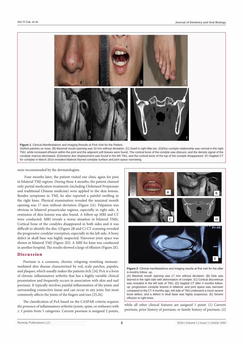
Surgery, Ninth People’s Hospital, Shanghai Jiao Tong University School of Medicine, with a chief complaint of limitation of mouth opening for 2 months. He had a history of psoriasis for 2 years with a main appearance of red patches on scalp and face. He had no complaint of clicking or pain in preauricular regions, and no toothache was mentioned. Physical examination revealed a symmetric face with some red patches on his nose (Figure 1A). The maximal mouth opening was 10 mm without deviation (Figure 1B). The occlusion was normal. Palpation was noticed in the right TMJ region. The patient had no history of trauma or rheumatoid arthrosis. His right little toe was swelling (Figure 1C), but no other joint was involved. Further physical examinations showed red scaly patches on the scalp. Laboratory test showed his full blood count was normal. The erythrocyte sedimentation was 10mm/1h (reference range was 0-15). Antistreptolysin O (ASO) was 301 IU/ml (reference range was 0-200). Rheumatoid factor was 21 IU/ml (reference range was 0-20).
In consideration of the existence of temporomandibular disorders, a MRI scan for TMJ region was conducted. The results revealed normal disc-condyle relationship in the right TMJ, but increased effusion within the joint and the adjacent soft tissues. The cortical bone of the condyle was obscure, and the density signal of the condylar marrow decreased. In the left TMJ, anterior disc displacement was found, and the cortical bone of the top of the condyle disappeared (Figure 1D and E). Furthermore, a CT scan for TMJ showed resorption of the right condyle and osteophytes in the joint space, together with blurred condylar surface and joint space narrowing in the left joint (Figure 1F). Based on his history of psoriasis, these findings suggested the diagnosis of right TMJ fibrous Ankylosis of the right TMJ caused by PsA. Local and systematic treatments for psoriasis
Progressive Fibrous Ankylosis of Temporomandibular Joint Caused by Psoricatic Arthritis: A Case Report
OPEN ACCESS
*Correspondence:Xieyi Cai, Department of Oral &
Maxillofacial Surgery, Shanghai Ninth People’s Hospital, Shanghai Jiaotong
University, 639 Zhi Zao Ju Road, Shanghai. 200011, P.R.China, Tel:
862123271699-5705;E-mail: [email protected] Date: 15 Jul 2016
Accepted Date: 01 Aug 2016Published Date: 10 Aug 2016
Citation: Chen Y, Xie-Yi Cai. Progressive Fibrous
Ankylosis of Temporomandibular Joint Caused by Psoricatic Arthritis: A Case Report. J Dent Oral Biol. 2016; 1(1):
1002.
Copyright © 2016 Xie-Yi Cai. This is an open access article distributed under
the Creative Commons Attribution License, which permits unrestricted
use, distribution, and reproduction in any medium, provided the original work
is properly cited.
Case ReportPublished: 10 Aug, 2016
AbstractPsoriatic arthritis (PsA) is a chronic, disabling inflammatory disease, associated with psoriasis. Involvement of the Temporomandibular Joint (TMJ) is very rare and may cause severe deformation or dysfunction. In this article, the authors presented a case of fibrous Ankylosis of TMJ caused by PsA and its natural history after 4 months. The findings of the report suggested that PsA in TMJ may progress rapidly in an early age without any treatments. Further study for the development of the TMJ lesion caused by PsA is quite necessary.
Keywords: Psoricatic arthritis; Temporomandibular joint; Ankylosis; Natural outcome
Ying Chen and Xie-Yi Cai*
Department of Oral and Maxillofacial Surgery, Shanghai Jiao Tong University School of Medicine, China
Xie-Yi Cai, et al. Journal of Dentistry and Oral Biology
Remedy Publications LLC. 2016 | Volume 1 | Issue 1 | Article 10022
were recommended by the dermatologists.
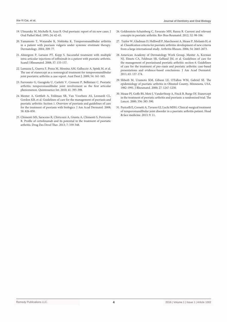
Four months later, the patient visited our clinic again for pain in bilateral TMJ regions. During these 4 months, the patient claimed only partial medication treatments (including Clobetasol Propionate and traditional Chinese medicine) were applied to the skin lesions. Besides symptoms in TMJ, he also reported a painful swelling in the right knee. Physical examination revealed the maximal mouth opening was 17 mm without deviation (Figure 2A). Palpation was obvious in bilateral preauricular regions, especially in right side. A remission of skin lesions was also found. A follow-up MRI and CT were conducted. MRI reveals a worse situation in bilateral TMJs. Cortical bone of the condyles disappeared in both sides and it was difficult to identify the disc ((Figure 2B and C) CT scanning revealed the progressive condylar resorption, especially in the left side. A bony defect in skull base was highly suspected. Narrower joint space was shown in bilateral TMJ (Figure 2D). A MRI for knee was conducted in another hospital. The results showed a large of effusion (Figure 2E).
DiscussionPsoriasis is a common, chronic relapsing remitting immune-
mediated skin disease characterized by red, scaly patches, papules, and plaques, which usually makes the patients itch [24]. PsA is a form of chronic inflammatory arthritis that has a highly variable clinical presentation and frequently occurs in association with skin and nail psoriasis. It typically involves painful inflammation of the joints and surrounding connective tissue and can occur in any joint, but most commonly affects the joints of the fingers and toes [25,26].
The classification of PsA based on the CASPAR criteria requires the presence of inflammatory arthritis (joints, spine, or entheses) with ≥ 3 points from 5 categories. Current psoriasis is assigned 2 points,
while all other clinical features are assigned 1 point: (1) Current psoriasis, prior history of psoriasis, or family history of psoriasis. (2)
Figure 1: Clinical Manifestations and Imaging Results at First Visit for the Patient. (A)Red patches on nose. (B) Maximal mouth opening was 10 mm without deviation. (C) Swell in right little toe. (D)Disc-condyle relationship was normal in the right TMJ, while increased effusion within the joint and the adjacent soft tissues were found. The cortical bone of the condyle was obscure, and the density signal of the condylar marrow decreased. (E)Anterior disc displacement was found In the left TMJ, and the cortical bone of the top of the condyle disappeared. (F) Sagittal CT for condylar in March 2014 revealed bilateral blurred condylar surface and joint space narrowing.
Figure 2: Clinical manifestations and imaging results at first visit for the after 4-months follow- up.(A) Maximal mouth opening was 17 mm without deviation. (B) Disk was blurred in the right side with deformation of condyle. (C) Cortical discontinue was revealed in the left side of TMJ. (D) Sagittal CT after 4 months follow-up ,progressive condylar lesions in bilateral ,and joint space was narrower compared to the CT 4 months ago, left side of TMJ underwent a much severe bone defect, and a defect in skull base was highly suspicious. (E) Severe effusion in right knee.

Xie-Yi Cai, et al. Journal of Dentistry and Oral Biology
Remedy Publications LLC. 2016 | Volume 1 | Issue 1 | Article 10023
Nail psoriasis. (3) Absence of serum rheumatoid factor. (4) Current or prior history of dactylitis. (5) Radiographic evidence of periarticular new bone formation (excluding osteophytes) on x-rays of the hand or foot [27].
In the present case, current psoriasis was diagnosed by rheumatologists. The serum rheumatoid factor was close to the critical value while dactylitis and radiographic evidence were both positive. Accordingly the diagnosis of PsA was established. The patient’s body surface area (BSA) involvement was only 1%-2% (<5%) being considered mild) [28]. After medication treatments for 4 months, the skin lesions remitted but the lesion in the joints progressed rapidly. According to the classification of PsA, the situation of joints’ involvements takes a more important place rather than BSA. A number of articles have described the TMJ Ankylosis caused by PsA, most of them were discovered in a quite late stage, with a long history of pain and limitation of mouth opening [4]. Our case was quite different. After 4-months follow-up, the mouth opening limitation seems to remit but joint pain was aggravated. Meanwhile condylar resorption was found with imaging studies. Combined the symptoms and the imaging results, we are more likely to consider this case in an active stage. In this particular stage, the condyles may undergo a relatively fast change of deformation and predictable eventual bony Ankylosis. In some research data from rheumatology referral centers indicate that erosive and deforming arthritis occurs in 40% to 60% of patients with PsA and may be progressive within the first year of diagnosis, [29] this may illustrate the rapid condylar deformation in our case.
In the guideline of care for management of psoriasis and psoriatic arthritis released in 2011, conservative treatments are the only suggestion in the treatments of PsA. First-line medicine including Adalimumab, Etanercept, Golimumab, Infliximab, Methotrexate, and TNF Blocker + MTX. (List in no particular order) [24]. In 2002, Philip J Mease [30] conducted a randomized trial and concluded that Etanercept was effective to PsA. In another case reported by L Lamazza [22], the use of Etanercept showed good results. The open-bite of the patient caused by TMJ PsA disappeared after 2 years’ treatment. However, in other cases finally induced bony Ankylosis in TMJ and caused severe limitation of mouth opening, surgeries may be necessary to improve the quality of life. Condylotomy has been reported in such a case and the postoperative course was approving [31].
It is common accepted that to the patients diagnosed with PsA of TMJ, conservative treatments should be put in the first position. In our opinion, when disease is in resting stage, surgical treatment is also acceptable, in the consideration of the life quality. Thus it is important to evaluate the stage of the disease before making treatment plan.
On the basis of our evaluation to the case, we had suggested conservative treatments to the patient, but he failed to follow our advice for the mouth open limitation was partly relieved. Since the rapid development in TMJ infusion was far beyond our expectation, 4-months observation period without performing standard medication resulted in severe deformation of the TMJ. Whether this kind of deformation is reversible remains unknown. Presently, taking account of the progressive change in TMJ, surgical treatment is definitely not suitable. Thus we again suggested the patient to refer to the rheumatologists and take conservative treatments. Besides, glucosamine sulfate and NSAID were recommended to relieve the symptoms of TMJ. Close follow-up in every 3 months for a
reevaluation was also recommended to the patient.
ConclusionIn summary, PsA is a rare disease that may cause Ankylosis of TMJ.
Several cases have been reported but rare showed the development or follow-up of the patients. PsA in TMJ may show different features in different stages, and the evaluation of the disease still requires more cases in further study. Diverse therapy to different patient is required in the treatments.
References1. Langham S, Langham J, Goertz HP, Ratcliffe M. Large-scale, prospective,
observational studies in patients with psoriasis and psoriatic arthritis: a systematic and critical review. BMC Med Res Methodol. 2011; 11: 32.
2. Wright V. Psoriatic arthritis: A comparative radiographic study of rheumatoid arthritis and arthritis associated with psoriasis. Ann Rheum Dis. 1961; 20: 123-132.
3. Moll JM, Wright V. Psoriasis and arthritis. Semin Arthritis Rheum. 1973; 3: 55-78.
4. Wang ZH, Zhao YP, Ma XC. Ankylosis of Temporomandibular joint caused by psoriatic arthritis: A report of four cases with literature review. Chin J Dent Res. 2014; 17: 49-55.
5. Lowry JC. Psoriatic arthritis involving the temporomandibular joint. J Oral Surg. 1975; 33: 206-208.
6. Franks AS. Temporomandibular joint arthrosis associated with psoriasis. Report of a case. Oral Surg Oral Med Oral Pathol. 1965; 19: 301-303.
7. Lundberg M, Ericson S. Changes in the temporomandibular joint in psoriasis arthropathica. Acta Derm Venereol. 1967; 47: 354-358.
8. Blair GS. Psoriatic arthritis and the temporomandibular joint. J Dent. 1976; 4: 123-128.
9. Rasmussen OC, Bakke M. Psoriatic arthritis of the temporomandibular joint. Oral Surg Oral Med Oral Pathol. 1982; 53: 351-357.
10. Stimson CW, Leban SG. Recurrent ankylosis of the temporomandibular joint in a patient with chronic psoriasis. J Oral Maxillofac Surg. 1982; 40: 678-680.
11. Wood N, Stankler L. Psoriatic arthritis of the Temporomandibular joint. Br Dent J. 1983; 154: 17-18.
12. Kudryk WH, Baker GL, Percy JS. Ankylosis of the temporomandibular joint from psoriatic arthritis. J Otolaryngol. 1985; 14: 336-338.
13. Avrahami E, Garti A, Weiss-Peretz J, Yaron M. Computerized tomographic findings in the temporomandibular joint in patients with psoriatic arthritis. J Rheumatol. 1986; 13: 1096-1098.
14. Baetz K, Klineberg I. Psoriatic arthritis of the temporomandibular joint. Case report. Aust Dent J. 1986; 31: 335-339.
15. Wilson AW, Brown JS, Ord RA. Psoriatic arthropathy of the temporomandibular joint. Oral Surg Oral Med Oral Pathol. 1990; 70: 555-558.
16. Koorbusch GF, Zeitler DL, Fotos PG, Doss JB. Psoriatic arthritis of the temporomandibular joints with ankylosis. Literature review and case reports. Oral Surg Oral Med Oral Pathol. 1991; 71: 267-274.
17. Larheim TA, Smith HJ, Aspestrand F. Rheumatic disease of temporomandibular joint with development of anterior disk displacement as revealed by magnetic resonance imaging. A case report. Oral Surg Oral Med Oral Pathol. 1991; 71: 246-249.
18. Miles DA, Kaugars GA. Psoriatic involvement of the temporomandibular joint. Literature review and report of two cases. Oral Surg Oral Med Oral Pathol. 1991; 71: 770-774.
Xie-Yi Cai, et al. Journal of Dentistry and Oral Biology
Remedy Publications LLC. 2016 | Volume 1 | Issue 1 | Article 10024
19. Ulmansky M, Michelle R, Azaz B. Oral psoriasis: report of six new cases. J Oral Pathol Med. 1995; 24: 42-45.
20. Yamamoto T, Watanabe K, Nishioka K. Temporomandibular arthritis in a patient with psoriasis vulgaris under systemic etretinate therapy. Dermatology. 2004; 209: 77.
21. Alstergren P, Larsson PT, Kopp S. Successful treatment with multiple intra-articular injections of infliximab in a patient with psoriatic arthritis. Scand J Rheumatol. 2008; 37: 155-157.
22. Lamazza L, Guerra F, Pezza M, Messina AM, Galluccio A, Spink M, et al. The use of etanercept as a nonsurgical treatment for temporomandibular joint psoriatric arthritis: a case report. Aust Dent J. 2009; 54: 161-165.
23. Farronato G, Garagiola U, Carletti V, Cressoni P, Bellintani C. Psoriatic arthritis: temporomandibular joint involvement as the first articular phenomenon. Quintessence Int. 2010; 41: 395-398.
24. Menter A, Gottlieb A, Feldman SR, Van Voorhees AS, Leonardi CL, Gordon KB, et al. Guidelines of care for the management of psoriasis and psoriatic arthritis: Section 1. Overview of psoriasis and guidelines of care for the treatment of psoriasis with biologics. J Am Acad Dermatol. 2008; 58: 826-850.
25. Chimenti MS, Saraceno R, Chiricozzi A, Giunta A, Chimenti S, Perricone R. Profile of certolizumab and its potential in the treatment of psoriatic arthritis. Drug Des Devel Ther. 2013; 7: 339-348.
26. Goldenstein-Schainberg C, Favarato MH, Ranza R. Current and relevant concepts in psoriatic arthritis. Rev Bras Reumatol. 2012; 52: 98-106.
27. Taylor W, Gladman D, Helliwell P, Marchesoni A, Mease P, Mielants H, et al. Classification criteria for psoriatic arthritis: development of new criteria from a large international study. Arthritis Rheum. 2006; 54: 2665-2673.
28. American Academy of Dermatology Work Group, Menter A, Korman NJ, Elmets CA, Feldman SR, Gelfand JM, et al. Guidelines of care for the management of psoriasisand psoriatic arthritis: section 6. Guidelines of care for the treatment of pso-riasis and psoriatic arthritis: case-based presentations and evidence-based conclusions. J Am Acad Dermatol. 2011; 65: 137-174.
29. Shbeeb M, Uramoto KM, Gibson LE, O'Fallon WM, Gabriel SE. The epidemiology of psoriatic arthritis in Olmsted County, Minnesota, USA, 1982-1991. J Rheumatol. 2000; 27: 1247-1250.
30. Mease PJ, Goffe BS, Metz J, VanderStoep A, Finck B, Burge DJ. Etanercept in the treatment of psoriatic arthritis and psoriasis: a randomised trial. The Lancet. 2000; 356: 385-390.
31. Puricelli E, Corsetti A, Tavares GJ, Luchi MHG. Clinical-surgical treatment of temporomandibular joint disorder in a psoriatic arthritis patient. Head & face medicine. 2013; 9: 11.