Progress in Organic Coatings - ilerimplant...M.J. Juan-Díaz et al. / Progress in Organic Coatings...
10
Progress in Organic Coatings 96 (2016) 42–51 Contents lists available at ScienceDirect Progress in Organic Coatings j o ur nal ho me pag e: www.elsevier.com/locate/porgcoat Development of hybrid sol–gel coatings for the improvement of metallic biomaterials performance M.J. Juan-Díaz a , M. Martínez-Ibá ˜ nez a , I. Lara-Sáez b , S. da Silva b , R. Izquierdo b , M. Gurruchaga a , I. Go˜ ni a , J. Suay b,∗ a Facultad de Ciencias Químicas, Universidad del País Vasco, P. M de Lardizábal, 3, San Sebastián 20018, Spain b Departamento de Ingeniería de Sistemas Industriales y Dise˜ no, Universidad de Castellón, Av. Sos Baynat s/n, Castellón 12071, Spain a r t i c l e i n f o Article history: Received 9 June 2015 Received in revised form 7 January 2016 Accepted 25 January 2016 Available online 2 February 2016 Keywords: Sol–gel hybrid coatings Si release Osteoinductive ability a b s t r a c t Ti implants osteointegration is widely recognized. However, silicon deficiency in animals leads to bone defects, since this element plays an important role in bone metabolism. Thus, hybrid (organic–inorganic) sol–gel coatings synthesis has been performed to create a material able to release silicon compounds under in vivo conditions, to promote a fast and good osseointegration. Synthesis procedures included acid-catalysed hydrolysis, sol preparation and the subsequent gelation and drying. To this end, methyl- trimethoxysilane (MTMOS) and 3-glycidoxypropyl-trimethoxysilane (GPTMS), alkoxide precursors with different molar ratios were used. After the determination of the optimal synthesis parameters to obtain homogeneous films, the materials were physicochemically characterized by 29 Si nuclear magnetic res- onance ( 29 Si NMR), Fourier transform infrared spectroscopy (FT-IR), contact angle measurements and electrochemical impedance spectroscopy (EIS) tests. The materials were assayed in vitro for their ability to release Si in a controlled manner. The sustained release of Si over long periods was demonstrated. Electrochemical analysis revealed the formation of pores and water uptake during the degradation. The degradation kinetics and Si release of coatings was mainly influenced by the amount of GPTMS. Among the cell types involved in bone regeneration, human adipose tissue-derived mesenchymal stem cells (AMSCs) are included; thus, the attachment and proliferation of these cells onto the coatings was analyzed. Furthermore, the osteoinduction capacity of the coatings was evaluated by establishing the mineralized extracellular matrix production by quantification of calcium-rich deposits. MSCs had good cell proliferation onto the hybrid coatings and could be able to produce mineralized extracellular matrix, evidencing an active osteoinduction process. After the in vitro tests, one formulation was selected to coat titanium implants and perform an in vivo test in rabbits. Although the in vivo results were not as good as those obtained in vitro, we demonstrated that the ability to utilize sol–gel coating processes on titanium implants opened up the opportunity to tailor surfaces to clinical requirements. Thus, a further research is proposed to include other precursors that enhance the coating degradation kinetics in order to obtain an early release of Si compounds that accelerate the osseointegration. © 2016 Elsevier B.V. All rights reserved. 1. Introduction The sol–gel technique has been used over the last decade to prepare functional hybrid materials for biomedical applications [1,2]. Among the characteristics that make it interesting in the bio- materials field, we can emphasize the versatility of the sol–gel process to coat different surfaces with complex shapes, good adhesion to many types of surfaces, biodegradability and biocom- patibility. An interesting application of these types of materials ∗ Corresponding author. E-mail address: [email protected] (J. Suay). is to use them to release silicon compounds from the hydrolytic degradation of the sol–gel matrix [3]. The major advantage of these materials is that the library of methods and chemicals with which one can design almost any desired surface property is practically endless (i.e. the degradation kinetic). One interesting siloxane pre- cursor is the 3-glycidoxypropyl-trimethoxy-silane (GPTMS) due to the oxirane ring that provides a functional and reactive group. GPTMS has been previously studied and proved to be a component able to give non-cytotoxic hybrid materials or scaffolds, which can stimulate bone cell proliferation [4,5]. In 1972, Carlisle [6] and Schwarz and Milne [7] first reported that silicon deficiency in chicks and rats led to abnormally shaped bones and defective cartilaginous tissue, both of which were restored http://dx.doi.org/10.1016/j.porgcoat.2016.01.019 0300-9440/© 2016 Elsevier B.V. All rights reserved.
Transcript of Progress in Organic Coatings - ilerimplant...M.J. Juan-Díaz et al. / Progress in Organic Coatings...

Dm
MMa
b
a
ARRAA
KSSO
1
p[mpap
h0
Progress in Organic Coatings 96 (2016) 42–51
Contents lists available at ScienceDirect
Progress in Organic Coatings
j o ur nal ho me pag e: www.elsev ier .com/ locate /porgcoat
evelopment of hybrid sol–gel coatings for the improvement ofetallic biomaterials performance
.J. Juan-Díaza, M. Martínez-Ibáneza, I. Lara-Sáezb, S. da Silvab, R. Izquierdob,
. Gurruchagaa, I. Goni a, J. Suayb,∗
Facultad de Ciencias Químicas, Universidad del País Vasco, P. M de Lardizábal, 3, San Sebastián 20018, SpainDepartamento de Ingeniería de Sistemas Industriales y Diseno, Universidad de Castellón, Av. Sos Baynat s/n, Castellón 12071, Spain
r t i c l e i n f o
rticle history:eceived 9 June 2015eceived in revised form 7 January 2016ccepted 25 January 2016vailable online 2 February 2016
eywords:ol–gel hybrid coatingsi releasesteoinductive ability
a b s t r a c t
Ti implants osteointegration is widely recognized. However, silicon deficiency in animals leads to bonedefects, since this element plays an important role in bone metabolism. Thus, hybrid (organic–inorganic)sol–gel coatings synthesis has been performed to create a material able to release silicon compoundsunder in vivo conditions, to promote a fast and good osseointegration. Synthesis procedures includedacid-catalysed hydrolysis, sol preparation and the subsequent gelation and drying. To this end, methyl-trimethoxysilane (MTMOS) and 3-glycidoxypropyl-trimethoxysilane (GPTMS), alkoxide precursors withdifferent molar ratios were used. After the determination of the optimal synthesis parameters to obtainhomogeneous films, the materials were physicochemically characterized by 29Si nuclear magnetic res-onance (29Si NMR), Fourier transform infrared spectroscopy (FT-IR), contact angle measurements andelectrochemical impedance spectroscopy (EIS) tests. The materials were assayed in vitro for their abilityto release Si in a controlled manner. The sustained release of Si over long periods was demonstrated.Electrochemical analysis revealed the formation of pores and water uptake during the degradation.The degradation kinetics and Si release of coatings was mainly influenced by the amount of GPTMS.Among the cell types involved in bone regeneration, human adipose tissue-derived mesenchymal stemcells (AMSCs) are included; thus, the attachment and proliferation of these cells onto the coatings wasanalyzed. Furthermore, the osteoinduction capacity of the coatings was evaluated by establishing themineralized extracellular matrix production by quantification of calcium-rich deposits. MSCs had goodcell proliferation onto the hybrid coatings and could be able to produce mineralized extracellular matrix,evidencing an active osteoinduction process. After the in vitro tests, one formulation was selected to coat
titanium implants and perform an in vivo test in rabbits. Although the in vivo results were not as good asthose obtained in vitro, we demonstrated that the ability to utilize sol–gel coating processes on titaniumimplants opened up the opportunity to tailor surfaces to clinical requirements. Thus, a further researchis proposed to include other precursors that enhance the coating degradation kinetics in order to obtainan early release of Si compounds that accelerate the osseointegration.. Introduction
The sol–gel technique has been used over the last decade torepare functional hybrid materials for biomedical applications1,2]. Among the characteristics that make it interesting in the bio-
aterials field, we can emphasize the versatility of the sol–gel
rocess to coat different surfaces with complex shapes, gooddhesion to many types of surfaces, biodegradability and biocom-atibility. An interesting application of these types of materials∗ Corresponding author.E-mail address: [email protected] (J. Suay).
ttp://dx.doi.org/10.1016/j.porgcoat.2016.01.019300-9440/© 2016 Elsevier B.V. All rights reserved.
© 2016 Elsevier B.V. All rights reserved.
is to use them to release silicon compounds from the hydrolyticdegradation of the sol–gel matrix [3]. The major advantage of thesematerials is that the library of methods and chemicals with whichone can design almost any desired surface property is practicallyendless (i.e. the degradation kinetic). One interesting siloxane pre-cursor is the 3-glycidoxypropyl-trimethoxy-silane (GPTMS) due tothe oxirane ring that provides a functional and reactive group.GPTMS has been previously studied and proved to be a componentable to give non-cytotoxic hybrid materials or scaffolds, which can
stimulate bone cell proliferation [4,5].In 1972, Carlisle [6] and Schwarz and Milne [7] first reported thatsilicon deficiency in chicks and rats led to abnormally shaped bonesand defective cartilaginous tissue, both of which were restored
n Organic Coatings 96 (2016) 42–51 43
ugm
hcllst[
icafbfiot
otigtctpetcartti
2
2
b
Table 1Thermal curing applied to the coatings and its conditions.
Formulation MTMOS GPTMS MTMOS:GPTMS
M.J. Juan-Díaz et al. / Progress i
pon the addition of soluble Si to their diet. This led to the sug-estion that Si may play an important role in connective tissueetabolism especially in bone and cartilage [8].Studies in vitro of the effects of soluble Si on human osteoblasts
ave been done using Zeolite A (ZA), which is a silicon-containingompound. Keeting et al. [9] showed that ZA stimulated the pro-iferation and differentiation of cultured cells of the osteoblastineage. Others studies demonstrated how the orthosilicic acidtimulates collagen type 1 synthesis and osteoblastic differentia-ion [10], showing the influence of Si in the activity of bone cells11], and improving the formation of bone nodules [12].
When bone regeneration is needed (i.e. the use of a dentalmplant), mesenchymal stem cells play the major role. When theells attach onto the surface of prostheses, they can drasticallyccelerate the integration of the implant [13], since they can dif-erentiate into osteoblastic cells, which start the formation of newone [14]. It is well known that many reasons for excluding patientsrom dental implant treatment are mainly due to their bone qual-ty. Consequently, dental implant that will lead to better and fastersseointegration and provide enduring stability is the key to reducehe cohort of excluded potential implant users.
The present study reports on the synthesis via sol–gel methodf hybrid (silica and organic chains) networks with a mixture ofwo alkoxysilanes, GPTMS and methyl-trimethoxysilane (MTMOS),n different molar proportions. The aim of the work is to investi-ate how changes in composition of the hybrid network influencehe degradation of the matrix and further the release of siliconompounds, and its effect on the proliferation and mineraliza-ion of MSCs. In order to characterize the network formation,hysicochemical analysis was performed by FTIR and RMN. Thelectrochemical impedance spectroscopy (EIS) technique was usedo study the initial pore structure of the materials and furtherhanges in the coating because of degradation, fundamentallyssessing its capacity to resist pore formation and water uptake,ecording both parameters as a function of time. Furthermore, allhese results together with those of the in vitro tests allowed uso choose one formulation to be applied as a coating on Ti dentalmplants and be tested in vivo.
. Materials and methods
.1. Materials
a) Sol–gel synthesisOrganic–inorganic hybrids coatings were synthesized from
MTMOS (Sigma–Aldrich) and GPTMS (Sigma–Aldrich). Themolar ratios MTMOS:GPTMS used were 10:0, 8:2, 5:5, 2:8 and0:10, giving rise to coatings named: MTMOS, 8M:2G, 5M:5G,2M:8G and GPTMS. In all cases, 2-propanol was used as co-solvent to obtain a miscible solution of the siloxanes, the volumeratio alcohol:siloxane was defined as 1:1 and the stoichiometricamount of acidified water as the catalyst of the reaction. The acidwater was prepared by mixing distilled water with 0.1 N HNO3until pH arrives to 1. Solutions were stirred for about 1 h and setfor another hour at room temperature before their depositionon the Petri dishes by solvent casting.
Three types of samples were obtained for the characterization:coatings deposited on a metal substrate, coatings on a glass disc,and free films (non-adherent to a substrate).
) Substrate preparation and sol depositionPetri dishes (Afora) were used as glass substrate for sol–gel
deposition after cleaning in an ethanol bath ultrasonicated for5 min with a power of 30 W using Sonoplus HD 3200, followedby distilled water rinsing, soaking in ethanol and, finally, dried at150 ◦C. Afterwards, to improve wettability, the glass Petri dishes
Cure temperature (◦C) 100 140 140Cure time (min) 120 120 120
were activated with an Argon plasma treatment (200 sccm)during 30 s (Plasma-Electronic PICCadheredOLO, 50 Pa, 300 W).After this treatment, a drop of sol of 10 �l was flowed horizon-tally to obtain a homogenous coating in the glass surface. Thethermal curing of the coatings was optimized until homoge-neous films, free of cracks and transparent, were obtained.
Stainless steel AISI 316-L plates (5 cm × 5 cm, RNSinox, S.L.) wereused as metal substrate for sol–gel deposition. The plates werecleaned with acetone to remove impurities and oil. A dipping device(KSV instrument-KSV DC) with a controlled withdrawal speed wasused for the film deposition. Plates were immersed into the solu-tion at a speed of 100 mm min−1, left for 1 min and then removedat the same speed.
In order to obtain a free sol–gel film, Teflon moulds were used.An appropriate quantity of sol was poured into the mould to obtaina homogeneous final film that was carefully removed.
The thermal process applied for all cases is described in Table 1.
2.2. Chemical characterization
29Si NMR solid spectroscopy was used to determine the Si O Sicross-linking density after curing treatment. Spectra were recordedon a Bruker 400 AVANCE II WB PLUS spectrometer, equipped witha CP-MAS (Cross Polarization Magic Angle Spinning) probe. Thesamples were placed in a rotor sample tube of 4 mm. The sam-ple spinning speed was 7.0 kHz. The pulse sequence used was theBruker standard, frequency 79.5 MHz, spectral width 55 KHz, con-tact time 2 ms and delay time 5 s.
The structure of the hybrid coatings was examined byFourier-transform infrared (FTIR) spectroscopy (Model FTIR 6700,NICOLET). The spectra were recorded on the attenuated totalreflectance (ATR) mode and the wavelength range was between4000 and 400 cm−1.
2.3. Contact angle
The contact angle of deionised water on the different coatingswas measured using an automatic contact angle meter (OCA20Goniometer). The wettability of the coatings (deposited on stainlesssteel AISI 316-L) was determined by the half angle method. A sessiledrop of 10 �l of deionised water was placed on the coated surfaces.The value given is the mean value of at least 11 measurements.
2.4. Si release assay
Si release was studied by the immersion of the coatingsdeposited on a glass substrate in 50 ml of Milli-Q water and keptat 37 ◦C for 5 weeks. Sample was taken once a week. The releasewas determined using an ICP-MS (Inductively coupled plasma massspectrometry) Instrument Agilent 7700 Series. Each data point isthe average of three individual measurements.
2.5. Electrochemical impedance spectroscopy (EIS)
Electrochemical impedance spectroscopy tests were carriedout on coated samples (3.2 ± 0.7 mm thickness) deposited on themetallic substrate exposed to 3.5% NaCl (by weight) in deionisedwater for up to 24 h. A three electrode electrochemical cell was
44 M.J. Juan-Díaz et al. / Progress in Organic Coatings 96 (2016) 42–51
rate an
ofi
tecarsTTtfiteagsutttatwtopaaetnbm
c[loootc
2
2
w
Fig. 1. Equivalent circuit employed to model (a) stain steel subst
btained by sticking a glass cylinder onto the sample sheet andlling it with the test solution.
The exposed surface area was 3.14 cm2. A carbon sheet acted ashe counter-electrode and an Ag/AgCl electrode was used as the ref-rence electrode. The AC impedance data were obtained at the freeorrosion potential using an IM6/6eX Zahner-elektrik potentiostatnd a frequency response analyser. The impedance tests were car-ied out over a frequency range of 100 kHz down to 10 mHz using ainusoidal voltage of 10 mV as the amplitude inside a Faraday cage.his was in order to minimize external interferences on the system.he impedance spectra were analyzed using Z-view software andhree different equivalent circuit models, as shown in Fig. 1. Therst model (Fig. 1a) consists on an equivalent electric circuit withhe solution resistance (RS) in series with the capacitive–resistivelements (CPEox and Rox) in parallel representing the metal oxidend its resistance for a stainless steel substrate. For fitting the solel coatings behaviour when they are applied over the metallic sub-trate, the equivalents circuits represented in Fig. 1a and b can besed depending on the quality of the sol gel film. CPEcoat representshe leaking capacitor modelling the coating and Rpo representinghe resistance of the electrolyte when pass through the pores ofhe hybrid layer [15,16]. The Chi-squared parameter of the fit waslways below 0.01. Fitting the EIS data to the circuits determineshe values of the characteristic parameters of the equivalent circuit,hich are generally assumed to be related to the corrosion proper-
ies of the system [17]. Fitting the impedance data to the parametersf the first time constant of the circuit (high frequencies) allows thearameters Rpo and Cc to be obtained. Rpo can be related to porositynd the deterioration of the coating while Cc is related to the waterbsorption and coating degradation [18,19]. When modelling thequivalent circuit with CPE, the software gives values of capaci-ance in sn/ units together with a parameter known as “n”. When
is close to 1 (ideal capacitor), as was the case in this study, it cane considered that the values of capacitances given by the softwareatch with the effective capacitances (ideal).It is generally assumed that the elements of the equivalent
ircuits are correlated to the corrosion properties of the system15,20]. Although it is necessary in order to fit correctly the equiva-ent circuit to all impedance spectra versus frequency, this study isnly related to the characterization of the hydrolysis degradationf the sol–gel coating. For this reason, we will focus our attentionn those results obtained at high frequency, where the response ofhe coating to electrolyte exposition (parameters of the equivalentircuit Rpo and Cc) is located.
.6. Biological characterization
.6.1. In vitro tests. Assays with MSCsThe biocompatibility and the osteoinduction of the coatings
ere tested with human adipose tissue-derived mesenchymal
d (b) and (c) sol gel coatings applied over the metallic substrate.
stem cells (AMSCs). To perform the cell culture onto the sam-ples, the sol–gel coatings were deposited on a glass substrate andsterilized by 30 min exposure to UV in a tissue culture cabin.All samples were preconditioned overnight dipping in Dulbecco’sModified Eagle’s Medium (DMEM-Glutamax) (Gibco) to ensureprotein adsorption.
To perform cell adhesion and proliferation assay about12500 cells/cm2 were seeded onto the sample surfaces andincubated during 14 days at 37 ◦C in 5% CO2/air atmosphere. Pro-liferation of cells was measured by analyzing the mitochondrialactivity using a colorimetric cell proliferation test kit (MTT, Roche)at different culture times (0, 7 and 14 days). The absorbance wasmeasured with a Multiskan Ascent plaque reader at � = 550 nm. Theexperiments were performed in triplicate.
In order to analyze the osteoinduction capacity of the coatings,the calcium deposits formed by cells in an osteogenic culturemedium were measured using Alizarin Red S staining. To performthe assay, about 2000 cell/cm2 were seeded onto the surface andincubated in DMEM-Glutamax containing 10% FBS during 7 daysat 37 ◦C in 5% CO2/air atmosphere. Then, the culture medium waschanged into an Osteoblast Differentiation Medium (Gibco) andincubated during 14 days, changing the medium every 72–96 hours.Finally, the calcium deposits were quantified using 2% Alizarin Red(Sigma–Aldrich) pH 4.1–4.3. The absorbance was measured with aMultiskan Ascent plaque reader at � = 570 nm.
2.6.2. In vivo test. Experimental design and animal modelTitanium dental screws covered with the sol–gel coating were
implanted in rabbit proximal tibia. All procedures followed theRules of Ethical Committee in University of Murcia (Spain) andthe European Directive. Both tibiae of twenty male and femalerabbits (New Zealand white) with an average weight of 2–3 kgwere used. The animals were individually housed and kept in acontrolled animal room with a 12 h light/dark cycle at 20.5 ± 0.5 ◦Cand 45–65% of relative humidity. Animals were fed with a standarddiet and filtered tap water ad libitum. One control sample (uncoatedscrew) and one 5M:5G coated screw were implanted per animal inopposite tibiae. Finally, surgical sites were closed in layers. Injurywas washed with saline solution and was covered with a plasticspray dressing (Nobecutan®, Inibsa Laboratories, Barcelona; Spain).The animals were randomly divided into four experimental periodgroups of 1, 2, 4 and 8 weeks after implantation, five animalsper study and period were implanted. Rabbits were euthanizedwith carbon monoxide inhalation at the end of the correspond-ing time. Three of the five samples per period were processed for
its histological analysis with light microscope. The other sampleswere processed for studying coating degradation and Si releaseby scanning electron microscope (SEM) images and EDX (energy-dispersive X-ray spectroscopy) microanalysis.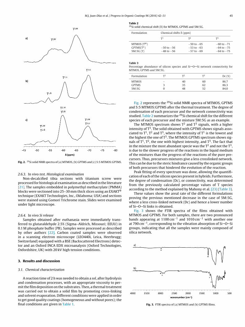
M.J. Juan-Díaz et al. / Progress in Organic Coatings 96 (2016) 42–51 45
F
2
p[btwu
2
f0biStO
3
3
amwatfi
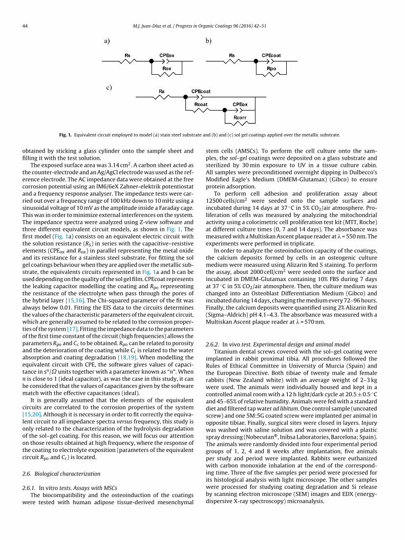
Table 229Si solid chemical shift (�) for MTMOS, GPTMS and 5M:5G.
Formulation Chemical shifts � (ppm)
T1 T2 T3
MTMOS (TM) −50 to −65 −60 to −71GPTMS(TG) −50 to −56 −53 to −63 −64 to −755M:5G (T) −48 to −56 −57 to −69 −64 to −75
Table 3Percentage abundance of silicon species and Si O Si network connectivity forMTMOS, GPTMS and 5M:5G.
Formulation T1 T2 T3 Dc (%)
MTMOS – 40 60 86.7
groups, indicating that all the samples were mainly composed ofsilica network.
ig. 2. 29Si solid NMR spectra of (a) MTMOS, (b) GPTMS and (c) 5:5 MTMOS:GPTMS.
.6.3. In vivo test. Histological examinationNon-decalcified tibia sections with titanium screw were
rocessed for histological examination as described in the literature21]. The samples embedded in polymethyl methacrylate (PMMA)locks were sectioned into 25–30 mm thick slices using an EXAKT®
echnique (EXAKT Technologies, Inc., Oklahoma; USA) and sectionsere stained using Gomori Trichrome stain. Slides were examinednder light microscope.
.6.4. In vivo Si releaseSamples obtained after euthanasia were immediately trans-
erred to glutaraldehyde 2.5% (Sigma–Aldrich, Missouri; EEUU) in.1 M phosphate buffer (PB). Samples were processed as describedy other authors [22]. Carbon coated samples were observed
n a scanning electron microscope (LEO440i, Leica, Heerbrugg;witzerland) equipped with a BSE (Backscattered Electrons) detec-or and an Oxford INCA EDX microanalysis (Oxford Technologies,xfordshire, UK) with 20 kV high tension conditions.
. Results and discussion
.1. Chemical characterization
A reaction time of 2 h was needed to obtain a sol, after hydrolysisnd condensation processes, with an appropriate viscosity to per-it the film deposition on the substrates. Then, a thermal treatmentas carried out to obtain a solid film by promoting cross-linking
nd solvent evaporation. Different conditions were applied in ordero get good quality coatings (homogeneous and without pores); thenal conditions are given in Table 1.
GPTMS 3 35 62 86.35M:5G 0 48 52 84.0
Fig. 2 represents the 29Si solid NMR spectra of MTMOS, GPTMSand 5:5 MTMOS:GPTMS after the thermal treatment. The degree ofcondensation of each precursor and the network connectivity wasstudied. Table 2 summarizes the 29Si chemical shift for the differentspecies of each precursor and the mixture 5M:5G as an example.
The MTMOS spectrum shows T2 and T3 signals, with a higherintensity of T3. The solid obtained with GPTMS shows signals asso-ciated to T1, T2 and T3, where the intensity of T1 is the lowest andthe highest the one of T3. The MTMOS:GPTMS spectrum shows sig-nals of T1, T2, the one with highest intensity, and T3. The fact thatin the mixture the most abundant specie was the T2 and not the T3,is due to the slower progress of the reactions in the liquid mediumof the mixtures than the progress of the reactions of the pure pre-cursors. Thus, precursors mixtures give a less crosslinked network.This can be due to the steric hindrance caused by the organic groupsof both precursors that hindered the evolution of the reaction.
Peak fitting of every spectrum was done, allowing the quantifi-cation of each of the silicon species present in hybrids. Furthermore,the degree of condensation (Dc), or connectivity, was determinedfrom the previously calculated percentage values of T speciesaccording to the method explained by Mahony et al. [23] (Table 3).
These values show the areal rate of the different formulationsproving the previous mentioned decrease in the case of 5M:5G,where a less cross-linked network (Dc) and hence a lower numberof Si O Si links is obtained.
Fig. 3 shows the FTIR spectra of the films obtained withMTMOS and GPTMS. For both samples, there are two pronouncedbands appearing at 1100 cm−1 and 1010 cm−1 with another oneat 790 cm−1, corresponding to the vibration absorption of Si O Si
Fig. 3. FTIR spectra of (a) MTMOS and (b) GPTMS films.

46 M.J. Juan-Díaz et al. / Progress in Orga
Table 4Representative bands of each precursor.
Formulation Wave number (cm−1) Assignment
MTMOS 2960 CH3
1275 Si CH3
GPTMS 3006 Epoxy ring2950 CH2
1205 Epoxy ring1150 CH2 O CH2
Fig. 4. FTIR spectra of (a) 8M:2G, (b) 5M:5G, (c) 2M:8G films.
Table 5Water contact angle results with different molar ratios MTMOS:GPTMS depositedon a metal substrate.
Formulation Wetting contact angle (◦)
MTMOS 75.9 ± 1.68M:2G 76.6 ± 0.65M:5G 66.9 ± 2.0
iooa1cgm
mdc1
3
a
intfbitfm
present less condensed networks. On the other hand, if we com-
2M:8G 68.4 ± 2.1GPTMS 64.8 ± 2.0
As expected, due to the organic groups, which are not involvedn the hydrolysis and condensation reactions, characteristic bandsf each precursors are developed in the spectra (the asteriskednes). In Table 4, these representative bands of each precursorre described. For the MTMOS coating, the band appearing at275 cm−1 confirmed the presence of the methyl group. In thease of GPTMS coating the band lying at 1205 cm−1 was due to thelycidoxypropyl group. This clearly demonstrated the successfulodification of the silica network by those organic groups.The materials with different molar ratios MTMOS:GPTMS (Fig. 4)
aintain the characteristic bands of the precursors alone, theifferences in the concentration is confirmed with the intensityhange in the bands at 1275 cm−1 of MTMOS and the ones of205 cm−1 and 3006 cm−1 associated with the GPTMS.
.2. Contact angle
The values of the contact angle for the set of materials obtainedre summarized in Table 5.
The GPTMS coating is more hydrophilic than the MTMOS coat-ng, this fact is due to the organic groups present in the polysiloxaneetwork, i.e. the hydrophilic glycidoxypropyl group of GPTMS andhe hydrophobic methyl group of MTMOS. The materials with dif-erent molar ratios MTMOS:GPTMS showed contact angle valuesetween those of MTMOS and GPTMS films. This range of values
s important since most literature suggests that protein adsorp-
ion that leads to a good biocompatibility tends to occur moreavourably on hydrophobic surfaces or on surfaces with an inter-ediate wettability (60–90◦) [24].
nic Coatings 96 (2016) 42–51
3.3. Electrochemical impedance spectroscopy (EIS)
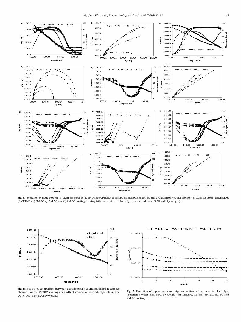
The EIS test was performed to study the isolating propertiesof the coatings along the time in an electrolyte. Samples of stain-less steel AISI 316L plates and samples of MTMOS, 8M:2G, 5M:5G,2M:8G and GPTMS obtained by dip-coating and applied on thestainless plates were used. Fig. 5 shows the Bode and Nyquist graphsof the impedance evolution with time over 24 h for coatings withdifferent molar ratios MTMOS:GPTMS.
Impedance spectra show that MTMOS’ impedance values arehigher than those obtained in coatings with GPTMS. As the quantityof GPTMS increases the impedance module decreases. This changein MTMOS impedance results at 24 hours is due probably to thehigher permeability of the film when the GPTMS is incorporated.
Impedance experimental results for the coatings were modelledaccording to the proposed equivalent circuits (Fig. 1). A good fitwas obtained for all coatings over the complete range of frequen-cies as is shown in Fig. 6. In the case of MTMOS coatings, only onetime constant was detected in the impedance response for shortexposure times (model shown in Fig. 1b). However, the coatingresistance decreased and the second time constant related to theoxide layer of the substrate appeared after 24 hours in contact withthe solution. Two time constants were detected for coatings in thepresence of GPTMS (Fig. 1c). This is attributed to a higher perme-ability in GPTMS coatings that lead to consider the second timeconstant related to the oxide of the metal. This fact was reflectedby the significant decrease of the impedance modulus value forGPTMS’ coatings when they are compared to the registered MTMOSimpedance.
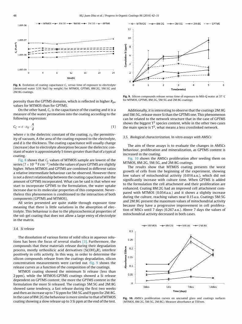
The aim of this study is to understand how the electrolyte expo-sure affects the material degradation. In this sense, the discussionis focused only on the first time constant parameters (Rpo and Cc),which are related to the coating behaviour. Fig. 7 shows Pore Resis-tance and Fig. 8 Coating Capacity parameters obtained from theresponse at high frequencies of all coatings.
Pore resistance Rpo is a measure of the porosity and deterio-ration of the coating. Rpo values have usually been related to thenumber of pores or capillary channels perpendicular to the sub-strate surface through which the electrolyte reaches the interface[25]. Although the Rpo can also increase with immersion time, prob-ably as a result of pore or defect blockage by corrosion products, itusually decreases (Fig. 6). Some authors have found three regionsin the time dependent trend of Rpo. It initially decreases rapidly,then slowly (displaying a plateau) and then again rapidly, coin-ciding with the appearance of the second semicircle. The plateauis explained by making the assumption that the number of path-ways formed is approximately constant with time. Thus, it can besaid that Rpo value is a measure of the ionic resistance throughthe pores of the coatings and is inversely proportional to theextent and number of defects in the coating. The evolution ofRpo with the exposure time in the electrolyte gives informationabout the coating capacity to avoid the formation of pores acrossthe film due to its degradation. High and constant Rpo valuesare attributed to coatings that do not degrade during electrolyteexposure.
Fig. 7 clearly shows that coatings with GPTMS have values ofRpo ranking from 3 to 4 magnitudes order being correlated to theincrease of its content. GPTMS containing coatings showed lessresistance to the flow of water through the coating than in the caseof MTMOS coating. The water is able to penetrate the coatings for-mulated with GPTMS much more than in MTMOS coating, possiblybecause of their bigger value of hydrophilicity and because they
pare MTMOS:GPTMS coatings with the GPTMS one, we can say thatdifferences in Rpo are due to heterogeneous porosity in mixturecoatings. In these matrixes the MTMOS domains present smaller
M.J. Juan-Díaz et al. / Progress in Organic Coatings 96 (2016) 42–51 47
Fig. 5. Evolution of Bode plot for (a) stainless steel, (c) MTMOS, (e) GPTMS, (g) 8M:2G, (i) 5M:5G, (k) 2M:8G and evolution of Nyquist plot for (b) stainless steel, (d) MTMOS,(f) GPTMS, (h) 8M:2G, (j) 5M:5G and (l) 2M:8G coatings during 24 h immersion in electrolyte (deionized water 3.5% NaCl by weight).
Fig. 6. Bode plot comparison between experimental (o) and modelled results (x)obtained for the MTMOS coating after 24 h of immersion in electrolyte (deionizedwater with 3.5% NaCl by weight).
Fig. 7. Evolution of a pore resistance Rpo versus time of exposure to electrolyte(deionized water 3.5% NaCl by weight) for MTMOS, GPTMS, 8M:2G, 5M:5G and2M:8G coatings.
48 M.J. Juan-Díaz et al. / Progress in Organic Coatings 96 (2016) 42–51
F(2
pv
mf
C
wia(sc
shaiasitc
mtti
3
tcppscr
2dfsaIc
because they have a progressive improvement in cell prolifera-tion of MSCs until 7 days (0.267 a.u.). Above 7 days the values ofmitochondrial activity decreased in both cases.
ig. 8. Evolution of coating capacitance Cc versus time of exposure to electrolytedeionized water 3.5% NaCl by weight) for MTMOS, GPTMS, 8M:2G, 5M:5G andM:8G coatings.
orosity than the GPTMS domains, which is reflected in higher Rpo
alues for MTMOS than for GPTMS.On the other hand, Cc is the capacitance of the coating and it is a
easure of the water permeation into the coating according to theollowing expression:
C = ε · ε0 · A
d(1)
here ε is the dielectric constant of the coating, ε0 the permittiv-ty of vacuum, A the area of the coating exposed to the electrolyte,nd d is the thickness. The coating capacitance will usually changeincrease) due to electrolyte absorption because the dielectric con-tant of water is approximately 5 times greater than that of a typicaloating.
Fig. 8 shows that Cc values of MTMOS sample are lowest of theeries (7 × 10⁻⁹ F cm−2) while the values of pure GPTMS are slightlyigher. When MTMOS and GPTMS are combined in different ratios
relative intermediate behaviour can be observed. However theres not a direct relationship between the coating capacitance and themount of GPTMS incorporated. What can be said is that when wetart to incorporate GPTMS to the formulation, the water uptakencrease due to its molecular properties of this component. Never-heless this phenomenon is conditioned to the interaction of bothomponents (GPTMS and MTMOS).
All series presented are quite stable through exposure timeeaning that there is little variations in the absorption of elec-
rolyte. This behaviour is due to the physicochemical properties ofhe sol–gel coating that does not allow a large entry of electrolyten the matrix.
.4. Si release
The dissolution of various forms of solid silica in aqueous solu-ions has been the focus of several studies [3]. Furthermore, theompounds that these materials release during their degradationrocess, mostly orthosilicic acid derivatives (Si(OH)3R), interfereositively in cells activity. In this way, in order to determine theilicon compounds release from the coatings degradation, silicononcentration measurements were carried out. Fig. 9 shows theelease curves as a function of the composition of the coatings.
MTMOS coating showed the minimum Si release (less than ppm), while the MTMOS:GPTMS coatings showed a Si releaseependent on GPTMS content; the more the GPTMS content in theormulation the more Si released. The coatings 5M:5G and 2M:8G
howed same tendency, a fast release during the first two weeksnd then an increase up to 7 Si ppm for 5M:5G and 9 ppm for 2M:8G.n the case of 8M:2G the behaviour is more similar to that of MTMOSoating showing a slow release up to 3 Si ppm at the end of the test.Fig. 9. Silicon compounds release versus time of exposure to Mili-Q water at 37 ◦Cfor MTMOS, GPTMS, 8M:2G, 5M:5G and 2M:8G coatings.
Additionally, it is interesting to observe that the coatings 2M:8Gand 5M:5G, release more Si than the GPTMS one. This phenomenoncan be related to the network structure that in the case of GPTMSshows the biggest T3 species content, while in the other two casesthe main specie is T2, what means a less crosslinked network.
3.5. Biological characterization. In vitro assays with AMSCs
The aim of these assays is to evaluate the changes in AMSCsbehaviour, proliferation and mineralization, as GPTMS content isincreased in the coating.
Fig. 10 shows the AMSCs proliferation after seeding them onMTMOS, 8M:2G, 5M:5G, and 2M:8G coatings.
The results show that MTMOS coating presents the worstgrowth of cells from the beginning of the experiment, showinglow values of mitochondrial activity (0.016 a.u.), which did notsignificantly increase with culture time. When GPTMS is addedto the formulation the cell attachment and their proliferation areenhanced. Coating 8M:2G had an improved cell attachment com-pared with MTMOS (0.054 a.u.) and it shows a slightly increaseduring the culture, reaching values near 0.15 a.u. Coatings 5M:5Gand 2M:8G present the maximum values of mitochondrial activity
Fig. 10. AMSCs proliferation curves on uncoated glass and coatings surfaces(MTMOS, 8M:2G, 5M:5G, 2M:8G). Measure absorbance at 550 nm.

M.J. Juan-Díaz et al. / Progress in Orga
F8
wtfud
ttmMwttw2cms
thSpShcots[tb
becular bone growth along its surface towards the endostium,
Ffit
ig. 11. Alizarin Red S staining at 7 and 14 days on coatings surfaces (MTMOS,M:2G, 5M:5G, 2M:8G). Values normalized with respect to MTMOS.
In order to analyze the mineralization capacity, calcium depositsere evaluated using an osteogenic medium culture. Fig. 11 shows
he quantification of these deposits using Alizarin Red S stainingor each material at two culture times (7 and 14 days). The val-es were normalized with respect to MTMOS coating for easieretermination of the influence of the GPTMS content.
Values of Alizarin Red presented big differences depending onhe composition. Values of mineralization of MTMOS coatings werehe lowest and formulations with GPTMS content presented higher
ineralization values in the two test periods. After 7 culture days,TMOS had the lower quantity of calcium, what can be relatedith the poor number of attached cells and the low Si release to
he medium from this type of coating. When GPTMS was added tohe formulation greater number of mineralized extracellular matrixas detected. Although the mitochondrial activity of 5M:5G and
M:8G coatings decreased above 7 days of culture (Fig. 9). Theseoatings after 14 days of culture reached the maximum value ofineralization. For this time, the results of mineralization of GPTMS
amples increased strong and independently of the composition.Reffitt et al. [9] founded that physiological concentrations of Si in
he form of orthosilicic acid stimulate collagen type 1 synthesis inuman osteoblast-like cells and skin fibroblasts. Treatment withi also enhanced osteoblastic differentiation (enhanced alkalinehosphatase activity and osteocalcin synthesis in the MG-63 cells).i is also known to bind to glycosaminoglycan macromolecules andas been shown to play a role in the formation of crosslinks betweenollagen and proteoglycans [26] thus resulting in the stabilizationf bone matrix molecules and preventing their enzymatic degrada-ion. It is likely that the mechanism of action of Si in bone matrixynthesis may involve a complex biochemical set of interactions
27] with biological molecules and the high values of mineraliza-ion showed by GPTMS containing coatings indicate that this maye another possible mode of action of soluble Si.ig. 12. Light microscopy images (10×) of 5M:5G in vivo samples after 8 weeks of implanbrous capsule with bone marrow contact, (a.2) fibrous capsule between bone and non rerabecular new bone without direct contact with 5M:5G sol–gel remaining coating (EXAK
nic Coatings 96 (2016) 42–51 49
Our results suggest that sol–gel coatings are able to produce Sirelease that can increase the differentiation process of the MSC ontoosteoblastic cells, and so production of calcium deposits could bedetected at 7 and 14 days.
3.6. Biological characterization. In vivo study
The good in vitro results obtained with GPTMS containingcoatings leaded to select the 5M:5G coating to be tested underin vivo conditions. To study the tissue response to sol–gel coatings,biocompatibility, osseointegration and degradation coating capa-bilities were evaluated through light microscope histological study(Fig. 12). In vivo Si release was observed by SEM images and EDXmicroanalysis (Fig. 13).
3.6.1. BiocompatibilityIn order to evaluate biocompatibility of 5M:5G coating,
three factors were kept in mind; bone marrow condition, pres-ence/absence of foreign-body giant cells and fibrous capsuleevolution. In the case of 5M:5G samples, bone marrow’s archi-tecture appeared modified with traumatic and aplasic tissue,particulary near the coating, at short periods. After, the architecturebegan to recuperate far of coating contact zone and, 8 weeks afterimplantation, bone marrow generally recovered the architectureand cellular charge, although not entirely.
Control samples showed normal bone marrow conditions afterimplantation with some traumatic signs after 4 weeks. However,after 8 weeks of implantation, bone marrow was recuperated.Foreign-body giant cells were found at all periods in case of controlsamples, but the number of cells did not exceed that of a normalforeign-body response. No foreign-body giant cells were found incase of 5M:5G. In all samples after 1 and 2 weeks of implantationa fibrous tissue relaxed (lax) band was found around the implantsin medullar contact zones. After 4 weeks the fibrous tissue turnedinto a dense fibrous capsule. Moreover, at 8 weeks, a thick cap-sule was also observed between cortical bone and dental screw. In5M:5G implants this capsule was found between trabecular boneand sol–gel coating, surrounding the implant.
3.6.2. Osseointegration and in vivo coating degradationIf cortical and trabecular bone repair in control implant and
5M:5G samples are compared, we could say that there is not atotal osseointegration at cortical level neither in the case of theTi control nor in the 5M:5G sample, because a thin fibrous layerappears between bone and screws. However, after 8 weeks ofimplantation, control screw osteoconduction allowed the new tra-
getting a total osseointegration. Nevertheless, if we observe 5M:5Gcoated implants we have to say that they did not get a goodosseointegration. First, a fibrous capsule surrounded the screw
tation (a.1) recovery of bone marrow condition in implant distant zones and densesorbable coating and surrounding the screw to protect the bone marrow, (a.3) thinT® cut and Gomori Trichrome stain).

50 M.J. Juan-Díaz et al. / Progress in Organic Coatings 96 (2016) 42–51
Fig. 13. (a.1, a.2) SEM images of in vivo 5M:5G interface after 1 week of implantation (1000×) and (a.3) EDX line profile crossing the different materials of the image,determining (from top to bottom); bone (Ca and P), an intermediate layer between coating and bone (Ca, P and Si), coating (Si), organic part of the coating (C) and titaniumimplant (Ti).
(itfptz
3
awrcpabc
sbtcic2b
Fig. 12a.1) and persisted between trabecular bone and coatedmplant (Fig. 12a.2). Second, a light level of osteoblastic activa-ion was observed. Third, the architecture of the trabecular spiculesormed was fine and slightly branched (Fig. 12a.3) and finally, a bigart of the sol–gel coating remained in the threads until the end ofhe studied period in the trabecular zone as well as in the corticalone (Fig. 12a.3).
.6.3. In vivo Si releaseIn vivo Si release was studied by SEM image with EDX micro-
nalysis and mapping of the histological samples (Fig. 13). A studyas made analyzing the different elements signals: Si signal cor-
esponding to sol–gel coating, Ca and P corresponding to bone, Tiorresponding to the implant and C corresponding to the organicart of the coating. The obtained images show a grey scale with
different grey colour for each zone, so that materials can easilye differentiated: light grey for Ti implant, dark grey for sol–geloating and medium grey for bone.
Thus, for the 5M:5G coated implants, we observe variousamples where Si, Ca and P signal coexisted in a same grey zoneetween bone and coating. This was detected in samples belongingo 1 week after implantation, in cortical and trabecular boneontact and in samples belonging to 2 weeks after implantation,
n cortical bone contact. Therefore, a migration of Si in the bone islearly observed indicating a biodegradation of the coating at 1 andweeks after implantation, what could contribute to stimulateone regeneration.
4. Conclusions
This study demonstrated that changes in the composition of theobtained sol–gel hybrid coatings highly influence their biodegrad-ability and hence their biomedical applications.
An increase in GPTMS content increases the wettability andwater absorption of the coating, due to either a decrease in thenetwork crosslinking density of the hybrid material or the increasein the number of polar groups. The presence of GPTMS, which is asilicon precursor with a big organic group, contributes to hinder-ing the Si O Si bond formation. Consequently, the degradabilityof the polysiloxane network in contact with water is higher due tothe formation of a more open and hydrophilic network.
As a result, the addition of GPTMS to MTMOS, significantlyincrease the release of silicon compounds, which is reflected in abetter AMSCs proliferation and mineralization. The fact of the for-mation of calcium rich deposits onto the coatings, suggests thatthe silicon compounds that are released during the degradationprocess of the material have in vitro osteoinduction ability. Never-theless, in vivo behaviour of 5M:5G did not correspond with cellularresults. Implanted coatings almost did not degrade, Si released wasonly detected by SEM-EDX, but thickness of coating did not reducein time significantly.
Therefore, at the sight of the good cells behaviour in vitro asso-ciated to the presence of GPTMS, and taking into account the
interest of obtaining materials with functional monomers we pro-pose to carry out further research in these materials. In that case,the addition of a precursor which adds biodegradability to thecoating, to enhance Si release from the earliest moments after
n Orga
itcr
A
pUafw
R
[
[
[
[
[
[[[[[
[
[
[
[
[
[25] S.J. Garcia, M.T. Rodriguez, R. Izquierdo, J. Suay, Prog. Org. Coat. 60 (4) (2007)
M.J. Juan-Díaz et al. / Progress i
mplantation should be a way to enhance the in vivo osteoinduc-ion capability of the coating. Furthermore, the addition of activeompounds attached to the GPTMS will be the next step of thisesearch.
cknowledgements
The supports of the Spanish Ministry of Economy and Com-etitiveness through project IPT-2012-0218-090000 and of theniversity of the Basque Country (UPV/EHU) through “UFI11/56”nd “IT-611-13” are kindly acknowledged. We are especially thank-ul to D. Antonio Coso and D. Jaime Franco (Ilerimplant S.L., http://ww.ilerimplant.com) for their cooperation.
eferences
[1] M. Manso-Silvan, C. Rodriguez-Navas, M.A. Hernandez, E. Lopez-Elvira, R.Gago, L. Vazquez, F. Agullo-Rueda, A. Climent, J.M. Martinez-Duart, J.P.Garcia-Ruiz, J. Biomed. Mater. Res. Appl. Biomater. 83B (1) (2007) 232–239.
[2] S.M. Hosseinalipour, A. Ershad-Langroudi, A.N. Hayati, A.M.Nabizade-Haghighi, Prog. Org. Coat. 67 (4) (2010) 371–374.
[3] M. Hernandez-Escolano, M. Juan-Diaz, M. Martinez-Ibanez, A.Jimenez-Morales, I. Goni, M. Gurruchaga, J. Suay, J. Sol–Gel Sci. Technol. 64 (2)(2012) 442–451.
[4] L. Ren, K. Tsuru, S. Hayakawa, A. Osaka, J. Sol–Gel Sci. Technol. 26 (1–3) (2003)1137–1140.
[5] K. Tsuru, Z. Robertson, B. Annaz, I.R. Gibson, S.M. Best, Y. Shirosaki, S.
Hayakawa, A. Osaka, Key Eng. Mater. 361–363 (Pt. 1, Bioceramics) (2008)447–450.[6] E.M. Carlisle, Science 178 (4061) (1972) 619–621.[7] K. Schwarz, D.B. Milne, Nature 239 (5371) (1972) 333–334.[8] X. Chatzistavrou, E. Kontonasaki, K.M. Paraskevopoulos, et al., 7 – sol–gel
[[
nic Coatings 96 (2016) 42–51 51
derived bioactive glass ceramics for dental applications, in: P Vallittu (Ed.),Non-Metallic Biomaterials for Tooth Repair and Replacement, WoodheadPublishing, 2013, pp. 194–231.
[9] P.E. Keeting, M.J. Oursler, K.E. Wiegand, S.K. Bonde, T.C. Spelsberg, B.L. Riggs, J.Bone Miner. Res. 7 (11) (1992) 1281–1289.
10] D.M. Reffitt, N. Ogston, R. Jugdaohsingh, H.F.J. Cheung, B.A.J. Evans, R.P.H.Thompson, J.J. Powell, G.N. Hampson, Bone 32 (2) (2003) 127–135.
11] Y. Shirosaki, K. Tsuru, S. Hayakawa, A. Osaka, M.A. Lopes, J.D. Santos, M.H.Fernandes, Biomaterials 26 (5) (2005) 485–493.
12] S.I. Anderson, S. Downes, C.C. Perry, A.M. Caballero, J. Mater. Sci. Mater. Med. 9(12) (1998) 731–735.
13] M. Manso, S. Ogueta, P. Herrero-Fernández, L. Vázquez, M. Langlet, J.P.García-Ruiz, Biomaterials 23 (19) (2002) 3985–3990.
14] C.J. Wilson, R.E. Clegg, D.I. Leavesley, M.J. Pearcy, Tissue Eng. 11 (1/2) (2005)1–18.
15] S.J. Garcia, J. Suay, Prog. Org. Coat. 59 (3) (2007) 251–258.16] S.J. Garcia, J. Suay, Prog. Org. Coat. 57 (3) (2006) 273–281.17] S.J. Garcia, J. Suay, Prog. Org. Coat. 66 (3) (2009) 306–313.18] A. Amirudin, D. Thierry, Prog. Org. Coat. 26 (1) (1995) 1–28.19] G.W. Walter, J. Electroanal. Chem. Interfacial Electrochem. 118 (1981)
259–273.20] E.P.M. van Westing, G.M. Ferrari, J.H.W. De Wit, Corros. Sci. 34 (9) (1993)
1511–1530.21] Peris Jl, J. Prat, R. Comín, R. Dejoz, I. Roger, P. Vera, Rev. Esp. Cir. Osteoart. 28
(1993) 231–238.22] J. Wierzchos, T. Falcioni, A. Kiciak, J. Wolinski, R. Koczorowski, P. Chomicki,
et al., Micron 39 (2008) 1363–1370.23] O. Mahony, S. Yue, C. Turdean-Ionescu, J.V. Hanna, M.E. Smith, P.D. Lee, J.R.
Jones, J. Sol Gel Sci. Technol. 69 (2014) 288–298.24] C.P. Stallard, K.A. McDonnell, O.D. Onayemi, J.P. O’Gara, D.P. Dowling,
Biointerphases 7 (2012) 31.
303–311.26] K. Schwarz, Proc. Natl. Acad. Sci. 70 (5) (1973) 1608–1612.27] S.D. Kinrade, N.J.W. Del, A.S. Schach, T.A. Sloan, K.L. Wilson, C.T.G. Knight,
Science 285 (5433) (1999) 1542–1545.