Programmed Cell Death (Apoptosis) · Apoptosis during the metamorphosis of a tadpole into a frog....
64
Programmed Cell Death (Apoptosis) Programmed cell-death (PCD) is death of a cell in any form, mediated by an intracellular program (Trends in Cell Biology)
Transcript of Programmed Cell Death (Apoptosis) · Apoptosis during the metamorphosis of a tadpole into a frog....
Programmed Cell Death (Apoptosis)
Programmed cell-death (PCD) is death of a cell in any form, mediated by an intracellular program
(Trends in Cell Biology)

The number of cells in this community is tightly regulated not simply
by controlling the rate of cell division, but also by controlling the rate
of cell death.
In humans, approx. 1010-1011 cells die every day
For every cell, there is a time to live and a time to die.
There are two ways in which cells die:
They are killed by injurious agents.
They are induced to commit suicide.
If cells are no longer needed, they commit suicide by activating an
intracellular death program. This process is therefore called
programmed cell death
“DEATH” is important to the “LIFE”
Apoptosis (programmed cell death): a physiological
process of cellular autodestruction, or cell suicide.
Strictly controlled in response to integrity of
prodeath signalling.
Critical roles of apoptosis in development, maintenance
of homeostasis and host defence in multicellular
organisms.
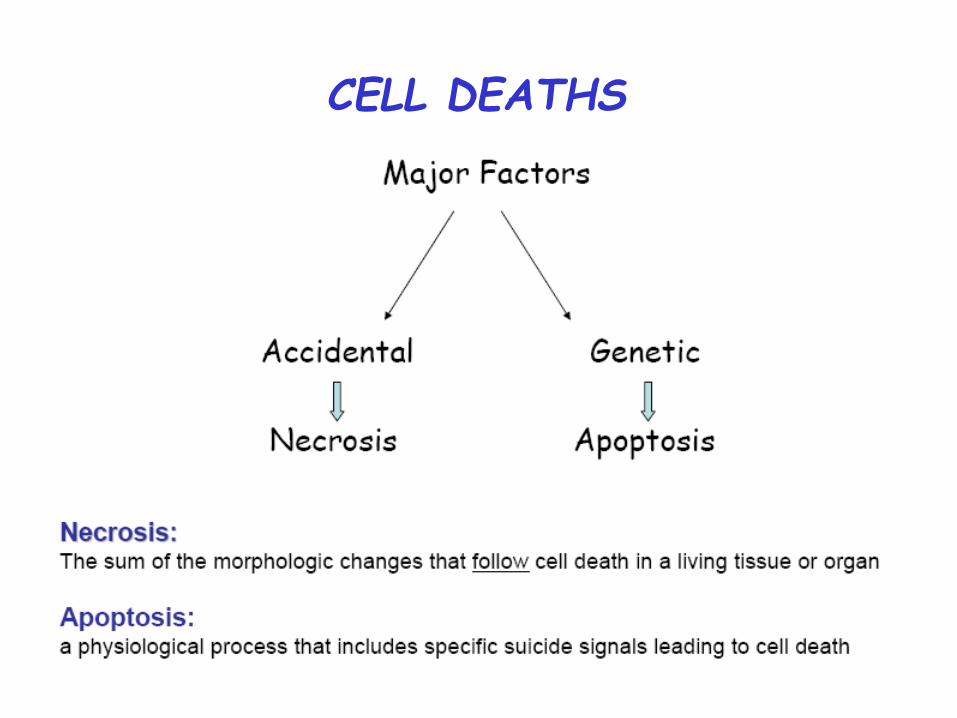
CELL DEATHS
Necrosis: Death by Injury Cells are damaged by injury, such as
Mechanical damage Exposure to toxic chemicals
Programmed Cell Death: Cell Suicide Cells are induced to commit suicide
Types of PCD
• Apoptosis or Type I cell-death
• Autophagic or Type II cell-death
(Cytoplasmic: characterized by the formation of large vacuoles
which eat away organelles in a specific sequence prior to the
nucleus being destroyed)
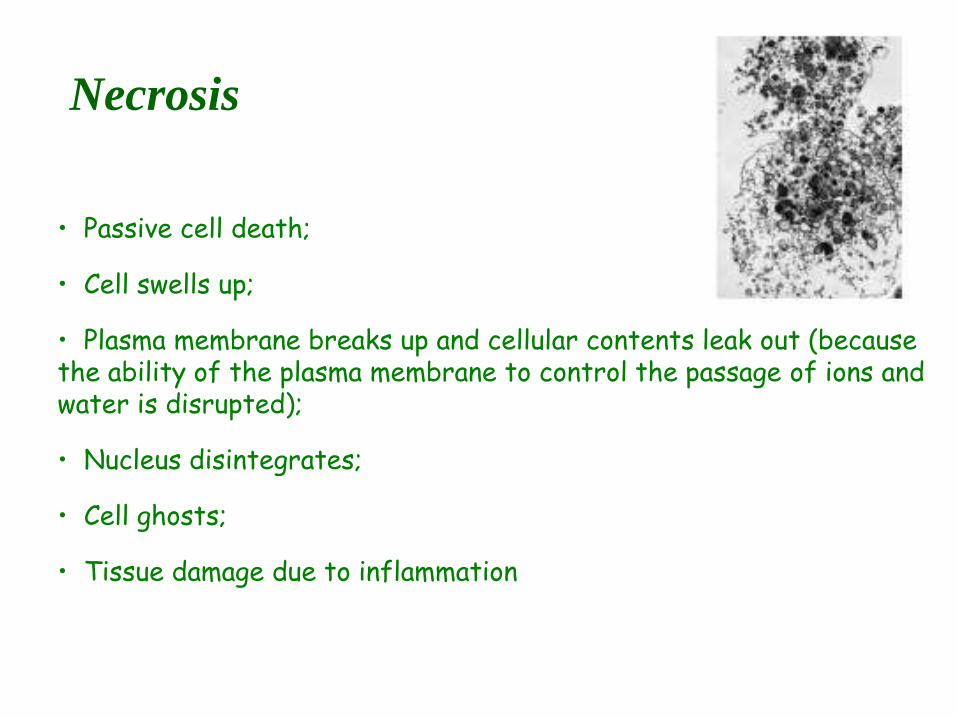
Necrosis
• Passive cell death;
• Cell swells up;
• Plasma membrane breaks up and cellular contents leak out (because the ability of the plasma membrane to control the passage of ions and water is disrupted);
• Nucleus disintegrates;
• Cell ghosts;
• Tissue damage due to inflammation
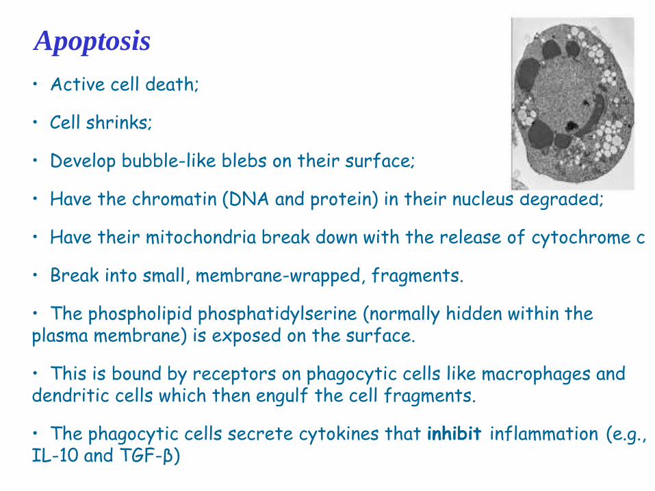
Apoptosis
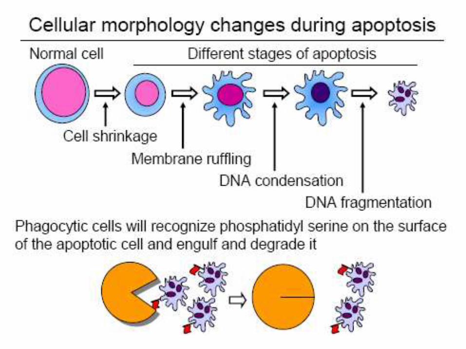
• Active cell death;
• Cell shrinks;
• Develop bubble-like blebs on their surface;
• Have the chromatin (DNA and protein) in their nucleus degraded;
• Have their mitochondria break down with the release of cytochrome c
• Break into small, membrane-wrapped, fragments.
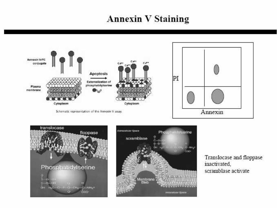
• The phospholipid phosphatidylserine (normally hidden within the plasma membrane) is exposed on the surface.
• This is bound by receptors on phagocytic cells like macrophages and dendritic cells which then engulf the cell fragments.
• The phagocytic cells secrete cytokines that inhibit inflammation (e.g., IL-10 and TGF-β)
CELL DEATHS
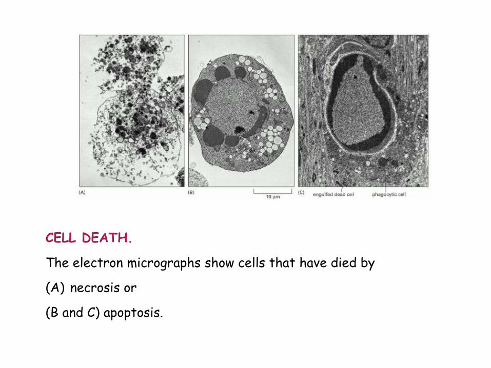
CELL DEATH. The electron micrographs show cells that have died by (A) necrosis or (B and C) apoptosis.
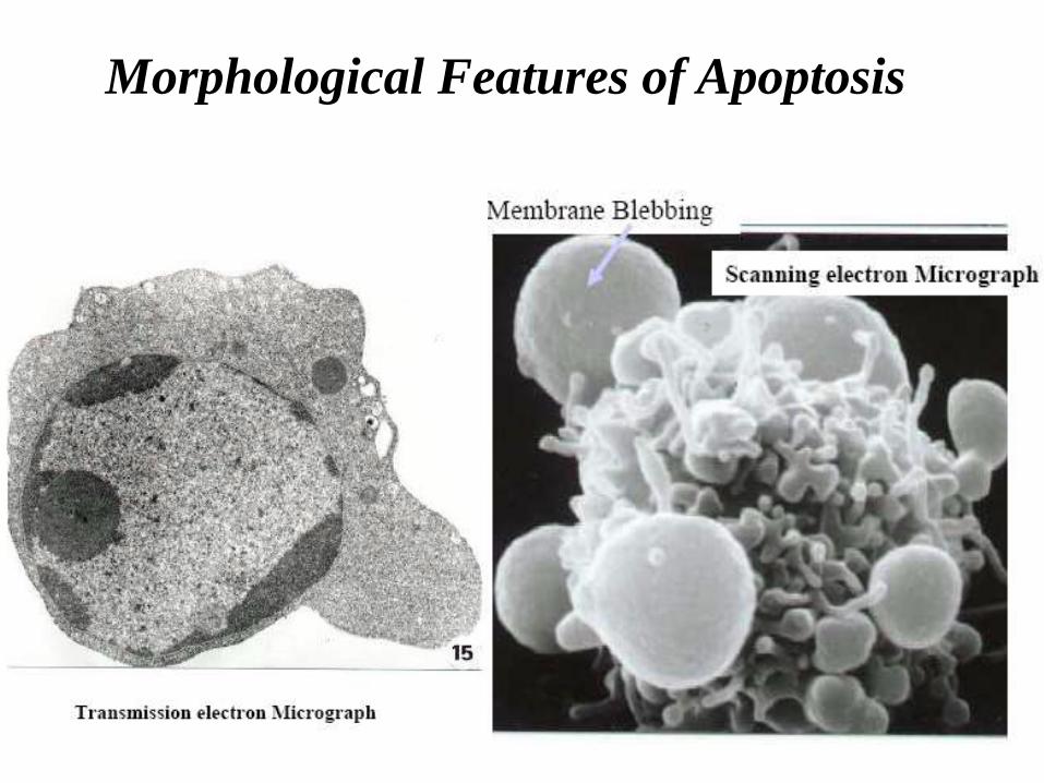
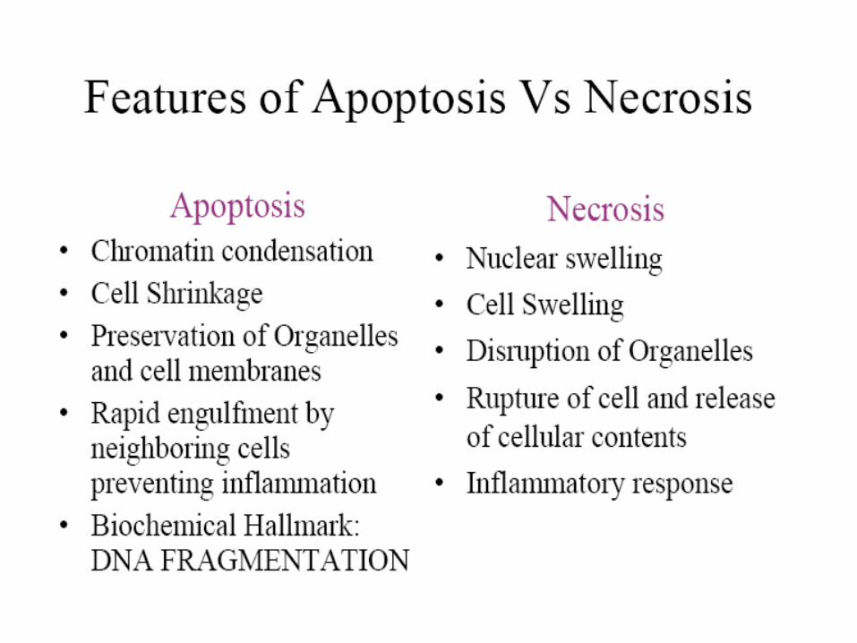
Morphological Features of Apoptosis
Why should a cell commit suicide?
There are two different reasons
1. Programmed cell death is as needed for proper development as
mitosis is.
Examples:
•The resorption of the tadpole tail at the time of its metamorphosis
into a frog occurs by apoptosis.
•The formation of the fingers and toes of the fetus requires the
removal, by apoptosis, of the tissue between them.
•The formation of the proper connections (synapses) between neurons
in the brain requires that surplus cells be eliminated by apoptosis
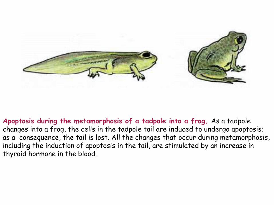
Apoptosis during the metamorphosis of a tadpole into a frog. As a tadpole changes into a frog, the cells in the tadpole tail are induced to undergo apoptosis; as a consequence, the tail is lost. All the changes that occur during metamorphosis, including the induction of apoptosis in the tail, are stimulated by an increase in thyroid hormone in the blood.
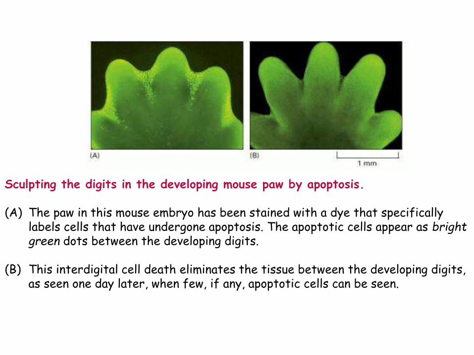
Sculpting the digits in the developing mouse paw by apoptosis. (A) The paw in this mouse embryo has been stained with a dye that specifically
labels cells that have undergone apoptosis. The apoptotic cells appear as bright green dots between the developing digits.
(B) This interdigital cell death eliminates the tissue between the developing digits, as seen one day later, when few, if any, apoptotic cells can be seen.

Apoptosis regulates nerve cell targeting
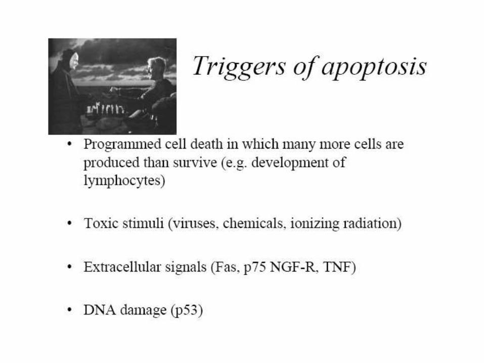
2. Programmed cell death is needed to destroy cells that represent a
threat to the integrity of the organism
Examples:
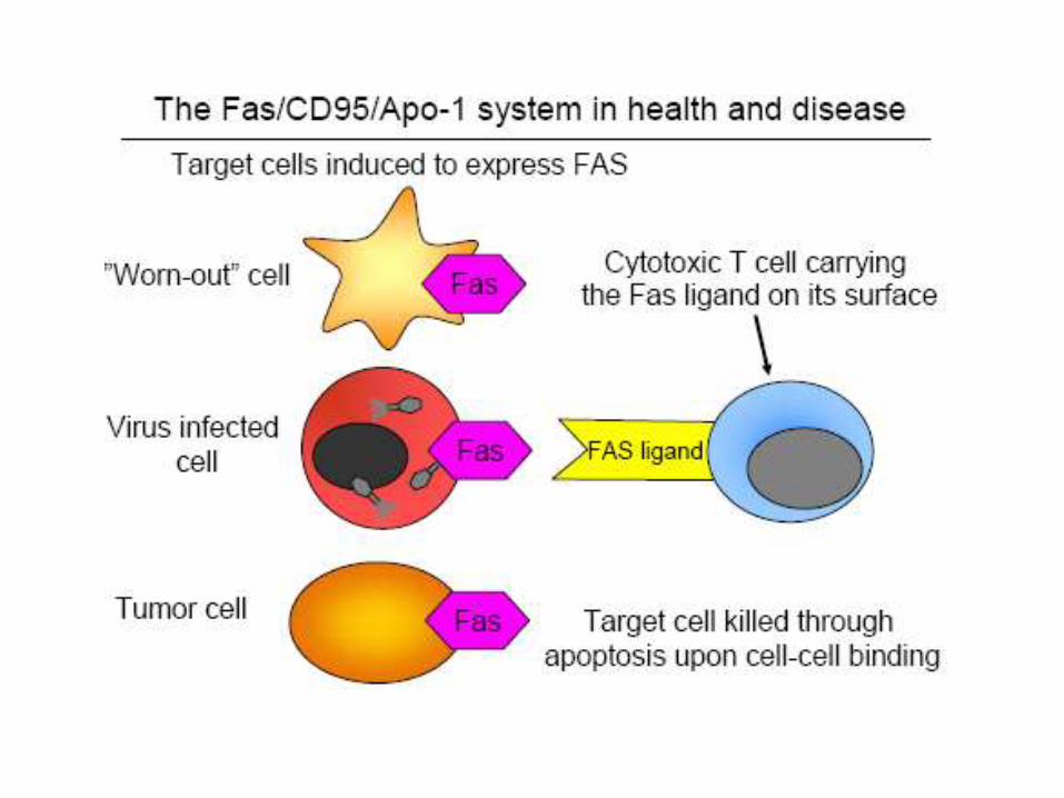
a) Cells infected with viruses
One of the methods by which cytotoxic T lymphocytes (CTLs) kill virus-
infected cells is by inducing apoptosis (some viruses mount
countermeasures to thwart it)
b) Cells of the immune system
As cell mediated immune responses wane, the effector cells must be
removed to prevent them from attacking body constituents. CTLs
induce apoptosis in each other and even in themselves. Defects in the
apoptotic machinery is associated with autoimmune diseases such as
lupus erythematosus and rheumatoid arthritis.
Immune response against its own tissues
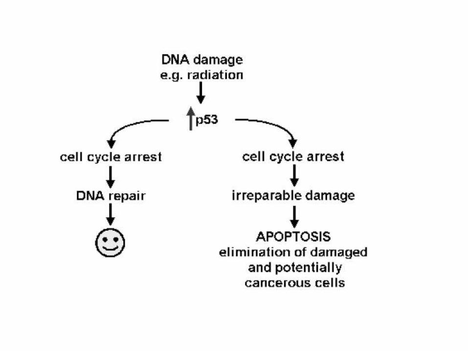
c) Cells with DNA damage
Damage to its genome can cause a cell
• to disrupt proper embryonic development leading to birth defects
• to become cancerous
Cells respond to DNA damage by increasing their production of p53
(A potent inducer of apoptosis).
Mutations in the p53 gene, producing a defective protein, are so often
found in cancer cells (that represent a lethal threat to the organism if
permitted to live)
d) Cancer cells
Radiation and chemicals used in cancer therapy induce apoptosis in some
types of cancer cells.
What makes a cell decide to commit suicide? The balance between: Withdrawal of positive signals and receipt of negative signals 1. Withdrawal of positive signals The continued survival of most cells requires that they receive continuous stimulation from other cells and, for many, continued adhesion to the surface on which they are growing. Positive signals:
• growth factors for neurons • Interleukin-2 (IL-2), an essential factor for the mitosis of lymphocytes
2. Receipt of negative signals
• Increased levels of oxidants within the cell
• Damage to DNA by these oxidants or other agents like
Ultraviolet light
x-rays
Chemotherapeutic drugs
• Accumulation of proteins that failed to fold properly into their proper tertiary structure
• Molecules that bind to specific receptors on the cell surface and signal the cell to begin the apoptosis program. ]
These death activators include:
Tumor necrosis factor-alpha (TNF-α ) that binds to the TNF receptors;
Lymphotoxin (also known as TNF-β ) that also binds to the TNF receptor;
Fas ligand (FasL), a molecule that binds to a cell-surface receptor named Fas (also called CD95).
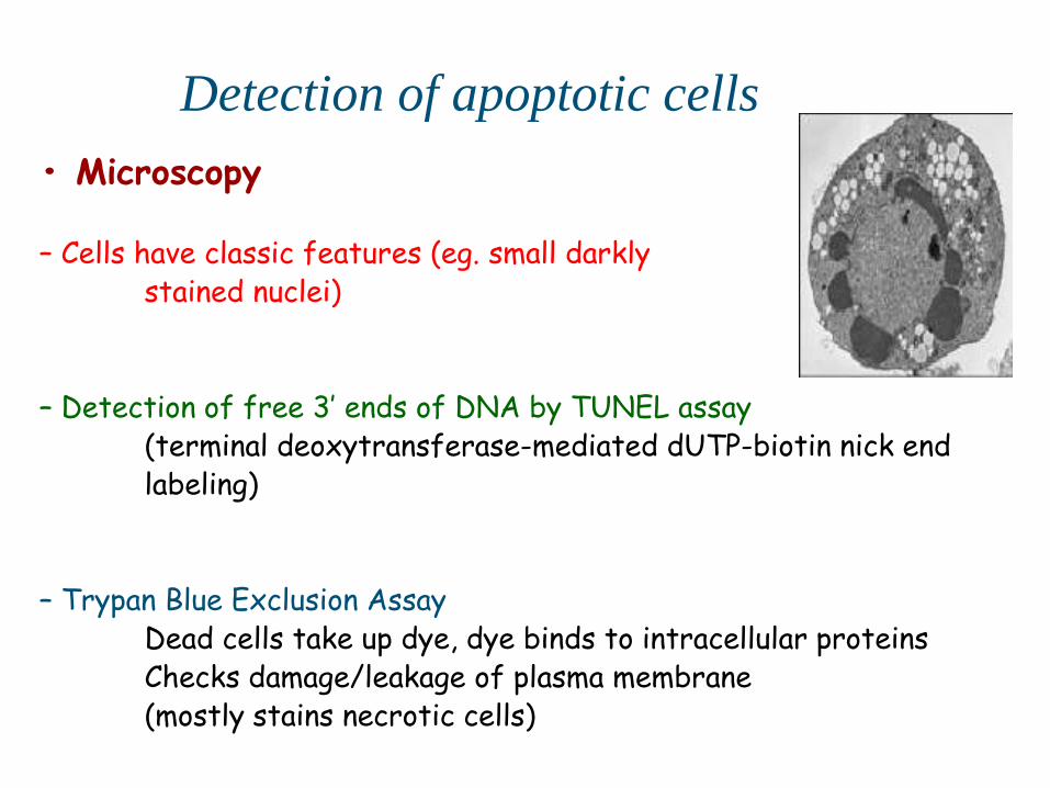
• Microscopy – Cells have classic features (eg. small darkly stained nuclei) – Detection of free 3’ ends of DNA by TUNEL assay (terminal deoxytransferase-mediated dUTP-biotin nick end labeling) – Trypan Blue Exclusion Assay Dead cells take up dye, dye binds to intracellular proteins Checks damage/leakage of plasma membrane (mostly stains necrotic cells)
Detection of apoptotic cells
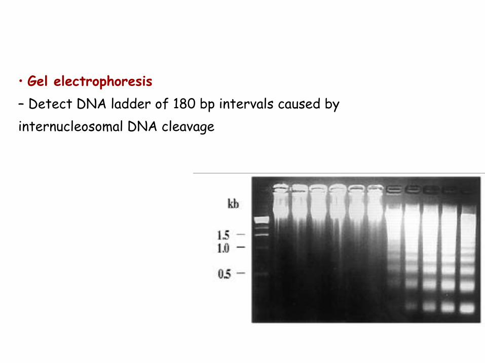
• Gel electrophoresis
– Detect DNA ladder of 180 bp intervals caused by
internucleosomal DNA cleavage
• Flow cytometry
– Measure externalization of phosphatidylserine (PS) with
fluorescently labeled Annexin-V (not very specific to apoptotic cells)
A DNA stain is added to distinguish necrotic cells
– Measure DNA fragmentation with propidium iodide fluorescence (not
specific for apoptotic cells)
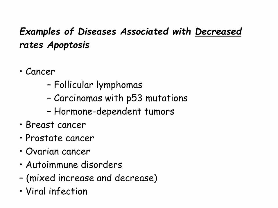
Examples of Diseases Associated with Decreased
rates Apoptosis
• Cancer
– Follicular lymphomas
– Carcinomas with p53 mutations
– Hormone-dependent tumors
• Breast cancer
• Prostate cancer
• Ovarian cancer
• Autoimmune disorders
– (mixed increase and decrease)
• Viral infection
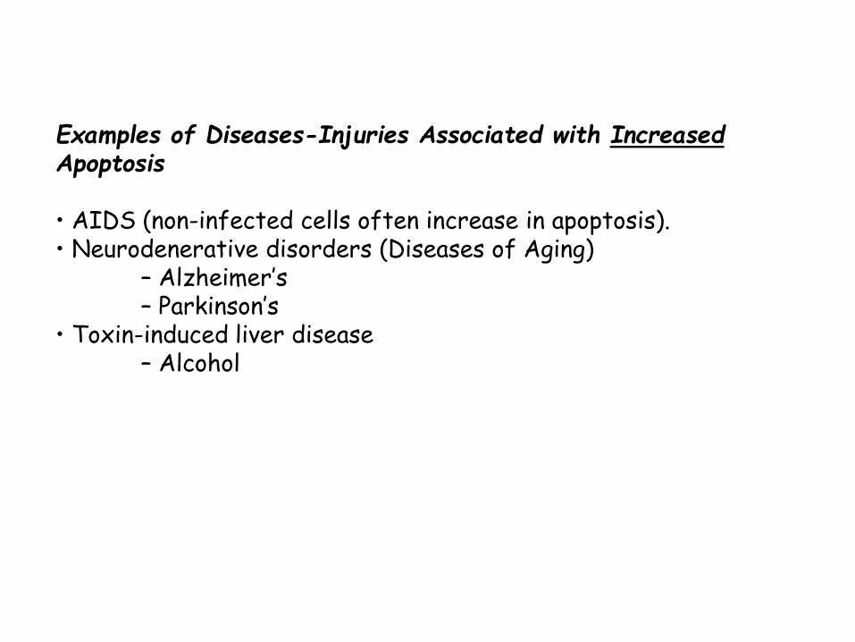
Examples of Diseases-Injuries Associated with Increased Apoptosis • AIDS (non-infected cells often increase in apoptosis). • Neurodenerative disorders (Diseases of Aging) – Alzheimer’s – Parkinson’s • Toxin-induced liver disease – Alcohol
Apoptosis and Cancer Development of Cancer: Increase in cell viability and decrease in apoptosis Proto-oncogenes regulate apoptosis Cancer therapy: Induce apoptosis
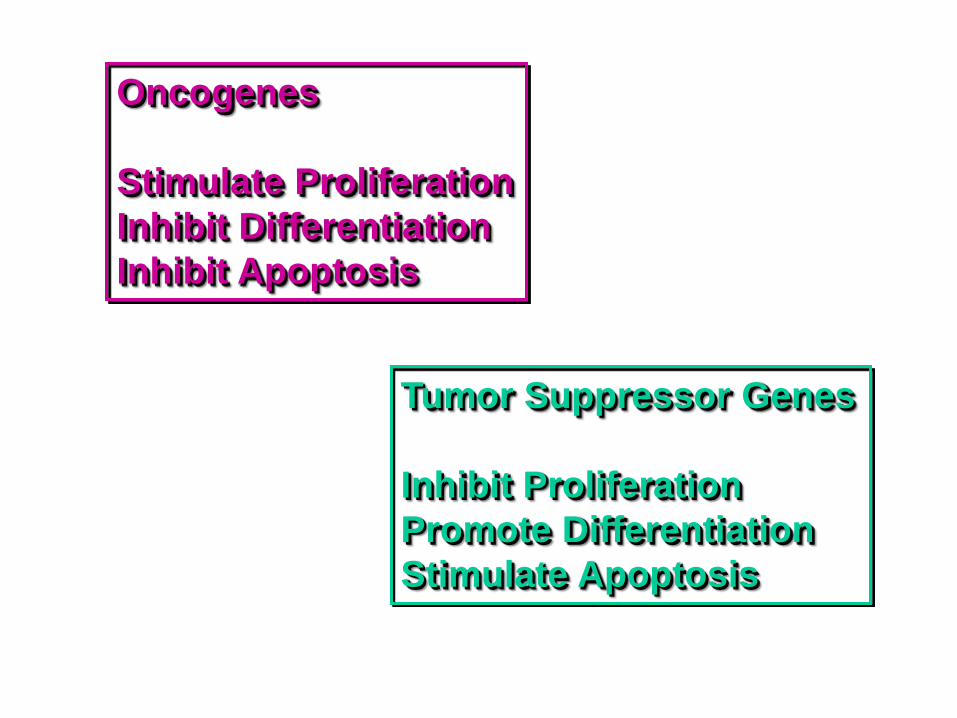
Oncogenes
Stimulate Proliferation
Inhibit Differentiation
Inhibit Apoptosis
Tumor Suppressor Genes
Inhibit Proliferation
Promote Differentiation
Stimulate Apoptosis
Apoptosis: Mediated by Intracellular Proteolytic Cascade
Cells that die as a result of acute injury typically swell and burst.
They spill their contents all over their neighbors a process called cell
necrosis causing a potentially damaging inflammatory response.
By contrast, a cell that undergoes apoptosis dies neatly, without
damaging its neighbors. The cell shrinks and condenses.
The cytoskeleton collapses,
The nuclear envelope disassembles,
The nuclear DNA breaks up into fragments,
The cell surface is altered, displaying properties that cause the dying cell to be rapidly phagocytosed, either by a neighboring cell or by a macrophage (a specialized phagocytic cell), before any leakage of its contents occurs.
The intracellular machinery responsible for apoptosis
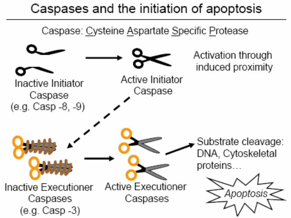
Involves a family of proteases that have a cysteine at their active site and
cleave their target proteins at specific aspartic acids called CASPASES
(Caspases: cysteine-dependent asparate-specific proteases)
CASPASES are synthesized in cell as inactive precursors, or PROCASPASES,
which are usually activated by cleavage at aspartic acids by other caspases.
Once activated, caspases cleave, and thereby activate, other procaspases,
resulting in an amplifying proteolytic cascade.
• Some of the activated caspases cleave other key proteins in the cell.
• Some cleave the nuclear lamins, for example, causing the irreversible breakdown of the nuclear lamina;
• Another cleaves a protein that normally holds a DNA-degrading enzyme (a DNAse) in an inactive form, freeing the DNAse to cut up the DNA in the cell nucleus.
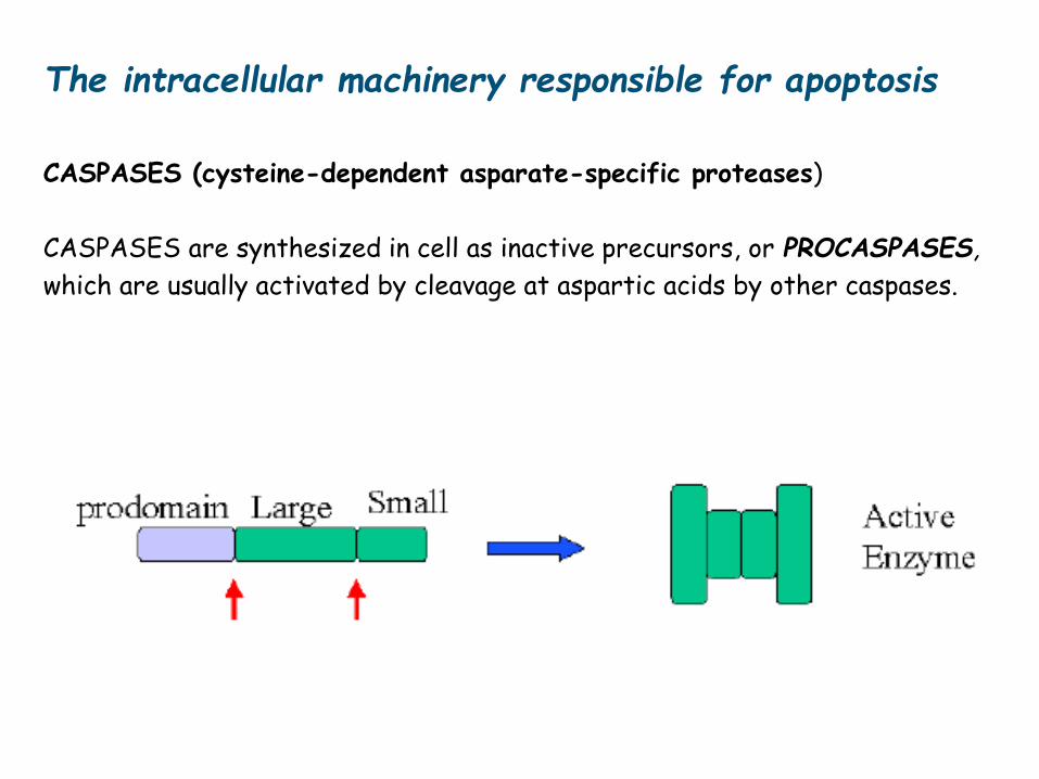
The intracellular machinery responsible for apoptosis
CASPASES (cysteine-dependent asparate-specific proteases)
CASPASES are synthesized in cell as inactive precursors, or PROCASPASES,
which are usually activated by cleavage at aspartic acids by other caspases.
Activation of the intracellular cell death pathway, like entry into a new stage
of the cell cycle, is usually triggered in a complete, all-or-none fashion.
The protease cascade is not only destructive and self-amplifying but also
irreversible, so that once a cell reaches a critical point along the path to
destruction, it cannot turn back.
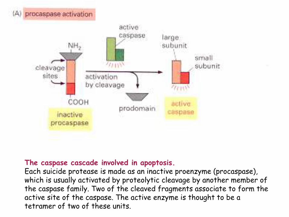
The caspase cascade involved in apoptosis. Each suicide protease is made as an inactive proenzyme (procaspase), which is usually activated by proteolytic cleavage by another member of the caspase family. Two of the cleaved fragments associate to form the active site of the caspase. The active enzyme is thought to be a tetramer of two of these units.
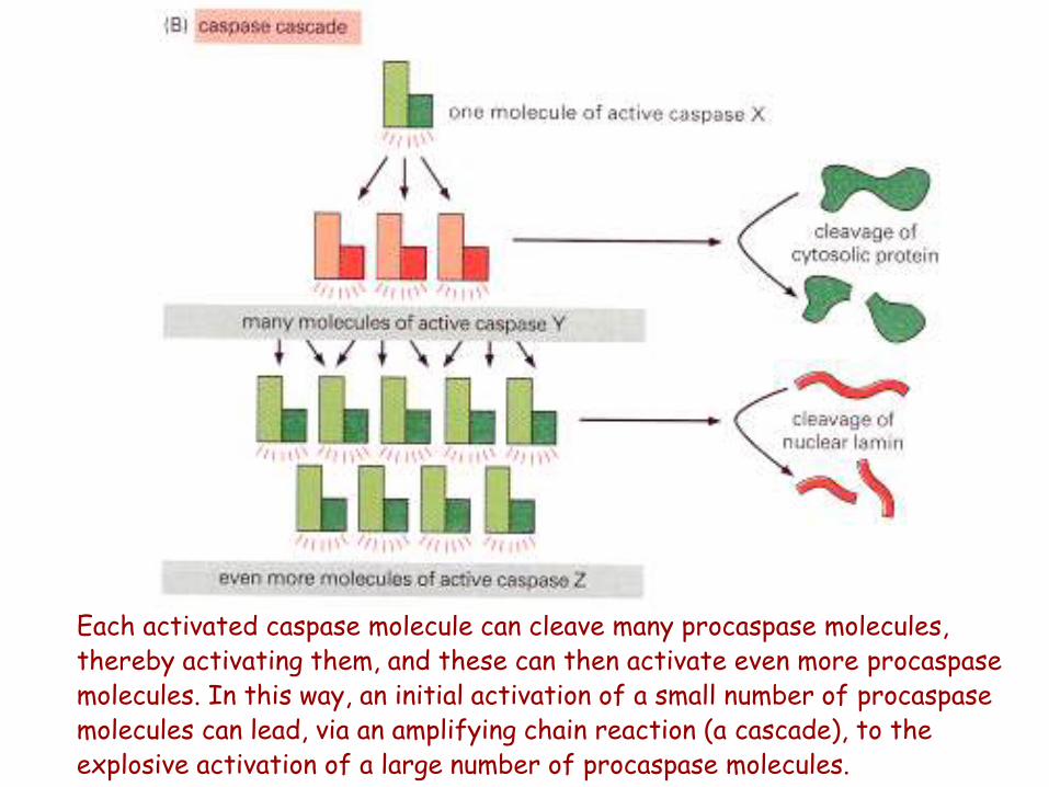
Each activated caspase molecule can cleave many procaspase molecules, thereby activating them, and these can then activate even more procaspase molecules. In this way, an initial activation of a small number of procaspase molecules can lead, via an amplifying chain reaction (a cascade), to the explosive activation of a large number of procaspase molecules.
All nucleated animal cells contain the seeds of their own destruction,
in the form of various inactive procaspases that lie waiting for a
signal to destroy the cell.
The caspase activity is tightly regulated inside the cell to ensure
that the death program is held in check until needed.
Activation of Procaspases: Binding to Adaptor Proteins
How are procaspases activated to initiate the caspase cascade?
What causes activation of caspases?
The activation is triggered by adaptor proteins that bring multiple
copies of specific procaspases, known as initiator procaspases, close
together in a complex or aggregate.
In some cases, the initiator procaspases have a small amount of
protease activity, and forcing them together into a complex causes
them to cleave each other, triggering their mutual activation.
In other cases, the aggregation is thought to cause a conformational
change that activates the procaspase. Within moments, the
activated caspase at the top of the cascade cleaves downstream
procaspases to amplify the death signal n spread it throughout the
cell.
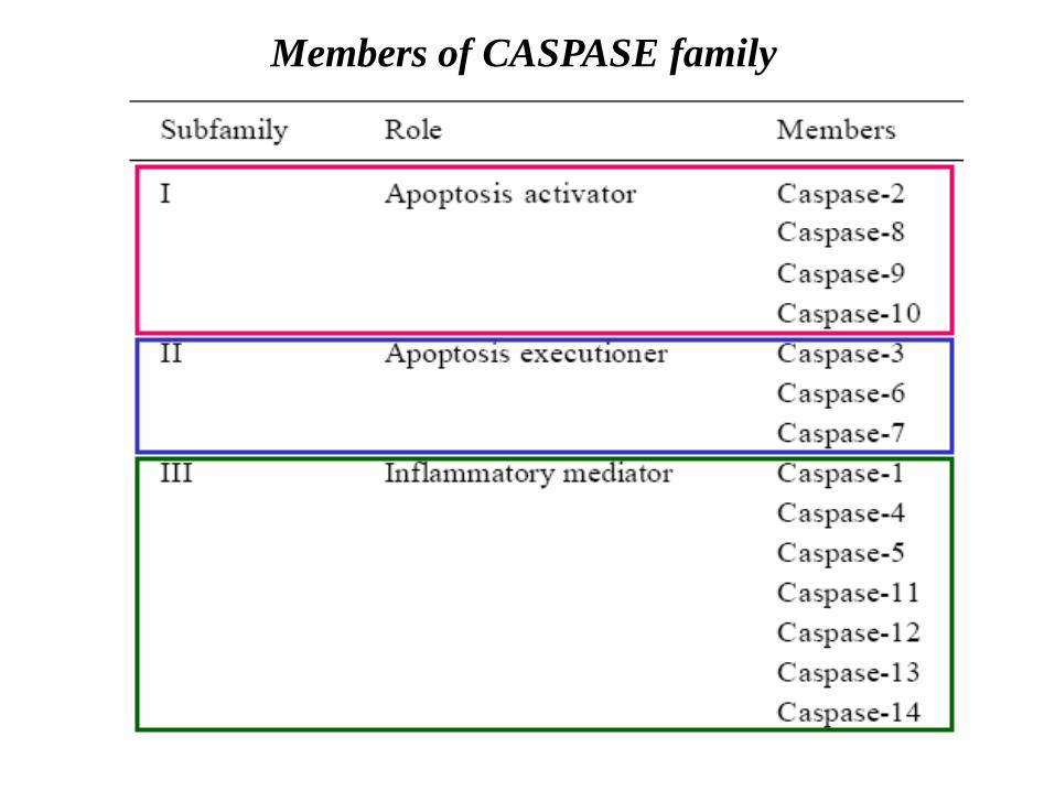
Members of CASPASE family
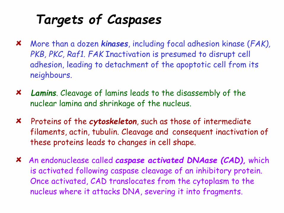
Targets of Caspases
More than a dozen kinases, including focal adhesion kinase (FAK), PKB, PKC, Raf1. FAK Inactivation is presumed to disrupt cell adhesion, leading to detachment of the apoptotic cell from its neighbours.
Lamins. Cleavage of lamins leads to the disassembly of the nuclear lamina and shrinkage of the nucleus.
Proteins of the cytoskeleton, such as those of intermediate filaments, actin, tubulin. Cleavage and consequent inactivation of these proteins leads to changes in cell shape.
An endonuclease called caspase activated DNAase (CAD), which is activated following caspase cleavage of an inhibitory protein. Once activated, CAD translocates from the cytoplasm to the nucleus where it attacks DNA, severing it into fragments.

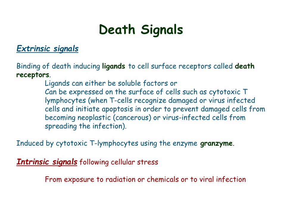
Death Signals
Extrinsic signals Binding of death inducing ligands to cell surface receptors called death receptors. Ligands can either be soluble factors or Can be expressed on the surface of cells such as cytotoxic T lymphocytes (when T-cells recognize damaged or virus infected cells and initiate apoptosis in order to prevent damaged cells from becoming neoplastic (cancerous) or virus-infected cells from spreading the infection). Induced by cytotoxic T-lymphocytes using the enzyme granzyme. Intrinsic signals following cellular stress From exposure to radiation or chemicals or to viral infection
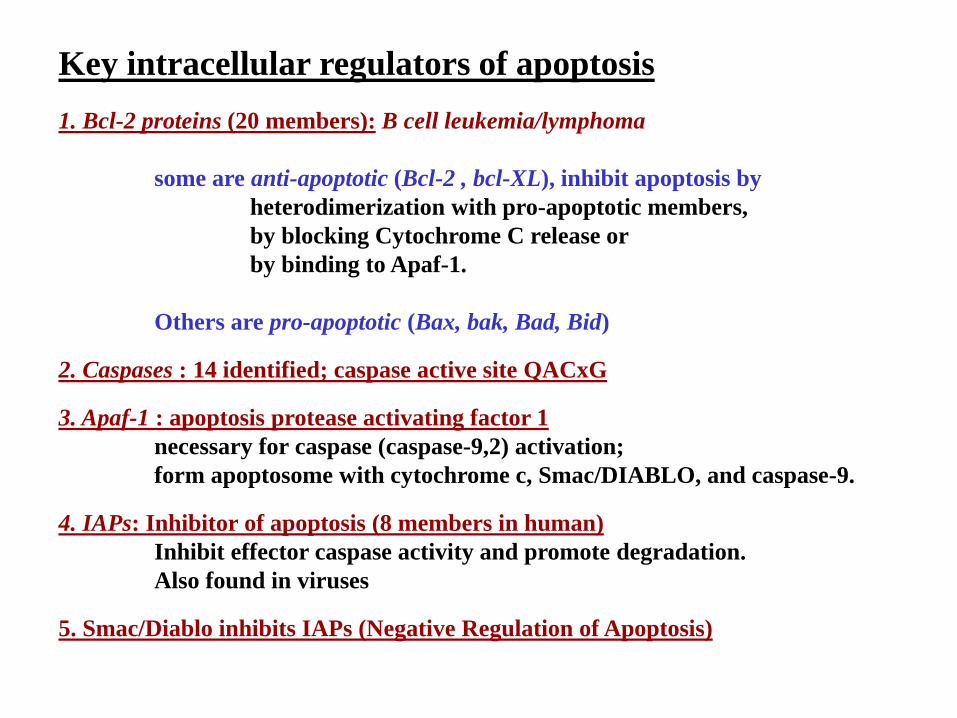
Key intracellular regulators of apoptosis
1. Bcl-2 proteins (20 members): B cell leukemia/lymphoma
some are anti-apoptotic (Bcl-2 , bcl-XL), inhibit apoptosis by
heterodimerization with pro-apoptotic members,
by blocking Cytochrome C release or
by binding to Apaf-1.
Others are pro-apoptotic (Bax, bak, Bad, Bid)
2. Caspases : 14 identified; caspase active site QACxG
3. Apaf-1 : apoptosis protease activating factor 1
necessary for caspase (caspase-9,2) activation;
form apoptosome with cytochrome c, Smac/DIABLO, and caspase-9.
4. IAPs: Inhibitor of apoptosis (8 members in human)
Inhibit effector caspase activity and promote degradation.
Also found in viruses
5. Smac/Diablo inhibits IAPs (Negative Regulation of Apoptosis)
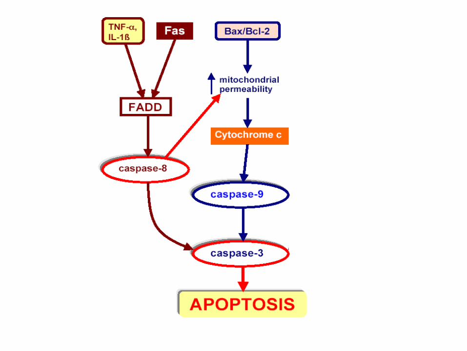
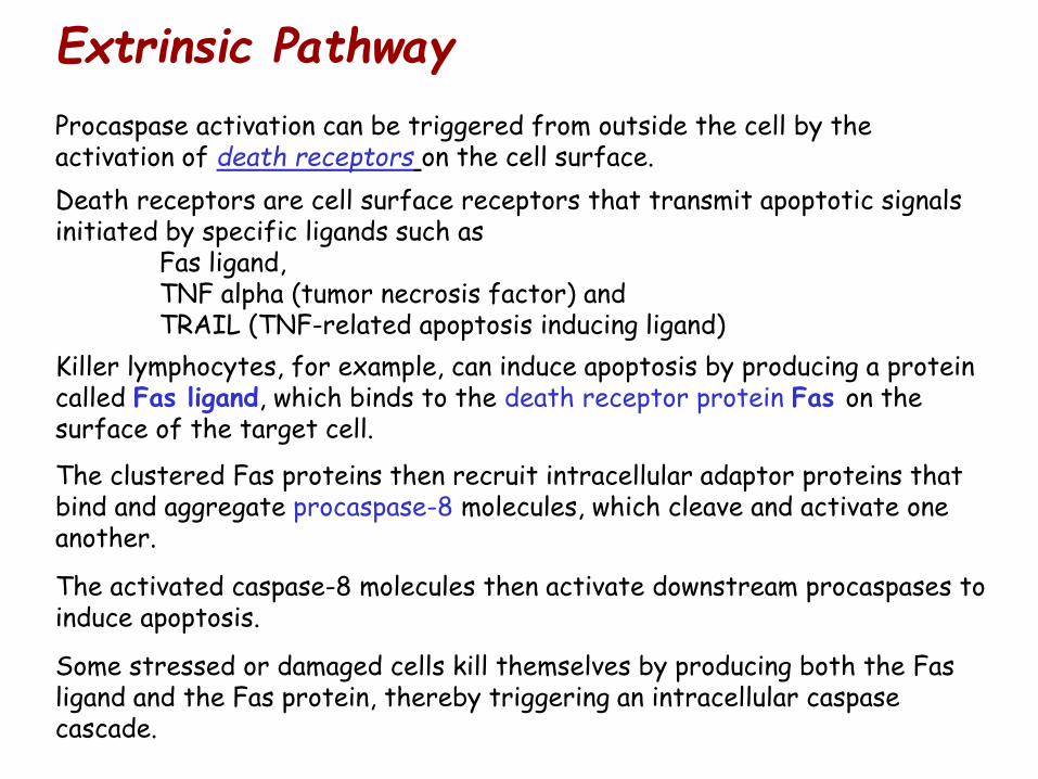
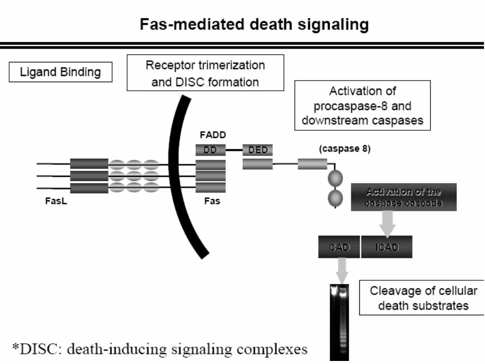
Procaspase activation can be triggered from outside the cell by the activation of death receptors on the cell surface.
Death receptors are cell surface receptors that transmit apoptotic signals initiated by specific ligands such as Fas ligand, TNF alpha (tumor necrosis factor) and TRAIL (TNF-related apoptosis inducing ligand)
Killer lymphocytes, for example, can induce apoptosis by producing a protein called Fas ligand, which binds to the death receptor protein Fas on the surface of the target cell.
The clustered Fas proteins then recruit intracellular adaptor proteins that bind and aggregate procaspase-8 molecules, which cleave and activate one another.
The activated caspase-8 molecules then activate downstream procaspases to induce apoptosis.
Some stressed or damaged cells kill themselves by producing both the Fas ligand and the Fas protein, thereby triggering an intracellular caspase cascade.
Extrinsic Pathway
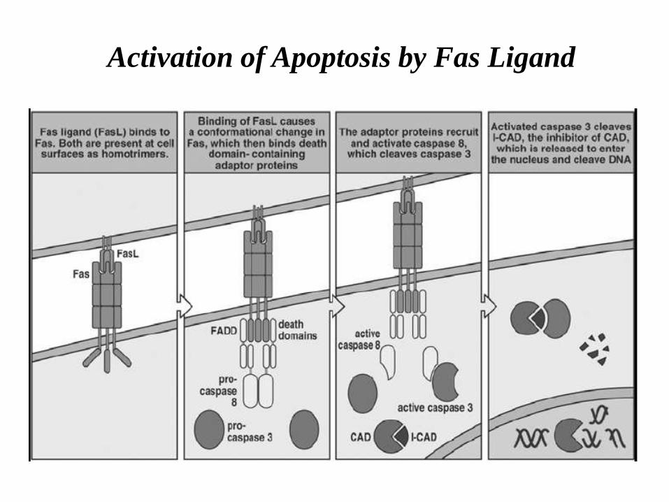
Activation of Apoptosis by Fas Ligand
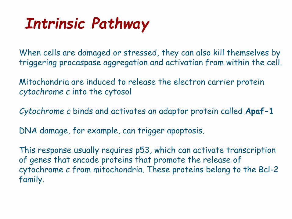
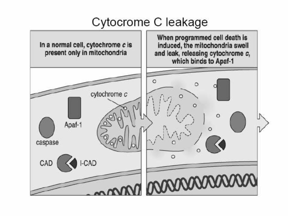
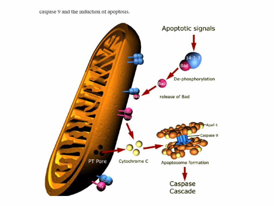
When cells are damaged or stressed, they can also kill themselves by triggering procaspase aggregation and activation from within the cell. Mitochondria are induced to release the electron carrier protein cytochrome c into the cytosol Cytochrome c binds and activates an adaptor protein called Apaf-1 DNA damage, for example, can trigger apoptosis. This response usually requires p53, which can activate transcription of genes that encode proteins that promote the release of cytochrome c from mitochondria. These proteins belong to the Bcl-2 family.
Intrinsic Pathway
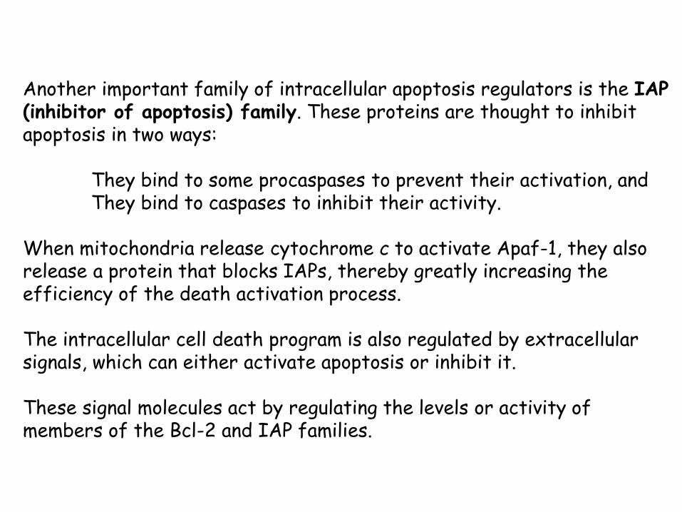
Another important family of intracellular apoptosis regulators is the IAP (inhibitor of apoptosis) family. These proteins are thought to inhibit apoptosis in two ways: They bind to some procaspases to prevent their activation, and They bind to caspases to inhibit their activity. When mitochondria release cytochrome c to activate Apaf-1, they also release a protein that blocks IAPs, thereby greatly increasing the efficiency of the death activation process. The intracellular cell death program is also regulated by extracellular signals, which can either activate apoptosis or inhibit it. These signal molecules act by regulating the levels or activity of members of the Bcl-2 and IAP families.
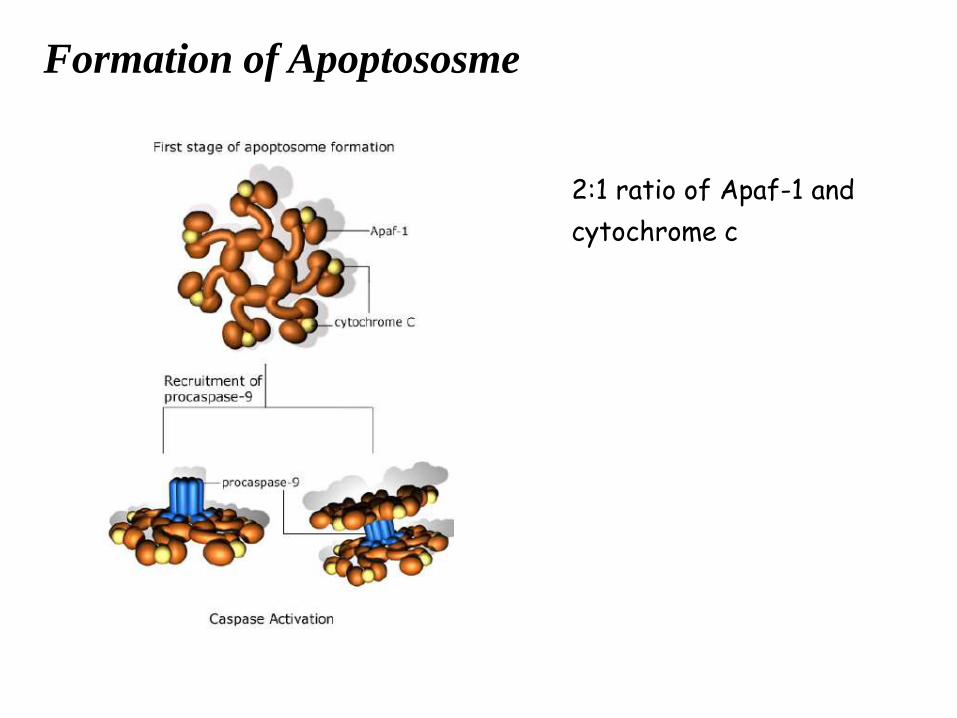
Formation of Apoptososme
2:1 ratio of Apaf-1 and
cytochrome c
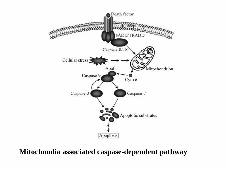
Mitochondia associated caspase-dependent pathway
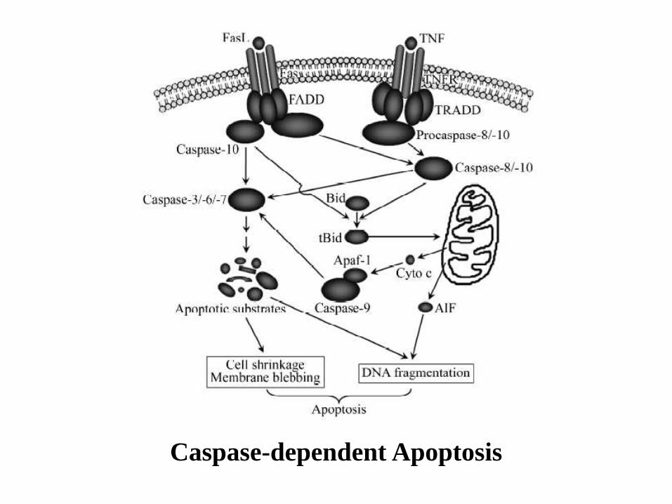
Caspase-dependent Apoptosis
Sensitivity of the cell to apoptotic stimuli depends on
•Expression of pro-apoptotic proteins ???
•Anti-apoptotic proteins (Bcl-2 or the inhibitor of apoptosis
protein)
•The severity of stimulus (viral infection, cell stress, DNA damage),
and
•The stage of the cell cycle
Caspases and chromatin breakdown
1) Inactivation of enzymes involved in DNA repair. The enzyme poly ADP-ribose polymerase, or PARP, is an important DNA repair enzyme (first proteins identified as a substrate for caspases). The ability of PARP to repair DNA damage is prevented following cleavage of PARP by caspase-3.
2) Breakdown of structural nuclear proteins. Lamins are intra-nuclear proteins that maintain the shape of the nucleus and mediate interactions between chromatin and the nuclear membrane. Degradation of lamins by caspase 6 results in the chromatin condensation and nuclear fragmentation.
3) Fragmentation of DNA. The fragmentation of DNA into nucleosomal units is caused by an enzyme known as CAD, or caspase activated DNase. Normally CAD exists as an inactive complex with ICAD (inhibitor of CAD). During apoptosis, ICAD is cleaved by caspases, such as caspase 3, to release CAD. Rapid fragmentation of the nuclear DNA follows.
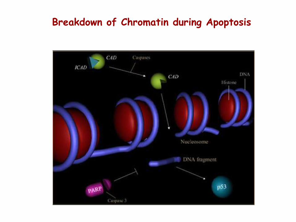
Breakdown of Chromatin during Apoptosis
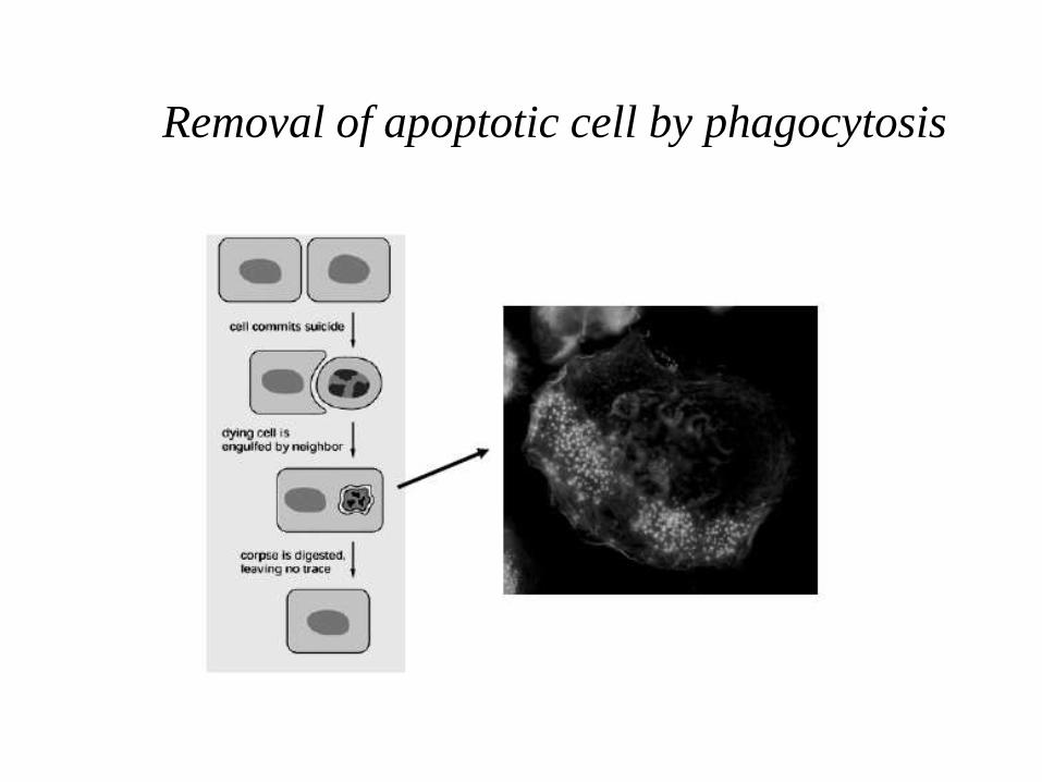
Removal of apoptotic cell by phagocytosis
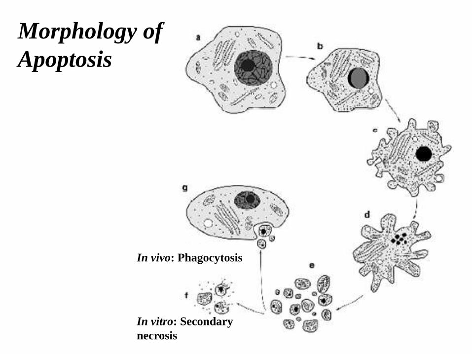
Morphology of
Apoptosis
In vivo: Phagocytosis
In vitro: Secondary
necrosis