Programme Booklet.doc
125
-
Upload
brucelee55 -
Category
Health & Medicine
-
view
841 -
download
1
description
Transcript of Programme Booklet.doc


1
Department of Allied Health
Professions
MSc Medical ImagingProgramme information

2

TABLE OF CONTENTS
Introduction 1
1.0
1.1
1.2
1.3
1.4
1.5
1.6
1.7
1.8
1.9
1.10
1.11
1.12
1.13
1.14
1.15
1.16
1.17
1.18
1.19
1.20
Programme
Information…………………………………………………………………………………..
Nature of the
Programme………………………………………………………………………………...
Programme Aims and Learning
Outcomes……………………………………………………………...
Modules of the Programme
……………………………………………………………………………..
Programme
Structure……………………………………………………………………………………..
Award
Titles………………………………………………………………………………………………..
Pattern of
Attendance…………………………………………………………………………………….
Programme
Content………………………………………………………………………………………
Credit
Exemption…………………………………………………………………………………………..
Learning and Teaching
Strategies………………………………………………………………………...
Assessment………………………………………………………………………………………………
…
Procedures and Criteria for
Assessment…………………………………………………………………
General Assessment Criteria for Banded Marking
Scheme…………………………………………….
Poster Assessment
Criteria………………………………………………………………………………..
Criteria for Assessment of
Portfolios……………………………………………………………………..
Summary Criteria for Research
Projects………………………………………………………………….
Assessment
Conventions………………………………………………………………………………….
Concession Requests
……………………………………………………………………………………...
Plagiarism, Copying and
Duplication…………………………………………………………………….
Responsibilities of Clinical
2
2
2
4
5
6
9
9
9
9
11
11
12
14
15
17
18
18
19
22
22

Supervisors…………………………………………………………………...
Guidelines for the Clinical Supervisor’s
Report………………………………………………………….
The Modules
Orientation .................................................................................................................................24
Magnetic Resonance Imaging Pathway
Principles of Science and Technology in Magnetic Resonance Imaging..........................................27
Clinical Applications and Management I (MRI of Brain, Spine and Knee)........................................29
Clinical Applications and Management II (Musculo-Skeletal MRI)...................................................32
Clinical Applications and Management III (Non-Routine Brain, Head & Neck).................................35
Clinical Applications and Management IV (MRI Body Imaging).......................................................38
Nuclear Medicine Pathway
Principles of Science and Technology in Nuclear Medicine.............................................................41
Clinical Applications and Management I (Standard Nuclear Medicine Procedures)..........................43
Clinical Applications and Management II (Complex & Non-Standard Nuclear Medicine Procedures)46
Clinical Applications and Management III (Positron Emission Tomography, PET).............................48
Clinical Applications and Management IV (Radionuclide Therapy).................................................50
Clinical Applications and Management V (Radiopharmacy and Non-Imaging Diagnostic Tests).......52
Medical Ultrasound Pathway
Principles of Science and Technology in Medical Ultrasound..........................................................54
Techniques in Musculo-skeletal Ultrasound Imaging
………………………………………………………………….57
Clinical Applications and Management I (Obstetrics & Pelvic Ultrasound)......................................58
Clinical Applications and Management II (Abdominal & Small Parts Ultrasound).............................60
Clinical Applications and Management III (Musculo-Skeletal Ultrasound)........................................62
Clinical Applications and Management IV (Peripheral Vascular Ultrasound)....................................64
Clinical Applications and Management V (Echocardiography)........................................................66
Modules available across all pathways
Open Module (work based learning).............................................................................................68
Developing a Research Proposal...................................................................................................70
Undertaking Systematic Reviews.................................................................................................72
Appendix 1 - Key Skills Matrix for the MSc Medical Imaging Programme …………………………….
…...........................75
Appendix 2 ....................................................................................................................................... ..77
- Validated Modules within the MSc Interprofessional Health & Social Care
- Modules available from other programmes
Appendix 3 - 2007/ 2008 fees…………….……..…………………….
……………………………………………………….78
1

Introduction
Welcome to Canterbury Christ Church University. This programme guide contains
specific details about the PgC/PgD/MSc Medical Imaging to supplement general
information in the Postgraduate (HE4) Interprofessional Framework Student
Information Booklet. I trust you will find it a comprehensive guide and welcome the
opportunity to discuss any aspect of the programme or your specific requirements
in more detail.
The programme offers a range of medical imaging qualifications, Postgraduate
Certificate/ Postgraduate Diploma / Master of Science, in either Medical Imaging,
Magnetic Resonance Imaging, Medical Ultrasound or Nuclear Medicine and
Postgraduate Certificate in Radiopharmacy Practice. It has been developed in
conjunction with service practitioners and forms part of the portfolio of the
Postgraduate Interprofessional Framework. Consonant with the ethos of the
Framework, all modules are available for stand alone study.
The programme is innovative in nature and requires significant and explicit
commitment from students who enter the programme, from all staff within the
student’s clinical department and from the relevant employing authorities.
As Programme Director I am here to offer individual advice and support about any
aspect of the Programme and I hope that you will make as much use of this service
as you deem necessary.
Position Name Telepho
ne
E-mail Room
Framework
Director
Keith
Piper
01227
782425
uk
Jg29
Johnson
Framework
Administrator
Susanna
h Russell
01227
782140
[email protected] Jf24
Johnson
Programme
Director
MSc Medical
Gill
Dolbear
01227
782085
[email protected] Jg30
Johnson
2
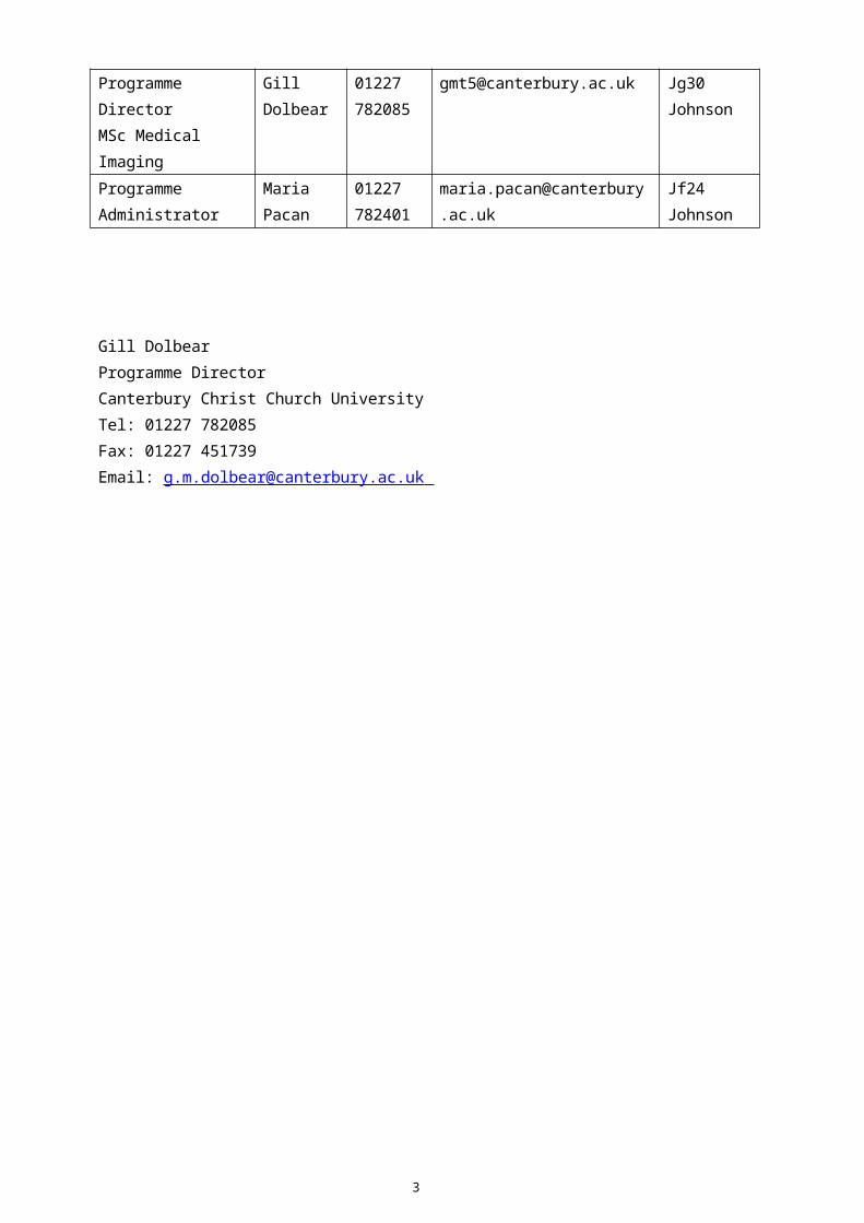
Imaging
Programme
Administrator
Maria
Pacan
01227
782401
c.uk
Jf24
Johnson
Gill Dolbear
Programme Director
Canterbury Christ Church University
Tel: 01227 782085
Fax: 01227 451739
Email: [email protected]
3
1.0 Programme Information
1.1 Nature of the Programme
The Programme consists of work-based Clinical Applications & Management
modules as well as taught Principles of Science and Technology, and Techniques in
Musculo-skeletal Ultrasound Imaging modules. The modules are studied at M
(Masters) HE4 level and each module carries 20 HE4 level credits.
Work-based learning requires commitment of the individual but, more significantly,
requires commitment of all staff in the workplace. Students are supported by the
Programme Director, their Academic Supervisor, their Clinical Supervisor and by
appropriate Learning Agreements.
Full and part time modes of study are offered. For example, a full time certificate
level programme may be undertaken in a minimum of one year or two years part
time, while a full MSc would be expected to take a minimum of two years full time
or up to six years part time. Approval for the programme has been gained from the
relevant accrediting and professional bodies.
1.2 Programme Aims and Learning Outcomes
Programme Aims
The overall purpose of the programme is to foster the individual personal and
professional development of health care practitioners to a higher level of practice
consonant with the ethos of interprofessional work and patient/client centred care.
It aims to:
Ensure practitioners develop the knowledge, competences, skills and attitudes
needed to demonstrate mastery in both academic and professional capability in
their chosen fields of practice;
Facilitate a challenging, interprofessional learning environment where
practitioners can develop further the critical, analytic and reflective cognitive
skills necessary for higher levels of patient/client centred practice and
interprofessional working;
4

Hone practitioners’ research and critical appraisal skills enabling them to make
innovative use of evidence for best practice;
Create autonomous, self directed learners who are able to sustain and advance
their continuing professional development beyond the programme and support
others in the practice environment to develop themselves;
Enable practitioners to be proactive in initiating and leading role developments
in their specific fields.
5

Programme Learning Outcomes
For all certificate, diploma and MSc awards, students who complete their chosen
award successfully will demonstrate the following outcomes. The breadth of their
achievement will be dependent on the specific scope of practice defined by the
pathway selected.
the clinical knowledge, skills and attributes to provide an imaging service within
a chosen modality or specialism;
critical and rigorous analysis of literature, research evidence, documentation
and policy in relation to higher levels of practice in their chosen field;
appropriate, person centred advice and support for clients/patients and carers;
the ability to engage in critical self evaluation and assessment;
personal skills of reflective critical awareness necessary to respond positively to
the challenges of the future through continuing professional development;
the knowledge and expertise to accept or decline service referrals and provide
appropriate comment and advice to referring colleagues;
the necessary knowledge, communication skills and critical understanding to
provide reports or interpretations on the imaging examinations undertaken,
within the scope of their professional practice;
critical awareness of the significance of interprofessional collaboration, liaison
and team working.
Successful completion of an MSc will require demonstration of;
the ability to plan, manage, execute and interpret a piece of independent
research in the student’s chosen field of practice.
In addition to the above transferable skills practitioners will be expected to
demonstrate in their assessments further development of the following key skills
where appropriate;
Communication
Application of number
Information technology
6

Working with others
Problem solving
Improving own learning and performance.
A key skills matrix for the programme is shown in Appendix One.
7
1.3 Modules of the Programme
The Programme consists of various modules offered at HE Level 4, with each
module attracting 20 credit points. The modules offered within the Programme are
listed below:-
Medical Ultrasound Pathway
Code Module Title
MMIHF4UPS Principles of Science and Technology in Medical Ultrasound
MMIHF4UTM Techniques in Musculo-Skeletal Ultrasound Imaging
MMIHF4UCP Clinical Applications and Management I (Obstetric & Pelvic
Ultrasound)
MMIHF4UCS Clinical Applications and Management II (Abdominal & Small
Parts Ultrasound)
MMIHF4UCM Clinical Applications and Management III (Musculo-Skeletal
Ultrasound)
MMIHF4UCV Clinical Applications and Management IV (Peripheral Vascular
Ultrasound)
MMIHF4UCE Clinical Applications and Management V (Echocardiography)
Magnetic Resonance Imaging Pathway
Code Module Title
MMIHF4MPS Principles of Science and Technology in Magnetic Resonance
Imaging
MMIHF4MCK Clinical Applications and Management I (MRI of Brain, Spine
and Knee)
MMIHF4MCM Clinical Applications and Management II (Musculo-Skeletal
MRI)
MMIHF4MCN Clinical Applications and Management III (Non-Routine Brain,
Head & Neck MRI)
MMIHF4MCB Clinical Applications and Management IV (MRI Body Imaging)
Nuclear Medicine Pathway
Code Module Title
MMIHF4NPS Principles of Science and Technology in Nuclear Medicine
MMIHF4NCS Clinical Applications and Management I (Standard Nuclear
8
Medicine Procedures)
MMIHF4NCC Clinical Applications and Management II (Complex & Non-
Standard Nuclear Medicine Procedures)
MMIHF4NCP Clinical Applications and Management III (Positron Emission
Tomography, PET)
MMIHF4NCR Clinical Applications and Management IV (Radionuclide
Therapy)
MMIHF4NRN Radiopharmacy and Non-Imaging Diagnostic Tests
The following modules are available across all pathways:-
Code Module Title
MZZHF4OP1 Open Module
MZZHF4USR Undertaking Systematic Reviews
MZZHF4DRP Developing a Research Proposal
MZZHF4DSS Research Project (two or three modules)
1.4 Programme Structure
Consonant with the MSc Interprofessional Health and Social Care (IPHSC) degree,
the programme is modular in structure and flexible. All modules attract 20 HE4
credit points and are offered at Master’s level. For the award of an MSc, students
are required to successfully complete 9 modules (180 credits). All pathways have a
discrete Postgraduate Certificate award comprising 3 modules (60 credits) and a
Postgraduate Diploma award comprising 6 modules (120 credits).
As befits a professional development programme where skill mix and cross
boundary, interprofessional working are integral to the aims, students are required
to complete the Orientation taught module in their certificate awards and include
one module from the MSc IPHSC in their diploma profile.
The choice of 3 exit points in the imaging pathways, Certificate, Diploma and MSc,
reflects the need to be service led in the provision of educational programmes. A
practitioner wishing to pursue their career in a specific field of imaging practice
requires, as a minimum, competence in standard procedures and a demonstrable
understanding of the scientific basis of the modality provided by the Postgraduate
Certificate. Thus, these awards offer little flexibility, the Orientation and Principles of
Science and Technology modules are compulsory, as is one Clinical Applications
module. Many will wish to develop their roles further, either by diversifying or
becoming competent in more complex procedures, and proceed to the Diploma
9

qualification. Moving from Certificate to Diploma will be a continuous process for
some practitioners, but for others, a break in study may be preferred. Some
practitioners will wish to continue studying and achieve a full MSc.
The modules themselves are a mixture of workplace based, taught and research
modules. Workplace based modules are clinically based with five hours contact with
the academic supervisor. The acquisition of specific competences takes place in the
work setting under the supervision of an expert practitioner, underpinned by a
learning agreement.
With the exception of Techniques in Musculo-skeletal Ultrasound Imaging and
Principles of Science and Technology, taught modules are from the existing MSc
Interprofessional Health and Social Care (IPHSC) framework. They comprise 40
hours contact time delivered in 3 x 2 day or 3 x 3 day blocks. This programme
offers a menu of approximately 12 modules each academic year, including the
compulsory Orientation and Developing a Research Proposal modules.
Research modules are independent enquiry, with six hours supervision from a
member of the academic staff. A research proposal is developed and assessed
within the compulsory taught Developing a Research Proposal module.
Integrating the medical imaging pathways with the MSc IPHSC framework means
that students can have some flexibility about their degree pathway. All pathways
contain modules which develop knowledge and skills in the specific imaging
modality. In each, there is a minimum number of competence based modules which
must be achieved to gain the certificate and diploma awards. Permitting students to
choose their remaining modules from the MSc IPHSC menu allows the programme
of study to be focussed on a particular professional interest and gives a broader,
interprofessional context to their degree profile. In addition, this approach enables
the recruitment of small numbers of students to different pathways whilst
maintaining viability.
1.5 Award Titles
Students are required to register for a specific pathway. The profile for the pathway
and associated award is given below.
Medical Ultrasound
Exit award: Postgraduate Certificate Medical Ultrasound
10

Module 1 Orientation (taught) (shared with MSc IPHSC)
Module 2 Principles of Science and Technology in Medical Ultrasound
(taught)
Module 3 Clinical Applications & Management (workplace based)
Modules 1 & 2 are compulsory, but students may choose any one of the Clinical
Applications and Management modules offered.
Exit award: Postgraduate Certificate in Musculo-skeletal Ultrasound
Module 1 Techniques in Musculo-skeletal Ultrasound Imaging
Module 2 Principles of Science and Technology in Medical Ultrasound
(taught)
Module 3 Clinical Applications & Management III (workplace based)
All modules are compulsory.
Exit award: Postgraduate Diploma Medical Ultrasound
Module 4 Clinical Applications & Management (workplace based)
Module 5 Clinical Applications & Management (workbased) OR Open
module OR one from the MSc IPHSC menu (taught)
Module 6 One from the MSc IPHSC menu (taught)
Students must complete six modules in total. Students must choose at least one
but may undertake two further Clinical Applications & Management modules in the
Diploma route. Only one open module will be permitted for students on the
medical ultrasound, nuclear medicine or MRI pathways.
Exit Award: MSc Medical Ultrasound
Module 7 Developing a Research Proposal (taught) (shared with MSc
IPHSC)
Modules 8 &
9
Research Project (two modules)
Magnetic Resonance Imaging (MRI)
Exit award: Postgraduate Certificate Magnetic Resonance Imaging
11

Module 1 Orientation (taught) (shared with MSc IPHSC)
Module 2 Principles of Science and Technology in MRI (taught)
Module 3 Clinical Applications & Management I (workplace based)
All modules are compulsory.
Exit award: Postgraduate Diploma Magnetic Resonance Imaging
Module 4 Clinical Applications & Management (workplace based)
Module 5 Clinical Applications & Management (workplace based)
OR Open module OR one from the MSc IPHSC menu
(taught)
Module 6 One from the MSc IPHSC menu (taught)
Students must complete six modules in total. Students must choose at least one
but may undertake two further Clinical Applications & Management modules in
the Diploma route. Only one open module will be permitted for students on the
medical ultrasound, nuclear medicine or MRI pathways.
Exit Award: MSc Magnetic Resonance Imaging
Module 7 Developing a Research Proposal (taught) (shared with
MSc IPHSC)
Module 8 &
9
Research Project
Nuclear Medicine Pathway
Exit award: Postgraduate Certificate Nuclear Medicine
Module 1 Orientation (taught) (shared with MSc IPHSC)
Module 2 Principles of Science and Technology in Nuclear Medicine
(taught)
Module 3 Clinical Applications & Management I (workplace based)
All modules are compulsory.
Exit award: Postgraduate Diploma Nuclear Medicine
Module 4 Clinical Applications & Management (workbased) OR
Radiopharmacy & Non Imaging Diagnostic Tests
12

(workbased)
Module 5 Clinical Applications & Management (workbased) OR
Radiopharmacy & Non Imaging Diagnostic Tests
(workbased) OR Open module OR one from the MSc
IPHSC menu (taught)
Module 6 One from the MSc IPHSC menu (taught)
Students must complete six modules in total. Students must choose at least one
but may undertake two further Clinical Applications & Management modules in the
Diploma route. Only one open module will be permitted for students on the medical
ultrasound, nuclear medicine or MRI pathways.
Please note: the Radiopharmacy and Non-Imaging Diagnostic Tests module is not
compulsory, however, students will be strongly advised to include it in their
Postgraduate Diploma.
Exit Award: MSc Nuclear Medicine
Module 7 Developing a Research Proposal (taught) (shared with
MSc IPHSC)
Modules 8 &
9
Research Project (Modality Specific)
Exit award: Postgraduate Certificate in Radiopharmacy Practice
Module 1 Orientation (taught) (shared with MSc IPHSC)
Module 2 Principles of Science and Technology in NM (taught)
Module 3 Radiopharmacy & Non Imaging Diagnostic Tests
(workbased)
All modules are compulsory.
Medical Imaging
13

Exit award: Postgraduate Certificate Medical Imaging
Module 1 Orientation (taught) (shared with MSc IPHSC)
Module 2 Open module or one from the MSc IPHSC menu (taught)
Module 3 Open module or one from the MSc IPHSC menu (taught)
Module 1 is compulsory. If two open modules are completed as modules 2 and 3,
no further open modules will be permitted.
Exit award: Postgraduate Diploma Medical Imaging
Module 4 Open module or one from the MSc IPHSC menu (taught)
Module 5 Open module or one from the MSc IPHSC menu (taught)
Module 6 One from the MSc IPHSC menu (taught)
Students must complete six modules in total. Only two open modules will be
permitted over all for students on the medical imaging pathway.
Exit Award: MSc Medical Imaging
Module 7 Developing a Research Proposal (taught) (shared with MSc
IPHSC)
Modules 8 &
9
Research Project (two modules)
1.6 Pattern of Attendance
Taught modules, including Developing a Research Proposal, are offered in 3 x 2day
blocks per semester. The Orientation taught module is offered in 3 x 3day blocks
per semester. Each semester lasts for approximately 18 weeks, from October to
February and February to June. Workplace based modules extend over a 26 week
period from the time of their registration.
14

Practitioners wishing to pursue a specific, in-depth, individual study may utilise the
Open module structure of the MSc Medical Imaging which includes 5 hours of
academic tutorial support over a six to nine month period.
Research modules may be registered at any time following assessment of the
proposal and approval by the Local Research Ethics Committee where applicable.
Students have 6 – 9 months to complete their research projects.
1.7 Programme Content
The programme content is based on:
Meeting the knowledge and skills base requirements to ensure participants are
competent to practise in their chosen imaging modality/field of practice.
The specific learning needs of individual students as identified in a learning
agreement developed on an individual basis at the start of the programme.
Providing an education platform from which students, on successful completion,
can provide a high quality service to patients / clients and also lead and respond
to role development challenges of the future.
The specific needs of the service as identified at the commencement of the
programme.
1.8 Credit Exemption
Consideration will be given to students holding relevant, specific academic credit or
having appropriate experience, utilising the Higher Education Institution’s existing
APL/APEL policies for up to 50% of the MSc award (90 credits maximum). This
includes work place based clinical applications modules, provided that evidence of a
match can be demonstrated.
There will normally be no exemption from the compulsory Orientation, Principles of
Science and Technology and Developing a Research Proposal modules or from the
research elements of the Master’s award.
1.9 Learning and Teaching Strategies
Consonant with the University Learning and Teaching Strategy, the programme is
learner centred and is oriented significantly towards workplace partnerships; using
work based materials and the normal work environment as a source and site of
15

learning. Following discussion with the Pathway Leader in conjunction with MSc
Medical Imaging Programme Director, the student will select their preferred
modules and a provisional pathway will be agreed and documented.
Student responsibility for managing their own learning is assumed, although it is
recognised that some support may be required to orientate students to work at this
level. Therefore some study skills are included in the compulsory Orientation
module.
Each of the work based clinical applications modules is founded on an individual
learning agreement between the student, the academic supervisor, the clinical
supervisor and the workplace manager. Effective work based learning requires such
formalised agreements, which clearly identify the respective responsibilities of all
parties involved. It also provides a significant opportunity for staff development in
relation to learning and teaching in the workplace.
The Learning Agreement sets out the nature and scope of the learning opportunities
in the individual setting and the duties and obligations of the student, the academic
tutor, the clinical supervisor and the line manager so that the roles and
responsibilities of each party are understood by all. Aims and learning outcomes are
specified together with identified learning activities and assessment. This four way
agreement will ensure that students are developing their practice in accordance
with Trust practices and protocols as well as securing the support of the workplace
as the main site and source of learning.
In most cases, the student is being sponsored by their workplace to develop skills in
line with service objectives and therefore the range of equipment and examinations
undertaken can normally meet the student’s development needs. It is part of the
role of the Academic Supervisor, as the learning facilitator, to ensure that the
clinical environment is adequate. If additional experience is required outside of the
normal workplace setting, then it is the responsibility of the student and/or their
Line Manager to negotiate an appropriate clinical placement and this is reflected in
the learning agreement.
Identification of an appropriate Clinical Supervisor is the responsibility of the
student and their Line Manager, subject to the approval of the Academic Supervisor.
This person will be proficient in the area of competence to be developed and may
16
therefore be a member of a different profession such as a medical practitioner.
Since modules are individually negotiated and may be begun at any time, it is
impractical to offer supervisors a group induction and training on site at the
University. A Guide for Clinical Supervisors has therefore been developed and, when
the Academic Supervisor visits the workplace to confirm the learning agreement, a
briefing meeting will be arranged with the Clinical Supervisor. Supervisors will be
encouraged to contact the Academic Supervisor for support and/or advice as often
as necessary.
The central learning and assessment tool for work based modules is a Portfolio of
Professional and Clinical Development. The Portfolio is a record of the student’s
learning and development as a competent practitioner measured against the
specified outcomes in the learning agreement. Its role in the formative
development of the student is as significant as its assessment function and it
therefore contains both formative and summative material in a mix of analytic and
reflective styles. The Academic Supervisor and student use the portfolio to monitor
and actively manage the student’s development, meeting together approximately
three times during the period of study.
Using all the learning opportunities that arise in the workplace, the aim is to enable
students to demonstrate their competence, analyse and reflect on their practice, as
well as develop their problem solving, judgement making, interpretation, report
writing and communication skills. Learning strategies may include self directed
learning activities, attendance at clinical review meetings, shadowing activities,
tutorials and supervised and unsupervised practice. Web technology will be utilised,
both for learning as in access to image banks, and for student support and
information. These should enable the integration of academic learning with
attainment of clinical competences such that that each informs the other and deep
learning is sustained.
1.10 Assessment
The rationale for assessment is to enable students to demonstrate a reflective,
critical understanding of the relationship between relevant theories and practice
knowledge and expertise to facilitate their professional and personal development
and contribute to service development. A familiarity with basic literature is
assumed and participants are expected to articulate a high level of academic ability
17
through reasoned argument and critical reflection on practice using a variety of
assessment modes.
Modules are weighted equally and the form of assessment is selected to match the
module content and reflect the crucial theory/practice interface and problem
solving emphasis in the programme. The assignment for each module is specified
in the module outline to ensure accurate and appropriate assessment of learning
outcomes. In the case of taught modules, it may be a case study, long essay,
situational analysis, practical exercise or report. In many modules students are
given the opportunity to present a preliminary plan of their assignment to their
peers. This is always formative in the sense that the discussion and feedback
generated is useful to students. In a few modules the presentation forms part of
the summative assessment schedule.
Where professional role development through the acquisition of specific
competences is an identified outcome, as in a work based competence
development module, students are required to demonstrate explicitly in their
written work and in practice, how these have been achieved. The Learning
Agreement and Portfolio of Professional Practice Development are the chief means
by which student attainment is defined, monitored, measured and verified. The
Learning Agreement sets out the specific learning outcomes and, where possible,
these are matched to occupational standards to ensure consistency.
1.11 Procedures and Criteria for Assessment
Each module is summatively assessed independently and equitably in accordance
with the programme's discrete structure. The module learning outcomes form the
basis for assessment and the general assessment criteria of the MSc Medical
Imaging are applied to these outcomes to form a judgement on the student's work.
To achieve accreditation at Master's level, written assignments will encompass all of
the following aspects; coherence in linking theory to practice, breadth of
understanding of issues within multiple frameworks, depth in evaluating a limited
range of key issues, consistency of argument, clarity of expression and good
presentation skills.
18

Breaches of Confidentiality
Students are expected to abide by relevant Codes of Practice and maintain
appropriate levels of confidence at all times. Patient/client identifying details should
be protected at all times. Appropriate consideration should be given to maintaining
anonymity of time, place, and persons e.g. carers, colleagues, peers. Repeated
breaches of confidentiality or breaches at Master’s level will result in a mark of zero
and subsequent requirement for resubmission. Where material is in the public
domain it is appropriate to recognise the source of the material- however no patient
details must be identifiable.
1.12 General Assessment Criteria for Banded Marking Scheme
The PgC/PgD/MSc Programme uses a marking system with the following bands and
categories:
Fail Unsatisfactory standard
Pass Satisfactory standard
Good Pass Very good standard
Distinction Exceptional standard
Use of Literature
Fail Limited, weak selection. Uncritically presented.
Limited recognition of significance for the professional context.
Pass Appropriate range and choice of literature.
Recognition and critical analysis of issues of significance for the
professional context
Good Pass Good selection of key primary texts with critical evaluation of
significant issues for the professional context.
Some, limited analysis of related, secondary texts.
Distinction Excellent, wide range of key and peripheral primary and
secondary texts, demonstrating critical evaluation and synthesis
with the professional context.
Interface between Theory and Practice in the Professional Context
Fail Limited use of theory. Lack of / limited awareness of the
relationship between theory and practice. Little integration or
19

critical analysis of the articulation between theory and practice.
Pass Clear articulation of the relationship between and critical
analysis / evaluation of the significance of relevant theory to
specific professional practice. Awareness of how each may be
informed by the other.
Good Pass Very good, critical analysis / evaluation of the relationship
between theory and practice. Some use of multiple theoretical
frameworks to evaluate professional practice. Demonstrable
synthesis to show how each is informing the other. Some
evaluation of their usefulness.
Distinction Rigorous critical analysis of the interface between theory and
practice, clearly elaborated to evaluate theoretical adequacy and
synthesize the development of professional practice. Excellent,
creative use of multiple frameworks for evaluation and synthesis
of own stance.
Knowledge and Clarity of Reasoning
Fail Knowledge circumscribed. Sense of argument but poorly
expressed lines of thought. Conclusions do not always arise from
premises.
Pass Appropriate, defended knowledge of current, relevant issues.
Logical development of arguments where lines of thought are
clearly discernible. Relevant, limited conclusions arising from
premises.
Good Pass Sound knowledge. Ability to discriminate and justify key issues.
Arguments are confidently expressed through clear, logical lines
of thought. Conclusions are firmly articulated, comprehensive,
relevant and arise directly from the premised arguments.
Distinction Excellent, comprehensive knowledge base. Ability to
discriminate key issues and relate them to the wider context.
Lines of thought are transparent and the arguments are
confidently expressed to develop and synthesize compelling
conclusions. Innovative thinker.
Organization of Material
20
Fail Poorly organized, incoherent structure. Poor presentation and
referencing. Appropriate supporting material not given /
disorganized.
Pass Organization and structure is sufficient to support and not
obscure the work. Appropriate presentation. All supporting
material present. Referencing is sound and appropriate.
Good Pass Organization is comprehensive and structure coherent. Well
presented, facilitating comprehension. Supporting material is
well presented and ordered. Accurate referencing.
Distinction Excellent coherent organization and structure which enhances
comprehension. Excellent presentation of all material.
Referencing is accurate to a high degree.
21

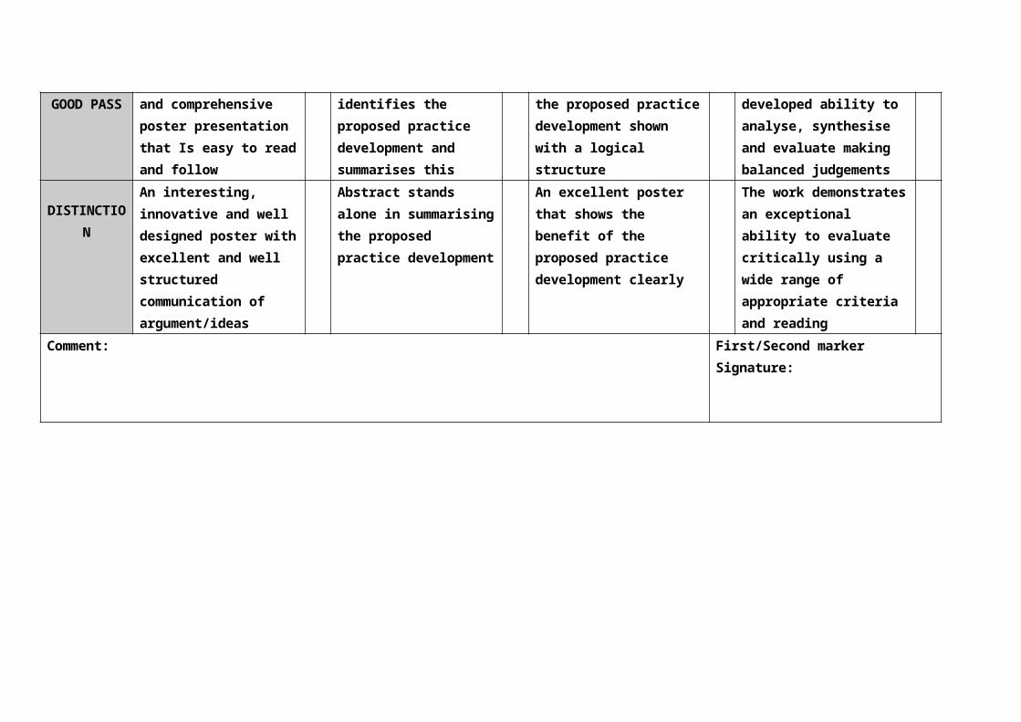
1.13 MSc Medical Imaging - Poster Assessment Criteria
Candidate Number Poster Number:
Title of Poster: The Development of Practice by the use of Musculoskeletal Ultrasound Imaging as a Clinical
Assessment Tool
Presentation &
Design
Abstract Practice Development Evidence of critical
evaluation
FAILStructure,
organisation and
presentation of
information on the
poster is disjointed
and difficult to follow
Abstract not present
or poorly structured
with only a limited
summary of the
proposed practice
development
Poster fails to convey
a coherent message
relating to the
proposed practice
development
Limited evidence of
appropriate reading
and inadequate
evidence to
substantiate
arguments. No critical
analysis shown
PASSA well designed
poster presentation
with good
communication and
logical flow
Abstract begins to
summarise the
proposed practice
development
Poster has some
application to the
proposed practice
development which is
clearly defined
Some evidence of the
ability to analyse and
synthesise concepts
with appropriate
application to practice
GOOD
PASS
A very well designed
and comprehensive
poster presentation
that Is easy to read
Abstract clearly
identifies the
proposed practice
development and
Good application to
the proposed practice
development shown
with a logical
Demonstrates a well
developed ability to
analyse, synthesise
and evaluate making

and follow summarises this structure balanced judgements
DISTINCTI
ON
An interesting,
innovative and well
designed poster with
excellent and well
structured
communication of
argument/ideas
Abstract stands
alone in summarising
the proposed
practice
development
An excellent poster
that shows the benefit
of the proposed
practice development
clearly
The work
demonstrates an
exceptional ability to
evaluate critically
using a wide range of
appropriate criteria
and reading
Comment: First/Second marker
Signature:
1.14 Criteria for Assessment of Portfolios
The Portfolio contains five elements of assessment, each of which must be
completed successfully for the student to pass.
Quantitative Record of Practice
For each pathway a specific number of examinations must be completed and
recorded (see module outlines for details). This number must reflect an
appropriate range and depth of experience and, by the time of submission of the
portfolio, a substantial number must have been completed unassisted. The
number of assisted examinations and the nature of the assistance given will
depend on the prior experience and ability of the specific student and will be
determined in conjunction with the Clinical Supervisor as the student progresses.
The combined total of unassisted and assisted examinations may well exceed the
minimum number specified in order to enable both the student and the Clinical
Supervisor to be confident that competence has been achieved.
Case Studies
These will be marked using the general assessment criteria for master’s level
work, as described in the Programme Guide.
The two 750 word case reports should be in the style of a case history presented
for submission to a peer reviewed journal. This will normally be under three
headings; a brief introduction to the topic, a synopsis of the case history and an
evaluation of the specific case in relation to current available evidence. Images
and/or video material will need to be submitted as part of each case study.
Patient information normally recorded must be removed from all material
submitted.
The 2000 word case study is an in-depth case study which evaluates the chosen
imaging strategy and its implications for the management of a chosen patient.
The study will be analysed in relation to current literature and must include a
reflective appraisal of the process and outcomes for the particular patient under
consideration. The case study chosen should be one that the student found
challenging, either in terms of its complexity or its unusualness. Again, all patient
identification information must be removed prior to submitting the case study.
Audit & Reflective Analysis of Reports or Commentaries (1500 words)

All unassisted examinations in each pathway must be accompanied by a report
or commentary on the findings in the style of a clinical report. This must be
produced by the student without assistance and prior to the production or sight
of the final report issued. (These are used as audit data and are not included in
the portfolio.)
Where the student’s reports are in addition to the medical report, as in the
magnetic resonance imaging and nuclear medicine pathways, they should be
audited against the medical report, although the patient’s case records may also
be used in the audit as additional audit evidence.
Unassisted examinations in medical ultrasound will generally result in the student
issuing the report. In this case, students must arrange for at least 10% (and a
minimum of 25) of their unassisted examinations to be verified after they have
produced the report to be issued.
The audit should be both quantitative and qualitative and written up in a properly
referenced report. Numerical data relating to the number of examinations
conducted, and the concordance or otherwise with the definitive or verifying
report must be included, together with an interpretation of the numerical data.
Additionally, students are required to reflectively and critically appraise their work,
evaluating particularly image quality, imaging appearances and the reports
produced. Within the audit report, students must use both the quantitative and
qualitative elements to evaluate and judge their development as competent
practitioners in the discipline. The written report will be assessed using relevant
aspects of the general assessment criteria for master’s level work.
Clinical Supervisor’s Report
This comprises a detailed appraisal of the student’s development in six
categories; knowledge/value base, skills development, capacity for professional
development, professional identity, learning to learn, and the Trust and the
Programme. The Clinical Supervisor will recommend a pass or a fail.
Personal reflection (500 words)
A personal reflection must be included on the role development undertaken
throughout the period of the module. The criteria for the assessment of reflection
in portfolios are as follows:
Fail Some limited reflection but no evidence of learning via
theorising and/or implementation of ideas/changes in
practice and only limited support for claims in the
presented portfolio. No reflection on implications for
future learning and development. Limited
understanding of role.
Pass/Good Pass Reflection on practice with evidence of learning via
theorising and/or implementation of ideas/changes in
practice with sound support for claims in the presented
portfolio. Able to appraise implications for future
learning and development. Good understand of role.
Distinction Self critical reflection on practice with evidence of
learning via theorising and/or implementation of
ideas/changes in practice plus excellent support for
claims in the presented portfolio. Able to identify
significant implications for future learning and
development. Advanced understanding of role.

1.15 Summary Criteria for Research Projects
Fail The research question is poorly thought out and the rationale is
not well articulated. There is insufficient literature to provide
balanced support for the scope of the study and a failure to link
the literature to the project aims and objectives. The research
design is inappropriate, poorly planned and executed, and not
clearly elaborated in the text. The results do not adequately
reflect the research question and are poorly demonstrated and
explained. There is minimal discussion of and reflection on the
findings in relation to the literature and to implications for
practice. Conclusions are limited and do not arise from the study
directly. Poor standard of presentation, clarity of expression and
referencing.
Pass The research question is of appropriate scope, adequately stated
and justified, and related to professional context. The literature
review utilises a range of key primary texts and journals to
provide a balanced if rather narrow range of competing
perspectives to support the project aims and objectives. The
research design is adequately planned and executed to produce
sound, reliable evidence which correlates with the research
question. Analysis is generally well focused on the evidence and
related to the literature. There is reflection on implications for
practice leading to sound conclusions. The work is well presented
with a clear style and accurate referencing and bibliography.
Good Pass The research question well articulated, set in the relevant
professional context and communicated clearly. A wide range of
literature is consulted, utilising primary and secondary texts. The
links between the literature and the student’s study are apparent
and a variety of perspectives provides a balanced context. An
appropriate methodology is justified and executed with accuracy
to provide valid data in respect of the research question. The data
is presented using appropriate descriptive/inferential statistics to
highlight intended outcomes. The analysis of data is rigorous and
comprehensive and includes reflective evaluation of implications
for the student’s own professional practice. Good, lucid writing

style, well presented with careful and accurate references and
bibliography.
Distinction There is clear evidence of original thought in constructing the
research question and design, building upon a consideration of
other researchers in the field of study. The research question is
carefully developed and demonstrates how it may add to existing
professional knowledge. Rich and detailed use of literature shows
original sources some of which are related more peripherally to
the intended focus. The review is balanced, analytic and
demonstrates perceptiveness. The methodology is fully justified
and executed with precision and detailed attention to its
reliability and validity. The data is precise in elaborating and
answering the research question and used insightfully with
appropriate statistical support. The quality of thought and
analysis contributes significantly to knowledge in the field and in
the development of professional practice. The conclusions may
have significant import for further development. The style is
transparent and the presentation excellent.
1.16 Assessment Conventions
To be eligible for the Post-graduate Certificate students must pass three
modules.
To be eligible for the Post-graduate Diploma students must pass six modules.
To be eligible for the MSc degree students must pass all nine modules.
Students are permitted to revise and resubmit a failed piece of work on one
occasion only, for a maximum of three modules. One of these three may be
the 2 module research module.
For taught modules the timescale for resubmission is normally within two
months of the notification of fail and for research and clinical applications
modules where the student may need to undertake further clinical
development, it may be up to six months.
Successfully resubmitted work can only achieve a basic pass mark.
Students who are required to withdraw from a specific award pathway may be
permitted to continue their studies within the generic MSc framework and

achieve the MSc Interprofessional Health and Social Care. They will not be
permitted to attempt more than eleven modules in the process.
The generic MSc Interprofessional Health and Social Care framework permits a
student to ‘carry’ up to two failed modules within their degree profile provided
that nine are completed successfully.
A student who fails either the Principles of Science and Technology module or a
Clinical Applications and Management module at Postgraduate Certificate level
will be required to withdraw from that specific pathway.
Any student who fails the Orientation module will be required to withdraw from
the programme.
Students will only be withdrawn from the programme after consultation with
the external examiner and will be informed in writing by the Academic
Registrar.
1.17 Concession requests
The possibility to self-certify an illness may not apply, so you may wish to make a
case that a longer illness or other serious misfortune has affected your work. In
this case you should make a request to the Board of Examiners by writing to the
Academic Registrar stating your case and giving appropriate details such as
dates and a statement of how your work was affected. This is called a request for
concessions. You must write a signed letter; an email will not be accepted.
Concessions requests should normally be accompanied with appropriate
documentary evidence (e.g. doctor’s note giving evidence of illness or incapacity
and dates, letter from a counsellor, evidence of accident or bereavement, etc)
and should be sent to the Academic Registrar or handed in at the Registry
Helpdesk in Beckett West. On receipt, a copy of the entire request will be sent to
your programme director to present to the Board of Examiners and you will
receive an acknowledgement.
Concessions requests can be made for coursework and/or examinations. For
coursework, normally an extension to the submission date would be granted by
the programme director but you must (a) request concessions as stated above
(b) contact the programme director before the submission date and must get the
agreement for a revised submission date in writing.
Other concessions requests are considered by your Board of Examiners and

appropriate adjustments may be made to your results. If you wish a restricted
number of persons on the Board to view your request you may make this clear in
your letter.
Please remember that for all concessions requests, you should provide a full
personal statement explaining the impact of the illness or other serious
misfortune on the assessments involved. It is not sufficient to supply a doctor’s
note or counsellor’s letter on its own.
Where to hand in a request for concessions
Requests for concessions must be submitted to the Academic Registrar. They
may be handed in at the Registry Helpdesk in Beckett West or posted.
Requests/evidence should NOT to be given to the University Medical Centre or
to a Departmental office or to any other office in the University. If you present
your request to any other office other than that of the Academic Registrar, the
Board of the Examiners may disregard it.
When to hand in a request for concessions
You must hand in your request within seven days of missing an examination or
within seven days of your return to University if you are absent. If your
circumstances are on-going you should hand in your request as soon as you
can.
Do NOT wait until you get your results from the Exam Board. The Board can
only consider your circumstances if you write about them at the proper time
and before they meet.
You must bear any costs entailed in the production of any concessions evidence.
1.18 Plagiarism, Copying, and Duplication (Summary)
A thesis, dissertation, report, essay or other form of assessment, which is
undertaken as part of an award-bearing programme, must be your own work and
must not contain plagiarised or duplicated material. If plagiarism is suspected in
your work, it will be investigated and adjudicated by a staff panel.
1. Definitions

1.1 Plagiarism is the act of presenting the material, ideas, and arguments of
another person/persons as one’s own. To copy sentences, phrases or even
particular striking expressions without acknowledgement in a manner which
may deceive the reader as to the source is plagiarism; to paraphrase in a
manner which may deceive the reader is likewise plagiarism. Plagiarism is
identified in the composition of the work submitted by a student for
assessment.
1.2 Copying is an act of plagiarism, incorporating into an assessment material
from books, journals, the Web, the work of another student or any other
source without acknowledgement and submitting it in verbatim or
paraphrased form as one’s own.
1.3 Collusion is an act of plagiarism through submission of work for assessment
that purports to be a student’s own work but is in fact jointly written with
another student or other students.
1.4 Duplication of material means the inclusion in coursework (including essays,
projects, reports, dissertations and theses) of a significant amount of
material that is identical or substantially similar to material which has
already been submitted by the student for the same or any other
programme or course at this University or elsewhere.
1.5 Minor and Serious offences :
Minor offences are cases where the amount of plagiarised material is
limited (e.g. less than 20% of the whole work) and cases where there
appears to have been a lack of diligence or understanding about
referencing conventions or about prohibitions to plagiarise.
Serious offences include most second and all further offences and all
offences where the plagiarism is extensive (e.g. more than 20% of the
whole work).
2. Student Obligations to prevent Plagiarism
2.1 In order to ensure that all the work you submit is your own, you should
ensure that:
(i) phrases, sentences and passages taken verbatim from a published
work are placed in quotation marks, or indented, and the source is
acknowledged;
(ii) paraphrasing, ideas and arguments taken from a published work are

clearly referenced;
(iii) the inclusion of any other intellectual property, for example,
illustrations, diagrams, proofs, designs, computer software, in written
text or project work is clearly identified and acknowledged;
(iv) the inclusion of material from electronic sources is carefully referenced
and only Web sites freely accessible to the marker must be used;
(v) the use of the work of others is not of such volume or importance to
the submitted work as to compromise your ownership of the work;
(vi) no significant collaboration has occurred where you are required to
submit the work as an individual piece. Where work is done
collaboratively and a single piece of work is submitted, the
collaboration must be permitted by the programme director and it
must be declared on the work.
(vii) You have not presented previously or simultaneously for assessment
in this University, or elsewhere, any work that you submit, or any
substantial amount of such work.
3. Penalties for minor offences
3.1Programme Panels may award the following penalties:
(i) The student’s mark for the piece of work may be reduced.
(ii) The student may be awarded a mark of 0 for the assignment.
3.2 Resubmission of work in cases of minor offences:
(i) For assignments at HE Level 1 and below the student may be given the
opportunity to resubmit, providing that the assignment is eligible for
resubmission; the opportunity to resubmit must be offered where the
mark reduction made would result in failure of a course or module. At
HE Level 2 and above resubmission is not normally permitted.
(ii) Resubmitted work can receive no more than the pass mark and counts
as a resubmission under those rules of the programme that permit
resubmissions. For resubmitted work, the student will be entitled to no
more than the pass mark for the whole course or module.
4. Penalties for serious offences

4.1 Penalties for serious offences are graduated in severity as shown in the list
below. The indications of the penalties below are provided as guidance;
Panels may exercise discretion in the award of penalties:
(i) For a first offence, the student may be awarded a mark of 0 for the
assignment normally with no opportunity to resubmit.
(ii) For a second offence, the student will be awarded a mark of 0 for the
assignment and will not be entitled to resubmit his/her work.
(iii) For very serious offences, the student may be awarded a mark of 0
for the course, module, or unit of which the assessed work was a
part. Normally no opportunity to retake the course or module will be
given and this could result in failure of the whole programme.
In addition, Plagiarism Review Panels may award the following penalties:
(iv) For very serious offences, particularly repeated offences, the panel
may recommend to the Board of Examiners that the degree be
reduced by a class.
(v) For repeated offences of serious plagiarism, the panel may
recommend to the Principal on behalf of the Academic Board that
the student be required to withdraw from the programme, forfeiting
the right to any exit or staged awards normally allowed from the
programme.
The complete University procedures for dealing with cases of suspected
plagiarism may be accessed in the Student Procedures Booklet (hard copy) and
under ‘Student Procedures’ on the University website.
1.19 Responsibilities of Clinical Supervisors
mentoring and assessment of practice to agreed standards;
contributing to the learning agreement development and review meetings;
meeting regularly together with the student and their Academic Supervisor to
discuss and moderate students’ practice, using the questions on the report
guidelines as points of discussion;
submission of an interim report;
submission of a final annual report with a recommendation to the Practice
Panel (see below).

1.20 Guidelines for the Clinical Supervisor’s Report
Student’s name Module Title
Clinical Supervisors are required to submit a report within the Portfolio,
commenting under the following headings:
A Knowledge/Value Base B Skills
Development
C Capacity for Professional Development D
Professional Identity
E Learning to Learn and Facilitating Learning in Others F The
Trust and the Programme
In order to assist Clinical Supervisors to address these areas and to increase
parity, criteria have been formulated under each of these headings. It is accepted
that not all criteria will be relevant for all areas of work.
A Knowledge Base
A1 Does the student have an identified knowledge base which can be
articulated and consciously applied in a range of clinical situations?
A2 Is the student able to identify appropriate research evidence relating to
their work and draw on relevant practice and policy development in the UK
and abroad?
A3 Does the student have a sound working knowledge of relevant legislation?
A4 Is the student able to think about and conceptualise her/his work
coherently to colleagues across all professions?
A5 Has the student demonstrated an understanding of the ethical basis of
work with patients and clients?
B Clinical Skills Development

B1 Has the student demonstrated competence in and appropriate use of a full
range of examinations?
B2 Is the student capable of sophisticated assessment of the value of the
examination performed within the context of the overall clinical
management of the patient/client?
B3 Is the student able to communicate well with patients/clients?
B4 Has the student demonstrated skills in communicating and working
effectively with other professionals/organisations?
C Capacity for Professional Development
C1 Is there evidence of the student’s creative thinking and practice?
C2 How has the student contributed to the development of practice and
protocols?
D Professional Identity – please comment on:
D1 The student’s level of professional competence and self-confidence in
her/his role.
D2 The student’s ability to use and own her/his knowledge and skills so as to
demonstrate professional responsibility.
D3 The student’s ability to provide leadership and to work autonomously while
remaining open and accountable in their practice.
E Learning to Learn and Facilitating Learning in Others
E1 What have been the most important learning developments in the
student’s practice?
E2 What evidence is there of the student disseminating her/his learning either
in formal or informal settings?
E3 Does the student support others in their professional development?
F The Trust and the Programme

F1 How have you experienced your role as a Clinical Supervisor?
F2 Have there been constraints not anticipated when the Learning Agreement
was negotiated, or has the work progressed broadly as planned?
F3 Have you any comments on links with the Programme?
FINAL RECOMMENDATION
Do you recommend that the applicant should pass/fail? If fail, what is your
recommendation for resubmission?
Clinical Supervisor’s signature Date
Module
Title:
Orientation
code: MZZHF4CCP
credit rating: 20 HE4 credits
duration: 40 hours taught & 160 hours independent study
academic
responsibility:
Mary Brown
MODULE AIM
The aim of the module is to develop a critical understanding of the key theoretical models, evidence
base and practical methods relevant to interprofessional collaboration and a patient/client centred
practice; to develop the characteristics of research mindedness and learner autonomy, and to enable
students to identify strategies for professional and career development.
LEARNING OUTCOMES
By the end of the module students should be able to demonstrate:
1. a systematic understanding of knowledge, and a critical awareness of current problems and/or
new insights, pertinent to interprofessional collaboration and client centred practice in their area
of professional practice (KS 1);
2. conceptual understanding to evaluate critically current research and advanced scholarship on
the subject of interprofessional collaboration in health and social care services, to evaluate

methodologies and develop critiques of them and, where appropriate, propose new hypotheses
(KS 2& 3);
3. the ability to examine complex issues both systematically and creatively, make informed
judgements in the absence of complete data and communicate their conclusions clearly to
specialist and non-specialist audiences (KS 1 & 5);
4. self-direction and originality in tackling and solving problems, acting autonomously in planning
and implementing tasks in an interprofessional context (KS 4 & %);
5. continued commitment to advance their knowledge and understanding and to develop new skills
to a high level (KS 6).
KEY SKILLS OUTCOMES
Demonstrated in assessment as indicated below:
Communication Evaluate critically empirical and theoretical literature, both orally and
in writing
Application of Number Critically interpret data in a research study (option in oral presentation)
Information Technology Access and use efficiently the internet, College databases and
computer software for assignment content and presentation
Working with Others Demonstrate interprofessionality and teamwork when writing about
practice (situational analysis)
Problem Solving Reflect critically on professional practice and strategies for service
improvement and development in an interprofessional context
Improving own learning and
performance
Determine an appropriate pathway for the achievement of academic
goals and professional development. (formative)
MODULE CONTENT
The module will provide a conceptual foundation from which students will be able to reflect upon and
examine their own professional education and practice. Students will examine and analyse, through
reflexive evaluation of their own practice experience:
Working at master’s level: the concept of masters’ worthiness, study skills and expectations;
using electronic learning environments and continuing professional development and
pathway planning.
Approaches to the concepts of knowledge and research, factivity and validity in the health
and social sciences and their application to evidence based practice.
Different perspectives of recent developments in health and social services, particularly,
policies strategies and practices intended to promote the modernisation of public services,
interprofessional collaboration and client centred care.
LEARNING AND TEACHING STRATEGIES
Formal lectures introducing novel concepts and theories; practical exercises to facilitate the
development and embedding of ideas and expertise and group discussions facilitated by tutors.
Workshops to assist study skills development
Group exercises and formative seminar presentations
Students will be encouraged throughout the course to reflect on their skills development so as to be
able to present a reflective and evidenced account within their assessed assignments of the
improvements they feel they have made in the six key areas during the module.
ILLUSTRATIVE ASSESSMENT
Group Oral Presentation (0.5 weighting)
Students, working collaboratively in small interprofessional groups select either a review article or a
report of an empirical study from a peer reviewed journal and present a critique of chosen work from
the perspective of interprofessional working, client centred practice, nature and quality of supporting
evidence, methodology and contribution to professional/practice development. (LO 1, 2 & 4 - KS 1, 2,
3, 4, 5, 6) 20 minutes
Situational Analysis (0.5 weighting)
A systematic analysis of a practice situation which has an interprofessional context or dimension.
Students will choose a situation with which they are closely involved and which seems problematic in
terms of interprofessional relations. Using either a case study or through collecting relevant data they
will reflect critically on the causes and influences using interprofessional theories to produce a
coherent evaluation and propose some solutions. (LO 3, & 5 - KS 1, 4, 5, 6)
1500 words
A 500 word minimum reflective account, illustrating key skills development throughout the module,
to contribute to the portfolio of skills development.
ILLUSTRATIVE BIBLIOGRAPHY
Benner, P. (1999) Clinical Wisdom & Interventions in Critical Care: a thinking in action approach.
Saunders.

Booth, A. (1996) The SCHARR Guide to Evidence Based Practice. Sheffield Centre for Health and
Related Research Occasional Paper.
Brown, S. (1999) Knowledge for Health Care Practice- a guide to using research evidence. Saunders.
Colyer, H. and Kamath, S. (1999) ‘Evidence Based Practice: a philosophical and political & analysis.’
Journal of Advanced Nursing 29 (1) 188-193
Downie, R. and Macnaughton, J. (2000) Clinical Judgement Evidence in Practice Oxford University
Press.DH (1997) The New NHS. H.M.S.O.
DH (1998) Modernising Social Services. H.M.S.O
DH (2000) The NHS Plan. H.M.S.O
DH (2001) Working Together, Learning Together. HMSO
Fink, A. (1998) Conducting Research Literature Reviews from Paper to the Internet. Sage.
Grahame-Smith, D. (1995) ‘Evidence-based medicine Socratic dissent’. British Medical Journal 310;
1126-7
Hill, A. (2000) What’s gone wrong with Health Care. Kings Fund.
Hornby, S. (2000) Collaborative Care. Blackwell.
Lockett, T. (1997) Evidence based and cost effective medicine. Radcliffe.
McSherry, R., Simmons, M. and Abbott, P. (eds). (2001) Evidence - informed Nursing Routledge.
Miller, C. and Freeman, M. (2001) Interprofessional Practice in Health and Social Care. Kingsley.
Ovretveit, J. et al (1997) Interprofessional Working for Health and Social Care Macmillan.
Pratt, J et al (1998) Partnership fit for Purpose? Kings Fund.
Rolfe, G., Freshwater, D., and Jasper, M.(2001) Critical Reflection For Nursing and The Helping
Professions. Basingstoke: Palgrave MacMillan
Rosenberg, W. and Donald, A. (1995) ‘Evidence-based medicine: an approach to clinical problem
solving.’ British Medical Journal 310 1122-6
Sackett, D., Strauss, S. (2000) Evidence Based Medicine. Churchill Livingstone.
Trinder, L. (2000) Evidence Based Practice: a critical approach. Blackwell.
JOURNALS
Critical Social Policy
Journal of Occupational and Organizational Psychology
Sociology
Journal of Interprofessional Care
Health Service Journal
Radiography
British Journal of Occupational Therapy
Journal of Advanced Nursing
British Journal of Social Work
Journal of Evidence Based Medicine

Evidence Based Nursing
WEBSITES
www.doh.gov.uk/nhs
www.cgsupport.org/
http://www.doh.gov.uk/cno/quality-assurance-jtstatementoct03.pdf
http://www.doh.gov.uk/agendaforchange/
http://www.doh.gov.uk/healthinequalities/programmeforaction/index.htm
www.nelh.nhs.uk
www.library.utoronto.ca/medicine/ebm
www.mc.duke.edu/mclibrary/respub/guides/question.html
Module
Title:
Principles of Science & Technology in Magnetic
Resonance Imaging
code: MMIHF4MPS
credit rating: 20 HE4 credits
duration: 40 hours taught & 160 hours independent study
academic
responsibility:
Dr Kevin Carlton
MODULE AIM
The aim of the module is to provide practitioners with a through knowledge and practical awareness
of the scientific principles and technology of MRI and its unique environment.
LEARNING OUTCOMES
In relation to magnetic resonance imaging, by the end of the module, students should be able to:
1. demonstrate knowledge, understanding and application of the science of MRI;
2. evaluate different MRI systems;
3. appraise critically image production, sequence choice, factor manipulation, image quality
and artefact reduction;
4. evaluate the principles of contrast agents in MRI;
5. understand the basic principles and applications of proton MR spectroscopy;
6. evaluate the relationship of MRI to other diagnostic imaging techniques, and recognise the
areas where MRI will have maximum benefit to patient management;
7. differentially appraise the roles and responsibilities of the interprofessional MRI team
KEY SKILLS OUTCOMES
1 Communication Evaluate critically empirical and theoretical literature, both orally and in
writing
2 Application of Number Critically interpret data in research studies
3 Information Technology Access and use efficiently the internet, College databases and computer
software for assignment content and presentation
4 Working with Others Relate and interact effectively with individuals and groups
5 Problem Solving Use relevant information sources. Identify and solve problems associated
with study skills and assessment at M level
6 Improving own learning
and performance
Manage own time in achieving objectives
MODULE CONTENT
Physical principles of magnetic resonance imaging;
Safety and the MRI environment;
MR system hardware and software choices;
Image production, sequences, sequence factor manipulation and their impact on the image;
Image quality, artefact recognition and reduction techniques;
Principles of contrast agents;
Basic principles and applications of spectroscopy.
Professional roles and relationships in the MR work environment.
LEARNING AND TEACHING STRATEGIES
Formal lectures, workshops, practical exercises, group tutorials.
ILLUSTRATIVE ASSESSMENT
The assessment will comprise:
A 20 minute oral presentation to the cohort (50% weighting) (LOs 1-7) (K/S 1-6)
A 2000 word fully referenced synopsis (50% weighting) (LOs 1-7) (K/S 1, 2, 3, 5, 6)

Students will present a critical evaluation of how imaging parameters may affect image quality and
perceptibility of diagnostic information. The presentation will be derived from material gathered
from the student’s clinical centre and make use of the equipment, quality control phantoms,
homemade phantoms and relevant clinical examples. Reflecting safe, current practice, it should
demonstrate a sound knowledge of the relevant fundamental physics, the features and capabilities
of the equipment, the nature of particular imaging parameters over which the practitioner has
control, the interrelationships between them and their influence on image quality.
ILLUSTRATIVE BIBLIOGRAPHY
Elster, A. (2001), Questions & Answers in Magnetic Resonance Imaging, Moseby
Hashemi, R. & Bradley, W (1997), MRI, the Basics. Williams & Wilkins
NRPB (1991), Board Statement on Clinical Magnetic Resonance Diagnostic Procedures, NRPB
NessAiver, M. (1997), All you really need to know about MRI physics. NessAiver
Shellock, F. & Kanal, E. (1996), Magnetic Resonance Bioeffects, Safety and Patient Management. Lippincott-
Raven
Westbrook, C & Kaut, C. (1998), MRI in Practice. Blackwell Science
WEBSITES:
www.mritutor.org
www.topspins.com - about MR angiography
www.mrieducation.com
www.med.harvard.edu
www.t2star.com
www.ismrm.org
www.mrisafety.com
www.cis.rit.edu
JOURNALS
Magnetic Resonance Imaging
Journal of Magnetic Resonance Imaging
Physics in Medicine and Biology
Module
Title:
Clinical Applications & Management I (MRI) (Brain,
spine & knee)
code: MMIHF4MCK
credit rating: 20 HE4 credits
duration: 5 hours academic support / 100 hours clinical supervision/ 95 hrs
independent study
academic
responsibility:
Gill Dolbear & Chris Jeffery
MODULE AIMS
The aims of the module are:
To enable the student to become a skilled, competent and confident practitioner of MRI in
relation to the ‘routine’ brain, spine and knee.
To enable the student to reflect on and critically evaluate the role of MRI in the management of
patients and clients.
LEARNING OUTCOMES
On completion of the module students should be able to:
1. demonstrate competence in the MR imaging of the ‘routine’ brain, the spine and the
knee, and a sound understanding of sequence choice and rationale;
2. interpret images and produce a written commentary in relation to clinical presentation;
3. demonstrate accurate recognition of common anatomy and pathology in the relevant
areas, and their MRI manifestations;
4. critically evaluate sequence choice and manipulation in relation to the relevant anatomy
and physiology;
5. examine critically the relationship of MRI to other diagnostic imaging techniques,
evaluating where MRI will have maximum benefit to patient management;
6. distinguish artefacts, image quality and sequence problems, and evaluate ways in which
they could be rectified;
7. demonstrate a comprehensive awareness of issues in relation to safe practices in MRI;
8. demonstrate a thorough understanding of the psychological issues surrounding MRI and
the patient/client.
KEY SKILLS OUTCOMES
1 Communication Evaluate critically empirical and theoretical literature in writing
2 Application of Number Critically interpret quantitative data in research and/or audit

studies
3 Information Technology Access and use efficiently the internet, College databases and
computer software for assignment content and presentation
4 Working with Others Demonstrate interprofessionality and teamwork
5 Problem Solving Reflect critically on professional practice and strategies for service
improvement and development in an interprofessional context
6 Improving own learning
and performance
Determine an appropriate pathway for the achievement of
academic goals and professional development
MODULE CONTENT
Anatomy, physiology and pathology of the brain, the knee and the spine;.
Comparative imaging techniques;
Patient management including psychological issues;
Caring for children having MRI;
Safety and the MRI environment;
The principles of MRI science and technology relative to the ‘routine’ brain, spine & knee;
Quality assurance and image quality pertaining to the brain, knee and spine, including artefact
recognition and reduction techniques;
Critical imaging; caring for patients in relation to sedation and general anaesthetics.
LEARNING AND TEACHING STRATEGIES
Work based supervision and practice, shadowing activities, clinical meetings and tutorials, expert
lectures, self directed study, web based materials.
ILLUSTRATIVE ASSESSMENT
Portfolio of Clinical and Professional Development, to meet module intended learning outcomes 1-8
and K/S 1-6, containing:
A record of 60 cases, demonstrating that the full range of magnetic resonance imaging
examinations has been carried out, of which a substantial number must have been completed
unassisted. Unassisted examinations must include a written commentary;
2 x 750 word case reports to the standard of an academic journal;
1 x 2000 word in depth case study;
Audit and comparative, reflective analysis of a randomly selected number of commentaries
with reports;
Clinical supervisor’s report and verification of competence.
ILLUSTRATIVE BIBLIOGRAPHY
Barkovich, A. (1995), Paediatric Neuroradiology 2 nd edition . Raven
Berquist, T. (1995), Pocket Atlas of MRI Body Anatomy. Lippincott-Raven
Durham, D. (1997), Atlas of MR Pathology. W.B.Saunders
Kelly, L. &. Petersen, M.(1997), Sectional Anatomy for Imaging Professionals. Mosby
Shellock, F. & Kanal, E. (1996), Magnetic Resonance Bioeffects, Safety and Patient Management. Lippincott-
Raven
St Amour, T. et al (1994), MRI of the Spine. Raven Press
Stark, D. & Bradley W. (Eds), (1998), Magnetic Resonance Imaging 3 rd edition , Mosby Year Book
Westbrook, C. (1999), Handbook of MRI Technique. Blackwell Science
WEBSITES:
www.mritutor.org
www.cis.rit.edu
www.ismrm.org
www.mrisafety.com
www.topspins.com - about MR angiography
www.mrieducation.com
www.med.harvard.edu
JOURNALS
American Journal of Roentgenology
British Journal of Radiology
Clinical Radiology
Journal of Magnetic Resonance Imaging
Magnetic Resonance Imaging
Physics in Medicine and Biology
RadioGraphics <http://radiographics.rsnajnls.org>
Radiologic Clinics of North America
Radiology http://radiology.rsnajnls.org>
Seminars in Ultrasound, CT and MR

Module
Title:
Clinical Applications & Management II (MRI) (Musculo-
skeletal)
code: MMIHF4MCM
credit rating: 20 HE4 credits
duration: 5 hours academic support / 100 hours clinical supervision/ 95 hrs
independent study
academic
responsibility:
Gill Dolbear & Chris Jeffery
MODULE AIMS
The aims of the module are:
To enable the student to become a skilled, competent and confident practitioner of MRI of the
musculo-skeletal system.
To enable the student to reflect on and critically evaluate the role of MRI in the management of
patients.
LEARNING OUTCOMES
On completion of the module students should be able to:
1. demonstrate competence in the MR imaging of the musculo-skeletal system and a sound
understanding of sequence choice and rationale and the appropriate use of contrast
agents;
2. interpret images and produce a written commentary in relation to clinical presentation;
3. demonstrate accurate recognition of common anatomy and pathology in the relevant
areas, and their MRI manifestations;
4. critically evaluate sequence choice and manipulation in relation to the relevant anatomy
and physiology;
5. demonstrate a sound, practical knowledge of sequence choice, factor manipulation,
image quality and image post processing techniques;
6. examine critically the relationship of MRI to other diagnostic imaging techniques,
evaluating where MRI will have maximum benefit to patient management.
KEY SKILLS OUTCOMES
1 Communication Evaluate critically empirical and theoretical literature in writing
2 Application of Number Critically interpret quantitative data in research and/or audit studies
3 Information Technology Access and use efficiently the internet, College databases and
computer software for assignment content and presentation

4 Working with Others Demonstrate interprofessionality and teamwork
5 Problem Solving Reflect critically on professional practice and strategies for service
improvement and development in an interprofessional context
6 Improving own learning
and performance
Determine an appropriate pathway for the achievement of academic
goals and professional development
MODULE CONTENT
Anatomy, physiology and pathology of the musculo-skeletal system;
Comparative imaging techniques;
The principles of MRI science and technology relative to the musculo-skeletal system;
Quality assurance and image quality pertaining to the musculo-skeletal system, including
artefact recognition and reduction techniques.
LEARNING AND TEACHING STRATEGIES
Work based supervision and practice, shadowing activities, clinical meetings and tutorials, expert
lectures, self directed study, web based materials.
ILLUSTRATIVE ASSESSMENT
Portfolio of Clinical and Professional Development, to meet module intended learning outcomes 1-6
and K/S 1-6, containing:
A record of 60 examinations demonstrating that a wide range of musculo-skeletal imaging
examinations has been carried out, of which a substantial number must have been completed
unassisted. Unassisted examinations must include a written commentary;
2 x 750 word case reports to the standard of an academic journal;
1 x 2000 word in depth case study;
Audit and comparative, reflective analysis of a randomly selected number of commentaries
with reports;
Clinical supervisor’s report and verification of competence.

ILLUSTRATIVE BIBLIOGRAPHY
Berquist, T. (1995), Pocket Atlas of MRI Musculo-skeletal Anatomy. Lippincott-Raven
Berquist, T. (Ed) (1995), MRI of the Musculo-skeletal system. Lippincott-Raven
Durham, D. (1997), Atlas of MR Pathology. W.B.Saunders
Kelly, L. &. Petersen, M.(1997), Sectional Anatomy for Imaging Professionals. Mosby
Pomeranz, S. (1991), Orthopaedic MRI: a teaching file. Lippincott-Raven
Stark, D. & Bradley W. (Eds), (1998), Magnetic Resonance Imaging 3 rd edition , Moseby Year Book
Stoller, D. (1997), Magnetic Resonance Imaging in Orthopaedics and Sports Medicine 2nd edition. Lippincott-
Raven
Westbrook, C. (1999), Handbook of MRI Technique. Blackwell Science
WEBSITES:
www.mritutor.org
www.cis.rit.edu
www.ismrm.org
www.mrisafety.com
www.topspins.com - about MR angiography
www.mrieducation.com
www.med.harvard.edu
www.t2star.com
JOURNALS
American Journal of Roentgenology
British Journal of Radiology
Clinical Radiology
Journal of Magnetic Resonance Imaging
Magnetic Resonance Imaging
Physics in Medicine and Biology
RadioGraphics <http://radiographics.rsnajnls.org>
Radiologic Clinics of North America
Radiology http://radiology.rsnajnls.org>
Seminars in Ultrasound, CT and MR

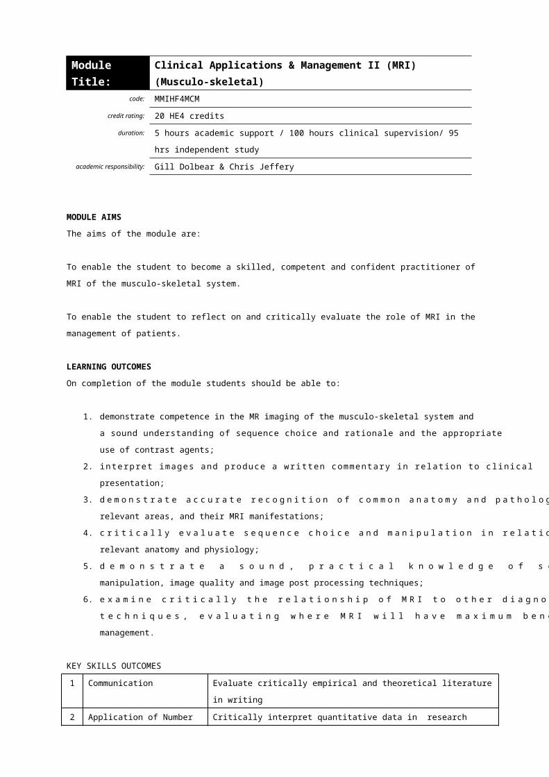
Module
Title:
Clinical Applications & Management III
(MRI) (Non-routine brain, head & neck)
code: MMIHF4MCN
credit rating: 20 HE4 credits
duration: 5 hours academic support / 100 hours clinical supervision/ 95 hrs
independent study
academic
responsibility:
Gill Dolbear & Chris Jeffery
MODULE AIMS
The aims of the module are:
To enable the student to become a skilled, competent and confident practitioner of MRI in the
non-routine brain, and head and neck, including the principles and applications of MR
Angiography (MRA), diffusion and perfusion imaging, and contrast agents.
To enable the student to reflect on and critically evaluate the role of MRI in the management of
patients.
LEARNING OUTCOMES
On completion of the module students should be able to:
1. demonstrate competence in the MR imaging of the brain and associated structures, and
head and neck, demonstrating a sound understanding of sequence choice and rationale
and the appropriate use of contrast agents;
2. interpret images and produce a written commentary in relation to clinical presentation;
3. demonstrate accurate recognition of common anatomy and pathology in the relevant
areas, and their MRI manifestations;
4. critically evaluate sequence choice and manipulation in relation to the relevant anatomy
and physiology;
5. demonstrate a sound, practical knowledge of sequence choice, factor manipulation,
image quality and image post processing techniques;
6. examine critically the relationship of MRI to other diagnostic imaging techniques,
evaluating where MRI will have maximum benefit to patient management;
7. appraise critically the principles and applications, current and future, of MRA.
KEY SKILLS OUTCOMES
1 Communication Evaluate critically empirical and theoretical literature in writing
2 Application of Number Critically interpret quantitative data in research and/or audit

studies
3 Information Technology Access and use efficiently the internet, College databases and
computer software for assignment content and presentation
4 Working with Others Demonstrate interprofessionality and teamwork
5 Problem Solving Reflect critically on professional practice and strategies for service
improvement and development in an interprofessional context
6 Improving own learning
and performance
Determine an appropriate pathway for the achievement of
academic goals and professional development
MODULE CONTENT
Anatomy, physiology and pathology of the brain and associated structures, head and neck;
Comparative imaging techniques;
The principles of MRI science and technology relative to the brain and associated structures, and
the head and neck;
The principles, sequences and applications of functional imaging (perfusion and diffusion);
The use of image registration (e.g. MRI, CT and PET), including post processing and image
manipulation techniques;
MR Angiography – time of flight, phase contrast and contrast enhanced studies;
Diffusion and perfusion imaging;
Principles and applications of proton MR spectroscopy;
MRI contrast agents;
Quality assurance and image quality pertaining to the non-routine brain, head and neck,
including artefact recognition and reduction techniques.
LEARNING AND TEACHING STRATEGIES
Work based supervision and practice, shadowing activities, clinical meetings and tutorials, expert
lectures, self directed study, web based materials.
ILLUSTRATIVE ASSESSMENT
Portfolio of Clinical and Professional Development, to meet module intended learning outcomes 1-7
and K/S 1-6, containing:
A record of 60 examinations demonstrating that a wide range of non-routine brain and head
and neck imaging examinations has been carried out, of which a substantial number must
have been completed unassisted. Unassisted examinations must include a written
commentary;

2 x 750 word case reports to the standard of an academic journal;
1 x 2000 word in depth case study;
Audit and comparative, reflective analysis of a randomly selected number of commentaries
with reports;
Clinical supervisor’s report and verification of competence.
ILLUSTRATIVE BIBLIOGRAPHY
Berquist, T. (1995), Pocket Atlas of MRI Body Anatomy. Lippincott-Raven
Durham, D. (1997), Atlas of MR Pathology. W.B.Saunders
Higgins, C. Hrikak, H. Helms, C. (Eds) (1997), Magnetic Resonance Imaging of the Body,
Lippincott-Raven
Kelly, L. &. Petersen, M. (1997), Sectional Anatomy for Imaging Professionals. Mosby
Stark, D. & Bradley W. (Eds), (1998), Magnetic Resonance Imaging 3 rd edition , Moseby Year Book
Westbrook, C. (1999), Handbook of MRI Technique. Blackwell Science
WEBSITES:
www.mritutor.org
www.cis.rit.edu
www.ismrm.org
www.mrisafety.com
www.topspins.com - about MR angiography
www.mrieducation.com
www.med.harvard.edu
www.t2star.com
JOURNALS
American Journal of Roentgenology
British Journal of Radiology
Clinical Radiology
Journal of Magnetic Resonance Imaging
Magnetic Resonance Imaging
Physics in Medicine and Biology
RadioGraphics <http://radiographics.rsnajnls.org>
Radiologic Clinics of North America
Radiology http://radiology.rsnajnls.org>
Seminars in Ultrasound, CT and MR


Module
Title:
Clinical Applications & Management IV (MRI) (Body
imaging)
code: MMIH4NPS
credit rating: 20 HE4 credits
duration: 5 hours academic support / 100 hours clinical supervision/ 95 hrs
independent study
academic
responsibility:
Gill Dolbear & Chris Jeffery
MODULE AIMS
The aims of the module are:
To enable the student to become a skilled, competent and confident practitioner of MR body
imaging, including cardiac and breast imaging;
To enable the student to reflect on and critically evaluate the role of MRI in the management of
patients.
LEARNING OUTCOMES
On completion of the module students should be able to:
1. demonstrate competence in body imaging, cardiac imaging and breast imaging,
including an evaluation of sequence choice and rationale and the appropriate use of MRI
contrast agents;
2. interpret images and produce a written commentary in relation to clinical presentation;
3. demonstrate accurate recognition of common anatomy and pathology in the relevant
areas, and their MRI manifestations;
4. critically evaluate sequence choice and manipulation in relation to the relevant anatomy
and physiology;
5. demonstrate sound knowledge of sequence choice, factor manipulation, image quality
and image post processing techniques;
6. examine critically the relationship of MRI to other diagnostic imaging techniques,
evaluating where MRI will have maximum benefit to patient management;
7. demonstrate a critical awareness of the principles and techniques used in fast imaging,
including their current and future applications, and their specific advantages in body
imaging;
8. evaluate current MRI systems and critically evaluate the future development
opportunities.
KEY SKILLS OUTCOMES

1 Communication Evaluate critically empirical and theoretical literature in writing
2 Application of Number Critically interpret quantitative data in research and/or audit
studies
3 Information Technology Access and use efficiently the internet, College databases and
computer software for assignment content and presentation
4 Working with Others Demonstrate interprofessionality and teamwork
5 Problem Solving Reflect critically on professional practice and strategies for service
improvement and development in an interprofessional context
6 Improving own learning
and performance
Determine an appropriate pathway for the achievement of
academic goals and professional development
MODULE CONTENT
Anatomy, physiology and pathology of the abdomen, pelvis, cardiac and breast;
Comparative imaging techniques;
The principles of MRI science and technology relative to imaging the body;
The principles, sequences and applications of cardiac imaging;
Fast scanning techniques (e.g. echo-planar imaging, breath-hold);
MRI contrast agents used in the body;
Future developments in MRI;
Quality assurance and image quality pertaining to body imaging, including artefact recognition
and reduction techniques.
LEARNING AND TEACHING STRATEGIES
Work based supervision and practice, shadowing activities, clinical meetings and tutorials, expert
lectures, self directed study, web based materials.
ILLUSTRATIVE ASSESSMENT
Portfolio of Clinical and Professional Development, to meet module intended learning outcomes 1-8
and K/S 1-6, containing:
A record of 60 examinations demonstrating that a wide range of magnetic resonance imaging
technique examinations has been carried out, of which a substantial number must have been
completed unassisted. Unassisted examinations must include a written commentary;
2 x 750 word case reports to the standard of an academic journal
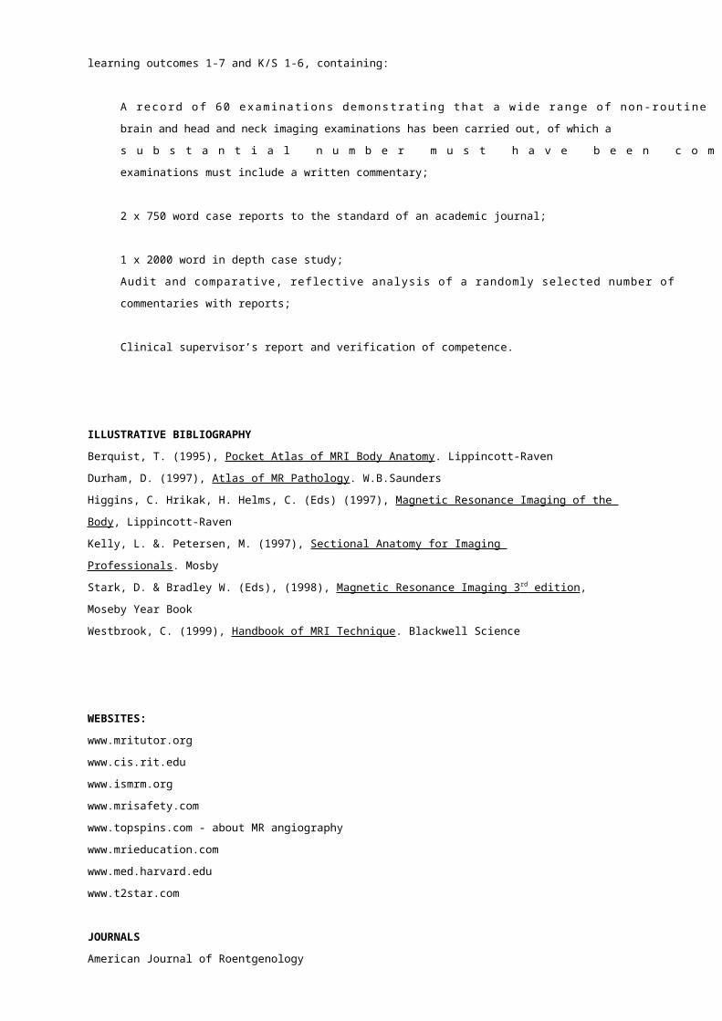
1 x 2000 word in depth case study;
Audit and comparative, reflective analysis of a randomly selected number of commentaries
with reports;
Clinical supervisor’s report and verification of competence.
ILLUSTRATIVE BIBLIOGRAPHY
Heywang-Kobrunner, S. & Beck, R. (1996), Contrast Enhanced MR of the Breast, Springer
Higgins, C. Hrikak, H. Helms, C. (Eds) (1997), Magnetic Resonance Imaging of the Body,
Lippincott-Raven
Lardo, A.C., Fayad, Z.A., Chronos, N.A.F. & Fuster V.(2003) Cardiovascular Magnetic Resonance,
Mirowitz, S. (1996), Pitfalls, Variants and Artefacts in Body MR Imaging, Moseby
Ros, P. & Bidgood, W. (1998), Abdominal Magnetic Resonance Imaging, Moseby
Stark, D. & Bradley W. (Eds), (1998), Magnetic Resonance Imaging 3 rd edition , Moseby Year Book
Westbrook, C. (1999), Handbook of MRI Technique. Blackwell Science
WEBSITES:
www.mritutor.org
www.cis.rit.edu
www.ismrm.org
www.mrisafety.com
www.topspins.com - about MR angiography
www.mrieducation.com
www.med.harvard.edu
www.t2star.com
JOURNALS
American Journal of Roentgenology
British Journal of Radiology
Clinical Radiology
Journal of Magnetic Resonance Imaging
Magnetic Resonance Imaging
Physics in Medicine and Biology
RadioGraphics <http://radiographics.rsnajnls.org>
Radiologic Clinics of North America
Radiology http://radiology.rsnajnls.org>
Seminars in Ultrasound, CT and MR

Module
Title:
Principles of Science & Technology in Nuclear Medicine
code: MMIHF4NPS
credit rating: 20 HE4 credits
duration: 40 hours taught & 160 hours independent study
academic
responsibility:
Chris Jeffery
MODULE AIM
The aim of the module is to provide students with a thorough knowledge of the science, technology
and mathematics necessary to become a competent practitioner in Nuclear Medicine
LEARNING OUTCOMES
At the end of the module, students should be able to:
1. discuss the various types of radioactive decay processes and their relationship to nuclear
medicine procedures;
2. demonstrate understanding of the interaction of radiation with matter and competence with
the associated mathematical functions;
3. understand the physical principles, chemical processes and radiation protection principles
applicable to radiopharmaceutical production;
4. evaluate the processes which contribute to degradation of radiopharmaceuticals;
5. appraise critically the methods for detecting and measuring radiation and the uses of various
devices in nuclear medicine and their continuing development.
6. evaluate methods available for storage and hard copy of images and discuss the
advantages and disadvantages of each.
KEY SKILLS OUTCOMES
1 Communication Evaluate critically empirical and theoretical literature, both orally
and in writing
2 Application of Number Critically interpret data in research studies
3 Information Technology Access and use efficiently the internet, College databases and
computer software for assignment content and presentation
4 Working with Others Relate and interact effectively with individuals and groups
5 Problem Solving Use relevant information sources. Identify and solve problems
associated with study skills and assessment at M level
6 Improving own learning
and performance
Manage own time in achieving objectives
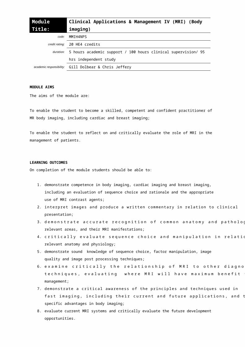
MODULE CONTENT
Atomic structure, radioactive decay processes, the interaction of radiation with matter and their
applications in nuclear medicine.
The mathematics of decay processes, isotope production and radionuclide generators.
The production of radiopharmaceuticals, metal-ligand chemistry and breakdown of
radiopharmaceuticals by radiation and chemical processes.
Radiation safety and protection.
The use of radiation detectors e.g. Geiger-Muller tubes and sodium iodide detectors. Gamma
cameras and their development including single, double and triple headed cameras, the latest
crystal detectors and digital imaging heads. The measurement of radioactivity using ionization
chambers.
Image generation, hard and soft copy and routine image manipulation, enhancement and display.
The use of computer technology to facilitate fast image transfer within and between imaging centres
and mass storage devices.
LEARNING AND TEACHING STRATEGIES
Formal lectures, case study presentations, group tutorials, practical demonstrations.
ILLUSTRATIVE ASSESSMENT
The assessment will comprise:
A 20 minute oral presentation to the cohort (50% weighting) (LOs 1-7) (K/S 1-6)
A 2000 word fully referenced synopsis (50% weighting) (LOs 1-7) (K/S1, 2, 3, 5, 6)
Students will present a critical evaluation of how imaging parameters may affect image quality and
perceptibility of diagnostic information. The presentation will be derived from material gathered
from the student’s clinical centre and make use of the equipment, quality control phantoms,
homemade phantoms and relevant clinical examples. Reflecting safe, current practice, it should
demonstrate a sound knowledge of the relevant fundamental physics, the features and capabilities
of the equipment, the nature of particular imaging parameters over which the practitioner has
control, the interrelationships between them and their influence on image quality.
ILLUSTRATIVE BIBLIOGRAPHY
Bernier, Christian & Langan (Eds), (1997), Nuclear Medicine - Technology and Techniques 4 th
Edition. Mosby
Chandra, R., (2005), Nuclear Medicine Physics the Basics, Lippincott, Williams &
Wilkins
Farr & Allisy-Roberts, (1997), Physics for Medical Imaging, W.B. Saunders
IPEMB report no73, (1996), Mathematical Techniques in Nuclear Medicine, IPEM
IPEM, (1991), Radiation Protection in Nuclear Medicine and Pathology
IPEM

Knoll, G., (1989), Radiation Detection and Measurement, Wiley
Steves, A., (1992), Review of Nuclear Medicine Technology, Society of
Nuclear Medicine
WEBSITES
www.bnms.org
www.mallinckrodt.nl/nucmed/noframes/nuclear/resources.htm
www.nycomed-amersham.com
www.nud.co.uk
www.asnc.org
www.snm.org
JOURNALS
The Journal of Nuclear Medicine Technology
Nuclear Medicine Communications
The European Journal of Nuclear Medicine
The Journal of Nuclear Medicine

Module
Title:
Clinical Applications & Management I (Standard NM
Procedures)
code: MMIHF4NCS
credit rating: 20 HE4 credits
duration: 5 hours academic support / 100 hours clinical supervision/ 95 hrs
independent study
academic
responsibility:
Peter Milburn & Gill Dolbear
MODULE AIMS
The aims of the module are:
To enable the student to become a competent and confident practitioner of Nuclear Medicine in
relation to standard imaging procedures.
To enable the student to reflect on and critically evaluate the role of standard nuclear medicine
images in the management of patients and clients.
LEARNING OUTCOMES
By the end of the module, students should be able to:
1. demonstrate the ability to produce images of diagnostic quality for standard nuclear
medicine procedures using safe working practices, identifying those factors which
optimize/degrade image production;
2. demonstrate the ability to interpret normal and abnormal appearances on standard nuclear
medicine images, recognise artefacts and produce a written commentary/report in relation
to clinical presentation;
3. demonstrate understanding of the underlying physiology and pathology pertaining to each
investigation;
4. discuss the relationship of nuclear medicine to other complementary or alternative imaging
modalities, evaluating benefits to patient management;
5. appraise critically the role of nuclear medicine with particular regard to cost, radiation safety
and availability;
6. demonstrate a comprehensive awareness of legislation governing nuclear medicine and its
implications for service delivery;
7. acknowledge the particular needs of patients and clients in relation to nuclear medicine
procedures and offer appropriate care.
KEY SKILLS OUTCOMES

1 Communication Evaluate critically empirical and theoretical literature in writing
2 Application of Number Critically interpret quantitative data in research and/or audit
studies
3 Information Technology Access and use efficiently the internet, College databases and
computer software for assignment content and presentation
4 Working with Others Demonstrate interprofessionality and teamwork
5 Problem Solving Reflect critically on professional practice and strategies for service
improvement and development in an interprofessional context
6 Improving own learning
and performance
Determine an appropriate pathway for the achievement of
academic goals and professional development
MODULE CONTENT
Image acquisition, optimization of image quality, quality control of images, image media, and
radiation protection.
Anatomy, physiology and pathology of standard nuclear medicine imaging including bone, renal,
lung, cardiac, (para)thyroid, brain, liver, infection imaging, oncology and bone densitometry.
Image processing, image enhancement and post acquisition techniques.
Identification of abnormal pathology and diagnostic pathways incorporating other imaging
modalities.
The legislative controls on the use of unsealed sources in nuclear medicine, radiation safety and
patient safeguarding.
Patient – professional interaction.
LEARNING AND TEACHING STRATEGIES
Work based supervision and practice, shadowing activities, clinical meetings and tutorials, expert
lectures, self directed study, web based materials.
ILLUSTRATIVE ASSESSMENT
Portfolio of Clinical and Professional Development, to meet module intended learning outcomes 1-7
and K/S 1-6, containing:
A record of 125 cases, demonstrating that the full range of standard nuclear medicine
procedures has been carried out, of which a substantial number must have been completed
unassisted. Unassisted examinations must include a written commentary/report;

2 x 750 word case reports to the standard of an academic journal;
1 x 2000 word in depth case study;
Audit and reflective analysis of a randomly selected number of reports;
Clinical supervisor’s report and verification of competence.
ILLUSTRATIVE BIBLIOGRAPHY
Bernier, Christian& Langan(Eds),(1997), Nuclear Medicine - Technology and Techniques 4 th Edition .
Mosby
Fogelman, I (Ed) (1987), Bone Scanning in Clinical Practice, Springer-Verlag
Gelfand, M. Thomas, S. (1988), Effective Use of Computers in Nuclear Medicine
Gerson, M., (1991), Cardiac Nuclear Medicine 2 nd Edition , Mc-Graw-Hill
Lee, K., (1991), Computers in Nuclear Medicine: A Practical Approach, Society of
Nuclear Medicine
Maisey, Britton, Collier. (1998), Clinical Nuclear Medicine 3rd Edition, Chapman and Hall Medical
Mistry, R., (1988), Manual of Nuclear Medicine Procedures, Chapman and Hall
Medical
Murray, I. & Ell, P. (1998), Nuclear Medicine in Clinical Diagnosis and Treatment,
Churchill Livingstone
Pennell, D. (1992), Thallium Myocardial Perfusion Tomography in Clinical
Cardiology, C. Livingstone
Peters, M. (2003) Nuclear Medicine In Radiological Diagnosis,
Woolf, Anthony D. (1988), Osteoporosis – A Clinical Guide, Dunitz
WEBSITES
www.bnms.org
www.snm.org
www.asnc.org
www.nycomed-amersham.com
www.nud.co.uk
www.mallinckrodt.nl/nucmed/noframes/nuclear/resources.htm

JOURNALS
The Journal of Nuclear Medicine Technology
Nuclear Medicine Communications
The European Journal of Nuclear Medicine
The Journal of Nuclear Medicine
Osteoporosis International
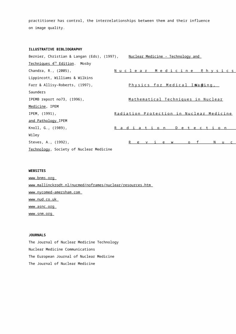
Module
Title:
Clinical Applications & Management II
(Complex & Non-standard NM Procedures)
code: MMIHF4NCC
credit rating: 20 HE4 credits
duration: 5 hours academic support / 100 hours clinical supervision/ 95 hrs
independent study
academic
responsibility:
Peter Milburn & Gill Dolbear
MODULE AIMS
The aims of the module are:
To enable the student to become a competent and confident practitioner of Nuclear Medicine in
relation to complex and non standard imaging procedures.
To enable the student to reflect on and critically evaluate the role of complex and non standard
nuclear medicine images in the management of patients and clients.
LEARNING OUTCOMES
At the end of the module, students should be able to:
1. demonstrate the ability to produce images of diagnostic quality, using safe working
practices, for complex and non standard nuclear medicine procedures;
2. demonstrate the ability to interpret normal and abnormal appearances on complex and non
standard nuclear medicine images, recognise artefacts and produce a written
commentary/report in relation to clinical presentation;
3. demonstrate understanding of the underlying physiology and pathology pertaining to each
investigation;
4. discuss the relationship of nuclear medicine to other complementary or alternative imaging
modalities, evaluating future developments and potential benefits to patient management;
5. acknowledge the particular needs of patients and clients in relation to complex and non
standard nuclear medicine procedures and offer appropriate care.
KEY SKILLS OUTCOMES
1 Communication Evaluate critically empirical and theoretical literature in writing
2 Application of Number Critically interpret quantitative data in research and/or audit
studies
3 Information Technology Access and use efficiently the internet, College databases and
computer software for assignment content and presentation
4 Working with Others Demonstrate interprofessionality and teamwork
5 Problem Solving Reflect critically on professional practice and strategies for service
improvement and development in an interprofessional context

6 Improving own learning
and performance
Determine an appropriate pathway for the achievement of
academic goals and professional development
INDICATIVE MODULE CONTENT
Image acquisition, optimization of image quality, quality control of images and radiation protection.
Anatomy, physiology and pathology of complex and non standard nuclear medicine imaging
including brain perfusion, antibody infection imaging and new developments in the diagnosis of
malignancy.
Image processing, image enhancement and post acquisition techniques including tomographic
reconstruction, filtering and 3D rendering.
Identification of abnormal pathology and diagnostic pathways incorporating other imaging modalities
and anticipation of the likely direction of advancements.
Patient – professional interaction.
LEARNING AND TEACHING STRATEGIES
Work based supervision and practice, shadowing activities, clinical meetings and tutorials, expert
lectures, self directed study, web based materials.
ILLUSTRATIVE ASSESSMENT
Portfolio of Clinical and Professional Development, to meet module intended learning outcomes 1-5
and K/S 1-6, containing:
A record of 75 cases, demonstrating that the full range of complex and advanced nuclear
medicine procedures has been carried out, of which a substantial number must have been
completed unassisted. Unassisted examinations must include a written commentary/report;
2 x 750 word case reports to the standard of an academic journal;
1 x 2000 word in depth case study;
Audit and reflective analysis of a randomly selected number of reports;
Clinical supervisor’s report and verification of competence.
ILLUSTRATIVE BIBLIOGRAPHY
Bernier, Christian& Langan(Eds) (1997), Nuclear Medicine - Technology and Techniques 4 th Edition .
Mosby
Gelfand, M. Thomas, S. (1988), Effective Use of Computers in Nuclear Medicine
George et al. (1991), Neuroactivation and Neuroimaging with SPET, Springer-Verlag
Gerson, M. (1991), Cardiac Nuclear Medicine 2 nd Edition , Mc-Graw-Hill

Goris, M. Bretille, J. (1992), A Colour Atlas of Nuclear Cardiology, Chapman & Hall
Larock et al. (1993), New Developments in Myocardial Imaging, Martin Dunitz
Lee, K (1991), Computers in Nuclear Medicine: A Practical Approach, Society of
Nuclear Medicine
Maisey, Britton, Collier (1998), Clinical Nuclear Medicine 3rd Edition, Chapman and Hall Medical
Mistry, R. (1988), Manual of Nuclear Medicine Procedures, Chapman and Hall
Medical
Murray, I. & Ell, P. (1998), Nuclear Medicine in Clinical Diagnosis & Treatment Vols I &
II. Churchill Livingstone
Pennell, D. (1992), Thallium Myocardial Perfusion Tomography in Clinical
Cardiology C. Livingstone
WEBSITES
www.bnms.org
www.mallinckrodt.nl/nucmed/noframes/nuclear/resources.htm
www.nycomed-amersham.com
www.nud.co.uk
www.asnc.org
www.snm.org
JOURNALS
The Journal of Nuclear Medicine Technology
The European Journal of Nuclear Medicine
The Journal of Nuclear Medicine
Nuclear Medicine Communications
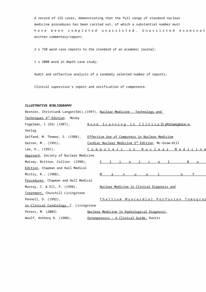
Module
Title:
Clinical Applications & Management III
(Positron Emission Tomography, PET)
code: MMIHF4NCP
credit rating: 20 HE4 credits
duration: 5 hours academic support / 100 hours clinical supervision/ 95 hrs
independent study
academic
responsibility:
Peter Milburn & Gill Dolbear
MODULE AIMS
The aims of the module are:
To enable the student to become a competent and confident practitioner of Positron Emission
Tomography (PET) imaging.
To enable the student to reflect on and critically evaluate the role of PET imaging in the management
of patients and clients.
LEARNING OUTCOMES
At the end of the module, students should be able to:
1. demonstrate the ability to produce PET images of diagnostic quality, using safe working
practices;
2. demonstrate the ability to interpret normal and abnormal image appearances, recognise
artefacts and produce a written commentary/report in relation to clinical presentation;
3. demonstrate understanding of the underlying anatomy, physiology and pathology;
4. demonstrate appreciation of the particular radiation protection requirements and radio-
pharmaceutical preparation;
5. evaluate the relative contribution of PET imaging to the management of patients and clients;
6. critically appraise PET imaging as a diagnostic tool, differentially evaluating its use in terms
of cost, patient radiation doses and availability;
7. acknowledge the particular needs of patients and clients in relation to PET imaging and offer
appropriate care.
KEY SKILLS OUTCOMES
1 Communication Evaluate critically empirical and theoretical literature in writing
2 Application of Number Critically interpret quantitative data in research and/or audit
studies
3 Information Technology Access and use efficiently the internet, College databases and
computer software for assignment content and presentation

4 Working with Others Demonstrate interprofessionality and teamwork
5 Problem Solving Reflect critically on professional practice and strategies for service
improvement and development in an interprofessional context
6 Improving own learning
and performance
Determine an appropriate pathway for the achievement of
academic goals and professional development
MODULE CONTENT
Anatomy, physiology and pathology of PET imaging, including brain, cardiac and whole body imaging
in oncology.
Cyclotron operation, PET radiopharmaceutical preparation and quality control.
The acquisition and processing of PET images.
Radiation protection.
The cost of PET and the role of PET imaging today and in the future. The merits of PET gamma
cameras.
Patient –professional interaction
LEARNING AND TEACHING STRATEGIES
Work based supervision and practice, shadowing activities, clinical meetings and tutorials, expert
lectures, self directed study, web based materials.
ILLUSTRATIVE ASSESSMENT
Portfolio of Clinical and Professional Development, to meet module learning intended outcomes 1-7
and K/S 1-6, containing:
A record of 50 cases, demonstrating that the full range PET nuclear medicine procedures has
been carried out, of which a substantial number must have been completed unassisted.
Unassisted examinations must include a written commentary/report;
2 x 750 word case reports to the standard of an academic journal;
1 x 2000 word in depth case study;
Audit and reflective analysis of a randomly selected number of reports;
Clinical supervisor’s report and verification of competence.
ILLUSTRATIVE BIBLIOGRAPHY
Larock et al (1993), New Developments in Myocardial Imaging, Martin Dunitz
Maisey, Wahl & Barrington Arnold (1999),Atlas of Clinical Positron Emission Tomography
Marcel P.M. Stokkel (1999), The Clinical Applicability of F18-FDG Detection with a Dual
Head Coincidence Camera, Ponsen & Looijen
Gerson, (1991), Cardiac Nuclear Medicine 2 nd Edition , Mc-Graw-Hill
WEBSITES
www.bnms.org
www.mallinckrodt.nl/nucmed/noframes/nuclear/resources.htm
www.nycomed-amersham.com
www.nud.co.uk
www.asnc.org
www.snm.org
JOURNALS
The Journal of Nuclear Medicine Technology
Nuclear Medicine Communications
The European Journal of Nuclear Medicine
The Journal of Nuclear Medicine

Module
Title:
Clinical Applications & Management IV (Radionuclide
Therapy)
code: MMIHF4NCR
credit rating: 20 HE4 credits
duration: 5 hours academic support / 100 hours clinical supervision/ 95 hrs
independent study
academic
responsibility:
Peter Milburn & Gill Dolbear
MODULE AIM
The aims of the module are:
To enable the student to become a competent and confident practitioner of radionuclide therapy.
To enable the student to reflect on and critically evaluate the contribution of radio nuclide therapy
procedures to the management of patients and clients.
LEARNING OUTCOMES
At the end of the module, students should be able to:
1. undertake the administration of therapeutic doses of radio nuclides using safe working
practices;
2. evaluate the role of radionuclide therapy and understand the technical and logistical
problems associated with unsealed source treatment;
3. be aware of the needs of the patient undergoing radionuclide therapy and examine
restrictions imposed on patients and their effects.
4. evaluate the differential uses of radionuclide therapy in patient management;
KEY SKILLS OUTCOMES
1 Communication Evaluate critically empirical and theoretical literature in writing
2 Application of Number Critically interpret quantitative data in research and/or audit studies
3 Information Technology Access and use efficiently the internet, College databases and
computer software for assignment content and presentation
4 Working with Others Demonstrate interprofessionality and teamwork
5 Problem Solving Reflect critically on professional practice and strategies for service
improvement and development in an interprofessional context
6 Improving own learning Determine an appropriate pathway for the achievement of academic
and performance goals and professional development
MODULE CONTENT
Anatomy, physiology and pathology relating to radionuclide therapy procedures.
The role of unsealed source therapy in relation to teletherapy and chemotherapy. Calculation of
radiation doses (MIRDOSE) and radiation protection and contamination issues during treatment.
Waste disposal and public dose restrictions relating to radionuclide radiotherapy, including the role of
carers and support workers.
Developments in therapeutic agents.
LEARNING AND TEACHING STRATEGIES
Work based supervision and practice, shadowing activities, clinical meetings and tutorials, expert
lectures, self directed study, web based materials.
ILLUSTRATIVE ASSESSMENT
Portfolio of Clinical and Professional Development, to meet module intended learning outcomes 1-4
and K/S 1-6, containing:
A record of 20-30 cases, demonstrating that the full range of standard radionuclide therapy
procedures has been observed. Participative procedures must include a written
commentary/report;
2 x 750 word case reports to the standard of an academic journal;
1 x 2000 word in depth case study;
Audit and reflective analysis of a randomly selected number of procedures;
Clinical supervisor’s report and verification of competence.
ILLUSTRATIVE BIBLIOGRAPHY
Bernier, Christian& Langan(Eds) (1997), Nuclear Medicine - Technology and Techniques 4 th Edition .
Mosby
Maisey, Britton, Collier (1998), Clinical Nuclear Medicine 3rd Edition Chapman and Hall Medical
Murray, I. & Ell, P. (1998), Nuclear Medicine in Clinical Diagnosis & Treatment Vols I &
II. Churchill Livingstone
Spencer, Richard P. (1978) Therapy in Nuclear Medicine , Grune and Stratton

WEBSITES
www.bnms.org
www.snm.org
www.asnc.org
www.nycomed-amersham.com
www.nud.co.uk
www.mallinckrodt.nl/nucmed/noframes/nuclear/resources.htm
JOURNALS
The Journal of Nuclear Medicine Technology
Nuclear Medicine Communications
The European Journal of Nuclear Medicine
The Journal of Nuclear Medicine

Module
Title:
Radiopharmacy and Non-imaging Diagnostic Tests
code: MMIHF4NRN
credit rating: 20 HE4 credits
duration: 5 hours academic support / 100 hours clinical supervision/ 95 hrs
independent study
academic
responsibility:
Peter Milburn & Gill Dolbear
MODULE AIMS
The aims of the module are:
To enable students to produce radiopharmaceutical products safely and accurately and to perform a
range of non-imaging investigations in nuclear medicine.
To enable students to reflect on and critically evaluate the use of non imaging investigation in the
management of patients and clients.
LEARNING OUTCOMES
At the end of the module, students should be able to:
1. produce sterile radiopharmaceutical products within the guidelines of good manufacturing
practice, incorporating aseptic technique, radiation protection practice and safe dispensing;
2. demonstrate a sound knowledge of factors affecting radiopharmaceutical manufacture;
3. demonstrate the ability to undertake quality control procedures such as environmental
monitoring, sterility testing of products and radio nuclide/radiochemical purity measurement;
4. evaluate the role of blood cell labelling procedures and perform leucocyte and red/platelet
radio labelling;
5. perform a range of non-imaging nuclear medicine procedures, GFR/ERPF measurement,
carbon-14 breath testing, vitamin B12 malabsorption tests, and red cell mass measurement.
KEY SKILLS OUTCOMES
1 Communication Evaluate critically empirical and theoretical literature in writing
2 Application of Number Critically interpret quantitative data in research and/or audit
studies
3 Information Technology Access and use efficiently the internet, College databases and
computer software for assignment content and presentation
4 Working with Others Demonstrate interprofessionality and teamwork
5 Problem Solving Reflect critically on professional practice and strategies for service

improvement and development in an interprofessional context
6 Improving own learning
and performance
Determine an appropriate pathway for the achievement of
academic goals and professional development
MODULE CONTENT
Legislation and MCA recommendations controlling the environment and working practices of radio
pharmacy units.
Radiopharmacy procedures, aseptic technique, quality assurance/control, radiation protection.
Handling of radio nuclides as regards receipt, storage and disposal.
Blood radio labelling techniques including leucocyte, erythrocyte and platelet labelling.
The physiology and pathology of nuclear medicine non-imaging tests.
Practical aspects of the tests and evaluation of the results. Critical appraisal of the tests in
comparison with complementary techniques.
LEARNING AND TEACHING STRATEGIES
Formal lectures, practical radiopharmacy sessions, group discussions.
ILLUSTRATIVE ASSESSMENT
Portfolio of Clinical and Professional Development, to meet module intended learning outcomes 1-5
and K/S 1-6, containing:
A record of 100 radiopharmaceutical preparations, 10 blood labelling procedures and 20-30
non-imaging case studies demonstrating a full range of types, of which a substantial number
must have been completed unassisted;
1x 2000 word case study on a non-imaging nuclear medicine investigation;
1 x 2000 word assignment on the working practices of a radiopharmacy;
Clinical supervisor’s report and verification of competence.
ILLUSTRATIVE BIBLIOGRAPHY
Bernier, Christian& Langan (Eds) (1997) Nuclear Medicine - Technology and Techniques 4 th Edition .
Mosby
IPEM (1991), Radiation Protection in Nuclear Medicine and Pathology, IPEM
Maisey, Britton, Collier (1998) Clinical Nuclear Medicine 3rd Edition, Chapman and Hall Medical
Medicines Control Agency (1997) Rules and Guidance for Radio pharmaceutical
Manufacturers and
Distributors 1997, HMSO
Mistry, R. (1988) Manual of Nuclear Medicine Procedures, Chapman and Hall Medical
Murray, I. & Ell, P. (1998) Nuclear Medicine in Clinical Diagnosis & Treatment Vols I &
II. Churchill Livingstone
Steves, A. (1992) Review of Nuclear Medicine Technology, Society of Nuclear
Medicine
WEBSITES
www.bnms.org
www.snm.org
www.asnc.org
www.nycomed-amersham.com
www.nud.co.uk
www.mallinckrodt.nl/nucmed/noframes/nuclear/resources.htm
JOURNALS
The Journal of Nuclear Medicine Technology
Nuclear Medicine Communications
The European Journal of Nuclear Medicine
The Journal of Nuclear Medicine
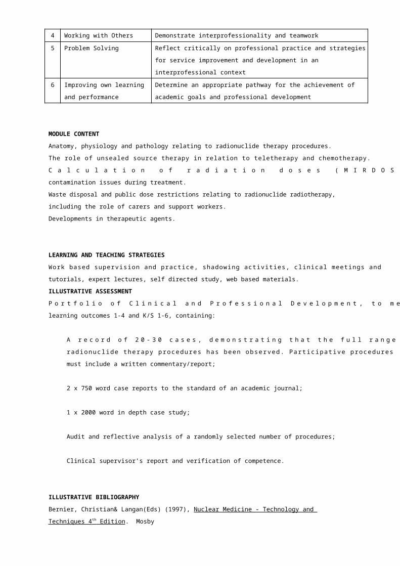
Module
Title:
Principles of Science & Technology in Medical
Ultrasound
code: MMIHF4UPS
credit rating: 20 HE4 credits
duration: 40 hours taught & 160 hours independent study
academic
responsibility:
Gill Dolbear
MODULE AIM
The aim of the module is to provide practitioners with a sound knowledge of the scientific principles
and technologies in ultrasound which underpin the provision of quality services to patients and
clients.
LEARNING OUTCOMES
In relation to medical ultrasound, by the end of the module, students should be able to:
1. Demonstrate knowledge, understanding and application of the scientific principles
underpinning medical diagnostic ultrasound imaging;
2. Evaluate ultrasound technologies used in medical ultrasound practice;
3. Utilise ultrasound technology effectively to produce appropriate diagnostic images and
spectra, ensuring that image quality is optimized and exposure to ultrasound minimised
according to clinical need;
4. Evaluate the formation of artefacts and differentiate between artefacts and true pathology;
5. Carry out effective quality monitoring in relation to ultrasound technology used in practice.
KEY SKILLS OUTCOMES
1 Communication Evaluate critically empirical and theoretical literature, both orally
and in writing
2 Application of Number Critically interpret data in research studies
3 Information Technology Access and use efficiently the internet, College databases and
computer software for assignment content and presentation
4 Working with Others Relate and interact effectively with individuals and groups
5 Problem Solving Use relevant information sources. Identify and solve problems
associated with study skills and assessment at M level
6 Improving own learning
and performance
Manage own time in achieving objectives
MODULE CONTENT
The physics of ultrasound and the scientific principles underpinning ultrasound imaging.
The nature of artefacts and the problems they can give rise to.
The range of ultrasound equipment that can and might be used, including the role of
Doppler ultrasound.
Ultrasound imaging techniques and the relative scientific roles of ultrasound imaging and other
imaging modalities.
Performance measurements needed to ensure technical quality assurance.
LEARNING AND TEACHING STRATEGIES
Formal lectures, practical work, exercises and demonstrations, group tutorials, student led seminars.
ILLUSTRATIVE ASSESSMENT
The assessment will comprise:
A 20 minute oral presentation to the cohort (50% weighting) (LOs 1-7) (K/S 1-6)
A 2000 word fully referenced synopsis (50% weighting) (LOs 1-7) (K/S 1, 2, 3, 5, 6)
Students will present a critical evaluation of how imaging parameters may affect image quality and
perceptibility of diagnostic information. The presentation will be derived from material gathered
from the student’s clinical centre and make use of the equipment, quality control phantoms,
homemade phantoms and relevant clinical examples. Reflecting safe, current practice, it should
demonstrate a sound knowledge of the relevant fundamental physics, the features and capabilities
of the equipment, the nature of particular imaging parameters over which the practitioner has
control, the interrelationships between them and their influence on image quality.
ILLUSTRATIVE BIBLIOGRAPHY
Evans, D.H., McDicken, W.N. (2000), Doppler Ultrasound: Physics, Instrumental, and Clinical
Applications
2nd Edition, John Wiley and Sons.
Fish, P. (1997), Physics and Instrumentation of Diagnostic Medical Ultrasound 5th
Edition, John Wiley and Sons.
Hedrick, W.R. (1994), Ultrasound Physics and Instrumentation Mosby.
Hoskins, P.R., Thrush, A., Diagnostic Ultrasound Physics and Equipment Greenwich Medical

Media.
Martin, K. & Whittingham T.A. (2003)
Kremkau, F.W. (1997), Diagnostic Ultrasound: Principles and Instruments, WB Saunders
Company.
Nelson, T.R. (1999), Three-dimensional Ultrasound, Lippincott, Williams and Wilkins.
Zagzebski, J.A. (1996), Essentials of Ultrasound Physics Mosby.
WEBSITES
www.ultrasound.net
www.aium.org/consumer/obus.htm
JOURNALS
Physics in Medicine and Biology
Module
Title:
Techniques in Musculo-skeletal Ultrasound Imaging
code: MMIHF4UTM
credit rating: 20 HE4 credits
duration: 40 hours taught/ 160 hrs independent study
academic
responsibility:
Kate Springett & Gill Dolbear
MODULE AIMS
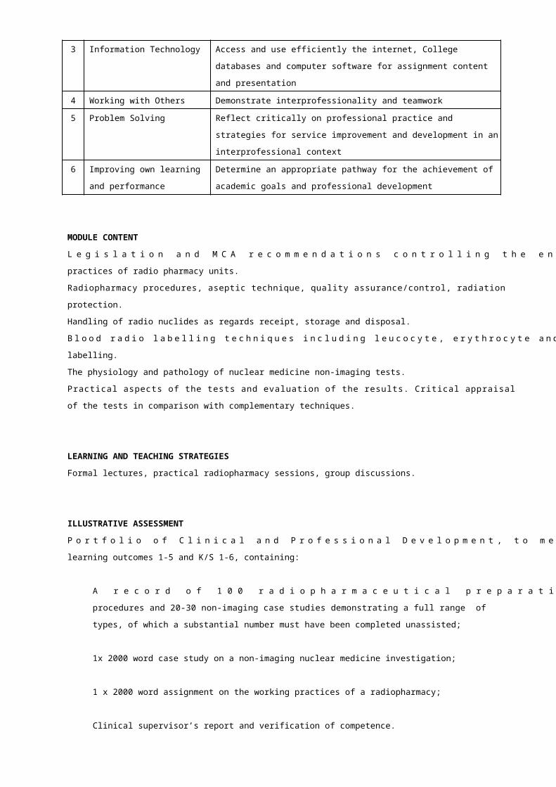
The aims of the module are:
To enable practitioners to transfer knowledge of physical principles of ultrasound imaging safely,
efficiently and appropriately to musculoskeletal ultrasound imaging techniques.
To facilitate development of ability in critical interpretation of musculoskeletal ultrasound images in
varying clinical contexts relevant to the student’s own scope of practice.
To appreciate the role of musculoskeletal ultrasound imaging in practice and research.
LEARNING OUTCOMES
In relation to musculo-skeletal ultrasound, by the end of the module, students should be able to:
1. Demonstrate a thorough knowledge of different ultrasound imaging techniques relevant to
different tissues, pathologies and body sites (K/S 1, 2, 3, 6);
2. Demonstrate an in depth understanding of visual interpretation, perception and orientation
of musculoskeletal ultrasound images as relevant to their area of work/scope of practice (K/S
1, 2, 3, 6);
3. Reflect critically on the ethical and medico-legal implications of musculoskeletal ultrasound
imaging in an interprofessional context (K/S 1, 2, 3, 4, 5, 6);
4. Critically appraise published research on musculoskeletal ultrasound imaging and analyse its
validity for health and social care practice (K/S 1, 2, 3, 4, 6).
KEY SKILLS OUTCOMES
1 Communication Evaluate critically empirical and theoretical literature,
both orally and in writing
2 Application of
Number
Critically interpret data in published research
3 Information
Technology
Access and use efficiently the Internet, College databases and
computer software for assignment content and presentation
4 Working with Others Demonstrate interprofessionality and teamwork when writing
about practice (poster)
5 Problem Solving Reflect critically on ethical and medico-legal aspects of musculo-
skeletal ultrasound in an interprofessional context
6 Improving own
learning and
performance
Determine an appropriate pathway for the achievement of
academic goals and professional development. (formative)

MODULE CONTENT
The appearance of pathology within different tissues when assessed using a range ultrasound
imaging techniques (including aspects of perception, spatial awareness, orientation of images, B-
mode imaging, Doppler imaging and movement during imaging).
A review of the different imaging modalities available for the demonstration of different pathologies
(purpose, cost effectiveness and impact on services and patients as individuals).
The ethics and medico-legal implications of using ultrasound imaging for the assessment and
diagnosis of musculo-skeletal disorders and injuries.
The validity of musculoskeletal ultrasound imaging research for health and social care practice.
LEARNING AND TEACHING STRATEGIES
Key note lectures, seminars, ‘hands-on’ workshops, demonstrations, web-based materials.
ILLUSTRATIVE ASSESSMENT
Poster design (LO 1, 3 and 4) (K/S 1-6) (40%) and presentation (LO 1, 2, 3 and 4) (K/S 1, 2, 3, 5, 6)
(60%) to demonstrate in depth understanding and critical awareness of musculoskeletal ultrasound
imaging as a clinical assessment tool.
(The poster and presentation marking criteria will be in accordance with the Faculty of Health Masters
Level Framework.)
A 500 word minimum reflective account, illustrating key skills development throughout the module.
ILLUSTRATIVE BIBLIOGRAPHY
Adler, R., Sofka, C.M., Positano, R.G. (2004) Atlas of Foot and Ankle Sonography. Lippincott,
Williams and Wilkins: Philadelphia.
Bennett, G.Y., El-Khoury D.L., Stanley, M.D. (2002) Essentials in Musculoskeletal Imaging.
Bradley, M. and O’Donnell P. (2004) Atlas of Musculoskeletal Ultrasound Anatomy . GMM:
London.
Brukner, P. and Khan, K. (2002) Clinical Sports Medicine. McGraw-Hill Publishing Co.
Eisenberg, R.L. (2003) Comprehensive Radiographic Pathology. Mosby.
Fletcher, C.D.M. (Editor) (2004) Pathology and Genetics of Tumours of Soft Tissue and
Bone (World Health Organisation Classification of

Tumours). Oxford University Press.
Fornage, B.D. (Editor) (1995) Musculoskeletal Ultrasound (Clinics in Diagnostic
Ultrasound). Churchill Livingstone: Philadelphia
McNally, E. (2004) Practical Musculoskeletal Ultrasound. Churchill
Livingstone: Philadelphia.
Miller, C. and Freeman, M. (2001) Interprofessional Practice in Health and Social Care.
Kingsley.
Sevenhuijsen, S. (1998) Citizenship and the Ethics of Care. Routledge.
WEBSITES
http://www.bmus.org
http://www.doh.gov.uk/nhs
http://www.podiatricultrasound.com/
http://www.ultrasound.net
JOURNALS
American Journal of Sports Medicine
Journal of Clinical Ultrasound
Journal of Interprofessional Care
Radiography
Skeletal Radiology
The Foot
Module
Title:
Clinical Applications & Management I (Obstetric & Pelvic
Ultrasound)
code: MMIHF4UCP
credit rating: 20 HE4 credits
duration: 5 hours academic support / 100 hours clinical supervision/ 95 hrs independent
study
academic
responsibility:
Gill Dolbear & Hazel Colyer
MODULE AIMS
The aims of the module are:
To ensure the student becomes a skilled, competent, confident practitioner in both obstetric and
pelvic ultrasound.
To enable the student to reflect on and critically evaluate the role of ultrasound in the management of
pregnant and non-pregnant women.
LEARNING OUTCOMES
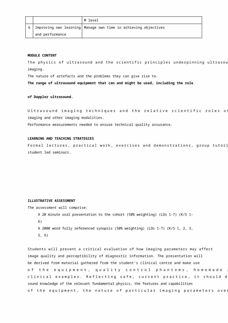
In relation to the pregnant abdomen and female pelvis, by the end of the module, students should be
able to:
1. demonstrate the ability to produce high quality diagnostic images using technique
appropriate to the clinical condition and history, and manipulating the ultrasound controls
effectively;
2. identify normal ultrasound appearances in relation to the relevant anatomy and physiological
processes;
3. demonstrate a thorough knowledge base of the pathology that can affect pregnancy, the
foetus and the female pelvic organs;
4. evaluate common pathological conditions demonstrated on ultrasound imaging, and discuss
the related image patterns and disease mechanisms;
5. demonstrate a critical awareness of the role of ultrasound in the clinical management of the
patient;
6. critically assess the relationship of the ultrasound report to other diagnostic tests, as well as
its impact on patient management;
7. appraise critically the role of other imaging modalities;
8. acknowledge the particular patient care demands of patients of all ages undergoing
ultrasound investigations.
KEY SKILLS OUTCOMES
1 Communication Evaluate critically empirical and theoretical literature in writing
2 Application of Number Critically interpret quantitative data in research and/or audit studies
3 Information Technology Access and use efficiently the internet, College databases and
computer software for assignment content and presentation
4 Working with Others Demonstrate interprofessionality and teamwork
5 Problem Solving Reflect critically on professional practice and strategies for service
improvement and development in an interprofessional context
6 Improving own learning
and performance
Determine an appropriate pathway for the achievement of academic
goals and professional development
MODULE CONTENT
The normal and abnormal anatomy, physiology and pathology of the pregnant abdomen and female
pelvis related to the ultrasound imaging appearances.
The relative roles of ultrasound imaging and other imaging modalities, including plain and contrast
studies, computed tomography, nuclear medicine and magnetic resonance imaging.
Particular patient care and management demands of patients of all ages undergoing ultrasound
examinations, paying particular attention to children and those with fertility or pregnancy
complications.
Ethical and legal constraints to the practice of sonographers in this field of ultrasound imaging.

LEARNING AND TEACHING STRATEGIES
Work based supervision and practice, shadowing activities, clinical meetings and tutorials, expert
lectures, self directed study, web based materials.
ILLUSTRATIVE ASSESSMENT
Portfolio of Clinical and Professional Development, to meet module intended learning outcomes 1-8
and K/S 1-6, containing:
A record of 250 examinations demonstrating that a wide range of obstetric and pelvic examinations
has been carried out. Of these, a substantial number must have been completed unassisted. Unaided
examinations must include a written report;
2 x 750 word case reports to the standard of an academic journal;
1 x 2000 word in depth case study;
Audit and reflective analysis of a random number of interpretations/reports;
Verification of competence in obstetric and pelvic ultrasound evidenced by the clinical supervisor’s
report.
ILLUSTRATIVE BIBLIOGRAPHY
Allan, L.D. (1986), Manual of Foetal Echocardiography, MTP Press
Limited.
Benaceraff. (1998), Ultrasound of Foetal Syndromes, Churchill Livingstone
Bonnar, J. (1998), Recent Advances In Obstetrics and Gynaecology,
Churchill Livingstone
Dewbury, K., Meire, H., Cosgrove, D. (1993), Ultrasound in Obstetrics and Gynaecology, Churchill
Livingstone.
Dodson, M. (1995), Transvaginal Ultrasound 2 nd Edition, Churchill
Livingstone
England, M.A. (1996), Life Before Birth 2nd Edition, Mosby-Wolfe.
Fleischer, A.C., Kepple, D.M. (1992), Transvaginal Sonography A Clinical Atlas, J.B.Lippincott
Company.
Impey, L. (1999), Obstetrics and Gynaecology Blackwell Science
Limited.
Johnston, P.G.B. (1998), The Newborn Child Churchill Livingstone
Nicolaides, K.H., Sebire, N.J., Snijders, J.M. (1999), The 11-14 week scan. The diagnosis of foetal
abnormalities, Parthenon.
Nyberg, D.A., Mahony, B.S., Pretorius, D.H. (1990), Diagnostic Ultrasound of Foetal Anomalies:
Text and Atlas Mosby.
Rumack, C.M., Wilson, S.R., Charboneau, J.W. (1998), Diagnostic Ultrasound 2nd Edition, Mosby.
Sanders, R.C., Smith, N. (1997), Clinical Sonography A Practical Guide, Lippincott,

Williams & Wilkins.
Sanders, R.C. (1996), Structural Foetal Abnormalities The Total Picture,
Mosby.
WEBSITES
www.ultrasound.net
www.ectopic.org.uk
w-cpc.org/fetal.html
www.genecare.com/amnio.html
JOURNALS
American Journal of Obstetrics and Gynaecology
British Journal of Obstetrics and Gynaecology
Gynaecologic Oncology
Journal of Clinical Ultrasound
Journal of Obstetrics and Gynaecology
British Medical Journal
Fertility and Sterility
Human Reproduction
Obstetric and Gynaecology Survey
Module
Title:
Clinical Applications & Management II
(Abdominal & Small Parts Ultrasound)
code: MMIHF4UCS
credit rating: 20 HE4 credits
duration: 5 hours academic support / 100 hours clinical supervision/ 95 hrs
independent study
academic
responsibility:
Gill Dolbear & Hazel Colyer
MODULE AIMS
The aims of the module are:
To ensure the student becomes a skilled, competent, confident practitioner abdominal ultrasound
To enable the student to carry out some small parts examinations competently
To enable the student to reflect on and critically evaluate the role of ultrasound in the management of
patients presenting for abdominal or small parts examinations.

LEARNING OUTCOMES
In relation to the abdomen and nominated small parts examinations, by the end of the module,
students should be able to:
1. demonstrate the ability to produce high quality diagnostic images using technique
appropriate to the clinical condition and history, and manipulating the ultrasound controls
effectively;
2. identify normal ultrasound appearances in relation to relevant anatomy and physiological
processes;
3. demonstrate a thorough knowledge base of the pathology that can affect the abdomen, the
breast, the thyroid gland and the testes;
4. evaluate common pathological conditions and discuss the related image patterns and
disease mechanisms;
5. demonstrate a critical awareness of the role of ultrasound in the clinical management of the
patient;
6. critically assess the relationship of the ultrasound report to other diagnostic tests, as well as
its impact on patient management;
7. appraise critically the role of other imaging modalities;
8. acknowledge the particular patient care demands of patients of all ages undergoing
ultrasound investigations.
KEY SKILLS OUTCOMES
1 Communication Evaluate critically empirical and theoretical literature in writing
2 Application of Number Critically interpret quantitative data in research and/or audit studies
3 Information Technology Access and use efficiently the internet, College databases and computer
software for assignment content and presentation
4 Working with Others Demonstrate interprofessionality and teamwork
5 Problem Solving Reflect critically on professional practice and strategies for service
improvement and development in an interprofessional context
6 Improving own learning
and performance
Determine an appropriate pathway for the achievement of academic
goals and professional development
MODULE CONTENT
The normal and abnormal anatomy, physiology and pathology of the abdomen and nominated small
parts, related to the ultrasound imaging appearances.
The relative roles of ultrasound imaging and other imaging modalities, including plain and contrast
studies, computed tomography, nuclear medicine and magnetic resonance imaging.
Particular patient care and management demands of patients of all ages undergoing ultrasound
examinations.
Ethical and legal constraints to the practice of sonographers in this field of ultrasound imaging.

LEARNING AND TEACHING STRATEGIES
Work based supervision and practice, shadowing activities, clinical meetings and tutorials, expert
lectures, self directed study, web based materials.
ILLUSTRATIVE ASSESSMENT
Portfolio of Clinical and Professional Development, to meet module intended learning outcomes 1-8
and K/S 1-6, containing:
A record of 250 examinations demonstrating that a wide range of abdominal examinations
and some nominated small part examinations has been carried out. A substantial number
must have been completed unassisted. Unaided examinations must include a written report;
2 x 750 word case reports to the standard of an academic journal;
1 x 2000 word in depth case study;
Audit and reflective analysis of a random number of interpretations/reports;
Verification of competence in abdominal and small parts ultrasound evidenced by the clinical
supervisor’s report.
ILLUSTRATIVE BIBLIOGRAPHY
Bates, J.A. (1999), Abdominal Ultrasound How, Why and When, Churchill
Livingstone.
Bissett, R.A.L., Khan, A.N. (1991), Differential Diagnosis in Abdominal Ultrasound, 2nd
Edition,
Bailliere Tindall.
Evans, R., Ahuja, A. (1999), Practical Head and Neck Ultrasound, Greenwich Medical
Media.
Hickey, J., Goldberg, F. (1999), Ultrasound Review of the Abdomen, Male Pelvis & Small
Parts, Lippincott.
Lees, W.R., Lyons, E.A. (1996), Invasive Ultrasound, Martin Dunitz
Rumack, C.M., Wilson, S.R., Charboneau, J.W. (1998), Diagnostic Ultrasound 2nd Edition, Mosby.
Sanders, R., Smith, N. (1997), Clinical Sonography a Practical Guide, Lippincott,
Williams & Wilkins
WEBSITES
www.ultrasound.net
www.limit.ac.uk/pancreas.htm
www.brisbio.ac.uk/roads/subject-listing/orchitis.html
www.prostatitis.org

JOURNALS
American Journal of Radiology British Medical Journal
British Journal of Radiologists British Journal of Surgery
British Journal of Urology Journal of Ultrasound Medicine
Lancet Paediatrics
Radiology Surgery

Module
Title:
Clinical Applications & Management III (Musculo-skeletal
Ultrasound)
code: MMIHF4UCM
credit rating: 20 HE4 credits
duration: 5 hours academic support / 100 hours clinical supervision/ 95 hrs independent
study
academic
responsibility:
Gill Dolbear & Peter Milburn
MODULE AIMS
The aims of the module are:
To ensure the student becomes a skilled, competent, confident practitioner in musculo-skeletal
ultrasound.
To enable the student to reflect on and critically evaluate the role of ultrasound in the management of
patients suffering from musculo-skeletal disease or injury.
LEARNING OUTCOMES
In relation to musculo-skeletal ultrasound, by the end of the module, students should be able to:
1. demonstrate the ability to produce high quality diagnostic images using technique
appropriate to the clinical condition and history, and manipulating the ultrasound controls
effectively;
2. identify normal ultrasound appearances in relation to relevant anatomy and physiological
processes;
3. demonstrate a thorough knowledge base of the pathology and trauma that may be present;
4. evaluate common traumatic and pathological conditions and discuss the related image
patterns and disease or trauma mechanisms;
5. demonstrate a critical awareness of the role of ultrasound in the clinical management of the
patient;
6. critically assess the relationship of the ultrasound report to other diagnostic tests, as well as
its impact on patient management;
7. appraise critically the role of other imaging modalities;
8. acknowledge the particular patient care demands of patients of all ages undergoing
ultrasound investigations.
KEY SKILLS OUTCOMES
1 Communication Evaluate critically empirical and theoretical literature in writing

2 Application of Number Critically interpret quantitative data in research and/or audit studies
3 Information Technology Access and use efficiently the internet, College databases and computer
software for assignment content and presentation
4 Working with Others Demonstrate interprofessionality and teamwork
5 Problem Solving Reflect critically on professional practice and strategies for service
improvement and development in an interprofessional context
6 Improving own learning
and performance
Determine an appropriate pathway for the achievement of academic
goals and professional development
MODULE CONTENT
The normal and abnormal anatomy, physiology and pathology of the musculo-skeletal system,
related to the ultrasound imaging appearances.
The relative roles of ultrasound imaging and other imaging modalities, including plain and contrast
studies, computed tomography, nuclear medicine and magnetic resonance imaging.
Particular patient care and management demands of patients of all ages undergoing ultrasound
examinations.
Ethical and legal constraints to the practice of sonographers in this field of ultrasound imaging.
LEARNING AND TEACHING STRATEGIES
Work based supervision and practice, shadowing activities, clinical meetings and tutorials, expert
lectures, self directed study, web based materials.
ILLUSTRATIVE ASSESSMENT
Portfolio of Clinical and Professional Development, to meet module intended learning outcomes 1-8
and K/S 1-6, containing:
A record of 250 examinations demonstrating that a wide range of musculo-skeletal
examinations has been carried out, of which a substantial number must have been completed
unassisted. Unaided examinations must include a written report;
2 x 750 word case reports to the standard of an academic journal;
1 x 2000 word in depth case study;
Audit and reflective analysis of a random number of interpretations/reports;
Verification of competence in musculo-skeletal ultrasound evidenced by the clinical
supervisor’s report.
ILLUSTRATIVE BIBLIOGRAPHY

Chhem, R., Cardinal, E. (1998), Guidelines and Gamuts in Musculoskeletal Ultrasound, John Wiley
and Sons
Enzinger, F.M., Weiss, S.W. (1995), Soft Tissue Tumours 3rd Edition, Mosby.
Fornage, B.D. (1989), Ultrasonography of muscles and tendons: Examination technique
and atlas of normal anatomy of the extremities Springer-Verlag
Gibbon, W.W. (1996), Musculoskeletal Ultrasound: The essentials, Oxford University Press
Harries, M., Williams, C., Stanish, W.D., Micheli, L.J., (1994), Oxford Textbook of Sports Medicine,
Oxford University Press.
Rumack, C.M., Wilson, S.R., Charboneau, J.W. (1998), Diagnostic Ultrasound 2nd Edition, Mosby.
WEBSITES
www.ultrasound.net
www.aium.org/consumer.htm
JOURNALS
American Journal of Neuroradiology
American Journal of Radiology
British Journal of Surgery
Journal of Bone and Joint Surgery
Journal of Clinical Ultrasound
Journal of Paediatric Orthopaedics
Journal of Neurosurgery
Lancet
Radiology
Skeletal Radiology
Surgery
Module
Title:
Clinical Applications & Management IV (Peripheral Vascular
Ultrasound)
code: MMIHF4UCV
credit rating: 20 HE4 credits
duration: 5 hours academic support / 100 hours clinical supervision/ 95 hrs independent
study
academic
responsibility:
Gill Dolbear & Peter Milburn
MODULE AIMS

The aims of the module are:
To ensure the student becomes a skilled, competent, confident practitioner in peripheral vascular
ultrasound.
To enable the student to reflect on and critically evaluate the role of ultrasound in the management of
patients with peripheral vascular disease.
LEARNING OUTCOMES
In relation to the peripheral vascular system, by the end of the module, students should be able to:
1. demonstrate the ability to produce high quality diagnostic images using technique
appropriate to the clinical condition and history, and manipulating the ultrasound controls
effectively;
2. identify normal ultrasound appearances in relation to relevant anatomy and physiological
processes;
3. demonstrate a thorough knowledge base of the pathology that can affect the peripheral
vascular system;
4. evaluate common pathological conditions and discuss the related image patterns and
disease mechanisms;
5. demonstrate a critical awareness of the role of ultrasound in the clinical management of the
patient;
6. critically assess the relationship of the ultrasound report to other diagnostic tests, as well as
its impact on patient management;
7. appraise critically the role of other imaging modalities;
8. acknowledge the particular patient care demands of patients of all ages undergoing
ultrasound investigations.
KEY SKILLS OUTCOMES
1 Communication Evaluate critically empirical and theoretical literature in writing
2 Application of Number Critically interpret quantitative data in research and/or audit studies
3 Information Technology Access and use efficiently the internet, College databases and computer
software for assignment content and presentation
4 Working with Others Demonstrate interprofessionality and teamwork
5 Problem Solving Reflect critically on professional practice and strategies for service
improvement and development in an interprofessional context
6 Improving own learning
and performance
Determine an appropriate pathway for the achievement of academic
goals and professional development

MODULE CONTENT
The normal and abnormal anatomy, physiology and pathology of the peripheral vascular system,
related this to the ultrasound imaging appearances.
The relative roles of ultrasound imaging and other imaging modalities, including plain and contrast
studies, computed tomography, nuclear medicine and magnetic resonance imaging.
Particular patient care and management demands of patients of all ages undergoing ultrasound
examinations.
Ethical and legal constraints to the practice of sonographers in this field of ultrasound imaging.
LEARNING AND TEACHING STRATEGIES
Work based supervision and practice, shadowing activities, clinical meetings and tutorials, expert
lectures, self directed study, web based materials.
ILLUSTRATIVE ASSESSMENT
Portfolio of Clinical and Professional Development, to meet module intended learning outcomes 1-8
and K/S 1-6, containing:
A record of 250 examinations demonstrating that a wide range of Peripheral Vascular
examinations has been carried out, of which a substantial number must have been completed
unassisted. Unaided examinations must include a written report;
2 x 750 word case reports to the standard of an academic journal;
1 x 2000 word in depth case study;
Audit and reflective analysis of a random number of interpretations/reports;
Verification of competence in peripheral vascular ultrasound evidenced by the clinical
supervisor’s report.
ILLUSTRATIVE BIBLIOGRAPHY
Jamieson, C.W., Yao, J.S.T.(1994), Vascular Surgery 5th Edition, Chapman and Hall
Medical.
Kim, D., Orron, D.E. (1992), Peripheral Vascular Imaging and Intervention,
Mosby.
Krebs, C.A. (1998), Ultrasound Atlas of Vascular Diseases, Appleton and
Lange.
Rumack, C.M., Wilson, Charboneau, J.W. (1998), S.R., Diagnostic Ultrasound 2nd Edition, Mosby.
Thrush, A., Hartshorne, T., (1999), Peripheral Vascular Ultrasound How, Why and When
Churchill Livingstone.
Zwiebel, W.J. (2000), Introduction to Vascular Ultrasonography 4th Edition,
W.B. Saunders.
WEBSITES
www.ultrasound.net
www.aium.org/consumer.htm
JOURNALS
American Journal of Medicine
American Journal of Radiology
British Journal of Surgery
Circulation
Clinical Radiology
Journal of Clinical Ultrasound
Journal of Ultrasound Medicine
Lancet
Medicine
Radiology
Stroke
Surgery
Module
Title:
Clinical Applications & Management V
(Echocardiography)
code: MMIHF4UCE
credit rating: 20 HE4 credits
duration: 5 hours academic support / 100 hours clinical supervision/ 95 hrs
independent study
academic
responsibility:
Gill Dolbear & Peter Milburn
MODULE AIMS
The aims of the module are:
To ensure the student becomes a skilled, competent, confident practitioner in adult
echocardiography.

To enable the student to reflect on and critically evaluate the role of ultrasound in the management of
patients with heart disease.
LEARNING OUTCOMES
In relation to the heart, by the end of the module, students should be able to:
1. demonstrate the ability to produce high quality diagnostic images using technique
appropriate to the clinical condition and history, and manipulating the ultrasound controls
effectively;
2. identify normal ultrasound appearances in relation to relevant anatomy and physiological
processes;
3. demonstrate a thorough knowledge base of the pathology that can affect the heart;
4. evaluate common pathological conditions and discuss the related image patterns and
disease mechanisms;
5. demonstrate a critical awareness of the role of ultrasound in the clinical management of the
patient;
6. critically assess the relationship of the ultrasound report to other diagnostic tests, as well as
its impact on patient management;
7. appraise critically the role of other imaging modalities;
8. acknowledge the particular patient care demands of patients undergoing ultrasound
investigations.
KEY SKILLS OUTCOMES
1 Communication Evaluate critically empirical and theoretical literature in writing
2 Application of Number Critically interpret quantitative data in research and/or audit studies
3 Information Technology Access and use efficiently the internet, College databases and computer
software for assignment content and presentation
4 Working with Others Demonstrate interprofessionality and teamwork
5 Problem Solving Reflect critically on professional practice and strategies for service
improvement and development in an interprofessional context
6 Improving own learning
and performance
Determine an appropriate pathway for the achievement of academic
goals and professional development
INDICATIVE MODULE CONTENT
The normal and abnormal anatomy, physiology and pathology of the adult heart related to the
ultrasound imaging appearances.
The relative roles of ultrasound imaging and other imaging modalities, including plain and contrast
studies, computed tomography, nuclear medicine and magnetic resonance imaging.
Particular patient care and management demands of patients of undergoing ultrasound
examinations.
Ethical and legal constraints to the practice of sonographers in this field of ultrasound imaging.
LEARNING AND TEACHING STRATEGIES
Work based supervision and practice, shadowing activities, clinical meetings and tutorials, expert
lectures, self directed study, web based materials.
ILLUSTRATIVE ASSESSMENT
Portfolio of Clinical and Professional Development, to meet module intended learning outcomes 1-8
and K/S 1-6, containing:
A record of 250 examinations demonstrating that a wide range of Echocardiography
examinations has been carried out, of which a substantial number must have been completed
unassisted. Unaided examinations must include a written report;
2 x 750 word case reports to the standard of an academic journal;
1 x 2000 word in depth case study;
(video/dynamic material is essential in each of the case studies.)
Audit and reflective analysis of a random number of interpretations/reports;
Verification of competence in echocardiography evidenced by the clinical supervisor’s report.
ILLUSTRATIVE BIBLIOGRAPHY
Boudreau Conover, M. (1996), Understanding Electrocardiography, 7th Edition,
Mosby.
Braunwald, E. (1997), Heart Disease Volume H2 5th Edition, B. Saunders
Company.
Chambers, J. (1995), Clinical Echocardiography British Medical
Journal.
Haslett, C., Chilvers, E.R., Hunter, J.A.A., Boon, N.A. (1999), Davidson’s Principles and Practice of
Medicine
18th Edition, Churchill Livingstone
Schmailzl, K.J.G., Ormerod, O. (1994), Ultrasound in Cardiology Iowa State,
University Press

WEBSITES
www.ultrasound.net
www.aium.org/consumer/obus.htm
JOURNALS
American Journal of Medicine
British Medical Journal
Circulation
Heart
Journal of Ultrasound Medicine
Journal of Clinical Ultrasound
Lancet
Medicine
Stroke

Module
Title:
Open Module (Work-based learning)
code: MZZHF4OP1
level: HE4
duration: 5 hours academic support plus 195 hrs independent work based practice
and/or private study
academic
responsibility:
Appropriate academic supervisor and professional/clinical supervisor
MODULE AIMS
The aim of an open module is to enable the accreditation of a negotiated profession development
experience conducted outside the College, assisting the practitioner in their autonomous self
development through a critical, systematic, circumscribed enquiry into an aspect of professional
development which includes the opportunity for considered reflection and self evaluation.
LEARNING OUTCOMES
By the end of the module and in relation to the specific topic or professional role development,
students should be able to:
1. organise documentation to convey clearly the processes undertaken and what has been
achieved, including their own learning and competence development where specified;
2. critically appraise contemporary relevant literature;
3. analyse the focus and processes of development through insightful recording of actions and
reactions in the practice situation;
4. synthesise theoretical perspectives in relation to the particular professional practice;
5. demonstrate rigorous self evaluation of the process and outcomes of the development
experience.
In addition, where competences are being developed and assessed, students should be able
to:
6. demonstrate the achievement of specified competences in the work place situation
KEY SKILLS OUTCOMES
Key Skills
1 Communication Evaluate critically empirical and
theoretical literature in writing
2 Application of Number Critically interpret data in research
studies
3 Information Technology Access and use efficiently the
internet, College databases and
computer software for assignment
content and presentation
4 Working with Others Demonstrate interprofessionality

and teamwork
5 Problem Solving Reflect critically on professional
practice and strategies for service
improvement and development in
an interprofessional context
6 Improving own Learning and Performance Determine an appropriate
pathway for the achievement of
academic goals and professional
development
MODULE CONTENT
The Learning Agreement
A learning agreement is formulated between the student, the academic institution and the work
place. The agreement outlines the background to the study, the aim of the module, how intended
learning outcomes will be met, the mode of study and the timescale for completion. It is signed by all
relevant parties.
THE ROLE OF THE COLLEGE
The College undertakes to provide academic support, relevant guidance materials and access to
resources. The Programme Director will facilitate the learning agreement and registration.
An Academic Supervisor will be appointed having the following duties:
1. to provide clear guidance on professional research and develop issues in the context of the
topic chosen;
2. to engage in approximately three one-to-one tutorials;
3. to facilitate links with other sources of information and expertise;
4. to provide detailed feedback on interim material submitted;
5. to act as the first marker of the final submission.
THE ROLE OF THE WORKPLACE
Students must ensure that they have the co-operation of their Line Manager prior to the commencing
study for an open module. The nature of the module should be such that the Line Manager can
appreciate the potential benefits to service provision and facilitate the enquiry without detriment to
service delivery.
Where competence development is part of the learning outcomes specified in the learning
agreement, the Clinical Supervisor will be required to verify evidence of the student’s professional
competence as part of their final Clinical Supervisors’ report.
LEARNING AND TEACHING STRATEGIES
These are specified in the individual learning agreement.
ASSESSMENT
The enquiry report, portfolio or essay will comprise 5000 words equivalent undertaken in one of the
following four modes over a period of six to nine months:
Work Based (without practice competence development):
1. Empirical Study with Report
This comprises a research based study of a practice situation. The accompanying report will
encompass literature review, justification of method, reflection and recommendations.
2. Action Research and Portfolio
A professional action is taken and a systematic evaluation of that action is undertaken. All the
relevant documentation of the evaluation and reflection process are included in a portfolio; e.g.
initial statement, reports on group discussions, support from manager, enquiry report, critical
analysis and reflection.
Work Based (with practice competence development):
3. Professional Role Development
The role to be developed is identified and a programme of professional practice is instigated,
underpinned by the development of the appropriate knowledge/value base. The nature of the
competence is generally at a higher level or advanced practice in line with published
professional standards. Over the study period, a Portfolio of Professional Practice Development
containing formative and summative relevant material forms the learning and assessment tool.
Specific elements for assessment include a record of practice, in-depth case studies, critical,
reflective evaluation and a practice supervisor’s verification report.
Literature Based:
4. Extended essay
An in depth, critical analysis by extended essay of the body of literature relevant to a specific
work place situation in order to elaborate and develop the arguments. The essay explores all
relevant perspectives, demonstrates personal development and proposes a practical response,
applicable to the workplace.
ILLUSTRATIVE BIBLIOGRAPHY/JOURNALS/WEBSITES
Appropriate to the students area of professional practice.
Module
Title:
Developing a Research Proposal
code: MZZHF4DRP
level: HE4
duration: 40 hours contact (including 5 hours of tutorials), 160 hours private
independent study
academic
responsibility:
Professor Stephen Clift
MODULE AIMS
The module provides students with practical guidance on the development of a research proposal
and enables them to develop practical skills in the use of common methods of data gathering and
analysis.
LEARNING OUTCOMES
By the end of the module students should be able to:
1. demonstrate ability to systematic search appropriate databases and produce a
comprehensive and critical review of recent and relevant literature related to their proposed
research (KS 1, 2, 3);
2. produce clear and specific aims and objectives for their project which are both grounded in
appropriate literature and related to professional practice (KS 1, 4, 5, 6);
3. demonstrate a critical understanding of research design and methods in health research and
justify their decisions regarding the approach to be adopted (KS 1, 2, 5);
4. demonstrate a critical understanding of the basic principles of quantitative and qualitative
analysis and justify their decisions regarding the analytic methods to be used (KS 1, 2, 5);
5. demonstrate a sound understanding of basic ethical principles governing health research,
taking account of current guidance from local ethics committees (KS 1, 4, 5, 6).
KEY SKILLS OUTCOMES
Key Skills
1 Communication Present in writing, a clear, succinct and evidence-based
rationale for the intended project, which demonstrates a critical
appraisal of a wide variety of information.
2 Application of Number Demonstrate an understanding of the role and limitations of
statistical techniques in health research. Be able to competently
interpret tables of data in research papers.
3 Information Technology Demonstrate competent and efficient use of the WWW, CLINIC
and other electronic databases.
4 Working with others Seek advice from peers, managers and colleagues associated
with the local R&D department and ethics committee, in the

development and refinement of research proposals.
5 Problem solving Formulate a plan of action for a project which will serve to
provide answers or solutions to professionally focused research
problems.
6 Improving own learning
and performance
Demonstrate self-reflection, self-motivation, advanced skills of
independent learning, and the ability to identify and seek
appropriate guidance for improving performance.
MODULE CONTENT
Useful starting points for identifying a research topic. Strategies for finding research information
relevant to proposed topic. Practical guidance in undertaking a systematic literature review. Use of
key research databases to locate literature. Approaches to the critical analysis of research. Practical
aspects of quantitative and qualitative research designs, specific methods of data collection and a
consideration of ethical principles in research. The importance of working within practical constraints,
resource limitations and a clear timetable.
LEARNING AND TEACHING STRATEGIES
Through lectures, discussions and group work, emphasis will be given to the practical issues involved
in using a range of methods currently employed in research in health and social care. Each student
will be expected to identify a recent research report relevant to their area of research interest for
critical discussion by the group within sessions.
ILLUSTRATIVE ASSESSMENT
A detailed research proposal of 4000 words encompassing rationale, a critical review of key literature,
research question(s)/hypothesis to be tested, and justified research design (LO 1-6) (K/S 1-6) (100%)
A 500 word minimum reflective account, illustrating key skills development throughout the module,
to contribute to the portfolio of skills development.
Research proposals must be assessed as satisfactory before students are permitted to proceed
with submission to the local research and development and ethics committees, if required, and the
undertaking of fieldwork.
ILLUSTRATIVE BIBLIOGRAPHY
Bowling, A. (1997) Research Methods in Health: Investigating Health and Health Services. Open
University Press.

Bryman, A. and Cramer, D. (1999) Quantitative Data Analysis for Social Scientists. London:
Routledge.
Dey, I. (1999) Qualitative Data Analysis: a user-friendly guide for social scientists. London: Routledge.
Dochartaigh, N.O. (2001) The Internet Research Handbook: A Practical Guide for Students and
Researchers in the Social Sciences. Sage.
Flick, U. (1998) An Introduction to Qualitative Research. London: Sage.
Gomm, R., Needham, G. and Bullman, A. (eds.)(2000) Evaluating Research in Health and Social Care.
Sage and Open University.
Grbich, C. (1999) Qualitative Research in Health: an introduction. London: Sage.
IUHPE (1999) The Evidence of Health Promotion Effectiveness. Brussels: European
Commission/IUHPE
Mason, J. (1996) Qualitative Researching. London: Sage.
May, T. & Williams, M. (eds), (1998). Knowing the Social World. Buckingham, Open University.
Robson, C. (1993) Real World Research: A Resource for Social Scientists and Practitioner-
Researchers. Oxford: Blackwell.
Scolari software (2000) Methodologist’s Toolchest. London: Sage.
Scolari software (2000) WinMax. London: Sage.
Tones, K. and Tilford, S. (2001)(3rd Edition) Health Promotion: Effectiveness, Efficiency and Equity.
Cheltenham: Nelson Thornes.
JOURNALS
Evidence based Healthcare. Published by Harcourt Publishers Ltd
Evidence Based Medicine. Published by American College of Physicians-American Society of Internal
Medicine
Evidence Based Mental Health. Published by HighWire Press
Evidence Based Nursing. Published by BMJ Publishing Group
WEBSITES
Centre for Reviews and Dissemination, University of York http://agatha.york.ac.uk/
CLINIC http://clinic.cant.ac.uk/
Health Development Agency Evidence Base http://www.hda.nhs.uk/evidence/
King’s Fund http://www.kingsfund.org.uk/
National Electronic Library for Health http://www.nelh.nhs.uk/cochrane.asp
National Statistics Office
http://www.statistics.gov.uk/cci/nugget.asp?id=313
South East Public Health Observatory http://www.sepho.org.uk/

Module
Title:
Undertaking Systematic Reviews
code: MZZHF4USR
level: HE4
duration: 40 hours taught plus 160 hours private independent study
academic
responsibility:
Douglas MacInnes
MODULE AIM
The aim of the module is to enable students to develop answerable questions in evaluating the
effectiveness of health care practices. The module will also focus on teaching the skills that are
needed to be able to conduct systematic reviews to address these questions. The module is also a
pre-requisite for those students wishing to undertake a systematic review for their practice
development project.
LEARNING OUTCOMES
By the end of the module students should be able to:
1. Understand the principles and methods underpinning systematic reviews (KS 1,2,3,5);
2. critically evaluate rationale/criteria for considering specific studies for inclusion in a review
(KS 1,2,5,6);
3. devise search procedures to locate relevant information relating to health care practices (KS
1,3,4,6);
4. evaluate critically the quality of studies(KS 1,2,3,4,6);
5. analyse the statistical meta-analysis procedures which determine the overall statistical
evidence of particular interventions/procedures(KS 2,5,6);
6. be aware of the methods of effectively disseminating the results of the systematic review (KS
1,4,5,6)
KEY SKILLS OUTCOMES
Key Skills
1 Communication Evaluate critically the systematic review and meta analyses
literature, in writing
2 Application of Number Analyse the statistical meta analyses and evaluate the statistical
data contained within the systematic review.
3 Information Technology Use extensively the internet, college databases, and search
engines to gain access to the relevant information for the
assignment; use of computer software to organise their
assignment content and presentation; and use of statistical
computer software to help with statistical analysis.
4 Working with others Show awareness of the implications of the findings of the review

for professional and inter professional practice
5 Problem solving Critically evaluate the efficacy of procedures used in
undertaking the systematic review and the confidence that
could be placed in the findings.
6 Improving own learning
and performance
Determine an appropriate pathway for the achievement of
academic goals and professional development (Formative)
MODULE CONTENT
The distinction between and uses of systematic reviews, overviews and meta analyses. Procedures to
develop a clear research question, devise a study protocol, cost the project and determine the length
of time need to complete the project.
Which sources to include and exclude from a study.
Developing search strategies to ensure that the review is comprehensive in its range of bibliographic
sources.
Methods to review the scope of the literature and ways of modifying the search, with examination of
the specialist tools that can be utilised to help with this process. Specific tools used to collect and
extract data.
Different approaches to appraising the quality of the studies collected for a review. Statistical meta-
analysis procedures which can be undertaken to statistically analyse data from a number of studies in
order to synthesise the results.
Key features of presenting and disseminating the results of a systematic review to ensure the target
audience is given clear and concise information relating to the topic under review.
LEARNING AND TEACHING STRATEGIES
The module will include a range of different strategies including formal lectures, group discussions,
workshops, formative seminar presentations, practical exercises and individual tutorials.
ILLUSTRATIVE ASSESSMENT
The module is assessed by a 4000 word assignment to meet the learning outcomes.
The assignment will develop a proposal to undertake a systematic review. This would require the
student to critically discuss the following issues and design specific procedures for the following areas
of the review.
1. Formulation of the review question
2. Definition of the inclusion and exclusion criteria
3. Development of search procedures (relating to obtaining research evidence)
4. Development of selection procedures (to evaluate the eligibility criteria of the studies).
5. Development of validity assessment (to evaluate the methodological quality of the studies)
6. Development of data extraction form (to detail the process for extracting all of the relevant

data from each study).
7. An overview of how the results would be analyzed and presented.
(KS 1-6), (LO 1-6)
A 500 word minimum reflective account, illustrating key skills development throughout the module,
to contribute to the portfolio of skills development.
Research proposals must be assessed as satisfactory before students are permitted to proceed with
submission to the local research and development and ethics committees, if required, and the
undertaking of fieldwork.
ILLUSTRATIVE BIBLIOGRAPHY
Chalmers. I. & Altman. D. (eds) (1996) Systematic reviews . BMJ Publishing.
Cook. D., Mulrow., C. & Haynes. R. (1997) Systematic reviews: synthesis of best evidence for clinical
decisions. Annals of Internal Medicine 126(5): 376-80.
Cooper. H., Carlisle. C., Watkins. C., & Gibbs. T. (2000) Using qualitative methods for conducting a
systematic review. Nurse Researcher 8: 28–38
Crombie. I. (1996) The Pocket Guide to Critical Appraisal. BMJ Publishing.
Eggars. M., Davey Smith. G., & Altman. D. (2001) Systematic Reviews in Health Care: Meta Analysis
in Context.
BMJ Books.
Entwistle. V., Sowden. A., & Watt. I. (1998) Evaluating interventions to promote patient involvement
in decision making: by what criteria should effectiveness be judged? Journal of Health Service
Research Policy 3: 100-7
Meade. M. & Richardson. S. (1997) Selecting and appraising studies for a systematic review. Annals
of Internal Medicine 127(7): 531-7
Milne. R. & Chambers. L. (1993) Assessing the scientific quality of review articles. Journal of
Epidemiological Community Health 47: 169-160.
Muir Gray, J. (1997) Evidence-Based Healthcare. Churchill Livingstone.
Mulrow. C. (1994) Rationale for systematic reviews. British Medical Journal 409: 597 –99.
Mulrow, C. & Oxman, A. (eds) (1997) Cochrane Collaboration Handbook. In; The Cochrane Library
(The Cochrane Collaboration). Update Software, Issue 4, Oxford
NHS Centre for Reviews and Dissemination (1996). Undertaking systematic reviews of research
effectiveness. CRD Report No. 4. York, York publishing services Ltd.
Sackett. D., Rosenberg. W., Muir Gray. J., Haynes. & Richardson. W. (1996) Evidence based medicine:
what it is and what it isn’t. British Medical Journal 312: 71–72
Sutton. A., Jones. D., Abrams. K., Sheldon. T., & Song. F. (1999) Systematic reviews and meta-
analysis: a structured review of the methodological literature. Journal of Health Services Research
Policy 4: 49–55.

JOURNALS
Evidence based Healthcare. Published by Harcourt Publishers Ltd
Evidence Based Medicine . Published by American College of Physicians-American Society of Internal
Medicine
Evidence Based Mental health. Published by HighWire Press
Evidence Based Nursing. Published by BMJ Publishing Group
WEBSITES
CASP (The Critical Appraisal Skills Programme) - http://www.phru.org.uk
Centre for Evidence Based Child health - http://www.ich.bpmf.ac.uk/ebm/ebm.htm
Centre for Evidence Based Medicine - http://www.cebm.jr2.ox.ac.uk/
Cochrane Database - http://www.cochrane.org/
NHS Centre for Reviews and Dissemination- http://www.york.ac.uk

Appendix 1
KEY TRANSFERABLE SKILLS MATRIX
KEY:
M1= Orientation module
M2= Science and Technology modules
M3= Clinical Applications modules
M4= Open modules
M5= Developing a Research Proposal module
M6= Research Dissertation
Key Transferable
Skill
Outcome Outcome Description M
1
M
2
M
3
M
4
M
5
M
6
Communication 1 Evaluate critically empirical and
theoretical literature in writing
√ √
2 Evaluate critically empirical and
theoretical literature, both orally
and in writing
√ √
3 Present, both orally and in writing, a
clear, succinct and evidence based
rationale for the intended project
which demonstrates a critical
appraisal of a wide variety of
information
√
4 Communicate research findings
through analysis and interpretation
of data
√
Application of
number
5 Critically interpret data in a
research study (option in oral
presentation)
√
6 Critically interpret data in research
studies
√ √
7 Critically interpret quantitative data
in research and/or audit studies
√
8 Have an understanding of the role
and limitations of statistical
techniques in health research. Be
able to competently interpret tables
of data in research papers
√
9 Critically interpret data and
undertake appropriate statistical
analysis in quantitative research
designs
√
Information
Technology
10 Access and use efficiently the
internet, College databases and
computer software for assignment
content and presentation
√ √ √ √ √
11 Competent and efficient use of the
WWW, CLINIC and other electronic
databases. Ability to access and
use at least one computer package
for statistical and for textual
analysis
√
Working with
Others
12 Demonstrate interprofessionality
and teamwork when writing about √

practice (situational analysis)
13 Relate and interact effectively with
individuals and groups
√
14 Demonstrate interprofessionality
and teamwork
√ √ √
15 Actively contribute towards peer
support and shared learning in the
development and refinement of
research proposals. Ability to offer
help and constructive criticism
√
Problem Solving 16 Use relevant information sources.
Identify and solve problems
associated with study skills and
assessment at M level
√
17 Reflect critically on professional
practice and strategies for service
improvement and development in
an interprofessional context
√ √ √
18 Ability to formulate a plan of action
which will serve to provide answers
or solutions to professionally
focused research problems
√
19 Reflect critically on research
findings for professional practice
and strategies for service
improvement and development in
an interprofessional context
√
Improving own
Learning and
Performance
20 Manage own time in achieving
objectives
√
21 Determine an appropriate pathway
for the achievement of academic
goals and professional
development
√ √ √
22 Demonstrate self reflection, self
motivation, advanced skills of
independent learning and the
ability to identify and seek
appropriate guidance for improving
performance
√
23 Plan and manage the research
process for the achievement of √

academic and professional
development goals

Appendix 2
Validated Modules within the MSc Interprofessional Health and Social Care
MZZHF4CCP Orientation
MZZHF4DRP Developing a Research Proposal
MZZH4USR Undertaking Systematic Reviews
MHSHF4MKP Knowledge and Power in Health and Social Care
MHSHF4ELH Ethic and Law in Health and Social Care
MHSHF4PHS Psychology in Health and Social Care
MHSHF4HWP Health and Welfare of Older People
MHSH4APP Advanced Professional Practice: Creativity and Conflict
MHSHF4JDM Judgement and Decision Making in Professional Practice
MHSHF4EQP Embedding Quality in Professional Practice
MHSHF4LGB Loss, Grief and Bereavement
MHSHF4FSM Solution Focused Methods in Health and Social Care
MHIHI4WTSC Working Together to Safeguard Children
MHSHF4DCD Discourses around Disability Chronic Illness and Physical Impairment
MHSHF4LMC Strategic Management and Leadership for Change
MHSHF4FPD Facilitating Practice Development
MHSHF4AMR Assessment, Management and Reduction of Falls in the Older Person
MHSHF4CLS Clinical Supervision: Multi-Professional Perspectives
MZZHF4OP1 Open Module Framework (including work based learning)
MZZHF4D40 Research Module; 2 modules
MZZHF4D60 Research Module; 3 modules
Modules available from other programmes
Health, Arts and Humanities
International Perspectives in Health Promotion and Public Health
Principles and Practice of Public Health and Health Promotion
Social and Political Contexts of Health Promotion and Public Health
Psychological Perspectives in Health Promotion and Public Health
Research and Evidence Based Practice in Health Promotion and Public Health
Teaching and Learning in Professional Practice
The Management and Assessment of Professional Practice
Teaching and Assessing in Professional Practice
82

Appendix 3
Price Information 2007/ 2008
Fee per module: £455 (11 PTDs)
Total price for complete PgC programme: £1365
Total price for complete PgD programme: £2730
Total price for complete MSc programme £4095
Fees for students employed in local trusts (Kent & Medway; and/or Surrey and Sussex Workforce
Development Directorates) may be recharged using Personal Training Days (PTDs). Please contact the
Programme Director for more information.
83