Program Book - Organization for Human Brain Mapping
72
June 16–20 Washington State Convention and Trade Center Program 19th Annual Meeting of the Organization for Human Brain Mapping www.humanbrainmapping.org/OHBM2013
Transcript of Program Book - Organization for Human Brain Mapping
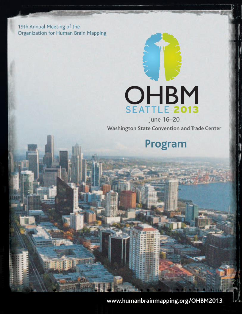
June 16–20
Washington State Convention and Trade Center
Program
19th Annual Meeting of the Organization for Human Brain Mapping
www.humanbrainmapping.org/OHBM2013

To some extent, Seattle remains a frontier metropolis,
a place where people can experiment with their lives,
and change and grow and make things happen.
— Tom Robbins
3
WELCOME
Welcome to Seattle, a city at the forefront of open neuroscience and information technology, and the 19th Annual Meeting of the Organization for Human Brain Mapping! It’s an amazing time to be a part of the OHBM community
as the field of human functional neuroimaging continues to move into the scientific mainstream
in all corners of the world. We are excited to spend the next several days together learning about
the latest scientific discoveries. We look forward to meeting you at one of several events designed
to foster networking and collaboration.
We have several suggestions to help you make the most of your Annual Meeting experience:
• Attend one of many educational courses offered on Sunday including: Advanced fMRI,
The Connectome, Introduction to Imaging Genetics, Anatomy, Computational Neuroscience
and Modeling of Neurodynamics (NEW), Resting-State Brain Networks, Neuroimaging
Meta-Analysis (NEW), Neuroimaging “Big Data” Challenges (NEW) and How Not to
Analyze Your Data (NEW).
• Learn from the scientific education offered throughout the four days of the meeting, including
the Talairach presentation, three member-initiated symposia, one LOC symposium, four daily
parallel oral sessions, twelve morning workshop sessions and I-Poster presentations.
• Take on a challenge at the HBM Hackathon — a meeting-long analysis and resource
building competition designed to accelerate the connection between open neuroscience
and cloud computing. To learn more about the HBM Hackathon, visit
http://ohbm-seattle.github.io.
• Engage in conversation with over 2,200 poster presenters sharing the latest research
in a variety of disciplines.
• Attend the Town Hall session on Wednesday to contribute your perspective on how the
Organization can best advance the study of human brain organization, and consider
the impact of the BRAIN Initiative (Brain Research through Advancing Innovative
Neurotechnologies), which will be discussed by guest Dr. Thomas Insel, Director of the
National Institute of Mental Health.
• Visit with our knowledgeable exhibitors to learn about the latest products and services
available for the brain mapping community.
• Enjoy the OHBM social events, including the opening reception, poster receptions,
and Club Night on Wednesday at the Experience Music Project.
• During and after the meeting, utilize OHBM resources including:
– The Annual Meeting mobile app at m.core-apps.com/ohbm2013
– The Onsite Career Resource room where job seekers can connect with employers
www.humanbrainmapping.org/2013Career
– The Online Library, which contains program presentations from this and past OHBM
meetings. https://cms.psav.com/library/ohbm/
– E-Posters, which contain hundreds of posters that you may have missed.
http://ww4.aievolution.com/hbm1301.
Don’t forget to let us know how we’re doing and what we can do to make your Annual
Meeting experience more valuable at future meetings by completing the online evaluation
forms found at www.humanbrainmapping.org/2013Evaluations.
Throughout this conference, we ask you to stay engaged, ask questions and help us shape
the future of human brain mapping. If you are not yet a member of OHBM, we invite you
to join our growing community by visiting the Membership area on our website at
www.humanbrainmapping.org.
We thank each of you for attending the OHBM meeting and bringing your expertise to the
gathering. We could not accomplish what we do without your support and leadership.
Sincerely,
Susan Bookheimer Peter Bandettini Tom Grabowski
Chair, Council Chair, Program Committee Chair, Local Organizing Committee
Program-At-A-Glance . . . . . . . . . . . . . . . . . . 4
General Information. . . . . . . . . . . . . . . . . . . . 6
Registration, Exhibit Hours,
Social Events, Speaker Ready Room,
Hack Room, Evaluations, Mobile App,
CME Credits, etc.
Daily Schedule
Sunday, June 16 . . . . . . . . . . . . . . . . . . . . . 8
Educational Courses:
Advanced fMRI . . . . . . . . . . . . . . . . . . . . . . . 8
Anatomy . . . . . . . . . . . . . . . . . . . . . . . . . . . 10
Computational Neuroscience and
Modeling of Neurodynamics . . . . . . . . 12
Introduction to Imaging Genetics . . . . . . 14
Neuroimaging Meta-Analysis. . . . . . . . . . 15
Resting State Brain Networks. . . . . . . . . . 17
The Connectome . . . . . . . . . . . . . . . . . . . . 18
Neuroimaging ‘Big Data’
Challenges andComputational
Workflow Solutions. . . . . . . . . . . . . . . . 20
How Not to Analyze Your Data:
A Skeptical Introduction to
Modeling Methods. . . . . . . . . . . . . . . . . 21
Opening Ceremony and
Talairach Lecture . . . . . . . . . . . . . . . . . . 22
Monday, June 17. . . . . . . . . . . . . . . . . . . . 23
Scientific Program
Tuesday, June 18 . . . . . . . . . . . . . . . . . . . 31
Scientific Program
Wednesday, June 19 . . . . . . . . . . . . . . . . 39
Scientific Program
Thursday, June 20. . . . . . . . . . . . . . . . . . . 48
Scientific Program
Trainee Abstract
Travel Award Winners. . . . . . . . . . . . . . . . . . 54
Abstract Review Committee. . . . . . . . . . . . 55
Acknowledgments . . . . . . . . . . . . . . . . . . . . 57
Exhibitor List . . . . . . . . . . . . . . . . . . . . . . . . . 58
Poster and Exhibit Hall . . . . . . . . . . . . . . . . 66
Floor Plans . . . . . . . . . . . . . . . . . . . . . . . . . . . 67
Council and Committees . . . . . . . . . . . . . . . 68
TABLE OF CONTENTS
4
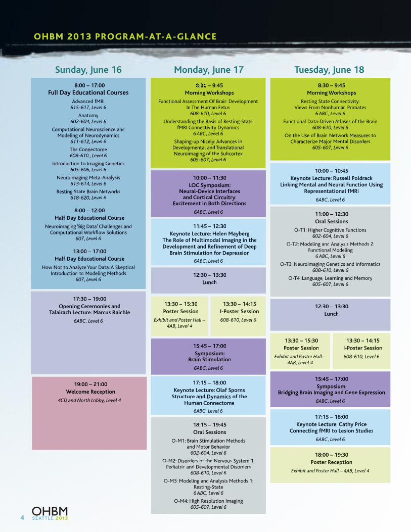
Sunday, June 16 Tuesday, June 18Monday, June 17
– 9:45
Morning Workshops
Functional Assessment Of Brainf DevelopmentIn The Human Fetus
608-610, Level 6
Understanding the Basis of Resting-StateffMRI Connectivity Dynamics
6 ABC, Level 6
Shaping-up Nicely: Advances inDevelopmental and TranslationalNeuroimaging of thef Subcortex
605-607, Level 6
8:00 – 17:00
Full Day Educational Courses
Advanced fMRI615-617, Level 6
Anatomy602-604, Level 6
Computational Neuroscience andModeling of Neurodynamicsf
611-612, Level 6
The Connectome608-610 , Level 6
Introduction to Imaging Genetics605-606, Level 6
Neuroimaging Meta-Analysis613-614, Level 6
Resting State Brain Networks618-620, Level 6
8:00 – 12:00
Half Dayf Educational Course
Neuroimaging ‘Big Data’ Challenges andComputational Workflow Solutions
607, Level 6
13:00 – 17:00
Half Dayf Educational Course
How Not to Analyze Your Data:r A SkepticalIntroduction to Modeling Methods
607, Level 6
10:00 – 11:30
LOC Symposium:Neural-Device Interfaces
and Cortical Circuitry:Excitement in Both Directions
6ABC, Level 6
11:45 – 12:30
Keynote Lecture: Helen MaybergThe Role of Multimodalf Imaging in theDevelopment and Refinement of Deepf
Brain Stimulation for Depressionr
6ABC, Level 6
12:30 – 13:30
Lunch
15:45 – 17:00
Symposium:Brain Stimulation
6ABC, Level 6
13:30 – 15:30
Poster Sessionr
Exhibit andt Posterd Hallr –4AB, Level 4
13:30 – 14:15
I-Poster Sessionr
608-610, Level 6
17:30 – 19:00
Opening Ceremonies andTalairach Lecture: Marcus Raichle
6ABC, Level 6
19:00 – 21:00
Welcome Reception
4CD and Northd Lobby, Level 4
10:00 – 10:45
Keynote Lecture: Russell PoldrackLinking Mental and Neural Function Using
Representational fMRI
6ABC, Level 6
11:00 – 12:30
Oral Sessions
O-T1: Higher Cognitiver Functions602-604, Level 6
O-T2: Modeling and Analysis Methods 2:Functional Modeling
6 ABC, Level 6
O-T3: Neuroimaging Genetics and Informatics608-610, Level 6
O-T4: Language, Learning and Memory605-607, Level 6
15:45 – 17:00
Symposium:Bridging Brain Imaging and Gene Expression
6ABC, Level 6
17:15 – 18:00
Keynote Lecture: Cathy PriceConnecting fMRI to Lesion Studies
6ABC, Level 6
18:00 – 19:30
Poster Receptionr
Exhibit andt Posterd Hallr – 4AB, Level 4
12:30 – 13:30
Lunch
13:30 – 15:30
Poster Sessionr
Exhibit andt Posterd Hallr –4AB, Level 4
13:30 – 14:15
I-Poster Sessionr
608-610, Level 6
8:30 – 9:45
Morning Workshops
Resting State Connectivity:Views From Nonhuman Primates
6 ABC, Level 6
Functional Data-Driven Atlases of thef Brain608-610, Level 6
On the Use of Brainf Network Measuresk toCharacterize Major Mentalr Disorders
605-607, Level 6
17:15 – 18:00
Keynote Lecture: Olaf SpornsfStructure and Dynamics of thef
Human Connectome
6ABC, Level 6
18:15 – 19:45
Oral Sessions
O-M1: Brain Stimulation Methodsand Motor Behaviorr
602-604, Level 6
O-M2: Disorders of thef Nervous System 1:Pediatric and Developmental Disorders
608-610, Level 6
O-M3: Modeling and Analysis Methods 1:Resting-State6 ABC, Level 6
O-M4: High Resolution Imaging605-607, Level 6
OHBM 2013 PROGRAM-AT-A-GL ANCE
5
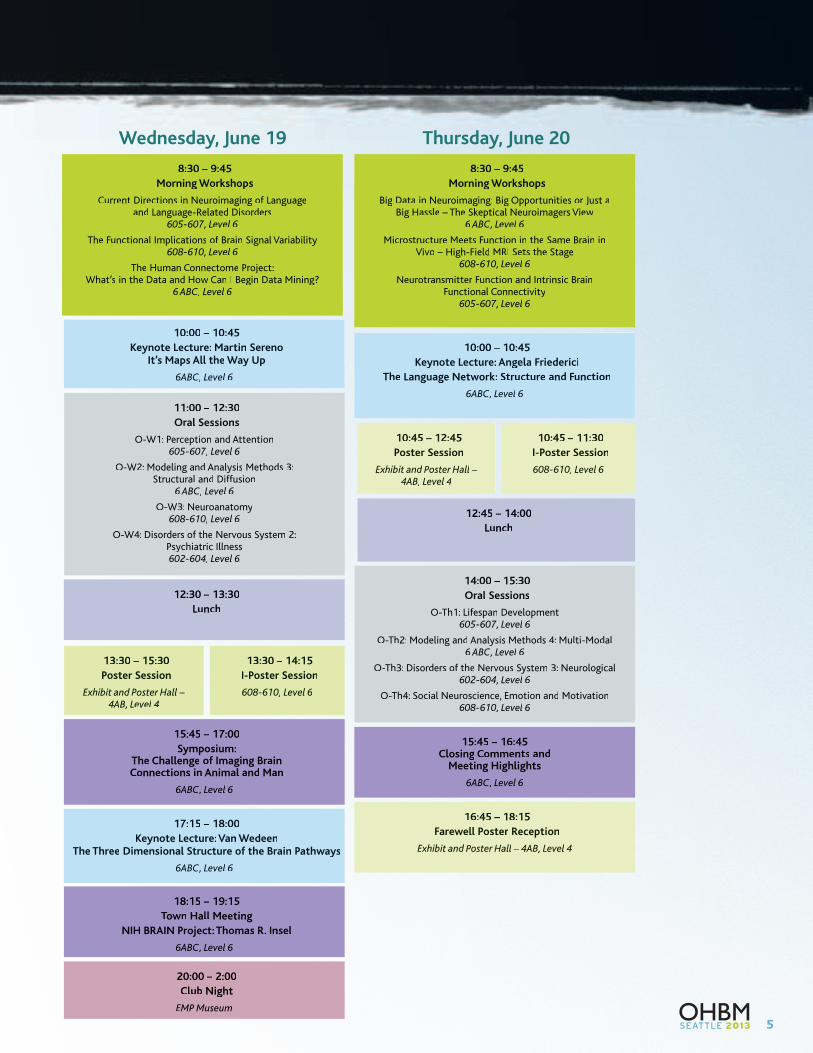
Thursday, June 20
8:30 – 9:45
Morning Workshops
Current Directions in Neuroimaging of Languagefand Language-Related Disorders
605-607, Level 6
The Functional Implications of Brainf Signal Variability608-610, Level 6
The Human Connectome Project:What’s in the Data and How Can I Begin Data Mining?
6 ABC, Level 6
8:30 – 9:45
Morning Workshops
Big Data in Neuroimaging: Big Opportunities or Justr aBig Hassle – The Skeptical Neuroimagers View
6 ABC, Level 6
Microstructure Meets Function in the Same Brain inVivo – High-Field MRI Sets the Stage
608-610, Level 6
Neurotransmitter Functionr and Intrinsic BrainFunctional Connectivity
605-607, Level 6
Wednesday, June 19
10:00 – 10:45
Keynote Lecture: Martin SerenoIt’s Maps All the Way Up
6ABC, Level 6
11:00 – 12:30
Oral Sessions
O-W1: Perception and Attention605-607, Level 6
O-W2: Modeling and Analysis Methods 3:Structural and Diffusion
6 ABC, Level 6
O-W3: Neuroanatomy608-610, Level 6
O-W4: Disorders of thef Nervous System 2:Psychiatric Illness602-604, Level 6
10:00 – 10:45
Keynote Lecture: Angela Friederici
The Language Network: Structure and Function
6ABC, Level 6
14:00 – 15:30
Oral Sessions
O-Th1: Lifespan Development605-607, Level 6
O-Th2: Modeling and Analysis Methods 4: Multi-Modal6 ABC, Level 6
O-Th3: Disorders of thef Nervous System 3: Neurological602-604, Level 6
O-Th4: Social Neuroscience, Emotion and Motivation608-610, Level 6
15:45 – 17:00
Symposium:The Challenge of Imagingf BrainConnections in Animal and Man
6ABC, Level 6
15:45 – 16:45Closing Comments and
Meeting Highlights
6ABC, Level 6
17:15 – 18:00
Keynote Lecture: Van WedeenThe Three Dimensional Structure of thef Brain Pathways
6ABC, Level 6
16:45 – 18:15
Farewell Poster Receptionr
Exhibit andt Posterd Hallr – 4AB, Level 4
18:15 – 19:15
Town Hall Meeting
NIH BRAIN Project: Thomas R. Insel
6ABC, Level 6
20:00 – 2:00
Club Night
EMP Museum
12:30 – 13:30
Lunch
12:45 – 14:00
Lunch
13:30 – 15:30
Poster Sessionr
Exhibit andt Posterd Hallr –4AB, Level 4
13:30 – 14:15
I-Poster Sessionr
608-610, Level 6
10:45 – 12:45
Poster Sessionr
Exhibit andt Posterd Hallr –4AB, Level 4
10:45 – 11:30
I-Poster Sessionr
608-610, Level 6
6
GENERAL INFORMATION
CONFERENCE VENUEWashington State Convention Center
800 Convention Place
Seattle, WA 98101-2350
Phone: 206-694-5000
Fax: 206-694-5399
Email: [email protected]
All events will take place at the Washington State Convention
Center unless otherwise noted.
REGISTRATION HOURSSouth Lobby, Level 4
Saturday, June 15: 15:00 – 18:00
Sunday, June 16: 7:00 – 19:30
Monday, June 17: 7:30 – 19:45
Tuesday, June 18: 8:00 – 18:00
Wednesday, June 19: 8:00 – 18:00
Thursday, June 20: 8:00 – 16:00
EXHIBIT HOURSExhibit and Poster Hall – 4AB, Level 4
Monday, June 17: 12:30 – 16:00
Tuesday, June 18: 12:30 – 19:30
Wednesday, June 19: 12:30 – 16:00
Thursday, June 20: 10:45 – 18:15
TOWN HALL MEETINGWednesday, June 18, 18:15 – 19:15
6ABC, Level 6
All OHBM meeting attendees are encouraged to participate in this
open forum where you will have an opportunity to ask questions
and give the OHBM leadership feedback. Updates on future meeting
sites and council elections will be presented. The Town Hall Forum
will include a presentation and discussion on the United States’
BRAIN Initiative.
WELCOME RECEPTIONSunday, June 16, 19:00 – 21:00
4CD and North Lobby, Level 4
Join us for the 2013 Annual Meeting Welcome Reception.
The reception will be held at the Washington State Convention
Center immediately following the Opening Ceremonies and
Talairach Lecture on Sunday, June 16th. Please make sure to wear
your name badge, which will serve as your ticket to the event.
Additional guest badges are $50.00 USD.
CLUB NIGHTWednesday, June 18, 21:00 – 2:00
EMP Museum | 325 5th Avenue N | Seattle, WA 98109
There will be a band and a DJ that will play dance music throughout
the evening. The party is complimentary to registrants. Please make
sure to bring your ticket to the EMP. Additional guest tickets are
$50.00 and must be purchased at the conference registration desk.
We encourage you to use the historic Seattle Monorail for
transportation to and from the event. The monorail is the most
economical option and will provide service until 2:00 am.
SPEAKER READY ROOMRoom 601, Level 6
Hours:
Saturday, June 15: 15:00 – 18:00
(located in the South Lobby near Registration on Saturday only)
Sunday, June 16: 6:30 – 19:30
Monday, June 17: 7:30 – 19:45
Tuesday, June 18: 7:30 – 18:00
Wednesday, June 19: 7:30 – 18:00
Thursday, June 20: 7:30 – 16:00
INTERNET AND DOCKING LOUNGELevel 6 Foyer
A limited number of complimentary terminals for computer access
and docking stations to charge your electronic devices will be
available. Please limit your time at a terminal to 15 minutes.
Hours:
Sunday, June 16: 7:30 – 19:30
Monday, June 17: 8:00 – 19:30
Tuesday, June 18: 8:00 – 19:30
Wednesday, June 19: 8:00 – 19:30
Thursday, June 20: 8:00 – 17:00
HBM Hackathon Exhibit and Poster Hall – 4AB, Level 4
OHBM 2013 will include an integrated hack room and cloud
computing contest called The HBM Hackathon: Open Brain
Mapping in the Cloud. HBM Hackathon will include a meeting-
long venue on the main poster/exhibit floor space, and dedicated
cloud-accessible data and software resources that will be available
to all interested attendees. The room will be available from Monday
through Thursday while the Exhibits and Posters are open.
The goals of HBM Hackathon are to accelerate the development
of a critical mass of cloud-based data, analytic, and computational
resources for human brain mapping, and to provide OHBM
attendees with access to and knowledge about them. To learn more
about the HBM Hackathon, visit http://ohbm-seattle.github.io or
www.humanbrainmapping.org/hackathon.
7
MOBILE APPThe 2013 Mobile App, powered by EventLink and created by
Core-Apps LLC, is a native application for smartphones (iPhone
and Android), a hybrid web-based app for Blackberry, and there’s
also a web-based version of the application for all other web
browser-enabled phones.
How to Download:
For iPhone (plus, iPod Touch & iPad) and Android phones: Visit your
App Store or Android Market on your phone and search for OHBM.
For All Other Phone Types (including BlackBerry and all other web
browser-enabled phones): While on your smartphone, point your
mobile browser to http://m.core-apps.com/ohbm2013. From there
you will be directed to download the proper version of the app for
your particular device, or, on some phones, you simply bookmark
the page for future reference.
TWITTERJoin the conversation on Twitter using the hash tag #OHBM2013
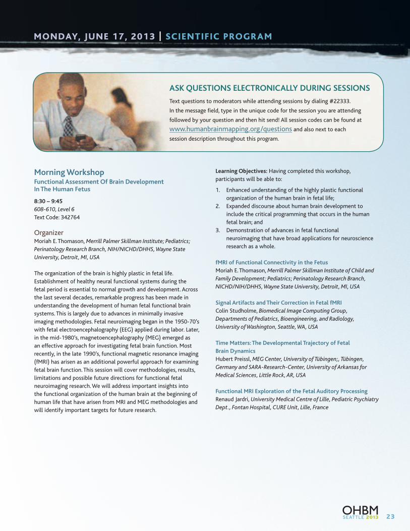
ASK QUESTIONS ELECTRONICALLY DURING SESSIONSText questions to moderators while attending
sessions by dialing #22333. In the message field,
type in the unique code for the session you are
attending followed by your question and then
hit send! All session codes can be found at
www.humanbrainmapping.org/questions
and also next to each session description throughout this program.
E-POSTERSIt is our goal to have all poster presentations uploaded on our
E-Poster format (as a pdf). To upload your poster, please go to
http://ww4.aievolution.com/hbm1301/.
WIRELESS CONNECTIONWireless connections will be available throughout the Washington
State Convention Center. Please connect to the wireless network
“OHBM 2013” to access the conference Wi-Fi.
ONSITE CAREER RESOURCESA popular feature every year at OHBM are bulletin boards heavy
with “positions available” notices. This year OHBM has created an
electronic board at http://www.humanbrainmapping.org/2013Career
Career where PIs can post positions available notices (under “Labs
Looking for People”) and trainees can post vitas (under “People
Looking for Jobs”) in advance of the meeting. OHBM has reserved
room 309 (Level 3) in the Washington State Convention Center from
Sunday, June 16th through Thursday, June 20th for attendees to
gather and discuss employment opportunities.
EVALUATIONS ONLINE!Conference evaluations will be conducted online only at
www.humanbrainmapping.org/2013Evaluations. It is only through
attendee’s feedback that we can continue to improve the content,
format, and schedule of the meeting. Your input is very important to
us, and we urge you to fill out these quick surveys.
ACCME ACCREDITATIONCME CREDIT: This activity has been planned and implemented
in accordance with the Essential Areas and Policies of the
Accreditation Council for Continuing Medical Education (ACCME)
through sponsorship of the Organization for Human Brain Mapping.
The OHBM is accredited by the ACCME to provide continuing medical
education for physicians.
The Organization for Human Brain Mapping designates this
educational activity for a maximum of 38.75 PRA Category 1
Credit(s)TM. Physicians should only claim credit commensurate with
the extent of their participation in the activity. CME forms will only
be available online at www.humanbrainmapping.org/CME2013.
EDUCATIONAL COURSES CREDITS
Advanced fMRI (Full Day). . . . . . . . . . . . . . . . . . . . . . . . . . . . . . . . . . . .7.00
Anatomy (Full Day). . . . . . . . . . . . . . . . . . . . . . . . . . . . . . . . . . . . . . . . .7.00
The Connectome (Full Day) . . . . . . . . . . . . . . . . . . . . . . . . . . . . . . . . . .7.00
Introduction to Imaging Genetics (Full Day) . . . . . . . . . . . . . . . . . . .7.00
Resting State Brain Networks (Full Day) . . . . . . . . . . . . . . . . . . . . . . .7.00
Computational Neuroscience and Modeling
of Neurodynamics (Full Day). . . . . . . . . . . . . . . . . . . . . . . . . . . . . .7.00
Neuroimaging Meta-Analysis (Full Day) . . . . . . . . . . . . . . . . . . . . . . .7.00
Neuroimaging ‘Big Data’ Challenges and
Computational Workflow Solutions (Half Day) . . . . . . . . . . . . . .3.50
How Not to Analyze Your Data:
A Skeptical Introduction to Modeling Methods (Half Day) . . . .3.50
Maximum number of possible credits earned at
Educational Courses . . . . . . . . . . . . . . . . . . . . . . . . . . . . . . . . . . .7.00
ANNUAL MEETING CREDITS
Talairach Lecture . . . . . . . . . . . . . . . . . . . . . . . . . . . . . . . . . . . . . . . . . . .0.75
Keynote Lectures. . . . . . . . . . . . . . . . . . . . . . . . . . . . . . . . . . . . . . 0.75 each
Morning Workshops . . . . . . . . . . . . . . . . . . . . . . . . . . . . . . . . . . . 1.25 each
Oral Sessions . . . . . . . . . . . . . . . . . . . . . . . . . . . . . . . . . . . . . . . . . 1.50 each
Poster Session Viewing . . . . . . . . . . . . . . . . . . . . . . . . . . . . . . .2.00 per day
Symposia. . . . . . . . . . . . . . . . . . . . . . . . . . . . . . . . . . . . . . . . . . . . . 1.25 each
LOC Symposia . . . . . . . . . . . . . . . . . . . . . . . . . . . . . . . . . . . . . . . . . . . . .1.50
Meeting Highlights . . . . . . . . . . . . . . . . . . . . . . . . . . . . . . . . . . . . . . . . .1.00
Town Hall Forum . . . . . . . . . . . . . . . . . . . . . . . . . . . . . . . . . . . . . . . . . . .0.50
Total number of possible credits earned at Annual Meeting . .31.75
TOTAL NUMBER OF POSSIBLE CREDITS. . . . . . . . . . . . . . . . . . . . .38.75

8
SUNDAY, JUNE 16, 2013 | EDUC ATIONAL COURSES
Advanced fMRI – Physics, Physiology, and Pattern InformationFULL-DAY COURSE | 8:00 – 17:00
615-617, Level 6
Text Code: 769863
OrganizersTor Wager, University of Colorado, Boulder, CO, USA
Nikolaus Kriegeskorte, MRC Cognition and Brain Sciences Unit,
Cambridge, UK
Functional magnetic resonance imaging (fMRI) has taken a central
role in the study of human brain function. fMRI is inherently
transdisciplinary, and data acquisition and analysis are constantly
evolving. Thus, there is a need for continuing education on new
methods and cutting-edge neuroscientific applications of fMRI.
The first part of the course covers the physics and physiology of
fMRI, and the relationship between neuronal and BOLD activity
patterns. The second part focuses on pattern-information analyses
and how they can be used to learn about neuronal population codes
and to test computational theories of brain information processing.
Learning Objectives: The course is designed to develop participants’
understanding of:
1. The physics and physiology underlying fMRI, and the resulting
potential and limitations of fMRI;
2. Pattern decoding, representational similarity analysis, and
voxel-receptive-field modelling; and
3. Computational modeling of brain information processing and
its integration into the analysis of fMRI data
Target Audience: This course is intended for an audience of
research scientists with intermediate to advanced knowledge of
fMRI techniques, who wish to extend the breadth and depth of
their understanding of the current state of the art.
Course Schedule8:00 – 8:10 Introduction to the Advanced fMRI Course
Tor Wager and Nikolaus Kriegeskorte
8:10 – 8:45 Introduction to MRI and fMRI Physics
Marta Correia, Cognition and Brain Sciences Unit,
Cambridge, UK
8:45 – 9:20 Basic Physiology of fMRI: Signal and Noise
Gary Glover, Stanford University, Stanford, CA, USA
9:20 – 9:55 The Physiology of fMRI and Its Relation to
Brain Information Processing
Amir Shmuel, MNI, McGill University,
Montreal, Canada
9:55 – 10:00 Questions and Discussion
10:00 – 10:30 Break
10:30 – 11:40 Pattern Decoding Analysis for fMRI:
Basic Steps and Advanced Techniques
Janaina Mourao-Miranda, University College
London, London, UK
11:40 – 12:00 Using Pattern Classification for
Psychological Inference
Tor Wager, University of Colorado, Boulder, CO, USA
12:00 – 13:00 Lunch
13:00 – 13:35 Computational Neuroscience with fMRI
and Coarse-Scale Contributions to
Orientation Decoding
Eli Merriam, New York University,
New York, NY, USA
ASK QUESTIONS ELECTRONICALLY DURING SESSIONS
Text questions to moderators while attending sessions by dialing #22333.
In the message field, type in the unique code for the session you are attending
followed by your question and then hit send! All session codes can be found at
www.humanbrainmapping.org/questions and also next to each
session description throughout this program.
9
13:35 – 14:10 Investigating Neuronal Population Codes
of Visual Objects with Representational
Similarity Analysis
Dwight Kravitz, NIH, Bethesda, MD, USA
14:10 – 14:45 Inferring Neuronal Tuning from fMRI:
Adaptation and Pattern Information
Geoffrey Aguirre, University of Pennsylvania,
Philadelphia, PA, USA
14:45 – 15:00 Questions and Discussion
15:00 – 15:30 Break
15:30 – 16:05 Voxel-Receptive-Field Modeling:
Testing Computational Theories with fMRI
Jack Gallant, University of California-Berkeley,
Berkeley, CA, USA
16:05 – 16:40 Engineering-Based Approaches to Machine
Learning Analysis of fMRI
Francois Meyer, University of Colorado,
Boulder, CO, USA
16:40 – 17:15 Depicting and Decoding Fine-Grained
Cortical Representations of Auditory Stimuli
Federico DeMartino, Maastricht University,
Maastricht, The Netherlands
17:15 – 17:30 Questions and Discussion
10
SUNDAY, JUNE 16, 2013 | EDUC ATIONAL COURSES
Anatomy and Its Impact on Structural and Functional ImagingFULL-DAY COURSE | 8:00 – 17:00
602-604, Level 6
Text Code: 769979
OrganizersKatrin Amunts, Institute of Neuroscience and Medicine,
Jülich, Germany
Karl Zilles, Institute of Neuroscience and Medicine,
Jülich, Germany
Results of neuroimaging studies cannot be understood without
knowing the anatomy of the brain, and the way how brain
structure influences the interpretation of the results through
interaction with image acquisition, processing and analysis.
The course will provide an introduction and critical overview
of classical and modern approaches for studying the anatomy
of the brain using neuroimaging techniques. It is aimed at a
multidisciplinary audience, and will provide an introduction to
brain macroscopy, gross anatomical landmarks and its intersubject
variability, the microstructural organization of the brain including
cortical segregation, and the representation of cognitive functions
with respect to organization principles. Neuroimaging methods
will be discussed with respect to their advantages, disadvantages
and potential pitfalls as it concerns anatomy. The relevance of
anatomical knowledge for the interpretation of structural and/or
functional imaging data will be made explicit.
Part one will consist of talks introducing anatomical concepts
and developmental aspects and show, how MRI contributes.
Part two will focus on organizational principles of the brain’s
microstructure, and critically reflect the perspectives and limits
of MR imaging with respect to microstructure. Part three will
elucidate the relationship between microstructure and brain
function, and provide an overview of some widely distributed
neuroimaging tools in this field.
Learning Objectives: Having completed this course, participants
will be able to:
1. Understand the organizational principles of the human brain
on a macroscopic and microscopic level, and their changes
during development;
2. Understand the advantages and limitations of
neuroanatomical techniques including receptor mapping
and cytoarchitectonics;
3. Understand methods for design and analysis of structural and
functional MRI data, and interpret the measures they provide
and their limitations; and
4. Give examples of applications of structural MRI for
understanding brain function and dysfunction.
Target Audience: The prime target audience is researchers with
an interest in understanding the relationship between brain
structure and function. This includes researchers with limited
previous anatomical knowledge. Prior experience of neuroimaging
is expected. Background will be provided for those without special
anatomical knowledge but some talks will address advanced issues
that would be of interest to people with experience in this field.
Course SchedulePart I: Introduction: Neuroanatomy, Development
and MRI
8:00 – 8:30 Surface Anatomy of the Brain and Landmarks
Thomas Naidich, Mt. Sinai Medical Center,
New York, NY, USA
8:30 – 9:00 Development of the Cerebral Cortex
David van Essen, Washington University,
St. Louis, MO, USA
9:00 – 9:30 MRT Imaging of Brain Development
Roger Woods, University of California-
Los Angeles, Los Angeles, CA, USA
9:30 – 10:00 High Resolution Imaging and Anatomy
Noam Harel, University of Minnesota,
Minneapolis, MN, USA
10:00 – 10:30 Break
Part II: Microstructure and Its Interpretation in MRI
10:30 – 11:00 Cytoarchitecture of the Human
Cerebral Cortex – Challenges for MRI
Katrin Amunts, Institute of Neuroscience and
Medicine, Research Center Jülich, Germany
11:00 – 11:30 Myeloarchitecture – a Window for MRI
Robert Turner, Max Planck Institute for Human
Cognitive and Brain Sciences, Leipzig, Germany
11:30 – 12:00 Receptorarchitecture and Neural Systems
Karl Zilles, Institute of Neuroscience and Medicine,
Jülich, Germany
12:00 – 13:00 Lunch

11
Part III: Structure, Function and Tools for Analysing
Their Relationship
13:00 – 13:30 Functional and Structural Architecture of the Brain
Christian Beckmann, NL Donders Institute for Brain,
Cognition & Behaviour, Radboud University Nijmegen,
Nijmegen, Netherlands
13:30 – 14:00 Tools to Combine Structural MRI with
Cytoarchitecture and Function
Simon Eickhoff, Heinrich-Heine University Düsseldorf,
Düsseldorf, Germany
14:00 – 14:30 Structural and Functional Segregation
of the Cortex
Jean-Francois Mangin, Neurospin, CEA,
Gif sur Yvette, France
14:30 – 15:00 Anatomical Conditions and MR-Morphometry
Christian Gaser, University of Jena, Jena, Germany
15:00 – 15:30 Break
15:30 – 16:00 Anatomical Background of Dynamic
Causal Modelling and Connectivity
Jakob Heinzle, University of Zurich &
ETH Zurich, Zurich, Switzerland
16:00 – 17:00 Question and Answer – Panel Discussion
12
SUNDAY, JUNE 16, 2013 | EDUC ATIONAL COURSES
Computational Neuroscience and Modeling of NeurodynamicsFULL-DAY COURSE | 8:00 – 17:00
611-612, Level 6
Text Code: 769980
OrganizersMichael Breakspear, Queensland Institute of Medical Research,
Brisbane, Australia
Stefan Kiebel, Friedrich-Schiller-University, Jena, Germany
Jean Daunizeau, Brain and Spine Institute, Paris, France
Computational neuroscience is a rapidly growing field that seeks
to understand the principles of neuronal dynamics and how
these underpin cognition. Computational neuroscience offers
fresh perspectives on the design, analysis and interpretation of
functional neuroimaging data, moving beyond static designs and
phenomenological heuristics. This course will provide a broad
overview of the field, moving from the foundations of dynamical
systems theory to large-scale computer platforms, the analysis of
imaging data and models of cognitive processes such as perception
and decision making.
Learning Objectives: Having completed this course, participants
will be able to:
1. Summarize the use of dynamic systems theory in modelling
neuroscience phenomena, ranging from single neuron models
to macroscopic modelling of networks;
2. Summarize new developments and research questions in
dynamic models of the brain;
3. Understand the link between models of cortical activity and
theories of brain function;
4. Understand the meaning and significance of stochastic
processes in cortical systems; and
5. Discuss how such computational approaches can lead to the
design and analysis of cognitive neuroscience experiments.
Target audience: This course is designed to guide both cognitive
neuroscientists and modellers through a variety of computational
approaches. The talks introduce and motivate dynamic systems
theory and other mathematical concepts as tools for modelling
various neuroscience phenomena, ranging from the single neuron to
the macroscopic network level. The participants do not require an
explicit mathematical background to follow the course but need to
bring a healthy interest in how ubiquitous neuroscience phenomena
can be explained mechanistically. Examples will be given of how
such approaches lead to the design and analysis of cognitive
neuroscience experiments.
Course SchedulePart I: Dynamic Systems Approach
Chair: Michael Breakspear, Queensland Institute of Medical Research,
Brisbane, Australia
8:00 – 8:40 Objectives of Large-Scale Computational
Neuroscience
Michael Breakspear, Queensland Institute
of Medical Research, Brisbane, Australia
8:40 – 9:20 Models for Dynamics from the Neural
Microcircuit to Cortical Regions
Peter Robinson, University of Sydney, Australia
9:20 – 10:00 Computational Models of Resting State Activity
Gustavo Deco, Universitat Pompeu Fabra,
Barcelona, Spain
10:00 – 10:30 Break
Part II: Computational Models of NeuroImaging Data
Chair: Viktor Jirsa, CNRS, Marseille, France
10:30 – 11:15 Investigating Neural Mechanisms with
Modelling and Imaging
Tim Behrens, University of Oxford, Oxford, UK
11:15 – 12:00 Computational Modelling in fMRI
John O’Doherty, California Institute of Technology,
Pasadena, USA
12:00 – 13:00 Lunch
Part III: Bayesian-Based Methods
Chair: Jean Daunizeau, Brain and Spine Institute, Paris, France
13:00 – 13:40 Dynamic Causal Modelling (Bayesian Inference,
Model Selection)
Jean Daunizeau, Brain and Spine Institute,
Paris, France
13:40 – 14:20 Dynamic Causal Modelling and Neurophysiology
Rosalyn Moran, Virginia Tech Carilion Research
Institute, Roanoke, VA, USA
14:20 – 15:00 Dynamics of Perceptual Decision Making
Sebastian Bitzer, Max Planck Institute for Human
Cognitive and Brain Sciences, Leipzig, Germany
15:00 – 15:30 Break

13
Part IV: Integrative Models
Chair: Peter Robinson, University of Sydney, Australia
15:30 – 16:10 Complex Brain Networks: Dynamics
and Structure
Mika Rubinov, University of New South Wales,
Australia
16:10 – 16:50 Platforms for Large-Scale Brain Simulations
Viktor Jirsa, CNRS, Marseille, France
16:50 – 17:00 Summary, Discussion, and Farewell
Michael Breakspear and Jean Daunizeau
Accelerating Your Research 24/7
alleninstitute.orgbrain-map.org
Special EventHBM Hackathon: Open Brain Mapping in the Cloud, co-lead sponsor
,
,
Educational Course - Sunday 16 JuneNuts and Bolts of the Allen Human Brain Atlas, Lydia Ng
LOC Symposium - Monday 17 JuneMapping the Neocortex at the Cellular Level in a Large-Scale and High-Throughput Manner, Christof Koch
Symposium - Tuesday 18 JuneBridging Brain Imaging and Gene Expression, Michael Hawrylycz, co-chair
An Anatomically Comprehensive Atlas of Gene Expression in Adult Human Brain, Ed Lein
PostersA high-resolution cyto- and chemo-architecture-based digital atlas for entire adult human brain, Ding et al., #1426
Altered gene expression in the dorsolateral prefrontal cortex of individuals with schizophrenia, Guillozet-Bongaarts et al., #3215
High-resolution histological and molecular reference atlases of the human prenatal brain, Royall et al., #3757
> Visit us at our booth
Come nd us at OHBM 2 1
14
SUNDAY, JUNE 16, 2013 | EDUC ATIONAL COURSES
Introduction to Imaging GeneticsFULL-DAY COURSE | 8:00 – 17:00
605-606, Level 6
Text Code: 769976
OrganizersThomas Nichols, University of Warwick, Coventry, UK
Jean-Baptiste Poline, CEA, France & UC Berkeley, US
This course will introduce the fundamentals of “Imaging Genetics,”
the process of modeling and understanding genetic variation in
brain image data. The course begins with a three-lecture genetics
tutorial in the morning, designed to give imaging practitioners a
quick overview of key genetics concepts and terminology.
The remainder of the course covers how imagers can use genetic
variables in their analyses. Specific topics include voxel-wise
genome-wide models, joint multivariate modeling of imaging
and genetic data, and heritability analyses of cortical surface
and thickness data. The course concludes with a case study
highlighting current imaging genetics research.
Learning Objectives: Having completed this course, participants
will be able to:
1. Understand the fundamentals of the molecular basis of
genetic variation, and how that variation is modeled in
traditional genetics studies;
2. Understand the difference between linkage, association and
heritability analyses;
3. Understand the relative strengths and weaknesses of each
different type of brain imaging phenotype used to find
genetic association; and
5. Understand how imaging genetics can be applied to an area
like major depression.
Target Audience: The course is designed for neuroimaging
practitioners who do not necessarily have a background
in genetics.
Course Schedule8:00 – 8:10 Introduction
Jean-Baptiste Poline, CEA, France &
UC Berkeley, USA
8:10 – 9:00 Molecular Basis of Genetic Variation
Elliot Hong, University of Maryland, Baltimore,
MD, USA
9:00 – 9:45 Structure and Analysis of Genetic Variation
Sven Cichon, Bonn University, Bonn, Germany
9:45 – 10:15 Overview of Neuroimaging Phenotypes
Anderson Winkler, Oxford University, Oxford, UK
10:15 – 10:30 Break
10:30 – 11:15 Nuts and Bolts of the Allen Brain Human Atlas
Lydia Ng, Allen Brain Institute, Seattle, WA, USA
11:15 – 12:00 Univariate Approaches: Multiple Testing &
Voxelwise WGA
Derrek Hibar, University of California,
Los Angeles,CA, USA
12:00 – 13:00 Lunch
13:00 – 13:45 Quantitative Traits: Heritability,
Linkage & Association
John Blangero, Texas Biomedical Research
Institute, San Antonio, TX, USA
13:45 – 14:30 Multivariate Approaches: Joint Modeling of
Imaging & Genetic Data
Giovanni Montana, Imperial College, London, UK
14:30 – 15:00 Multivariate Phenotypes for Association
and Linkage
Peter Kochunov, University of Maryland,
Baltimore, MD, USA
15:00 – 15:30 Break
15:30 – 16:15 ENIGMA & Large Scale Imaging Association
Jason Stein, University of California,
Los Angeles, CA, USA
16:15 – 17:00 Case Study: Identifying Informative
Phenotypes in Large Functional Imaging
Studies: An Application of Genome-Wide
Complex Trait Analysis
Tomáš Paus, University of Toronto,
Toronto, Canada

15
Neuroimaging Meta-AnalysisFULL-DAY COURSE | 8:00 – 17:00
613-614, Level 6
Text Code: 769982
OrganizersSimon B. Eickhoff, Heinrich-Heine University
Düsseldorf, Düsseldorf, Germany
Tor D. Wager, University of Colorado, Boulder, CO, USA
Functional neuroimaging has provided a wealth of information
on the cerebral localization of mental functions. In spite of its
success, however, several limitations restrict the amount of
knowledge that may be gained from each individual experiment.
These include a usually rather small sample size, limited reliability
of an indirect signal and the need to infer knowledge from specific
contrasts. Such limitations have raised some concerns, whether
neuroimaging may ultimately provide fundamental insight into
problems from cognitive psychology or clinical neurosciences.
In turn, however, they also encouraged the development of
quantitative meta-analysis approaches that allow statistically
summarizing a vast amount of neuroimaging findings across a large
number of participants and diverse experimental settings. Such
integration then enables statistically defensible generalizations on
the neural basis of psychological processes in health and disease.
They also allow relating different tasks or processes to each other
and modeling interacting networks. Quantitative meta-analysis
therefore represents a powerful tool to gain a synoptic view of
distributed neuroimaging findings in an objective and impartial
fashion and address the above concerns. This course is set out
to cover the burgeoning field of meta-analytic modeling and
database-driven syntheses. In order to provide a comprehensive
overview, this course spans both basic and advanced topics, from
the foundations allowing the synthesis of neuroimaging data
to cutting-edge methodological developments and emerging
psychological clinical applications. This broad coverage will
thus provide both a deeper understanding of the statistical and
methodological underpinnings as well as concrete ideas for how to
apply meta-analytic techniques to advance brain science.
Learning Objectives:
1. Methodological foundations of database-driven systems
neuroscience;
2. Established and innovative approaches for multi-study
integration by meta-analyses;
3. Methods for large scale data-mining and the meta-analytic
investigation of brain networks;
4. Emerging approaches to cognitive psychology based on
computational neurobiology; and
5. The possibilities of meta-analytic modeling provides to
understand brain organization.
Target Audience: Imaging researchers interested in databases,
meta-analyses and functional atlassing of the brain as well
as cognitive psychologists who wish to learn about emerging
computational approaches to understanding mental functions.
While some background in neuroimaging will be helpful, this course
does introduce basic concepts and approaches before moving on to
advanced methods and applications.
Course SchedulePart I: Methodological Foundations
8:00 – 8:30 Coordinates and Templates
Jack L. Lancaster, University of Texas Health Science
Center at San Antonio, San Antonio, TX, USA
8:30 – 9:00 Neuroimaging Activation Databases
Angela R. Laird, Florida International University,
Miami, FL, USA
9:00 – 9:30 Overview of Meta-Analysis Approaches
Thomas Nichols, University of Warwick,
Coventry, UK
9:30 – 10:00 Bringing New Techniques from Statistics into
Neuroimaging Meta-Analysis
Timothy Johnson, University of Michigan,
Ann Arbor, MI, USA
10:00 – 10:30 Break
Part II: Informatics Approaches to Psychological Constructs
10:30 – 11:00 Cognitive Ontologies as Top-Down Descriptions
Jessica A. Turner, MIND Research Network,
Albuquerque, NM, USA
11:00 – 11:30 Text Mining and Machine Learning for
Neuroinformatics
Tal Yarkoni, University of Colorado, Boulder,
CO, USA
11:30 – 12:00 Inferring Mental States from
Neuroimaging Data
Russell Poldrack, University of Texas at Austin,
Austin, TX, USA
12:00 – 13:00 Lunch
16
SUNDAY, JUNE 16, 2013 | EDUC ATIONAL COURSES
Part III: Applications: Understanding the Structure
of the Mind
13:00 – 13:30 Learning From the Past: Using Prior
Neuroimaging Literature to Constrain
Predictions of Psychological States
Tor Wager, University of Colorado,
Boulder, CO, USA
13:30 – 14:00 Using Neuroimaging Meta-Analysis to
Understand the Structure of Emotion
Lisa Feldman Barrett, Northeastern University,
Boston, MA, USA
14:00 – 14:30 Meta-Analysis for Consolidation of the
Literature: Cognitive and Clinical Applications
Claudia Rottschy, RWTH Aachen University,
Aachen, Germany
Part IV: Applications: Understanding the Structure of
Brain Networks
14:30 – 15:00 Meta-Analytic Connectivity: Concepts and
Task-Dependent Application
Jennifer L. Robinson, Auburn University,
Auburn, AL, USA
15:00 – 15:30 Break
15:30 – 16:00 Meta-Analytic Connectivity:
Comparison to Resting-State and DTI
Simon B. Eickhoff, Heinrich-Heine University
Düsseldorf, Düsseldorf, Germany
16:00 – 16:30 Co-Activation Based Seed-Region Parcellation
Danilo Bzdok, Research Center Jülich,
Jülich, Germany
16:30 – 17:00 Combining Meta-Analysis with Other
Modalities: Clinical and Basic Examples
Peter T. Fox, University of Texas Health Science
Center at San Antonio, San Antonio, TX, USA
17
Resting State Brain NetworksFULL-DAY COURSE | 8:00 – 17:00
618-620, Level 6
Text Code: 769981
OrganizersBharat Biswal, UMDNJ, Newark, NJ, USA
Yu Feng Zang, Hangzhou Normal University, Hangzhou, China
This course is designed to teach users how to design, analyze, and
interpret resting state brain connectivity. Due to its increasing
popularity, a large number of investigators are collecting MRI
data from healthy and clinical subjects during rest. A novelty of this
course will be that actual data from a large study will be used to
show the user, all points of the study. In the first part of the course,
users will be taught how to design an experiment for a resting
state study. The importance of initial instruction given and the
subject’s behavioral and physiological parameters including satiety,
and emotional state on the baseline signal will be discussed. In the
second part, pre-processing and post-processing steps their relative
advantages and disadvantages will be demonstrated. During this
process, their software implementation will also be demonstrated.
In the third part, data integration with other clinical and
connectivity measures including DTI will also be shown.
Learning Objectives: Having completed this course, participants
will be able to:
1. Design a resting state study, with full knowledge as to how the
various behavioral or physiological states would affect RSFC;
2. Understand the sources of variation both within and between
subjects. Also, they will be aware of the various pre processing
methods used, including their advantages and disadvantages;
3. Generate various measures of connectivity, including seed
based, data driven approached including ICA/PCA, aggregate
properties including ALFF, small world, etc. Different software
implementation including AFNI, FSL, REST, GIFT and CONN will
be covered;
4. Integrate the RSFC results with other measures including
DTI, EEG, etc; and
5. Analyze Single subject and Group level analysis.
Target Audience: This course is designed for neuroimaging
practitioners interested in resting state fMRI studies.
Course Schedule8:00 – 8:20 Introduction
Bharat Biswal, New Jersey Institute of Technology
8:20 – 8:50 Frequency-Dependent Analysis of
Resting-State fMRI Signal
Yu-Feng Zang, Hangzhou Normal University
8:50 – 9:20 Pre-Processing Steps and Considerations
Christian Windischberger, Medical University
of Vienna, Vienna, Austria
9:20 – 9:50 Analysis of Resting-State Data Using ICA
Christian Beckmann, NL Donders Institute
for Brain, Cognition & Behaviour, Radboud
University Nijmegen, Nijmegen, Netherlands
9:50 – 10:25 Global Correlations: What You Don’t Know
Will Hurt You
Ziad Saad, National Institute of Health,
Bethesda, MD, USA
10:25 – 10:35 Break
10:35 – 11:10 Analysis: Granger Causality and Other SEM
Xiaoping Hu, Georgia Institute of Technology,
Atlanta, GA, USA
11:10 – 11:45 Functional Connectomics and Network
Analysis with Resting-State fMRI
Yong He, Beijing Normal University, Beijing, China
11:45 – 12:25 Putting Clinical Applications of R-fMRI
Into Perspective
Mike Milham, Child Mind Institute,
New York, NY, USA
12:25 – 13:25 Lunch
13:25 – 14:00 Functional Brain Organization in Typical and
Atypical Development: Insights from
Resting-State fMRI
Vinod Menon, Stanford University,
Stanford, NJ, USA
14:00 – 14:35 Combining Diffusion-Based
Structural Connectivity with RSFC:
Methodological Approaches
Paul Taylor, African Institute for
Mathematical Sciences, Cape Town, South Africa
continued on page 18
18
SUNDAY, JUNE 16, 2013 | EDUC ATIONAL COURSES
Resting State Brain Networks, continued
14:35 – 15:10 Multimodal Integration: Combining
DTI and fcMRI
Ching-Po Lin, National Yang-Ming
University, Taipei
15:25 – 16:00 Integrating Intracranial Electrodes and
Diffusion Tractography to Study Resting
State Networks
Timothy Ellmore, The City College of New York,
New York, NY, USA
16:00 – 16:45 Case Study: Single Subject and Group Analysis
Suril Gohel and Xin Di, UMDNJ, Newark, NJ, USA
16:45 – 17:00 Resting State Studies: A Pharmaceutical
Industry Perspective
Richard Baumgartner, Merck Inc
The ConnectomeFULL-DAY COURSE | 8:00 – 17:00
608-610, Level 6
Text Code: 769865
OrganizersEd Bullmore, University of Cambridge, Cambridge, UK
Randy McIntosh, Rotman Research Institute, Toronto, Canada
This course provides an introduction to the emerging science
of brain ‘Connectomics’, the study of large-scale networks of
structural and functional brain connections. Brain imaging data
can provide powerful information for building maps of the
‘Human Connectome’.
The first part of the course, Building Connectomes, will provide
methodological introductions to the types of data that can be used
to define the connectome, including diffusion MRI, resting state
FMRI, EEG and MEG.
Session II, Processing Connectomes, will introduce methods for
modelling distributed brain networks, progressing from introductory
concepts to more advanced discussions of challenging issues
such as defining network nodes, integrating across modalities and
grouping across individuals.
Connectomics raises new challenges for informatics and
visualisation and Session III will include talks highlighting
approaches to mining and visualising these complex datasets.
Finally, Session IV will review how the connectomics approach has
already provided novel insights into human brain organisation and
its breakdown in disease.
Learning Objectives: Having completed this course, participants
will be able to:
1. Understand network modelling methods for connectomics;
2. Give examples of approaches to visualising connectomes; and
3. Give examples of applications of connectomics to
understanding brain function and dysfunction.
Target Audience: The target audience is researchers with an interest
in using human imaging data for studying the connectome. Prior
experience of human neuroimaging is expected. Background will be
provided for those without experience of network modelling but
some talks will address advanced methodological issues that would
be of interest to people with experience in this field.
19
Course Schedule8:00 – 8:10 Welcome
Part I. Building Connectomes
8:10 – 8:35 Diffusion Tractography and Structural Measures
Heidi Johansen-Berg, University of Oxford,
Oxford, UK
8:35 – 9:00 Overview of Intrinsic Connectivity Networks
Vince Calhoun, University of New Mexico,
Albuquerque, NM, USA
9:00 – 9:25 EEG/MEG and Brain Networks
Jan-Mathijs Schoffelen, Radboud University,
Nijmegen, Netherlands
9:25 – 9:50 MRI Acquisition and Analysis Strategies for
Connectomics
Anastasia Yendiki, Martinos Center for Biomedical
Imaging, Charlestown, MA, USA
9:50 – 10:20 Break
Part II. Processing Connectomes
10:20 – 10:45 Overview of FMRI Network Modelling Methods in
Task and Rest
Randy McIntosh, Rotman Research Institute, Toronto,
ON, Canada
10:45 – 11:10 Edge-Based Parcellation: Concept and Validation
Steve Petersen, Washington University, St. Louis, MO,
USA
11:10 – 11:35 Advanced Network Modelling I: Dynamic Models;
Multimodal Integration
Mark Woolrich, University of Oxford, Oxford, UK
11:35 – 12:00 Advanced Network Modelling II
Gael Varoquaux, INSERM, Neurospin,
Gif-sur-Yvette, France
12:00 – 12:30 Panel Discusssion
12:30 – 13:30 Lunch
Part III. Mining and Visualising Connectomes
13:30 – 13:55 Complex Network Models to the Human
Connectome
Ed Bullmore, University of Cambridge,
Cambridge, UK
13:55 – 14:20 Data Mining and Visualisation
Angela R. Laird, Florida International University,
Miami, FL, USA
14:20 – 14:45 Neuroinformatics for Connectomics
David van Essen, Washington University,
St. Louis, MO, USA
14:45 – 15:15 Break
15:15 – 15:40 State-Dependent and Disease-Related Variations
in Functional Networks
Silvina Horovitz, NINDS, NIH, Bethesda, MD, USA
15:40 – 16:05 Brain Networks in Health and Disease
Alex Fornito, University of Melbourne, Melbourne,
Australia
16:05 – 16:30 The Future of Connectomics
Olaf Sporns, Indiana University, Bloomington,
IN, USA
16:30 – 17:00 Panel Discussion
20
SUNDAY, JUNE 16, 2013 | EDUC ATIONAL COURSES
Neuroimaging ‘Big Data’ Challenges and Computational Workflow SolutionsFULL-DAY COURSE | 8:00 – 17:00
607, Level 6
Text Code: 769983
OrganizersIvo D. Dinov, UCLA, Los Angeles, CA, USA
Jack D. Van Horn, UCLA, Los Angeles, CA, USA
There are Peta bytes of neuroimaging data, 10,000’s of
computational algorithms reported in the literature, 1,000’s of
independently developed software tools, and 100’s of protocols
for analyzing structural, functional, diffusion and spectroscopic
neuroimaging data. The demand for sophisticated data
management skills, choice of appropriate software tools and
reliable computational protocol, and the broad gamut of possible
result interpretations require significant multidisciplinary expertise
and robust computational infrastructure. Rather than presenting
a forum for discussing the theoretical and methodological aspects
of neuroimaging and brain mapping, the focus of this education
workshop will be on training, practical usage, functionality and
applications illustrating tool utilization, software scope and
limitations, and available computational infrastructure.
This course will include paired training and application
demonstrations on using different graphical and script-based
pipeline workflow architectures to manage, process, analyze
and visualize large volumes of neuroimaging and genetics data.
Attendees will learn to use several concrete end-to-end pipeline
workflow solutions for imaging (sMRI, fMRI, DTI) and phenotypic
(demographic, genetic, clinical) data in development, aging
and pathology. Examples of workflow solutions that will be
demonstrated include the LONI Pipeline, Neuroimaging in
Python (NiPy), Pipeline system for Octave and Matlab (PSOM)
and SWIFT.
Learning Objectives: Having completed this course, participants
will be able to:
1. Understand the benefits of employing a pipeline workflow
infrastructure for large-scale Neuroinformatics, and
identification of differences between alternative workflow
architectures;
2. Gain the ability to find, modify, execute, monitor and interpret
the results of common computational pipeline protocols; and
3. Have a working knowledge of validating, sharing and
reviewing computational neuroimage processing protocols
as pipeline workflows.
Target Audience: Three types of learners would benefit from
this training workshop – experienced investigators (interested in
sharing their computational protocol with wider audiences), novice
users (looking for high-throughput data processing capabilities),
neuroimaging system administrators (searching for distributed,
computationally efficient and efficient mechanism to support
heterogeneous image computing cluster systems).
Course Schedule8:00 – 8:25 The Pipeline Workflow Environment
Ivo Dinov, UCLA, Los Angeles, CA, USA
8:30 – 8:55 PTSD/TBI Morphometrics Using the Pipeline
David Gutman, Emory University, Atlanta, GA, USA
9:00 – 9:25 Single Subject fMRI Workflow
Satrajit Ghosh, MIT, Cambridge, MA, USA
9:30 – 10:00 Neuroimaging in Python (NiPy) Architecture
Jarrod Millman, University of California, Berkeley,
Berkeley, CA, USA
10:00 – 10:30 Break
10:30 – 10:55 Pipeline System for Octave and Matlab (PSOM)
Pierre Bellec, l‘institut de gériatrie de Montréal, and
Université de Montréal, Montréal, QC, Canada
11:00 – 11:25 Configurable PSOM Pipeline for the Analysis of
Connectomes (C-PAC)
Cameron Craddock, Virginia Tech Carilion Research
Institute, Roanoke, VA, USA
11:30 – 11:55 The Swift Parallel Scripting Language and
Computational Neuroscience Applications
Justin Wozniak, Argonne National Laboratory,
Argonne, IL, USA
11:55 – 12:00 Conclusion/Evaluations
21
How Not to Analyze Your Data: A Skeptical Introduction to Modeling MethodsHALF-DAY COURSE | 13:00 – 17:00
607, Level 6
Text Code: 769984
OrganizersTom Nichols, University of Warwick, Coventry, UK
Victor Solo, Electrical Engineering, University of New South Wales,
Sydney, Australia
While the explosive growth of neuroimaging over the last 20 years
is now a commonplace, less remarked is the similar growth of
neuroimaging data analysis methodology. Indeed since the beginning
of the HBM conference about 20% of the posters have been on
methodology demonstrating emphatically the enduring importance
of methodology.
Further the intense recent interest in connectivity has put pressure
on the methodology to deal coherently with the complementary
information supplied by different modalities such as MEG, EEG,
DTI and so on.
But even though the whole neuroimaging community of necessity
uses methods, only fractions are experts. Yet rigorous science
requires the scientist to be critical of all aspects of the science and
this includes methodology. But how to do this for those who lack
the expertise without handing all responsibility to the ’quants’?
This course will tackle that challenge from a number of angles.
But an underlying theme will be a bottom-up approach that starts
with realistic neuroimaging data and allows the issues to thereby
emerge naturally.
Learning Objectives: Having completed this course, participants
will be able to:
1. Learn to view neuroimaging methodology from a coherent
framework rather than in an adhoc way;
2. Learn simple model criticism techniques including residuals
analysis to help deconstruct neuroimaging data analyses; and
3. Understand how to use the physics behind methods to help
formulate critical approaches to data analysis.
Target Audience: PhD students, Post-doctoral fellows and junior
faculty in all neuroimaging sub disciplines.
Course Schedule13:00 – 13:30 Introduction and Philosophy and Examples
of Skeptical Neuroimaging
Victor Solo, University of New South Wales,
Sydney, Australia
13:30 – 14:00 Efficient Modeling of fMRI Data Avoiding
Misspecification, Bias and Power Loss
Martin Lindquist, Johns Hopkins University,
Baltimore, MD, USA
14:00 – 14:30 Building Confidence in fMRI Results with
Model Diagnosis
Tom Nichols, University of Warwick, Coventry, UK
14:30 – 15:00 Beyond Univariate Analyses: Multivariate
Modeling of Functional Neuroimaging Data
DuBois Bowman, Emory University, Atlanta,
GA, USA
15:30 – 16:00 Network Modelling and Connectivity in
Functional Neuroimaging – Keeping It Real
Mark Woolrich, University of Oxford, Oxford, UK
16:00 – 16:30 Avoiding Bias in Longitudinal Image Processing
Martin Reuter, MIT, Boston, MA, USA
16:30 – 17:00 Direct Non-Invasive Measurements of
Neural Currents with MEG and EEG
Matti Hamalainen, Martinos Center,
Harvard Medical School, Boston, MA, USA
22
SUNDAY, JUNE 16, 2013 | EVENING EVENTS
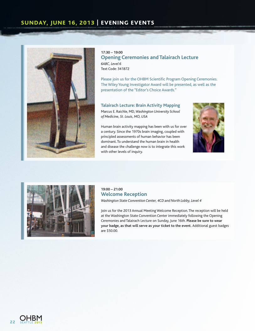
17:30 – 19:00
Opening Ceremonies and Talairach Lecture6ABC, Level 6
Text Code: 341872
Please join us for the OHBM Scientific Program Opening Ceremonies.
The Wiley Young Investigator Award will be presented, as well as the
presentation of the “Editor’s Choice Awards.”
Talairach Lecture: Brain Activity Mapping
Marcus E. Raichle, MD, Washington University School
of Medicine, St. Louis, MO, USA
Human brain activity mapping has been with us for over
a century. Since the 1970s brain imaging, coupled with
principled assessments of human behavior has been
dominant. To understand the human brain in health
and disease the challenge now is to integrate this work
with other levels of inquiry.
19:00 – 21:00
Welcome ReceptionWashington State Convention Center, 4CD and North Lobby, Level 4
Join us for the 2013 Annual Meeting Welcome Reception. The reception will be held
at the Washington State Convention Center immediately following the Opening
Ceremonies and Talairach Lecture on Sunday, June 16th. Please be sure to wear
your badge, as that will serve as your ticket to the event. Additional guest badges
are $50.00.
23
MONDAY, JUNE 17, 2013 | SCIENTIFIC PROGRAM
Morning WorkshopFunctional Assessment Of Brain Development In The Human Fetus
8:30 – 9:45
608-610, Level 6
Text Code: 342764
OrganizerMoriah E. Thomason, Merrill Palmer Skillman Institute; Pediatrics;
Perinatology Research Branch, NIH/NICHD/DHHS, Wayne State
University, Detroit, MI, USA
The organization of the brain is highly plastic in fetal life.
Establishment of healthy neural functional systems during the
fetal period is essential to normal growth and development. Across
the last several decades, remarkable progress has been made in
understanding the development of human fetal functional brain
systems. This is largely due to advances in minimally invasive
imaging methodologies. Fetal neuroimaging began in the 1950-70’s
with fetal electroencephalography (EEG) applied during labor. Later,
in the mid-1980’s, magnetoencephalography (MEG) emerged as
an effective approach for investigating fetal brain function. Most
recently, in the late 1990’s, functional magnetic resonance imaging
(fMRI) has arisen as an additional powerful approach for examining
fetal brain function. This session will cover methodologies, results,
limitations and possible future directions for functional fetal
neuroimaging research. We will address important insights into
the functional organization of the human brain at the beginning of
human life that have arisen from MRI and MEG methodologies and
will identify important targets for future research.
Learning Objectives: Having completed this workshop,
participants will be able to:
1. Enhanced understanding of the highly plastic functional
organization of the human brain in fetal life;
2. Expanded discourse about human brain development to
include the critical programming that occurs in the human
fetal brain; and
3. Demonstration of advances in fetal functional
neuroimaging that have broad applications for neuroscience
research as a whole.
fMRI of Functional Connectivity in the Fetus
Moriah E. Thomason, Merrill Palmer Skillman Institute of Child and
Family Development; Pediatrics; Perinatology Research Branch,
NICHD/NIH/DHHS, Wayne State University, Detroit, MI, USA
Signal Artifacts and Their Correction in Fetal fMRI
Colin Studholme, Biomedical Image Computing Group,
Departments of Pediatrics, Bioengineering, and Radiology,
University of Washington, Seattle, WA, USA
Time Matters: The Developmental Trajectory of Fetal
Brain Dynamics
Hubert Preissl, MEG Center, University of Tübingen;, Tübingen,
Germany and SARA-Research-Center, University of Arkansas for
Medical Sciences, Little Rock, AR, USA
Functional MRI Exploration of the Fetal Auditory Processing
Renaud Jardri, University Medical Centre of Lille, Pediatric Psychiatry
Dept., Fontan Hospital, CURE Unit, Lille, France
ASK QUESTIONS ELECTRONICALLY DURING SESSIONS
Text questions to moderators while attending sessions by dialing #22333.
In the message field, type in the unique code for the session you are attending
followed by your question and then hit send! All session codes can be found at
www.humanbrainmapping.org/questions and also next to each
session description throughout this program.

24
MONDAY, JUNE 17, 2013 | SCIENTIFIC PROGRAM
Morning WorkshopUnderstanding the Basis of Resting-State fMRI Connectivity Dynamics
8:30 – 9:45
6 ABC, Level 6
Text Code: 343708
OrganizersDaniel A. Handwerker, Section on Functional Imaging Methods,
NIMH, Bethesda, MD, USA
Catie Chang, Section on Advanced Magnetic Resonance Imaging,
NINDS, Bethesda, MD, USA
Resting-state fMRI has become a promising tool for better
diagnosing and monitoring many mental and neurological disorders,
as well as for elucidating the functional architecture of the human
brain. Although resting-state fMRI is widely applied toward such
clinical and scientific goals, many questions regarding analysis
practices and interpretation remain open. One such question is the
potential biological significance of dynamic changes in connectivity
patterns observed at short temporal scales (on the order of
seconds to minutes), and how this dynamic behavior may impact
the acquisition, analysis, and interpretation of resting-state data.
To better understand the biological significance of connectivity
dynamics, our session will focus on studies aimed at determining
relationships between connectivity changes and measures of
neuronal activity, cognition, and behavior. The four speakers in
this symposium probe changes in fMRI connectivity using distinct
approaches. Shella Keilholz investigates the neural basis of
resting-state dynamics using simultaneous MRI and microelectrode
recordings; Olaf Sporns will describe how computational models of
neuronal networks predict changes in network connectivity across
time; Javier Gonzalez-Castillo conducts behavioral interventions
(i.e., tasks with different cognitive demands) to evaluate whether
dynamic changes in fMRI resting state connectivity at short
time scales correlate with experimentally controlled changes in
mental processes; and Stephen LaConte applies real-time fMRI to
determine whether subjects can modulate resting state networks,
and also uses network activity levels to control experimental
events. These talks will provide perspectives on new ways to
study spontaneous activity and how to best link the insights from
task-based and resting fMRI studies.
Learning Objectives: Having completed this workshop, participants
will be able to:
1. Learn about current research attempting to elucidate
the potential biological significance of fMRI resting state
connectivity dynamics;
2. Gain awareness of how dynamic changes in resting state
connectivity can inform analysis practices and interpretation
of resting state data; and
3. Learn about potential applications for resting state dynamics.
Neural Basis of Dynamic Network Activity
Shella Keilholz, Wallace H. Coulter Department of Biomedical
Engineering, Georgia Tech and Emory University, Atlanta, GA, USA
EEG Correlates of Functional Connectivity States
Elena Allen, K.G. Jebsen Center for Research on Neuropsychiatric
Disorders and the Department of Biological and Medical Psychology
at the University of Bergen, Norway; Mind Research Network,
Albuquerque, New Mexico, USA
When Does a Task Disturb Rest?
Javier Gonzalez-Castillo, Section on Functional Imaging Methods,
NIMH, NIH, Bethesda, MD, USA
Directly Testing the Roles of Resting-State Networks with
Real-Time fMRI
Stephen LaConte, Virginia Tech, Carilion School of Medicine,
Roanoke, VA, USA
25
Morning WorkshopShaping-up Nicely: Advances in Developmental and Translational Neuroimaging of the Subcortex
8:30 – 9:45
605-607, Level 6
Text Code: 343607
OrganizerM. Mallar Chakravarty, The Centre for Addiction and Mental Health,
Toronto, Canada
Armin Raznahan, Child Psychiatry Branch, National Institute of
Mental Health, Bethesda, MS, USA
Sub-cortical systems sometimes take a backseat to the cerebral
cortex as a focus for basic and clinical neuroimaging studies.
However, structures such as the striatum and thalamus are
evolutionarily ancient components of the brain that not only play
a fundamental role in sensorimotor processing, but also diverse
domains of higher mental function. Despite the clear importance
of sub-cortical systems for developmentally dynamic, sexually
differentiated and disease-sensitive aspects of brain function,
sub-cortical maturation and sexual dimorphism in humans remain
relatively uncharted, and clinical studies rooted in these normative
models are scarcer still. This symposium will bring together some of
the latest work from labs in Europe and North American that have
been developing and applying new tools for sub-cortical analysis in
order to (i) unlock the wealth of shape-related information hidden
within classical measures of sub-cortical volume, (ii) create four-
dimensional maps of sub-cortical maturation using longitudinal
data in healthy youth, (iii) dissect-out patterns of structural and
functional connectedness between sub-cortical structures and the
rest of the brain, and (iv) leverage these newly-built normative
models to arrive at mechanistically informative and clinically
useful sub-cortical signatures of neuropsychiatric disorders across
the lifespan.
Learning Objectives:
1. The unique challenges faced in MRI-based analysis of
sub-cortical systems, and the latest strategies being adopted
to address these;
2. How the volume and shape of sub-cortical systems change
between childhood and adolescence in healthy males and
females, the way in which age and sex-biased illnesses impact
typical sub-cortical development, and the structural and
functional connections that tie developmental dynamic and
disease-sensitive sub-cortical “hot-spots” into other brain
systems to underpin behavior; and
3. The power of high-resolution, high-field image acquisition
techniques in fine-mapping sub-cortical connectivity in
humans as a parallel to animal research and strategies for
wielding sub-cortical analyses in order to generate clinical
useful predictions in disease states.
Developmental Deformations of the Subcortex in Healthy:
Localizing “Hotspots” of Dynamic Change and Sexual
Dimorphism in Childhood and Adolescence
Armin Raznahan, Child Psychiatry Branch, National Institute of
Mental Health, Bethesda, MD, USA
Compromised Neuroanatomical Developmental Trajectories
and the Translational Utility of Subcortical Anatomy
M. Mallar Chakravarty, The Centre for Addiction and Mental Health,
Toronto, ON, Canada
Ultra-High 7T MRI of Structural Age-Related Changes of the
Subthalamic Nucleus
Birte U. Forstmann, Cognitive Science Center Amsterdam,
University of Amsterdam, Amsterdam, The Netherlands
Structural and Functional Cortical-Subcortical Interactions
and Their Relationship to Typical and Atypical Development
Damien Fair, Oregon Health and Science University, Portland,
OR, USA
Break9:45 – 10:00

26
MONDAY, JUNE 17, 2013 | SCIENTIFIC PROGRAM
LOC SymposiumNeural – Device Interfaces and Cortical Circuitry: Excitement in Both Directions
10:00 – 11:30
6ABC, Level 6
Text Code: 341605
OrganizerTom Grabowski, University of Washington, Seattle, WA, USA
The LOC will showcase Seattle as a center of neuroscientific
innovation with a symposium that brings together leading local
efforts to fathom the circuitry of the cortex, and to develop novel
brain-machine interface technology. Advances in machine learning,
sensor technology, and optogenetics have made brain-computer
interfaces increasingly feasible, but an understanding of brain
systems architecture and its cortical basis is critical for advancing
useful BCI and this is in turn informs systems neuroscience.
Mapping the Neocortex at the Cellular Level in a Large-Scale
and High Throughput Manner
Christof Koch, Allen Institute for Brain Science, Seattle, WA, USA
Bidirectional Interactions Between the Brain and
Implantable Computers
Eberhard Fetz, Departments of Physiology & Biophysics and
Bioengineering, University of Washington, Seattle, WA, USA
Dynamic Learning Networks Support Brain-Machine
Interface Adaptation
Jeffrey Ojemann, Department of Neurological Surgery,
University of Washington, Seattle, WA, USA
Break11:30 – 11:45
Keynote LectureThe Role of Multimodal Imaging in the Development and Refinement of Deep Brain Stimulation for Depression
11:45 – 12:30
6ABC, Level 6
Text Code: 341001
Helen Mayberg, Emory University, Atlanta, GA, USA
Deep Brain Stimulation is an emerging treatment strategy for
patients with intractable depression with imaging playing a crucial
role in the development, testing and refinement of the procedure.
Combined with real-time behavioral and physiological metrics,
these studies offer a unique perspective on critical pathways and
mechanisms mediating antidepressant effects of DBS, and on the
pathophysiology of treatment resistant depression more generally.
We cordially invite you to our Philips Lunch Symposium during OHBM. On Monday, June 17th 2013, 12.45-13.45. Room 602-640, we will update you on our fMRI portfolio. Listen to our keynote Neuroscience speakers who will present some of their current cutting edge activities. The symposium is free to attend and lunch will e provided to the rst attendees. We are looking forward to seeing you!
Philips Neuroscience MRI Symposium
27
Lunch12:30 – 13:30
Interactive Poster Presentations13:30 – 14:15
608-610, Level 6
Text Code: 614743
I-Poster presentations highlight top ranked submitted abstracts.
Authors will present their abstracts in a short, “datablitz” format.
The objective of the I-Poster session is to arrive at a hybrid of
posters and oral sessions.
Moderator: Marco Catani, Natbrainlab, King’s College London,
London, UK
13:30 – 13:35
3511: Combining ZOOPPA and blipped CAIPIRINHA for
highly accelerated Diffusion Weighted Imaging at 7T & 3T
Cornelius Eichner, Athinoula A. Martinos Center for
Biomedical Imaging, Charlestown, MA, USA
13:35 – 13:40
3741: The Human Cerebral Cortex Flattens During Adolescence
Yasser Alemán-Gómez, Instituto de Investigación Sanitaria
Gregorio Marañón, IiSGM, HGUGM, CIBERSAM, Madrid, Spain
13:40 – 13:45
3880: Attentional base response in human superior colliculus
measured using high-resolution fMRI
Sucharit Katyal, University of Texas at Austin, Austin, TX, USA
13:45 – 13:50
3958: Reciprocal anti-correlation underlies multi-functionality
of the temporo-parietal junction
Danilo Bzdok, Research Center Jülich, Germany
13:50 – 13:55
4027: Electrocorticography of the Face and Place Network
Connectivity
Mehmet Kadipasaoglu, University of Texas Medical School
at Houston, Houston, TX, USA
13:55 – 14:00
4030: Functional cross-subject mapping reveals common
dimensions of visually evoked brain activity
Natalia Bilenko, University of California, Berkeley, CA, USA
14:00 – 14:15
Questions and Answers
Poster Session13:30 – 15:30
Exhibit and Poster Hall – 4AB, Level 4
Poster Numbers #1000-2119: Even Numbered Posters Stand-By
Brain Stimulation Methods: Deep Brain Stimulation, Direct
Electrical/Optogenetic Stimulation, TDCS, TMS
Disorders of the Nervous System: Autism, Developmental
Disorders, Other Disorders, Stroke, Obsessive-Compulsive Disorder
and Tourette Syndrome, Parkinson’s Disease and Movement
Disorders, Sleep Disorders
Genetics: Genetic Association Studies, Genetic Modeling and
Analysis Methods, Neurogenetic Syndromes
Higher Cognitive Functions: Decision Making, Executive Function,
Imagery, Music, Reasoning and Problem Solving, Space, Time and
Number Coding
Informatics: Atlases, Databasing and Data Sharing, Pipelines
Language: Language Acquisition, Language Comprehension
and Semantics, Reading and Writing, Speech Perception,
Speech Production
Learning and Memory: Implicit Memory, Long-Term Memory
(Episodic and Semantic), Neural Plasticity and Recovery of
Function, Skill Learning, Working Memory
Modeling and Analysis Methods: Bayesian Modeling, Classification
and Predictive Modeling, Diffusion MRI Modeling and Analysis,
EEG/MEG Modeling and Analysis, Exploratory Modeling and
Artifact Removal, fMRI Connectivity and Network Modeling, Image
Registration and Computational Anatomy, Motion Correction and
Preprocessing, Multivariate modeling, Other Methods, PET Modeling
and Analysis, Segmentation and Parcellation, Task-Independent and
Resting-State Analysis, Univariate Modeling
Motor Behavior: Brain Machine Interface, Mirror System,
Motor Planning and Execution, Visuo-Motor Functions
Physiology, Metabolism and Neurotransmission:
Cerebral Metabolism and Hemodynamics, Neurophysiology
of Imaging Signals, Pharmacology and Neurotransmission
Break15:30 – 15:45
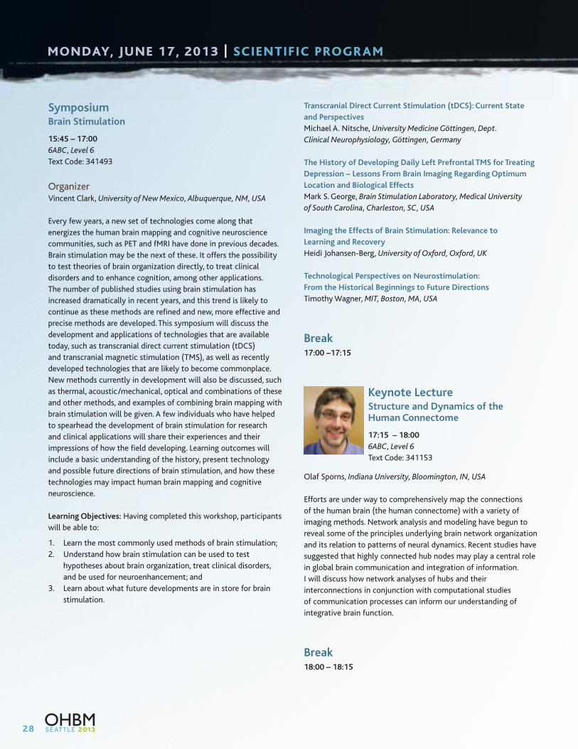
28
MONDAY, JUNE 17, 2013 | SCIENTIFIC PROGRAM
SymposiumBrain Stimulation
15:45 – 17:00
6ABC, Level 6
Text Code: 341493
OrganizerVincent Clark, University of New Mexico, Albuquerque, NM, USA
Every few years, a new set of technologies come along that
energizes the human brain mapping and cognitive neuroscience
communities, such as PET and fMRI have done in previous decades.
Brain stimulation may be the next of these. It offers the possibility
to test theories of brain organization directly, to treat clinical
disorders and to enhance cognition, among other applications.
The number of published studies using brain stimulation has
increased dramatically in recent years, and this trend is likely to
continue as these methods are refined and new, more effective and
precise methods are developed. This symposium will discuss the
development and applications of technologies that are available
today, such as transcranial direct current stimulation (tDCS)
and transcranial magnetic stimulation (TMS), as well as recently
developed technologies that are likely to become commonplace.
New methods currently in development will also be discussed, such
as thermal, acoustic/mechanical, optical and combinations of these
and other methods, and examples of combining brain mapping with
brain stimulation will be given. A few individuals who have helped
to spearhead the development of brain stimulation for research
and clinical applications will share their experiences and their
impressions of how the field developing. Learning outcomes will
include a basic understanding of the history, present technology
and possible future directions of brain stimulation, and how these
technologies may impact human brain mapping and cognitive
neuroscience.
Learning Objectives: Having completed this workshop, participants
will be able to:
1. Learn the most commonly used methods of brain stimulation;
2. Understand how brain stimulation can be used to test
hypotheses about brain organization, treat clinical disorders,
and be used for neuroenhancement; and
3. Learn about what future developments are in store for brain
stimulation.
Transcranial Direct Current Stimulation (tDCS): Current State
and Perspectives
Michael A. Nitsche, University Medicine Göttingen, Dept.
Clinical Neurophysiology, Göttingen, Germany
The History of Developing Daily Left Prefrontal TMS for Treating
Depression – Lessons From Brain Imaging Regarding Optimum
Location and Biological Effects
Mark S. George, Brain Stimulation Laboratory, Medical University
of South Carolina, Charleston, SC, USA
Imaging the Effects of Brain Stimulation: Relevance to
Learning and Recovery
Heidi Johansen-Berg, University of Oxford, Oxford, UK
Technological Perspectives on Neurostimulation:
From the Historical Beginnings to Future Directions
Timothy Wagner, MIT, Boston, MA, USA
Break17:00 –17:15
Keynote LectureStructure and Dynamics of the Human Connectome
17:15 – 18:00
6ABC, Level 6
Text Code: 341153
Olaf Sporns, Indiana University, Bloomington, IN, USA
Efforts are under way to comprehensively map the connections
of the human brain (the human connectome) with a variety of
imaging methods. Network analysis and modeling have begun to
reveal some of the principles underlying brain network organization
and its relation to patterns of neural dynamics. Recent studies have
suggested that highly connected hub nodes may play a central role
in global brain communication and integration of information.
I will discuss how network analyses of hubs and their
interconnections in conjunction with computational studies
of communication processes can inform our understanding of
integrative brain function.
Break18:00 – 18:15

29
Oral Sessions18:15 – 19:45
Oral session presentations are chosen by the Program Committee
from submitted abstracts using criteria of quality and timeliness;
a wide spectrum of investigation is represented.
O – M1: Brain Stimulation Methods and Motor Behavior
602-604, Level 6
Text Code: 341201
Chair: Christian Ruff, University of Zurich, Zurich, Switzerland
18:15 – 18:30
1047: Topological correlates of motor improvements after
repetitive transcranial magnetic stimulation
Chang-hyun Park, UCL Institute of Neurology, London, UK
18:30 – 18:45
2057: The role of the anterior midcingulate cortex in
neurofeedback training
Tibor Auer, Biomedizinische NMR GmbH am Max Planck Institute
for Biophysical Chemistry, Goettingen, Germany
18:45 – 19:00
2061: Exploring the Neural Basis of Observational Motor
Learning using Resting-state fMRI
Heather McGregor, Western University, London, Canada
19:00 – 19:15
1018: Establishing a causal link between oscillatory coupling
of brain activity and cognitive performance
Rafael Polania, Laboratory for Social and Neural Systems
Research, Department of Economics, University of Zurich,
Zurich, Switzerland
19:15 – 19:30
1037: Impact of 5Hz rTMS is related to volume of white
matter in the sensory cortex after stroke
Sonia Brodie, University of British Columbia, Vancouver, Canada
19:30 – 19:45
1003: Fornix Deep Brain Stimulation Induces Functional
Activation in Hippocampal Circuitry
Erika Ross, Mayo Clinic, Rochester, MN, USA
O – M2: Disorders of the Nervous System 1: Pediatric and Developmental Disorders
608-610, Level 6
Text Code: 341222
Chair: Damien Fair, Oregon Health & Science University,
Portland, OR, USA
18:15 – 18:30
3252: Modules of synchronized cortical maturation in typical
development and childhood-onset schizophrenia
Aaron F. Alexander-Bloch, Brain Mapping Unit, Department of
Psychiatry, University of Cambridge, Cambridge, UK
18:30 – 18:45
1064: Diagnostic classification of autism using particle
swarm optimization for fMRI feature selection
Colleen Chen, San Diego State University, San Diego, CA, USA
18:45 – 19:00
3147: Assaulted Adolescents Fail to Recruit Domain-Specific
Neural Networks during Conflict Processing
James Steele, University of Arkansas for Medical Sciences, Little
Rock, AR, USA
19:00 – 19:15
3155: Childhood Maltreatment Contributes to Altered Resting
State Connectivity in Young Combat Veterans
Remi Patriat, University of Wisconsin Madison, Madison, USA
19:15 – 19:30
1081: Robust antero-posterior default mode
hypoconnectivity in ASDs despite motion scrubbing
Tuomo Starck, Oulu University, Oulu, Finland
19:30 – 19:45
3002: Abnormal Patterns of Gyrification in Fetal Alcohol
Spectrum Disorder
Shantanu Joshi, UCLA, Los Angeles, CA, USA
30
MONDAY, JUNE 17, 2013 | SCIENTIFIC PROGRAM
O – M3: Modeling and Analysis Methods 1: Resting – State
6 ABC, Level 6
Text Code: 341226
Chair: Elena Allen, Mind Research Network, Albuquerque, NM, USA
18:15 – 18:30
1775: Boundaries on Functional Connectivity Boundaries
Fenna Krienen, Harvard University, Cambridge, MA, USA
18:30 – 18:45
1796: Detection of Spontaneous Co-activation Patterns by
Selectively Grouping Resting-State fMRI Volumes
Xiao Liu, NIH, Bethesda, MD, USA
18:45 – 19:00
2019: Inferring Transiently Synchronising Networks using a
Hidden Markov Model
Adam Baker, University of Oxford, Oxford, UK
19:00 – 19:15
2011: Frequency characteristics of large scale resting state
networks using 7T Spin Echo EPI
Erik van Oort, MIRA Institute, University of Twente, Donders
Institute, Radboud University Nijmegen, Nijmegen, Netherlands
19:15 – 19:30
1996: Characterizing Intrinsic Connectivity Microstates
Within The Posteromedial Cortex
Zhen Yang, Center for the Developing Brain, Child Mind Institute,
New York, NY, USA
19:30 – 19:45
1974: Convergent functional organization of Broca’s area
across multi-sites rs-fMRI datasets
Yu Zhang, Institute of Automation, Chinese Academy of Sciences,
Beijing, China
O – M4: High Resolution Imaging
605-607, Level 6
Text Code: 341264
Chair: Natalia Petridou, UMC Utrecht, Utrecht, Netherlands
18:15 – 18:30
3384: Fine details of brain anatomy revealed in-vivo by
ultra-high resolution quantitative T1 mapping
Pierre-Louis Bazin, Max Planck Institute for Human Cognitive and
Brain Sciences, Leipzig, Germany
18:30 – 18:45
3815: Detailed laminar characteristics of the human
neocortex revealed by NODDI and histology
Michiel Kleinnijenhuis, University Medical Centre St. Radboud,
Nijmegen, Netherlands
18:45 – 19:00
2115: Variable Couplings between Neural Activity and
Flow-metabolism across Cortical Lamina
Fahmeed Hyder, Yale University, New Haven, CT, USA
19:00 – 19:15
2090: A hemodynamic model for layered BOLD signals
Jakob Heinzle, Translational Neuromodelling Unit,
University Zurich & ETH Zurich, Zurich, Switzerland
19:15 – 19:30
2104: Correspondence of Spontaneous and Evoked
Inter-Laminar Functional Connectivity of Current Sources
Shmuel Naaman, MNI, McGill University, Montreal, Canada
19:30 – 19:45
1920: Decoding cell-type specific spatial representations in
human entorhinal layers
Tobias Navarro Schroeder, Donders Institute for Brain, Cognition
and Behaviour; Radboud University Nijmegen; The Netherlands,
Nijmegen, Netherlands
31
Morning WorkshopResting State Connectivity: Views From Nonhuman Primates
8:30 – 9:45
6 ABC, Level 6
Text Code: 343605
OrganizerAnna W. Roe, Dept of Psychology, Vanderbilt University, Nashville,
TN, USA
Correlated spontaneous activity in the resting brain (or resting
state activity, RSA) is increasingly recognized as a useful index
for inferring underlying functional-anatomic architecture. However,
fundamental questions remain regarding the interpretation of
RSA; such questions can be informed by studies of RSA in the
nonhuman primate. The first two speakers will first address the
fundamental issue of whether RSA can be equated with underlying
anatomical connectivity and whether the size of the network scale
under consideration (local or global) affects this relationship.
Dr. Anna Roe will show at a very local mm-based scale within area
3b and 1 in squirrel monkey somatosensory cortex, that local digit
connectivity revealed by anatomical tracers and neuronal cross
correlation strongly parallels BOLD RSA. Dr. Yasushi Miyashita
brings this analysis to a larger scale and will show in anesthetized
macaques that RSA may be predicted by interareal short
connection patterns but, with larger global networks, it deviates
from direct underlying anatomy and is substantially influenced
by network-level cortical architecture. The second two speakers
will address RSA-inspired studies of homologies between monkey
and man. Dr. Sebastian Neggers presents new data suggesting
commonalities between monkey and human fronto-striatal systems
underlying eye movement behavior; surprisingly, this data calls
for a revision of previous models of oculomotor circuitry in the
monkey. Using both resting state and natural-vision fMRI data,
Dr. Wim Vanduffel examines homologies between monkey and
human and identifies signatures for conserved brain circuits as
well as distinct networks which may have been adapted for new
functions over evolution.
Learning Objectives:
1. What is the anatomical and neurophysiological basis of resting
state connectivity? Does this basis differ between the size
(local vs global) of network examined?
2. Are there evolutionarily conserved networks?
3. What novel networks have emerged over evolution?
4. How do resting state networks relate to behavioral networks?
The Anatomical and Functional Basis of Resting State
Connectivity in Primate Somatosensory Cortex
Anna W. Roe, Dept of Psychology, Vanderbilt University,
Nashville, TN, USA
Differences in Anatomical Basis of Local vs Global Resting
State Networks
Yusuke Adachi, Dept of Physiology, University of Tokyo, Tokyo, Japan
Comparison of Human and Monkey Networks Reveal Conserved
and Evolutionarily Novel Networks
Wim Vanduffel, Massachusetts General Hospital, Charlestown,
MA, USA
Comparing the Human and Macaque Fronto-Striatal
Oculomotor Network
Bas Neggers, University Medical Center Utrecht, Utrecht,
The Netherlands
ASK QUESTIONS ELECTRONICALLY DURING SESSIONS
Text questions to moderators while attending sessions by dialing #22333.
In the message field, type in the unique code for the session you are attending
followed by your question and then hit send! All session codes can be found at
www.humanbrainmapping.org/questions and also next to each
session description throughout this program.
TUESDAY, JUNE 18, 2013 | SCIENTIFIC PROGRAM
32
TUESDAY, JUNE 18, 2013 | SCIENTIFIC PROGRAM
Morning WorkshopFunctional Data-Driven Atlases of the Brain
8:30 – 9:45
608-610, Level 6
Text Code: 342778
OrganizerBertrand Thirion, Parietal team, INRIA Saclay, Gif sur Yvette, France
The exploration of brain structure and function through various
functional and anatomical neuroimaging modalities includes
its segmentation into areas that are characterized by different
cytoarchitecture, connectivity and functional organization. In
this respect, mounting evidence suggests that region-specific
features are, to some extent, reflected in the measurements
provided by various neuroimaging modalities. Conversely, emerging
neuroimaging and modeling approaches allow the in-vivo
parcellation of the brain volume (or cortical surface) as a set of
multiple pieces or modules. Parcellations are a concise way to
map information conveyed by various modalities, as they relate
these observations with a certain ontology of brain structures and
organization. They play a key role in the understanding of structure-
function relationships, i.e. in the identification of the neural
implementation of various cognitive functions. The practical value
of brain parcellations is that intersubject anatomical variability as
well as the additional spatial variability of functional regions cannot
be completely overcome by current spatial normalization methods.
However, the impact of such residual variability can be mitigated
by the use of extended regions instead of voxels when brain image
analysis procedures are performed at the group level. An additional
benefit of parcellations is that they make it possible to run complex
analysis procedures (e.g. the estimation of graphical models in
connectivity studies) in an informed fashion rather than arbitrarily
dividing the brain into small units. They also make it easier to run
multivariate pattern analyses and to provide benefits in sensitivity
by limiting the multiple comparison problem. This particular aspect
might gain importance with the advent of high-resolution data on
the one hand, and of under-powered neuroimaging-genetic studies
on the other hand. In spite of the emerging success of the data-
driven definition of brain regions, this approach also carries some
ambiguities: subdivision of some structures into sub-structures
is debatable and sharp borders are not necessarily observed with
a high confidence due to limited resolution and signal-to-noise
ratio of current neuroimaging data. Current challenges from group
models to individual models: to make parcellations usable, a major
issue is to adapt a model (atlas) to a given subject under study.
Data-driven procedures are particularly useful because they can
take into account the characteristics of new subjects. Yet, it is still
unclear how to model properly this between-subject variability.
Link data-driven models with an ontology of brain regions: the
neuroimaging community deals with many different ontologies of
the brain territories, that provide more or less consistent labellings
of the image data. As a community, we need to find rules to decide
on the evolution of the ontology; on the other hand, data-driven
parcellations need to take into account prior knowledge associated
with current ontologies. Improve the data-driven definition of brain
regions: parcellation models need to be inferred from and compared
across different modalities, protocols and methods. They can take
various priors or constraints into account. The procedures used to
infer them have to be made available to the community.
Model selection: Given a certain amount of data, the model that
represents the best bias-variance compromise can be considered
as the “best” model. In the case of brain parcellations, the problem
consists typically in estimating the right number of regions.
However defining this best model this is intrinsically an ill-posed
problem, and rather indirect approaches and surrogate criteria have
to be used instead.
Learning Objectives: Having completed this workshop,
participants will be able to:
1. Understand the neuroscientific underpinnings of brain
parcellation models and the limitations as well as open issues
in current parcellation schemes and models;
2. Know what combinations of imaging modalities and
computational tools are currently used to perform brain
parcellation, as well as the best practices;
3. Refer to adequate tools and packages to find the necessary
resource; and
4. Understand the impact of choosing adapted spatial models for
connectivity inference, multivariate pattern analysis or group
studies (e.g. neuroimaging-genetics).
Data Driven Methods for Neuroimaging Phenotypes:
Progress and Pitfalls
Jessica A. Turner, Director of Operations, 3T MRI Center,
Mind Research Network, Albuquerque, NM, USA
Areas and Clusters: What Are We Mapping?
Simon B. Eickhoff, Heinrich-Heine University Düsseldorf,
Düsseldorf, Germany
Brain Parcellations for the Modeling of Brain Functional
Connectivity
Steve Petersen, Director, McDonnell Center for Systems Neuroscience
Depts. of Neurology and Psychology Washington University Medical
School, Saint Louis, MO, USA
A Statistical Perspective on Brain Parcellations
Bertrand Thirion, Parietal team, INRIA Saclay, Gif sur Yvette, France
33
Morning WorkshopOn the Use of Brain Network Measures to Characterize Major Mental Disorders
8:30 – 9:45
605-607, Level 6
Text Code: 343577
OrganizerVince D. Calhoun, The Mind Research Network & The University of
New Mexico, Albuquerque, NM, USA
There has been a considerable amount of work recently on the
characterization of brain structure and function in the context of
“networks”. This includes identifying correlated changes, defining
various network properties (such as small worldness, rich club
behavior, or more general information theoretic measures) and
evaluating changes in these properties in various patient groups.
We propose to bring together experts in the use of network-based
approaches to characterize neuropsychiatry illness. Dr. Bullmore
will present an overview of how network approaches can inform
a study of schizophrenia, from characterizing resting and task
networks, pharmacological interventions, and even generative
models. Dr. Calhoun will discuss some exciting findings showing
state versus trait aspects are captured in different intrinsic
network properties in schizophrenia and bipolar disorder. He will
also discuss the issue of overlapping symptoms in the patient
groups. Dr. Hendler utilizes a new approach for portraying
individual emotional experience via dynamics in network
cohesion to characterize the interaction between emotional
and cognitive aspects of mental processing in schizophrenia
patients and their healthy siblings. And Dr. Jiang will discuss a
multi-level framework for characterizing mental illness from
whole-brain networks to genes.
Learning Objectives: Having completed this workshop, participants
will be able to:
1. Realize the superiority of using network-based approach for
translating brain imaging finding to mental illness practice;
2. Understand the differences between the neural correlates
of state and trait aspect of mental illness as well as the
importance of these concepts for effective personalized
psychairty; and
3. Identify several types of network measures in brain imaging
and understand their differential advantages and disadvantages
for the diagnosis and/or treatment of mental disorders.
Brain Network Disorganization in Schizophrenia
Ed Bullmore, University of Cambridge, Cambridge, UK
Intrinsic Functional Networks are Sensitive Markers of State
versus Trait Changes in Bipolar Disorder and Schizophrenia
Vince D. Calhoun, The Mind Research Network & The University
of New Mexico, Albuquerque, NM, USA
Network Probing of Emotional Dynamics in Schizophrenia
Patients and Their Healthy Siblings
Talma Hendler, Tel Aviv Medical Center and Tel Aviv University,
Tel Aviv, Israel
Brainnetome-Wide Association Studies in Schizophrenia
Tianzi Jiang, The Chinese Academy of Sciences, Beijing, China
Break9:45 – 10:00
Keynote LectureLinking Mental and Neural Function Using Representational fMRI
10:00 – 10:45
6ABC, Level 6
Text Code: 340739
Russell Poldrack, University of Texas at Austin, TX, USA
A continuing debate has centered around the question of whether
neuroimaging can inform questions about psychological function.
I will show how representational similarity analysis with fMRI data
provides the ability to test the claims of psychological models
regarding the similarity of mental representations, and discuss how
these methods allow the establishment of powerful isomorphisms
between psychological and neural representations.
Break10:45 – 11:00
34
TUESDAY, JUNE 18, 2013 | SCIENTIFIC PROGRAM
Oral Sessions11:00 – 12:30
Oral session presentations are chosen by the Program Committee
from submitted abstracts using criteria of quality and timeliness;
a wide spectrum of investigation is represented.
O – T1: Higher Cognitive Functions
602-604, Level 6
Text Code: 341278
Chair: Simon Eickhoff, Heinrich-Heine University Düsseldorf,
Düsseldorf, Germany
11:00 – 11:15
1297: A within-attribute comparison strategy in
multi-attribute choice
Laurence Hunt, University College London, London, UK
11:15 – 11:30
1417: Analogue coding of nonsymbolic numbers and digital
coding of symbolic numbers in the human brain
Ian Lyons, University of Chicago, Chicago, IL, USA
11:30 – 11:45
1317: Informatic Parcellation of the Network Involved
in the Neural Computation of Value
John Clithero, California Institute of Technology, Pasadena,
CA, USA
11:45 – 12:00
1367: Post-error adjustments are modulated by
acetylcholine
Markus Ullsperger, Radboud University, Nijmegen, Netherlands
12:00 – 12:15
1352: Functional Maturation of the Executive System
in Adolescence
Theodore Satterthwaite, UPenn, Philadelphia, PA, USA
12:15 – 12:30
1400: Interactions between nucleus accumbens and
sensory cortices predict music reward value
Valorie N. Salimpoor, McGill University, Montreal, Canada
O – T2: Modeling and Analysis Methods 2: Functional Modeling
6 ABC, Level 6
Text Code: 341353
Chair: Stephen LaConte, Virginia Tech, Carilion School of Medicine,
Roanoke, VA, USA
11:00 – 11:15
1708: EEG modeling of the sleep wake transition using
physiologically based neural field theory
Romesh Abeysuriya, University of Sydney, Sydney, Australia
11:15 – 11:30
1752: Spatiotemporal neural dynamics from fMRI:
Deconvolution with a spatiotemporal HRF
Kevin Aquino, University of Sydney, Sydney, Australia
11:30 – 11:45
1786: Connectivity-based neurofeedback: dynamic
causal modeling for real-time fMRI.
Yury Koush, EPFL Lausanne, Lausanne, Switzerland
11:45 – 12:00
1662: Sparse network-based discriminative models for
depression using fMRI
Maria Joao Rosa, UCL, London, UK
12:00 – 12:15
1823: Granger causality analysis of fMRI is invariant to
hemodynamic convolution but not downsampling
Anil Seth, University of Sussex, Brighton, UK
12:15 – 12:30
1800: Dynamic causal modeling for arterial spin
labeling data
Martin Havlicek, Maastricht University, Maastricht, Netherlands
35
O – T3: Neuroimaging Genetics and Informatics
608-610, Level 6
Text Code: 341377
Chair: Jason Stein, UCLA, Los Angeles, CA, USA
11:00 – 11:15
4110: Model-based neurogenetic characterization of social
information processing in an interactive game
Andreea Oliviana Diaconescu, Translational Neuromodeling Unit
(TNU), University & ETH Zürich, Zurich, Switzerland
11:15 – 11:30
1285: SOLAR-Eclipse computational tools for imaging
genetic and mega-genetic analysis
Peter Kochunov, Maryland Psychiatric Research Center,
Baltimore, MD, USA
11:30 – 11:45
1284: Multi-Site Genetic Analysis of 1151 Diffusion MRI
Scans from the ENIGMA–DTI Working Group
Neda Jahanshad, UCLA, Los Angeles, CA, USA
11:45 – 12:00
1288: VGWAS revisited: A novel and powerful approach to
voxelwise genome-wide association studies
Jonathan Rosenblatt, Tel Aviv University, Tel Aviv, Israel
12:00 – 12:15
1289: Voxel-wise and Cluster-based Heritability Inferences
of fMRI Data
Xu Chen, University of Warwick, Coventry, UK
12:15 – 12:30
1282: Genetic control of resting state default mode network
derived from task fMRI using a TWINs dataset
Mayuresh Korgaonkar, University of Sydney, Sydney, Australia
O – T4: Language, Learning and Memory
605-607, Level 6
Text Code: 341227
Chair: Svenja Caspers, Institute for Neuroscience and Medicine,
Research Center Jülich, Jülich, Germany
11:00 – 11:15
1556: Layer-specific encoding processes in the human
medial temporal lobe
Anne Maass, Institute of Cognitive Neurology and Dementia
Research, Otto-von-Guericke-University Magdeburg,
Magdeburg, Germany
11:15 – 11:30
1598: A longitudinal study of fronto-parietal and
fronto-striatal networks and working memory development
Fahimeh Darki, Neuroscience Department, Stockholm, Sweden
11:30 – 11:45
1608: Feedback associated with reward normalizes
responses in ADHD children during working memory tasks
Rubi Hammer, Northwestern University, Evanston, IL, USA
11:45 – 12:00
1472: The neural segregation of syntax from semantics in
the developing brain
Michael Skeide, MPI CBS, Leipzig, Germany
12:00 – 12:15
1542: Sensory-Motor Integration in Speech Production,
a Voxel-Based Lesion-Symptom Mapping Study
Tasha Poppa, University of California, Irvine, Irvine, CA, USA
12:15 – 12:30
3951: Electrocorticography of visual cortex responses to
multisensory speech
Inga Schepers, University of Texas Health Science Center at
Houston, Houston, TX, USA
36
TUESDAY, JUNE 18, 2013 | SCIENTIFIC PROGRAM
Lunch12:30 – 13:30
Interactive Poster Presentations13:30 – 14:15
608-610, Level 6
Text Code: 675686
Moderator: Peter Bandettini, National Institutes of Health,
Bethesda, MD, USA
I-Poster presentations highlight top ranked submitted abstracts.
Authors will present their abstracts in a short, “datablitz” format.
The objective of the I-Poster session is to arrive at a hybrid of
posters and oral sessions.
13:30 – 13:35
3376: Background-Suppressed Myelin Water Imaging
Jongho Lee, University of Pennsylvania, Philadelphia, PA, USA
13:35 – 13:40
3465: Physiological Noise Reduction in 7 T fMRI using
Concurrent Magnetic Field Monitoring
Jakob Heinzle, University of Zurich/ETH Zurich, Zurich, Switzerland
13:40 – 13:45
3416: BOLD mapping of finger movement compares with
electrophysiology; a combined 7T fMRI and ECoG study
Jeroen Siero, UMC Utrecht, Utrecht, Netherlands
13:45 – 13:50
3549: Colocalizing EEG and fMRI in Space
Pamela Douglas, UCLA, Los Angeles, CA, USA
13:50 – 13:55
3552: EEG dynamic source imaging based on general linear
modeling and non-parametric statistical testing
Anna Custo, University of Geneva, Switzerland
13:55 – 14:00
3593: A combined portable multimodal imaging system
for electric and hemodynamic activity of the brain
Javad Safaie, UPJV, Amiens, France
14:00 - 14:15
Questions and Answers
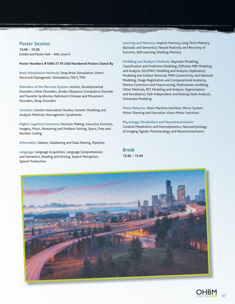
37
Poster Session13:30 – 15:30
Exhibit and Poster Hall – 4AB, Level 4
Poster Numbers #1000-2119: Odd Numbered Posters Stand-By
Brain Stimulation Methods: Deep Brain Stimulation, Direct
Electrical/Optogenetic Stimulation, TDCS, TMS
Disorders of the Nervous System: Autism, Developmental
Disorders, Other Disorders, Stroke, Obsessive-Compulsive Disorder
and Tourette Syndrome, Parkinson’s Disease and Movement
Disorders, Sleep Disorders
Genetics: Genetic Association Studies, Genetic Modeling and
Analysis Methods, Neurogenetic Syndromes
Higher Cognitive Functions: Decision Making, Executive Function,
Imagery, Music, Reasoning and Problem Solving, Space, Time and
Number Coding
Informatics: Atlases, Databasing and Data Sharing, Pipelines
Language: Language Acquisition, Language Comprehension
and Semantics, Reading and Writing, Speech Perception,
Speech Production
Learning and Memory: Implicit Memory, Long-Term Memory
(Episodic and Semantic), Neural Plasticity and Recovery of
Function, Skill Learning, Working Memory
Modeling and Analysis Methods: Bayesian Modeling,
Classification and Predictive Modeling, Diffusion MRI Modeling
and Analysis, EEG/MEG Modeling and Analysis, Exploratory
Modeling and Artifact Removal, fMRI Connectivity and Network
Modeling, Image Registration and Computational Anatomy,
Motion Correction and Preprocessing, Multivariate modeling,
Other Methods, PET Modeling and Analysis, Segmentation
and Parcellation, Task-Independent and Resting-State Analysis,
Univariate Modeling
Motor Behavior: Brain Machine Interface, Mirror System,
Motor Planning and Execution, Visuo-Motor Functions
Physiology, Metabolism and Neurotransmission:
Cerebral Metabolism and Hemodynamics, Neurophysiology
of Imaging Signals, Pharmacology and Neurotransmission
Break15:30 – 15:45
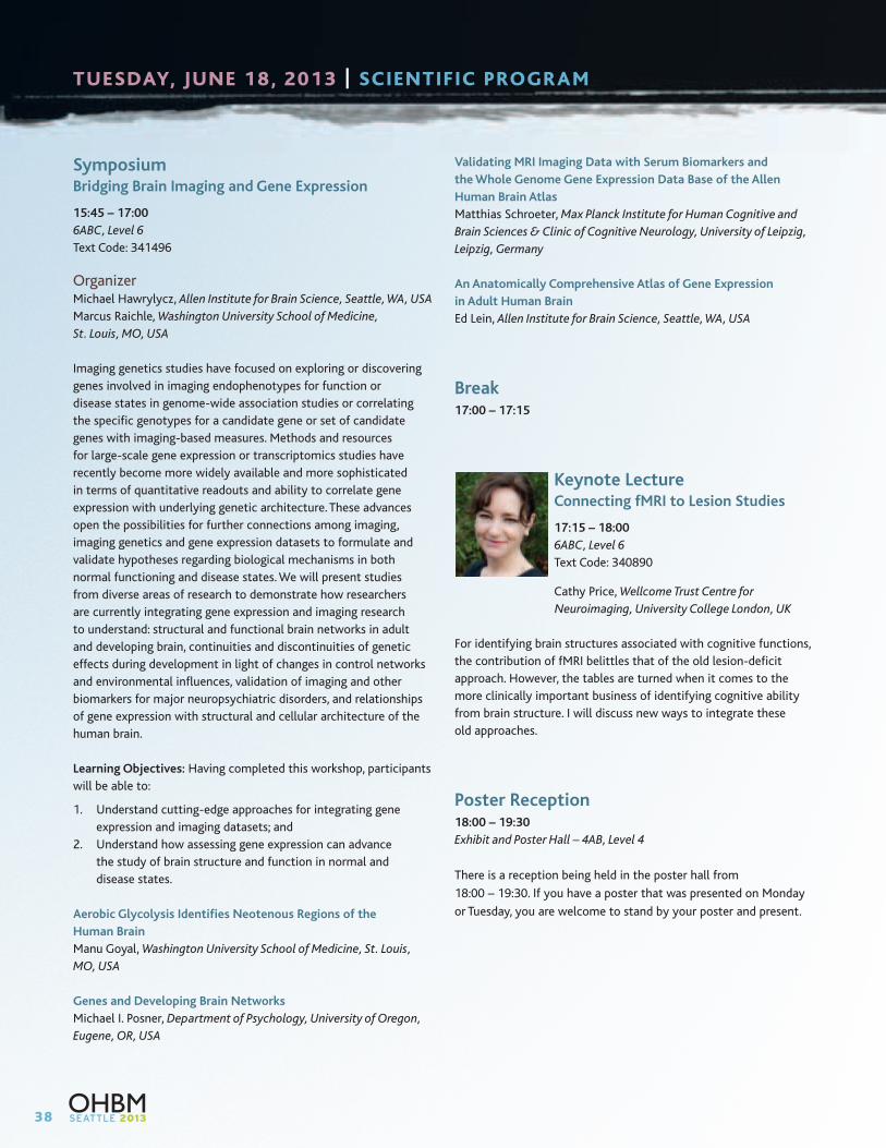
38
SymposiumBridging Brain Imaging and Gene Expression
15:45 – 17:00
6ABC, Level 6
Text Code: 341496
OrganizerMichael Hawrylycz, Allen Institute for Brain Science, Seattle, WA, USA
Marcus Raichle, Washington University School of Medicine,
St. Louis, MO, USA
Imaging genetics studies have focused on exploring or discovering
genes involved in imaging endophenotypes for function or
disease states in genome-wide association studies or correlating
the specific genotypes for a candidate gene or set of candidate
genes with imaging-based measures. Methods and resources
for large-scale gene expression or transcriptomics studies have
recently become more widely available and more sophisticated
in terms of quantitative readouts and ability to correlate gene
expression with underlying genetic architecture. These advances
open the possibilities for further connections among imaging,
imaging genetics and gene expression datasets to formulate and
validate hypotheses regarding biological mechanisms in both
normal functioning and disease states. We will present studies
from diverse areas of research to demonstrate how researchers
are currently integrating gene expression and imaging research
to understand: structural and functional brain networks in adult
and developing brain, continuities and discontinuities of genetic
effects during development in light of changes in control networks
and environmental influences, validation of imaging and other
biomarkers for major neuropsychiatric disorders, and relationships
of gene expression with structural and cellular architecture of the
human brain.
Learning Objectives: Having completed this workshop, participants
will be able to:
1. Understand cutting-edge approaches for integrating gene
expression and imaging datasets; and
2. Understand how assessing gene expression can advance
the study of brain structure and function in normal and
disease states.
Aerobic Glycolysis Identifies Neotenous Regions of the
Human Brain
Manu Goyal, Washington University School of Medicine, St. Louis,
MO, USA
Genes and Developing Brain Networks
Michael I. Posner, Department of Psychology, University of Oregon,
Eugene, OR, USA
Validating MRI Imaging Data with Serum Biomarkers and
the Whole Genome Gene Expression Data Base of the Allen
Human Brain Atlas
Matthias Schroeter, Max Planck Institute for Human Cognitive and
Brain Sciences & Clinic of Cognitive Neurology, University of Leipzig,
Leipzig, Germany
An Anatomically Comprehensive Atlas of Gene Expression
in Adult Human Brain
Ed Lein, Allen Institute for Brain Science, Seattle, WA, USA
Break17:00 – 17:15
Keynote LectureConnecting fMRI to Lesion Studies
17:15 – 18:00
6ABC, Level 6
Text Code: 340890
Cathy Price, Wellcome Trust Centre for
Neuroimaging, University College London, UK
For identifying brain structures associated with cognitive functions,
the contribution of fMRI belittles that of the old lesion-deficit
approach. However, the tables are turned when it comes to the
more clinically important business of identifying cognitive ability
from brain structure. I will discuss new ways to integrate these
old approaches.
Poster Reception18:00 – 19:30
Exhibit and Poster Hall – 4AB, Level 4
There is a reception being held in the poster hall from
18:00 – 19:30. If you have a poster that was presented on Monday
or Tuesday, you are welcome to stand by your poster and present.
TUESDAY, JUNE 18, 2013 | SCIENTIFIC PROGRAM
39
WEDNESDAY, JUNE 19, 2013 | SCIENTIFIC PROGRAM
Morning WorkshopCurrent Directions in Neuroimaging of Language and Language-Related Disorders
8:30 – 9:45
605-607, Level 6
Text Code: 342534
OrganizerWilliam W. Graves, Rutgers University, Newark, NJ, USA
The neural bases of typical and disordered language were
among the first applications of functional neuroimaging, and
discoveries have continued apace ever since. The goal of this
workshop is to give a snapshot of the current state of the art in
the functional brain imaging of language and language-related
disorders. Although much is now known about the neural regions
that support typical language and areas of damage that lead to
language deficits, less is known about how such regions work
together as a network to support language functions. Connectivity
among regions can also change over time, whether in terms of
developmental trajectory over years, as a result of an advancing
neurodegenerative disease process, or even with different
information processing demands within a single experiment.
Language networks reported at the group level can also vary
among individuals, whether pathological or within the typical
performance range. In some cases these differences may be more
clearly elucidated using recent network-level and/or dynamic time
course analyses than with traditional static mapping approaches.
This workshop will present experiments aimed at these issues and
more using a range of brain imaging techniques including fMRI,
VBM, DTI, EEG, and MEG.
Learning Objectives: Having completed this workshop,
participants will be able to:
1. Learn about the role of individual differences in neuroimaging
studies of language, including how measuring such differences
can be informative beyond exclusively reporting group means;
2. Learn about the dynamic arrangement of language-related
brain regions into networks and how such networks may
change over various time scales; and
3. Become acquainted with contemporary approaches to
analyzing neuroimaging data that are used to understand
language function and dysfunction. These include analyses
of fMRI data with multiple continuous regressors, as well
as analyses of functional, effective, and structural
connectivity. Approaches integrating fMRI with EEG and
MEG will also be discussed.
Automatic and Predictive Contextual Facilitation in
Comprehension: Evidence from Multimodal Neuroimaging
Ellen Lau, Department of Linguistics, University of Maryland,
College Park, MD, USA
Mapping the Language Network: Multimodal Neuroimaging in
Primary Progressive Aphasia
Stephen M. Wilson, Department of Speech, Language, and Hearing
Sciences and Department of Neurology, University of Arizona,
Tucson, AZ, USA
Identifying Static and Dynamic Neural Indices of Individual
Differences in Comprehension Abilities
Chantel S. Prat, Department of Psychology and Institute for Learning
& Brain Sciences, University of Washington, Seattle, WA, USA
Influences of Individual Differences and Performance on the
Neural Reading Network
William W. Graves, Department of Psychology, Rutgers University,
Newark, NJ, USA
ASK QUESTIONS ELECTRONICALLY DURING SESSIONS
Text questions to moderators while attending sessions by dialing #22333.
In the message field, type in the unique code for the session you are attending
followed by your question and then hit send! All session codes can be found at
www.humanbrainmapping.org/questions and also next to each
session description throughout this program.

40
WEDNESDAY, JUNE 19, 2013 | SCIENTIFIC PROGRAM
Morning WorkshopThe Functional Implications of Brain Signal Variability
8:30 – 9:45
608-610, Level 6
Text Code: 343659
Organizers:Biyu Jade He, National Institute of Neurological Disorders and Stroke,
NIH, Bethesda, MD, USA
Douglas Garrett, Center for Lifespan Psychology, Max Planck Institute
for Human Development, Berlin, Germany
Brain signal variability has been traditionally considered noise.
While the field of resting-state networks has revealed an impressive
and rich hierarchical-organization within spontaneous brain
activity via studying connectivity patterns among brain regions,
much remains unknown regarding the exact mechanisms and
functional roles of variability per se. The four speakers in this
proposal have studied signal variability from different angles and
with a variety of techniques and methodology, including functional
magnetic resonance imaging (fMRI), electroencephalography (EEG),
magnetoencephalography (MEG), electrocorticography (ECoG),
neuronal spiking, and neural network modeling. A particular
emphasis will be placed on the functional implications of signal
variability, including its relation to cognition and behavioral
performance, as well as changes of brain variability during
development, aging and certain clinical conditions. Neural network
models will further shed light upon the mechanisms of signal
variability. Converging results from these diverse methods suggest
that signal variability represents a complemenary, if not orthogonal
dimension of information processing in the brain. More practically,
we suggest that as an alternative to mean-signal-based brain
mapping that has brought tremendous successes over the past
two decades, brain signal variability provides a promising new
approach to mapping the brain in the context of resting-state or
cognitive experiments.
Learning Objectives: Having completed this workshop, participants
will be able to:
1. Understand the various emerging approaches in the study of
brain signal variability;
2. Understand the relationship between brain signal variability and
traditional signal-mean-based measurements; and
3. Recognize the functional implications of brain signal variability
across task types, cognitive domains, as well as developmental
and clinical groups.
Brain Signal Variability is a Robust Marker of Aging and Cognition
Douglas Garrett, Center for Lifespan Psychology, Max Planck Institute
for Human Development, Berlin, Germany
The Development of a Noisy Brain
A. Randy McIntosh, Department of Psychology, University of Toronto,
Toronto, ON, Canada
Brain Variability and Task Performance
Biyu Jade He, National Institute of Neurological Disorders and Stroke,
NIH, Bethesda, MD, USA
The Dynamical Structure of Brain Fluctuations At Rest
Gustavo Deco, Center for Brain and Cognition, University Pompeu
Fabra, Barcelona, Spain
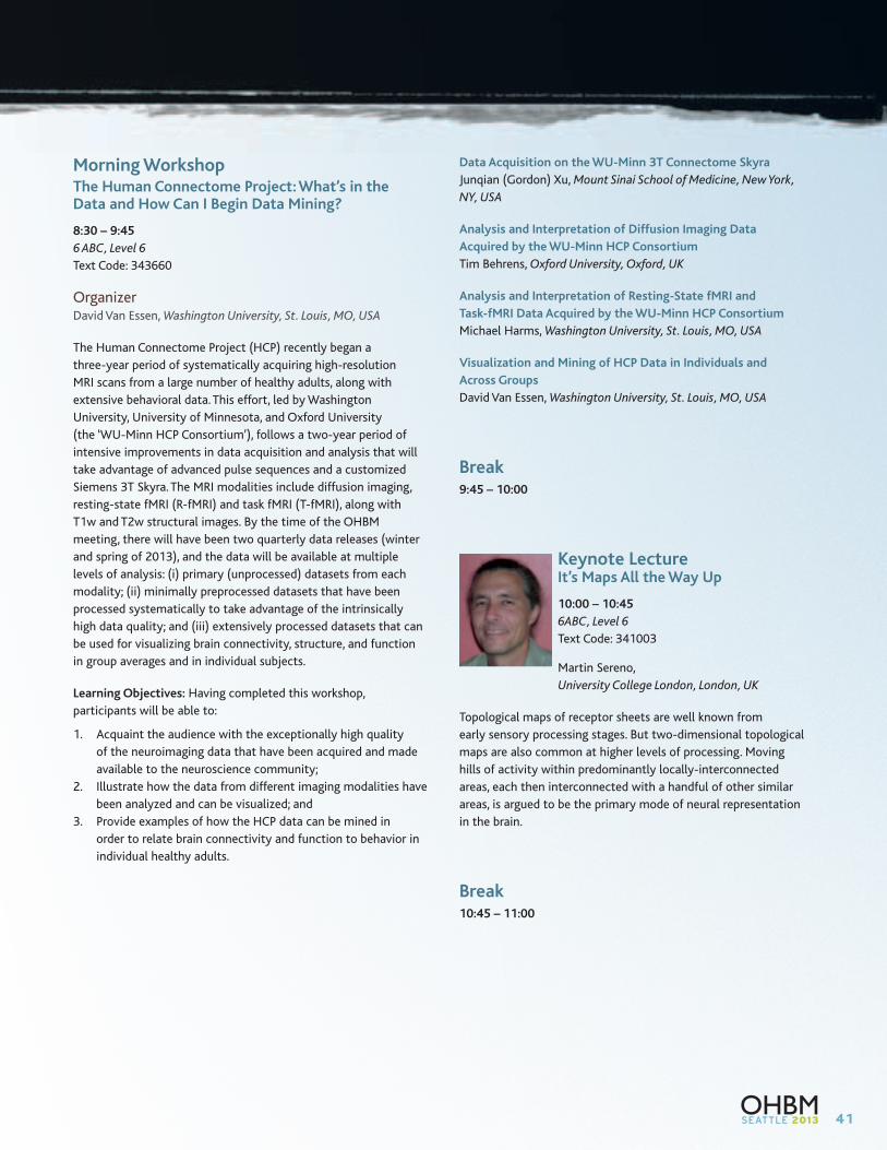
41
Morning WorkshopThe Human Connectome Project: What’s in the Data and How Can I Begin Data Mining?
8:30 – 9:45
6 ABC, Level 6
Text Code: 343660
OrganizerDavid Van Essen, Washington University, St. Louis, MO, USA
The Human Connectome Project (HCP) recently began a
three-year period of systematically acquiring high-resolution
MRI scans from a large number of healthy adults, along with
extensive behavioral data. This effort, led by Washington
University, University of Minnesota, and Oxford University
(the ‘WU-Minn HCP Consortium’), follows a two-year period of
intensive improvements in data acquisition and analysis that will
take advantage of advanced pulse sequences and a customized
Siemens 3T Skyra. The MRI modalities include diffusion imaging,
resting-state fMRI (R-fMRI) and task fMRI (T-fMRI), along with
T1w and T2w structural images. By the time of the OHBM
meeting, there will have been two quarterly data releases (winter
and spring of 2013), and the data will be available at multiple
levels of analysis: (i) primary (unprocessed) datasets from each
modality; (ii) minimally preprocessed datasets that have been
processed systematically to take advantage of the intrinsically
high data quality; and (iii) extensively processed datasets that can
be used for visualizing brain connectivity, structure, and function
in group averages and in individual subjects.
Learning Objectives: Having completed this workshop,
participants will be able to:
1. Acquaint the audience with the exceptionally high quality
of the neuroimaging data that have been acquired and made
available to the neuroscience community;
2. Illustrate how the data from different imaging modalities have
been analyzed and can be visualized; and
3. Provide examples of how the HCP data can be mined in
order to relate brain connectivity and function to behavior in
individual healthy adults.
Data Acquisition on the WU-Minn 3T Connectome Skyra
Junqian (Gordon) Xu, Mount Sinai School of Medicine, New York,
NY, USA
Analysis and Interpretation of Diffusion Imaging Data
Acquired by the WU-Minn HCP Consortium
Tim Behrens, Oxford University, Oxford, UK
Analysis and Interpretation of Resting-State fMRI and
Task-fMRI Data Acquired by the WU-Minn HCP Consortium
Michael Harms, Washington University, St. Louis, MO, USA
Visualization and Mining of HCP Data in Individuals and
Across Groups
David Van Essen, Washington University, St. Louis, MO, USA
Break9:45 – 10:00
Keynote LectureIt’s Maps All the Way Up
10:00 – 10:45
6ABC, Level 6
Text Code: 341003
Martin Sereno,
University College London, London, UK
Topological maps of receptor sheets are well known from
early sensory processing stages. But two-dimensional topological
maps are also common at higher levels of processing. Moving
hills of activity within predominantly locally-interconnected
areas, each then interconnected with a handful of other similar
areas, is argued to be the primary mode of neural representation
in the brain.
Break10:45 – 11:00
42
WEDNESDAY, JUNE 19, 2013 | SCIENTIFIC PROGRAM
Answers for life.
www.siemens.com/neuro-MRI
At the frontier of neuroscience with cutting-edge technologiesOHBM Lunch symposium sponsored by Siemens
Prof. Rainer Goebel
“Cracking the functional code of the human brain at 7 Tesla”.
Professor, Cognitive Neuroscience, University of Maastricht, NL / Director, M-BIC, Maastrich, NL / Research Director, FPN Maastrich Research Institute, Maastrich, NL
Prof. Lawrence L. Wald
“Technology game changers in human brain mapping”.
Director, MGH NMR Core at Martinos Center, Department of Radiology, Boston, Massachusetts, USA
Wednesday, June 19th 1:00 p.m. – 3:30 p.m. Room: 602-604 Lunch will be provided for the first 200 attendees
Leading. With MAGNETOM.
Oral Sessions11:00 – 12:30
Oral session presentations are chosen by the Program Committee
from submitted abstracts using criteria of quality and timeliness;
a wide spectrum of investigation is represented.
O – W1: Perception and Attention
605-607, Level 6
Text Code: 341500
Chair: Geoff Boynton, University of Washington, Seattle, WA, USA
11:00 – 11:15
3960: Reorganization of auditory motion direction encoding
in early blind humans
Fang Jiang, University of Washington, Seattle, WA, USA
11:15 – 11:30
4015: Broadly Tuned Face and Hand Representations in
Human Ventral Temporal Cortex
Nicolas Davidenko, UC Santa Cruz, Santa Cruz, CA, USA
11:30 – 11:45
3953: Is this really an orange? Effects of crossmodal
semantic congruency on olfactory perception
Janina Seubert, Monell Chemical Senses Center, Philadelphia,
PA, USA
11:45 – 12:00
3861: Predicting Response Time Variations from Anticipatory
Modulation of Large-scale Brain Networks
Matthias Ekman, Donders Institute for Brain, Cognition and
Behaviour, Nijmegen, Netherlands
12:00 – 12:15
3896: White matter damage in healthy aging disrupts neural
oscillations underlying top-down attention
David Ziegler, UCSF, San Francisco, CA, USA
12:15 – 12:30
3936: Measurement of auditory frequency selectivity in the
depth of human inferior colliculus
David Ress, The University of Texas at Austin, Austin, TX, USA
43
O – W2: Modeling and Analysis Methods 3: Structural and Diffusion
6 ABC, Level 6
Text Code: 341507
Chair: Ged Ridgway, UCL Institute of Neurology, London, UK
11:00 – 11:15
1623: Analysis of Multiple Sclerosis Lesions via Spatially
Varying Coefficients
Tian Ge, University of Warwick, Coventry, UK
11:15 – 11:30
1657: Predicting favorable vs non-favorable surgery outcome
in patients with mesial temporal lobe epilepsy
Delia-Lisa Feis, Max Planck Institute for Neurological Research,
Cologne, Germany
11:30 – 11:45
1883: Automated Analysis of the Shape of Sulcal Curves
using the Anisotropic Helmholtz Equation
Anand Joshi, University of Southern California, Los Angeles,
CA, USA
11:45 – 12:00
1885: Brain Image and Fiber Log-demons Registration
with Currents
Viviana Siless, Parietal Team, INRIA, Saclay, Paris, France
12:00 – 12:15
1676: DoubleAx: In-vivo Axon Measurement in the
Human Corpus Callosum Using Angular Double-PFG MRI
Wenjin Zhou, Oakland University, Rochester Hills, MI, USA
12:15 – 12:30
1898: Towards joint morphometry of white matter tracts
and gray matter surfaces
Pietro Gori, Inria – ICM, Paris, France
44
WEDNESDAY, JUNE 19, 2013 | SCIENTIFIC PROGRAM
O – W3: Neuroanatomy
608-610, Level 6
Text Code: 341547
Chair: Gaolang Gong, Montreal Neurological Institute and Hospital,
McGill University, Montreal ,Canada
11:00 – 11:15
3851: MR diffusion-based histology and micro-tractography
reveal mesoscale details of the human cerebellum
Flavio Dell’Acqua, King’s College London – Institute of
Psychiatry, London, UK
11:15 – 11:30
3844: Estimation of fiber orientation distribution functions
in 3D-polarized light imaging
Melanie Dohmen, Institute for Neuroscience and Medicine,
Research Center Juelich, Juelich, Germany
11:30 – 11:45
3759: Learning to read improves the structure of the
arcuate fasciculus
Michel Thiebaut de Schotten, Institute of Psychiatry,
London, UK
11:45 – 12:00
3816: Identifying Heavily Myelinated Areas of the Cortex
using subject-specific Cortical Profiles of T1
Miriam Waehnert, Max-Planck Institute for Human Cognitive
and Brain Sciences, Leipzig, Germany
12:00 – 12:15
3765: Pupil diameter indexes locus coeruleus activity
in humans
Joshua Balsters, ETH Zurich, Zurich, Switzerland
12:15 – 12:30
3840: Connectome based classification of brain-derived
neurotrophic factor Met allele carriers
Christophe Phillips, Cyclotron Research Centre, University of
Liege, Sart Tilman, Liege, Belgium
O – W4: Disorders of the Nervous System 2: Psychiatric Illness
602-604, Level 6
Text Code: 341593
Chair: Vaibhav Diwadkar, Wayne State University, Detroit, MI, USA
11:00 – 11:15
3755: Gray matter volume analysis in major depression
based on cytoarchitectonic maps of the frontal pole
Sebastian Bludau, Institute of Neuroscience and Medicine,
INM-1, Juelich, Germany
11:15 – 11:30
3250: Mapping thalamo-cortical function in psychiatric
disease: focus on schizophrenia and bipolar illness
Alan Anticevic, Yale University School of Medicine, New Haven,
CT, USA
11:30 – 11:45
3217: An fMRI study of impaired sensory prediction in
schizophrenia
Thomas White, Institute of Psychiatry, King’s College London,
London, UK
11:45 – 12:00
3197: Real-time fMRI Neurofeedback Training of Amygdala
Modulates Frontal EEG Asymmetry in MDD Patients
Vadim Zotev, Laureate Institute for Brain Research, Tulsa,
OK, USA
12:00 – 12:15
3165: Disrupted effective connectivity between amygdala
and OFC in social anxiety disorder revealed by DCM
Ronald Sladky, MR Centre Of Excellence, Medical University
of Vienna, Austria
12:15 – 12:30
3009: Altered Brain Network Interactions Are Associated
With Nicotine Withdrawal
Elliot Stein, NIH, Baltimore, MD, USA
Lunch12:30 – 13:30
45
Interactive Poster Presentations13:30 – 14:15
608-610, Level 6
Text Code: 36265
I-Poster presentations highlight top ranked submitted abstracts.
Authors will present their abstracts in a short, “datablitz” format.
The objective of the I-Poster session is to arrive at a hybrid of
posters and oral sessions.
Moderator: Ziad Saad, National Institute of Health,
Bethesda, MD, USA
13:30 – 13:35
1628: Using Bayesian priors to improve power of whole
brain voxel- and connexelwise inferences
Krzysztof Gorgolewski, Max Planck Institute for Human Brain
and Cognitive Sciences, Leipzig, Germany
13:35 – 13:40
1622: A Bayesian Random Shape model for fMRI and
MRI Brain Activity Analysis
Lijun Zhang, Emory University, Atlanta, GA, USA
13:40 – 13:45
1710: Frequency Specific Interactions of MEG activity
within and across resting state networks as revealed
Laura Marzetti, Department of Neuroscience and Imaging,
University ‘G. d’Annunzio’ Chieti Pescara, Chieti, Italy
13:45 – 13:50
1834: Inferring Ontologies of Mind-Brain Relations from
Neuroimaging Data
BT Thomas Yeo, Duke-NUS, Singapore
13:50 – 13:55
1790: Contradictory Conclusions in Analysis of Brain
Functional Networks: the Role of Image Registration
Maxime Taquet, Harvard Medical School, Boston, MA, USA
13:55 – 14:00
1811: Frequency-Dependent Resting-State Network Modules
Revealed in MR-Encephalography
Hsu-Lei Lee, University Medical Center Freiburg, Freiburg, Germany
14:00 – 14:15
Questions and Answers
Poster Session13:30 – 15:30
Exhibit and Poster Hall – 4AB, Level 4
Poster Numbers #3000-4119: Even Numbered Posters Stand-By
Disorders of the Nervous System: Addictions, Alzheimer’s Disease
and Other Dementias, Epilepsy, Mood and Anxiety Disorders,
Schizophrenia and Psychotic Disorders, Traumatic Brain Injury
Emotion and Motivation: Emotional Learning, Emotional
Perception, Reward and Punishment, Sexual Behavior
Imaging Methods: Anatomical MRI, BOLD fMRI, Diffusion MRI,
EEG, MEG, MR Spectroscopy, Multi-Modal Imaging, Non-BOLD
fMRI, Optical Imaging/NIRS, PET
Lifespan Development: Aging, Normal Brain Development:
Fetus to Adolescence
Neuroanatomy: Anatomy and Function, Brain Networks, Cortical
Anatomy and Segregation, Subcortical Structures, White Matter
Anatomy, Fiber Pathways and Connectivity
Perception and Attention: Attention: Auditory/Tactile/Motor,
Attention: Visual, Chemical Senses: Olfaction, Taste, Consciousness
and Awareness, Perception: Auditory/ Vestibular, Perception:
Multisensory and Crossmodal, Perception: Pain and Visceral,
Perception: Tactile/Somatosensory, Perception: Visual, Sleep
and Wakefulness
Social Neuroscience: Self Processes, Social Cognition,
Social Interaction
Break15:30 – 15:45

46
WEDNESDAY, JUNE 19, 2013 | SCIENTIFIC PROGRAM
SymposiumThe Challenge of Imaging Brain Connections in Animal and Man
15:45 – 17:00
6ABC, Level 6
Text Code: 341686
OrganizerMarco Catani, Natbrainlab, King’s College London, London, UK
Current approaches to brain function rest on the empirical
evidence that the pattern of connectivity defines the functional
specialization of individual cerebral regions. Despite the
increasingly number of methods available to visualize connections
the analysis of structural connectivity remains a difficult task.
In this symposium we will try to define connections at different
levels from single neuronal morphology to large scale networks.
Current methods based on electron microscopy are providing
unprecedented access to local connectivity of animal models
and relatively simple human structures (e.g. retina). The main
limitation of these approaches is the relatively low volume of data
that can be acquired in a reasonable amount of time, which makes
the connectome of the Drosophila an ambitious goal. Axonal
tracing methods have been extensively used to define short and
long axonal connections in the animal brain. These approaches
provide an opportunity for the connectome of the monkey brain.
There is however a risk of translating these findings blindly to
humans without further verification. Tractography methods
based on diffusion imaging could help to identify pathways
that are unique to humans and help to correlate interindividual
variability with cognitive performance and behaviour. Nevertheless
tractography remains an indirect quantitative method prone to
many biases, which raise some doubts on whether this method
will ever be able to reproduce the real anatomy of human
connections. Our symposium will provide a balanced overview of
this timely topic with ample time for discussion and reflection.
Learning Objectives: Having completed this workshop,
participants will be able to:
1. Learn about imaging structural connectivity at different scales;
2. Recognise the importance of developing reliable methods
to provide solid foundations to different connectome
projects; and
3. Identify emerging approaches to obtain high resolution
anatomical connectivity for the human brain.
Defining Networks
Olaf Sporns, Department of Psychological and Brain Sciences,
Indiana University, Bloomington, IN, USA
Imaging Neuronal Microcircuits
Kevin Briggman, Circuit Dynamics and Connectivity Unit, NIH,
Bethesda, MD, USA
Tracing Connections in the Monkey Brain
Jeremy Schmahmann, Laboratory for Neuroanatomy and Cerebellar
Neurobiology, Boston, MA, USA
Tracking Pathways in the Human Brain
Marco Catani, Natbrainlab, King’s College London, London, UK
Break17:00 – 17:15
Keynote LectureThe Three Dimensional Structure of the Brain Pathways
17:15 – 18:00
6ABC, Level 6
Text Code: 341158
Van Wedeen, Massachusetts
General Hospital, Martinos Imaging Center,
Charlestown, MA, USA
We present the thesis that the brain pathways conform to the
three axes of a curved approximately orthogonal coordinate
system related or identical to the natural coordinates of the
body-plan. The evidence, implications for brain organization and
particularly for the imaging of connectivity will be discussed.
Break18:00 – 18:15
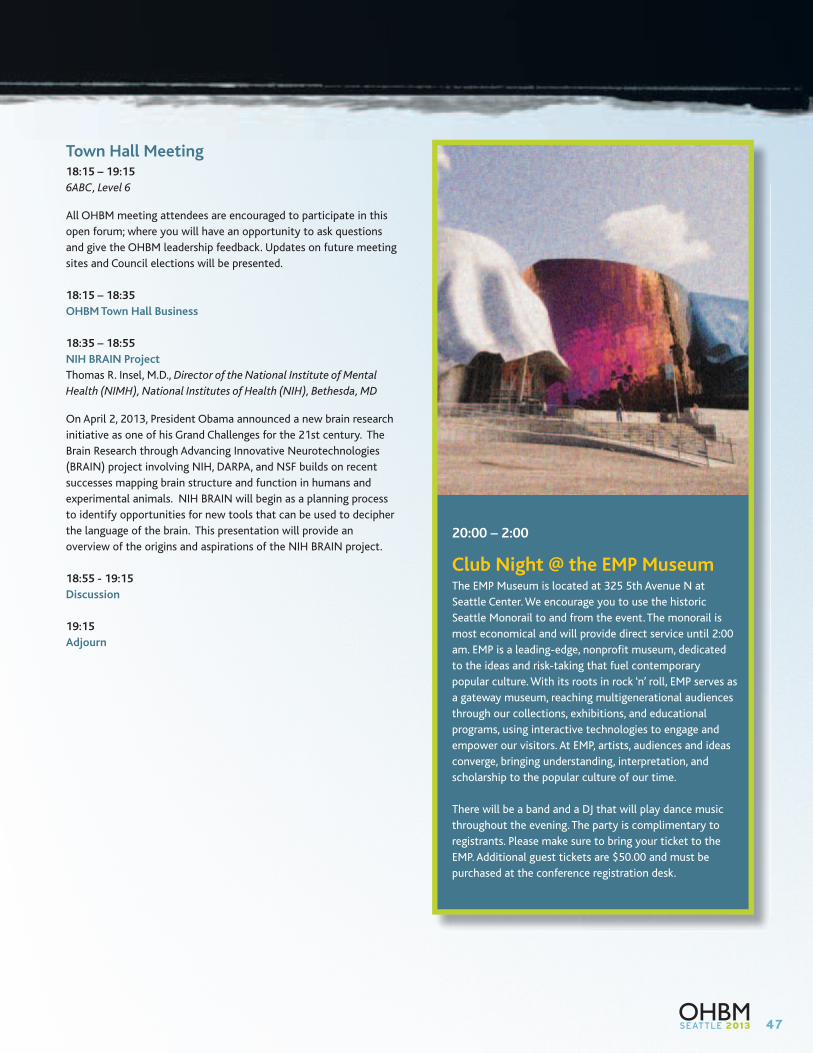
47
Town Hall Meeting18:15 – 19:15
6ABC, Level 6
All OHBM meeting attendees are encouraged to participate in this
open forum; where you will have an opportunity to ask questions
and give the OHBM leadership feedback. Updates on future meeting
sites and Council elections will be presented.
18:15 – 18:35
OHBM Town Hall Business
18:35 – 18:55
NIH BRAIN Project
Thomas R. Insel, M.D., Director of the National Institute of Mental
Health (NIMH), National Institutes of Health (NIH), Bethesda, MD
On April 2, 2013, President Obama announced a new brain research
initiative as one of his Grand Challenges for the 21st century. The
Brain Research through Advancing Innovative Neurotechnologies
(BRAIN) project involving NIH, DARPA, and NSF builds on recent
successes mapping brain structure and function in humans and
experimental animals. NIH BRAIN will begin as a planning process
to identify opportunities for new tools that can be used to decipher
the language of the brain. This presentation will provide an
overview of the origins and aspirations of the NIH BRAIN project.
18:55 - 19:15
Discussion
19:15
Adjourn
20:00 – 2:00
Club Night @ the EMP MuseumThe EMP Museum is located at 325 5th Avenue N at
Seattle Center. We encourage you to use the historic
Seattle Monorail to and from the event. The monorail is
most economical and will provide direct service until 2:00
am. EMP is a leading-edge, nonprofit museum, dedicated
to the ideas and risk-taking that fuel contemporary
popular culture. With its roots in rock ‘n’ roll, EMP serves as
a gateway museum, reaching multigenerational audiences
through our collections, exhibitions, and educational
programs, using interactive technologies to engage and
empower our visitors. At EMP, artists, audiences and ideas
converge, bringing understanding, interpretation, and
scholarship to the popular culture of our time.
There will be a band and a DJ that will play dance music
throughout the evening. The party is complimentary to
registrants. Please make sure to bring your ticket to the
EMP. Additional guest tickets are $50.00 and must be
purchased at the conference registration desk.
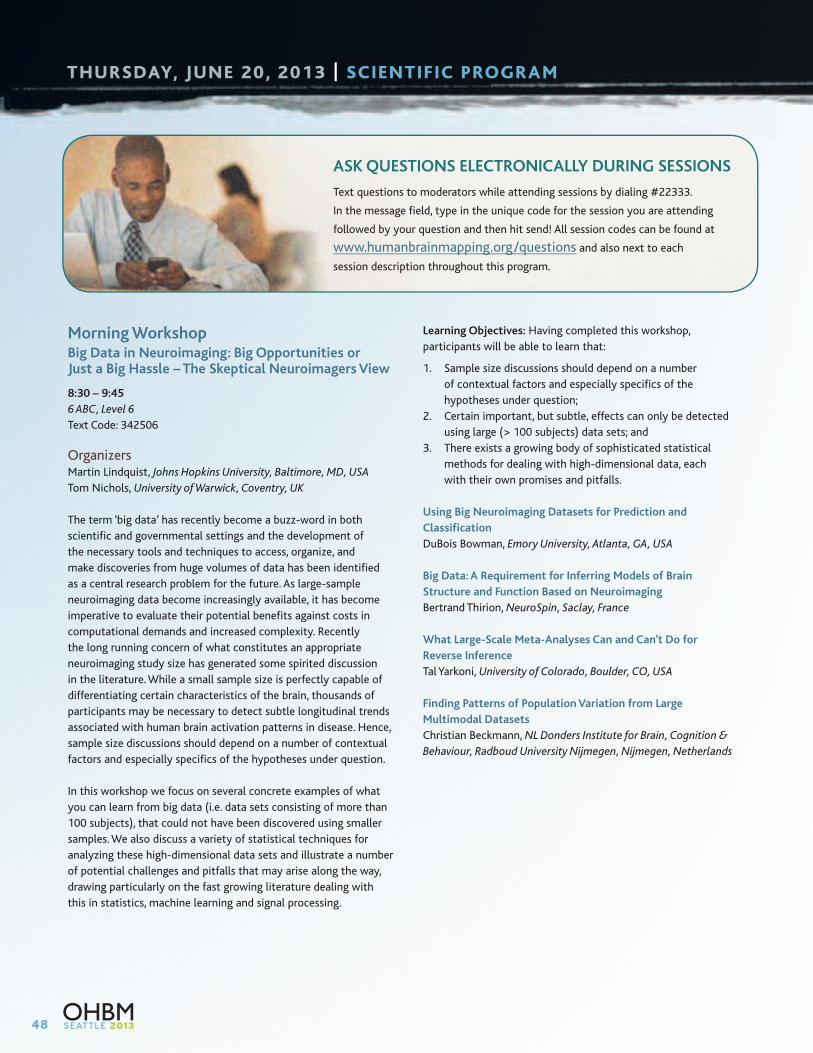
48
THURSDAY, JUNE 20, 2013 | SCIENTIFIC PROGRAM
Morning WorkshopBig Data in Neuroimaging: Big Opportunities or Just a Big Hassle – The Skeptical Neuroimagers View
8:30 – 9:45
6 ABC, Level 6
Text Code: 342506
OrganizersMartin Lindquist, Johns Hopkins University, Baltimore, MD, USA
Tom Nichols, University of Warwick, Coventry, UK
The term ‘big data’ has recently become a buzz-word in both
scientific and governmental settings and the development of
the necessary tools and techniques to access, organize, and
make discoveries from huge volumes of data has been identified
as a central research problem for the future. As large-sample
neuroimaging data become increasingly available, it has become
imperative to evaluate their potential benefits against costs in
computational demands and increased complexity. Recently
the long running concern of what constitutes an appropriate
neuroimaging study size has generated some spirited discussion
in the literature. While a small sample size is perfectly capable of
differentiating certain characteristics of the brain, thousands of
participants may be necessary to detect subtle longitudinal trends
associated with human brain activation patterns in disease. Hence,
sample size discussions should depend on a number of contextual
factors and especially specifics of the hypotheses under question.
In this workshop we focus on several concrete examples of what
you can learn from big data (i.e. data sets consisting of more than
100 subjects), that could not have been discovered using smaller
samples. We also discuss a variety of statistical techniques for
analyzing these high-dimensional data sets and illustrate a number
of potential challenges and pitfalls that may arise along the way,
drawing particularly on the fast growing literature dealing with
this in statistics, machine learning and signal processing.
Learning Objectives: Having completed this workshop,
participants will be able to learn that:
1. Sample size discussions should depend on a number
of contextual factors and especially specifics of the
hypotheses under question;
2. Certain important, but subtle, effects can only be detected
using large (> 100 subjects) data sets; and
3. There exists a growing body of sophisticated statistical
methods for dealing with high-dimensional data, each
with their own promises and pitfalls.
Using Big Neuroimaging Datasets for Prediction and
Classification
DuBois Bowman, Emory University, Atlanta, GA, USA
Big Data: A Requirement for Inferring Models of Brain
Structure and Function Based on Neuroimaging
Bertrand Thirion, NeuroSpin, Saclay, France
What Large-Scale Meta-Analyses Can and Can’t Do for
Reverse Inference
Tal Yarkoni, University of Colorado, Boulder, CO, USA
Finding Patterns of Population Variation from Large
Multimodal Datasets
Christian Beckmann, NL Donders Institute for Brain, Cognition &
Behaviour, Radboud University Nijmegen, Nijmegen, Netherlands
ASK QUESTIONS ELECTRONICALLY DURING SESSIONS
Text questions to moderators while attending sessions by dialing #22333.
In the message field, type in the unique code for the session you are attending
followed by your question and then hit send! All session codes can be found at
www.humanbrainmapping.org/questions and also next to each
session description throughout this program.
49
Morning WorkshopMicrostructure Meets Function in the Same Brain in Vivo – High-Field MRI Sets the Stage
8:30 – 9:45
608-610, Level 6
Text Code: 342788
OrganizerStefan Geyer, Max Planck Institute for Human Cognitive and Brain
Sciences, Department of Neurophysics, Leipzig, Germany
Unraveling the functional properties of structural brain elements
in an individual-specific way is one of the fundamental goals of
neuroscientific research. In the cerebral cortex this is not so easy
to accomplish, since cortical areas are classically defined by their
microstructure in post-mortem brains (cf. the famous map of
Brodmann) but functionally in living brains with electrophysiological
or neuroimaging techniques – and cortical areas vary in their
topography considerably across individual brains.
One attempt to overcome this problem in the past has been to
generate probabilistic cytoarchitectonic maps from post-mortem
brains (ex vivo) in standard anatomical (e.g., MNI) space where
they can be matched with co-registered functional imaging data
(in vivo). However, due to interindividual variability, the population
maps of adjacent areas overlap considerably and only after extensive
thresholding – and thus discarding structural information – is it
possible to unequivocally assign a given voxel in standard space to a
particular population map (or cortical area).
It would be a huge leap forward if it were possible to remove this
guesswork and generate an individual-specific map of cortical
microstructure (areas) in vivo and correlate it with cortical
function in vivo in the same brain.
In recent years, two advances have brought us closer to this
ambitious goal. The first is the dramatic improvement in the quality
of in vivo MRI scans. With 7 Tesla magnets and high sensitivity
radiofrequency receive coils, the current state of the art allows
structural images of entire brains to be obtained with 0.4 mm
isotropic resolution (or even less) and BOLD contrast changes of
blood oxygenation with an isotropic resolution much better than
1 mm. The second advancement is based on the observation that
maps of the longitudinal relaxation time T1 effectively indicate the
presence of myelin and closely resemble myelin-stained histological
sections, whereas differences in cytoarchitecture are detectable
with MRI only in rare instances. Research by Cécile and Oskar Vogt,
two pioneers in the field of myeloarchitecture in the first half of the
20th century has shown that there is a great degree of concordance
between structural parcellations of the cortex based on differences
in myelin (myeloarchitecture) and maps based on differences in cell
bodies (cytoarchitecture – the basis of Brodmann’s map). Myelo- and
cytoarchitecture are not two parallel universes but two different
views of the same universe.
This sets the stage for an important new impulse for “The future of
fMRI” – inspired by the seminal NeuroImage Special Issue “Twenty
years of functional MRI” published in August 2012. We would like
to share our enthusiasm with the neuroimaging community in this
workshop entitled “Microstructure Meets Function in the Same Brain
in Vivo – High-Field MRI Sets the Stage”.
The first talk by Stefan Geyer introduces a “triple jump” approach
that validates the myelin-based in vivo cortical maps with “classical”
histology data ex vivo. Matt Glasser combines in vivo microstructure
with resting state and task-related data from the Human
Connectome Project. Rosa Sanchez-Panchuelo and Frederic Dick
focus on more specific visual, somatomotor, and auditory regions
where they link structure to function in single individuals.
Learning Objectives: Having completed this workshop, participants
will be able to:
1. Understand that cortical areas and their boundaries are
“classically” defined by their myelo- and cytoarchitectonic
pattern ex vivo, i.e., in post-mortem brains;
2. Learn that T1-based maps obtained with high-field MRI
represent individual-specific in vivo maps of cortical
microstructure, based on differential grey matter myelination
between areas; and
3. Appreciate that the ultimate goal is direct structure-function
correlation in the same subjects, which can now be achieved
by matching microstructural and functional maps in the same
living brains.
The “Triple Jump” Myeloarchitectural Approach for In Vivo
Cortical Parcellation
Stefan Geyer, Max Planck Institute for Human Cognitive and Brain
Sciences, Department of Neurophysics, Leipzig, Germany
Identifying Cortical Areas with In Vivo Architectonic and
Functional Data from the Human Connectome Project
Matthew F. Glasser, Washington University Medical School,
Department of Anatomy and Neurobiology, St. Louis, MO, USA
Linking Structure to Function In Vivo in Primary Visual and
Somatosensory Cortex
Rosa Sanchez-Panchuelo, University of Nottingham, Sir Peter
Mansfield Magnetic Resonance Centre, Nottingham, UK
Myeloarchitectural and Functional Mapping of Visual, Auditory,
and Somatomotor Areas in Single Individuals
Frederic Dick, Birkbeck/UCL Centre for NeuroImaging, Birkbeck
College, London, UK
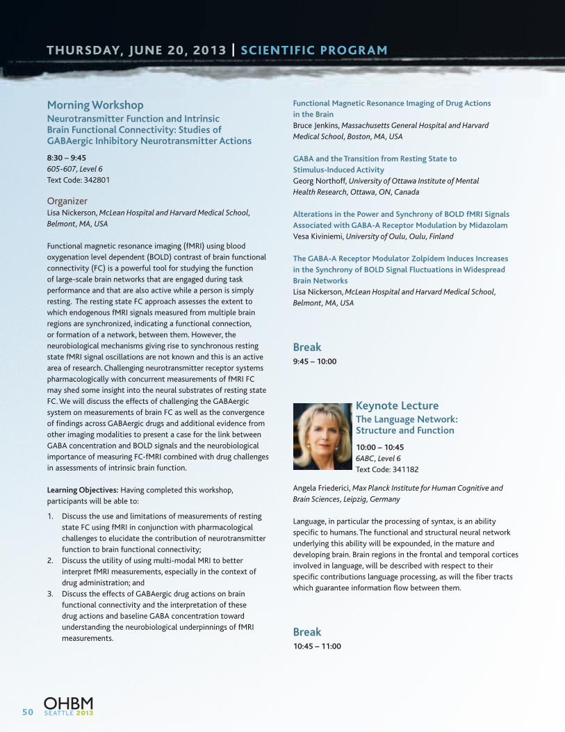
50
THURSDAY, JUNE 20, 2013 | SCIENTIFIC PROGRAM
Morning WorkshopNeurotransmitter Function and Intrinsic Brain Functional Connectivity: Studies of GABAergic Inhibitory Neurotransmitter Actions
8:30 – 9:45
605-607, Level 6
Text Code: 342801
OrganizerLisa Nickerson, McLean Hospital and Harvard Medical School,
Belmont, MA, USA
Functional magnetic resonance imaging (fMRI) using blood
oxygenation level dependent (BOLD) contrast of brain functional
connectivity (FC) is a powerful tool for studying the function
of large-scale brain networks that are engaged during task
performance and that are also active while a person is simply
resting. The resting state FC approach assesses the extent to
which endogenous fMRI signals measured from multiple brain
regions are synchronized, indicating a functional connection,
or formation of a network, between them. However, the
neurobiological mechanisms giving rise to synchronous resting
state fMRI signal oscillations are not known and this is an active
area of research. Challenging neurotransmitter receptor systems
pharmacologically with concurrent measurements of fMRI FC
may shed some insight into the neural substrates of resting state
FC. We will discuss the effects of challenging the GABAergic
system on measurements of brain FC as well as the convergence
of findings across GABAergic drugs and additional evidence from
other imaging modalities to present a case for the link between
GABA concentration and BOLD signals and the neurobiological
importance of measuring FC-fMRI combined with drug challenges
in assessments of intrinsic brain function.
Learning Objectives: Having completed this workshop,
participants will be able to:
1. Discuss the use and limitations of measurements of resting
state FC using fMRI in conjunction with pharmacological
challenges to elucidate the contribution of neurotransmitter
function to brain functional connectivity;
2. Discuss the utility of using multi-modal MRI to better
interpret fMRI measurements, especially in the context of
drug administration; and
3. Discuss the effects of GABAergic drug actions on brain
functional connectivity and the interpretation of these
drug actions and baseline GABA concentration toward
understanding the neurobiological underpinnings of fMRI
measurements.
Functional Magnetic Resonance Imaging of Drug Actions
in the Brain
Bruce Jenkins, Massachusetts General Hospital and Harvard
Medical School, Boston, MA, USA
GABA and the Transition from Resting State to
Stimulus-Induced Activity
Georg Northoff, University of Ottawa Institute of Mental
Health Research, Ottawa, ON, Canada
Alterations in the Power and Synchrony of BOLD fMRI Signals
Associated with GABA-A Receptor Modulation by Midazolam
Vesa Kiviniemi, University of Oulu, Oulu, Finland
The GABA-A Receptor Modulator Zolpidem Induces Increases
in the Synchrony of BOLD Signal Fluctuations in Widespread
Brain Networks
Lisa Nickerson, McLean Hospital and Harvard Medical School,
Belmont, MA, USA
Break9:45 – 10:00
Keynote LectureThe Language Network: Structure and Function
10:00 – 10:45
6ABC, Level 6
Text Code: 341182
Angela Friederici, Max Planck Institute for Human Cognitive and
Brain Sciences, Leipzig, Germany
Language, in particular the processing of syntax, is an ability
specific to humans. The functional and structural neural network
underlying this ability will be expounded, in the mature and
developing brain. Brain regions in the frontal and temporal cortices
involved in language, will be described with respect to their
specific contributions language processing, as will the fiber tracts
which guarantee information flow between them.
Break10:45 – 11:00
51
Interactive Poster Presentations10:45 – 11:30
608-610, Level 6
Text Code: 675633
I-Poster presentations highlight top ranked submitted abstracts.
Authors will present their abstracts in a short, “datablitz” format.
The objective of the I-Poster session is to arrive at a hybrid of
posters and oral sessions.
Moderator: Nathalie Tzourio-Mazoyer, CEA – CNRS – Université
Bordeaux Ségalen, Bordeaux, France
10:45 – 10:50
1279: Estimating heritability of the fMRI response to faces
using Whole-Genome Complex-Trait Analysis
Erin Dickie, Rotman Research Institute at Baycrest, Toronto, Canada
10:50 – 10:55
1336: A Fundamental Causal Mechanism in Cognitive
Control within the Fronto-Cingulo-Parietal Network
Weidong Cai, Stanford University School of Medicine,
Palo Alto, CA, USA
10:55 – 11:00
1394: A cross-sectional and longitudinal analysis of
functional asymmetries mediated by musical training
Robert Ellis, Beth Israel Deaconess Medical Center / Harvard
Medical School, Boston, MA, USA
11:00 – 11:05
1435: The Second Generation of Dense Individualized and
Common Connectivity-based Cortical Landmarks
Xi Jiang, The University of Georgia, Athens, GA, USA
11:05 – 11:10
1554: Hippocampal-neocortical reorganization and maturation
of fact retrieval: a longitudinal fMRI study
Shaozheng Qin, Stanford University, Stanford, CA, USA
11:10 – 11:15
3776: The role of fronto-parietal networks in mental imagery
Henrietta Howells, Institute of Psychiatry, London, United Kingdom
11:15 – 11:30
Questions and Answers
Poster Session10:45 – 12:45
Exhibit and Poster Hall – 4AB, Level 4
Poster Numbers #3000-4119: Odd Numbered Posters Stand-By
Disorders of the Nervous System: Addictions, Alzheimer’s Disease
and Other Dementias, Epilepsy, Mood and Anxiety Disorders,
Schizophrenia and Psychotic Disorders, Traumatic Brain Injury
Emotion and Motivation: Emotional Learning, Emotional
Perception, Reward and Punishment, Sexual Behavior
Imaging Methods: Anatomical MRI, BOLD fMRI, Diffusion MRI,
EEG, MEG, MR Spectroscopy, Multi-Modal Imaging, Non-BOLD
fMRI, Optical Imaging/NIRS, PET
Lifespan Development: Aging, Normal Brain Development:
Fetus to Adolescence
Neuroanatomy: Anatomy and Function, Brain Networks,
Cortical Anatomy and Segregation, Subcortical Structures,
White Matter Anatomy, Fiber Pathways and Connectivity
Perception and Attention: Attention: Auditory/Tactile/Motor,
Attention: Visual, Chemical Senses: Olfaction, Taste, Consciousness
and Awareness, Perception: Auditory/ Vestibular, Perception:
Multisensory and Crossmodal, Perception: Pain and Visceral,
Perception: Tactile/Somatosensory, Perception: Visual, Sleep
and Wakefulness
Social Neuroscience: Self Processes, Social Cognition,
Social Interaction
Lunch12:45 – 14:00
52
THURSDAY, JUNE 20, 2013 | SCIENTIFIC PROGRAM
Oral Sessions14:00 – 15:30
Oral session presentations are chosen by the Program Committee
from submitted abstracts using criteria of quality and timeliness;
a wide spectrum of investigation is represented.
O – Th1: Lifespan Development
605-607, Level 6
Text Code: 341229
Chair: Brad Schlaggar, Washington University School of Medicine,
St. Louis, MO, USA
14:00 – 14:15
3723: Dynamic changes in heritability of brain morphology:
a longitudinal pediatric twin study
Eric Schmitt, University of Pennsylvania, Philadelphia, PA, USA
14:15 – 14:30
3738: Spatial-temporal atlas of fetal brain development
during the early second trimester
Shuwei Liu, Research Center for Sectional and Imaging
Anatomy, Shandong University School of Medicine, Jinan, China
14:30 – 14:45
3681: Effect of chronic smoking on brain atrophy:
assessment in a cohort of 1,184 healthy elderly
Quentin Duriez, Bordeaux University, Bordeaux, France
14:45 – 15:00
3737: Quantitative biological measurements of white
matter development
Jason Yeatman, Stanford University, Stanford, CA, USA
15:00 – 15:15
3709: A reference curve of neuroanatomical development:
Identifying normal and abnormal brain maturation
Katja Franke, Structural Brain Mapping Group, Jena University
Hospital, Jena, Germany
15:15 – 15:30
3705: White matter lesion burden and cognitive function:
a voxel-wise analysis in late middle age
Alex Birdsill, University of Wisconsin-Madison, Madison, WI, USA
O – Th2: Modeling and Analysis Methods 4: Multi-Modal
6 ABC, Level 6
Text Code: 341441
Chair: Sepideh Sadaghiani, University of California, Berkeley,
CA, USA
14:00 – 14:15
3572: A multi-variate method for assessment of dynamic
functional connectivity in MEG
Matthew Brookes, University of Nottingham, Nottingham, UK
14:15 – 14:30
3615: Neuroelectrical Decomposition of Resting State fMRI
Zhongming Liu, National Institutes of Health, Bethesda,
MD, USA
14:30 – 14:45
3657: Using Optical Coherence Tomography to Validate
Diffusion MRI
Caroline Magnain, Athinoula A. Martinos Center for Biomedical
Imaging, MGH, Charlestown, MA, USA
14:45 – 15:00
2096: Quantification of baseline oxygen metabolism at
7T using QUO2
Claudine Gauthier, Max Planck Institute for Human Cognitive
and Brain Sciences, Leipzig, Germany
15:00 – 15:15
3645: Mapping higher order resting-state networks with
high-density diffuse optical tomography
Adam Eggebrecht, Washington University School of Medicine,
St Louis, MO, USA
15:15 – 15:30
2110: The Amplitude of the Resting State fMRI Global
Signal Is Related to EEG Vigilance Measures
Thomas Liu, University of California San Diego, San Diego,
CA, USA
53
O – Th3: Disorders of the Nervous System 3: Neurological Disease
602-604, Level 6
Text Code: 341442
Chair: Stephen Wilson, University of Arizona, Tucson, AZ, USA
14:00 – 14:15
1162: Motor rehabilitation impacts white matter
microstructure in patients with Multiple Sclerosis
Laura Bonzano, University of Genoa, Genoa, Italy
14:15 – 14:30
1255: Support vector machine classification of motor
impairment after stroke based on resting-state fMRI
Anne Rehme, Max-Planck-Institute for Neurological Research,
Cologne, Germany
14:30 – 14:45
1189: Atomoxetine and citalopram enhance action inhibition
systems in Parkinson’s disease
Zheng Ye, University of Cambridge, Cambridge, UK
14:45 – 15:00
3837: Chronic neglect and disconnection of white matter
pathways: A longitudinal study
Marine Lunven, Hôpital Pitié Salpétrière U975, Paris, France
15:00 – 15:15
3050: Carriers of TREM2 Alzheimer risk gene show
accelerated temporal lobe atrophy and cognitive decline
Priya Rajagopalan, Laboratory of Neuro Imaging, Department of
Neurology, UCLA School of Medicine, Los Angeles, CA, USA
15:15 – 15:30
3055: Conceptualizing frontotemporal dementia with
data-driven multimodal imaging meta-analyses
Matthias Schroeter, Max Planck Institute for Human Cognitive
and Brain Sciences, Leipzig, Germany
O – Th4: Social Neuroscience, Emotion and Motivation
608-610, Level 6
Text Code: 341467
Chair: Michael Treadway, McLean Hospital/Harvard Medical School,
Belmont, MA, USA
14:00 – 14:15
4060: A penny for your thoughts – Revealing the valence
of spontaneous self-generated thoughts during rest
Anita Tusche, Bernstein Center for Computational Neuroscience
Charité – Universitätsmedizin, Berlin, Germany
14:15 – 14:30
3306: Neural pattern similarity predicts long-term
fear memory
Renée Visser, University of Amsterdam, AMSTERDAM,
Netherlands
14:30 – 14:45
4108: Increasing honesty by stimulation of the right DLPFC
Giuseppe Ugazio, University of Zurich, Zurich, Switzerland
14:45 – 15:00
4098: Using social immersion to induce embarrassment
in real-life person-group interactions
Laura Müller-Pinzler, University of Marburg, Marburg, Germany
15:00 – 15:15
4083: Neuronal coding of assessing another person’s
knowledge based on nonverbal cues
Carsten Bogler, Bernstein Center for Computational
Neuroscience, Berlin, Germany
15:15 – 15:30
4071: Brain Activity during Empathy Tasks Predicts
Prosocial Behavior in the Dictator Game
Leonardo Christov-Moore, UCLA, Los Angeles, CA, USA
Break15:30 – 15:45
Closing Comments and Meeting Highlights15:45 – 16:45
6ABC, Level 6
Maurizio Corbetta, Washington University, St. Louis, MO, USA
Farewell Poster Reception16:45 – 18:15
Exhibit and Poster Hall – 4AB, Level 4
There is a reception being held in the poster hall from
16:45 – 18:15. If you have a poster that was presented on
Wednesday or Thursday, you are welcome to stand by your
poster and present.
54
OHBM 2013 TRAINEE ABSTRACT TRAVEL AWARDS
Congratulations to the following 2013 Trainee Abstract Travel Award Recipients
Abbas, Kausar
Abeysuriya, Romesh
Ajina, Sara
Alemán-Gómez, Yasser
Alexander-Bloch, Aaron
Allen, Elena
Aquino, Kevin
Archer, Jo
Arnold, Aiden
Auer, Tibor
Baker, Adam
Balsters, Joshua
Benson, Noah
Bigdely-Shamlo, Nima
Bilenko, Natalia
Bludau, Sebastian
Bogler, Carsten
Brodie, Sonia
Bzdok, Danilo
Cai, Weidong
Cao, Miao
Carolan, Patrick
Chen, Colleen
Chen, Xu
Cheung, Connie
Christov-Moore, Leonardo
Clithero, John
Conner, Christopher
Cukur, Tolga
Custo, Anna
Darki, Fahimeh
Davidesco, Ido
Diaconescu, Andreea
Dickie, Erin
Dickinson, Philip
Dinse, Juliane
Douglas, Pamela
Duriez, Quentin
Eggebrecht, Adam
Eichner, Cornelius
Ekman, Matthias
Ellis, Robert
Fan, Yang
Forkel, Stephanie
Franke, Katja
Frankort, Astrid
Gauthier, Claudine
Ge, Tian
Gorgolewski, Krzysztof
Gori, Pietro
Guillaume, Bryan
Hacker, Carl
Hammer, Rubi
Havlicek, Martin
Howells, Henrietta
Huber, Elizabeth
Hunt, Laurence
Iuculano, Teresa
Jahanshad, Neda
Jiang, Xi
Kadipasaoglu, Mehmet
Kalyanam, Ravi
Kasper, Lars
Katyal, Sucharit
Kearney-Ramos, Tonisha
Khosrowabadi, Reza
Kleinnijenhuis, Michiel
Koessler, Laurent
Korgaonkar, Mayuresh
Koush, Yury
Krienen, Fenna
Lee, Hsu-Lei
Li, Chia-Wei
Liang, Xia
Limanowski, Jakub
Liu, Xiao
Liu, Zhongming
Lomakina-Rumyantseva,
Ekaterina
Lunven, Marine
Lv, Jinglei
Lyons, Ian
Madugula, Sasidhar
Magnain, Caroline
Marusak, Hilary A.
Marzetti, Laura
McGregor, Heather
Mejias, Sandrine
Miller, Robyn
Miriam, Waehnert
Müller-Pinzler, Laura
Navarro Schroeder, Tobias
Oh, Sehong
Olulade, Olumide
O’Neill, George
Owen, Julia
Park, Chang-hyun
Polania, Rafael
Portman, Nataliya
Qin, Shaozheng
Raja, Anjali
Ramot, Michal
Rehme, Anne K
Roalf, David
Robic, Suzanne
Rosa, Maria Joao
Rosenblatt, Jonathan
Ross, Erika
Safaie, Javad
Salimpoor, Valorie
Schepers, Inga
Schmitt, James
Seeber, Martin
Sethi, Arjun
Seubert, Janina
Siero, Jeroen
Siless, Viviana
Skeide, Michael
Sladky, Ronald
Steele, James
Taquet, Maxime
Tardif, Christine
Tusche, Anita
Ugazio, Giuseppe
van Oort, Erik
Visser, Renee
White, Thomas
Witt, Suzanne
Wong, Angelita Pui-Yee
Wotruba, Diana
Yang, Zhen
Ye, Zheng
Yeatman, Jason
Yeo, Boon Thye Thomas
Zhang, Jiaxiang
Zhang, Lijun
Zhang, Tianhao
Zhang, Yu
Zhu, Xun
Ziegler, David
55
2013 ABSTRACT REVIEW COMMITTEE
Abbott, David
Abdul-Kareem, Ihssan
Abdullah, Jafri
Achim, Amelie
Ackerley, Rochelle
Adapa, Ram
Adhimoolam, Babu
Afacan, Onur
Alahyane, Nadia
Amaral, Selene
Andoh, Jamila
Andrade, Alexandre
Archer, Josephine
Aribisala, Benjamin
Arnold Anteraper, Sheeba
Bagshaw, Andrew
Balasubramanian, Mukund
Balteau, Evelyne
Bandettini, Peter
Barrios, Fernando
Bastin, Christine
Batrancourt, Bénédicte
Bayle, Dimitri
Bazin, Pierre-Louis
Beall, Erik
Beauchamp, Michael
Beissner, Florian
Beisteiner, Roland
Bellec, Pierre
Benavides-Varela, Silvia
Benjamin, Christopher
Benuzzi, Francesca
Binkofski, Ferdinand
Bishop, Courtney
Björnsdotter, Malin
Bora, Samudragupta
Borich, Michael
Boukrina, Olga
Boulanouar, Abdel-Kader
Bourke, Patrick
Bowyer, Susan
Brauer, Jens
Bridwell, David
Brozzoli, Claudio
Budisavljevic, Sanja
Burggren, Alison
Burianova, Hana
Campbell, Darren
Caspers, Svenja
Catani, Marco
Caverzasi, Eduardo
Chakravarty, Mallar
Chang, Catie
Chang, Yu-Teng
Chen, Annabel
Chen, Mei-Yen
Chen, Tianwen
Chen, Xu
Chen, Zhang
Cheng, Hu
Cheng, Kang
Cheng, Xi
Cheung, Teresa
cho, sang soo
Choi, Ki Sueng
Christopoulos, George
Chung, Chun Kee
Cirstea, Carmen M
Clark, Vince
Cordes, Dietmar
Cox, Christine
Cramer, Steven
Cribben, Ivor
Cristia, Alex
Damoiseaux, Jessica
de Ruiter, Michiel
Dehaes, Mathieu
Dell’Acqua, Flavio
Demos, Kathryn
Di, Xin
Diaconescu, Andreea Oliviana
Diaz, Michele
Dickie, Erin
Diksic, Mirko
Ding, Lei
Dobryakova, Ekaterina
Douglas, Pamela
Duong, Timothy
Eickhoff, Simon
Eslamy, Hedieh
Evans, Jennifer
Ewen, Joshua
Fan, Yong
Fang, Xiaoping
Fennema-Notestine, Christine
Fernandez-Espejo, Davinia
Ferretti, Antonio
Figley, Chase
Fishman, Inna
Forkel, Stephanie
Franco, Alexandre
Frank, Sabine
Frederick, Blaise
Freiherr, Jessica
Fukuyama, Hidenao
Funane, Tsukasa
Gaab, Nadine
Gallagher, Anne
Gao, Junling
Gao, Wei
Garrett, Douglas
Gauthier, Claudine
Ge, Jianqiao
Geng, Joy
Ghaderi, Halleh
Ghahremani, Dara
Ghosh, Satrajit
Gitelman, Darren
Gollub, Randy
Gomez, David
Gonzalez-Castillo, Javier
Graff, Ariel
Gramfort, Alexandre
Graves, William
Grefkes, Christian
Greicius, Michael
Grosvald, Michael
Gullapalli, Rao
Haldar, Justin
Haley, Andreana
Hamalainen, Matti
Handwerker, Daniel
Hashmi, Javeria
Havlicek, Martin
Hayashi, Takuya
He, Huiguang
He, Lili
He, Qinghua
He, Xiaofu
Herholz, Sibylle
Hernandez-Garcia, Luis
Holland, Scott
Holt, Anna
Homae, Fumitaka
Hong, Zhaoping
Hubbard, Catherine
Hui, Sai Kam
Huijbers, Willem
Iannilli, Emilia
Ingeholm, John
Inglese, Matilde
Ishai, Alumit
Jabbi, Mbemba
Jahanshad, Neda
Jamadar, Sharna
James, George Andrew
Jao, Tun
Jeong, bum seok
Jia, zhiyun
Jiang, Fang
Jiang, Tianzi
Jo, Hang Joon
Joshi, Anand
Joshi, Shantanu
Jung, Wi Hoon
Just, Nathalie
Kadkhodaeian
Bakhtiari, Shahab
Kamagata, Koji
Keilholz, Shella
Kemmer, Laura
Kilpatrick, Lisa
Kirsch, Peter
Kiviniemi, Vesa
Klein, Johannes
Knutson, Kris
Koenig, Katherine
Kota, Srinivas
Kottlow, Mara
Koyama, Maki
Kurata, Jiro
LaConte, Stephen
Laird, Angie
Lamm, Claus
Lange, Joachim
Langers, Dave
Langner, Robert
Larson-Prior, Linda
Le BIhan, Denis
Lecce, Francesca
Lee, Hyekyoung
Lee, Jong-Hwan
Lee, Ray
Lee, Yune-Sang
Leff, Daniel
Lei, Xu
Leith, Cameron
Lenartowicz, Agatha
Leonard, Christiana
Leung, Ada
Li, Kaiming
Li, Yansong
Liao, Yu
Liew, Sook-Lei
Lin, Chun-Yu
Lin, Jo-Fu
Lin, Pei-Yi
Lin, Yuan-Pin
Lindenberg, Robert
Liu, Bing
Liu, Ning
Liu, Thomas

56
2013 ABSTRACT REVIEW COMMITTEE, c o n t i n u e d
Liu, Xiao
Liu, Xin
Liu, Yong
Lloyd, William
Lori, Nicolas
lubrano, vincent
Lui, Fausta
Luo, Qian
Lützkendorf, Ralf
Ma, Ning
Makris, Stergios
Malaia, Evie
Malinen, Sanna
Marenco, Stefano
Markides, Loizos
Marsman, Jan Bernard
Masdeu, Joseph
Matsuo, Kayako
Mattout, Jérémie
Mayberg, Helen
Mazaika, Paul
Mazerolle, Erin
McGuire, Joseph
McLaren, Donald
Medvedev, Andrei V.
Meintjes, Ernesta
Melzer, Tracy
Mendola, Janine
Meng, Xiangxiang
Mennes, Maarten
Meunier, Fanny
Milazzo, Anna-Clare
Mincic, Adina
Mishina, Masahiro
Mohamed, Ismail
Morawetz, Carmen
Muckli, Lars
Mumford, Jeanette
Naci, Lorina
Nakai, Toshiharu
Natu, Vaidehi
Neumann, Jane
Nichols, Thomas
Niu, Haijing
Niziolek, Caroline
Noirhomme, Quentin
Noll-Hussong, Michael
Nugent, Allison
Oei, Nicole
Ofen, Noa
Ogawa, Akitoshi
Olulade, Olumide
Orban, Pierre
Ouchi, Yasuomi
Palanca, Ben
Pan, Wen-Ju
Pang, Elizabeth
Papinutto, Nico
Park, Chang-hyun
Parlatini, Valeria
Paulesu, Eraldo
Peter, Varghese
Petit, Laurent
Petro, Lucy
Phillips, Christophe
Pietrini, Pietro
Poldrack, Russell
Pomares, Florence
Poser, Benedikt
Poston, Kathleen
Preissl, Hubert
Prior, Fred
Puce, Aina
Rajagopalan, Priya
Ramon, Meike
Ratnanather, John
Reddick, Wilburn
Reishofer, Gernot
Renvall, Ville
Ress, David
Ricciardi, Emiliano
Richards, Todd
Ridgway, Gerard
Righart, Ruthger
Ritzl, Eva
Robin, Donald
Rodrigue, Karen
Roe, Anna
Rogers, Baxter
Rombouts, Serge
Rosa, Maria Joao
Rose, Emma
Rosenblatt, Jonathan
Rosenthal, Clive
Ross, Thomas
Roteneberg, David
Roth, Robert
Ruf, Matthias
Saggar, Manish
Sakaie, Ken
Sämann, Philipp
Sanefuji, Masafumi
Sankar, Tejas
Scherling, Carole
Schira, Mark
Schwarzbauer, Christian
Seiferth, Nina
Seitz, Rudiger
Seminowicz, David
Seo, Dongju
Serag, Ahmed
Seubert, Janina
Shen, Elaine
Shen, Kelly
Shen, Li
Shinkareva, Svetlana
Shou, Guofa
Sibilla, Luisella
Sjoerds, Zsuzsika
Skudlarski, Pawel
Slater, David
Smith, David
Solesio, Elena
Soltysik, David
Stamatakis, Emmanuel
Stephan, Klaas Enno
Strafella, Antonio
Szaflarski, Jerzy
Tahmasebi, Amir
Tak, Sungho
Takemura, Hiromasa
Tesan, Graciela
Thiebaut de Schotten, Michel
Thiel, Christiane
Tie, Yanmei
Tittgemeyer, Marc
Tobia, Michael
Tohka, Jussi
Tomson, Steffie
Toussaint, Paule
Tsvetanov, Kamen
Tungaraza, Rosalia
Turner, Jessica
Turner, Robert
Tzourio-Mazoyer, Nathalie
Tzyy-Ping, Jung
Vachon-Presseau, Etienne
Vaina, Lucia Maria
Valente, Giancarlo
Valero-Cabre, Antoni
van der Zwaag, Wietske
van Tol, Marie-Jose
Vatsalya, Vatsalya
Vink, Matthijs
Voisin, Julien
Voss, Patrice
Waggoner, Allen
Walter, Henrik
Wang, Defeng
Wang, Jianli
Wang, Jin
Wang, Jinhui
Wang, Lei
Wang, Liang
Wang, Lin
Wang, Ling
Wang, Xiang
Wang, Yijun
Wang, Yingying
Weiskopf, Nikolaus
Wey, Hsiao-Ying
Whelan, Robert
Wilson, Stephen
Windischberger, Christian
Wink, Alle Meije
Winkler, Anderson
Witt, Suzanne
Wolf, Robert Christian
Wu, Changwei
Wu, Kai
Xia, Mingrui
Xia, Shugao
Xu, Duan
Xu, Jing
Yacoub, Essa
Yan, Chao-Gan
Yan, Lirong
Yan, Xiaodan
Yang, Hongyu
Yang, Jie
Yang, Yihong
Yang, Zhen
Yang, Zhi
Yao, Zeshan
You, Xiaozhen
Yu, Qingbao
Zachariou, Valentinos
Zhang, Jiaxiang
Zhang, Lijuan
Zhang, Lijun
zhang, sheng
Zhang, Tianhao
Zhang, Zhiqiang
Zhou, Dongming
Zhou, Juan
Zhou, Yuxiang
Zhu, Min
Zou, Ping
Zumer, Johanna
Zuo, Xi-Nian
57
ACKNOWLEDGEMENTS
The Organization for Human Brain Mapping wishes to thank the following companies for their generous financial
support of the OHBM 2013 Program:
Silver Level SponsorsSiemens Healthcare
Philips
GE Healthcare
Hack RoomAmazon Web Services
Allen Institute
National Database for Autism Research (NDAR)
Young Investigator Award and
Trainee Abstract Travel AwardsWiley-Blackwell
Travel AwardsNIH Grant
Industry Supported SymposiaElectrical Geodesics, Inc
Student/Post-Doc SocialMillisecond Software
Mobile App SponsorshipAustralasian Cognitive Neuroscience Society
58
EXHIBITOR LIST
The Allen Institute for Brain ScienceBooth #124
551 North 34th Street
Seattle, WA, USA 98103
USA
Email: [email protected]
Web: www.alleninstitute.org
The Allen Institute for Brain Science is a nonprofit medical
research organization dedicated to accelerating understanding of
the human brain in health and disease. The Allen Institute makes
its large-scale datasets and associated tools openly available to
fuel discovery for researchers worldwide. The Institute’s public
resources are available at www.brain-map.org.
Australasian Cognitive Neuroscience SocietyBooth #121
c/o A/Prof Ross Cunnington, Queensland Brain Institute
University of Queensland
Brisbane, St. Lucia 4072 Queensland
Australia
Email: [email protected]
Web: www.icon2014.org
The ACNS will host the International Conference on Cognitive
Neuroscience (ICON) in Brisbane, Australia, 27-31 July 2014.
The conference will feature an intensive program of keynote
lectures, symposia, posters sessions, and pre-conference
workshops and satellite events in Australia and New Zealand.
See us at Booth 121 to collect your ICON Koala.
Avotec, Inc.Booth #224
603 N. W. Buck Hendry Way
Stuart, FL 34994
USA
Email: [email protected]
Web: www.avotecinc.com
3T, 4T, 7T, 9.4T… for more than 20 years Avotec has been setting
the standard for MR compatible audio, video, and eye tracking
systems. In-Bore Inert™ design philosophy ensures our systems
are indistinguishable from background noise, as well as offering
exceptional reliability.
Axilum RoboticsBooth #325
1, place de l’hôpital, c/o IRCAD
Strasbourg 92500
France
Email: [email protected]
Web: axilumrobotics.com
Axilum Robotics is a French company developping and
commercializing robotic solutions to assist researchers and
healthcare professionals with technical procedures. Axilum
Robotics TMS Robot is the first and only robot developed
specifically for Transcranial Magnetic Stimulation. Visit us at
our booth for a demonstration. www.axilumrobotics.com.
BESA GmbHBooth #212, 214
Freihamer Strasse 18
Grafelfing 82166
Germany
Email: [email protected]
Web: www.besa.de
BESA, the leading innovator in EEG/MEG analysis software
for clinical and research applications, presents its
user-friendly products: BESA Research, MRI, Statistics and
Epilepsy, offering data analysis from preprocessing to source
coherence, advanced imaging, individual head model generation
(FEM) and coregistration with individual MRI data as well as
cross-subject statistics.
Biomedical Informatics Research Network (BIRN)Booth #116
Email: [email protected]
Web: http://www.birncommunity.org
The Biomedical Informatics Research Network (BIRN) is a national
initiative toadvance biomedical research through data sharing
and online collaboration. Funded by the National Institute of
General Medicine Sciences (NIGMS), BIRN provides data-sharing
infrastructure, software tools, strategies and advisory services —
all from a single source.
59
BIOPAC Systems, Inc.Booth #219
42 Aero Camino
Goleta, CA 93117
USA
Email: [email protected]
Web: http://www.biopac.com/
BIOPAC industry-standard data acquisition amplifiers, stimulus
delivery, transducers & electrodes plus powerful software &
automated analysis. Complete physiological data solutions
include fNIR Spectroscopy to monitor hemodynamic changes in
the prefrontal cortex, MRI-optimized amplifiers for cleaner data,
wireless EEG, and more. Cited in thousands of publications
— ask for a demo today!
Brain Image Analysis, LLCBooth #322
3138 Meldoy Lane
Simi Valley 93063
USA
Email: [email protected]
Web: www.brainimageanalysis.com
Brain Image Analysis, LLC provides premium quality image
processing software and services for neuroscience researchers and
CRO’s. We have recently focused our SBIR supported development
efforts on monitoring Alzheimer’s disease progression. Stop by
and register with us to obtain the free release of our latest image
analysis pipeline!
Brain Innovation bvBooth #207, 209
Oxfordlaan 55
Maastricht 6229 EV
The Netherlands
Email: [email protected]
Web: www.BrainVoyager.com
Brain Innovation fulfills all your needs for multi-modal —
fMRI/MRI, DTI, TMS, EEG/MEG, NIRS — brain imaging research.
The comprehensive and user-friendly neuroimaging software
tools of the BrainVoyager Family are running on many
computer platforms ranging from mobile devices up to
powerful multi-processor workstations. Visit our exhibition
on OHBM2013 conference.
Brain Products GmbHBooth #109
Zeppelinstrasse 7
Gilching 82205
Germany
Web: www.brainproducts.com
Brain Products is a leading manufacturer of solutions for
neurophysiological research. Brain Vision LLC is Brain Products’
distributor in the US & Canada. We offer amplifier systems,
electrode caps, recording and analysis software as well as sensors
& accessories for the following application fields:
• EEG / ERP / EP
• EEG & fMRI, EEG & TMS, EEG & MEG
• BCI & Bio-/Neurofeedback
• Sleep & Behavioral Research
At HBM 2013 we present our latest product developments —
don’t miss to stop by at our booth!
www.brainproducts.com
www.brainvision.com
Brain Vision LLCBooth #107
2500 Gateway Centre Blvd, Suite 400
Morrisville, NC 27560
USA
Web: www.brainvision.com
Brain Products is a leading manufacturer of solutions for
neurophysiological research. Brain Vision LLC is Brain Products’
distributor in the US & Canada. We offer amplifier systems,
electrode caps, recording and analysis software as well as sensors
& accessories for the following application fields:
• EEG / ERP / EP
• EEG & fMRI, EEG & TMS, EEG & MEG
• BCI & Bio-/Neurofeedback
• Sleep & Behavioral Research
At HBM 2013 we present our latest product developments —
don’t miss to stop by at our booth!
www.brainproducts.com
www.brainvision.com
60
EXHIBITOR LIST
BrainMapBooth #320
7703 Floyd Curl Drive
San Antonio, Texas 78229
USA
Web: http://brainmap.org
BrainMap is an electronic environment for meta-analysis of
functional and structural neuroimaging results published in
standardized coordinates. The BrainMap environment includes
an on-line database, software tools for data entry (Scribe),
data retrieval (Sleuth), and meta-analysis (GingerALE), with an
active forum for user support. Recent releases and applications
(e.g., co-activation mapping) will be displayed.
BrainnetomeBooth #218
No. 95, Zhong-Guan-Cun east Road
Beijing 100190
China
Email: [email protected]
Web: www.brainnetome.org
Brainnetome Initiative is launched to reveal how the brain
works as well as to understand the pathophysiological
mechanism of psychiatric and neurological disorders from a
perspective of brain networks on different scales with various
brain imaging technologies.
Cambridge Research SystemsBooth #125
80 Riverside Estate, Sir Thomas Longley Road
Rochester, Kent ME24BH
United Kingdom
Email: [email protected]
Web: www.crsltd.com
At Cambridge Research Systems, we collaborate openly with
academic partners and other like-minded companies, enabling us
to deliver integrated, single-source solutions from a broad range
of selected, high quality research equipment. We offer the
flexibility and choice including MRI-compatible LCD Displays,
Eyetracking equipment and much more. Please visit
www.crsltd.com
Compumedics IncBooth #324
6605 W WT Harris Blvd, Suite F
Charlotte, NC 28269
USA
Web: www.compumedics.com
Compumedics Neuroscan provides complete systems for acquiring
and integrating neuroimaging data from almost all functional
and structural data modalities. Our product line includes CURRY7
Neuroimaging Suite software with advanced image processing
and source localization capabilities, SynAmps RT digital amplifier
systems, and the MicroMagLink RT package for simultaneous
EEG/fMRI data acquisition.
Cortech Solutions, Inc.Booth #323
1409 Audubon Blvd, Ste B1
Wilmington, NC 28403
USA
Email: [email protected]
Web: www.cortechsolutions.com
The most innovative brain research solutions, including the cortiQ
ECoG functional mapping system by g.tec, the NIRScout and
NIRSport systems for dynamic optical tomography by NIRx, the
g.HIamp 256 channel active EEG/ERP system by g.tec and the
ActiveTwo 280 channel active EEG / ERP system by Biosemi.
Current Designs Inc.Booth #217
3950 Haverford Ave
Philadelphia, PA 19104
USA
Web: www.curdes.com
Current Designs’ fORP offers the best solution for computer
response in the MR/MEG room. At over 1100 sites since 1996,
our fiber optic response systems provide many options for
complex tasks including 10 buttons, joystick, trackball, gripforce,
scrollwheel and pedals. Best of all, there is no metal and nothing
magnetic in the MR/MEG room, so no worries that the fORP will
add noise to the images or raise safety concerns.

61
Electrical Geodesics, Inc. (EGI)Booth #105, 204, 206
1600 Millrace Drive, Suite 200
Eugene, OR 97403
USA
Email: [email protected]
Web: www.egi.com
Whole-head, fMRI-compatible EEG with 32, 64, 128, or 256
channels. Complete systems include the Geodesic Sensor Net for
easy and comfortable application, amplifier, and software with
Metafile Format that facilitates interoperation with third party
software. EGI also offers source estimation software, experimental
control workstations, and integrated eye tracking systems.
ElektaBooth #115
400 Perimeter Center Terrace, Suite 50
Atlanta, GA 30346
USA
Email: [email protected]
Web: http://www.elekta.com
Elekta Neuromag is the global leader in advanced, wholecortex
magnetoencephalography (MEG) instrumentation. MEG is a
completely non-invasive technology to map activity within the
human brain with millimeter-millisecond resolution. Clinically,
MEG is finding broad acceptance for presurgical planning,
especially for epilepsy. In neuroscience, it continues to offer
unique insights.
ElsevierBooth #316
360 Park Avenue South
New York, NY 10010
USA
Web: www.elsevier.com
As your gateway to the latest advancements in Neuroscience
research, Elsevier books, eBooks, and journals provide you
with leading-edge data from experts around the world. Our
Neuroscience publications cover a wide range of topics, making
them ideal for professionals, students and academics alike.
Human Connectome ProjectBooth #112, 114
MS 8108, 660 S. Euclid
Saint Louis, MO 63110
USA
Email: [email protected]
Web: humanconnectome.org
The Human Connectome Project (HCP) is comprehensively
mapping region-to-region brain connections and variability
in 1,200 healthy adults using cutting-edge neuroimaging and
extensive behavioral and heritability measures. All data and tools
for analysis, data mining, and visualization are being made freely
available by HCP to advance discovery of brain circuitry in humans.
International Neuroinformatics Coordinating Facility (INCF)Booth #120
Nobels väg 15 A
Stockholm SE-171 77
Sweden
Email: [email protected]
Web: www.incf.org
The International Neuroinformatics Coordinating Facility (INCF),
together with its 17 member countries, coordinates collaborative
informatics infrastructure for neuroscience and manages scientific
programs to develop standards for data sharing, analysis, modeling
and simulation in order to catalyze insights into brain function in
health and disease.
The Magstim Company LimitedBooth #113
Spring Gardens
Whitland, Carmarthenshire SA34 0HR
United Kingdom
Email: [email protected]
Web: www.magstim.com
The leading provider of TMS neurostimulation products.
Magstim TMS products and services allow researchers to pursue
a vast array of Neuromodulation studies utilizing the very latest
TMS techniques.
62
EXHIBITOR LIST
MagVenture Inc.Booth #222
303 Perimeter Center North, Suite 300
Atlanta, GA 30346
USA
Email: [email protected]
Web: www.magventure.com
MagVenture Inc. is the Atlanta, Georgia based exclusive US
distributor of MagPro magnetic stimulators and Localite MRI
guided neuronavigation systems. Previously marketed as Dantec
or Medtronic MagPro devices, MagVenture Inc. formed in 2010 to
take on direct US distribution of MagPro products; manufactured
since 1991 by MagVenture A/S (Denmark).
Mega Electronics LtdBooth #215
Pioneerinkatu 6
Kuopio FI-70800
Finland
Email: [email protected]
Web: www.megaemg.com
Mega Electronics Ltd presents NeurOne EEG/ERP system,
MRI compatible NeurOne Tesla EEG system and new Brain
Storm technology enabling EEG systems of 1200 channels.
Mega launches new Heart Rate Variability (HRV) offline/online
software called “HRV Scanner” and compact wireless physiological
signal monitoring system (a new version of WBA system).
Millisecond SoftwareBooth #213
1508 E Prospect St
Seattle, WA 98112
USA
Email: [email protected]
Web: www.millisecond.com
MILLISECOND SOFTWARE — makers of INQUISIT, software for
administering cognitive and neuropsychological measures in the
lab and over the web. Choose from a library of dozens of well-
known tasks or program novel paradigms. Supports both Windows
and Mac. Visit http://www.millisecond.com for more information
and a free trial.
The MIT PressBooth #318
55 Hayward Street
Cambridge, MA 02142
USA
Email: [email protected]
Web: mitpress.mit.edu
The MIT Press publishes extensively in neuroscience and related
fields. Please visit our booth to browse new and classic titles at
30% discount.
National Database for Autism Research (NDAR)Booth #118
6001 Executive Boulevard, Room 7162, MSC 9640
Rockville, MD 20852
USA
Email: [email protected]
Web: http://ndar.nih.gov
The National Database for Autism Research is an NIH-funded
research data repository that aims to accelerate progress in
autism spectrum disorders (ASD) research through data sharing,
data harmonization, and the reporting of research results. NDAR
also serves as a scientific community platform and portal to
multiple other research repositories, allowing for aggregation and
secondary analysis of data.
NeuGRID for you: expansion of NeuGRID services and outreach to new user communities (N4U)Booth #216
Web: www.neugrid4you.eu
N4U (www.neugrid4you.eu) is an image processing and data
mining service infrastructure in the Cloud, aiming to become
the “Google for brain imaging”. N4U provides users with the
innovative online neuGRID functional environment, where to
securely upload, use, share brain feature extraction algorithms
paired with access to computational power, large image datasets
and specialized support & training. N4U services are directed
towards 4 communities: researchers, clinicians, algorithm
developers, and pharmaceutical companies.
Acknowledgments: neuGRID4 you has received funding from
the European Commission’s Seventh Framework Programme
(FP7/2007-2013) under grant agreement n°283562
63
NIRx Medical TechnologiesBooth #123
15 Cherry Lane
Glen Head, NY 11545
USA
Email: [email protected]
Web: www.nirx.net
NIRx Medical Technologies, LLC. is a world-leader in providing
integrated fNIRS neuroimaging systems to the research market.
Through our offices in New York and Berlin, the NIRx team is
working to support a growing number of investigative teams
with comprehensive technology solutions for the most
demanding applications.
NITRC-Neuroimaging Informatics Tools and Resources ClearinghouseBooth #122
306 Florida Ave., NW
Washington, DC 2001
USA
Email: [email protected]
Web: www.nitrc.org
For MR, EEG, MEG, PET/SPECT, CT, optical neuroimaging, genomic
imaging, and clinical neuroinformatics, NeuroInformatics Tools and
Resources Clearinghouse is the “go to” place to find and compare
resources. Go to www.nitrc.org. New services include: NITRC
Image Repository with community-generated data sets and NITRC
Computational Environment offering pay-as-you-go computation
via AWSMarketplace.
NordicNeuroLabBooth #117
234 W. Florida St
Milwaukee, WI 53204
USA
Email: [email protected]
With over a decade of experience, NordicNeuroLab (NNL)
provides products and solutions that define the field of
functional MR imaging. We understand the growing need for
reliable and innovative tools in this growing field. As a result,
we closely collaborate with research and clinical teams from
both academic and medical centers, MR system manufacturers,
and third party vendors to develop and manufacture hardware
and software solutions that meet the needs of very experienced
centers while developing training programs to make fMRI easy to
adopt for more novice users. From state of the art post-processing
and visualization software for BOLD, Diffusion/DTI, and Perfusion/
DCE imaging to fMRI hardware for audio and visual stimulation,
eye tracking, and patient response collection, NNL’s products
are used around the world by researchers and clinicians alike.
Ultimately, we are dedicated to bringing the most advanced
neuro-imaging tools to market while making functional MRI
programs easy to implement.
Oxford University PressBooth #314
198 Madison Avenue
New York, NY 10016
USA
Email: [email protected]
Web: www.oup.com/us
Featuring Oxford’s leading list in neurology including:
Shulman BRAIN IMAGING; Catani ATLAS OF HUMAN BRAIN
CONNECTIONS; Passingham THE NEUROBIOLOGY OF THE
PREFRONTAL CORTEX and much more! Complimentary copies
of Oxford journals available including Brain and Cerebral Cortex.
Psychology Software Tools, Inc.Booth #223, 225
311 23rd Street
Pittsburgh, PA 15215
USA
Web: www.pstnet.com
Psychology Software Tools, Inc. is a world leader in stimulus
presentation software with their flagship product E-Prime®.
Their hardware product line includes advanced solutions for fMRI
and eye tracking research. Their customer base is comprised of
more than 3,000 institutions in over 50 countries.
64
EXHIBITOR LIST
Resonance TechnologyBooth #306, 308
18121 Parthenia Street, Unit A
Northridge, CA 91325
USA
Email: [email protected]
Web: http://www.mrivideo.com
Resonance Technology offers a complete modular state-of-the-
art fMRI solution combining functional imaging task presentation
with fully automated data processing, eliminating complex,
time-intensive manual analysis. VisuaStim Digital with advanced
Eye-tracker provides true stereoscopic display with 500,000 pixels
per 0.25 square-inch, combined with ultra-realistic digital sound.
Rogue Research and Rogue ResolutionsBooth #313, 315, 317, 319
4398 St. Laurent., Suite 206
Montreal, Quebec H2W 125
Canada
Email: [email protected]
Web: www.rogue-research.com
Rogue Resolutions Limited: Rogue Resolutions specializes in
offering integrated solutions for the neuroscience field. This
includes Rogue Research’s Brainsight neuronavigation for TMS
and EEG and Brainsight NIRS as well as tDCS and EEG solutions
from neuroConn GmbH.
Rogue Research Inc.: Rogue Research develops the Brainsight
family of products including Brainsight Vet, a complete
neuronavigation system and suite of neurosurgical tools for a
variety of applications. We also offer design and manufacturing
services for custom surgical tool or implants.
Sensomotoric Instruments, Inc.Booth #221
236 Lewis Wharf
Boston, MA 02110
USA
Email: [email protected]
Web: www.smivision.com
SMI is a leading provider of eye and gaze tracking systems
to a global market. Our advanced analysis software provides
visualizations that simplify the interpretation of eye tracking data.
Let us show you how to add an eye tracker to your existing set of
tools: www.smivision.com/egts.
Shimadzu CorporationBooth #220
1-3, Kanda-Nishikicho, Chiyoda-ku
Tokyo 101-8448
Japan
Web: www.shimadzu.com
SHIMADZU contribute to society through science and technology.
We provide wide possibility for brain science with functional
Near Infrared Spectroscopy (fNIRS). In SHIMADZU booth, we
will introduce the advantage features of fNIRS system: LABNIRS
and variety of applications. We are looking forward to see you
in our booth.
Siemens AGBooth #208
Allee am Rothelheimpark 2
Erlangen 91052
Germany
Web: www.siemens.com
The Siemens Healthcare Sector is one of the world’s largest
suppliers to the healthcare industry and a trendsetter in medical
imaging, laboratory diagnostics, medical information technology
and hearing aids. Siemens offers its customers products and
solutions for the entire range of patient care from a single source
— from prevention and early detection to diagnosis, and on to
treatment and aftercare.
Smart Eye ABBooth #321
Första Långgatan 28 B
Göteborg 413 27
Sweden
Email: [email protected]
Web: www.smarteye.se
SMART EYE provides ROBUST 3D Binocular Eye Tracking systems
for psychology research or usability tests — among others.
It is fully integrated with EEG nets that monitor brain activity.
The systems provide REAL-TIME 3D information on gaze
direction, head position and angles, eyelid opening, pupil size
and many more.

65
VPixx Technologies Inc.Booth #119
1494 Montarville suite 206
Saint-Bruno, QC J3V 3T5
Canada
Email: [email protected]
Web: www.vpixx.com
VPixx Technologies welcomes the research community to OHBM
2013. We are excited to demonstrate our new PROPixx LED
DLP projector. The PROPixx has been specifically designed with
vision and neuroscience in mind. It features up to 500Hz refresh
rate, MRI-compatible passive 3D glasses, and deterministic
synchronization of I/Os to video.
WileyBooth #312
350 Main Street
Malden, MA 02148
USA
Email: [email protected]
Web: www.wiley.com
Wiley is the leading society publisher. We publish on behalf of
more societies and membership associations than anybody else,
and offer libraries and individuals 1250 online journals, thousands
of books and e-books, reviews, reference works, databases, and
more. For more information, visit www.wiley.com, or our online
resource: onlinelibrary.wiley.com.
NONPROFIT TABLE TOPS
NIH Neuroscience Blueprint6001 Executive Boulevard, Suite 7162
Rockville, MD 20892
USA
Email: [email protected]
Web: neuroscienceblueprint.nih.gov
The NIH Blueprint for Neuroscience Research is a cooperative
effort among the NIH Institutes that support neuroscience
research. By pooling resources and expertise, the Blueprint
identifies cross-cutting areas of research and confronts challenges
too large for any single Institute. Since its inception, the
Blueprint has supported the development of new tools, training
opportunities and other resources to assist neuroscientists.
Northwest NeuroNeighborhoodSeattle, WA
USA
Web: http://nwnn.org
Mission: Promote the Growth of Neuro Industry in the
Northwest NWNN is a non-profit organization formed in 2012
to foster the development of our emerging Neuro-industry cluster.
Our membership reflects:
• The Northwest’s neuro institutions’ worldwide presence
• Our culture of cooperative competition
• Abundant opportunities for innovation in neuro
Seattle Children’s Research Institute1900 9th Avenue
Seattle, WA 98121
USA
Web: www.seattlechildrens.org/research
Located in downtown Seattle’s biotech corridor, Seattle
Children’s Research Institute is pushing the boundaries of medical
research to find cures for pediatric diseases and improve outcomes
for children all over the world. Internationally recognized
investigators and staff at the research institute are advancing new
discoveries in cancer, genetics, neurology, immunology, pathology,
infectious disease, injury prevention and bioethics, among others.
As part of Seattle Children’s Hospital, the research institute brings
together leading minds in pediatric research to provide patients
with the best care possible. Seattle Children’s serves as the
primary teaching, clinical and research site for the Department
of Pediatrics at the University of Washington School of Medicine,
which consistently ranks as one of the best pediatric
departments in the country. For more information, visit
http://www.seattlechildrens.org/research.

66
POSTER AND EXHIBIT HALL
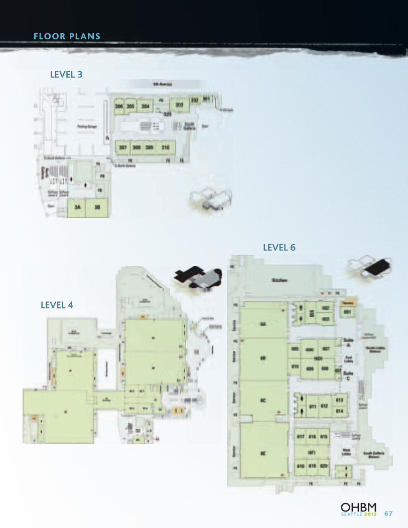
67
FLOOR PL ANS
LEVEL 3
LEVEL 4
LEVEL 6
68
COUNCIL AND COMMITTEES
CouncilSusan Bookheimer, Chair
Steve Smith, Chair Elect
Maurizio Corbetta, Past Chair
Julien Doyon, Treasurer
Jia-Hong Gao, Treasurer Elect
Christian Beckmann, Past Treasurer
Thomas Nichols, Secretary
Simon Eickhoff, Secretary Elect
Hugh Garavan, Past Secretary
Peter Bandettini, Meetings Liaison
Pedro Valdes Sosa, Meetings Liaison Elect
Katrin Amunts, Past Meetings Liaison
Program CommitteePeter Bandettini, Chair
Pedro Valdes Sosa, Chair Elect
Katrin Amunts, Past Chair
Klaas Enno Stephan, Chair Education Subcommittee
Jack Van Horn, Chair Elect Education Subcommittee
Michael Beauchamp, Past Chair Education Subcommittee
Bradley Schlaggar
Marco Catani
Nathalie Tzourio-Mazoyer
Vince Clark
Simon Eickhoff
Maurizio Corbetta
Susan Bookheimer
Steve Smith
Jia-Hong Gao
Thomas Grabowski
Arno Villringer
2013 Local Organizing CommitteeThomas Grabowski, Chair
Geoffrey Boynton
Allan Jones
Elizabeth Aylward
Jeffrey Ojemann
Kenneth Maravilla
Adrian K.C. Lee
Elaine Shen
Todd Richards
Colin Studholme
Executive Office5841 Cedar Lake Road, Suite 204
Minneapolis, MN, USA 55416
Telephone: (952) 646-2029
Fax: (952) 545-6073
Email: [email protected]
Web: www.humanbrainmapping.org
JoAnn Taie, Executive Director
Kayla Stidger, Conference Meeting Planner
Sam Fortin, Administrative Coordinator

69
NOTES

70
NOTES

Organization for Human Brain Mapping
PLEASE JOIN US FOR THE
CCH – Congress Center HamburgAm Dammtor / Marseiller Str. | 20355 Hamburg, Germany
20th Annual Meeting
Hamburg, Germany
June 8-12, 2014
Hawaii Convention Center1801 Kalakaua Avenue | Honolulu, HI 96815
21st Annual Meeting
Honolulu, Hawaii, USA
June 14 –18, 2015
5841 Cedar Lake Road, Suite 204
Minneapolis, MN 55416 USA
www.humanbrainmapping.org
Phone: 952.646.2029
Fax: 952.545.6073
Email: [email protected]