PROGNOSTIC FACTORS IN GASTROINTESTINAL STROMAL …
64
PROGNOSTIC FACTORS IN GASTROINTESTINAL STROMAL TUMOURS Dissertation submitted to THE TAMILNADU Dr. MGR MEDICAL UNIVERSITY in partial fulfillment of the requirements for the award of degree of MCh (BRANCH VII) SURGICAL ONCOLOGY COLLEGE OF ONCOLOGICAL SCIENCES CANCER INSTITUTE (WIA) ADYAR CHENNAI – 600 020 FEBRUARY 2008
Transcript of PROGNOSTIC FACTORS IN GASTROINTESTINAL STROMAL …

PROGNOSTIC FACTORS IN
GASTROINTESTINAL STROMAL TUMOURS
Dissertation submitted to
THE TAMILNADU
Dr. MGR MEDICAL UNIVERSITY
in partial fulfillment of the requirements for the award of degree of
MCh (BRANCH VII)
SURGICAL ONCOLOGY
COLLEGE OF ONCOLOGICAL SCIENCES
CANCER INSTITUTE (WIA)
ADYAR
CHENNAI – 600 020
FEBRUARY 2008

ii
CERTIFICATE
I hereby certify that this is the bonafide work done by
Dr. AQIB K. SHAICK who is appearing for MCh Surgical Oncology
(branch VII) final examination in February 2008, under my guidance in
the College of Oncological Sciences, Cancer Institute (WIA), Adyar
Chennai.
Dr. HEMANTH RAJ MCh. PhD.
Professor and Chairman
Division of Surgical Oncology
College of Oncological Sciences,
Cancer Institute (WIA)
Adyar, Chennai.

iii
ACKNOWLEDGMENT
I am very grateful to all the patients, whom I have served and from whom
I have learnt.
I express my gratitude to Dr. S. Krishnamurthy, Advisor and
Dr. V. Shantha, Executive Chairman, College of Oncological Sciences, Cancer
Institute (WIA), Adyar, for providing the facilities to carry out this study.
I am thankful to my teacher and guide in this project, Dr Hemant Raj,
Professor and chairman Division of Surgical Oncology, College of Oncological
Sciences, Cancer Institute (WIA), Adyar.
Iam also thankful to Dr A.S. Ramakrishnan associate professor Division of
Surgical Oncology and Dr. Urmila Majhi, professor and Dr. Shirley. S associate
professor Department of Pathology for their inputs.
I am also thankful for the support given by the administration of the Cancer
Institute (WIA), headed by the Director and Scientific Director Dr. T Rajkumar.
I acknowledge the help rendered by the staff at the Tumor Registry and
Epidemiology Division at the Cancer Institute (WIA).

iv
CONTENTS
CHAPTER
NO TITLE
PAGE
NO
1. INTRODUCTION 1
2. AIMS & OBJECTIVES 3
3. MATERIALS & METHODS 4
4. REVIEW OF LITERATURE 5
5. RESULTS 40
6. DISCUSSION 49
7. CONCLUSION 54
8. REFERENCES 55
9. PROFORMA 60

v
INTRODUCTION
Gastrointestinal stromal tumours (GIST) are the most common
mesenchymal tumours of the digestive tract. Most gastrointestinal soft
tissue neoplasms, previously classified as leiomyomas, schwannomas,
leiomyoblastomas or leiomyosarcomas, are today classified as GIST on
the basis of molecular and immunohistological features. They originate
from gastrointestinal pacemaker cells and are characterised by over-
expression of the tyrosine kinase receptor KIT. Overall 5-year survival
after surgical resection of GIST is approximately 60%. However, these
tumours span a wide clinical spectrum from benign to highly malignant.
Prognostic factors have recently been identified for GIST and include
tumour size, mitotic rate and other minor factors. At present, surgery is
the standard treatment for primary resectable GIST. Benign GIST have
an excellent prognosis after primary surgical treatment, with over 90% 5-
year survival. While recurrent or malignant GIST, which are resistant to
radiotherapy and chemotherapy, had until recently an extremely poor
prognosis even after surgical resection, with median survival of 12

vi
months. The development of a tyrosine kinase inhibitor has changed the
management of unresectable malignant cases. This new tyrosine kinase
inhibitor, imatinib mesylate, which inhibits the c-kit receptor, has proved
highly effective against GIST and has improved survival in metastatic
GIST.

vii
AIMS & OBJECTIVES
1. To find out the actual number of Gastrointestinal stromal tumours
treated in our institute based on c-kit positivity.
2. To identify the prognostic factors influencing recurrence and
survival.
3. To identify subgroup of patients, who might benefit from adjuvant
therapy.

viii
MATERIALS & METHODS
All the case records of patients who were diagnosed to have GIST
(on the basis of c-kit positivity) were analysed individually. Case records
of all the patients who were diagnosed to have intraabdominal sarcoma
from the year 1999 to june 2007 were scrutinized. Paraffin blocks and
slides of the above patients were retrieved from pathology dept. IHC
study for c-kit were performed on the slides. All the c-kit positive cases
were also included in the study. Of the 32 patients with GIST only 31
were available for analysis.
Data was analysed using SPSS 10.0.1 structured package.
Survival was calculated by life table method.
Comparison of survival by different categories were done by log
rank test.

ix
REVIEW OF LITERATURE
Gastrointestinal stromal tumours (GISTs) are rare tumours of the
gastrointestinal tract, mesentery, and omentum. However, malignant
GIST is the most common sarcoma of the gastrointestinal tract and
accounts for about 5% of all sarcomas.1 The estimated annual incidence
is 10–20 cases per million, of which 20–30% are malignant.2 However,
these estimates may need to be revised after the recent clearer definition
of diagnostic criteria for GIST.3 Clinical, histopathological,
ultrastructural, and molecular-biological findings, have made clear that
GIST is completely separate from leiomyoma and leiomyosarcoma,
which were previously thought to be the most common types of
softtissue neoplasms in the gastrointestinal tract. Recent studies suggest
that true gastrointestinal-tract leiomyomas and leiomyosarcomas are
rare.2
The term GIST was first used by Mazur and Clark in 1983 to
describe gastrointestinal non-epithelial neoplasms that lacked the

x
immunohistochemical features of Schwann cells and did not have the
ultrastructural characteristics of smooth-muscle cells.4 The discovery of
gain-of-function mutations in the KIT proto-oncogene in GISTs by
Hirota and colleagues in 1998 was of crucial importance in terms of the
genesis and classification of these tumours.5 This finding led to the
development of rational, molecularly targeted therapy of GISTs with the
KIT-receptor tyrosine-kinase inhibitor, imatinib mesylate (formerly
known as STI571).
The KIT protein is the transmembrane receptor for the cytokine
known as stem-cell factor (SCF); the intracytoplasmic portion of KIT
functions as a tyrosine kinase. At present, GISTs are defined as spindle-
cell, epithelioid, or occasionally pleiomorphic mesenchymal tumours of
the gastrointestinal tract that express the KIT protein.2,3 The KIT
protein is often detected clinically by immunohistochemical assays for
CD117 antigen. The definition of KIT-negative GISTs remains a focus
of research, but for clinical purposes the important point is that the vast
majority of GISTs express KIT.

xi
CLINICAL FEATURES
Most studies of the clinicopathological entity referred to as GIST
before the year 2000, are likely to include a group of patients with true
GISTs as well as other histological subtypes of spindle-cell sarcoma
such as leiomyosarcoma. GISTs occur in both sexes at a similar
frequency, but some data show male predominance.2,6 The median age
at diagnosis is about 60. GISTs are occasionally found in young adults,
but they are very rare in children.2 Nearly all GISTs arise as a result of a
somatic mutation, but rare familial cases associated with mutated KIT
have been identified.
The hereditary forms are characterised by the presence of multiple
tumours and in some cases hyperpigmentation of the skin and the
mucous membranes, systemic mast-cell disease, multiple naevi, urticaria
pigmentosa, and diffuse spindle-cell hyperplasia in the myenteric plexus
layer of the gastrointestinal tract.7–9 GISTs may also be a feature of the
Carney triad, a very rare syndrome of unknown cause affecting mainly
young women.

xii
The triad includes gastric stromal sarcoma (generally epithelioid
type), extra-adrenal paraganglioma, and pulmonary chondroma. Familial
occurrence has been suggested for the Carney triad, but no detailed
molecular genetic mechanism is known.10 A pathogenetic relation has
also been suggested between neurofibromatosis type 1 (von
Recklinhausen’s disease) and GISTs because of the high frequency of
nonrandom association of these diseases.11 However, the vast majority
of GISTs are sporadic, and predisposing factors are unknown.
GISTs are most commonly found in the stomach (40–70%), but
they can occur in all other parts of the gastrointestinal tract. About 20–
40% of GISTs arise from the small intestine, and 5–15% from the colon
and rectum.1,2,12,13 GISTs can also be found in the oesophagus (<5%),
omentum (<5%), mesentery, or retroperitoneum.2,3,12 They typically
grow in an endophytic way parallel to the bowel lumen, commonly with
overlying mucosal necrosis and ulceration, and they vary in size from a
few millimetres to 40 cm in diameter.12 Larger, high-grade GIST
lesions, can be necrotic and haemorrhagic and show more mucosal
ulceration than smaller GISTs, which might have been diagnosed in the
past as purely benign lesions. Many GISTs are well confined by a very

xiii
thin surrounding pseudocapsule.14 Over 95% of patients present with a
solitary primary tumour; in 10–40% of cases these tumours directly
invade the surrounding organs.15,16
Many small GISTs are discovered incidentally during endoscopy
or laparotomy done for other reasons such as submucosal or subserosal
nodules, or during imaging examinations. At presentation, the most
common symptoms of GISTs are vague abdominal discomfort or pain,
presence of a palpable abdominal mass, feeling of abdominal fullness,
and secondary symptoms resulting from tumour bleedingand associated
anaemia. GISTs can also cause altered bowel function, bowel obstruction
or perforation, dysphagia, and fever. Duodenal GISTs occasionally cause
obstructive jaundice. GISTs are commonly discovered during emergency
surgery for unexpected perforation of the gastrointestinal tract and
consequent intra-abdominal blood loss.17 15–50% of GISTs present
with overtly metastatic disease.1, 16, 18.
A peculiar feature of GISTs is that the great majority of
recurrences are solely intra-abdominal. Macroscopic extraabdominal
metastases are uncommon even in advanced disease, and they rarely

xiv
occur in the absence of intraabdominal recurrence. This feature contrasts
with true leiomyosarcomas of the abdomen and gastrointestinal tract,
which commonly give rise to pulmonary metastases.6,19 40–80% of
GISTs recur despite histopathologically complete tumour resection. The
most common sites of metastases are the peritoneum and the liver,1,6
whereas regional lymph-node metastases are extremely rare.1,20 In one
review of 60 patients with recurrent GIST, local recurrence occurred in
76% of patients, half of whom had synchronous liver metastases, 15%
liver metastases, and 7% peritoneal metastases.21 None had extra-
abdominal metastases at first recurrence.
Peritoneal metastases are most probably a result of tumourcell
seeding from the primary tumour directly into the peritoneal cavity.
Similarly, liver metastases most probably result from haematogenous
seeding into the portal vein.
Histopathology and immunohistochemistry
On histolgical analysis most GISTs look fairly benign, which is
surprising in view of the malignant potential of the disease. However, the

xv
histological appearance of GISTs can vary greatly among patients, and
their malignant potential ranges from clinically benign tumours to
aggressive cancers. The spindle-cell variant of malignant GIST (70%)
corresponds to tumours previously classified as leiomyosarcoma, and
many of the epithelioid or round-cell variants (30%) were previously
thought to be leiomyoblastoma. Most tumours previously diagnosed as
gastrointestinal autonomic nerve tumours (GANTs) are in fact
GISTs.2,3,22 GANTs have been described as cells with axon-like
cytoplasmic processes and synapse-like structures (seen on electron
microscopy), with dense core granules and intercellular fibrils.
Tumours previously diagnosed as GANTs have subsequently been
found to express KIT and to harbour essentially identical KIT mutations
to GISTs.23 Also, electron microscopy shows that a substantial
proportion of GISTs have similar ultrastructural features to GANTs.
Thus, GANT should be regarded as a type of GIST and no longer be
classed as a separate entity.3It is important to differentiate between
GISTs, which constitute about 80% of gastrointestinal mesenchymal
tumours, and the less common gastrointestinal nonepithelial neoplasms,
leiomyoma, leiomyosarcoma (10–15% of mesenchymal tumours),

xvi
schwannomas (5%), and other malignant disorders such as melanomas,
so that appropriate clinical decisions can be made (table 1). GISTs
characteristically stain strongly for the CD117 antigen, an epitope of the
KIT-receptor tyrosine kinase. Smooth-muscle neoplasms (leiomyoma
and leiomyosarcoma), neurogenic tumours (schwannomas), and desmoid
fibromatoses typically do not show this positive expression of
CD117.2,3,22
Thus, CD117 immunostaining is an important method for
diagnostic distinction. Typically, in GISTs, KIT is widespread
throughout the entire tumour and is highlighted by cytoplasmic staining,
sometimes showing a dot-like ‘golgi’ pattern.3 60%–70% of GISTs stain
for CD34, a sialylated transmembrane glycoprotein also found in
haemopoietic progenitor cells and endothelial cells.3,5,22,24 Up to 40%
of GISTs are also positive for smooth muscle actin (SMA).
They rarely express desmin, an intermediate filament protein
typical of muscle, or S100, a neural (schwann) cell marker, and they are
negative for neurofilaments and glial fibrillary acidic protein.2 By
contrast, leiomyosarcomas are positive for SMA and desmin but
negative for KIT, and schwannomas are positive for S100 but negative

xvii
for CD117.3,22 Overall, strong KIT expression in the absence of
smooth-muscle differentiation-related proteins is characteristic for
GISTs, and these features aid the differential diagnosis between GISTs
and most other types of gastrointestinal mesenchymal tumours.
KIT expression is not limited to GISTs, but some other neoplasms
may stain positively for KIT in immunohistochemical assays. These
tumours include a subset of soft-tissue sarcomas such as Ewing’s
sarcoma and angiosarcoma, as well as other neoplasms such as
melanoma, small-cell lung cancer, adenoid cystic carcinoma, ovarian
carcinoma, sinonasal natural-killer/T-cell lymphoma, anaplastic large-
cell lymphoma, acute myelogenous leukaemia, seminoma,
neuroblastoma, and mastocytomas.
However, these tumours are rarely included in the differential
diagnosis of GISTs.2,25,26 Immunopositivity for KIT does not
necessarily indicate KIT activation or the presence of a KIT mutation,
and there is, as yet, little reason to believe that these other tumours
would show a response to imatinib mesylate.

xviii
Cell of origin
Kindblom and colleagues suggested in 1998 that GISTs originate
from stem cells that differentiate towards the interstitial cells of Cajal
(ICCs), and that GISTs should be called gastrointestinal pacemaker-cell
tumours.24 ICCs arisefrom precursor mesenchymal cells and are the
pacemaker cells that bring about autonomous movement of the
gastrointestinal tract.27 They intercalate between nerve fibres and
muscle cells and can be seen in the adult intestine in and around the
myenteric plexus.2 Both ICCs and GISTs express KIT protein, have
similar ultrastructural features, and express the embryonic form of the
heavy chain of smoothmuscle myosin;24,28 all these features support a
common histogenesis.

xix
The precursor-cell hypothesis could also explain why KIT-
expressing mesenchymal tumours, with similar histology to GISTs, can
arise outside the gastrointestinal tract in the omentum, mesentery, and
retroperitoneum.29,30 ICC-like cells have also been identified in the
omentum.31 The precursor-cell hypothesis could also account for the
coexpression of KIT, SMA, and even desmin in some GISTs. In studies
of immunohistochemistry and confocal microscopy of normal intestinal
muscle, CD34 immunopositivity has been found in fibroblast-like cells
located near the ICCs. Conversely, ICCs did not stain for CD34 in these
studies, and no cells with concomitant CD34 and KIT positivity were
found. These findings also support the hypothesis that GISTs originate
from a more primitive precursor cell.32
Diagnosis
The clinical prediagnostic investigation of GISTs is similar to that
of other gastrointestinal malignant disorders. A doublecontrast series of
radiographs may show a characteristic smooth-lined filling defect with
clearly demarcated borders. On endoscopic examination, there may be a
smooth protrusion of the bowel wall, lined with mucosa, which can also

xx
show signs of bleeding and ulceration.14 Endoscopic ultrasonography
may show a hypoechoic mass that is contiguous with the muscularis
propria of the normal gut wall. In one ultrasonography study, presence of
malignant GIST was suggested by the presence of a large (>4 cm)
tumour with irregular extraluminal border, echogenic foci, and cystic
spaces.33 Computed tomography (CT) and magnetic resonance imaging
(MRI) are essential in assessment of primary tumour extension and the
presence of metastases.
Most GISTs are submucosal and grow endophytically, which
decreases the likelihood that a tissue diagnosis can be obtained
preoperatively. Only about 50% of patients assessed by endoscopy are
given a histological diagnosis preoperatively. Percutaneous fine-needle
aspiration has been suggested as an initial diagnostic technique, if
feasible, but it is not universally recommended because intra-abdominal
tumour spillage is possible.14,34 Most oesophageal stromal tumours are
benign,22 and endoscopic biopsy, for cases in which the mucosa
surrounding the tumour is intact, is controversial because of the potential
high risk of intraoperative oesophageal perforation.14,35

xxi
During laparotomy, all resected margins should be carefully
oriented and examined, and biopsy samples from grossly different areas
of the excised tumour should be evaluated pathologically. Solid and firm
areas should be included, as well as necrotic and haemorrhagic parts of
the tumour. A minimum of one tissue section per centimetre of tumour
diameter has been recommended for microscopic assessment of fixed
tissue.14 KIT immunostaining should be done with appropriate positive
and negative control samples to avoid falsely positive and negative
results. Mast cells and ICCs stain strongly positively for KIT and may be
used as positive internal controls.
Immunohistochemical staining for KIT (CD117), CD34, desmin,
SMA, and S100 should be carried out. Mutation analysis of KIT is not
considered mandatory in routine diagnostics, but this technique is
currently being evaluated to see whether it might provide predictive
information about the likelihood of a response to imatinib mesylate
therapy. Additionally, a KIT mutation gives further support to the
diagnosis of GIST.

xxii
Prognostic features
Assessment of the malignant potential of a primary GIST lesion is
difficult in many cases, and even small GISTs (less than 2 cm in
diameter) have uncertain malignant potential.3 The criteria used to
predict biological behaviour also vary significantly with tumour location,
for example, smoothmuscle tumours arising from the small bowel, colon,
rectum, omentum, or mesentery are generally associated with a less
favourable outcome than those arising in the stomach.12 Most
oesophageal and colonic GISTs are malignant, whereas in the case of
gastric GISTs, more indolent tumours seem to outnumber overtly
malignant ones. However, with long follow-up their outcome may not
differ greatly. Almost all incidental small (<1 cm) GISTs are clinically
benign, whereas tumours larger than about 5 cm in diameter are
generally malignant. Nonetheless, no cut-off diameter predicts
subsequent malignant behaviour with certainty, and the optimum cut-off
size may vary for different sites.12,13,15,36
The mitotic rate is one of the more reliable single factors in
differentiating between GISTs of varying malignant potential. In general,

xxiii
most tumours with 0–1 mitoses per 10–50 high-power fields (HPFs) will
not give rise to metastases, those with over five mitoses per 50 HPF are
considered as malignant, and tumours with over 20–50 mitoses per 50
HPF are classed as high-grade malignant.2,13–15.
However, a low mitotic count does not rule out malignancywith
certainty, and vice versa, and the mitotic count is of limited value
especially in assessment of the malignant potential of small-bowel
GISTs.12 Other factors suggested to be associated with an adverse
outcome include: incomplete surgical resection and tumour rupture at
surgery;6 infiltration of tumour to the neighbouring structures; location
of the primary tumour in the intestine;22 presence of coagulative tumour
necrosis, high cellularity, and pronounced pleiomorphism; a high S-
phase fraction and DNA aneuploidy in flow cytometry or image
cytometry; a high Ki-67 score; proliferating-cell nuclear-antigen
expression; and presence of telomerase activity. In some studies the
presence of KIT mutation has also been implicated.12,18. However,
many of these features are predictive of outcome only in statistical
analysis of large series of cases, and their usefulness on an individual-
case basis is limited.

xxiv
Altogether, there are many borderline malignant tumours within
the spectrum of GISTs, ranging from indolent tumours to clearly
malignant cancers. All GISTs should be considered as having some low
malignant potential, and they should be described in terms of risk
assessment, rather than using distinct benign and malignant categories.3
The only certain indication of malignancy is tumour spread beyond the
organ of origin at the time of diagnosis.13 However, most primary
GISTs larger than 5 cm in diameter and with a mitotic count higher than
five per 50 HPF are likely to behave in a malignant way. Similarly,
GISTs larger than 10 cm in diameter have a high risk of aggressive
behaviour whatever the mitotic count, and GISTs of any size with a high
mitotic count (more than ten per 50 HPF) are also deemed to be high-risk
tumours.3
Outcome
Before about the year 2000, studies of GISTs included tumours
that would not presently be classified as GISTs, and data are therefore
contaminated by these cases. However, since GISTs constitute the
majority of gastrointestinal sarcomas, the survival data from these

xxv
studies probably largely reflect the experience of patients with true
GISTs. Most recurrences take place within 5 years of the primary
diagnosis,12 but in the slowly proliferating subset of GISTs, metastases
can appear more than 10 years after the primary diagnosis.
Outcome depends on the histopathological and clinical features.
The reported overall or disease-specific 5-year survival is 28–60%
among patients with malignant GIST; the median disease-specific
survival is about 5 years for primary disease, and 10–20 months in
recurrent or metastatic disease.1,6,16,18. According to one study,
patients who have been diagnosed with GISTs with one to five mitoses
per 50 HPF have median survival of 98 months, whereas those with
GISTs with more than ten mitoses per ten HPFs have median survival of
25 months.42 In another study, patients who had undergone curative
surgery had 8-year disease-free survival of 80% when fewer than ten
mitoses were present per 50 HPF compared with an 18- month median
survival in patients with more high-grade lesions.
xxvi
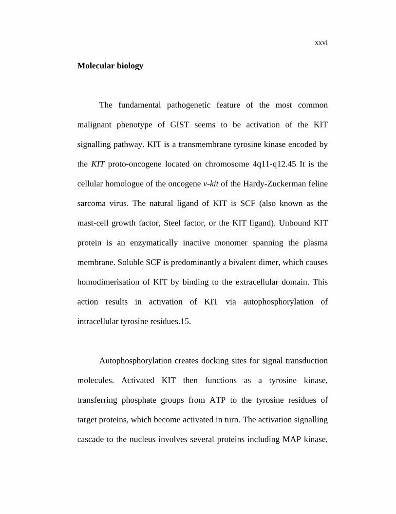
Molecular biology
The fundamental pathogenetic feature of the most common
malignant phenotype of GIST seems to be activation of the KIT
signalling pathway. KIT is a transmembrane tyrosine kinase encoded by
the KIT proto-oncogene located on chromosome 4q11-q12.45 It is the
cellular homologue of the oncogene v-kit of the Hardy-Zuckerman feline
sarcoma virus. The natural ligand of KIT is SCF (also known as the
mast-cell growth factor, Steel factor, or the KIT ligand). Unbound KIT
protein is an enzymatically inactive monomer spanning the plasma
membrane. Soluble SCF is predominantly a bivalent dimer, which causes
homodimerisation of KIT by binding to the extracellular domain. This
action results in activation of KIT via autophosphorylation of
intracellular tyrosine residues.15.
Autophosphorylation creates docking sites for signal transduction
molecules. Activated KIT then functions as a tyrosine kinase,
transferring phosphate groups from ATP to the tyrosine residues of
target proteins, which become activated in turn. The activation signalling
cascade to the nucleus involves several proteins including MAP kinase,

xxvii
PI3 kinases, STAT5, RAS, and JAK2,15,48-51 which have been
implicated in KIT-induced mitogenesis and differentiation.
Structurally, KIT tyrosine kinase is a type III tyrosine kinase
receptor, similar to receptors of macrophage colonystimulating factor,
platelet-derived growth factor, and the haemopoietic growth factor FLT3
ligand, each possessing five immunoglobulin-like extracellular repeats
and a tyrosine kinase domain split into two by an insert sequence of
variable length (figure 2). In addition to ICCs, KIT is normally expressed
in mast cells, melanocytes, Leydig cells, spermatogonia, spermatids,
haemopoietic stem cells, cutaneous basal cells, and breast epithelial
cells,25,52 and it has important roles in haemopoiesis, melanogenesis,
gametogenesis, and the development of mast cells and ICCs.27.
Almost all GISTs have constitutive activation (phosphorylation)
of the KIT protein, and most have inframe mutations that preserve the
expression of KIT. Stable transfection of mutated KIT cDNAs into
murine lymphoid cells causes malignant transformation.5. Unlike the
normal KIT protein, mutated KIT may not require SCF for dimerisation
or autophosphorylation. This ligandindependent activation causes a shift

xxviii
in the balance between cell survival and proliferation away from
apoptosis. The frequency of KIT mutations in GISTs varies in different
studies, according to the tumour type, and probably because of
differences in the methods used to analyse the tumours. Up to 80–90% of
metastatic GISTs have been reported to have mutated KIT, and in some
studies KIT mutations have been found in benign, borderline, and
malignant GISTs at about equal frequency. Corless and co-workers
found identical KIT mutations to those previously found in larger GISTs,
in 11 (85%) of 13 of morphologically benign GISTs that were found
incidentally at autopsy, endoscopy, or laparotomy, by use of a sensitive
mutation-detection method (denaturing high-performance liquid
chromatography). These small GISTs were all immunohistochemically
positive for KIT, and ranged in size from 4 mm to 10 mm. If confirmed,
these findings suggest that KIT mutations are acquired very early in the
development of most GISTs and that KIT mutations alone may be of
limited prognostic importance. KIT mutations have not been found in
leiomyomas and leiomyosarcomas.
Family members with germline mutations of KIT have indolent
and malignant GISTs from a young age and have the same mutation in

xxix
the germline, indolent GISTs, and malignant GISTs, which also suggests
that KIT mutation is an early oncogenic event.15 KIT has 21 exons, and
in sporadic GISTs the KIT mutations have been found in exon 11,
encoding an intracellular juxtamembrane region of the receptor, in 50–
77% of cases. Exon-9 mutations, encoding a region located in the
extracellular domain, are found in 3–18% of GISTs, and a few GISTs
have mutations in exons 13 and 17, encoding the intracellular part of the
receptor. Mutation of NF2 gene has also been described in GISTs. This
finding is consistent with high incidence of GISTs in patients with
neurofibromatosis. Loss of chromosome 1p or complete or partial loss of
chromosomes 14 and 22 occurs in 50% or more of GISTs and may be
involved in GIST pathogenesis and progression. However, such changes
may be secondary, and activating mutations of KIT seem to have a
central role in GIST pathogenesis.44 This idea is supported by a recent
study that used 13, 826-element, cDNA microarrays to analyse gene-
expression patterns of KIT-mutation-positive GISTs.
In this study the expression profiles of GISTs were remarkably
uniform with the KIT gene ranking the highest on the discriminator list
and being highly expressed in every tumour studied. Therefore, mutated

xxx
KIT is an excellent molecular target for therapeutic interventions with
KIT-selective tyrosine-kinase inhibitors.
Surgery
Surgery remains the standard initial treatment for nonmetastatic
GISTs. Careful pathological assessment should be done to differentiate
GISTs from carcinomas and lymphomas. As with other soft-tissue
sarcomas, a true capsule does not exist, and the tumour should be
removed en-bloc with its pseudocapsule and, if possible, an adjacent
margin of normal soft tissue or bowel. In cases where contiguous organs
xxxi
are involved, en-bloc resection has been recommended wherever
feasible.14 Since this process may require total gastrectomy in the case
of gastric GISTs, pancreaticoduodenectomy for a duodenal GIST, or an
abdominoperineal resection for tumours involving the rectum, associated
morbidity can be substantial. In such cases, preoperative, neoadjuvant
treatment with imatinib mesylate may eventually be considered, but at
present no data are available to support this practice. A trial evaluating
this treatment option is in progress in the USA.
In several studies, patients who had complete tumour resection
had better overall survival than those who had less radical surgery.16.
Although this difference may partly reflect aggressive biological features
of GISTs that cannot be totally removed, an effort should be made to
obtain histologically tumour-free tissue margins. The optimum width of
the tumour-free margin has not been defined. Tumour rupture,
spontaneously or during surgery, may be associated with an increased
risk of development of peritoneal implants and should be avoided.
Regional lymph-node resection is of unproven value, and extensive
lymphadenectomy is not recommended.14.

xxxii
There are few data about the usefulness of resection of recurrent
disease or intra-abdominal metastases. In some studies, tumour-specific
mortality and overall survival have not differed significantly between
patients who underwent complete resection of recurrent disease and
those who had partial resection or biopsy alone.21 However, there is
evidence that metastasectomy may improve survival in selected patients.
Patients with well or moderately differentiated GIST, a disease-free
interval between the diagnosis and detection of metastases of longer than
12 months, and isolated resectable liver metastases are more likely to
benefit from metastasectomy than patients who have rapidly progressing
or widespread GIST. Many patients who have a bleeding tumour or
tumour-related bowel or biliary-tract obstruction achieve efficient
palliation with surgery.
However, since imatinib mesylate was developed as a therapeutic
alternative, surgery for metastatic GIST has largely been replaced by
drug therapy, and primary surgery for metastatic GIST should probably
be attempted only in patients who have bleeding or obstructive disease.
The question of whether surgical resection should be done to remove
residual masses, after imatinib mesylate therapy, is unanswered and

xxxiii
requires further research. We suspect that this approach would be a very
reasonable option for patients with low-volume metastatic disease.
Radiotherapy
The impact of radiotherapy on outcome is unknown. Many
visceral sarcomas are not readily amenable to radiotherapy because of
organ motility, and postoperatively contaminated bowel loops may
relocate to remote sites. The large target volumes needed and the limited
radiation tolerance of the intra-abdominal organs limit the usefulness of
radiotherapy. Fixed lesions on the abdominal wall or adjacent organs
have been treated with postoperative radiotherapy, but recurrences both
within and outside the radiation field have been frequent.16 At present,
radiotherapy is not a standard postoperative therapy for GIST, and in
most cases should be reserved for limited palliative settings or for
research of new strategies.
Chemotherapy
Attempts to treat malignant GISTs with systemic chemotherapy
have been almost universally unsuccessful. In one study, only 3 of 43

xxxiv
(7%) patients with gastrointestinal soft-tissue sarcomas (most tumours
probably GISTs) responded to a combination of doxorubicin and
dacarbazine, whereas 22% of patients with uterine leiomyosarcomas and
21% of patients with leiomyosarcomas of other sites responded to this
combination (p=0·05), suggesting relative chemoresistance of
gastrointestinal soft-tissue sarcomas. Similar results have been obtained
in other studies. Only 1 of 21 (5%) patients with GISTs treated with
combination chemotherapy consisting of dacarbazine, mitomycin c,
doxorubicin, cisplatinum, and sargramostine showed a response, and no
gastrointestinal soft-tissue sarcomas responded to a combination of
etoposide and ifosfamide.
The unresponsiveness of GISTs to drugs commonly used in the
treatment of soft-tissue sarcomas may be explained partly by the
frequent expression of P-glycoprotein and multidrug resistance protein 1
(MDR1) in GISTs. study, 38·4% of GISTs expressed P-glycoprotein and
35·4% expressed MDR1 protein, whereas only 13·4% and 13·3% of
gastrointestinal leiomyosarcomas, respectively, stained positively for
these proteins.19In onestudy, 38·4% of GISTs expressed P-glycoprotein
and 35·4% expressed MDR1 protein, whereas only 13·4% and 13·3% of

xxxv
gastrointestinal leiomyosarcomas, respectively, stained positively for
these proteins.19
Imatinib mesylate
Imatinib mesylate is a competitive inhibitor of certain tyrosine
kinases including the intracellular kinases ABL and BCR-ABL fusion
protein present in some leukaemias, KIT, and the platelet-derived growth
factor receptors. Imatinib mesylate inhibits these tyrosine kinases at
submicromolar concentrations, but has little or no effect on many other
tyrosine or serine/threonine kinases. It is a small multiringed molecule,
which competes with ATP for its kinase-binding site, and prevents the
kinase from transferring phosphate from ATP to tyrosine residues of the
substrates. This action inhibits downstream signalling from the kinase,
which switches the balance towards apoptosis. Imatinib mesylate is very
well absorbed after oral administration and is available as capsule
formulations. It is metabolised mainly in the liver by the P450
isoenzyme CYP3A4, and the metabolites are mostly excreted via the bile
into the stools. The half-life in the circulation is about 20 h, which is
compatible with oncedaily administration. Preclinical studies suggest
that maintenance of imatinib mesylate serum concentrations above 1

xxxvi
_mol/L are needed for optimum therapeutic effects, and such
concentrations are obtained in most patients with daily doses of 300 mg
or greater.
Effectiveness of imatinib mesylate in advanced GIST
Based on two studies, imatinib mesylate is the first effective drug
in the treatment of metastatic GIST. The US–Finland study reported a
response rate of 54% among 147 patients with inoperable or metastatic
GIST treated with a daily dose of 400 mg or 600 mg with follow-up of at
least 6 months. In addition, 28% had minor response or stable disease,
and only 14% showed primary resistance to the drug. Similar results
were reported from a trial by the European Organization for Research
and Treatment of Cancer, in which 36 patients with advanced GISTs
were treated with a daily dose of 400–1000 mg. 53% had confirmed
partial responses and 17% had as yet unconfirmed partial responses or
more than20% regression, and only 11% of patients had progression. In
these studies, about 90% of patients with symptoms had marked relief of
them. In both studies, inclusion required histology compatible with GIST
and KIT expression verified by immunohistochemistry.

xxxvii
Response to imatinib mesylate can occur rapidly, even in patients
with a large tumour burden but regression can also be slow, particularly
after an initial rapid phase of tumour regression. The median time to
response is about 13 weeks. Interestingly, tumour lysis syndrome has not
been described in these patients, even though hydration or allopurinol
have not been routinely administered alongside imatinib mesylate. Liver
lesions commonly acquire a cyst-like appearance after the start of
treatment, and may thus seem better delineated on magnetic resonance
(MR) or computed tomography (CT) images they should not be confused
with new lesions or progressive disease. Histologically, the cyst-like
lesions consist of hyaline degeneration, but a few remaining KIT-
positive cells could represent dormant or slowly proliferating GIST cells
and may persist for several months in these lesions. The malignant
potential of the few persistent intralesional KIT-positive cells is not
currently known.
In addition to CT and MRI, fluorodeoxyglucose (labeled with
fluorine-18) positron emission tomography (FDG-PET) may be a useful
imaging technique to assess response to imatinib mesylate. Glucose
uptake of GIST lesions decreases within a few hours to a few days after

xxxviii
the start of imatinib mesylate treatment, which can be verified by FDG-
PET. The technique could be helpful in problematic cases to make a
distinction between intratumoral bleeding and disease progression. The
PET scan responses can also predict subsequent tumour volume
reductions found on CT or MRI. In one study, patients with an exon-11
KIT mutation had a significantly higher rate of response to imatinib
mesylate therapy (72%) than patients whose tumour had an exon-9
mutation (32%) or no detectable mutation (12%), and the time to
treatment failure was also longer in patients with an exon-11 mutation.
The optimum dose of imatinib mesylate in the treatment of GISTs
is not yet known and is being studied in current randomised phase III
trials. Toxicity increases with increasing dose, and in one study, 800 mg
was the maximum tolerated dose when taken for 8 weeks. No statistical
difference in the response rate was found in another study of 400 mg and
600 mg daily doses, although these data are underpowered and the
results of ongoing large-scale studies are needed before conclusions are
reached.

xxxix
Data from studies on chronic myeloid leukaemia suggest that
doses less than 300 mg may be too small for the competitive inhibition
of the BCR-ABL kinase and should be avoided in clinical practice.
Overall, present data suggest that a daily dose of400 mg or higher should
be used in the treatment of patients with metastatic or unresectable
GIST.
The optimum treatment duration remains unknown, but in
metastatic disease administration of imatinib mesylate for several
months to a few years is likely to be needed. In the US–Finland study,
objective major responses were durable and generally continuing at
follow-up of at least 9 months, with no median duration of responses yet
evident. So far, responses lasting up to 24 months have been observed
but secondary resistance to imatinib mesylate has been seen in some
patients who initially responded to the drug. The proportion of patients
who will relapse after a response is not currently known.

xl
Tolerability and safety of imatinib mesylate
Tolerability has been acceptable with daily doses of 800 mg or
less. The most common adverse effects include periorbital and leg
oedema transient nausea associated with drug ingestion, muscle cramps,
diarrhoea, headache, dermatitis, fatigue, anaemia, and neutropenia. Most
of these side-effects are mild to moderate in severity, and grade 3–4
toxic effects occur in less than 30% of patients at a dose of 400–600 mg
per day.A few patients (5%) have had a tumour-associated bleeding
either into the abdominal cavity or into the bowel, and patients who
develop symptoms that suggest acute bleeding may need emergency
care.
Most adverse effects resolve within a few days to weeks after
cessation of treatment, and most patients can continue at a lower dose.
Imatinib-mesylate-associated oedema can be treated with diuretics.
Drug-ingestion-related nausea rarely requires antinausea medication,
although some patients are given a divided dose.

xli
The muscle cramps typically occur in the fingers and legs, and are
transient, most requiring no specific therapy. Interactions with other
drugs metabolized by CYP3A4 are possible. In particular, concomitant
use of paracetamol or warfarin is not recommended
Future directions and unresolved questions
Imatinib mesylate is a major breakthrough in the treatment of
advanced GISTs and is the first effective systemic therapy for this
disease. However, several important questions remain unanswered: the
required duration of imatinib mesylate therapy; the proportion (if any) of
the patients with metastatic disease who will achieve long-term disease
control, and whether treatment results can be improved with combination
therapies.
In the treatment of BCR-ABLexpressing leukaemia, resistance to
imatinib mesylate is commonly associated with reactivation of BCR-
ABL signalling due either to a secondary mutation resulting in
substitution of threonine with isoleucine at a critical binding site of the
drug or to progressive BCR-ABL gene amplification.80 Little is known

xlii
about the resistance mechanisms of imatinib mesylate in the treatment of
GISTs, but these are currently being studied.
An important strategy, which is likely to affect cure rates, is
whether adjuvant therapy will benefit patients after macroscopically
complete removal of malignant GIST, or after excision of GIST with
intra-abdominal metastases amenable to surgical removal. Many such
patients will develop inoperable systemic metastases within a few years
of primary surgery, and adjuvant therapy with imatinib mesylate for
subclinical metastatic disease might lead to a better cure rate than
treatment for overtly metastatic disease. In the absence of data from
clinical trials, there is no objective basis for recommending adjuvant use
of imatinib mesylate, but the potency of the drug in metastatic disease
certainly justifies the rapid application and study in the adjuvant setting.
Similarly, the role of neoadjuvant imatinib mesylate needs to be studied
especially in cases where extensive surgery resulting in loss of organ
function is the only other option. Combination therapies with
conventional chemotherapeutic drugs and other signal transduction
inhibitors also need to be investigated.

xliii
The molecular targeting of the critical pathogenetic mechanism
underlying GIST has given patients new hope, and has provided
physicians with a highly effective and well tolerated therapeutic option
for a disease for which no systemic therapy existed previously.

xliv
RESULTS
Thirty one patients with GIST treated at our institute were
included in the study. The median age was 55 years (14-70 years). From
the 31 patients were 8 (25.8%) females and 23 (74.2%) males. Among
these, 24 patients has first time surgery for a GIST at our institute and 7
patients were presented with advanced metastatic disease which were
deemed inoperable. All the 7 patients were started on Imatinib.
Out of the 24 operated patients, 2 patients presented with
metastasis, primary alone was resected. Detailed tumour localizations are
shown in Table 1. Applying the Fletcher classification, 4 patient had low
risk, 4 had intermediate risk and 16 patient had high risk. This reflects
that the majority of the patients had an intermediate or high risk of an
aggressive behaviour of their GIST. Regarding tumour size 5/24 of all
tumours were smaller and 19/24 larger than 5 cm.

xlv
Table 1 Tumour characteristics
Stomach Small intestine Rectum TotalTumuor size
< 5cm 2 2 1 5 5-10cm 7 1 1 9
10-15cm 2 - - 2 15-20cm 3 3 1 7
>20cm 1 - - 1 Total (n) 15 6 3 24 Mitosis
<5/50 hpf 7 2 - 9 >5/50 hpf 8 4 3 15
Fletcher classification Very low risk - - - 0
Low risk 2 2 - 4 Intermediate 3 - 1 4
High 10 4 2 16 Necrosis + 12 2 1 15
Type of resection
Segmental /TG 10 4 - 14 APR 2 2
Multi organ 5 2 1 8 Status
Disease free 12 1 1 14
Recurrent disease 3
5 2
10

xlvi
Surgical procedures
The surgical procedures carried out in this study varied from
wedge resection of the tumour to complex multivisceral surgery.
Multiorgan resection were performed in 8/24 patients. All the surgical
procedures are summarized in Table 2. Complete tumour resection (R0)
was achieved in 22/40 patients, whereas 2/24 patients had incomplete
tumour resection (R2). One patient who underwent APR had positive
iliac nodes (2/5), which is a rare feature of GIST.
Table 2 Surgical procedures
Resection of stomach
Wedge resection 13 Total gastrectomy 2
Small bowel resection Segmental resection 5 Whipple procedure 1
Rectal surgery Abdominoperineal resection 2
APR+ prostatectomy 1 Splenectomy 5 Distal pancreatectomy 2 Partial liver resection 1 Partial cystectomy 1 Tumour spill 4
R0 resection 22 R1 resection - R2 resection 2

xlvii
Complications
Post-operative complications were observed in 3/24 patients.
There was no post-operative mortality (until day 30). One patient had
urinary fistula following APR and another patient had faecal fistula
following multiorgan resection. Both these patient had incomplete
tumour resection (R2). The third patient had persistent post-operative
fever, managed appropriately.
Survival and tumour recurrence
The median disease free survival time was 43.6 months with a
median followup of 13 months (6- 88 months). At 5 years the probability
of disease free survival was 45.8%. 10 out of 24 patients developed
recurrence on follow up. In five patients liver alone was the site of
metastasis. One patient developed metastases in liver and peritoneum.
One developed recurrence in the small bowel, one on the dome of the
urinary bladder and one patient developed pelvic recurrence. One patient
developed lung metastasis. Among the 10 patients who developed
recurrent disease three underwent secondary surgery, resulting in R0

xlviii
resection. The secondary resections were, one liver metastasectomy, one
segmental resection of jejunum and one partial cystectomy. Among the
recurrences the primary sites of GIST were small intestine in 5 patients,
stomach in 3 patients and rectum in 2 patients. All the 10 patients were
started on Imatinib. Out of this 10 patients, one patient died after 4
months of primary surgery. Remaining 9 patients are alive with disease
till date.
Survival Function
DFS
706050403020100-10
Cum
Sur
viva
l
1.2
1.0
.8
.6
.4
.2

xlix
The probability of DFS at 5 years is 100% for tumours with
mitotic count <5/50 hpf and 26.8% at 3 years for >5/50 hpf (p=0.006).
Survival Function
DFS
706050403020100-10
Cum
Sur
viva
l
1.2
1.0
.8
.6
.4
.2
0.0
MITOSIS1
2
1

l
The probability of DFS at 5years for tumours without necrosis is
80% and 26% at 3 years for tumours with necrosis (p=0.0046).
Survival Function
DFS
706050403020100-10
Cum
Sur
viva
l
1.2
1.0
.8
.6
.4
.2
0.0
-.2
NEC
2.00
1.00

li
Tumour size >5cm was also found to be a poor prognostic factor
although it did not reach statistical significance. The probability of DFS
at 5 years for tumours <5 cm is 80% and 36.8% at 5 years for tumours
>5 cm (p=0.54).
Survival Function
DFS
706050403020100-10
Cum
Sur
viva
l
1.2
1.0
.8
.6
.4
.2
0.0
-.2
C
2.00
1.00

lii
Positive margin is a strong prognostic factor for recurrence. Out of
the 24 operated patients, 2 patients had positive margins and both of
them recurred. The primary tumour site was also found to be a strong
prognostic factor for recurrence. Out of the 6 patients with primary GIST
of small intestine 5 recurred (one patient had R2 resection). Among the 6
patients with rectal primary 2 have recurred (one had R2resection). Out
of the 15 patients with primary GIST of stomach, only 3 have recurred.
The extend of surgery and tumour spill were not found to be of any
prognostic significance. Patient characteristics like age and sex showed
no prognostic value for the development of a recurrent disease.

liii
DISCUSSION
Most studies of the clinicopathological entity referred to as GIST
before the year 2000, are likely to include a group of patients with true
GISTs as well as other histological subtypes of spindle-cell sarcoma
such as leiomyosarcoma.
Our study included patients on the basis of c-kit positivity.
GISTs occur in both sexes at a similar frequency, but some data
show male predominance.2,6 The median age at diagnosis is about 60.
Our male: female ratio is 4:1, and the median age is 55 years.
GISTs are most commonly found in the stomach (40–70%), but
they can occur in all other parts of the gastrointestinal tract. About 20–
40% of GISTs arise from the small intestine, and 5–15% from the colon
and rectum.1,2,12,13 GISTs can also be found in the oesophagus (<5%),
omentum (<5%), mesentery, or retroperitoneum.2,3,12.

liv
In our series also the commonest site is stomach, constituting
62.5%. 40–80% of GISTs recur despite histopathologically complete
tumour resection. The most common sites of metastases are the
peritoneum and the liver,1,6 whereas regional lymph-node metastases
are extremely rare.1,20.
In our study also 41.66% developed recurrence. Commonest site
of recurrence is liver. One patient with GIST of rectum had lymph node
metastasis.
Despite the great success of Imatinib in the treatment of metastatic
GIST, primary surgery remains the cornerstone in the treatment of
localized and resectable GIST. Recurrent disease is still a great problem.
Therefore primary risk adapted surgery is the most important factor to
avoid an early tumour recurrence and it is important to identify patients
who may be candidates to receive an adjuvant treatment after complete
tumour resection.
The criteria used to predict biological behaviour vary significantly
with tumour location, for example, smooth muscle tumours arising from
the small bowel, colon, rectum, omentum, or mesentery are generally
associated with a less favourable outcome than those arising in the
stomach.12

lv
Our series supports the theory that GIST arising in sites other than
stomach has a more aggressive behaviour. In our series Out of the 6
patients with primary GIST of small intestine 5 recurred (one patient had
R2 resection). Among the 6 patients with rectal primary 2 have recurred
(one had R2resection). Out of the 15 patients with primary GIST of
stomach, only 3 have recurred.
As per our results primary GIST of small intestine is the most
aggressive and GIST of stomach is the least aggressive, similar to results
shown by most of the studies. 6,12.
The mitotic rate is one of the more reliable single factors in
differentiating between GISTs of varying malignant potential. In general,
most tumours with 0–1 mitoses per 10–50 high-power fields (HPFs) will
not give rise to metastases, those with over five mitoses per 50 HPF are
considered as malignant, and tumours with over 20–50 mitoses per 50
HPF are classed as high-grade malignant.2,13–15. However, a low
mitotic count does not rule out malignancy with certainty, and vice
versa, and the mitotic count is of limited value especially in assessment
of the malignant potential of small-bowel GISTs.12

lvi
Our results showed strong correlation of mitotic count with
tumour recurrence, which reached statistical significance. The
probability of DFS at 5 years is 100% for tumours with mitotic count
<5/50 hpf and 26.8% at 3 years for >5/50 hpf (p=0.006).
Other factors suggested to be associated with an adverse outcome
include: incomplete surgical resection and tumour rupture at surgery;6
infiltration of tumour to the neighbouring structures; multiorgan
resection, location of the primary tumour in the intestine;22 presence of
coagulative tumour necrosis, high cellularity, and pronounced
pleiomorphism; a high S-phase fraction and DNA aneuploidy in flow
cytometry or image cytometry; a high Ki-67 score; proliferating-cell
nuclear-antigen expression; and presence of telomerase activity. 12,18.
In our series also incomplete resection is a strong predictive factor
of recurrence. Out of the 24 operated patients, 2 patients had positive
margins and both of them recurred.
Presence of necrosis in the tumour showed strong correlation with
tumour recurrence, which reached statistical significance. The
probability of DFS at 5years for tumours without necrosis is 80% and
26% at 3 years for tumours with necrosis (p=0.0046).

lvii
Tumour spill alone, with complete resection of the tumour did not
show a prognostic value for recurrence in our study. Similarly extend of
surgery did not influence DFS in our series.
Currently adjuvant therapy trials are being conducted to evaluate
the benefit of Imatinib in those GIST patients who have a substantial risk
of relapse after complete surgery. Furthermore the administration of
Imatinib in patients with advanced GIST preoperatively to avoid
multivisceral surgery is still an individual decision as data from a
controlled trial are still missing.

lviii
CONCLUSION
Primary surgery remains the cornerstone in the treatment of
localized and resectable GIST. Recurrent disease is still a great problem.
Summarizing our results with the influence of high mitotic count,
presence of tumour necrosis, large tumour size, incomplete surgical
resection and primary site other than stomach on the DFS, which is
reflected by a high rate of recurrences, we support the concept that all
patients with these high risk features need to be assessed for adjuvant
treatment with Imatinib.
Considering the 100% chance of recurrence with an incomplete
resection, neoadjuvant treatment with Imatinib followed by surgical
resection and continuing of Imatinib administration is a reasonable
option. This combination strategy may provide survival benefit. But it
remains to be determined whether this multimodal approach may exceed
the benefits associated with Imatinib monotherapy.

lix
REFERENCES
1. Beghini A, Tibiletti M, Roversi G, et al. Germline mutation in the
juxtamembrane domain of the KIT gene in a family with
gastrointestinal stromal tumors and urticaria pigmentosa. Cancer
2001; 92: 657–62.
2. Bonavina L, Segalin A, Rosati R, et al. Surgical therapy of
esophageal leiomyoma. J Am Coll Surg 1995; 181: 257–62.
3. Carney JA. Gastric stromal sarcoma, pulmonary chondroma, and
extra-adrenal paraganglioma (Carney triad): natural history,
adrenocortical component, and possible familial occurrence. Mayo
Clin Proc 1999; 74: 543–52.
4. Catena F, Pasqualine E, Campione O. Gastrointestinal stromal
tumors: experience of an emergency surgery department. Dig Surg
2000; 17: 503–07.
5. Chak A, Canto MI, Rosch T, et al. Endosonographic
differentiation of benign and malignant stromal cell tumors.
Gastrointest Endosc 1997; 45: 468–73.
6. Clary BM, DeMatteo RP, Lewis JJ, et al. Gastrointestinal stromal
tumors and leiomyosarcoma of the abdomen and retroperitoneum:
a clinical comparison. Ann Surg Oncol 2001; 8: 290–99.

lx
7. Crosby JA, Catton CN, Davis A, et al. Malignant gastrointestinal
stromal tumors of the small intestine: a review of 50 cases from a
prospective database. Ann Surg Oncol 2001; 8: 50–59.
8. DeMatteo RP, Lewis JJ, Leung D, et al. Two hundred
gastrointestinal stromal tumors: recurrence patterns and prognostic
factors for survival. Ann Surg 2000; 231: 51–58.
9. Emory TS, Sobin LH, Lukes L, et al. Prognosis of gastrointestinal
smooth-muscle (stromal) tumors. Am J Surg Pathol 1999; 23: 82–
87.
10. Fletcher CDM, Berman JJ, Corless C, et al. Diagnosis of
gastrointestinal stromal tumors: a consensus approach. Hum
Pathol 2002; 33: 459–65.
11. Fong Y, Coit DG, Woodruff JM, et al. Lymph node metastasis
from soft tissue sarcoma in adults: analysis of data from a
prospective database of 1772 sarcoma patients. Ann Surg 1993;
217: 72–77.
12. Heinrich MC, Blanke CD, Druker BJ, et al. Inhibition of KIT
tyrosine kinase activity: a novel molecular approach to the
treatment of KIT-positive malignancies. J Clin Oncol 2002; 20:
1692–703. 26 Hornick JL, Fletcher CDM. Immunohistochemical
staining for KIT (CD117) in soft tissue sarcomas is very limited in
distribution. Am J Clin Pathol 2002; 117: 188–93.
13. Hirota S, Isozaki K, Moriyama Y, et al. Gain-of-function
mutations of c-KIT in human gastrointestinal stromal tumors.
Science 1998; 279: 577–80.
lxi
14. Ishida T, Wada I, Horiuchi H, et al. Multiple small intestinal
stromal tumors with skeinoid fibers in association with
neurofibromatosis 1 (von Reclinghausen’s disease ). Pathol Int
1996; 46: 689–95.
15. Kindblom LG, Remotti HE, Aldenborg F, et al. Gastrointestinal
pacemaker cell tumor (GIPACT): gastrointestinal stromal tumors
show phenotypic characteristics of the intestinal cells of Cajal. Am
J Pathol 1998; 152: 1259–69.
16. Lecoin L, Gabella G, Le Douarin N. Origin of the c-kit positive
intestinal cells in the avian bowel. Development 1996; 122: 725–
33.
17. Lee JR, Joshi V, Griffin JW Jr, et al. Gastrointestinal autonomic
nerve tumor: immunohistochemical and molecular identity with
gastrointestinal stromal tumor. Am J Surg Pathol 2001; 25: 979–
87.
18. Maeyama H, Hidaka E, Ota H, et al. Familial gastrointestinal
stromal tumor with hyperpigmentation: association with a
germline mutation of the c-kit gene. Gastroenterology 2001; 120:
210–15.
19. Mazur MT, Clark HB. Gastric stromal tumors: reappraisal of
histogenesis. Am J Surg Pathol 1983; 7: 507–19.
20. Miettinen M, Lasota J. Gastrointestinal stromal tumors—
definition, clinical, histological, immunohistochemical, and
molecular genetic features and differential diagnosis. Virchows
Arch 2001; 438: 1–12.

lxii
21. Miettinen M, Monihan JM, Sarlomo-Rikala M, et al.
Gastrointestinal stromal tumors/smooth muscle tumors/GISTs in
the omentum and mesentery: clinicopathologic and
immunohistochemical study of 26 cases. Am J Surg Pathol 1999;
23: 1109–18.
22. Miettinen M, Sarlomo-Rikala M, Lasota J. Gastrointestinal
stromal tumours: recent advances in understanding of their
biology. Hum Pathol 1999; 30: 1213–20.
23. Mudan SS, Conlon KC, Woodruff JM, et al. Salvage surgery for
patients with recurrent gastrointestinal sarcoma: prognostic factors
to guide patient selection. Cancer 2000; 88: 66–74.
24. Nishida T, Hirota S, Taniguchi M, et al. Familial gastrointestinal
stromal tumours with germline mutation of the KIT gene. Nat
Genet1998; 19: 323–24.
25. Nishida T, Hirota S. Biological and clinical review of stromal
tumors in the gastrointestinal tract. Histol Histopathol 2000; 15:
1293–301.
26. Pidhorecky I, Cheney RT, Kraybill WG, et al. Gastrointestinal
stromal tumors: current diagnosis, biologic behavior, and
management. Ann Surg Oncol 2000; 7: 705–12.
27. Plaat BEC, Hollema H, Molenaar WM, et al. Soft tissue
leiomyosarcomas and malignant gastrointestinal stromal tumors:
differences in clinical outcome and expression of multidrug
resistance proteins. J Clin Oncol 2000; 18: 3211–20.

lxiii
28. Reith JD, Goldblum JR, Lyles RH, et al. Extragastrointestinal
(soft tissue) stromal tumors: an analysis of 48 cases with emphasis
on histologic predictors of outcome. Mod Pathol 2000; 13: 77–85.
29. Rudolph P, Gloeckner K, Parwaresch R, et al. Immunophenotype,
proliferation, DNA ploidy, and biological behavior of
gastrointestinal stromal tumors: a multivariate clinicopathologic
study. Hum Pathol 1998; 29: 791–800.
30. Sakurai S, Fukusawa T, Chong J-M, et al. Embryonic form of
smooth muscle myosin heavy chain (Seenb/MCH-B) in
gastrointestinal stromal tumor and intestitial cells of Cajal. Am J
Pathol 1999; 154: 23–28.
31. Sakurai S, Hishima T, Takazawa Y, et al. Gastrointestinal stromal
tumors and KIT-positive mesenchymal cells in the omentum.
Pathol Int 2001; 51: 524–31.
32. Sanders L, Silverman M, Rossi R, et al. Gastric smooth muscle
tumors: diagnostic dilemmas and factors affecting outcome. World
J Surg 1996; 20: 992–95.
33. Strickland L, Letson GD, Muro-Cacho CA. Gastrointestinal
stromal tumors. Cancer Control 2001; 8: 252–61.
34. Vanderwinden JM, Rumessen JJ, De Laet MH, et al. CD34
immunoreactivity and interstitial cells of Cajal in the human and
mouse gastrointestinal tract. Cell Tissue Res 2000; 302: 145–53.
35. Wieczorek TJ, Faquin WC, Rubin BP, et al. Cytologic diagnosis
of gastrointestinal stromal tumor with emphasis on the differential
diagnosis with leiomyosarcoma. Cancer 2001; 93: 276–87.

lxiv
PROFORMA
C.I
Name
Age
Sex
Tobacco
Alcohol
Family History
Abd. Pain
Distension
Bleeding
Others
Endoscopy
Biopsy Number
HPR
IHC
CT/Usg
CXR
Metastasis
Site
Date of surgery
Type
Blood loss
Tumour spll
Complication
Biopsy number
HPR
Grade
Size
Mitosis
Necrosis
Margins
CKit
S100p
CD 34
Vimentin
SMA
Keratin / others
Adjuvant Treatment
Recurrence
Date of Last follow up
Condition