Progess report for CDFA contract number Project Title ... · Progess report for CDFA contract...
15
Progess report for CDFA contract number 08-0170 Project Title: Exploiting pathogen signal molecules for control of Pierce’s Disease Principal Investigator: Steven Lindow University of California Department of Plant and Microbial Biology 111 Koshland Hall Berkeley, CA 94720-3102 [email protected] 510-642-4174 Cooperators: Dr. Dirk Trauner and Ellen Beaulieu Department of Chemistry Dr. Michael Ionescu and Clelia Baccari Department of Plant and Microbial Biology University of California Berkeley, CA 94720-1460 [email protected] 510 643-5507 Period of this report: July 1, 2009 – July, 2010 Objectives: 1) Identification and characterization of low molecular weight signaling molecule (DSF) central to behavior of X. fastidiosa 2) Design and synthesize low molecular weight compounds capable of interfering with signal molecule function in X. fastidiosa 3) Evaluate efficacy of signal analogs for control of disease and insect transmission of X. fastidiosa Summary of research accomplishments: The movement of Xylella fastidiosa in plants and insect transmission is controlled by a small diffusible signal factor (DSF) that accumulates when cells are at high cell densities. Pathogen behavior can be dramatically changed and disease reduced by altering the abundance of DSF in plants in a form of “pathogen confusion”. To enable new strategies of pathogen confusion we have chemically characterized the DSF produced by grape strains of X. fastidiosa under the control of the rpfF gene as 2-Z-tetradecenoic acid (hereafter called C14-cis). The DSF is structurally related to, but distinct from, the DSF made by Xanthomonas campestris pv. campestris (Xcc). While an Xcc eng:gfp based biosensor for DSF can detect as little as about 1 uM of DSF produced by Xcc, more than about 100 uM of C14-cis is required for detection.
Transcript of Progess report for CDFA contract number Project Title ... · Progess report for CDFA contract...
Progess report for CDFA contract number 08-0170 Project Title: Exploiting pathogen signal molecules for control of Pierce’s Disease Principal Investigator: Steven Lindow University of California Department of Plant and Microbial Biology 111 Koshland Hall Berkeley, CA 94720-3102 [email protected] 510-642-4174 Cooperators: Dr. Dirk Trauner and Ellen Beaulieu Department of Chemistry Dr. Michael Ionescu and Clelia Baccari Department of Plant and Microbial Biology University of California Berkeley, CA 94720-1460 [email protected] 510 643-5507 Period of this report: July 1, 2009 – July, 2010 Objectives: 1) Identification and characterization of low molecular weight signaling molecule (DSF) central to behavior of X. fastidiosa 2) Design and synthesize low molecular weight compounds capable of interfering with signal molecule function in X. fastidiosa 3) Evaluate efficacy of signal analogs for control of disease and insect transmission of X. fastidiosa Summary of research accomplishments: The movement of Xylella fastidiosa in plants and insect transmission is controlled by a small diffusible signal factor (DSF) that accumulates when cells are at high cell densities. Pathogen behavior can be dramatically changed and disease reduced by altering the abundance of DSF in plants in a form of “pathogen confusion”. To enable new strategies of pathogen confusion we have chemically characterized the DSF produced by grape strains of X. fastidiosa under the control of the rpfF gene as 2-Z-tetradecenoic acid (hereafter called C14-cis). The DSF is structurally related to, but distinct from, the DSF made by Xanthomonas campestris pv. campestris (Xcc). While an Xcc eng:gfp based biosensor for DSF can detect as little as about 1 uM of DSF produced by Xcc, more than about 100 uM of C14-cis is required for detection.

Biological assays for the presence of C14-cis are being developed in X. fastidiosa. As the expression of genes conferring type IV pili and thus twitiching are suppressed while those involved in EPS production and production of various cell adhesins are induced in the presence of DSF in X. fastidiosa, we are developing X. fastidiosa-based bioassays for C14-cis using an rpfF mutant of X. fastidiosa that cannot produce DSF but which can respond to exogenous C14-cis. Twitching motility of the rpfF mutant was suppressed in the presence of as little as 1 uM exogenous C14 cis while cell-cell adhesiveness and cell-surface adhesiveness was enhanced. Preliminary results indicate that X. fastidiosa responds to C14-cis concentrations that are at least 10-fold less than that of the DSF produced by Xcc suggesting that indicating that the responsiveness of different DSF-producing bacteria is likely species specific; eg. they respond best to the DSF that they produce. Further bioassays based on immunological detection of cell surface adhesins or EPS as well as by quantifying mRNA associated with these genes in X. fastidiosa are being developed. Initial results suggest that the responsiveness of X. fastidiosa to C14-cis is dependent on the physiological state of cells; young, actively-growing cells appear to respond much less than older cells. Tests of fractionated cell extracts of wild-type cultures of X. fastidiosa suggest that other fatty acids produced by X. fastidiosa such as a branched chain, C13 fatty acid may also confer changes in expression of genes such as hxfA and fimA. We are currently exploring the relative activity of such molecules with C14 cis and also determining if such molecules cooperate in regulating gene expression in X. fastidiosa. Sufficiently large amounts of C14-cis, as well as the Sodium salt of this fatty acid which is highly water soluble, have been produced and have been used as topical and injected treatments of grape that have subsequently been challenge inoculated with X. fastidiosa for tests of disease control. We have designed and synthesized some DSF-analogs and will soon test them for their ability to alter pathogen gene expression and behavior in culture as well as control disease. Introduction: Research in the Lindow lab has provided considerable evidence for a diffusible signal factor (DSF) encoded by rpfF, which was considered likely to be a fatty acid derivative, that operates in quorum sensing and biofilm initiation in Xylella fastidiosa (Xf). Xf rpfF- mutants, blocked in production of DSF, exhibit increased virulence to plants, however, they are unable to be spread from plant to plant by their insect vectors. We found that Xf colonizes grapevine xylem extensively, with many vessels harboring relatively few Xf cells and only a minority blocked by Xf.. We thus believe that Xf has evolved as an endophyte that colonizes the xylem; blockage of xylem would reduce its ability to multiply and thus the DSF-mediated virulence system in Xf constrains virulence when cell density increases to high levels in the plant. Preliminary data indicate that DSF perception is central to the expression of a large number of genes in Xf, including those that are involved in virulence to plants as well as acquisition by insect vectors. DSF accumulation results in the expression of several fimbrial and afimbrial adhesins, resulting in the cells becoming “sticky” in the plant. DSF accumulation also results in the suppression of expression of extracellular enzymes such as polygalacturonases and endoglucanases that are required for erosion of pit membranes and hence movement through the plant. As the pathogen apparently acquires substantial nutrition from the degradation products of the pit membranes, DSF thus suppresses the multiplication in vessels as cell numbers, and hence DSF, accumulate. Xf thus appears to coordinate its behavior in a plant to have both an “exploratory” phase (non-sticky cells highly expressing pit membrane-macerating enzymes) that enable it to spread widely through the plant but not be easily acquired and transmitted by insect vectors, that occurs until
Figure 1. Process by which X. fastidiosa DSF as detected in Xcc was isolated and characterized.
cells start to become locally abundant. This phase is followed by an “acquisition phase” (sticky cells that no longer express extracellular enzymes) in a subset of the cells that are maximally transmitted by insects. Thus, because the plant lifestyle (as an endophyte) conflicts with its ability to adhere to insects and be transmitted the pathogen apparently takes on a “bi-polar” lifestyle of two different physiologies that are adapted for plant invasion and insect transmission, respectively. DSF serves as the switch coordinate the plant lifestyle and convert cells into the insect acquisition phase. Our earlier work demonstrated that the severity of Pierce’s disease is reduced when the levels of DSF are increased inhte plant in various ways. For example, the severity of Pierce’s disease is greatly reduced when DSF-producing bacteria are co-inoculated with Xf into grape or when DSF expression is enhanced in Xf itself. In a direct approach to altering DSF levels in plants we have transformed grape with the rpfF gene from Xf. Large numbers of clonal rpfF-expressing grapes have been produced and inoculated with Xf to test for susceptibility to Pierce’s disease. In very exciting results, the DSF-expressing grape are MUCH less susceptible to Pierce’s disease. The severity of disease was reduced over 10-fold compared to non-transformed plants. While Xf spread throughout non-transformed plants causing disease on petioles located great distances from the point of inoculation, disease was observed only very close to the point of inoculation in rpfF-expressing plants. A major goal of this proposal is to determine the structure of Xf DSF so that it and analogs can be evaluated in a strategy of control of diseases caused by Xf that rely on “pathogen confusion”. Synthetic DSF and analogs will be made and tested for efficacy in controlling Pierce’s disease by introducing these materials on or into the plant in various ways. Summary of results: Objective 1. Characterization of DSF. We determined the conditions that led to optimum production of DSF by Xf and surrogate hosts. An rpfC mutant of Xf that is de-repressed for DSF production was cultured in defined media for the harvest of signal molecules. We found that an RpfC- mutant of Xf produces about 11-fold more DSF than a wild type strain and that optimum production is on solidified media after growth for 10 days or more. We also expressed rpfF from Xf in E. coli and Erwinia herbicola strain 299R under strong promoters. The yield of DSF as detected in Xcc from these surrogate hosts was much larger than even from the rpfC mutant of Xf because of the much larger number of cells that could be produced in culture. We obtained more than 100-fold more DSF than normally produced by a comparable number of Xf cells in such surrogate hosts, and found that that E. herbicola is a superior surrogate host compared to E. coli.
The scheme depicted in Figure 1 was used to isolate and characterize the DSF from Xf. Initial characterization of DSF was made from the large amounts of DSF produced in surrogate hosts. DSF was extracted from culture media using ethyl acetate partitioning. Among several fractions from separations of

materials made from these crude extracts made by flash column chromatography, the fraction containing organic acids showed higher activity in an Xcc DSF bioassay than other fractions above the background. The Xf DSF isolated from reverse phase HPLC of the active fraction showed NMR spectral data consistent with a fatty acid containing one site of unsaturation. The DEPT 135 indicates that this is a straight chain acid with no branching. Spectral data suggest the Xf DSF has a molecular formula of C14H26O2. The methyl ester was synthesized for GCMS analysis. The methyl ester has a molecular formula of C15H28O2 which means the Xf DSF has a formula of C14H26O2. DSF was then extracted from Xf and used to verify that the compounds made by Xf and the surrogate hosts are the same. X. fastidiosa was grown on periwinkle wilt (PW) gel in solid culture. From 200 plates (~4 L volume), we were able to obtain 0.8 mg of the Xf DSF. The gel medium was cut into 0.4 x 0.4 cm squares and sonicated with twice the volume of Ethyla acetate. Extracts were purified by flash column chromatography and HPLC as described above. The isolable active compound (DSF) from Xf was identified as 2-Z-tetradecenoic acid (hereafter called C14-cis). Isolates from an rpfF mutant of X. fastidiosa strain did not produce C14-cis. The putative Xf DSF was synthesized using a Still-Gennari olefination followed by saponification (Figure 2).. The spectral data for the acid isolated from E. herbicola match those obtained for the synthetic 2-Z-tetradecenoic acid.
Figure 2. Synthesis of C14-cis
Based on the finding that the DSF from the E. coli and E. herbicola surrogate hosts
harboring X. fastidiosa rpfF , and that isolated from rpfC mutants of X. fastidiosa were the same and that all matched that the synthetic material, we tentatively conclude that DSF from X. fastidiosa is C14-cis (Figure 3). The putative DSF from X. fastidiosa differs somewhat from the DSF made by Xcc in that it has a longer, but unbranched acyl chain (Figure 4).
Figure 3. Putative structure of C14-cis, the DSF made by X. fastidiosa that can be detected in Xcc.
Figure 4. Structure of DSF made by Xanthomonas campestris.
The biological activity of C14 cis was assessed using the Xcc based biosensor Xcc 8523 (pKLN55). In this biosensor gfp fluorescence conferred by cells harboring an eng:gfp reporter
Figure 6. Twitching motility of X. fastidiosa evident as a fringe around the coloniy of an rpfF mutant (top) on PWG medium but not around the colony when grown on medium containing C14-cis.
gene fusion that is responsive to Xcc DSF is measured. While the Xcc-based biosensor for DSF can detect as little as about 1 uM of DSF produced by Xcc, more than about 100 uM of C14-cis is required for detection. (Figure 5). It is important to note that the biological activity of C14-cis was much less than that of that of Xcc DSF; this was expected as earlier work had revealed that while the Xcc biosensor could detect DSF from X. fastidiosa the signal was much lower than from a corresponding amount of cells of Xcc. It is also clear that the trans form of the C14 enoic acid has no biological activity in this assay in Xcc (Figure 5).
Biological assays for the activity of C14-cis are also being developed in X. fastidiosa to ensure that the C14-cis molecule detected in Xcc is also biologically active in X. fastidiosa. As the expression of genes conferring type IV pili and thus twitiching are suppressed while those involved in EPS production and production of various cell adhesins are induced in the presence of DSF in X. fastidiosa, we are developing bioassays for C14-cis using an rpfF mutant of X. fastidiosa that cannot produce DSF but which should respond to exogenous C14-cis. Twitching motility of the rpfF mutant was suppressed in the presence of as little as 1 uM exogenous C14 cis while cell-cell adhesiveness and cell-surface adhesiveness was enhanced (Figure 6). Preliminary results indicate that X. fastidiosa responds to C14-cis concentrations that are at least 10-fold less than that of the DSF produced by Xcc (Figure 7) suggesting that indicating that the responsiveness of different DSF-producing bacteria is likely species specific; eg. they respond best to the DSF
Figure 5. Dose response relationship for DSF from Xcc and that from X. fastidiosa as well as other related enoic acids.
Figure 7. Inhibition of twitching activity of an rpfF mutant of X. fastidiosa in the presence of different concentrations of DSF from X. fastidiosa and Xcc.
that they produce. The twitching assay tends to be highly variable from one assay to another, presumably due to small differences in the physiological state of the X. fastidiosa indicator bacteria or of the agar surface on which twitching is being assayed. More quantitative assays based on expression of the genes involved in twitching motility and in adhesion to surfaces are being developed using quantitative RT-PCR to assess expression of genes such as hxfA, fimA, and pilA. Since it is possible that a functional rpfF gene may be needed to properly respond to DSF, the responsiveness of these genes to exogenous DSF is being assessed in both a WT strain as well as an rpfF mutant of X. fastidiosa. Initial results suggest that the responsiveness of X. fastidiosa to C14-cis is dependent on the physiological state of cells; young, actively-growing cells appear to respond much less than older cells. We are continuing to optimize the assay methods for DSF in X. fastidiosa by varying the culture media on which the cells are exposed to DSF and the age of cells that are assayed. Since RT-PCR assays are time consuming and expensive, we are also exploring the use of cell “dot blots” to directly test for expression of EPS and afimbrial adhesins using antibodies obtained from the Kirkpatrick lab.
Further bioassays based on
immunological detection of cell surface adhesins or EPS as well as by quantifying mRNA associated with these genes in X. fastidiosa are being developed to better assess the activity of DSF and synthetic analogs in future experiments. The current biodetector for DSF that we developed earlier is based on an eng:gfp fusion that is expressed in Xanthomonas campestris pv. campestris (Xcc) (it was known that the endoglucanase gene of Xcc was induced in the presence of DSF). The Xcc DSF biosensor (8523/PKLN55) will detect
DSF of Xf but we have now shown it to be much less responsive to C14-cis. This may be due to considerable differences in the components involved in DSF sensing like
RpfC and RpfG which are hybrid two-component sensor and response regulators in Xcc and Xf.. We thus have done considerable work on developing an improved DSF biosensor. Much of our work has focused on producing a chimeric rpfC that will recognize DSF from X. fastidiosa but will function in signal transduction in Xcc. Our analysis of RpfC from X. fastidiosa indicates that it has a similar cytoplasmic domain as that from RpfC from Xcc (Figure 8). In fact, models that predict the 3-dimentional structure of proteins predict that the cytoplasmic domain of these two proteins will have very similar structure (Figure 9).
Figure 8. Structure of RpfC from Xcc (top) and from X. fastidiosa Temecula (Bottom) Figure 9 (Below) – Predicted structure of RpfC from Xcc (yellow) and from X. fastidiosa (red).
Xcc
Histidine-containingPhosphotransfer domain
CheY-homologous receiver domain
Histidine kinase-like ATPases
His Kinase A (phosphoacceptor)
domain

In contrast to the cytoplasmic domain which is predicted to function in signal
transduction by phosphorelay, the transmembrane domain of the RpfC of X. fastidiosa is somewhat shorter than that of RpfC from Xcc (Figure 10). Given that the transmembrane domain is thought to serve as the DSF binding domain, we hypothesized that an improved DSF biosensor could be made in Xcc by replacing its native RpfC with a chimeric RpfC which had the N-terminal transmembrane domain of X. fastidisoa with the cytoplasmic phospho-transfer domain of Xcc. This has now been accomplished by forming the hybrid protein with the fusion point shown in Figure 11.
Figure 10. Hydrophobicity plot of RpfC from Xcc (left) and from X. fastidiosa (right)
indicating a more extensive transmembrane region in Xcc.
Xcc Xf1-112 1-167
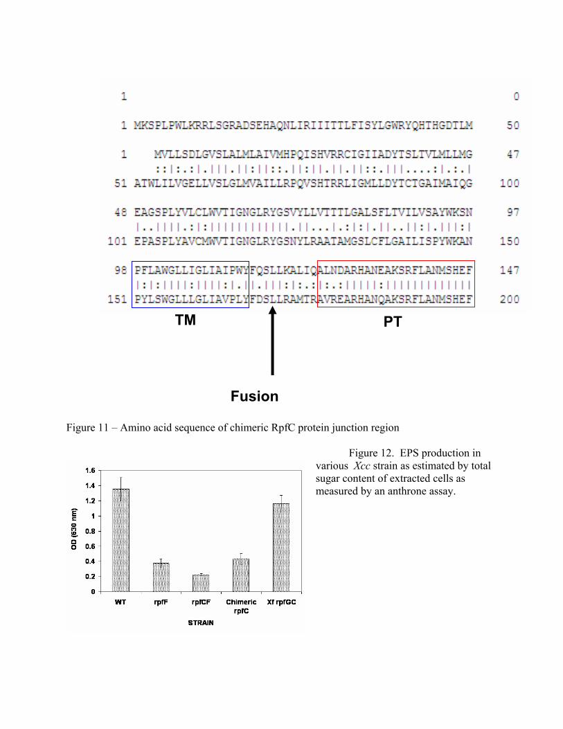
Figure 11 – Amino acid sequence of chimeric RpfC protein junction region
Figure 12. EPS production in various Xcc strain as estimated by total sugar content of extracted cells as measured by an anthrone assay.
PTTM
Fusion

The production of EPS in Xcc normally increases in response to accumulation of DSF. We therefore assessed the regulation of EPS production wild type and rpfF mutants of Xcc. There is a large reduction of EPS production as measured by total sugar concentration in extracted cells of an rpfF mutant compared to wild type, while EPS content of an rpfC mutant is even lower (Figure 12). Introduction of a chimeric rpfC into the rpfFC double mutant in trans restored production of EPS to levels similar to that in an rpfF mutant (Figure 12), suggesting that it functioned in a manner similar to the native Xcc rpfC. In contrast, the rpfC from X. fastidiosa conferred high levels of EPS production in this mutant background, suggesting that it was inappropriately de-repressed in Xcc. Thus the chimeric RpfC appears to be functioning properly in Xcc. We are currently determining the levels of EPS production in an rpfFC double mutant of Xcc harboring the chimeric rpfC in the presence and absence of added DSF; we how to see elevated EPS levels with added DSF.
We have done extensive work to develop alternative reporter genes for use in X. fastidiosa. In our past work we found that gfp and ice nucleation reporter genes were not efficiently expressed, and significant expression of these reporter genes could be detected only when transcription was driven by strong promoters such as the 16S rRNA promoter. We thus have explored the use of two other reporter genes. In one example, we have cloned the gene encoding alkaline phosphatase (phoA) from X. fastidiosa and introduced it into a stable plasmid vector suitable for introduction into X. fastidiosa (Figure 13). We also have knocked out the expression of the indigenous alkaline phosphatase gene in X. fastidiosa since such a background would be required to measure expression of this gene in response to an environmentally-responsive gene such as those encoding EPS production, adhesins or other genes that are regulated upon increases in DSF concentration. We are in the process of determining if alkaline phosphatase activity can be detected in strains harboring various fusions to this phoA reporter gene. We also have obtained variants of a gfp reporter gene that confer much higher levels of expression in E. coli than the native gfp reporter gene. We are determining whether fusions to these gfp variants yield sufficient green fluorescence for detection in X. fastidiosa.
Figure 13. Expression vector harboring phoA from X. fastidiosa to be tested for
expression in a phoA mutant of X. fastidiosa.
rpfFhxfBpglA
gumB16S rRNA
::phoA
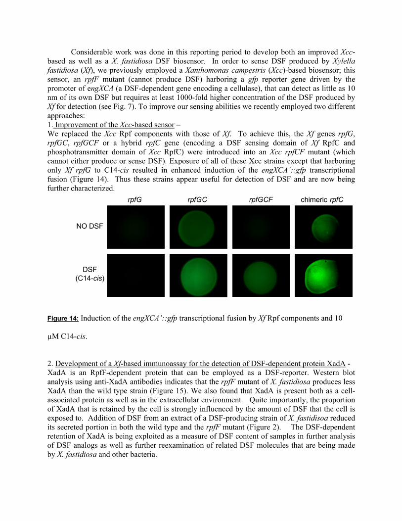
Considerable work was done in this reporting period to develop both an improved Xcc-based as well as a X. fastidiosa DSF biosensor. In order to sense DSF produced by Xylella fastidiosa (Xf), we previously employed a Xanthomonas campestris (Xcc)-based biosensor; this sensor, an rpfF mutant (cannot produce DSF) harboring a gfp reporter gene driven by the promoter of engXCA (a DSF-dependent gene encoding a cellulase), that can detect as little as 10 nm of its own DSF but requires at least 1000-fold higher concentration of the DSF produced by Xf for detection (see Fig. 7). To improve our sensing abilities we recently employed two different approaches: 1. Improvement of the Xcc-based sensor – We replaced the Xcc Rpf components with those of Xf. To achieve this, the Xf genes rpfG, rpfGC, rpfGCF or a hybrid rpfC gene (encoding a DSF sensing domain of Xf RpfC and phosphotransmitter domain of Xcc RpfC) were introduced into an Xcc rpfCF mutant (which cannot either produce or sense DSF). Exposure of all of these Xcc strains except that harboring only Xf rpfG to C14-cis resulted in enhanced induction of the engXCA’::gfp transcriptional fusion (Figure 14). Thus these strains appear useful for detection of DSF and are now being further characterized.
rpfG rpfGC rpfGCF chimeric rpfC
NO DSF
DSF(C14-cis)
Figure 14: Induction of the engXCA’::gfp transcriptional fusion by Xf Rpf components and 10
µM C14-cis.
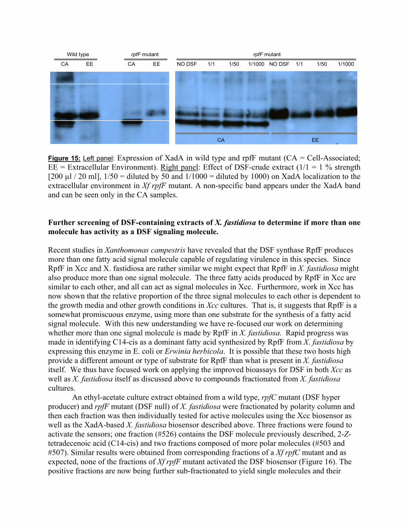
2. Development of a Xf-based immunoassay for the detection of DSF-dependent protein XadA - XadA is an RpfF-dependent protein that can be employed as a DSF-reporter. Western blot analysis using anti-XadA antibodies indicates that the rpfF mutant of X. fastidiosa produces less XadA than the wild type strain (Figure 15). We also found that XadA is present both as a cell- associated protein as well as in the extracellular environment. Quite importantly, the proportion of XadA that is retained by the cell is strongly influenced by the amount of DSF that the cell is exposed to. Addition of DSF from an extract of a DSF-producing strain of X. fastidisoa reduced its secreted portion in both the wild type and the rpfF mutant (Figure 2). The DSF-dependent retention of XadA is being exploited as a measure of DSF content of samples in further analysis of DSF analogs as well as further reexamination of related DSF molecules that are being made by X. fastidiosa and other bacteria.
rpfF mutant
NO DSF 1/1 1/50 1/1000 NO DSF 1/1 1/50 1/1000
CA EE
Wild type rpfF mutant
CA EE CA EE
Figure 15: Left panel: Expression of XadA in wild type and rpfF mutant (CA = Cell-Associated; EE = Extracellular Environment). Right panel: Effect of DSF-crude extract (1/1 = 1 % strength [200 µl / 20 ml], 1/50 = diluted by 50 and 1/1000 = diluted by 1000) on XadA localization to the extracellular environment in Xf rpfF mutant. A non-specific band appears under the XadA band and can be seen only in the CA samples.
Further screening of DSF-containing extracts of X. fastidiosa to determine if more than one molecule has activity as a DSF signaling molecule. Recent studies in Xanthomonas campestris have revealed that the DSF synthase RpfF produces more than one fatty acid signal molecule capable of regulating virulence in this species. Since RpfF in Xcc and X. fastidiosa are rather similar we might expect that RpfF in X. fastidiosa might also produce more than one signal molecule. The three fatty acids produced by RpfF in Xcc are similar to each other, and all can act as signal molecules in Xcc. Furthermore, work in Xcc has now shown that the relative proportion of the three signal molecules to each other is dependent to the growth media and other growth conditions in Xcc cultures. That is, it suggests that RpfF is a somewhat promiscuous enzyme, using more than one substrate for the synthesis of a fatty acid signal molecule. With this new understanding we have re-focused our work on determining whether more than one signal molecule is made by RpfF in X. fastidiosa. Rapid progress was made in identifying C14-cis as a dominant fatty acid synthesized by RpfF from X. fastidiosa by expressing this enzyme in E. coli or Erwinia herbicola. It is possible that these two hosts high provide a different amount or type of substrate for RpfF than what is present in X. fastidiosa itself. We thus have focused work on applying the improved bioassays for DSF in both Xcc as well as X. fastidiosa itself as discussed above to compounds fractionated from X. fastidiosa cultures.
An ethyl-acetate culture extract obtained from a wild type, rpfC mutant (DSF hyper producer) and rpfF mutant (DSF null) of X. fastidiosa were fractionated by polarity column and then each fraction was then individually tested for active molecules using the Xcc biosensor as well as the XadA-based X. fastidiosa biosensor described above. Three fractions were found to activate the sensors; one fraction (#526) contains the DSF molecule previously described, 2-Z-tetradecenoic acid (C14-cis) and two fractions composed of more polar molecules (#503 and #507). Similar results were obtained from corresponding fractions of a Xf rpfC mutant and as expected, none of the fractions of Xf rpfF mutant activated the DSF biosensor (Figure 16). The positive fractions are now being further sub-fractionated to yield single molecules and their

structure is being determined with NMR. Later they will be tested for biological activity. We also will ensure that these molecules are not produced by an Xf rpfF mutant.
Figure 16: Response of the new Xylella fastidiosa DSF specific sensor to fractions of Xylella fastidiosa wild type culture extract. Fraction 501 is the most polar one and 531 is the less. Positive (100 µM 2-Z-tetradecenoic acid) and negative (MeOH) controls are shown at the bottom-right.
These results suggest that more than one molecule is being made by RpfF in X. fastidiosa. It will be important to determine whether the different froms of what presumably are related fatty acid signal molecules regulate the same traits in X. fastidiosa and their relative activity. Further support for the possibility that more than one fatty acid signal molecule is made by RpfF was obtained by the use of a Thin Layer Chromatography (TLC) method to assess the fatty acids produced by X. fastidiosa. In this method, acidified ethyl acetate extracts of culture supernatants of a wild-type X. fastidiosa strain and an RpfF- mutant and a RpfB mutant were subjected to TLC and fatty acids visualized by iodine vapors. Interestingly, three different fatty acids were visualized in the wild type strain, while these were largely missing in an RpfF- mutant, with only very small amounts of two other putative fatty acids present (Figure 17). It also was of interest to see that the RpfB mutant produced an altered pattern of putative fatty acids, with the major chemical species produced by the WT strain missing, and much larger amounts of one of the other species produced. This supports the model that RpfB, a putative long-chain fatty acyl CoA ligase, serves to produce suitable substrates for RpfF, the DFS synthase. Based on this and other data, we are expressing both RpfF and RpfB simultaneously in transgenic grape and in surrogate bacterial hosts for optimum production of suitable DSF molecules.
501 502 503 505 506 507 520 521 523
524 525 526 527 528 529 530 531 C14‐cis MeOH
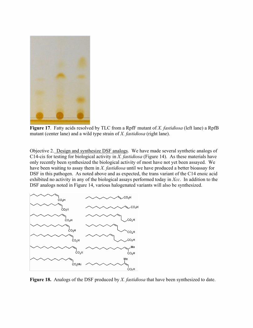
Figure 17. Fatty acids resolved by TLC from a RpfF mutant of X. fastidiosa (left lane) a RpfB mutant (center lane) and a wild type strain of X. fastidiosa (right lane). Objective 2. Design and synthesize DSF analogs. We have made several synthetic analogs of C14-cis for testing for biological activity in X. fastidiosa (Figure 14). As these materials have only recently been synthesized the biological activity of most have not yet been assayed. We have been waiting to assay them in X. fastidiosa until we have produced a better bioassay for DSF in this pathogen. As noted above and as expected, the trans variant of the C14 enoic acid exhibited no activity in any of the biological assays performed today in Xcc. In addition to the DSF analogs noted in Figure 14, various halogenated variants will also be synthesized.
Figure 18. Analogs of the DSF produced by X. fastidiosa that have been synthesized to date.
Objective 3. Synthesis of sufficient DSF analogs for in planta evaluations. We have synthesized gram quantities of C14 cis as well as the Sodium salt of this fatty acid which is highly water soluble. These quantities are sufficiently large for initial greenhouse studies. Initial studies were carried out in greenhouse studies in the summer of 2008. These materials were sprayed onto leaves as well as injected into stems and used as a soil drench in initial studies to determine their efficacy for disease control. The Sodium salt is quite water-soluble and has mild surfactant characteristics which make its topical application to leaves relatively easy. However, the solubility of this material in water is not sufficiently high that milligram quantities could be injected into plants in small (microliter) volumes of water. We thus have injected high concentrations of the acid form dissolved in methanol for such studies. After treatment plants were challenge inoculated with X. fastidiosa. Unfortunately, a malfunction in the greenhouse cuased damage to all of the treated grapes such that it masked our ability to monitor disease symptoms on leaves. Thus the initial tests made in Fall, 2008 were inconclusive because of damage to plants that occurred during pest control activities in the greenhouse obscured Pierce’s disease symptoms. More extensive studies involving culturing of the pathogen have been initiated. Research Relevance Statement: Since we have shown that DSF accumulation within plants is a major signal used by Xf to change its gene expression patterns and since DFS-mediated changes all lead to a reduction in virulence in this pathogen we have shown proof of principle that disease control can be achieved by a process of “pathogen confusion”. This study addresses an obvious means of achieving pathogen confusion since direct introduction of DSF via topical application to plants should enable us to alter this signal molecule. While the principle of disease control by altering DSF levels has been demonstrated, this work addresses the feasibility of how achieve this goal using synthetic DSF. Our continuing work will address whether this is a practical means to achieve disease control by pathogen confusion. Lay Summary X. fastidiosa produces an unsaturated fatty acid signal molecule called DSF that changes its gene expression in cells as they reach high numbers in plants. Accumulation of DSF in Xf cells, which presumably normally occurs as cells become numerous within xylem vessels, causes a change in many genes in Xf, but the overall effect is to suppress the virulence of Xf in plants. The DSF produced by grape strains of X. fastidiosa has tentatively been characterized as a 14 carbon, unsaturated molecule we will refer to as C14-cis. Both its relatively higher biological activity as assessed in Xcc and X. fastidiosa than that of the DSF from Xcc and lesser activity in an Xcc bioassay is as expected, indicating that there is considerable specificity in the structure-function relationships between different bacterial DSF signal molecules. The production of sufficient X. fastidiosa for testing for pathogen confusion has been shown to be possible and we now conducting greenhouse tests in which the synthetic DSF is being applied as a topical spray to plants, as a soil drench, and by direct injection into the stems of plants before inoculation with X. fastidiosa. We will be measuring disease severity in treated plants to determine if topical applications of the material can lead to disease control via pathogen confusion. Summary and Status of Intellectual Property: No new intellectual property was developed in this reporting period.