Organization of The Nervous System By Prof. Saeed Abuel Makarem& Dr. Sanaa Sharawy.
Upload
angelica-hutchinsonCategory
view
220download
0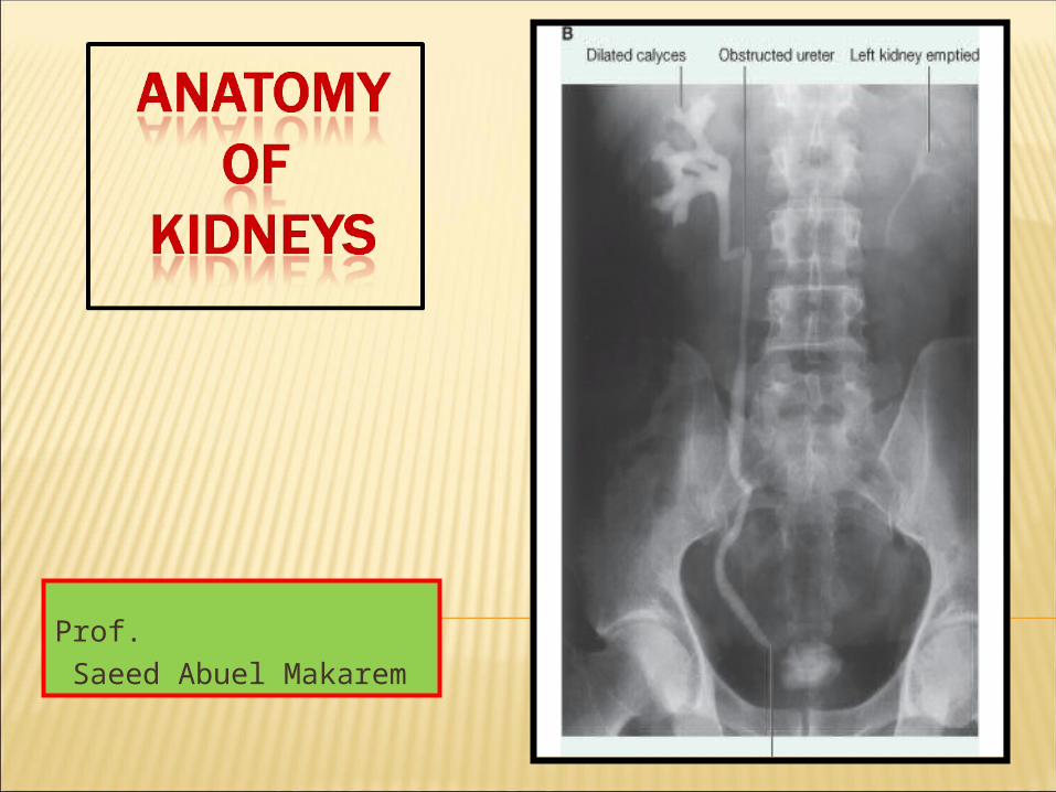
Prof. Saeed Abuel Makarem

By the end of this course you should be able to discuss: COMPONENTS OF
THE URINARY SYSTEM (kidney, ureter ,urinary bladder, urethra)
By the end of this lecture you should be able to discuss :
KIDNEY SHAPE & POSITION. SURFACE ANATOMY. EXTERNAL FEATURES. HILUM and its CONTENTS. RELATIONS. INTERNAL STRUCTURE. BLOOD SUPPLY LYMPH DRAINAGE.. NERVE SUPPLY.

3
Every day, each Every day, each kidney filters liters of kidney filters liters of fluid from the fluid from the bloodstreambloodstream. .
Although the Although the lungslungs and and the the skin skin also play roles also play roles in excretion, the in excretion, the kidneys has major kidneys has major responsibility for responsibility for eliminating eliminating nitrogenous (nitrogen-nitrogenous (nitrogen-containing) wastes, containing) wastes, toxins, and drugs from toxins, and drugs from the body.the body.

Functions:1. Excretes most of the waste
products of metabolism.
2. Controls water & electrolyte balance of the body.
3. Maintain acid-base balance of the blood.
4. Stimulate bone marrow for RBCs formation by Erythropoietin hormone.
5. Regulates blood pressure by Rennin enzyme.
6. Converts vitamin D to its active form.
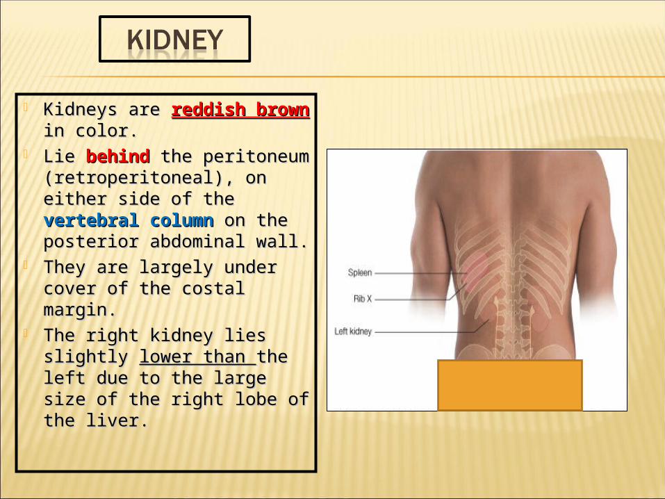
Kidneys are Kidneys are reddish reddish brown brown in color.in color.
Lie Lie behindbehind the the peritoneum peritoneum (retroperitoneal), on (retroperitoneal), on either side of the either side of the vertebral column vertebral column on the on the posterior abdominal wall.posterior abdominal wall.
They are largely under They are largely under cover of the costal cover of the costal margin.margin.
The right kidney lies The right kidney lies slightly slightly lower than lower than the left the left due to the large size of due to the large size of the right lobe of the liver.the right lobe of the liver.

With contraction of the With contraction of the diaphragm the kidney diaphragm the kidney moves downward as much moves downward as much as 2.5 cm.as 2.5 cm.
The lateral border is The lateral border is convex, while the medial convex, while the medial border is convex at both border is convex at both ends but its middle pat ends but its middle pat shows a vertical slit called shows a vertical slit called the hilumthe hilum.
The hilum extends into a The hilum extends into a large cavity called the large cavity called the renal sinus.renal sinus.
The hilum transmits the The hilum transmits the renal renal veinvein,, two branches two branches of renal of renal artery,artery, ureter, ureter, and the third branch of and the third branch of renal renal arteryartery from the front from the front backward backward ((V.V.A.A.U.U.AA.)
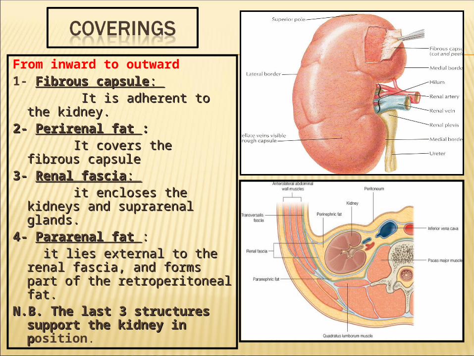
From inward to outward1- Fibrous capsuleFibrous capsule: : It is adherent to the It is adherent to the
kidney.kidney.2-2- Perirenal fat Perirenal fat : : It covers the fibrous It covers the fibrous
capsulecapsule3-3- Renal fasciaRenal fascia: : it encloses the kidneys it encloses the kidneys
and suprarenal glands.and suprarenal glands.4-4- Pararenal fatPararenal fat :: it lies external to the renal it lies external to the renal
fascia, and forms part of the fascia, and forms part of the retroperitoneal fat.retroperitoneal fat.
N.B. The last 3 structures N.B. The last 3 structures support the kidney in support the kidney in pposition.
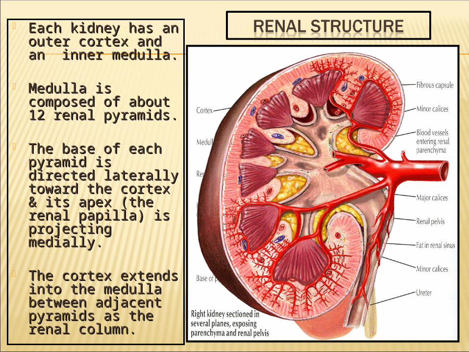
Each kidney has an Each kidney has an outer cortex and outer cortex and an inner medulla.an inner medulla.
Medulla is Medulla is composed of about composed of about 12 renal pyramids.12 renal pyramids.
The base of each The base of each pyramid is directed pyramid is directed laterally toward the laterally toward the cortex & its apex cortex & its apex (the renal papilla) (the renal papilla) is projecting is projecting medially.medially.
The cortex extends The cortex extends into the medulla into the medulla between adjacent between adjacent pyramids as the pyramids as the renal column.renal column.

Extending from the bases Extending from the bases of the renal pyramids of the renal pyramids into the cortex are into the cortex are striations known striations known as as medullary raysmedullary rays..
The renal sinus within the The renal sinus within the hilum, contains the upper hilum, contains the upper expanded end of the expanded end of the ureter, the renal pelvisureter, the renal pelvis.
Renal pelvis divides into Renal pelvis divides into two or three two or three major major calyces.calyces.
Each Each major calyces major calyces divides into two or three divides into two or three minor calyces.minor calyces.

( Last rib + 4muscles + 3 ( Last rib + 4muscles + 3 nerves)nerves)
Diaphragm, Diaphragm, (last intercostal space)(last intercostal space)
Costodiaphragmatic pleural Costodiaphragmatic pleural recess.recess.
Twelfth rib,Twelfth rib, Psoas major muscle,Psoas major muscle, Quadratus lamborum m.,Quadratus lamborum m., Transversus abdominis m., Transversus abdominis m.,
1.1. Subcostal nerve (T12),Subcostal nerve (T12),
2.2. Iliohypogastric (L1) Iliohypogastric (L1) nerve.nerve.
3.3. Ilioinguinal (L1) Ilioinguinal (L1) nervenerve NB. The left kidney reaches NB. The left kidney reaches
up to the 11up to the 11thth rib rib..

Posterior Relation

Right Kidney :Right Kidney : 11- Right suprarenal gland- Right suprarenal gland 2- Liver,2- Liver, 33- Second part of the- Second part of the duodenumduodenum 44- Right colic flexure- Right colic flexure 5- Coils of small intestine5- Coils of small intestine
Left Kidney : Left Kidney : 11- Left suprarenal gland,- Left suprarenal gland, 2- Stomach, 2- Stomach, 3- Spleen3- Spleen, , 44- Pancreas, - Pancreas, 55- Left colic flexure,- Left colic flexure, 6-6- Descending colon Descending colon 7- Coils of jejunum7- Coils of jejunum
ANTERIOR
RELATION

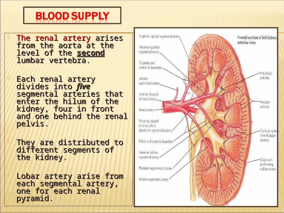
The renal artery The renal artery arises arises from the aorta at the level from the aorta at the level of the of the second second lumbar lumbar vertebra.vertebra.
Each renal artery divides Each renal artery divides into into five five segmental segmental arteries that enter the arteries that enter the hilum of the kidney, four hilum of the kidney, four in front and one behind in front and one behind the renal pelvis.the renal pelvis.
They are distributed to They are distributed to different segments of the different segments of the kidney.kidney.
Lobar artery arise from Lobar artery arise from each segmental artery, each segmental artery, one for each renal one for each renal pyramid.pyramid.
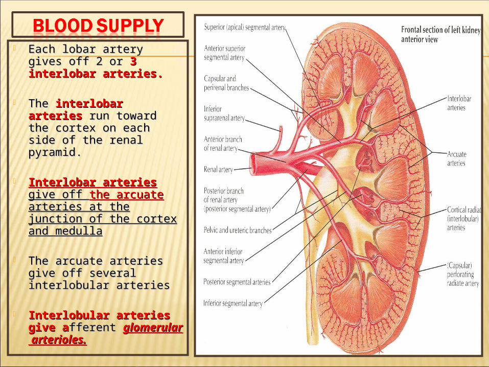
Each lobar artery gives Each lobar artery gives off 2 or off 2 or 3 interlobar 3 interlobar arteries.arteries.
The The interlobar arteries interlobar arteries run toward the cortex on run toward the cortex on each side of the renal each side of the renal pyramid.pyramid.
Interlobar arteries Interlobar arteries give off give off the arcuate the arcuate arteries at the junction of arteries at the junction of the cortex and medullathe cortex and medulla
The arcuate arteries give The arcuate arteries give off several interlobular off several interlobular arteriesarteries
Interlobular arteries Interlobular arteries give agive afferent fferent glomerular arterioles.glomerular arterioles.

Apical segmental artery
Caudal segmental artery
Segmental Branches of the Renal Artery
Anterior superior segmental artery
The renal artery divides into 5 segmental branches
Posterior segmental artery
Anterior inferiorAnterior inferior segmental arterysegmental artery
The renal arteryThe renal artery
17
Prof. Saeed Abuel Makarem

Which give
a number of lobar arteries
Branches of the Segmental arteryBranches of the Segmental artery
the interlobar arteries give offEach segmental arterysegmental artery divides into
2 or 3 interlobar arteriesthe arcuate arteries
The segmental arteryThe segmental artery
several interlobular branches
Which give
the afferent glomerular arterioles
Each lobar arterylobar artery divides into
18Prof. Saeed Abuel Makarem
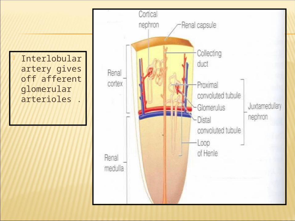
Interlobular artery gives off afferent glomerular arterioles .

20
Each Nephron is associated with two capillary beds:1. The glomerulus and 2. The peritubular
capillary bed. The glomerulus is both
fed and drained by arterioles. The afferent
arteriole, which arises from an interlobular artery, is the "feeder vessel," and
the efferent arteriole receives blood that has passed through the glomerulus.

VENOUS VENOUS DRAINAGEDRAINAGE
Both renal veins drain to the inferior vena cava.The left is three times longer than the right (7.5 cm and 2.5 cm).So, for this reason the left kidney is the preferred side for live donor nephrectomy. It runs from its origin in the renal hilum, posterior to the splenic vein and the body of pancreas, and then across the anterior aspect of the aorta, just below the origin of the superior mesenteric artery.
21
Prof. Saeed Abuel Makarem
Left renal Vein

VENOUS VENOUS DRAINAGEDRAINAGE
The left gonadal vein enters the left renal vein from below while the left suprarenal vein, enters it from above but nearer to the midline. The left renal vein enters the inferior vena cava a little above the right vein. The right renal vein is behind the 2nd part of the duodenum and sometimes the lateral part of the head of the pancreas
22Prof. Saeed Abuel Makarem

Lymph Drainage:
Lateral aortic lymph nodes around the origin of the renal artery.
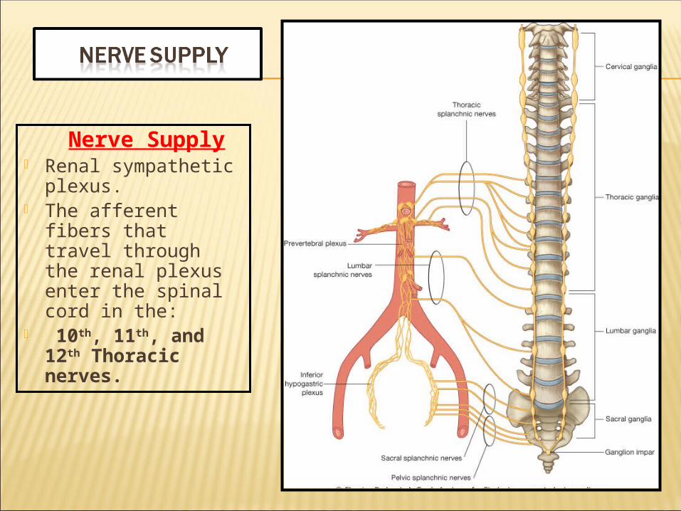
Nerve Supply Renal sympathetic
plexus. The afferent fibers
that travel through the renal plexus enter the spinal cord in the:
10th, 11th, and 12th Thoracic nerves.


11 - -Apical segmentApical segment
22 - -Caudal segmentCaudal segment
Segments of the kidneysSegments of the kidneys
4 Anterior superior segment
Each kidneyEach kidney consists of 5 segments5 segments
33--Posterior segmentPosterior segment
55 - -Anterior inferior segmentAnterior inferior segment
26
Prof. Saeed Abuel Makarem

Apical segment
Caudal segment
Segments of the kidneys (Rt.)Segments of the kidneys (Rt.)
Anterior superior segment
Each kidneyEach kidney consists of 5 segments each has its own blood supply5 segments each has its own blood supply
Posterior segmentPosterior segment
Anterior inferior segmentAnterior inferior segment
Lateral viewLateral view
Anterior Anterior
PosteriorPosterior
Upper Upper
Lower Lower
27
Prof. Saeed Abuel Makarem