Prof Govind Narayan Purohit€¦ · •Prof Govind Narayan Purohit Head, Department of Veterinary...
46
Placental formation, Growth and functions in domestic animals • Prof Govind Narayan Purohit Head, Department of Veterinary Gynecology and Obstetrics, College of Veterinary and Animal Sciences, RAJUVAS, Bikaner, Rajasthan, India
Transcript of Prof Govind Narayan Purohit€¦ · •Prof Govind Narayan Purohit Head, Department of Veterinary...

Placental formation, Growth and functions in domestic animals
• Prof Govind Narayan PurohitHead, Department of Veterinary Gynecology and Obstetrics, College of Veterinary and Animal Sciences, RAJUVAS, Bikaner, Rajasthan, India
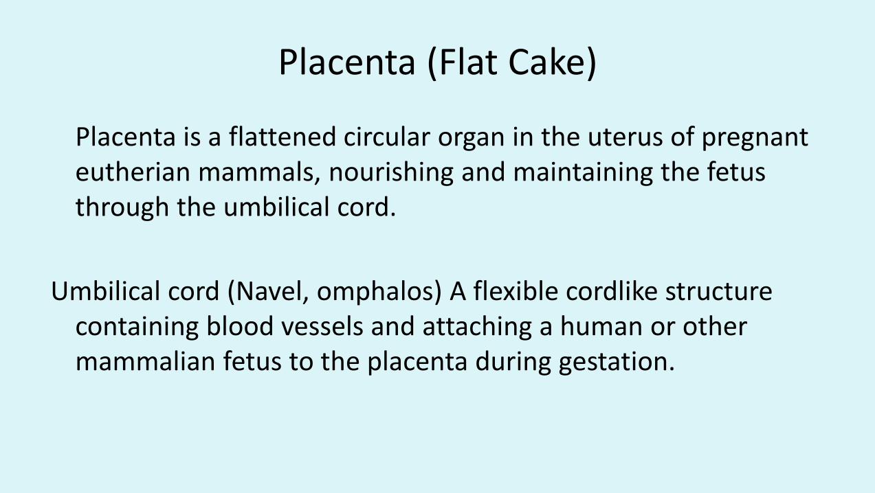
Placenta (Flat Cake)
Placenta is a flattened circular organ in the uterus of pregnant eutherian mammals, nourishing and maintaining the fetus through the umbilical cord.
Umbilical cord (Navel, omphalos) A flexible cordlike structure containing blood vessels and attaching a human or other mammalian fetus to the placenta during gestation.
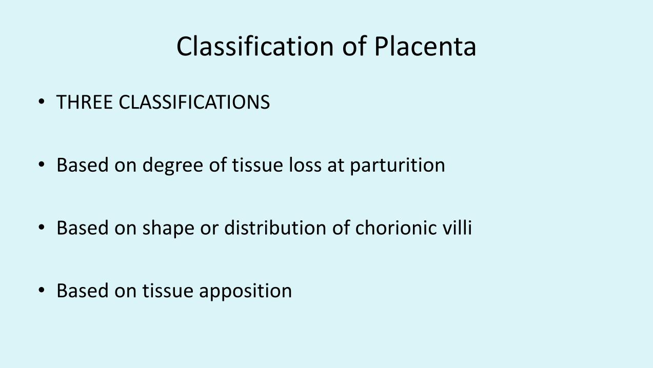
Classification of Placenta
• THREE CLASSIFICATIONS
• Based on degree of tissue loss at parturition
• Based on shape or distribution of chorionic villi
• Based on tissue apposition
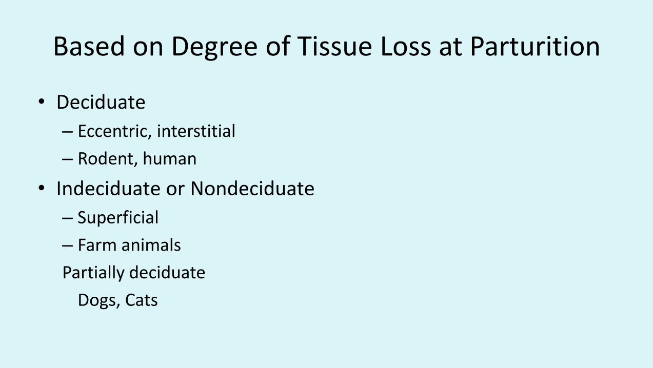
Based on Degree of Tissue Loss at Parturition
• Deciduate
– Eccentric, interstitial
– Rodent, human
• Indeciduate or Nondeciduate
– Superficial
– Farm animals
Partially deciduate
Dogs, Cats

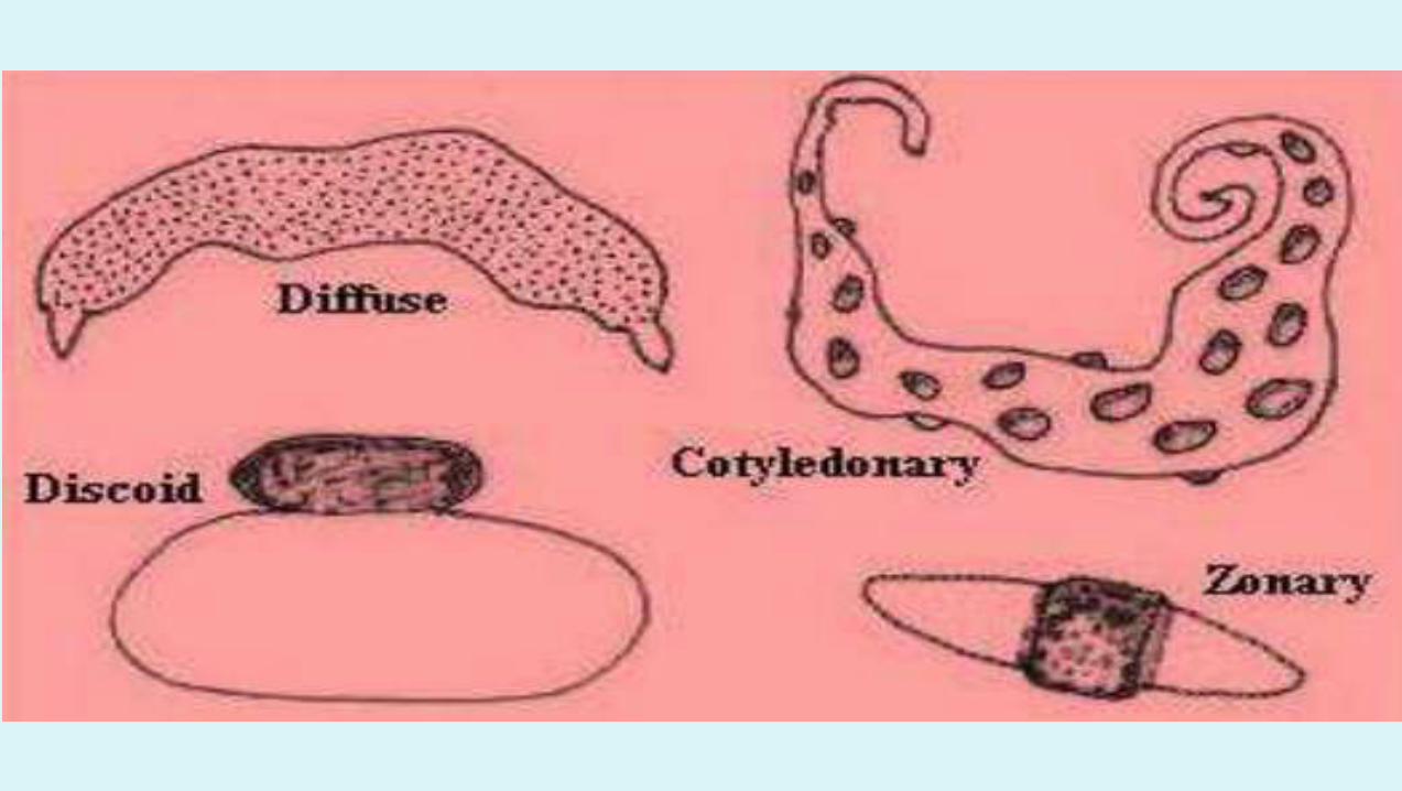
Shape or Distribution of Chorionic Villi
• Cotelydonary - cow, sheep
• Diffuse - pig, horse
• Zonary - dog, cat
• Discoid - human

Ruminant placenta
• Instead of having a single large area of contact between maternal and fetal vascular systems, these animals have numerous smaller placentae. The terminology used to describe ruminant placentation is cotyledonarry: Cotyledon: the fetal side of the placenta. Caruncle: the maternal side of the placenta. Placentome: a cotyledon and caruncle together


Cotelydonary Placenta
Cow
Ewe
Concave
Convex
Endometrium
Caruncle
Cotelydon (Chorion)
90 - 10070 - 120

Chorion
Uterine
Epithelium
Stroma
Capillary
BinucleateCell
Syncytium
Migrate andfuse withuterine
epithelium
Fusion ofBinucleatecells and uterine
epithelium
(multinucleate)
Placental Attachment in Ruminant

Binuclear Giant Cells
• 20% of fetal placenta
• Invade endometrium
• Source
– Placental lactogen
– Pregnancy specific protein B
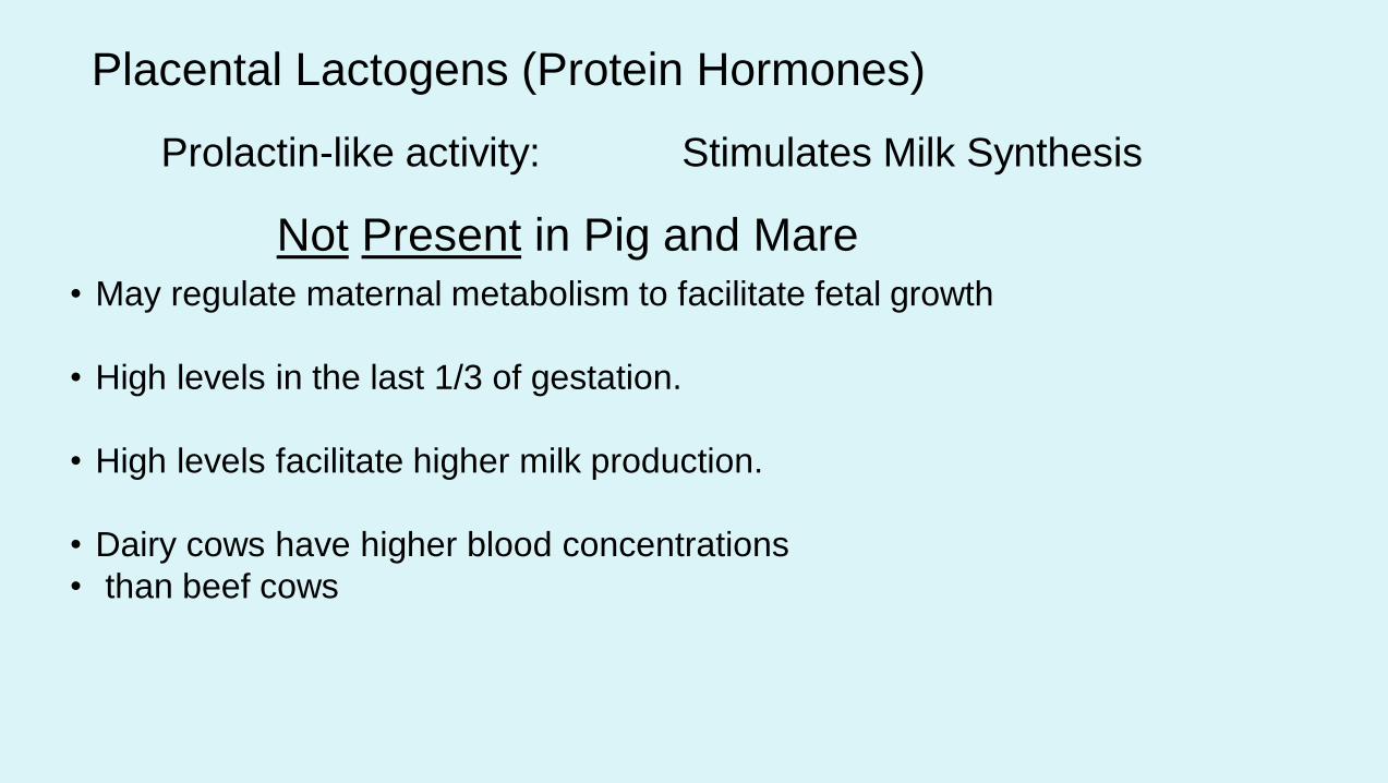
Placental Lactogens (Protein Hormones)
Prolactin-like activity: Stimulates Milk Synthesis
Not Present in Pig and Mare
• May regulate maternal metabolism to facilitate fetal growth
• High levels in the last 1/3 of gestation.
• High levels facilitate higher milk production.
• Dairy cows have higher blood concentrations
• than beef cows

Placental Lactogens
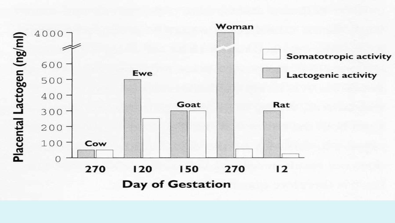
• The bovine PL hormone is detected in maternal serum at about 4 months of gestation and remains low through parturation. In contrast, ovine placental lactogen is secreted in whopping quantities beginning at about day 50 and remains high through gestation. Placental lactogen also accumulates to high concentrations in the serum of fetal sheep
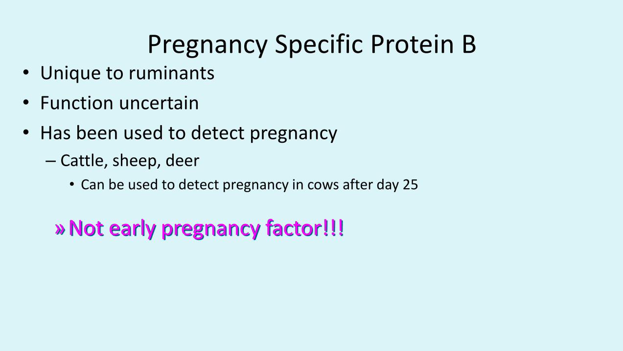
Pregnancy Specific Protein B• Unique to ruminants
• Function uncertain
• Has been used to detect pregnancy
– Cattle, sheep, deer
• Can be used to detect pregnancy in cows after day 25
» Not early pregnancy factor!!!

ZONARY PLACENTA DOG

• The canine placenta looks very similar to that of cats. A feature usually seen in the placentae of both species is marginal hematomas(hematophagus zones). These are bands of maternal hemorrhage at the margins of the zonary placenta. The products of hemoglobin breakdown give them a distinctly green coloration due to uteroverdin in dogs, whereas in cats they are brownish and usually less obvious. The canine placenta is said to produce little if any quantity of steroid hormones. As with other species, maintenance of pregnancy is dependent on continued secretion of progesterone during gestation, but corpora lutea appear to be the exclusive source of progesterone in the bitch. Luteal secretion of progesterone is, in turn, dependent on secretion of luteinizing hormoneand probably prolactin from the anterior pituitary. Removal of the ovaries at any time during canine gestation leads to termination of the pregnancy. Also, progesterone profiles in pregnant and pseudopregnant bitches are indistinguishable until late in gestation or diestrus.

Discoid Placenta(human)
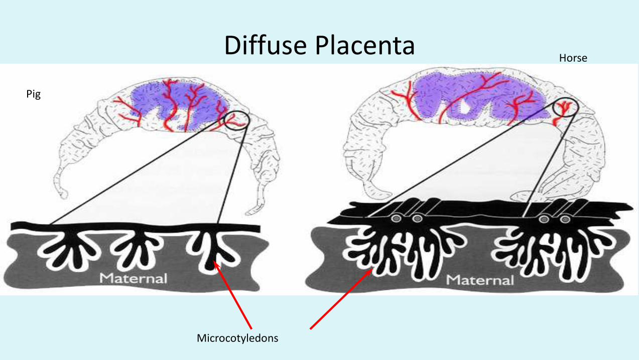
Diffuse Placenta
Pig
Horse
Microcotyledons

Microcotelydons
Microcotelydon
Microcotelydon(Fetal)
Microcotelydon(Maternal)
Uterine Arteries
Uterine Veins
Endometrial Glands
End
om
etrium
Epithelium
Increase placental surface area
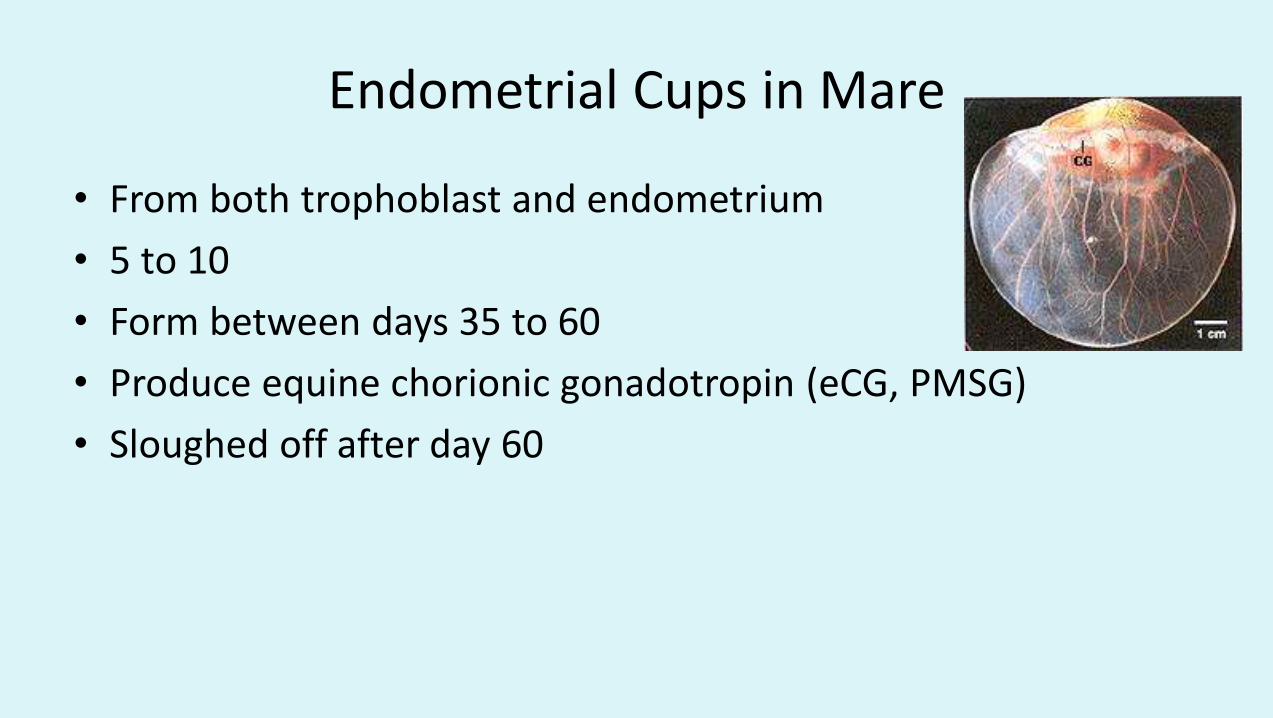
Endometrial Cups in Mare
• From both trophoblast and endometrium
• 5 to 10
• Form between days 35 to 60
• Produce equine chorionic gonadotropin (eCG, PMSG)
• Sloughed off after day 60
Endometrial cups develop from cells of the chorionic
girdle, which can first be detected histologically at
roughly 25 days of gestation. Initially, this structure
is a narrow band of thicked trophoblast that
develops circumferentially around the conceptus at
a point where the membranes of the allantois and
yolk sac meet.

Girdle cell invasion and proliferation result in formation of tightly packed mass of trophoblast-derived cells containing little stroma - these are the endometrial cups. Invasion of endometrial glands leads to destruction of their apical epithelium; deeper segments of those glands are spared, but their lumens are obstructed by cup cells and they become distended with secretions. Endometrial cups are destroyed by Day 100-140
Immunological destruction of the endometrial cups
appears to be a response to paternal class I MHC antigens,
which are highly expressed on invading girdles cells. In conjunction with the cellular response is a vigorous
humoral immune response to these antigens.
• Several interesting observations on endometrial cup biology have been made in inter-specific equine pregnancies. In mares carrying donkey conceptuses, the chorionic girdle fails to invade the endometrium, and endometrial cups do not develop. Most of these pregnancies are aborted between days 80 and 90, but the roughly 30% that survive and are carried to term do so in the absence of eCG. However, in donkeys carrying a hinny fetus, the cups develop to a much larger size and considerably higher concentrations of eCG are achieved than in donkeys carrying a donkey fetus.
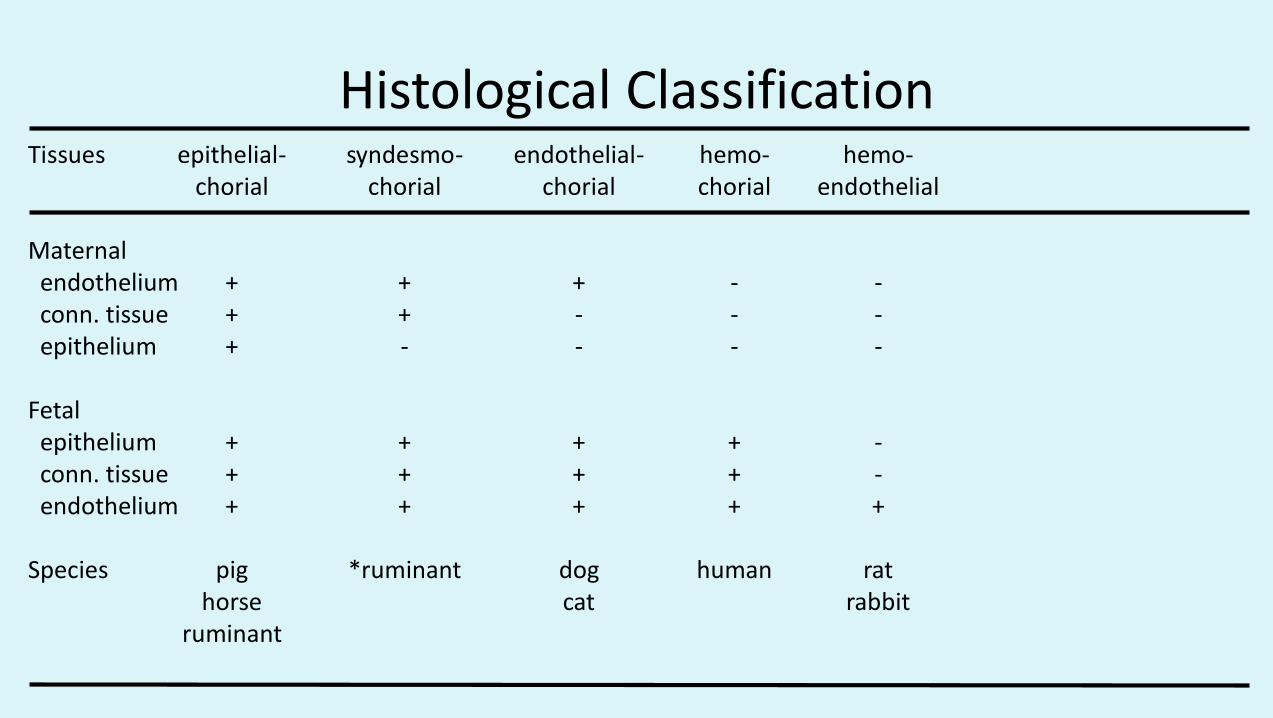
Histological ClassificationTissues epithelial- syndesmo- endothelial- hemo- hemo-
chorial chorial chorial chorial endothelial
Maternalendothelium + + + - -conn. tissue + + - - -epithelium + - - - -
Fetalepithelium + + + + -conn. tissue + + + + -endothelium + + + + +
Species pig *ruminant dog human rathorse cat rabbit
ruminant

Important!!!!
• In all cases, fetal and maternal blood does not mix.

Type of Placenta Common Examples
Diffuse, epitheliochorial Horses and pigs
Cotyledonary, epitheliochorial Ruminants (cattle, sheep, goats, deer)
Discoid, hemochorial Humans, apes, monkeys and rodents
Zonary, endotheliochorial Carnivores (dog, cat, ferret)
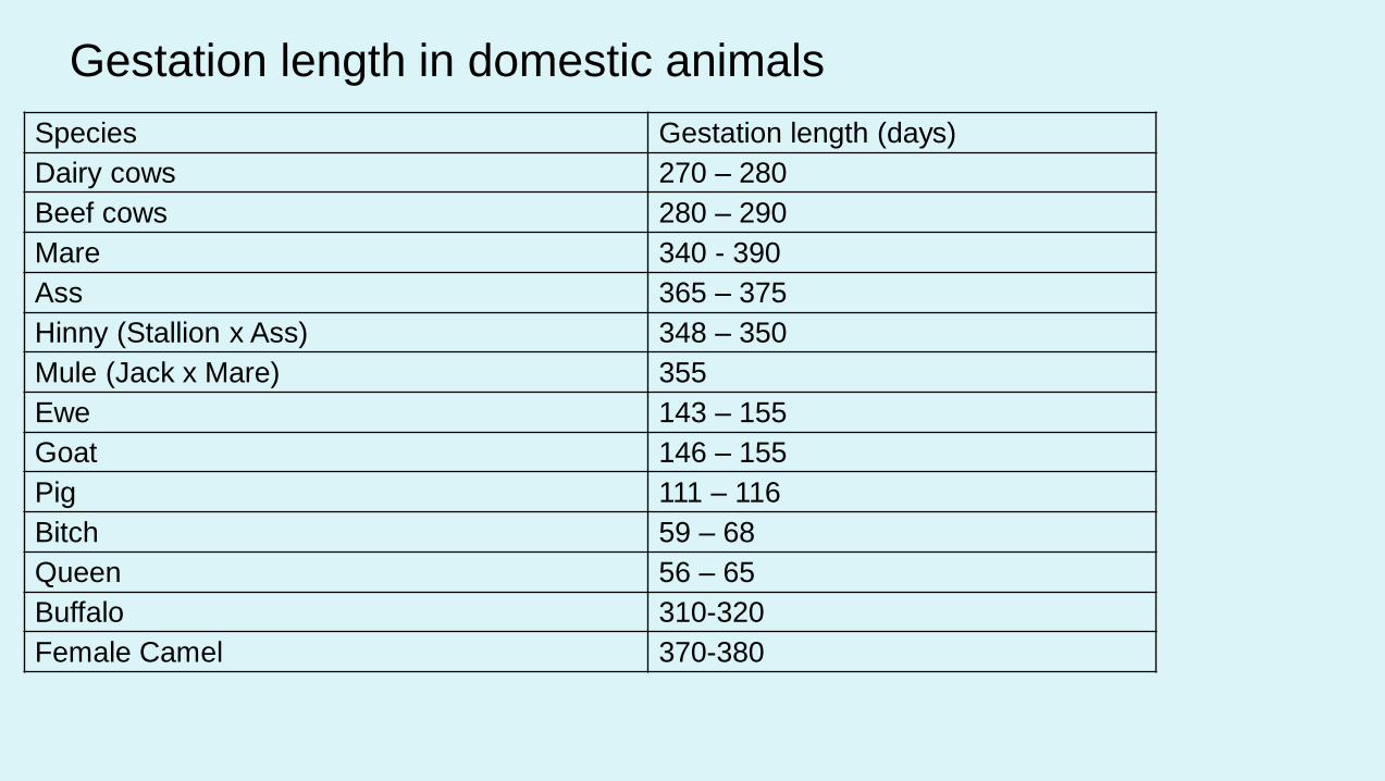
Species Gestation length (days)
Dairy cows 270 – 280
Beef cows 280 – 290
Mare 340 - 390
Ass 365 – 375
Hinny (Stallion x Ass) 348 – 350
Mule (Jack x Mare) 355
Ewe 143 – 155
Goat 146 – 155
Pig 111 – 116
Bitch 59 – 68
Queen 56 – 65
Buffalo 310-320
Female Camel 370-380
Gestation length in domestic animals
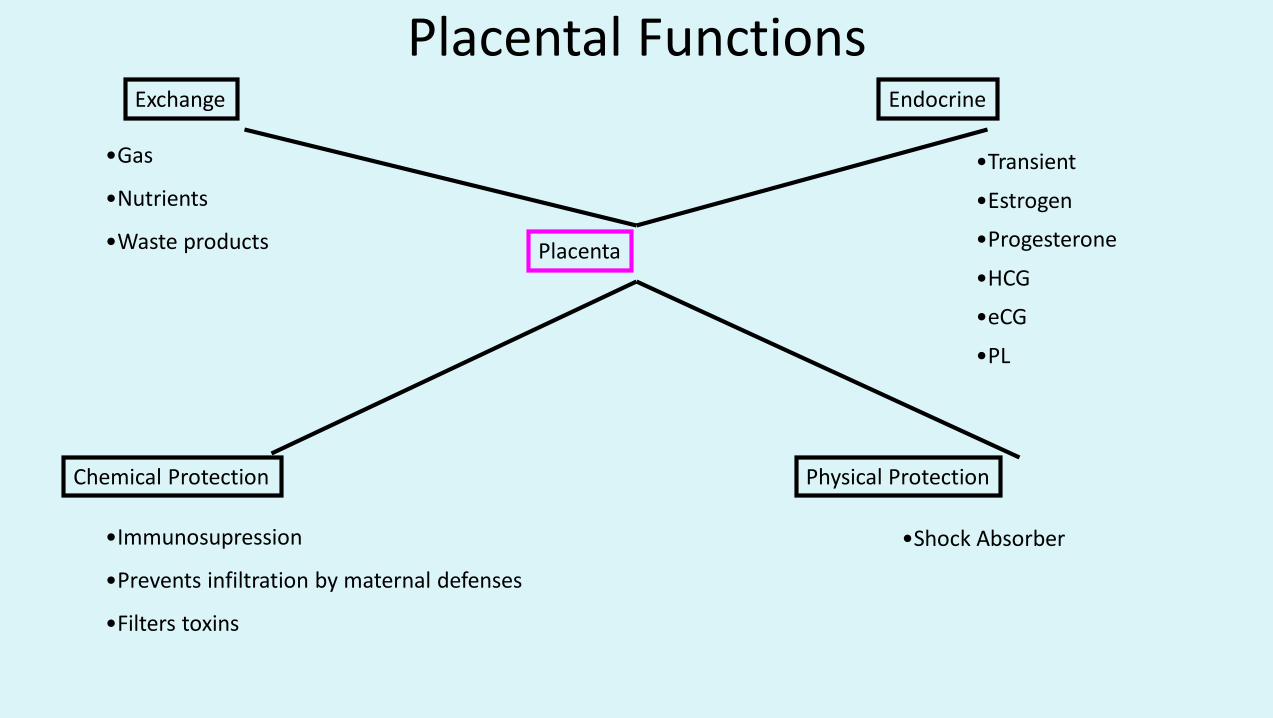
Placental Functions
Placenta
Chemical Protection Physical Protection
EndocrineExchange
•Gas
•Nutrients
•Waste products
•Immunosupression
•Prevents infiltration by maternal defenses
•Filters toxins
•Shock Absorber
•Transient
•Estrogen
•Progesterone
•HCG
•eCG
•PL
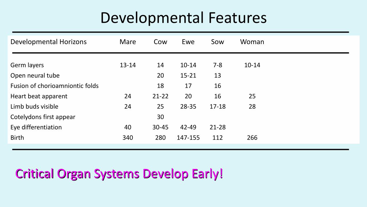
Developmental Horizons Mare Cow Ewe Sow Woman
Germ layers 13-14 14 10-14 7-8 10-14
Open neural tube 20 15-21 13
Fusion of chorioamniontic folds 18 17 16
Heart beat apparent 24 21-22 20 16 25
Limb buds visible 24 25 28-35 17-18 28
Cotelydons first appear 30
Eye differentiation 40 30-45 42-49 21-28
Birth 340 280 147-155 112 266
Developmental Features
Critical Organ Systems Develop Early!
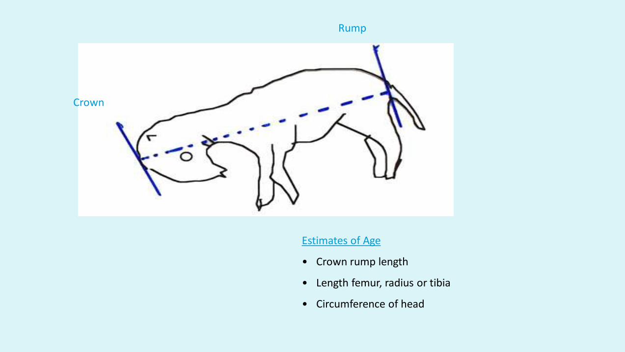
Crown
Rump
Estimates of Age
• Crown rump length
• Length femur, radius or tibia
• Circumference of head

Factors Influencing Fetal Growth
Genetics
Species
Breed
Litter size
Genotype
Fetal Hormones
Thyroid
Insulin
Growth hormone
Environment
MotherNutritionSize, Parity
PlacentaBlood flowSize

Genetics
Species
Breed
Litter size
Genotype
Factors Influencing Fetal Growth
Certains lines of animals may grow faster.

Factors Influencing Fetal Growth
Fetal Hormones
Thyroid
Insulin
Growth hormone
Skeletal and muscular development
Increased energy substrate availability and stimulates placental growth
Stimulates fetal growth
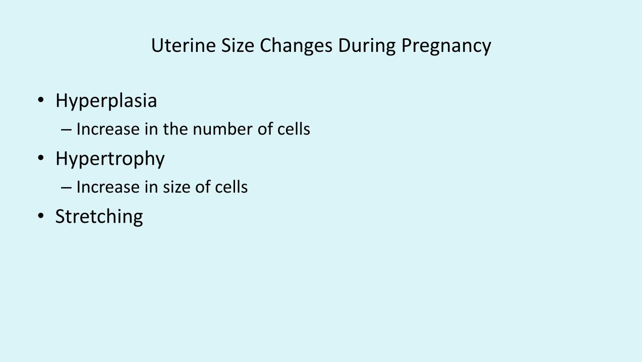
Uterine Size Changes During Pregnancy
• Hyperplasia
– Increase in the number of cells
• Hypertrophy
– Increase in size of cells
• Stretching
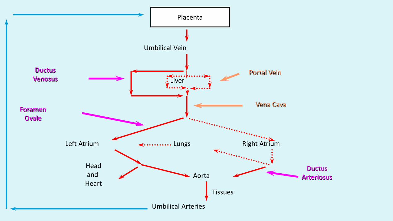
Placenta
Liver
Left Atrium Lungs Right Atrium
ForamenOvale
DuctusVenosus
DuctusArteriosusAorta
Umbilical Arteries
Umbilical Vein
Portal Vein
Vena Cava
Headand
HeartTissues
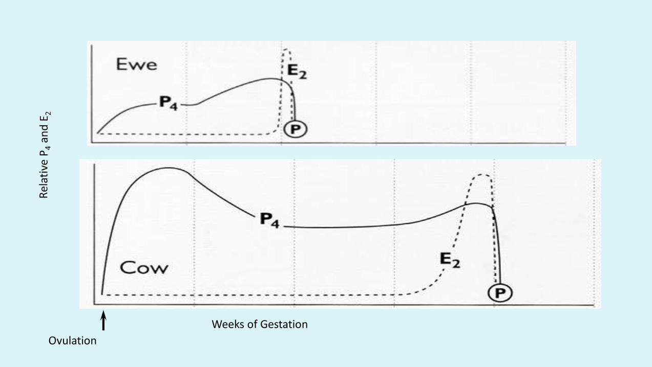
Weeks of Gestation
Ovulation

Weeks of Gestation
Ovulation


Weeks of Gestation
Ovulation

Weeks of Gestation
Ovulation
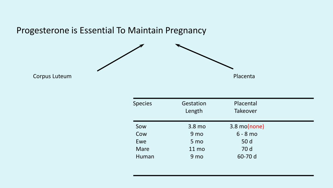
Progesterone is Essential To Maintain Pregnancy
Corpus Luteum Placenta
Sow 3.8 mo 3.8 mo(none)Cow 9 mo 6 - 8 moEwe 5 mo 50 dMare 11 mo 70 dHuman 9 mo 60-70 d
Species Gestation PlacentalLength Takeover

Other Species in Which Placenta Does not Take-over Progesterone Production
• Bitch
• Queen
• Alpaca, Llama, Camel
• Rabbit
• Goat

Thank YouKindly share the video and subscribe to my You tube
channel Govind Narayan Purohit if you like them