Production of Giant Mouse Oocyte Nucleoli and Assessment ... › f7e3 › 818b8c8fc0bb... ·...
6
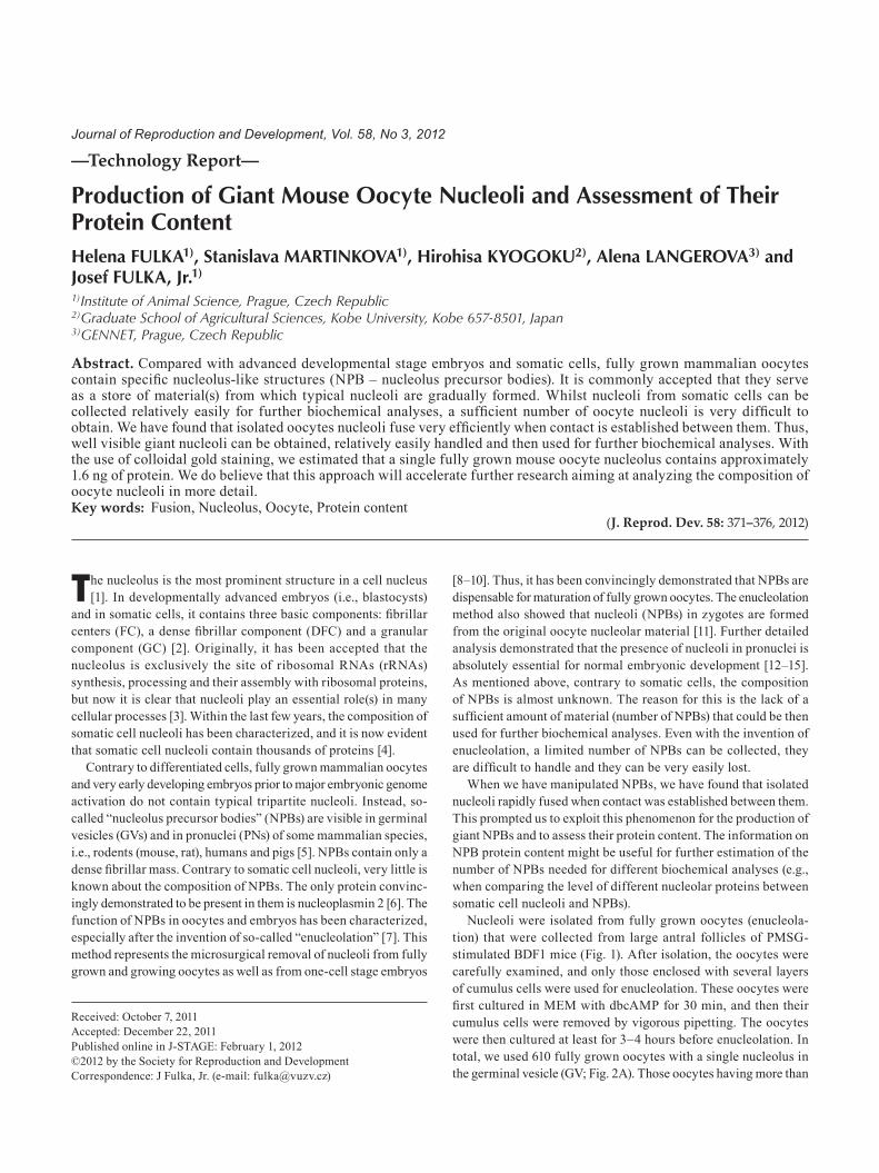
—Technology Report— Production of Giant Mouse Oocyte Nucleoli and Assessment of Their Protein Content Helena FULKA 1) , Stanislava MARTINKOVA 1) , Hirohisa KYOGOKU 2) , Alena LANGEROVA 3) and Josef FULKA, Jr. 1) 1) Institute of Animal Science, Prague, Czech Republic 2) Graduate School of Agricultural Sciences, Kobe University, Kobe 657-8501, Japan 3) GENNET, Prague, Czech Republic Abstract. Compared with advanced developmental stage embryos and somatic cells, fully grown mammalian oocytes contain specific nucleolus-like structures (NPB – nucleolus precursor bodies). It is commonly accepted that they serve as a store of material(s) from which typical nucleoli are gradually formed. Whilst nucleoli from somatic cells can be collected relatively easily for further biochemical analyses, a sufficient number of oocyte nucleoli is very difficult to obtain. We have found that isolated oocytes nucleoli fuse very efficiently when contact is established between them. Thus, well visible giant nucleoli can be obtained, relatively easily handled and then used for further biochemical analyses. With the use of colloidal gold staining, we estimated that a single fully grown mouse oocyte nucleolus contains approximately 1.6 ng of protein. We do believe that this approach will accelerate further research aiming at analyzing the composition of oocyte nucleoli in more detail. Key words: Fusion, Nucleolus, Oocyte, Protein content (J. Reprod. Dev. 58: 371–376, 2012) T he nucleolus is the most prominent structure in a cell nucleus [1]. In developmentally advanced embryos (i.e., blastocysts) and in somatic cells, it contains three basic components: fibrillar centers (FC), a dense fibrillar component (DFC) and a granular component (GC) [2]. Originally, it has been accepted that the nucleolus is exclusively the site of ribosomal RNAs (rRNAs) synthesis, processing and their assembly with ribosomal proteins, but now it is clear that nucleoli play an essential role(s) in many cellular processes [3]. Within the last few years, the composition of somatic cell nucleoli has been characterized, and it is now evident that somatic cell nucleoli contain thousands of proteins [4]. Contrary to differentiated cells, fully grown mammalian oocytes and very early developing embryos prior to major embryonic genome activation do not contain typical tripartite nucleoli. Instead, so- called “nucleolus precursor bodies” (NPBs) are visible in germinal vesicles (GVs) and in pronuclei (PNs) of some mammalian species, i.e., rodents (mouse, rat), humans and pigs [5]. NPBs contain only a dense fibrillar mass. Contrary to somatic cell nucleoli, very little is known about the composition of NPBs. The only protein convinc- ingly demonstrated to be present in them is nucleoplasmin 2 [6]. The function of NPBs in oocytes and embryos has been characterized, especially after the invention of so-called “enucleolation” [7]. This method represents the microsurgical removal of nucleoli from fully grown and growing oocytes as well as from one-cell stage embryos [8–10]. Thus, it has been convincingly demonstrated that NPBs are dispensable for maturation of fully grown oocytes. The enucleolation method also showed that nucleoli (NPBs) in zygotes are formed from the original oocyte nucleolar material [11]. Further detailed analysis demonstrated that the presence of nucleoli in pronuclei is absolutely essential for normal embryonic development [12–15]. As mentioned above, contrary to somatic cells, the composition of NPBs is almost unknown. The reason for this is the lack of a sufficient amount of material (number of NPBs) that could be then used for further biochemical analyses. Even with the invention of enucleolation, a limited number of NPBs can be collected, they are difficult to handle and they can be very easily lost. When we have manipulated NPBs, we have found that isolated nucleoli rapidly fused when contact was established between them. This prompted us to exploit this phenomenon for the production of giant NPBs and to assess their protein content. The information on NPB protein content might be useful for further estimation of the number of NPBs needed for different biochemical analyses (e.g., when comparing the level of different nucleolar proteins between somatic cell nucleoli and NPBs). Nucleoli were isolated from fully grown oocytes (enucleola- tion) that were collected from large antral follicles of PMSG- stimulated BDF1 mice (Fig. 1). After isolation, the oocytes were carefully examined, and only those enclosed with several layers of cumulus cells were used for enucleolation. These oocytes were first cultured in MEM with dbcAMP for 30 min, and then their cumulus cells were removed by vigorous pipetting. The oocytes were then cultured at least for 3–4 hours before enucleolation. In total, we used 610 fully grown oocytes with a single nucleolus in the germinal vesicle (GV; Fig. 2A). Those oocytes having more than Received: October 7, 2011 Accepted: December 22, 2011 Published online in J-STAGE: February 1, 2012 ©2012 by the Society for Reproduction and Development Correspondence: J Fulka, Jr. (e-mail: [email protected]) Journal of Reproduction and Development, Vol. 58, No 3, 2012
Transcript of Production of Giant Mouse Oocyte Nucleoli and Assessment ... › f7e3 › 818b8c8fc0bb... ·...
—Technology Report—
Production of Giant Mouse Oocyte Nucleoli and Assessment of Their Protein ContentHelena FulkA1), Stanislava MArTiNkOvA1), Hirohisa kyOGOku2), Alena lANGerOvA3) and Josef FulkA, Jr.1)
1)Institute of Animal Science, Prague, Czech Republic2)Graduate School of Agricultural Sciences, Kobe University, Kobe 657-8501, Japan3)GENNET, Prague, Czech Republic
Abstract. Compared with advanced developmental stage embryos and somatic cells, fully grown mammalian oocytes contain specific nucleolus-like structures (NPB – nucleolus precursor bodies). It is commonly accepted that they serve as a store of material(s) from which typical nucleoli are gradually formed. Whilst nucleoli from somatic cells can be collected relatively easily for further biochemical analyses, a sufficient number of oocyte nucleoli is very difficult to obtain. We have found that isolated oocytes nucleoli fuse very efficiently when contact is established between them. Thus, well visible giant nucleoli can be obtained, relatively easily handled and then used for further biochemical analyses. With the use of colloidal gold staining, we estimated that a single fully grown mouse oocyte nucleolus contains approximately 1.6 ng of protein. We do believe that this approach will accelerate further research aiming at analyzing the composition of oocyte nucleoli in more detail.Key words: Fusion, Nucleolus, Oocyte, Protein content
(J. Reprod. Dev. 58: 371–376, 2012)
The nucleolus is the most prominent structure in a cell nucleus [1]. In developmentally advanced embryos (i.e., blastocysts)
and in somatic cells, it contains three basic components: fibrillar centers (FC), a dense fibrillar component (DFC) and a granular component (GC) [2]. Originally, it has been accepted that the nucleolus is exclusively the site of ribosomal RNAs (rRNAs) synthesis, processing and their assembly with ribosomal proteins, but now it is clear that nucleoli play an essential role(s) in many cellular processes [3]. Within the last few years, the composition of somatic cell nucleoli has been characterized, and it is now evident that somatic cell nucleoli contain thousands of proteins [4].
Contrary to differentiated cells, fully grown mammalian oocytes and very early developing embryos prior to major embryonic genome activation do not contain typical tripartite nucleoli. Instead, so-called “nucleolus precursor bodies” (NPBs) are visible in germinal vesicles (GVs) and in pronuclei (PNs) of some mammalian species, i.e., rodents (mouse, rat), humans and pigs [5]. NPBs contain only a dense fibrillar mass. Contrary to somatic cell nucleoli, very little is known about the composition of NPBs. The only protein convinc-ingly demonstrated to be present in them is nucleoplasmin 2 [6]. The function of NPBs in oocytes and embryos has been characterized, especially after the invention of so-called “enucleolation” [7]. This method represents the microsurgical removal of nucleoli from fully grown and growing oocytes as well as from one-cell stage embryos
[8–10]. Thus, it has been convincingly demonstrated that NPBs are dispensable for maturation of fully grown oocytes. The enucleolation method also showed that nucleoli (NPBs) in zygotes are formed from the original oocyte nucleolar material [11]. Further detailed analysis demonstrated that the presence of nucleoli in pronuclei is absolutely essential for normal embryonic development [12–15]. As mentioned above, contrary to somatic cells, the composition of NPBs is almost unknown. The reason for this is the lack of a sufficient amount of material (number of NPBs) that could be then used for further biochemical analyses. Even with the invention of enucleolation, a limited number of NPBs can be collected, they are difficult to handle and they can be very easily lost.
When we have manipulated NPBs, we have found that isolated nucleoli rapidly fused when contact was established between them. This prompted us to exploit this phenomenon for the production of giant NPBs and to assess their protein content. The information on NPB protein content might be useful for further estimation of the number of NPBs needed for different biochemical analyses (e.g., when comparing the level of different nucleolar proteins between somatic cell nucleoli and NPBs).
Nucleoli were isolated from fully grown oocytes (enucleola-tion) that were collected from large antral follicles of PMSG-stimulated BDF1 mice (Fig. 1). After isolation, the oocytes were carefully examined, and only those enclosed with several layers of cumulus cells were used for enucleolation. These oocytes were first cultured in MEM with dbcAMP for 30 min, and then their cumulus cells were removed by vigorous pipetting. The oocytes were then cultured at least for 3–4 hours before enucleolation. In total, we used 610 fully grown oocytes with a single nucleolus in the germinal vesicle (GV; Fig. 2A). Those oocytes having more than
Received: October 7, 2011Accepted: December 22, 2011Published online in J-STAGE: February 1, 2012©2012 by the Society for Reproduction and DevelopmentCorrespondence: J Fulka, Jr. (e-mail: [email protected])
Journal of Reproduction and Development, Vol. 58, No 3, 2012
FULKA et al.372
one nucleolus were discarded. We successfully enucleolated 505 (83%) fully grown oocytes, and the remaining oocytes were either damaged or their nucleoli could not be completely removed. The nucleoli were isolated in the form of so-called nucleoloplasts, i.e., they were enclosed with a minimal volume of the oocyte cytoplasm surrounded by the oocyte plasma membrane. Some nucleoloplasts were damaged when released from the enucleolation pipette (75). The remaining nucleoloplasts (430) were used for the production of giant nucleoli or labeling. As the oocytes used for enucleola-tion contain nucleoli that are enclosed with a ring of chromatin (SN – surrounded nucleoli), we first verified the absence of chro-matin contamination of isolated nucleoli. To do that, the isolated nucleoloplasts were kept under zonae pellucidae of the oocytes from which they were isolated. The nucleoloplasts were stuck to enucleolated oocytes by a short incubation in phytohemagglutinin. Zonae pellucidae were then dissolved, and the oocyte-nucleoloplast aggregates were fixed and labeled with anti-trimethyl H4/K20 antibody. In the mouse, this antibody detects predominantly the pericentric heterochromatin–a chromatin domain that is specifi-cally associated with NPBs. We evaluated 45 oocyte-nucleoloplasts aggregates. In 42 of them, positive labeling was detected only
inside GVs, whilst in nucleoloplasts, there was no labeling (Fig. 2B). In three cases, the labeling was also visible in nucleoloplasts. Additionally, the staining of live oocyte-nucleolus aggregates (57) by Hoechst 33342 gave essentially the same results. In 54 cases, no blue fluorescence was visible in nucleoloplasts. Thus, in almost all cases, the isolated nucleoli do not contain residual chromatin. Finally, it must be noted that ultimate proof that the enucleolation does not cause DNA damage was reported by Ogushi et al. [11] – when NPBs are returned to previously enucleolated oocytes, live mice can be obtained after fertilizing the oocytes.
As mentioned, in some cases, during the enucleolation procedure, the nucleolus was split into two parts, which rapidly fused when the nucleoloplast was released from the pipette into the medium. This prompted us to test if nucleoli isolated from several oocytes can also fuse. First, we isolated nucleoli from nucleoloplasts by breaking their membranes with several piezo pulses. These nucleoli, however, rapidly decreased in volume and disappeared in the me-dium. Thus, we kept them in the form of nucleoloplasts from which they were released immediately before the induction of fusion. So, before the induction of fusion, the nucleoplast was aspirated into an enucleolation pipette and then several piezo pulses were ap-
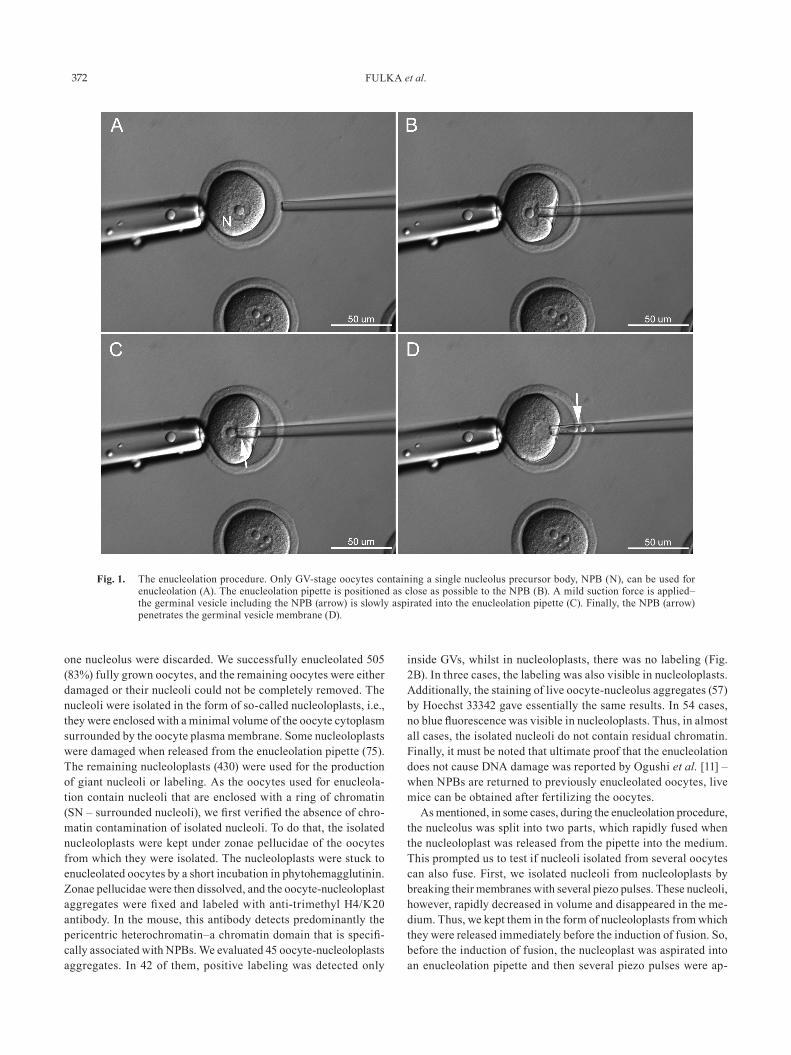
Fig. 1. The enucleolation procedure. Only GV-stage oocytes containing a single nucleolus precursor body, NPB (N), can be used for enucleolation (A). The enucleolation pipette is positioned as close as possible to the NPB (B). A mild suction force is applied–the germinal vesicle including the NPB (arrow) is slowly aspirated into the enucleolation pipette (C). Finally, the NPB (arrow) penetrates the germinal vesicle membrane (D).
GIANT MOUSE OOCYTE NUCLEOLI 373
plied. This breaks the nucleoloplast membrane and removes some cytoplasmic debris from the nucleolus. The isolated nucleolus was then released into a manipulation medium. After the next nucleolus was isolated in the same way, it was released into the medium and immediately pushed against the first isolated nucleolus, with which it fused when contact was achieved. Thereafter, additional nucleoli could be added (fused) to the previously obtained giant nucleoli produced by fusion (Fig. 2C). The success of fusion is absolute. In contrast to a single isolated nucleolus, the giant nucleoli are well visible and can thus be relatively easily handled.
The giant nucleoli can be removed from the manipulation medium and used for further analysis.
During previous experiments, we noticed that oocyte nucleoli are sensitive to pepsin digestion. This indicates that NPBs might be composed primarily from different proteins. However, the amount of protein in NPBs is unknown, and common methods used for protein quantification (Lowry, BCA assay, etc.) are not sensitive enough to
reliably detect these small amounts of protein. However, silver or colloidal gold staining is known to detect nanogram quantities of protein. Therefore, we reasoned that it might be feasible to use one of these methods to quantify the protein content of giant nucleoli.
Because the manipulation medium contains bovine serum albu-min (BSA), which would ultimately interfere with quantification, we first needed to select a suitable wash solution. After testing several different solutions, we selected phosphate buffered saline (PBS) supplemented with polyvinyl alcohol (PVA), as it does not react with colloidal gold and proteins are not washed through the carrier membrane. Thus, the giant nucleoli (composed of 10 fused NPBs) were quickly washed in PBS/PVA, and stored in 1 μl of this solution in Protein LoBind Tubes and then transferred to a PVDF membrane. At the same time, a protein standard, composed of dif-ferent solutions with known BSA concentrations, was also spotted onto the membrane. After transfer, the whole PVDF membrane was stained by colloidal gold solution (Fig. 2D). The next day,
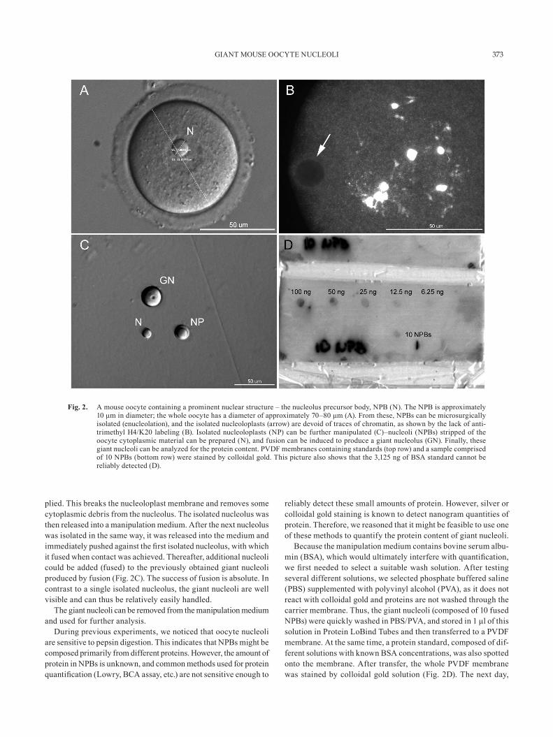
Fig. 2. A mouse oocyte containing a prominent nuclear structure – the nucleolus precursor body, NPB (N). The NPB is approximately 10 μm in diameter; the whole oocyte has a diameter of approximately 70–80 μm (A). From these, NPBs can be microsurgically isolated (enucleolation), and the isolated nucleoloplasts (arrow) are devoid of traces of chromatin, as shown by the lack of anti-trimethyl H4/K20 labeling (B). Isolated nucleoloplasts (NP) can be further manipulated (C)–nucleoli (NPBs) stripped of the oocyte cytoplasmic material can be prepared (N), and fusion can be induced to produce a giant nucleolus (GN). Finally, these giant nucleoli can be analyzed for the protein content. PVDF membranes containing standards (top row) and a sample comprised of 10 NPBs (bottom row) were stained by colloidal gold. This picture also shows that the 3,125 ng of BSA standard cannot be reliably detected (D).
FULKA et al.374
membranes were removed from the staining solution, washed and scanned, and the integrated optical density (IOD) was measured for each standard as well as the sample (Fig. 3). The optical density of the sample was plotted against the standards, and the approxi-mate amount of protein per one oocyte nucleolus was established (Fig. 4). Our analysis indicated that one oocyte nucleolus contains roughly 1.6 ± 0.3 ng of protein. However, it must be noted that the protein content is likely to be higher, as part of the proteins is probably lost during the wash steps. Unfortunately, this cannot be avoided. In our experiment, we labeled both freshly prepared NPB samples as well as NPB samples that were stored at –20 C for at least one month. No differences in sample quality were observed. Thus, the main concern of this procedure is the contamination of NPB samples by BSA originating from the manipulation media.
Our approach describes a simple way to collect a sufficient amount of nucleolar materials from fully grown mammalian oocytes that can then be used for further biochemical analyses. The methods for isolation of a sufficient number of somatic cell nucleoli have already been published, and the isolated nucleoli were used for proteomic analysis. As far as we are aware, no similar approach has been reported for the isolation of mammalian oocyte nucleoli.
The method of oocyte enucleolation was invented by Fulka, Jr. et al. [7], who demonstrated that nucleoli from fully grown porcine oocytes can be microsurgically removed. Subsequently, essentially the same approach was used for enucleolation of mouse oocytes [8, 11, 13]. Oocyte enucleolation thus enabled us to clarify the role of nucleoli during the process of oocyte maturation and in early embryonic development. Logically, the next step is a detailed bio-chemical characterization of NPBs. When compared with somatic cell nucleoli, mammalian oocyte nucleoli are relatively difficult to isolate in sufficient numbers. Their visibility is also limited. Moreover, when collected from the manipulation medium, they can be easily lost, for example, by sticking to a pipette wall. The
production of giant nucleoli eliminates these problems, as giant nucleoli are well visible and can be easily transferred into a test tube and frozen. However, attention must be paid to some issues. First, the enucleolation method requires good equipment and certain skill. Second, the volume of the oocyte cytoplasm enclosing the nucleolus must be kept to a minimum as the nucleoli reinjected into the oocyte cytoplasm disappear (are dissolved) readily. Therefore, we must assume that the same will happen in the nucleoloplast cytoplasm if the volume of cytoplasm is too large. Third, the work required for fusing the isolated nucleoli must be performed very quickly. The isolated nucleoli dissolve in the manipulation medium, so we must keep in mind that some nucleolar material will also be degraded when fusion of nucleoli is performed. Logically, it is thus better to produce relatively smaller giant nucleoli and to freeze them quickly than to produce supergiants from hundreds of nucleoli but lose some nucleolar material. Nevertheless, we do believe that our approach will accelerate the work aiming at characterizing the composition of fully grown mammalian oocyte nucleoli.
Methods
Preparation of oocytesBDF1 (B6D2F1/Crl) females were stimulated with 7.5 IU of
PMSG (pregnant mare serum gonadotropin, Calbiochem, EMD Chemicals USA, Gibbstown, NJ, USA). After about 44-46 h, their ovaries were isolated, and oocytes were released from large antral follicles into HTF (Human Tubal Fluid, Zenith Biotech, Guilford, CT, USA) supplemented with 4 mg/ml of BSA (bovine serum albumin). Thereafter, only those oocytes surrounded by several layers of cumulus cells were selected, transferred into MEM (mini-mal essential medium) supplemented with gentamicin (50 µg/ml), Na-pyruvate (0.2 mM), BSA (4 mg/ml) and dbcAMP (150 µg/ml; dibutyryl cyclic AMP) and cultured in an incubator at 37 C/5%
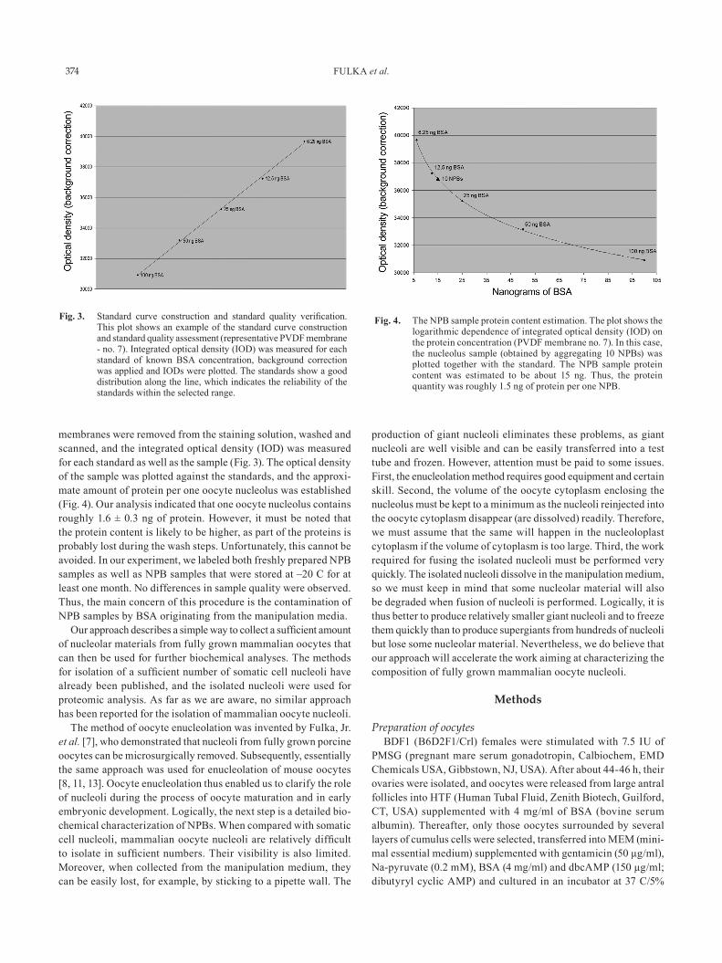
Fig. 3. Standard curve construction and standard quality verification. This plot shows an example of the standard curve construction and standard quality assessment (representative PVDF membrane - no. 7). Integrated optical density (IOD) was measured for each standard of known BSA concentration, background correction was applied and IODs were plotted. The standards show a good distribution along the line, which indicates the reliability of the standards within the selected range.
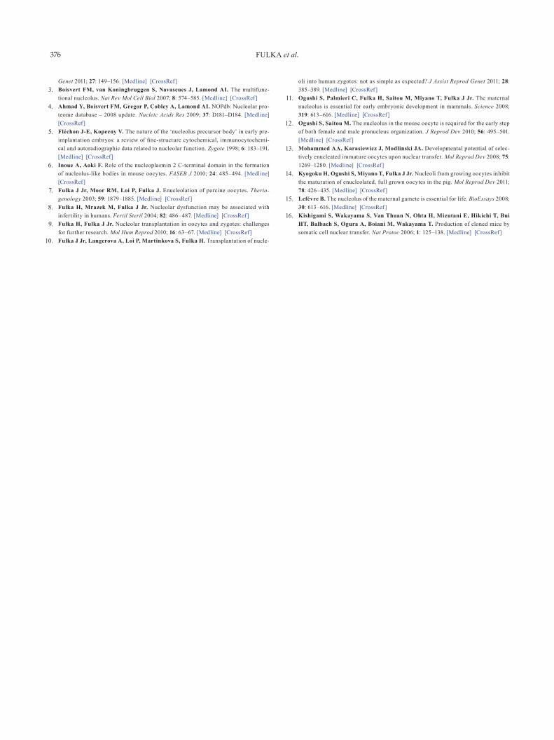
Fig. 4. The NPB sample protein content estimation. The plot shows the logarithmic dependence of integrated optical density (IOD) on the protein concentration (PVDF membrane no. 7). In this case, the nucleolus sample (obtained by aggregating 10 NPBs) was plotted together with the standard. The NPB sample protein content was estimated to be about 15 ng. Thus, the protein quantity was roughly 1.5 ng of protein per one NPB.

GIANT MOUSE OOCYTE NUCLEOLI 375
CO2 for 30 min. The cumulus cells were then removed by vigorous pipetting, and only healthy looking oocytes with intact germinal vesicles were further cultured for another 3–4 h. The remaining oocytes were discarded.
Oocyte enucleolationThe nucleoli from oocytes were isolated exactly as described in
our previous papers [7, 11]. Briefly, the oocytes were transferred into 10 µl droplets of KSOM/HEPES (Zenith Biotech, Guilford, CT, USA) supplemented with cytochalasin D (7.5 µg/ml), BSA (4 mg/ml) and dbcAMP (150 µg/ml). The droplets were covered with paraffin oil. The basic micromanipulation procedure was es-sentially the same as that described by Kishigami et al. [16]. The manipulation was performed on an Olympus IX-71 stage with the help of Narishige micromanipulators. The oocytes were stabilized with a holding pipette. The injection (enucleolation) pipette fixed to piezo unit (Prime Tech, Tsuchiura, Ibaraki, Japan) first penetrated through the zona pellucida and then was slowly pushed against the GV membrane. As soon as the tip was in close vicinity to the nucleolus, a very mild suction was applied. The nucleolus typically penetrates through GV membrane and is slowly translocated into the oocyte cytoplasm. A nucleolus enclosed by a minimum volume of the cytoplasm and surrounded by an oocyte plasma membrane (nucleoloplast) was obtained by withdrawing the enucleolation pipette from the oocyte. The nucleoloplasts are then either expelled from the pipette into the medium or left under zonae pellucidae (Hoechst staining, immunolabeling). As soon as a chosen number of nucleoloplasts was prepared, nucleoli were isolated from them. A single nucleoloplast was slowly aspirated into the enucleolation pipette, and then several high frequency pulses were applied. This breaks the membrane and concomitantly removes some cytoplas-mic debris from the nucleolus. The nucleolus was then released from the pipette. Next, another nucleolus that was prepared in the same way was pushed against the first nucleolus. Typically, as soon as minimal contact was established between these nucleoli, fusion occurred very rapidly. The procedure was repeated until a giant nucleolus with the desired amount of nucleolar material was collected.
Verifying the absence of chromatin incorporation around isolated nucleoli
As mentioned above, nucleoli in the best oocytes are surrounded closely by the chromatin (SN – surrounded nucleolus). In order to verify whether this chromatin is or is not attached to isolated nucleoli in nucleoloplasts, two approaches were used. First, oocytes with nucleoloplasts under zonae pellucidae were stained with Hoechst 33342 (20 µg/ml) for 20 min at 37 C. Then they were washed several times in HTF/BSA and examined under a fluorescence microscope.
The second approach used labeling with anti-trimethyl H4/K20 antibody. Briefly, the enucleolated oocytes with nucleoloplasts under zonae pellucidae were incubated in PBS supplemented with phytohemagglutinin (200 µg/ml). This sticks the nucleoloplast to the oocyte. Zonae pellucidae were then dissolved in acid Tyrode’s solution, and then oocytes with nucleoloplasts were fixed in 4% paraformaldehyde in PBS for 15 min at room temperature. The fixed specimens were then stored in PBS (4 C). Before labeling,
the oocyte-nucleoloplasts aggregates were permeabilized with Triton-TX100 (0.2%) in PBS for 30 min and then blocked 1 h (4 C) in 1% BSA/PBS. Samples were then incubated with anti-trimethyl H4/K20 antibody (Abcam, Cambridge, UK) overnight at 4 C, washed several times in PBS/BSA and incubated with Cy2 conju-gated secondary antibody (Jackson ImmunoResearch, Newmarket, UK) for 2 h (RT). After this incubation and several washes in PBS/BSA, samples were mounted on slides in VECTASHIELD (Vector Laboratories, Peterborough, UK) and evaluated under a fluorescence microscope.
Assessing the protein content in nucleoliOocyte nucleoli (NPBs) were isolated and fused as described.
Samples of 10 NPBs were taken for analysis. A giant nucleolus was removed from the manipulation medium containing BSA and quickly washed in phosphate buffered saline (PBS) supplemented with 0.1% polyvinyl alcohol (PVA). The washed giant nucleolus was transferred into an Eppendorf Protein LoBind tube (Eppendorf, Prague, Czech Republic) under a stereomicroscope, snap-frozen in liquid nitrogen and analyzed or stored at –20 C until analysis. Standards were prepared from a BSA solution containing 2 mg/ml protein (Pierce, Rockford, IL, USA) by serial dilution at the fol-lowing concentrations: 100, 50, 25, 12.5, 6.25 and 3.125 ng/μl. A PVDF membrane (Bio-Rad, Prague, Czech Republic) was activated by a brief incubation in methanol. The membrane was washed in distilled water and then in Towbin Transfer Buffer containing 20% methanol. Excess buffer was removed by vacuum. One microliter of each standard was spotted onto the membrane; finally, the sample was transferred onto the membrane, and the membrane was stained by colloidal gold solution as described by the manufacturer (Bio-Rad, Prague, Czech Republic). The next day, the membrane was washed in distilled water and scanned. Signal quantification was performed using the Image-Pro software (Media Cybernetics, Bethesda, MD, USA). Parameters collected were the area and integrated optical density (IOD), and samples of the background were also analyzed. Background intensity was subtracted, and standard concentrations of proteins were plotted against the signal. In total, we evaluated 9 PVDF membranes, each containing a single sample equivalent to 10 NPBs (this number of NPBs was estimated to be the lowest amount of material sufficient for reliable analysis by the colloidal gold staining method). From this, the unknown protein concentra-tion of the sample was calculated. Results are given as the average ± standard deviation.
Unless stated otherwise, all chemicals were purchased from Sigma.
Acknowledgments
This study was supported by GACR P302/11/P069. HK was supported by a Research Fellowship for Young Scientists.
References
1. Mao YS, Zhang B, Spector DL. Biogenesis and function of nuclear bodies. Trends Genet 2011; 27: 295–306. [Medline] [CrossRef]
2. Németh A, Langst G. Genome organization in and around the nucleolus. Trends
FULKA et al.376
Genet 2011; 27: 149–156. [Medline] [CrossRef] 3. Boisvert FM, van Koningbruggen S, Navascues J, Lamond AI. The multifunc-
tional nucleolus. Nat Rev Mol Cell Biol 2007; 8: 574–585. [Medline] [CrossRef] 4. Ahmad Y, Boisvert FM, Gregor P, Cobley A, Lamond AI. NOPdb: Nucleolar pro-
teome database – 2008 update. Nucleic Acids Res 2009; 37: D181–D184. [Medline] [CrossRef]
5. Fléchon J-E, Kopecny V. The nature of the ‘nucleolus precursor body’ in early pre-implantation embryos: a review of fine-structure cytochemical, immunocytochemi-cal and autoradiographic data related to nucleolar function. Zygote 1998; 6: 183–191. [Medline] [CrossRef]
6. Inoue A, Aoki F. Role of the nucleoplasmin 2 C-terminal domain in the formation of nucleolus-like bodies in mouse oocytes. FASEB J 2010; 24: 485–494. [Medline] [CrossRef]
7. Fulka J Jr, Moor RM, Loi P, Fulka J. Enucleolation of porcine oocytes. Therio-genology 2003; 59: 1879–1885. [Medline] [CrossRef]
8. Fulka H, Mrazek M, Fulka J Jr. Nucleolar dysfunction may be associated with infertility in humans. Fertil Steril 2004; 82: 486–487. [Medline] [CrossRef]
9. Fulka H, Fulka J Jr. Nucleolar transplantation in oocytes and zygotes: challenges for further research. Mol Hum Reprod 2010; 16: 63–67. [Medline] [CrossRef]
10. Fulka J Jr, Langerova A, Loi P, Martinkova S, Fulka H. Transplantation of nucle-
oli into human zygotes: not as simple as expected? J Assist Reprod Genet 2011; 28: 385–389. [Medline] [CrossRef]
11. Ogushi S, Palmieri C, Fulka H, Saitou M, Miyano T, Fulka J Jr. The maternal nucleolus is essential for early embryonic development in mammals. Science 2008; 319: 613–616. [Medline] [CrossRef]
12. Ogushi S, Saitou M. The nucleolus in the mouse oocyte is required for the early step of both female and male pronucleus organization. J Reprod Dev 2010; 56: 495–501. [Medline] [CrossRef]
13. Mohammed AA, Karasiewicz J, Modlinski JA. Developmental potential of selec-tively enucleated immature oocytes upon nuclear transfer. Mol Reprod Dev 2008; 75: 1269–1280. [Medline] [CrossRef]
14. Kyogoku H, Ogushi S, Miyano T, Fulka J Jr. Nucleoli from growing oocytes inhibit the maturation of enucleolated, full grown oocytes in the pig. Mol Reprod Dev 2011; 78: 426–435. [Medline] [CrossRef]
15. Lefévre B. The nucleolus of the maternal gamete is essential for life. BioEssays 2008; 30: 613–616. [Medline] [CrossRef]
16. Kishigami S, Wakayama S, Van Thuan N, Ohta H, Mizutani E, Hikichi T, Bui HT, Balbach S, Ogura A, Boiani M, Wakayama T. Production of cloned mice by somatic cell nuclear transfer. Nat Protoc 2006; 1: 125–138. [Medline] [CrossRef]