PROCEEDINGS Open Access How does heparin prevent the pH ... · Results: Our pKa calculations of...
15
PROCEEDINGS Open Access How does heparin prevent the pH inactivation of cathepsin B? Allosteric mechanism elucidated by docking and molecular dynamics Mauricio GS Costa 1* , Paulo R Batista 1 , Cláudio S Shida 1,2 , Charles H Robert 3 , Paulo M Bisch 1 , Pedro G Pascutti 1 From 5th International Conference of the Brazilian Association for Bioinformatics and Computational Biology (X-meeting 2009) Angra Dos Reis, RJ, Brazil. 18-22 October 2009 Abstract Background: Cathepsin B (catB) is a promising target for anti-cancer drug design due to its implication in several steps of tumorigenesis. catB activity and inhibition are pH-dependent, making it difficult to identify efficient inhibitor candidates for clinical trials. In addition it is known that heparin binding stabilizes the enzyme in alkaline conditions. However, the molecular mechanism of stabilization is not well understood, indicating the need for more detailed structural and dynamic studies in order to clarify the influence of pH and heparin binding on catB stability. Results: Our pKa calculations of catB titratable residues revealed distinct protonation states under different pH conditions for six key residues, of which four lie in the crucial interdomain interface. This implies changes in the overall charge distribution at the catB surface, as revealed by calculation of the electrostatic potential. We identified two basic surface regions as possible heparin binding sites, which were confirmed by docking calculations. Molecular dynamics (MD) of both apo catB and catB-heparin complexes were performed using protonation states for catB residues corresponding to the relevant acidic or alkaline conditions. The MD of apo catB at pH 5.5 was very stable, and presented the highest number and occupancy of hydrogen bonds within the inter-domain interface. In contrast, under alkaline conditions the enzyme’s overall flexibility was increased: interactions between active site residues were lost, helical content decreased, and domain separation was observed as well as high- amplitude motions of the occluding loop – a main target of drug design studies. Essential dynamics analysis revealed that heparin binding modulates large amplitude motions promoting rearrangement of contacts between catB domains, thus favoring the maintenance of helical content as well as active site stability. Conclusions: The results of our study contribute to unraveling the molecular events involved in catB inactivation in alkaline pH, highlighting the fact that protonation changes of few residues can alter the overall dynamics of an enzyme. Moreover, we propose an allosteric role for heparin in the regulation of catB stability in such a manner that the restriction of enzyme flexibility would allow the establishment of stronger contacts and thus the maintenance of overall structure. * Correspondence: [email protected] 1 Instituto de Biofísica Carlos Chagas Filho, Universidade Federal do Rio de Janeiro, 21949-901, Rio de Janeiro, Brasil Full list of author information is available at the end of the article Costa et al. BMC Genomics 2010, 11(Suppl 5):S5 http://www.biomedcentral.com/1471-2164/11/S5/S5 © 2010 Costa et al; licensee BioMed Central Ltd. This is an open access article distributed under the terms of the Creative Commons Attribution License (http://creativecommons.org/licenses/by/2.0), which permits unrestricted use, distribution, and reproduction in any medium, provided the original work is properly cited.
Transcript of PROCEEDINGS Open Access How does heparin prevent the pH ... · Results: Our pKa calculations of...
PROCEEDINGS Open Access
How does heparin prevent the pH inactivation ofcathepsin B? Allosteric mechanism elucidated bydocking and molecular dynamicsMauricio GS Costa1*, Paulo R Batista1, Cláudio S Shida1,2, Charles H Robert3, Paulo M Bisch1, Pedro G Pascutti1
From 5th International Conference of the Brazilian Association for Bioinformatics and Computational Biology(X-meeting 2009)Angra Dos Reis, RJ, Brazil. 18-22 October 2009
Abstract
Background: Cathepsin B (catB) is a promising target for anti-cancer drug design due to its implication in severalsteps of tumorigenesis. catB activity and inhibition are pH-dependent, making it difficult to identify efficientinhibitor candidates for clinical trials. In addition it is known that heparin binding stabilizes the enzyme in alkalineconditions. However, the molecular mechanism of stabilization is not well understood, indicating the need formore detailed structural and dynamic studies in order to clarify the influence of pH and heparin binding on catBstability.
Results: Our pKa calculations of catB titratable residues revealed distinct protonation states under different pHconditions for six key residues, of which four lie in the crucial interdomain interface. This implies changes in theoverall charge distribution at the catB surface, as revealed by calculation of the electrostatic potential. We identifiedtwo basic surface regions as possible heparin binding sites, which were confirmed by docking calculations.Molecular dynamics (MD) of both apo catB and catB-heparin complexes were performed using protonation statesfor catB residues corresponding to the relevant acidic or alkaline conditions. The MD of apo catB at pH 5.5 wasvery stable, and presented the highest number and occupancy of hydrogen bonds within the inter-domaininterface. In contrast, under alkaline conditions the enzyme’s overall flexibility was increased: interactions betweenactive site residues were lost, helical content decreased, and domain separation was observed as well as high-amplitude motions of the occluding loop – a main target of drug design studies. Essential dynamics analysisrevealed that heparin binding modulates large amplitude motions promoting rearrangement of contacts betweencatB domains, thus favoring the maintenance of helical content as well as active site stability.
Conclusions: The results of our study contribute to unraveling the molecular events involved in catB inactivationin alkaline pH, highlighting the fact that protonation changes of few residues can alter the overall dynamics of anenzyme. Moreover, we propose an allosteric role for heparin in the regulation of catB stability in such a mannerthat the restriction of enzyme flexibility would allow the establishment of stronger contacts and thus themaintenance of overall structure.
* Correspondence: [email protected] de Biofísica Carlos Chagas Filho, Universidade Federal do Rio deJaneiro, 21949-901, Rio de Janeiro, BrasilFull list of author information is available at the end of the article
Costa et al. BMC Genomics 2010, 11(Suppl 5):S5http://www.biomedcentral.com/1471-2164/11/S5/S5
© 2010 Costa et al; licensee BioMed Central Ltd. This is an open access article distributed under the terms of the Creative CommonsAttribution License (http://creativecommons.org/licenses/by/2.0), which permits unrestricted use, distribution, and reproduction inany medium, provided the original work is properly cited.

BackgroundCathepsin B (EC 3.4.22.1) (catB) is one of the most well-characterized cysteine proteases, belonging to the clanCA (papain superfamily). In humans, its physiologicalrole is implicated in bone resorption, antigen processingand protein turnover [1]. However, catB also participatesin pathological processes such as cardiovascular distur-bances [2], parasitic infections [3], Alzheimer’s disease[4], osteoarthritis [5], tumor invasion and metastasisdevelopment [6,7]. Its main roles in cancer are i) itsactivity in directly cleaving extracellular matrix (ECM)components, ii) its activation of other ECM degradingproteases, which promotes tumor cell invasion into thesurrounding tissue and bloodstream escape [8], and iii)stimulating angiogenesis which provides increased nutri-ents and oxygen supplies to cancer cells [9]. Thus, catBregulates several crucial steps in tumorigenesis and is apromising target for anti-cancer drug design [10].Structurally, catB possesses the regular fold of papain-
like enzymes, enclosing two distinct domains stabilizedby six disulfide bridges, forming a large polar interfaceinto which project the side chains of a few charged resi-dues such as E171 and E36 (see Fig. 1). This interdo-main interface is extremely important to catB overallactivity as it comprises the active site residues (C29,H199 and N219). Unlike other members of the papainfamily, catB exhibits both exo- and endo-proteolytic
activities. Its exo-activity is dependent on the presenceof two adjacent histidine residues (H110 and H111)located at an insertion region called the “occludingloop”. These residues provide the necessary positivecharge to anchor the negatively-charged C-terminal car-boxylate of exo-substrates [11,12]. This region is onlyfound in catB within its family, and it controls theaccess of large substrates to the active site [12]. Site-directed mutagenesis studies confirmed the role of theoccluding loop since deletion of this entire regionimpairs exo- but not endo-proteolytic activity [13].Additionally, this region confers thermal stability to catBand resistance against endogenous inhibitors such ascystatin C [13,14].Currently the most potent and selective structure-
based designed compounds available are derived fromE-64 targeting the unusual occluding loop present in thecatB 3D-structure [15]. However, enzymatic assays haveshown that these inhibitors are strongly pH dependentas their optimal binding affinities are considerablydiminished under neutral/alkaline conditions [16]. SincecatB can be found in several cellular compartments withdistinct pH values, these inhibitors are not effective invivo. When catB is within the lysosomal or endosomalvesicles it confronts acidic conditions, in contrast to theneutral/alkaline environment when it is attached tomembranes (mainly in caveolar microdomains) [17,18]
Figure 1 Localization of differentially protonated residues in catB Cartoon representation of the catB tertiary structure showing differentiallyprotonated residues as green sticks. Protonation states were assigned to represent acidic (pH 5.5) or alkaline conditions (pH 8) based on pKacalculations with PROPKA on the catB crystal structure. The L and R domains of the protein are highlighted blue and red, respectively. Theoccluding loop (residues 106-124) a structural element found only in catB within its family, is represented in yellow.
Costa et al. BMC Genomics 2010, 11(Suppl 5):S5http://www.biomedcentral.com/1471-2164/11/S5/S5
Page 2 of 15

or secreted in the ECM [19]. Although catB is rapidlyinactivated under alkaline pH conditions, it was reportedthat membrane-associated forms are resistant to thisprocess [20]. This peculiarity is believed to occur due toits interaction with heparan sulfate glycosaminoglycans(GAGs) on the cell surface [19]. This polysaccharide,structurally related to heparin, acts on the ECM as a co-receptor for several molecules such as growth factorsand proteases [21] . Interaction between catB andheparin-like GAGs was already shown to prevent theenzyme’s inactivation in high pH [22]. The mainreported outcome was the maintenance of catB helicalcontent in the presence of heparin at high pH, whichwas observed for papain as well [22,23]. Nevertheless, ithas not been possible to precisely define the catB/heparin interaction sites and the molecular mechanismresponsible for this protective effect.Structural and molecular modelling studies can give
insight into the molecular events concerning the modifi-cation of catB activity by pH changes and heparin bind-ing. Some attempts have already been made towardsdesigning new catB inhibitors using molecular modelingtechniques [24,25], taking into account dynamicalaspects of binding. However, these studies did not evalu-ate the influence of pH nor that of heparin binding onthe modulation of the enzyme intrinsic flexibility.In order to understand the heparin protective effect
over catB structure we performed molecular dynamics(MD) simulations using an approach in which differentpH conditions were taken into account by consideringdifferent protonation states of titratable groups on theprotein surface. Further, docking analyses resulted in theidentification of two heparin binding sites on catB struc-ture. The MD calculations confirmed an increase of theoverall catB flexibility and the loss of stability of the apocatB inter-domain interface in alkaline pH. The destabi-lization and the increased flexibility, notably in theoccluding loop, were prevented by interaction withheparin, again in agreement with experimental data. Weobserved a role of active site residues in enzyme stabili-zation and in maintaining the helical content, and wepropose an allosteric mechanism for the stabilizingeffect promoted by GAG interaction. Taken together,our data provides an improved understanding of themolecular mechanisms responsible for both pH-inducedinactivation and protection against inactivation byheparin binding.
Results and discussionpH changes result in distinct protonation profilesPrediction of pKa’s for protein ionizable residues is animportant tool for understanding features and catalyticmechanisms of pH-dependent enzymes [26]. We appliedthe PROPKA program to estimate pKa values in the
catB structure and to determine the most probable cor-responding protonation states of the enzyme underacidic and alkaline conditions. Two different conditionswere studied: acidic (pH 5.5), and alkaline (pH 8.0),which allow comparison to available experimental data[22].It is known that catalytic residues often present unu-
sual pKa values compared to those of free amino acidsin solution [27]. Accordingly, our results for catB pre-dicted a pKa of 2.14 for the catalytic C29, in contrast to8.0 expected in solution. Previous work on the papaincatalytic cysteine (structurally related to C29 in catB)showed pKa values around 2.5 to 3.5 [28], in accordanceto our estimation for C29.Comparing the acidic and alkaline conditions we
observed that six key residues are differentially proto-nated. Table 1 gives the pKa values for these residuesand Fig. 1 shows their positions in the catB structure.We note that four of these residues belong to the inter-domain interface (E36, H199, E171 and H110). The pre-dicted pKa for E171 is 7.71, as carboxyl groups usuallyexhibit high pKa values in buried hydrophobic environ-ments [26]. From our prediction we observed that bothE171 and E36 are likely to be protonated at pH 5.5 butnot in alkaline conditions.As expected, most of the residues with differing proto-
nation states were histidines, with a pKa in solution of6.5 and an ionization state that is very susceptible to pHchanges in the physiological range. H110 is a key resi-due for stabilization of the occluding loop and is alsocrucial for the exo-proteolytic activity of catB, sincealong with H111 it anchors the carboxyl terminus ofsubstrates. Interestingly, we observed an pKa of 7.79 forthe catalytic H199, which implies that this residue isprotonated only in acidic conditions. This result is cor-respondent to the measured pKa of H159 in papain[28]. In papain, it was previously reported that the
Table 1 Differentially protonated residues in catB andpKa values
Average values during 40ns MD
Residue predicted crystal pH 5,5 pH 8
C29 2,14 3.7 ± 1.5 7.3 +1.6
E36 5,9 5.4 ± 0.5 3.4 ± 1.2
H97 6,9 7.1 ± 0.2 7.3 ± 0.4
H110 6,6 6.6 ± 0.8 8.1 ± 0.7
E171 7,7 7.4 ± 0.2 6.5 ± 0.4
H190 7,2 7.1 ± 0.2 7.4 ± 0.7
H199 7,8 8.1 ± 2 4.5 ± 1.7
pKa values predicted by PROPKA and the state of ionizable groups underdifferent pH conditions are indicated. The protonation state of C29 at pH 8were obtained taking into account deprotonation of H199 as previouslyproposed for papain. The protonation states of the catalytic residues (C29 andH199) are highlighted.
Costa et al. BMC Genomics 2010, 11(Suppl 5):S5http://www.biomedcentral.com/1471-2164/11/S5/S5
Page 3 of 15

ionization of the catalytic C25 and H159 residues arecoupled [29]. There the deprotonation of H159 shiftsthe pKa of C25 from ~3.3 to 7.6, while the neutraliza-tion of C25 decreases the pKa of H159 from ~8.5 to 4.3.Due to the direct relationship between catB and papainwe assigned the protonation states of these residuesaccording to this proposed mechanism.The pKa values predicted with PROPKA thus appear
to be sensitive to local microenvironment changesaround the ionizable residues. In order to verify if theapplied protonation profiles accurately represent the dis-tinct pH conditions simulated, we collected 1000 snap-shots (one at each 400 ps) from each moleculardynamics (MD) trajectory in order to perform pKa cal-culations. Table 1 shows the average pKa values duringthe MD simulations. From both pH conditions weobtained values consistent with the protonation statespredicted from the crystal structure, thus confirmingtheir validity. In this Table we have also highlighted thepKa shift in the catalytic residues C29 and H199 asobserved for papain.
Electrostatic potential calculation and docking analysesreveal two heparin binding sites on catB structureThe electrostatic potential at the catB surface revealedthe influence of the different protonation states for eachof the pH conditions considered. Overall, independentof the conditions, the catB surface is mostly negative,presenting one region of positive potential in eachdomain (Fig. 2). Under alkaline conditions the overallsurface is qualitatively more negative. The positive sitesare composed of R85, K86, K130, K141 and K144 in theL-domain, and the catB N-terminus together with K154and R235 in the R-domain. The protein’s total chargechanges from -11 at pH 8 to -5 at pH 5.5, with six keyresidues protonated exclusively under acidic conditions.Experimental studies have provided knowledge about
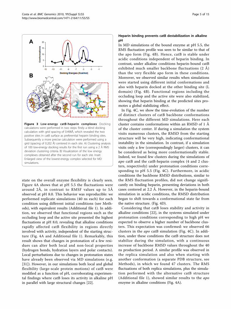
the affinity and kinetics of the catB-heparin interaction[22]. However, the precise binding site(s) has(have) notbeen defined. Taking into account that heparin-proteininteractions are mainly driven by charge interactionsdue to the high number of sulfate groups found in thepolysaccharide, we visually identified two regions ofpositive potential at the catB surface as potential heparinbinding sites. Blind docking calculations confirmed thisprediction, as we found two low energy clusters of dock-ing poses coincident with these positive regions (Fig. 3Aand B). Almeida and coworkers proposed that heparinmainly interacts with the occluding loop [22]. From astructural view this hypothesis appears unlikely due tothe lack of charge and shape complementarity in thisregion of the enzyme, and is consistent with the absenceof low-energy clusters for such a mode of binding in thedocking results. Moreover, we carried out another
docking calculation verifying the atomic interactions inthe heparin binding sites more precisely (see Methods).In the lowest energy complex obtained, heparin interactswith catB in the R-domain with an energy of -11 Kcal/mol (Fig. 3B). Although the positive potential at this siteis smaller than that found in the L-domain, in R-domainsite the van der Waals interactions were more favorable(contributed mainly by L1 at the N-terminus, A3, K154and R231). We suggest that for a disaccharide the R-domain site is more important for this interaction; forlonger sugar chains the L-domain could be also impor-tant for proper heparin accommodation.
catB protonation profile influences protein stabilityAs flexibility plays a key role in protein biological func-tions [30], we analyzed the root mean square fluctuation(RMSF) of catB backbone residues during MD for eachcondition simulated. A direct influence of protonation
Figure 2 Electrostatic surface of catB in distinct protonationstates reveals two possible heparin binding sites Theelectrostatic surface of catB using protonation states correspondingto acidic and alkaline pH conditions. Blue, red, and white indicatepositively charged regions, negative areas and neutral regions,respectively, with the scale indicating ± kbT/ec, where kb is theBoltzmann constant, T is the temperature and ec is the charge ofone electron. The heparin binding sites correspond to the basicregions localized in each domain.
Costa et al. BMC Genomics 2010, 11(Suppl 5):S5http://www.biomedcentral.com/1471-2164/11/S5/S5
Page 4 of 15
state on the overall enzyme flexibility is clearly seen.Figure 4A shows that at pH 5.5 the fluctuations werearound 2Å, in contrast to RMSF values up to 5Åobserved at pH 8.0. This behavior was reproducible: weperformed replicate simulations (40 ns each) for eachcondition using different initial conditions (see Meth-ods), with equivalent results (Additional file 1). In addi-tion, we observed that functional regions such as theoccluding loop and the active site presented the highestfluctuations at pH 8.0, revealing that alkaline conditionsrapidly affected catB flexibility in regions directlyinvolved with activity, independent of the starting struc-ture (Fig. 4A and Additional file 1). Remarkably, thisresult shows that changes in protonation of a few resi-dues can alter both local and non-local properties(hydrogen bonds, hydration layers and polar contacts).Local perturbations due to changes in protonation stateshave already been observed via MD simulations (e.g.[31]). However, in our simulations both local and globalflexibility (large-scale protein motions) of catB weremodified as a function of pH, corroborating experimen-tal findings where catB loses its activity in alkaline pHin parallel with large structural changes [22].
Heparin binding prevents catB destabilization in alkalinepHIn MD simulations of the bound enzyme at pH 5.5, theRMS fluctuation profile was seen to be similar to that ofthe apo form (Fig. 4B). Hence, catB is stable underacidic conditions independent of heparin binding. Incontrast, under alkaline conditions heparin-bound catBexhibited much smaller backbone fluctuations (2 Å)than the very flexible apo form in these conditions.Moreover, we observed similar results when simulationswere started using different initial conformations andalso with heparin docked at the other binding site (Ldomain) (Fig. 4B). Functional regions including theoccluding loop and the active site were also stabilized,showing that heparin binding at the predicted sites pro-motes a global stabilizing effect.In Fig. 4C, we show the time-evolution of the number
of distinct clusters of catB backbone conformationsthroughout the different MD simulations. Here eachcluster contains conformations within an RMSD of 1 Åof the cluster center. If during a simulation the systemvisits numerous clusters, the RMSD from the startingstructure will be very high, indicating conformationalinstability in the simulation. In contrast, if a simulationvisits only a few (correspondingly larger) clusters, it canbe considered as being more conformationally stable.Indeed, we found few clusters during the simulations ofapo catB and the catB-heparin complex (4 and 2 clus-ters, respectively) under protonation conditions corre-sponding to pH 5.5 (Fig. 4C). Furthermore, in acidicconditions the backbone RMSD distributions, similar tothe RMS fluctuation profiles, did not change signifi-cantly on binding heparin, presenting deviations in bothcases centered at 2.2 Å. However, in the heparin-boundsimulation in acidic conditions, the RMSD distributionbegan to shift towards a conformational state far fromthe native structure. (Fig. 4D).Considering that catB loses stability and activity in
alkaline conditions [22], in the systems simulated underprotonation conditions corresponding to high pH weexpected to observe a higher number of backbone clus-ters. This expectation was confirmed: we observed 66clusters in the apo catB simulation (Fig. 4C). In addi-tion, under these conditions the catB structure does notstabilize during the simulation, with a continuousincrease of backbone RMSD values throughout the 40ns production period. A similar profile was observed inthe replica simulation and also when starting withanother conformation (a separate PDB structure, seeMethods), in which we found 47 clusters. The RMSfluctuations of both replica simulations, plus the simula-tion performed with the alternative catB structure(Additional file 1), showed similar results to the apoenzyme in alkaline conditions (Fig. 4A).
Figure 3 Low-energy catB-heparin complexes Dockingcalculations were performed in two steps: firstly a blind dockingcalculation with grid spacing of 0.498Å, which revealed the twopositive sites in catB surface as preferential heparin binding sites.Subsequently a more precise calculation were performed using agrid (spacing of 0.202 Å) centered in each site. A) Clustering analysisof 100 low-energy docking results for the first run using a 2 Å RMSdeviation clustering criteria. B) Visualization of the low energycomplexes obtained after the second run for each site. Inset:Enlarged view of the lowest-energy complex selected for MDsimulations.
Costa et al. BMC Genomics 2010, 11(Suppl 5):S5http://www.biomedcentral.com/1471-2164/11/S5/S5
Page 5 of 15
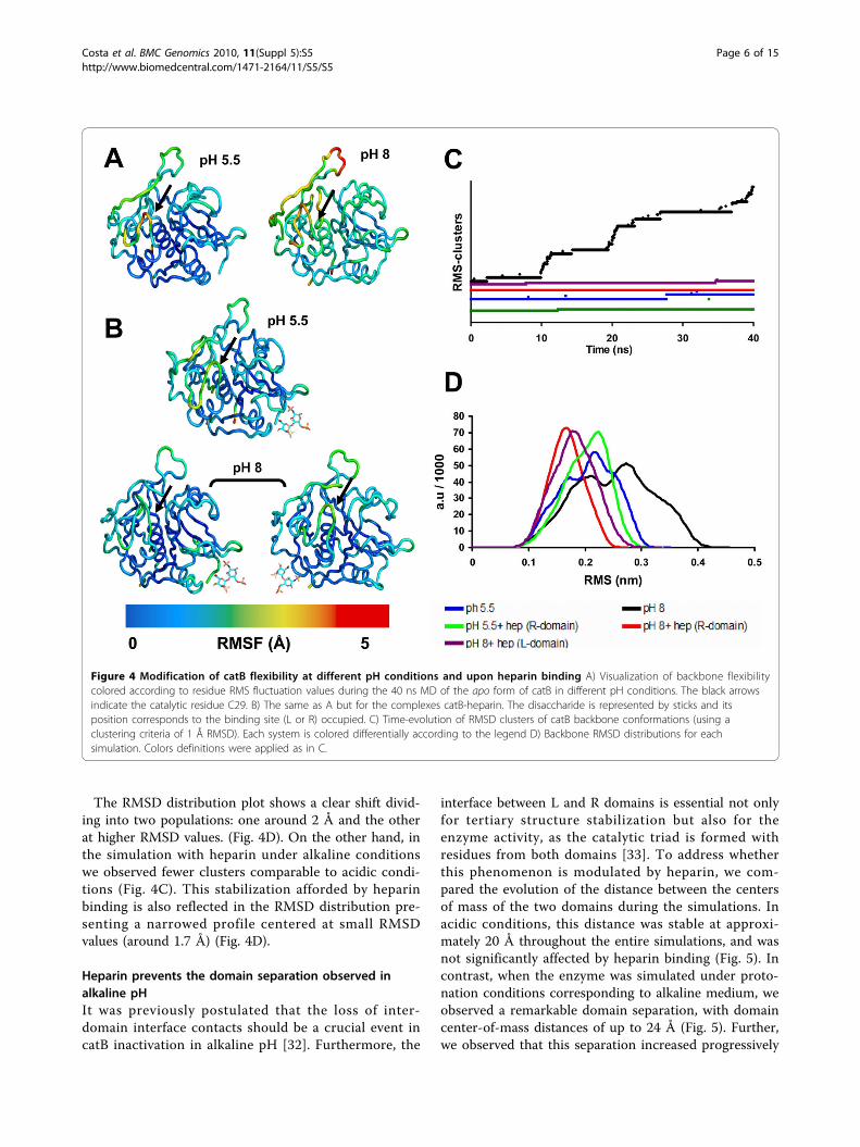
The RMSD distribution plot shows a clear shift divid-ing into two populations: one around 2 Å and the otherat higher RMSD values. (Fig. 4D). On the other hand, inthe simulation with heparin under alkaline conditionswe observed fewer clusters comparable to acidic condi-tions (Fig. 4C). This stabilization afforded by heparinbinding is also reflected in the RMSD distribution pre-senting a narrowed profile centered at small RMSDvalues (around 1.7 Å) (Fig. 4D).
Heparin prevents the domain separation observed inalkaline pHIt was previously postulated that the loss of inter-domain interface contacts should be a crucial event incatB inactivation in alkaline pH [32]. Furthermore, the
interface between L and R domains is essential not onlyfor tertiary structure stabilization but also for theenzyme activity, as the catalytic triad is formed withresidues from both domains [33]. To address whetherthis phenomenon is modulated by heparin, we com-pared the evolution of the distance between the centersof mass of the two domains during the simulations. Inacidic conditions, this distance was stable at approxi-mately 20 Å throughout the entire simulations, and wasnot significantly affected by heparin binding (Fig. 5). Incontrast, when the enzyme was simulated under proto-nation conditions corresponding to alkaline medium, weobserved a remarkable domain separation, with domaincenter-of-mass distances of up to 24 Å (Fig. 5). Further,we observed that this separation increased progressively
Figure 4 Modification of catB flexibility at different pH conditions and upon heparin binding A) Visualization of backbone flexibilitycolored according to residue RMS fluctuation values during the 40 ns MD of the apo form of catB in different pH conditions. The black arrowsindicate the catalytic residue C29. B) The same as A but for the complexes catB-heparin. The disaccharide is represented by sticks and itsposition corresponds to the binding site (L or R) occupied. C) Time-evolution of RMSD clusters of catB backbone conformations (using aclustering criteria of 1 Å RMSD). Each system is colored differentially according to the legend D) Backbone RMSD distributions for eachsimulation. Colors definitions were applied as in C.
Costa et al. BMC Genomics 2010, 11(Suppl 5):S5http://www.biomedcentral.com/1471-2164/11/S5/S5
Page 6 of 15

throughout the simulation, suggesting that total L and Rdomain separation could occur at longer time-scales.This certainly would affect catB catalysis, since theactive site is found inserted into a V-shaped cleftbetween the L and R domains. A more detailed analysisprovided by use of the DynDom software [34] revealedthe separation of the L-domain as the principal overallmotion of the enzyme in this condition, thus reinforcingthis conclusion (Fig. 5). This domain motion wasobserved in all simulations except those containingheparin, in which it was not possible to identify largedomain motions with the DynDom program. Remark-ably, in these last systems, the interdomain distanceremained at approximately 21 Å in alkaline conditions,comparable to acidic conditions. This shows thatheparin binding governs the inter-domain stability evenunder unfavorable alkaline conditions and would pre-sumably also allow the maintenance of protein activity.
Essential dynamics analysis reveals an allosteric role forheparinThe results shown above suggest that catB as a wholeand in particular the critical domain interface andoccluding loop are stabilized by heparin binding to arelatively distant site in the R domain. From basic
thermodynamic principles, preferential binding of ligandto the native state will generally confer stability to amacromolecule in what has been described as an allos-teric mechanism, without necessity for direct interactionbetween the ligand binding site and the active site resi-dues, and has been extensively described in phenomeno-logical terms [35]. The structural information availablefor catB, coupled with macromolecular simulations, pro-vides a means for investigating details of such amechanism directly.We applied an essential dynamics analysis to examine
the most relevant global macromolecular motionsoccurring during the simulations. Such analyses havebeen largely applied in the understanding of biologicalfunctions in proteins since they provide an evaluation oflarge-scale movements that are often related to domainmotions and conformational changes [36]. We thusdiagonalized the atomic positional covariance matrix toobtain the eigenvectors and corresponding eigenvalues.Selecting the first five (largest amplitude) principal com-ponents, we could recover around 60% of the totalmotions for apo catB and around 56% for heparin-bound catB. Our analysis focused on the systems simu-lated under alkaline conditions in order to address howheparin influences catB flexibility and prevents domainseparation as shown above. We point out that certainanalyses involving averaged quantities such as the essen-tial dynamics and clustering approaches can be bestinterpreted for stable systems; the precise results for theapo catB under alkaline conditions will to some extentdepend on the starting conformation. However, evenunder these conditions the overall results obtained weresimilar for replicated runs.We examined the RMS fluctuations of each trajectory
projected onto its five most representative principalcomponents (PC). When comparing apo and complexedcatB, we observed considerably lower fluctuations in thelatter system for all PC analyzed (Fig. 6A). Further, weidentified that the region around C29 and the occludingloop presented dramatically higher fluctuations in apocatB (reaching 3 Å in the two first PC), while in thecomplex these regions were shown to be very stable(deviations around 1Å in all PC). The movements corre-sponding to the first two components were seen tocomprise relevant motions of both the occluding loopand active site. Figure 6B shows the directions of indivi-dual residue movements and also their amplitudes,which are proportional to the lengths of the arrows inthis representation.By inspecting other motions corresponding to the first
component (PC1) in the R-domain complexes, we iden-tified local changes in the binding region. The residuesclose to the polysaccharide (L1, A3 K156 and R233)adopted a new conformation. To verify that these
Figure 5 Heparin prevents domain separation in alkaline pHTime evolution of the distance between the centers-of-mass ofeach catB domain (L and R) during the MD. The domainnomenclature follows those adopted for papain. The L-domaincorresponds to the amino terminus of catB (with exception of thefirst 10 residues) up to Y148 and the last four carboxy-terminalresidues. The R-domain comprises the first 10 residues and thesegment that extends from Y148 until the last four residues. Inset:Domain definitions of catB (colored red and blue respectively) weresubmitted to Domain Select analysis and the main axis of overallmovement for apo catB (pH 8) showing the direction of the domainseparation effect. We selected for this analysis the starting structureand the average structure of the 40ns MD simulation.
Costa et al. BMC Genomics 2010, 11(Suppl 5):S5http://www.biomedcentral.com/1471-2164/11/S5/S5
Page 7 of 15

motions would not lead to loss of binding interactionbetween catB and heparin, we measured the distancebetween the center of mass of heparin and the positivesite during MD (Additional file 2) and found stablebehavior: heparin remained bound during the entiresimulation. Remarkably, however, this local motion iscoupled to movements far from the heparin bindingsite. We observed that the residues 58 to 75 in the L-domain moved towards the interdomain interface (Fig.6C). This motion certainly contributes to the stabiliza-tion of the overall structure, thus helping explain theresults described above (Fig 4 and Fig 5). We did notfind equivalent behavior in the L-domain complexes,which led us to investigate more deeply the correlatedmotions of catB in the apo form and in complex withheparin.The analysis of cross correlation coefficients between
pairs of residues is a useful method to identify corre-lated collective motions [37,38]. The correlation matrixrepresents the linear correlation between pairs of C-alpha atoms as they move about their average positionsduring dynamics. Positive correlations are related tomotions occurring in the same direction whereas nega-tive correlations indicate motion in opposite directions.We compared the correlation matrix of apo catB and ofthe complexes. In the apo form we observed positiveintra-domain correlations but negative inter-domaincorrelations (Additional file 3). This pattern is notobserved in the complexes, in which we identified amore diffuse pattern of correlations regardless of thebinding site occupied. This result shows that heparinacts mainly as a stabilizing element, and its bindingseems to restrict the anti-correlated collective motionsthat would be responsible for domain separation.Concerning the occluding loop, we observed that the
opening-closing movement is clearly represented by thetwo first components in apo catB. On the other hand,upon heparin binding these motions were not observed,independent of the binding site occupied. In addition,the first PC reveals the increasing distance between C29and the other catalytic residues in apo catB, while in thecomplex this motion is absent. Therefore, heparin stabi-lizes catB motions in its functional regions. Theseresults are consistent with experimental findings, which,although they revealed the implications of binding onthe activity of the enzyme, did not provide a structuralview of the phenomenon [22]. Heparin binding, despitethe distance separating it from active site residues, pre-vents their disorganization as would otherwise be pro-duced under alkaline conditions. The principalcomponents obtained from the essential dynamics analy-sis of our MD simulations suggest that global concertedmovements in the macromolecule leading to domainseparation are suppressed by heparin binding, thus
Figure 6 Allosteric effect of heparin: stabilization of motions infunctional regions of catB A) RMS fluctuations of the trajectoriesin alkaline conditions calculated from projection of the MDtrajectories onto the 5 principal components. B) Visualization of themotions of the active site and occluding loop provided by the firsttwo principal components. The directions and amplitudes of themotions are represented by red arrows. C) Concerted motions inthe R-domain complex. The regions with large amplitudemovements are represented in yellow. The representation ofdirections and amplitudes is the same as B.
Costa et al. BMC Genomics 2010, 11(Suppl 5):S5http://www.biomedcentral.com/1471-2164/11/S5/S5
Page 8 of 15

helping explain an allosteric role for heparin in this bio-logical system.
Heparin prevents loss of helical content and stabilizes theoccluding loopLoss of helical content was reported as the main effectrelated to alkaline-pH-induced inactivation of catB [22].In the same study, it was reported that heparin pre-vented the loss of helical structure, which led to thepostulate that the maintenance of activity in the catB-heparin complexes is strictly related to this structuralaspect.In analyzing the time evolution of secondary structural
elements throughout the simulations, we observed stabi-lity of secondary structure elements in acidic conditions,independent of heparin binding (data not shown). Incontrast, this same analysis for alkaline conditionsrevealed differences in the region of the main alphahelix (residues 28-45), which is located in the inter-domain interface. Figure 7A shows the phi-psi distribu-tion plot for the first residues of the main helix in alka-line conditions. It is clear that heparin bindingmaintained this region in helical form under alkalineconditions, whereas in the apo simulations the sameregion assumed a random coil profile. The fact thatheparin was bound far form this helical region againemphasizes an allosteric mechanism for this stabiliza-tion. We also believe that the loss of helical contentseen here on the 40 ns timescale is just a precursor of amajor loss of secondary structure at longer timescales.It is discussed in the literature that papain-like
enzymes, which possess a similar domain organization,present structural characteristics that confer some rigid-ity to the active site region [29]. The most importantelement supporting this characteristic is the network ofinteractions in the interdomain interface. We verifiedthe stability of the interaction between catalytic residuesC29-H199 and found that disorganization of the mainalpha helix in apo catB under alkaline conditions iscoupled with the separation of the catalytic residues(Additional file 5). These events are strongly correlatedwith the overall domain motions observed. The bindingof heparin at a substantial distance form the interfacenevertheless stabilizes the interdomain contacts main-taining the structuring of the active site, and wouldexplain the results obtained in biochemical assays [22].The occluding loop controls access to the active site
and also confers the exo-proteolytic activity of theenzyme. In our simulations, we verified the open-closemechanism of this region and found that in acidic con-ditions a stable closed state is maintained regardless ofheparin binding. However, at pH 8.0 the occluding loopexhibited high flexibility in the absence of heparin (Fig.7B) as expected after RMSF analysis (Fig 4A).
Nevertheless, under alkaline conditions, when heparinwas bound the occluding loop exhibited a more stableconformation (Fig. 7B). In this conformation, besidesclosing the active site the occluding loop interacts withthe R-domain in a distinct fashion from that observed inapo catB (Additional file 4). The occurrence of this con-formation was also independent of the positive siteoccupied by heparin since the RMSD between the differ-ent complexes is around 1.4 Å. On the other hand, theloop conformations observed in the apo form simula-tions exhibit higher RMSD values (around 4.5 Å) com-pared to the complexes.This result allows a discussion of experimental find-
ings in which it was shown that heparin inhibits theexo-proteolytic activity of catB [22]. In that study, theauthors proposed a competitive mechanism of inhibitiondue to the possible interaction of heparin with H110and H111, which are the most important residuesinvolved in accommodation of the negatively-charged C-terminal substrate carboxylate groups. From our analysiswe conclude that the mechanism of inhibition promotedby heparin appears to be related also to the induction ofconformational changes in catB, mainly in the occludingloop region, which adopts a distinct stable conformationthat may impair proper binding of small substrates.However, we cannot exclude the possibility that large
heparin polymers may also bind to the occluding loop,thus inhibiting the exo-proteolytic activity of theenzyme, especially if the concentration of heparinincreases as shown in ref. [22].
A rearrangement of contacts explains heparin-inducedstability at alkaline pHThe catB interface is mainly polar and is stabilized byion pairs and hydrogen bonds between buried residues[12]. Since we see that pH changes induce distinct pro-tonation profiles mainly at the interface, and also thatheparin binding affects the interface stability, we ana-lyzed the atomic interactions in this region. In particu-lar, we verified the permanency of interactions betweeninterfacial residues in order to see the influence ofheparin binding on their stability (Table 2). We firstchecked certain interactions suggested by experimentalstudies, such as W30- E171. This interaction wasobserved only in the acidic pH simulations (87% apocatB / 85% catB-hep), and does not occur in alkaline pH(see Fig. 1 and Table 1) due to deprotonation of theE171 carboxyl group which interacts with the backboneof W30. The protonation state of E171 also affects theinteraction between this residue and W80, since theobserved occupancies were higher in acidic pH (88%apo catB / 87% catB-hep) than in alkaline conditions(37% apo catB / 68% catB-hep). This interaction wassuggested to be important in the overall maintenance of
Costa et al. BMC Genomics 2010, 11(Suppl 5):S5http://www.biomedcentral.com/1471-2164/11/S5/S5
Page 9 of 15

the catB interface [12] and it was significantly stabilizedby heparin binding. Other interactions found to be sta-bilized by heparin binding are detailed in Table 2.An important aspect observed through the H-bonding
analysis was the network of contacts between Q23, E36and S220 (Fig. 8). Concerning the Q23 – S220 contact,we observed high stability in acidic pH. We identifiedtwo stable H-bonds between these residues in acidicsimulations, while in alkaline conditions only one weakinteraction was found. Further, the occupancy for eachof these contacts was dependent on heparin binding,since their prevalence increasing from 65% to 82% inacidic conditions and 6% to 29% at higher pH. Itappears that the lack of one H-bond between Q23 and
S220 in alkaline conditions is compensated by an inter-action achieved between E36 and S220 only in suchconditions. This occurs due to the distinctive protona-tion state of E36, which only when ionized (i.e., at pH 8)is able to interact with S220 through an H-bond.Remarkably, the occupancy of the E36-S220 interactionincreases from 38% to 72% upon heparin binding. Thisresult may be explained by the global stabilizing effectobserved (see Essential dynamics analysis) which is cru-cial in the establishment of this distinct interdomaincontact. The D22-H110 pair is another example of aninteraction that was stabilized by the same mechanism.We measured the occupancy of this H-bonded stateduring the 40ns MD simulation. While in acidic pHconditions the occupancy of the hydrogen-bonded statewas very high (80% apo catB / 79% catB-heparin), inalkaline conditions heparin binding significantlyincreased the stability of this interaction, which passedfrom 6% in apo catB to 67% in the catB-hep complex.These results are correlated with the distinct loop
conformation observed in the heparin bound complexes(Additional file 4). In addition, as shown, heparin bind-ing at the R-domain induces a conformational changethat leads to a more compact structure (Fig 6C).Further, heparin restricts collective motions involved indomain separation, which allows the stabilization of adistinct pattern of interactions formed in the interfaceunder alkaline conditions.
ConclusionsHeparin-protein interactions are known to regulate sev-eral biological processes including protease regulation[39], growth factor activity [40], macromolecular assem-bly [41] and viral mechanisms [42]. In this paper weunraveled the molecular mechanism of heparin protec-tion against pH-induced inactivation of catB. This phe-nomenon was previously demonstrated throughbiochemical assays without a full understanding of theprocess because of the lack of structural informationconcerning heparin binding. Herein we were able tomimic the different pH conditions by performing MDsimulations of catB with different protonation statesaccording to pKa calculations. We confirmed experi-mental findings showing that under acidic conditionscatB is stable. In contrast, at alkaline pH six residues aredeprotonated, increasing the negative charge of theinterdomain interface. This leads to charge repulsionand subsequent separation of the L and R domain. Inaddition, we observed the effect of the alkaline condi-tions in terms of destabilization of helical content,active-site disruption and increases in the flexibility ofthe occluding loop. All these events are closely relatedto loss of enzyme activity. Heparin binding counteractedthese effects by a long-distance, allosteric mechanism
Figure 7 Heparin binding stabilizes helical content in theactive site and occluding loop A) Phi-Psi distributions for the firstresidues of the main alpha helix (residues 28 to 45) in simulationswith protonation corresponding to alkaline conditions. In black :apo catB; In red catB + heparin (R-domain). We present here theonly the first four residues of the helix (S28, C29, W30 and A31)since the analysis on the subsequent residues did not shownobservable differences in the distributions. B) Superposition ofstructural snapshots collected during MD, highlighting motions ofthe occluding loop. The color code from red to blue reflects thetime evolution of the trajectory.
Costa et al. BMC Genomics 2010, 11(Suppl 5):S5http://www.biomedcentral.com/1471-2164/11/S5/S5
Page 10 of 15

which appears to be associated with (i) conformationalchanges leading to a more compact interface, and (ii)the restriction of catB flexibility, allowing stabilizationand rearrangement of interdomain contacts under neu-tral/alkaline conditions. These new findings may be cru-cial for the design of new catB inhibitors.
MethodsParameters for cathepsin B and heparinThe three-dimensional atomic coordinates of heparinwere taken from the Protein Data Bank (entry 1HPN)[43]. From this structure we considered a heparin disac-charide for our studies. For docking and moleculardynamics we used the partial charges and force fieldparameters for heparin described in ref. [44]. Thehuman cathepsin B crystal structure (PDB entry: 1HUC)[12] contains two proteins per asymmetric unit; the pro-tein consisting of chains A and C was used as the start-ing structure for docking and MD simulations. Thecoordinates of catB complexed with CA-030 (PDB entry:1CSB) [45] were also used to increase conformationalsampling. We removed the coordinates of the inhibitorand the enzyme was treated as an apo form.
pKa calculationsTo obtain the pKa values for titratable catB residues weapplied the PROPKA web server [26], using the catBcrystal structure as input. This tool uses empirical para-meters to estimate pKa values of each titratable residuein its chemical micro-environment, considering the con-tribution of H-bond formation, charge interactions anddesolvation effects. This method was recently comparedto other pKa predictive tools obtaining excellent results[46].
Electrostatic surface analysisWith the results of the pKa analysis, we determine themost probable protonation state for each pH conditionand added hydrogen atoms accordingly. Electrostatic
surface calculations were performed using APBS soft-ware [47]. This analysis combines the solvent accessiblesurface (SAS) calculation with the values of the electro-static potential for each atom at the surface. This lattercalculation is obtained by the resolution of the linear-ized Poisson-Boltzmann equation to obtain the surfacecharge distribution. Images were generated using PyMol[48].
Molecular dockingThe heparin disaccharide was docked as a ligand withfree rotation for all its torsion angles (sulfate, hydroxylgroups and glycosidic bonds). Docking calculations wereperformed using the Lamarckian Genetic Algorithm(LGA) implemented in Autodock 3.0 [49], a programextensively used to predict proteins/polysaccharide bind-ing modes [50]. We applied a two-step protocol. Initiallywe performed blind docking with a grid of probe-atominteraction energies and the electrostatic potential gen-erated covering the whole protein with a spacing of0.498 Å. This step was necessary to obtain the putativeheparin binding sites on catB structure. Subsequently,we carried out runs taking into consideration only theregions around the most populated low energy clustersobtained from the blind docking. In this step we used agrid spacing of 0.202 Å to evaluate more precisely theatomic interactions in the binding sites. In all, we per-formed a total of 100 docking runs using a populationof 200 individuals. The solutions within 2 Å root meansquare deviation of each other were grouped in thesame cluster, which were then ranked according to theirdocking energy values. We selected docked structures ofheparin from the lowest energy clusters obtained in ourcalculations to assemble two distinct catB-heparin com-plexes, in which heparin was bound to either one or theother of the two catB domains. We identify these com-plexes according to the domain occupied by the sugar(L or R) and used these structures as starting points forthe energy minimization / MD protocols.
Table 2 Occupancy of hydrogen bonds between interacting interface residues
Percentage of occupancy of hydrogen bonds between interface residues
Interaction Type apo catB (pH 5,5) Apo catB (pH 8,0) catB + hep (pH 5,5) catB + hep (pH 8,0)
W30 – E171 MC-SC 87% — 85% —
W80 – E171 SC-SC 88% 37% 87% 68%
D81 – S150 SC-SC 48% 6% 62% 55%
R41 – P169 SC-MC 70% 42% 83% 91%
Q23 – S220 MC-MC 65% 5% 82% 29%
SC-SC 96% — 90% —
E36-S220 SC/SC — 38% — 72%
D22-H110 SC-SC 80% 6% 79% 67%
Interactions are broken down into main-chain (MC) and sidechain (SC). Domain definitions were used following papain domain organization.
Costa et al. BMC Genomics 2010, 11(Suppl 5):S5http://www.biomedcentral.com/1471-2164/11/S5/S5
Page 11 of 15

Molecular dynamics simulation detailsMolecular dynamics (MD) simulations, energy minimi-zation and trajectory analyses were carried out with theGROMACS 3.1 package [51,52] using the GROMOS96(G53a6) forcefield [53]. Different pH conditions weremimicked by changing the protonation states of six keyresidues accordingly to pKa calculations and experimen-tal data as described above. Explicit SPC water
molecules [54] were used in all simulations, in which a14 Å layer of water molecules were added around thesolute molecules within a cubic water box, using peri-odic boundary conditions. Counter ions were insertedfor system neutralization. LINCS [55] and SETTLE [56]were applied to constrain solute and solvent bonds,respectively. Temperature and pressure were kept at 310K and 1 atm respectively by the Berendsen approach[57]. Electrostatic interactions were calculated with thePME method [58], using non-bonded cutoffs of 1.0 nmfor Coulomb and 1.2 nm for van der Waals. The MDintegration timestep was 2fs.A three-step energy minimization protocol was used
to avoid artifacts in atomic trajectories due to conver-sion of potential into kinetic energies: firstly, we appliedthe steepest-descent algorithm: i) 5000 steps with soluteheavy atom positions restrained to their initial positionsusing a harmonic constant of 1 kJ/mol.nm in each Car-tesian direction, allowing unrestrained water and hydro-gen movement; and ii). 5000 steps with all atoms free tomove. Subsequently, the conjugate-gradients algorithmwas applied for further energy minimization until anenergy gradient fell to 42 KJ/mol.nm. A preliminary MD(1 ns) with heavy atom positions restrained was per-formed to achieve solvent equilibration and system heat-ing to 310 K. In this step, initial velocities weregenerated for each simulation. A replica simulation wasperformed for each system using different initial veloci-ties. Then we performed a 3 ns unrestrained equilibra-tion MD for each system followed by a 40ns productionMD.In the simulations of the heparin-bound complexes,
we verified the heparin-catB interaction stability by mea-suring the time-evolution of the distances between thecenters of masses of the heparin and catB positivedomains (Additional file 2). This control is important toguarantee that the effects observed result from themaintenance of a stable complex during the entiresimulation.
Essential dynamics analysisWe used the last 20 ns of the catB trajectories to obtainthe covariance matrix of C-alpha atomic positions. Inthis step we applied the g_covar module of GROMACSpackage. Rotation and translation motions wereremoved prior to covariance matrix calculation by least-squares superposition. All analyses were performed withthe g_anaeig module of GROMACS.
Cross correlation analysisWe projected the MD trajectory onto the first principalcomponent, corresponding to the largest eigenvector, ofthe covariance matrix in order to visualize the extremestructures and the major fluctuations of the correlated
Figure 8 Rearrangement of contacts explains the changes inflexibility of catB View of the contacts established under thedistinct pH conditions. In apo catB in acidic conditions two stableinteractions are estabilished between Q23 and S220. Due todeprotonation of E36 in alkaline conditions, the pattern ofinteractions is changed and a new interaction is formed (E36 –S220). However, there is a loss of 1 hydrogen bond between !36and S220 in this condition. Residues involved in the rearrangementare represented in stick representation and numbered. Theconformation of D22 is represented since this residue is crucial instabilization of the occluding loop.. Domains are colored blue andred (L and R, respectively). Hydrogen bonds are represented bydashed lines.
Costa et al. BMC Genomics 2010, 11(Suppl 5):S5http://www.biomedcentral.com/1471-2164/11/S5/S5
Page 12 of 15

motions. The correlation matrix, a N×N array whose i-jentry Corrij summarizes the correlation between themotions of atoms i and j, is obtained from the reductionand normalization of the covariance matrix.
Corrr r r r
r r r rij
i i ave j j ave
i i ave j j ave
=−( ) • −( )− −
, ,
, ,2 2
Domain motions analysisDomain motions were analyzed with the Domain Selectmodule of the DynDom software. This program com-pares two available structures of the same protein anddeduces the rigid-body movement of one domain (themoving domain) relative to the other domain (the fixeddomain) in the same way as the DynDom program. Weselected the starting structure of each MD and the aver-age structure of the respective simulation to becompared.
Clustering analysisWe applied the g_cluster module of GROMACS pack-age to calculate the RMS clusters of catB backbone con-formations. Our clustering criteria was 1Å RMSD.
Additional file 1: Similar RMS profiles of deviations are independentof the starting structure. A) Visualization of backbone flexibility coloredaccording to residue RMS fluctuation values during the 40 ns MD of theapo form of catB using the crystal structure with entry:1HUC . B) Same asA but using the crystal structure of catB entry 1CSB.
Additional file 2: Heparin binding is stable throughout all thesimulations Mean values of the distances between the centers of massof each binding site and heparin. Deviations are colored according tothe binding site.
Additional file 3: Cross correlation analysis of catB C-alpha atoms A)Cross correlation matrix for the apo catB C-alpha atoms. B) Same as Abut for the R-domain complex. C) Same as B but for the L-domaincomplex. Correlations close to 1 (colored in red) are related to motionsin the same direction, whereas negative correlations are related tomotions in opposite directions. In the upper left of each matrix arerepresented correlations with absolute values higher than 0.5. Thedomain organization of catB is represented close to each axis to clarifythe interpretation of domain motions.
Additional file 4: Heparin binding induces a distinct conformationof the occluding loop Structural alignment of the average structuresobtained from the MD simulations in alkaline conditions. Colordefinitions are represented. We represented the occluding loop region intubes to highlight the distinct conformation adopted by this structuralelement when catB binds heparin.
Additional File 5: Active site behavior during MD of apo catB at pH5.5 Close-up view of the active-site region .The C-alpha of each catalyticresidue (C29, H199 and N219) is represented as an orange sphere.
Additional File 6: Active site behavior during MD of apo catB at pH8.0 Close-up view of the active-site region .The C-alpha of each catalyticresidue (C29, H199 and N219) is represented as an orange sphere.
Additional File 7: Active site behavior during MD of catB-heparincomplex at pH 5.5 Close-up view of the active-site region .The C-alpha
of each catalytic residue (C29, H199 and N219) is represented as anorange sphere.
Additional File 8: Active site behavior during MD of catB-heparincomplex at pH 8.0 Close-up view of the active-site region .The C-alphaof each catalytic residue (C29, H199 and N219) is represented as anorange sphere.
AcknowledgementsWe are grateful to MSc Roberta S Faccion for critical reading of themanuscript. We thank FAPERJ, CNPq and CAPES for financial support.This article has been published as part of BMC Genomics Volume 11Supplement 5, 2010: Proceedings of the 5th International Conference of theBrazilian Association for Bioinformatics and Computational Biology. The fullcontents of the supplement are available online at http://www.biomedcentral.com/1471-2164/11?issue=S5.
Author details1Instituto de Biofísica Carlos Chagas Filho, Universidade Federal do Rio deJaneiro, 21949-901, Rio de Janeiro, Brasil. 2Universidade de Mogi das Cruzes,08780-911, Mogi das Cruzes, Brasil. 3CNRS Laboratoire de BiochimieThéorique, Institut de Biologie Physico Chimique, 75005 Paris, France.
Authors’ contributionsMGSC designed the study, carried out all simulations and drafted themanuscript. PRB designed and coordinated the study, worked on dataanalysis and drafted the manuscript. CSS performed preliminary moleculardocking and dynamics studies. CHR participated in interpreting the studyand drafted the manuscript. PMB designed and coordinated the study. PGPdesigned and coordinated the study.
Competing interestsThe authors declare that they have no competing interests
Published: 22 December 2010
References1. Chapman HA, Riese RJ, Shi GP: Emerging roles for cysteine proteases in
human biology. Annual review of physiology 1997, 59:63-88.2. Lutgens SP, Cleutjens KB, Daemen MJ, Heeneman S: Cathepsin cysteine
proteases in cardiovascular disease. Faseb J 2007, 21(12):3029-3041.3. McKerrow JH: Development of cysteine protease inhibitors as
chemotherapy for parasitic diseases: insights on safety, target validation,and mechanism of action. International journal for parasitology 1999,29(6):833-837.
4. Haque A, Banik NL, Ray SK: New insights into the roles of endolysosomalcathepsins in the pathogenesis of Alzheimer’s disease: cathepsininhibitors as potential therapeutics. CNS & neurological disorders drugtargets 2008, 7(3):270-277.
5. Martel-Pelletier J, Cloutier JM, Pelletier JP: Cathepsin B and cysteineprotease inhibitors in human osteoarthritis. J Orthop Res 1990, 8(3):336-344.
6. Gocheva V, Joyce JA: Cysteine cathepsins and the cutting edge of cancerinvasion. In Cell cycle. Volume 6. Georgetown, Tex; 2007:(1):60-64.
7. Jedeszko C, Sloane BF: Cysteine cathepsins in human cancer. Biologicalchemistry 2004, 385(11):1017-1027.
8. Joyce JA, Hanahan D: Multiple roles for cysteine cathepsins in cancer. InCell cycle. Volume 3. Georgetown, Tex; 2004:(12):1516-1619.
9. Kostoulas G, Lang A, Nagase H, Baici A: Stimulation of angiogenesisthrough cathepsin B inactivation of the tissue inhibitors of matrixmetalloproteinases. FEBS letters 1999, 455(3):286-290.
10. Palermo C, Joyce JA: Cysteine cathepsin proteases as pharmacologicaltargets in cancer. Trends in pharmacological sciences 2008, 29(1):22-28.
11. Krupa JC, Hasnain S, Nagler DK, Menard R, Mort JS: S2’ substrate specificityand the role of His110 and His111 in the exopeptidase activity ofhuman cathepsin B. The Biochemical journal 2002, 361(Pt 3):613-619.
12. Musil D, Zucic D, Turk D, Engh RA, Mayr I, Huber R, Popovic T, Turk V,Towatari T, Katunuma N, et al: The refined 2.15 A X-ray crystal structureof human liver cathepsin B: the structural basis for its specificity. TheEMBO journal 1991, 10(9):2321-2330.
Costa et al. BMC Genomics 2010, 11(Suppl 5):S5http://www.biomedcentral.com/1471-2164/11/S5/S5
Page 13 of 15
13. Illy C, Quraishi O, Wang J, Purisima E, Vernet T, Mort JS: Role of theoccluding loop in cathepsin B activity. The Journal of biological chemistry1997, 272(2):1197-1202.
14. Nagler DK, Storer AC, Portaro FC, Carmona E, Juliano L, Menard R: Majorincrease in endopeptidase activity of human cathepsin B upon removalof occluding loop contacts. Biochemistry 1997, 36(41):12608-12615.
15. Frlan R, Gobec S: Inhibitors of cathepsin B. Current medicinal chemistry2006, 13(19):2309-2327.
16. Cathers BE, Barrett C, Palmer JT, Rydzewski RM: pH Dependence ofinhibitors targeting the occluding loop of cathepsin B. Bioorganicchemistry 2002, 30(4):264-275.
17. Cavallo-Medved D, Rudy D, Blum G, Bogyo M, Caglic D, Sloane BF: Live-cellimaging demonstrates extracellular matrix degradation in associationwith active cathepsin B in caveolae of endothelial cells during tubeformation. Experimental cell research 2009, 315(7):1234-1246.
18. Cavallo-Medved D, Sloane BF: Cell-surface cathepsin B: understanding itsfunctional significance. Current topics in developmental biology 2003,54:313-341.
19. Mohamed MM, Sloane BF: Cysteine cathepsins: multifunctional enzymesin cancer. Nature reviews 2006, 6(10):764-775.
20. Sloane BF, Rozhin J, Lah TT, Day NA, Buck M, Ryan RE, Crissman JD,Honn KV: Tumor cathepsin B and its endogenous inhibitors inmetastasis. Advances in experimental medicine and biology 1988,233:259-268.
21. Coombe DR, Kett WC: Heparan sulfate-protein interactions: therapeuticpotential through structure-function insights. Cell Mol Life Sci 2005,62(4):410-424.
22. Almeida PC, Nantes IL, Chagas JR, Rizzi CC, Faljoni-Alario A, Carmona E,Juliano L, Nader HB, Tersariol IL: Cathepsin B activity regulation. Heparin-like glycosaminogylcans protect human cathepsin B from alkaline pH-induced inactivation. The Journal of biological chemistry 2001,276(2):944-951.
23. Almeida PC, Nantes IL, Rizzi CC, Judice WA, Chagas JR, Juliano L, Nader HB,Tersariol IL: Cysteine proteinase activity regulation. A possible role ofheparin and heparin-like glycosaminoglycans. The Journal of biologicalchemistry 1999, 274(43):30433-30438.
24. Lim IT, Meroueh SO, Lee M, Heeg MJ, Mobashery S: Strategy in inhibitionof cathepsin B, a target in tumor invasion and metastasis. Journal of theAmerican Chemical Society 2004, 126(33):10271-10277.
25. Zhou Z, Wang Y, Bryant SH: Computational analysis of the cathepsin Binhibitors activities through LR-MMPBSA binding affinity calculationbased on docked complex. Journal of computational chemistry 2009.
26. Li H, Robertson AD, Jensen JH: Very fast empirical prediction andrationalization of protein pKa values. Proteins 2005, 61(4):704-721.
27. Harris TK, Turner GJ: Structural basis of perturbed pKa values of catalyticgroups in enzyme active sites. IUBMB life 2002, 53(2):85-98.
28. Pinitglang S, Watts AB, Patel M, Reid JD, Noble MA, Gul S, Bokth A,Naeem A, Patel H, Thomas EW, et al: A classical enzyme active centermotif lacks catalytic competence until modulated electrostatically.Biochemistry 1997, 36(33):9968-9982.
29. Dardenne LE, Werneck AS, de Oliveira Neto M, Bisch PM: Electrostaticproperties in the catalytic site of papain: A possible regulatorymechanism for the reactivity of the ion pair. Proteins 2003,52(2):236-253.
30. Huber R, Bennett WS Jr.: Functional significance of flexibility in proteins.Biopolymers 1983, 22(1):261-279.
31. Varma S, Chiu SW, Jakobsson E: The influence of amino acid protonationstates on molecular dynamics simulations of the bacterial porin OmpF.Biophysical journal 2006, 90(1):112-123.
32. Turk B, Dolenc I, Zerovnik E, Turk D, Gubensek F, Turk V: Human cathepsinB is a metastable enzyme stabilized by specific ionic interactionsassociated with the active site. Biochemistry 1994, 33(49):14800-14806.
33. Turk B, Turk V, Turk D: Structural and functional aspects of papain-likecysteine proteinases and their protein inhibitors. Biological chemistry1997, 378(3-4):141-150.
34. Hayward S, Berendsen HJ: Systematic analysis of domain motions inproteins from conformational change: new results on citrate synthaseand T4 lysozyme. Proteins 1998, 30(2):144-154.
35. Robert CH, Colosimo A, Gill SJ: Allosteric formulation of thermaltransitions in macromolecules, including effects of ligand binding andoligomerization. Biopolymers 1989, 28(10):1705-1729.
36. Berendsen HJ, Hayward S: Collective protein dynamics in relation tofunction. Current opinion in structural biology 2000, 10(2):165-169.
37. de Oliveira CAF, Guimaraes CRW, Barreiro G, de Alencastro RB: Humancytomegalovirus protease: Why is the dimer required for catalyticactivity? Journal of Chemical Theory and Computation 2007, 3(1):278-288.
38. Hunenberger PH, Mark AE, Vangunsteren WF: Fluctuation and Cross-Correlation Analysis of Protein Motions Observed in NanosecondMolecular-Dynamics Simulations. Journal of molecular biology 1995,252(4):492-503.
39. Jin L, Abrahams JP, Skinner R, Petitou M, Pike RN, Carrell RW: Theanticoagulant activation of antithrombin by heparin. Proceedings of theNational Academy of Sciences of the United States of America 1997,94(26):14683-14688.
40. Mach H, Volkin DB, Burke CJ, Middaugh CR, Linhardt RJ, Fromm JR,Loganathan D, Mattsson L: Nature of the interaction of heparin withacidic fibroblast growth factor. Biochemistry 1993, 32(20):5480-5489.
41. Chen Y, Maguire T, Hileman RE, Fromm JR, Esko JD, Linhardt RJ, Marks RM:Dengue virus infectivity depends on envelope protein binding to targetcell heparan sulfate. Nature medicine 1997, 3(8):866-871.
42. Harrop HA, Coombe DR, Rider CC: Heparin specifically inhibits binding ofV3 loop antibodies to HIV-1 gp120, an effect potentiated by CD4binding. In AIDS. Volume 8. London, England; 1994:(2):183-192.
43. Mulloy B, Forster MJ, Jones C, Davies DB: N.m.r. and molecular-modellingstudies of the solution conformation of heparin. The Biochemical journal1993, 293(Pt 3):849-858.
44. Verli H, Guimaraes JA: Molecular dynamics simulation of a decasaccharidefragment of heparin in aqueous solution. Carbohydrate research 2004,339(2):281-290.
45. Turk D, Podobnik M, Popovic T, Katunuma N, Bode W, Huber R, Turk V:Crystal structure of cathepsin B inhibited with CA030 at 2.0-A resolution:A basis for the design of specific epoxysuccinyl inhibitors. Biochemistry1995, 34(14):4791-4797.
46. Davies MN, Toseland CP, Moss DS, Flower DR: Benchmarking pK(a)prediction. BMC biochemistry 2006, 7:18.
47. Baker NA, Sept D, Joseph S, Holst MJ, McCammon JA: Electrostatics ofnanosystems: application to microtubules and the ribosome. Proceedingsof the National Academy of Sciences of the United States of America 2001,98(18):10037-10041.
48. Delano WL: The PyMOL Molecular Graphics System. 1998.49. Morris GM, Goodsell DS, Halliday RS, Huey R, Hart WE, Belew RK, Olson AJ:
Automated docking using a Lamarckian genetic algorithm and anempirical binding free energy function. Journal of computational chemistry1998, 19(14):1639-1662.
50. Bitomsky W, Wade RC: Docking of Glycosaminoglycans to Heparin-Binding Proteins: Validation for aFGF, bFGF, and Antithrombin andApplication to IL-8. Journal of the American Chemical Society 1999,121(13):3004-3013.
51. Hess B, Kutzner C, Van Der Spoel D, Lindahl E: GROMACS 4: Algorithms forHighly Efficient, Load-Balanced, and Scalable Molecular Simulation.Journal of chemical theory and computation 2008, 4(2):435.
52. Van der Spoel D, Lindahl E, Hess B, van Buuren AR, Apol E, Meulenhoff PJ,Tieleman DP, Sijbers aLTM, Feenstra KA, van Drunen R, Berendsen H.J.C.:Gromacs User Manual version 3.2. 2004.
53. Oostenbrink C, Villa A, Mark AE, van Gunsteren WF: A biomolecular forcefield based on the free enthalpy of hydration and solvation: theGROMOS force-field parameter sets 53A5 and 53A6. Journal ofcomputational chemistry 2004, 25(13):1656-1676.
54. Berendsen HJC PJ, Van Gusteren WF, Hermans J: “Intermolecular Forces”.1981.
55. Hess B, Bekker H, Berendsen HJC, Fraaije JGEM: LINCS: A linear constraintsolver for molecular simulations. Journal of computational chemistry 1997,18:1463-1472.
56. Miyamoto S, Kollman PA: Settle - an Analytical Version of the Shake andRattle Algorithm for Rigid Water Models. Journal of computationalchemistry 1992, 13:952-962.
Costa et al. BMC Genomics 2010, 11(Suppl 5):S5http://www.biomedcentral.com/1471-2164/11/S5/S5
Page 14 of 15

57. Berendsen HJC, Postma JPM, Vangunsteren WF, Dinola A, Haak JR:Molecular-Dynamics with Coupling to an External Bath. J Chem Phys1984, 81:3684-3690.
58. Ulrich E, Lalith P, Max LB, Tom D, Hsing L, Lee GP: A smooth particle meshEwald method. J Chem Phys 1995, 103:8577-8593.
doi:10.1186/1471-2164-11-S5-S5Cite this article as: Costa et al.: How does heparin prevent the pHinactivation of cathepsin B? Allosteric mechanism elucidated bydocking and molecular dynamics. BMC Genomics 2010 11(Suppl 5):S5.
Submit your next manuscript to BioMed Centraland take full advantage of:
• Convenient online submission
• Thorough peer review
• No space constraints or color figure charges
• Immediate publication on acceptance
• Inclusion in PubMed, CAS, Scopus and Google Scholar
• Research which is freely available for redistribution
Submit your manuscript at www.biomedcentral.com/submit
Costa et al. BMC Genomics 2010, 11(Suppl 5):S5http://www.biomedcentral.com/1471-2164/11/S5/S5
Page 15 of 15