PROCEEDINGS OF UGC NATIONAL SEMINAR - ISCA 978-93-84648-46-6.pdf · PROCEEDINGS OF UGC NATIONAL...
191
Transcript of PROCEEDINGS OF UGC NATIONAL SEMINAR - ISCA 978-93-84648-46-6.pdf · PROCEEDINGS OF UGC NATIONAL...

PROCEEDINGS OF UGC NATIONAL SEMINAR
On
“RECENT TRENDS IN LIFE SCIENCES 2014”
August 7th
& 8th
2014
Organized & Edited by
Dr.K.R.SHANMUGAM,
Lecturer in Zoology,
Department of Zoology,
T.R.R. Government Degree College,
Kandukur – Prakasam District, A.P
PIN 523105
2014
International E - Publication
www.isca.me , www.isca.co.in
Page ii RTLS - 2014
International E - Publication 427, Palhar Nagar, RAPTC, VIP-Road, Indore-452005 (MP) INDIA
Phone: +91-731-2616100, Mobile: +91-80570-83382
E-mail: [email protected] , Website: www.isca.me , www.isca.co.in
© Copyright Reserved
2014
All rights reserved. No part of this publication may be reproduced, stored, in a
retrieval system or transmitted, in any form or by any means, electronic,
mechanical, photocopying, reordering or otherwise, without the prior permission
of the publisher.
ISBN: 978-93-84648-46-6

Page iii RTLS - 2014
OBJECTIVES OF THE SEMINAR
To develop academic aptitude and scientific temper among the students with regard
to life sciences and inspire them to make their career out of it.
To inculcate learning spirit in students and motivate them to keep abreast of
advanced knowledge apart maid routine subject.
To bring the students to the closed vicinity of ever developing folds of life sciences
and exposing them to current discoveries by providing a common platform for the
interaction with eminent scientists, expertise resource persons, researchers and
managers of life sciences.
To create awareness about the multifarious applied aspects of life sciences in
designing and formulating new products of commercial value.
To prepare the students mindset through intellectual stimulation to take up
academic as well as research activities in the unexplored or unfolded areas of life
sciences.
To integrate all students, researchers, scientists, academicians, industry policy
makers, biology teachers from colleges, university teaching faculties and others
interested in life sciences for overall development of the country.
To train and develop skillful and technical human resource to face the emerging
challenges in the competitive global scenario where major concern is for quality and
cost effectiveness.
To make a right path way between the instruction and job market requirement by
building up relevant institutional learning, research innovation and industrial
requirement in order to suit the competitive world market.
Page iv RTLS - 2014
MAJOR THEMES
1. Animal Physiology
2. Toxicology
3. Pharmacoology
4. Exercise Physiology
5. Sericulture
6. Wild life biology
7. Environmental Biology
8. Aquaculture
9. Ecology

Page v RTLS - 2014
S.No Title of the Research Article Page No
1.
Bio Medical Sciences and Biotechnology for Disease Control: A review of
Past, Present and Future by Prof. K. Sathyavelu Reddy
1
2. Novel strategy for enhancing Therapeutic efficacy of Standard
Chemotherapeutic agents in treating Cancer by Dr.Riyaz Basha 6
3. Animal cell Culture - Production of Biotherapeutics by Prof. K.R.S.Sambhasiva
Rao 7
4. Metastasis and Angiogensis in Cancer by Dr. C. Thirunavukkarasu 8
5. White Blood cells Safeguard our Health by Prof.S.Krupanidhi 9
6. Stem cell therapy for peripheral artery disease by Dr. AlavalaMatta Reddy 12
7. Bio medical waste management scenario in India with special reference to
Andhrapradesh state by Dr. Rajasekhar, 13
8. Prospects in Diatom research for new bioactive compounds and biofuels by
Dr.Thomas Kiran 14
9. Challenges of Ionizing Radiation on Tumor Treatment and Role of
Angiogenesis - Subir Kumar Das 15
10. Assessment of Microbial Contamination of Ethiopian Currency Notes by Dr.
HarikrishnaRamaprasad Saripalli 16
11. Phytochemical analysis and antihepatitic activity of phenolic constituents of
Phyllathus Niruri - B. Shanmugam 26
12. Aluminium-induced oxidative stress in kidney tissue of rats: protective effect of
vitamin E P. Harish
35
13. Deterioration of physicochemical and antioxidant properties of Malaysian
Tualang honeys on long term storage - R. Krishna Kishore 50
14. Therapeutic potential of ginger on cardiac antioxidant defense system and blood
glucose levels in experimental diabetic rats - Chilakala Ramakrishna 67
15. Bioaccumulation of heavy metals in fish (Rastrelliger ) collected
fromRoyapuram and Kovalam waters of Chennai, India - Ramesh Babu 77
16. A Survey of Medicinally Important Plants in around Venkatapur mandal of
Bhadrachalam area; Khammam District; (A.P) - Dr.S.Rama mohana Rao 83
17. Exercise physiology – Effects of cardiovascular functions - M. NIRMALA
DEVI 88
18. Effect of antijuvnile hormone precocene-ii on the ovarion development of Chilo
partellus - H.R. Anitha 94
19. Seri culture –a preferred rural employment and enterprise -
UpputuriVenkateswarlu,E.Ramaraju 98
20. Herbal Immunostimulants for sustainable Aquaculture - N.Ankamma
107
21. Effect of 2-Methyl furan on sperm head morphology of mouse - Dr.Rathna
Prabha 112

Page vi RTLS - 2014
22. Phytochemical analysis of Bacopa monnieri - Dr. G. Swathi 117
23. Oxidative stress induced renal damage: A pharmacological appraisal of Annona
squamosa leaves extract under STZ induced diabetic rats - Mohan.P 124
24. Sub-cloning and expression of nucleocapsid protein in E.coliDH5α - T. Madhavi 135
25. Curcuma longa in combination with Trigonella foenum graecum restored altered
lipid metabolic profiles in alloxan induced type 1 diabetic rat kidneys M.Guru
Sekhar
146
26. A Review on Cord Stem cell banking ,a present day Life saving technology -
Dr.V.Gurumurthy 161
27. Some ascepects of applied statistical methods in genetics and zoology -P.Sudha 162
28. A STUDY on Capsaicin content and some nutritional components of male
sterile hybrids and their parents of capsicum - Dr. Ch. G. Gupta 163
29. Fluoride induced biochemical changes in albino mice - Dr. P. Ravi Sekhar 164
30. Effects of Aflotoxicosis in fresh water fish Cyprinus carpio- Shaik Mannur
Ismail 165
31. Effect of Cadmium on antioxidant metabolic modulationsin heart and muscle of
female rabbits - K. Subba Rao
166
32. Chlorpyrifos induced Toxic stress on free amino acids and AAT, ALAT enzyme
activity levels in albino rat - Y. Savithri 167
33. Insilico Molecular Modelling and Docking Studies on Therapeutic Target Non
Structural Protein3 (NS3) of Dengue Virus1. Komala S 168
34. Screening of Marine Bioactive Compounds as Anti Lung Cancer Agents with
Reference to Proline Rich Protein 11 (PRR11): An Approach using Molecular
Modelling, Structure Based Virtual Screening.- Ravi S
169
35. Effect of culture filtrate of Trichoderma species against important seed borne
fungal pathogens of paddy - Lalitha.V 170
36. Antimicrobial activity of aqueous and solvent extract of Seeds of Psoralea
corylifolia L. against some important biodegrading microorganisms of Maize
and Sorghum - Kiran.B
171
37. Functions of Red Grape Extract on Nicotine Induced Oxidative Stress on
Antioxidants Defence Mechanism in the Heart Tissue of Male Albino Rat With
Reference to Aging - M. Jayachandrudu
172
38. Assuring safety and quality of milk and Dairy foods - Rajkumar.Y 173
39. Invivo anticholinergic effect of Withania somnifera in non-transgenic
Alzheimer’s disease animal model - G. Visweswari 174
40. The Study of Neuro protective role of Chloroform and aqeous extracts of
Bacopamonnieri on Catecholamine metabolism during Pentylenetetrazole -
induced Epilepsy in different brain regions - E. Komali
175
41. Therapeutic approach on Functional Characterization of 3-Deoxy Glucosone by
Molecular Modeling and Virtual Screening. - Usnantaj P 176
42. Ginger extract attenuates ethanol- induced oxidative stress in rat small
intestine - Himabindhu J
177

Page vii RTLS - 2014
43. Thin layer chromatographic enzyme inhibition method for the detection of lead
compound.- G. Seethamma 178
44. Alcohol induced Exaggeration in plasma lipid profile and cardiac biomarkers
repealed by ginger treatment in myocardial infarction rats - Ganjikunta
Venkata Subbaiah
179
45. A Survey of Diabetic complications in Kandukur town: a Study with reference
to Socio-Economic Status - P. Abgigna 180
46. A Project Report on the medicinal plants to cure diseases in Oguru and
Kandukur in 2014 - U.Yedukondalu 181

Page 1RTLS - 2014
Key Note Address
Bio Medical Sciences and Biotechnology for Disease Control:
A review of Past, Present and Future
By
Prof. K.Sathyavelu Reddy,
Department of Zoology, S.V.University, Tirupati.
Introduction:As we move into the 21st Century it is becoming increasingly clear that biomedical
Sciences are entering the most exciting phase of their development.
AIDS, tuberculosis and malaria each kill over a million people annually. HIV remains without a
cure or vaccine, and is growing rapidly in India and much of the African continent. Antibiotic
resistance is a growing concern for organisms such as tuberculosis. Other diseases, such
as SARS, Ebola, and flu variations, are also causes for concern. The World Health
Organization has warned of a possible coming flu pandemic resulting from bird flu mutations. In
2009, there was an outbreak of swine flu whose country of origin is still unknown.
Industrial countries have not been able to solve the problem of the spiraling costs of
health care resulting from technological development, public expectations, and—in particular—
the rapidly increasing size of their elderly populations. The people of many developing countries
are still living in dire poverty with dysfunctional health care systems and extremely limited
access to basic medical care. Against this complex background, today we examine the role of
science and technology for disease control in the past and present and assess the potential of the
remarkable developments in the basic biomedical sciences for global health care.
Medicine before 20th
Century:
Advances in science and philosophy throughout the 16th and 17th centuries led to equally
momentous changes in medical sciences. The elegant anatomical dissections of Andreas Vesalius swept
away centuries of misconceptions about the relationship between structure and function of the human
body; the work of Isaac Newton, Robert Boyle, and Robert Hooke disposed of the basic Aristotelian
elements of earth, air, fire, and water; and Hooke, through his development of the microscope, showed a
hitherto invisible world to explore. In 1628, William Harvey described the circulation of the blood, a
discovery that, because it was based on careful experiments and measurement, signaled the beginnings of
modern scientific medicine. After steady progress during the 18th century, the biological and medical
sciences began to advance at a remarkable rate during the 19th century, which saw the genuine
beginnings of modern scientific medicine. Charles Darwin changed the whole course of biological

Page 2 RTLS - 2014
thinking, and Gregor Mendel laid the ground for the new science of genetics, which was used later to
describe how Darwinian evolution came about.
Science, Technology and Medicine in 20th
Century:
Although rapid gains in life expectancy followed social change and public health measures,
progress in the other medical sciences was slow during the first half of the 20th century, possibly because
of the debilitating effect of two major world wars. The control of communicable disease has been the
major advance of the 20th century in scientific medicine. It reflects the combination of improved
environmental conditions and public health together with the development of immunization, antimicrobial
chemotherapy, and the increasing ability to identify new pathogenic organisms. Currently, live or killed
viral or bacterial vaccines—or those based on bacterial polysaccharides or bacterial toxoids—are licensed
for the control of 29 common communicable diseases worldwide. The highlight of the field was the
eradication of smallpox by 1977. The next target of the World Health Organization (WHO) is the global
eradication of poliomyelitis.
Summary of Scientific Medicine in the 20th Century:
In summary, although the 20th century witnessed remarkable advances in the control of
communicable disease, the current position is uncertain. The emergence of new infectious agents, as
evidenced by the severe acute respiratory syndrome (SARS) epidemic in 2002, is a reminder of the
constant danger posed by the appearance of novel organisms; more than 30 new infective agents have
been identified since 1970. Effective vaccines have not yet been developed for some of the most common
infections—notably tuberculosis, malaria, and HIV—and rapidly increasing populations of organisms are
resistant to antibacterial and antiviral agents. Furthermore, development of new antibiotics and effective
antiviral agents with which to control such agents has declined. The indiscriminate use of antibiotics, both
in the community and in the hospital populations of the industrial countries, has encouraged the
emergence of resistance, a phenomenon exacerbated in some of the developing countries by the use of
single antimicrobial agents when combinations would have been less likely to produce resistant strains.
Finally, public health measures have been hampered by the rapid movement of populations and by war,
famine, and similar social disruptions in developing countries. In short, the war against communicable
disease is far from over.
Although some of the diseases that produce this enormous burden may be at least partially preventable by
the more effective control of risk factors, to what extent such control will be achievable is unclear, and for
many diseases these factors have not been identified. In short, scientific medicine in the 20th century, for

Page 3 RTLS - 2014
all its successes, has left a major gap in the understanding of the pathogenesis of disease between the
action of environmental risk factors and the basic disease processes that follow from exposure to them
and that produce the now well-defined deranged physiology that characterizes them. It is against this
rather uncertain background that the role of science and technology for medical care in the future has to
be examined.
Science, Technology and Medicine in the Future:
Without question the fields of molecular and cell biology were the major developments in the
biological sciences in the second half of the 20th century. The announcement of the partial completion of
the human genome project in 2001 was accompanied by claims that knowledge gained from this field
would revolutionize medical practice over the next 20 years. After further reflection, some doubts have
been raised about this claim, not in the least the time involved; nevertheless, considerable reason for
optimism still exists. Although the majority of common diseases clearly do not result from the
dysfunction of a single gene, most diseases can ultimately be defined at the biochemical level; because
genes regulate an organism's biochemical pathways, their study must ultimately tell us a great deal about
pathological mechanisms.
The genome project is not restricted to the human genome but encompasses many infectious
agents, animals that are extremely valuable models of human disease, disease vectors, and a wide variety
of plants. However, obtaining a complete nucleotide sequence is one thing; working out the regulation
and function of all the genes that it contains and how they interact with each other at the level of cells and
complete organism presents a much greater challenge. The human genome, for example, will require the
identification and determination of the function of the protein products of 25,000 genes (proteomics) and
the mechanisms whereby genes are maintained in active or inactive states during development
(methylomics). It will also involve the exploration of the roles of the family of regulatory ribonucleic acid
(RNA) molecules that have been discovered recently. All this information will have to be integrated by
developments in information technology and systems biology. These tasks may take the rest of this
century to carry out. In the process, however, valuable fallout from this field is likely to occur for a wide
variety of medical applications. The first applications of DNA technology in clinical practice were for
isolating the genes for monogenic diseases. Either by using the candidate gene approach or by using DNA
markers for linkage studies, researchers have defined the genes for many monogenic diseases. This
information is being used in clinical practice for carrier detection, for prenatal diagnosis, and for defining
of the mechanisms of phenotypic variability.
Stem Cell and Organ Therapy:

Page 4 RTLS - 2014
Stem cell therapy, or, to use its more popular if entirely inappropriate title, therapeutic cloning, is
an area of research in cellular biology that is raising great expectations and bitter controversies.
Transplant surgery has its limitations, and the possibility of a ready supply of cells to replace diseased
tissues, even parts of the brain, is particularly exciting. Stem cells can be obtained from early embryos,
from some adult and fetal tissues, and (at least theoretically) from other adult cells.
Embryonic stem cells, which retain the greatest plasticity, are present at an early stage of the
developing embryo, from about the fourth to seventh day after fertilization. Although some progress has
been made in persuading them to produce specific cell types, much of the potential for this field so far has
come from similar studies of mouse embryonic stem cells. For example, mouse stem cells have been
transplanted into mice with a similar condition to human Parkinson's disease with some therapeutic
success, and they have also been used to try to restore neural function after spinal cord injuries.
Many adult tissues retain stem cell populations. Bone marrow transplantation has been applied to
the treatment of a wide range of blood diseases, and human marrow clearly contains stem cells capable of
differentiating into the full complement of cell types found in the blood. Preliminary evidence indicates
that they can also differentiate into other cell types if given the appropriate environment; they may, for
example, be a source of heart muscle or blood vessel cell populations. Although stem cells have also been
found in brain, muscle, skin, and other organs in the mouse, research into characterizing similar cell
populations from humans is still at a very early stage
Education:
The central theme of the previous sections is that the potential fruits of the exciting developments in the
biomedical sciences will be achieved only if a complete change in attitude occurs on the part of industrial
countries, with the evolution of a much more global attitude to the problems of medical research and
health care. Change will have to start in the Universities of the industrial countries, which will need to
incorporate a more global perspective in medical education so that the next generation of young people is
more motivated to develop research careers that take a more international view of the problems of
medical research. A major change of emphasis in education will be required and will be difficult to
achieve unless those who control the university education and research programs can be convinced that
funding is available for further development in these new directions.
Of course, much broader issues involving education need to be resolved for the better exploitation
of medical research. People are increasingly suspicious of modern biological science and of modern high-
technology medicine, a factor that, together with concerns over the pastoral skills of today's doctors, is
Page 5 RTLS - 2014
probably playing a role in driving many communities in industrial countries toward complementary
medicine. These trends undoubtedly are attributable to inadequacies of medical education and the way
that science is taught in schools—reflected by the lack of scientific literacy both in the general public and
in governments. If trust is to be restored between the biomedical sciences and the public, significant
efforts will have to be made to improve the level of scientific literacy, and a much more open dialogue
will need to be developed between scientists and the community. This requirement will be increasingly
important as work on basic biomedical sciences impinges on areas such as gene therapy, stem cell
research, and the collection of large DNA databases to be used for both research and therapeutic purposes
in the future.
Summary:
Research in basic human biology and the biomedical sciences is entering the most exciting phase
of its development. However, it is difficult to anticipate when the gains of this explosion in scientific
knowledge will become available for the prevention and treatment of the major killers of mankind. Thus,
medical research must strike a balance between the well-tried approaches of epidemiology, public health,
and clinical investigation at the bedside with the application of discoveries in the completely new fields of
science that have arisen from the genome revolution.
If this balanced approach toward the future provision of health care is not to continue to worsen
the gap between North and South, however, a complete change of attitude is necessary toward health care
research and practice on the part of the industrial countries. A major effort will be required to educate all
parties—international nongovernmental organizations, governments, universities, and the private sector—
in global health problems. Equally important will be a major change of emphasis in the Universities of
industrial countries toward education programs in science and medicine to provide medical scientists of
the future with a more global perspective of health and disease. If this transformation can be achieved—if
it can form the basis for the establishment of networks for sustainable research programs between
universities and related bodies in the North and South—much progress will be made toward distributing
the benefits of biomedical research and good practice among the populations of the world. However, the
great potential of advances in the biomedical sciences for global health will not come to full fruition
without much closer interaction between the fields of basic and clinical research and the fields of public
health, health economics, and the social sciences.

Page 6 RTLS - 2014
NOVEL STRATEGY FOR ENHANCING THERAPEUTIC EFFICACY OF
STANDARD CHEMOTHERAPEUTIC AGENTS IN TREATING CANCER
Plenary Talk - 1
Morbidities associated with cancer treatment is a serious concern. Improving the therapeutic
efficacy of current standard chemotherapeutic agents is urgently needed. We are working on a
strategy to improve the therapeutic efficacy of certain chemotherapeutic agents by testing
combination therapies involving a small molecule, Tolfenamic Acid (TA). TA targets Specificity
protein (Sp) transcription factors that play critical role(s) in the growth and metastasis of cancer.
Sp proteins also regulate the expression of Survivin, a member of Inhibitor of Apoptosis Protein
family that is associated with resistance to chemo- and radiation therapies and impacts the
prognosis. Our pre-clinical using the models for both adult (esophageal, ovarian, pancreatic and
prostate) and pediatric (leukemia, medulloblastoma and neuroblastoma) malignancies
demonstrated promising results. TA inhibits cancer cell growth through inducing apoptosis and
causing cell cycle arrest. We also found that by suppressing survivin, TA augmented the
response of human cancer cells and mouse tumors to radiation therapy by inducing
radiosensitization. We also tested the efficacy of TA in a combination therapy along with
chemotherapeutic agents (eg., 5FU, Cisplatin, Gemzar, Toptecan and Vincristine). These
combination therapies resulted in higher therapeutic response in pre-clinical studies suggesting
that TA sensitizes the malignant cells to chemotherapeutic agents. These findings are crucial in
developing novel strategies for treating human cancers.
Financial Support: UNTHSC, HyndaiHopeOnWhells, Orlando Health, and Shirley E. Noland
Foundation.
Presenting Author
Riyaz M Basha, Ph.D.
Associate Professor
Department of Pediatrics &
Institute for Cancer Research
University of North Texas Health Science Center
Fort Worth, TX
USA
Dr. Basha is working on investigational new drugs focusing on developing novel therapeutic
strategies for treating various human cancers. He received young scientist travel award from Asian
Pacific Society for Neurochemistry, four research presentation awards from the Society of
Toxicology and a research presentation award (1st place) at the International Conference on Drug
Discovery & Therapy. Dr. Basha delivered several invited talks in Canada, Dubai, Korea, India, and
USA. He co-authored about 60 peer reviewed publications and serving as a reviewer for more than 25
journals. He served as Guest Editor for two journals and co-Editor for a book.
ie
Page 7 RTLS - 2014
Plenary Talk - 2
Animal cell Culture - Production of Biotherapeutics
K.R.S.Sambasiva Rao Department of Biotechnology, Acharya Nagarjuna University
Nagarjunanagar, Guntur – 522 510, A.P., India
The large scale in vitroculture of animal cells including human cells for manufacture of
various biotherapeutics and for screening of several new drugs has taken up a pivotal role in the
field of biotechnology. And the cell culture technology has become a basis for development of
vaccines. The in vitro culture of cells for the production of highly effective vaccines against a
wide variety of human and animal viral diseases is now playing a significant role in human
health. The recombinant DNA technology has made possible to transfer a gene from one species
into the cells of another and these transformed cells can produce the encoded gene products for
therapeutic use. And in the case where animal proteins cannot be synthesized in bacteria, such
proteins can be expressed successfully and potentially in cultured animal cells. In the early
eighties the majority of biotherapeutics produced were being from E. coli (86%), whereas in
early ninety this figure had been dropped to around 40%, whereas 50-60% were being produced
using animal cells such as Chinese Hamster Ovary (CHO) cells and Baby Hamster Kidney
(BHK) cells. Majority of therapeutic products which are being used for the treatment of
cardiovascular diseases (tPA, reteplase), cystic fibrosis (DNases), anemia (erythropoietin),
haemophilia (coagulation factors VIII and IX), cancer and viral infections (interferons and
interleukins), multiple sclerosis (interferon-beta2) and dwarfism (human growth hormone) were
being produced using animal cell culture technology. Monoclonal antibodies which are being
used for treatment of various diseases, required in relatively very large amounts and demand
large scale manufacturing. Despite the fact that the animal cells considered as production
factories for various therapeutics, the major disadvantage is that the production cost is relatively
very high and only small production scale when compared with bacteria or yeast cells. The in
vitro large scale culture of animal cells need complex and specific balanced culture media which
contain substrates such as glucose, amino acids, vitamins, salts, trace elements and others. And
the large scale culture for industrial purpose is mainly performed in specific bioreactors.
Currently, a major development is to design large scale cultivation procedures using the media
free of serum additives or protein growth factors is considered to be safer as the transfer of
biological contamination can be prevented. Particularly, potential contamination of
biopharmaceutical end products with agents that may transfer the factors which transfer the
various contaminating factors can be avoided by developing protein/serum free media for related
cell culturing processes during large scale culture.

Page 8 RTLS - 2014
Invited Talk – 1
Metastasis and Angiogensis in Cancer
Dr. C. ThirunavukkarasuM. Sc., Ph. D., FABMS., Assistant Professor (Stage II)
Department of Biochemistry and Molecular Biology PondicherryUniversity
Puducherry, India.
Cancer is the leading cause of death globally and accounts for 13% of all lethality as per
WHO report. The mechanism of initiation and spread of cancer in the living system is quite
complicated. The defects in suppressor genes and unregulated growth of cells leads to their
dedifferentiation and uncontrolled proliferation. The aggressive properties of cancer cells are
marked by its potential of metastasis and angiogenesis. The progression of cancer growth
initiates from a primary tumor, where the cells undergo proto-oncogene activation further
aggravated by defects in tumor suppression (p53 gene expression).
The primary tumor cells continue to replicate locally at the site of formation in an
uncontrolled fashion. The tumor cells overcome the spatial restriction and invade locally by
mesenchymal transition mode. Epithelial mesenchymal transition (EMT) aids normal
embryogenic morphogenesis of cancer stem cells, which is mediated by the transcription factors
ZEB1 and ZEB2. The EMT triggers MMPs which aid in dissolution of β-integrins and cahderins
and dissociation of cancer cells within epithelial cell sheets to individual cells, which exhibit
multiple mesenchymal properties including aggressive invasiveness. The secretion of SDF1 by
stromal cells induce MMP2 and MMP9 by CXCR4 positive cancer cells, which drives the
migration of cancer cells.
The locally invading cancer cells further spread by intravasation into lymphatic or
vascular circulation, induced by TGFβ and enhanced by tumor associated macrophages (TAM)
and epithelial growth factor (EGF). The metastasis from primary site is further promoted by
secretion of vasculo endothelial growth factor (VEGF) secreted by the tumor cells to promote
formation of new blood vessels. These blood vessels are more tortuous and prone to leakage thus
promoting the transport and dislodging of cancer cells to secondary site. Additionally
neovasculature is also essential for nutrient supply, drainage of metabolites and supply of
humoral factors and immune cells to the tumor growth site.
Metastasis and angiogenesis are thus indispensible for sustenance and effective spread
and establishment of cancer. Hence modern therapeutic strategies are being developed targeting
the factors involved in neoangeogenesis and EMT. A deeper insight into these pathways and
their regulation would pave way in multitargeted therapeutic design to an effective cure for
cancer.
Page 9 RTLS - 2014
Invited Talk - 2
WHITE BLOOD CELLS SAFEGUARD OUR HEALTH
Prof.S.Krupanidhi
Head, Dept. of Biotechnology
Vignan’s University, VFTRU
Vadlamudi 522213 AP
Email:[email protected]
Vertebrate blood is composed of both cellular and fluid components. The cellular
components include RBCs and WBCs. The former is involved in the transportation of
oxygen from the ventilating region to the deeper parts of organs and tissues. The latter are
more important primarily because of their defined role in safeguarding the host organism.
Their normal count varies between 4000 to 11,000/cmm. Doctors look at WBC numbers for
diagnosis:
If number goes up there is some kind of infection and surgery might be needed.
Clinics will count the number of WBC’s in a blood sample, this is called differential
count.
A decrease in the number of white blood cells is leukopenia
An increase in the number of white blood cells is leukocytosis.
WBCs take their origin from the haemotopoietic tissue in the bone marrow and mature in
lymph glands. They are boon to our health. The WBCs count determines the state of our health.
They are classified as granulocytes and lymphocytes. Granulocytes are: neutrophils, eosinophils,
basophils, mast cells and monocytes (which mature as macrophages). They are involved in
innate immunity i.e., they encounter pathogens destined to be characterized by pathogen
associated molecular patterns (PAMPs) and further the cascade of reactions prompt them to
devour the pathogen and process its antigens. To perform this action of safeguarding the host,
granulocytes need not be taught. They are all equipped with the receptors popularly known as
Toll Like Receptors.At least 5 to 20 bacteria would be killedin the life time of a monocyte
(within 10 days).

Page 10 RTLS - 2014
The lymphocytes are involved in adaptive immunity. They are B cells and T cells.
Natural killer (NK) cells also come under lymphocytes. B cells are so called because they are
invented in relation to the Bursa Fabricius in birds. It is a gland in the cloaca of birds, whose
removal made birds to deprive the potential of adaptive immunity. B-cells respond to T helper
cells and as a consequence of promptingB-cells secrete immunoglobulinsthat neutralize the toxic
influence and invasion of pathogens. The T-cells are so named because these lymphocytes
undergo maturation in the thymus gland. T-cells are very important in the adaptive immunity
because they receive the information about bacterial peptides from macrophages and convey to
B-cells to synthesize the tailored-made immunoglobulins, as an end product of adaptive
immunity which takes care of the pathogen invasion.
Types of WBCs Normal
prevalence
in human blood
• Neutrophils • 62%
• Eosinophils • 2-3 %
• Basophils • 0.4 %
• Monocytes • 5%
• Lymphocytes • 30 %
– NK cells – 7%
– Th cells – 46%
– Tc cells – 19%
– B-cells – 23%

Page 11 RTLS - 2014
CD4 T cells (T helper cells) are the target cells for the HIV. Hence, there is the depletion
of T helper cells in AIDs patient. As a result the patient looses the capacity of adaptive immunity
and therefore, he is vulnerable to mycotic infections like candida, etc. Yet another incident
where the kidney transplant is made to tolerate/accept upon reducing the number of CD 8 T cells
(Tc cells). To facilitate the same, anti CD3 treatment regimens are being given in addition to the
immunosuppressive drugs. One more inherent immunodeficiency namely X-Linked
Agammaglobulinemia (XLA) a condition unable to produce antibodies that are needed to defend
against bacteria and viruses. A genetic mistake in a gene called Bruton's Tyrosine Kinase (BTK)
deprives B cells from developing normally which causes infection due toStreptococcus,
Staphylococcus and Haemophilus.
Therefore, essentially the varied WBCs and their relative roles in combating pathogens
and ultimately in supporting life made them an important discipline in both clinical and medical
sciences and also became a decision making in the diagnosis of diseases and disorders.
***

Page 12 RTLS - 2014
Invited Talk -3
Stem cell therapy for peripheral artery disease
AlavalaMatta Reddy
School of Life and Health Sciences,
AdikaviNannaya University, Rajahmundry-533296,
AP, India, Email: [email protected]
Peripheral artery disease (PAD) is a kind of established inflammatory disease mainly
caused by atherosclerosis where arteries become narrow, hardened and occluded. Patients with
severe stage of PAD may develop critical limb ischemia (CLI) with symptoms of rest pain and
non-healing ulcerations. Despite of available treatment options like endovascular therapy and
surgical revascularization, many CLI patients still could not overcome major amputation of legs,
cardiovascular diseases or death. Gene therapy with vascular endothelial growth factor (VEGF)
is the first step of modern therapeutic options for vascular regeneration, but the results are not
promising. In these patients, stem cell therapy is a novel and promising option to promote
vasculogenesis, improve vascular growth factors and also tissue perfusion. Mesenchymal stem
cells (MSCs) are able to differentiate into a variety of cell types including vascular cells.
Mononuclear cells (MNC) component isolated from bone marrow, peripheral blood and
umbilical cord blood is the common source of MSC for regenerative medicine. A long-term
clinical trial of therapeutic angiogenesis by transplantation of granulocyte colony-stimulating
factor (GMCSF) stimulated peripheral blood stem cells (PBSC) in critical limb ischemia patient
resulted in limb salvage, decreased pain, improved ankle brachial index and also enhanced peak
systolic velocity. Advances in stem cell therapy and angiogenic factors show promise to
vascular regeneration therapy, although further basic studies are required to understand
molecular mechanisms in detail.
Key words: Critical limb ishchemial, Peripheral blood stem cells, Mesenchymal stem cells,
Vasculogenesis

Page 13 RTLS - 2014
Invited Talk –4
BIO MEDICAL WASTE MANAGEMENT SCENARIO IN INDIA WITH SPECIAL
REFERENCE TO ANDHRAPRADESH STATE.
G.Latha1, M.Rajasekhar
2, DV.Ramana
1
1. Department of Management Studies, S.V.University, Tirupati
2. Department of Zoology, S.V.University, Tirupati
Hospital waste has been considered as bio hazardous waste. Improper methods of waste
segregation, transportation and disposal are the causatives for several infectious diseases to the
biota and poses risk to the environment. Hospital waste has not received enough attention to
dispose and incinerate in India. The objective of the present study is to evaluate the growth of
health care facilities, amount of hospital waste generated and the usage of Common Biomedical
Waste Treatment Facilities (CBMWTF) by health care facilities in India. A comprehensive
study has been carried out in Andhra Pradesh. It was estimated that 2,10,680kgs /day is being
left untreated in India. Average hospital waste generated in South India is 1.04 kgs /bed/ day.
The average waste generation per bed per day is very less in central India. It was identified that
CBMWTF’s are working well all over the India. All Government Health Care Facilities are
properly using CBMWTF’s in Chittoor district when compared to Private Health Care Facilities
for their waste disposal. Waste handling practices, segregation methods, and usage of CBMWTF
in Chittoor District were thoroughly evaluated and found significant results. It is concluded that
the hospital waste generation rate is high and increasing year to year. Waste handling practices
and segregation methods need to be improved among all private Health Care Facilities. Health
risk of occupational exposure and incidence of infections related to hospital waste were at
concerned levels. Education, training and practice guidelines for waste management are very
much needed.
Key words: Hospital Waste, Bio hazardous waste, Waste Management Practices, Health care
waste, Medical waste

Page 14 RTLS - 2014
Invited Talk - 5
Prospects in Diatom research for new bioactive compounds and biofuels.
Thomas Kiran.M.
Chief technology officer, Kadambari Consultants Pvt. Ltd., Hyderabad, India
In recent years, there has been tremendous interest in microalgal metabolites among
researchers such as natural products chemists, pharmacologists, and biochemists. There are two
major reasons for this surge of interest. First, it has been recognized that microalgae can be a
source of new types of metabolites or potential drugs. In the past, drug searches have been
focused mostly on organisms such as actinomycetes, fungi, and higher plants. Here, people are
increasingly isolating known compounds or close analogues of known compounds, and the task
is becoming more and more repetitive and wasteful. Meanwhile, microalgae have yielded new
types of structures not found in higher plants or other traditional drugs sources. The second
reason that microalgae have been attracting so much attention is the realization that they may be
a primary source of some exciting molecules found in marine invertebrates. In the past two
decades, a great number of new structures with unique biological activity have been found in
marine invertebrates. Many of them are potential therapeutic drugs, but their supplies are very
limited. Of all the micro algal species diatoms is a major group having enormous potential in
neutraceutical and biofuel industry.
A number of commercial developments have occurred in microalgal biotechnology in
recent years. New products are being developed for use in the mass commercial markets as
opposed to the health food markets. These include algal derived long chained polyunsaturated
fatty acids, docosahexanoic acid (DHA) and eicosapentanoic acid (EPA) for use as supplements
in human nutrition and animals, pigments in food and pharmaceutical industry, aquaculture and
poultry, fertilizers and agrochemicals, for effluent treatment and algae for other bioactive
compounds (Borowitzka, 1992, Lebeauetal., 2003). Diatom extracts demonstrated anti-tumoral
activities against human lung cancer and also had anti-HIV effects (Berge, 1997, 1999).
In this article we want to explore the enormous potential of microalgae as precursors to
life saving bioactive compounds, Natural food in Aquaculture, Bioremediating agents and
precursors for biofuel production. This will provide a new insight on nano technology assisted
Growth, Biomass production, Lipid production and bioactive molecule production in indigenous
microalgae.

Page 15 RTLS - 2014
Invited Talk - 6
Challenges of Ionizing Radiation on Tumor Treatment and Role of Angiogenesis
Subir Kumar Das
Department of Biochemistry
College of Medicine & JNM Hospital, WBUHS
Kalyani, Nadia 741235, West Bengal
e-mail: [email protected]
Ionizing radiation is a non-specific, but most widely used therapeutic method for
cancer treatment. However a minor fraction of tumor cell population manages to survive
after radiation. Radiation efficacy depends on adequate oxygen supply. Rapid growing
tumors cause hypoxia that up regulates many pro-survival pathways. At clinical doses,
radiation activates inflammatory pathways and causes oxidative stress that plays a
positive role during angiogenesis. Selective targeting of signaling mechanisms may
radiosensitize tumors.

Page 16 RTLS - 2014
Invited Talk - 7
Assessment of Microbial Contamination of Ethiopian Currency Notes
Harikrishna Ramaprasad Saripalli1, Lydia Swapna Nandam
2, Zenebe Teka
3&Haftom Kebede
4
1Associate Professor, Dept. of Biology and Biotechnology,
College of Natural and Computational Sciences,
Aksum University, Axum, P.O.Box 1010, Ethiopia, N E Africa. 2
Assistant Professor, Dept. of Biology and Biotechnology,
College of Natural and Computational Sciences,
Aksum University, Axum, P.O.Box 1010, Ethiopia, N E Africa. 3Dean, College of Natural and Computational Sciences, Aksum University,
Axum, P.O.Box 1010, Ethiopia, N E Africa. 4
Head, Dept. of Biology & Biotechnology, College of Natural and Computational Sciences,
Aksum University, Axum, P.O.Box 1010, Ethiopia, N E Africa.
Contact e-mail: [email protected]
ABSTRACT
Microbial contaminations of the Ethiopian currency have been receiving very poor attention.The
objective of this study was to investigate the extent of degree of bacterial contamination of
Ethiopian paper currency notes. A total of 25 old paper currency notes and 15 fresh paper notes,
with five representatives of each denomination, were collected from artisans (supermarkets,
bakeries, butcheries, taxi drivers, milk sellers, merchants, fruits and vegetable sellers and bajaj
drivers) and commercial bank of Ethiopia respectively. By means of broth wash and appropriate
culturing the microbes were isolated, characterized, and then identified to genus level. Results
showed that all the samples had bacterial growth of which count varied based on the degree of
dirtiness of the birr. Old paper notes bear much more microbial colonies and gram positive
bacteria were much higher in number than those of the gram negative ones. But there was no any
relationship between the degree of contamination and denominations which probably might have
resulted from the same constructional substance of the paper notes. As confirmed by biochemical
tests the following pathogenic and potentially pathogenic bacterial genera were isolated from the
currency notes: Escherichia coli, Staphylococcus spp., Bacillus, Klebsiella, Streptococcus,
Serratia, Salmonella, Pseudomonas, Citrobacter, Shigella, Listeria, Enterobacter and
Micrococcus. The only two fungal genera found on Ethiopian paper notes investigated here
were Aspergilus spp and Penicillium spp. Moreover the estimated number of microbes on each
paper note is also given. The Ethiopian currency notes are found contaminated with pathogenic
or potentially pathogenic microbes; in rare cases these contaminated paper currencies could act
as a vehicle of transmission of diseases. So, an efficient public awareness and raising conciseness
seem to be necessary. Therefore, it seems that creating public awareness on this issue may help
minimize the occurrence of disease from microbial contamination.
Key words: Microbial Contamination, Ethiopian currency notes, Public health

Page 17 RTLS - 2014
INTRODUCTION
The Ethiopian birr (Amharic) is the nomenclature of currency in Ethiopia. Before 1976 dollar
was the official English translation of birr. Today it is officially referred to as birr in English as
well. In 1931 the Emperor of Ethiopia, Haileselassie formally requested that the international
community use the name Ethiopia instead of Abyssinia and the issuing bank of Abyssinia also
became the bank of Ethiopia. The Ethiopia birr is the second most used currency in Africa with
88 million users after Nigerian Naira. One hundred eighty six billion birr was in circulation in
2008(en.wikipidan. org).
Paper currency is widely exchanged for goods and services in Ethiopia and in most other
countries worldwide. In 1999 the United States department of Treasury printed more than 35
billion one dollar bills each with life span of about 18 months. In recent study 94% of United
States one dollar bills were found to be contaminated with potentially pathogenic
microorganisms. The possibility that currency notes might act as vehicles or fomites for
transmission of pathogenic microorganisms was suggested in 1970s (Pope et al., 2002, Pinner, et
al., 1996).
Bacteria have been shown to be spread from person to person via contact with fomites. Paper
currency is commonly and routinely passed among individuals. Thus bacteria could be spread on
the surface of paper currency. Paper currency can be contaminated by droplets during coughing,
sneezing, touching with previously contaminated hands or other materials and placement on dirty
surface and it is commonly handled by various categories of people during transaction.
Contamination of objects by pathogenic microorganisms is much public health concern that can
be source of transmitting pathogens (Pope, et al., 2002).
Paper currencies are widely used and each currency is exchanged many times during the time it
circulates. If some of these papers are contaminated with pathogenic bacteria, there is a
possibility to spread these microorganisms, to uncontaminated ones.
Comprising a large number of organisms, microbes are found everywhere including on paper
notes frequently exchanged among different people of different social classes and

Page 18 RTLS - 2014
occupations; even among children. Therefore contaminated paper notes can result in spreading
dangerous diseases caused by pathogens. People with different skins, sweat pH, skin
secretions, activities and diseases (skin, digestive and respiratory tract diseases) bear too many
different and probably disease causing microbes which can be transferred easily via
handling money (Abrams BL and Waterman NG, 1972).
There are several studies on the microbial contamination of currency notes worldwide (Abrams
BL and Waterman NG, 1972; El- Dars FM and Hassan WM, 2005; Goktas P and Oktay G,
1992; Jakir hosen M et al., 2006; Khin NO et al., 1989; Oyero OG and Emikpe BO, 2007; Pope
TM et al., 2002; Shekarforoush Sh et al., 2009; Umeh EU et al., 2007 and Xu J, Moore JE and
Millar B Ch, 2005) and many pathogens have been identified including Staphylococcus,
Escherichia, Klebsiella, Shigella, Salmonella, Bacillus, Pseudomonad, Diphtroids, Citrobacter
and so on. However, degrees of contamination and types of microbes may be area-dependent
because of the texture of paper notes, sanity condition and microbe endemism (El, Dars, FM
and Hassan WM, 2007).
Microbial contamination of paper money is not only confined to developing nations. Several
studies from the United States reported contamination of coins and paper bills and identification
of the presence of pathogenic microbes like Staphylococcus aureus, E.coliKlebseille,
Enterobacter and others (Abrams BL and Waterman NG, 1972).
A study in Egypt also reported that 65% of the paper bills had bacteria like Staphylococcus
albus, Staphylococcus aureus and Klebseille pneumonia. In addition to this various pathogenic
microbes associated with tuberclosis, meningitis, pneumonia, tonsillitis, peptic vlcers, genitial
tract infections, gastroenteritis and throat infection had been identified in damaged or spoiled
notes held together with bits of sticky tapes (Goktas P and Oktay G, 1992).
In Bangladesh, research studies reported high rates of microbial contamination of
currency notes in circulation. The micro organisms implicated included members of family
Enterobacteriaceae, Mycobacterium sps.,Vibrio sps., Bacillus species, Staphylococcus species,
Micrococcus species and Corynebacterium species. Most likely contaminants of paper money

Page 19 RTLS - 2014
are environmental organisms such as gram positive flora (especially bacillus species) and those
arising from human normal skin flora such as Staphylococcus aureus (Charnock. C, 2005).
Since money is very important for human life as it facilitates the needs and currency
notes have vital role for exchange of goods and services worldwide, these paper notes and coins
can be contaminated by microbes when they come in to contact with skin, anal region, wounds,
nasal secretions generated by sneezing, coughing and some persons entering these paper notes
and coins in to their mouth. So this microbial contamination of currency notes could lead to
transmission of diseases from person to person that causes different types of diseases such as
tuberculosis, pneumonia and other communicable diseases. These can be spread by different
means such as food, water, air, soil etc., and thus have risk on public health of the world
population. To minimize the risk of microbial contamination by paper currency notes, it requires
special consideration to conduct complex studies and to prove the pathogenic nature of the
microbes.
The main concern of this study was to confirm whether the Ethiopian paper currency
notes are contaminated by bacteria & fungi; then to isolate, characterize, identify the microbes
and then raising public awareness during currency handling.
MATERIALS AND METHODS
Sample collection:
A total of 25 readily available old paper notes (paper notes in circulation) and 15 intact fresh
paper notes (paper notes newly minted and were not entered in currency system) in Axum,
Ethiopia were used in this study. The notes selected for the study comprised nine series of
5 currency notes each. Three of which included Birr 100 (units of currency in Ethiopia is Birr)
fresh notes and the rest included old notes of Birr 1, Birr 5, Birr 10, Birr 50 and Birr 100 (table
1). Samples were collected wearing sterile gloves from different occupation groups such as
supermarkets, bakeries, butcheries, taxi drivers, milk sellers, merchants, fruits and vegetable
sellers, Bajaj drivers and so on and fresh paper notes from Commercial Bank of Ethiopia,

Page 20 RTLS - 2014
Axum Branch, Ethiopia. Each paper note was kept in separate sterile nylon bag until the
preparations took place.
Table 1. Numbers of old and fresh paper notes examined for each denomination
Type of
Currency
Notes
Birr 1 Birr 5 Birr 10 Birr 50 Birr 100 Birr 100
(1)
Birr 100
(2)
Birr 100
(3)
New Notes 0 0 0 0 0 5b 5
b 5
b
Old notes 5a
5a 5
a 5
a 5
a 0 0 0
aPaper notes collected in sterile condition from artisans
bPaper notes collected in sterile condition from bank treasury
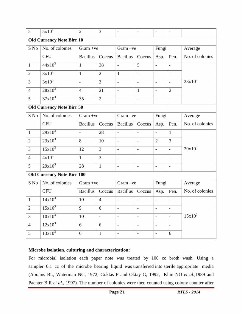
Table 2. Data acquired on old paper notes: the number of colonies on each paper
note, the types of gram positive and gram negative bacteria and the number of fungal
colonies.
Old Currency Note Birr 1
S No No. of colonies
CFU
Gram +ve Gram –ve Fungi Average
No. of colonies Bacillus Coccus Bacillus Coccus Asp. Pen.
1 8x10³
1 1 - 1 - 5
9x10³
2 6x10³ 2 2 - - 2 -
3 15x103 9 3 2 - - 1
4 7x10³ 3 1 1 - - 2
5 9x10³ 6 3 - - - -
Old Currency Note Birr 5
S No No. of colonies
(CFU)
Gram +ve Gram –ve Fungi Average
No. of colonies Bacillus Coccus Bacillus Coccus Asp. Pen.
1 21x103
12 9 - - - -
24x103
2 58x103 4 - 1 - 30 23
3 4x10³ 4 - - - - -
4 32x103 26 3 2 1 - -
Page 21 RTLS - 2014
5 5x10³ 2 3 - - - -
Old Currency Note Birr 10
S No No. of colonies
CFU
Gram +ve Gram –ve Fungi Average
No. of colonies Bacillus Coccus Bacillus Coccus Asp. Pen.
1 44x103
1 38 - 5 - -
23x103
2 3x10³ 1 2 1 - - -
3 3x10³ - 3 - - - -
4 28x103 4 21 - 1 - 2
5 37x103 35 2 - - - -
Old Currency Note Birr 50
S No No. of colonies
CFU
Gram +ve Gram –ve Fungi Average
No. of colonies Bacillus Coccus Bacillus Coccus Asp. Pen.
1 29x103
- 28 - - - 1
20x103
2 23x103 8 10 - - 2 3
3 15x103 12 3 - - - -
4 4x10³ 1 3 - - - -
5 29x103 28 1 - - - -
Old Currency Note Birr 100
S No No. of colonies
CFU
Gram +ve Gram –ve Fungi Average
No. of colonies Bacillus Coccus Bacillus Coccus Asp. Pen.
1 14x103
10 4 - - - -
15x103
2 15x103 9 6 - - - -
3 10x103 10 - - - - -
4 12x103 6 6 - - - -
5 13x103 6 1 - - - 6
Microbe isolation, culturing and characterization:
For microbial isolation each paper note was treated by 100 cc broth wash. Using a
sampler 0.1 cc of the microbe bearing liquid was transferred into sterile appropriate media
(Abrams BL, Waterman NG, 1972; Goktas P and Oktay G, 1992; Khin NO et al.,1989 and
Pachter B R et al., 1997). The number of colonies were then counted using colony counter after

Page 22 RTLS - 2014
incubation for 72 hours at 37ºC and pure colonies were isolated by using streaking and
restreaking method where pure colonies can be identified by using colony morphology, growth
patterns, colony pigmentation, etc. Once pure colonies were isolated, they were categorized into
gram positive and gram negative by using the gram-staining technique. After gram staining the
colonies were identified to genus level by using biochemical tests. Here to identify the microbes
to genus level Mac-conkey agar, blood agar, and chocolate agar were used for the bacterial
colonies. For the fungal identification, potato dextrose agar (PDA) was used and they were
identified based on their morphological features, growth pattern, spore formation and others. The
total number of cells on each paper note was calculated by using the formula which was given
below; the results were tabulated in table2 and table3.
DISCUSSION
Noting the significant occurrence of microbes on old paper notes and their negligible occurrence
on the fresh ones gave clues that their sources should be the places where the paper notes were
collected. The skin, respiratory system, digestive system and also other places where notes
are deposited or come into occasional contact such as earth, mud and so on, are the major
sources of the microbes.
Owing to their constructional substance (which is pure cotton), the
Ethiopian paper notes` cannot be suitable media for microbial growth, and multiplication. So
notes may play a major role as a vehicle and a surface area for microbial transfer. It means that
frequency and duration of presence of a specific microbe should not be expected to prevail the
others. Naturally, microbes found on paper notes come from the sources with which they come
into contact.
Since Paper notes are commonly contaminated with frequently high pathogenic or
potentially pathogenic microbes, they may cause serious diseases. As reported earlier, there
is no strong relationship between demonstrations and degrees of contamination, probably
because of the same substance (pure cotton). However, use of polymer paper notes may
decrease contamination to a large extent; it has been well shown that currency provides a surface
area for microbe establishment and functions as a strong vehicle in their transmition. The
microbial load of the currency note may vary depending on the source of the sample. For
example, the highest intestinal pathogen contamination was reported in butcheries and the
Page 23 RTLS - 2014
lowest isolates were observed on paper notes obtained from office workers. As a result, the
poorer the sanity condition, the more the contamination will be. The matter of great importance
is that not only children and foods are susceptible to microbial infections, but also
vulnerable people and patients of weakened immune system as well as healthy people are
prone to serious dangers, due to presence of different and abundant pathogens on paper notes.
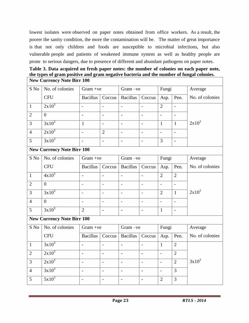
Table 3. Data acquired on fresh paper notes: the number of colonies on each paper note,
the types of gram positive and gram negative bacteria and the number of fungal colonies.
New Currency Note Birr 100
S No No. of colonies
CFU
Gram +ve Gram –ve Fungi Average
No. of colonies Bacillus Coccus Bacillus Coccus Asp. Pen.
1 2x10³
- - - - 2 -
2x10³
2 0 - - - - - -
3 3x10³ 1 - - - 1 1
4 2x10³ - 2 - - - -
5 3x10³ - - - - 3 -
New Currency Note Birr 100
S No No. of colonies
CFU
Gram +ve Gram –ve Fungi Average
No. of colonies Bacillus Coccus Bacillus Coccus Asp. Pen.
1 4x10³
- - - - 2 2
2x10³
2 0 - - - - - -
3 3x10³ - - - - 2 1
4 0 - - - - - -
5 3x10³ 2 - - - 1 -
New Currency Note Birr 100
S No No. of colonies
CFU
Gram +ve Gram –ve Fungi Average
No. of colonies Bacillus Coccus Bacillus Coccus Asp. Pen.
1 3x10³
- - - - 1 2
3x10³
2 2x10³ - - - - - 2
3 2x10³ - - - - - 2
4 3x10³ - - - - - 3
5 5x10³ - - - - 2 3
Page 24 RTLS - 2014
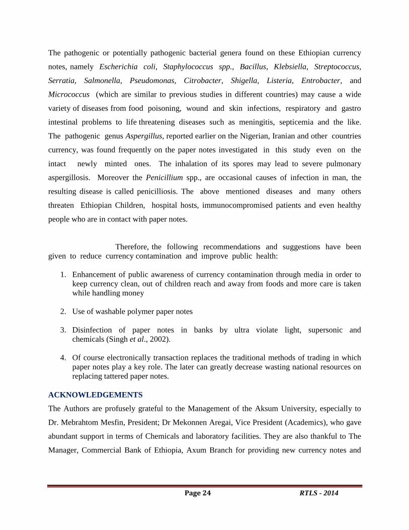
The pathogenic or potentially pathogenic bacterial genera found on these Ethiopian currency
notes, namely Escherichia coli, Staphylococcus spp., Bacillus, Klebsiella, Streptococcus,
Serratia, Salmonella, Pseudomonas, Citrobacter, Shigella, Listeria, Entrobacter, and
Micrococcus (which are similar to previous studies in different countries) may cause a wide
variety of diseases from food poisoning, wound and skin infections, respiratory and gastro
intestinal problems to life threatening diseases such as meningitis, septicemia and the like.
The pathogenic genus Aspergillus, reported earlier on the Nigerian, Iranian and other countries
currency, was found frequently on the paper notes investigated in this study even on the
intact newly minted ones. The inhalation of its spores may lead to severe pulmonary
aspergillosis. Moreover the Penicillium spp., are occasional causes of infection in man, the
resulting disease is called penicilliosis. The above mentioned diseases and many others
threaten Ethiopian Children, hospital hosts, immunocompromised patients and even healthy
people who are in contact with paper notes.
Therefore, the following recommendations and suggestions have been
given to reduce currency contamination and improve public health:
1. Enhancement of public awareness of currency contamination through media in order to
keep currency clean, out of children reach and away from foods and more care is taken
while handling money
2. Use of washable polymer paper notes
3. Disinfection of paper notes in banks by ultra violate light, supersonic and
chemicals (Singh et al., 2002).
4. Of course electronically transaction replaces the traditional methods of trading in which
paper notes play a key role. The later can greatly decrease wasting national resources on
replacing tattered paper notes.
ACKNOWLEDGEMENTS
The Authors are profusely grateful to the Management of the Aksum University, especially to
Dr. Mebrahtom Mesfin, President; Dr Mekonnen Aregai, Vice President (Academics), who gave
abundant support in terms of Chemicals and laboratory facilities. They are also thankful to The
Manager, Commercial Bank of Ethiopia, Axum Branch for providing new currency notes and
Page 25 RTLS - 2014
also to Mr. Bahalibi G/Abezgi and Mr Gebru Tekle for their technical help in the laboratory for
successful completion of this Research work.
REFERENCES
1. http:// www.en.wikipedia.org/wiki/ethiopian birr.
2. Pinner, R. W, Toutsch S. M and Simonsen, L. 1996. Trends in infectious diseases
mortality in the United States, Jama. Med. Ass. 275: 189-193.
3. Pope TM, Ender PT, Woelk WK, Koroscil MA, Koroscil, TM. 2002. Bacterial
contamination of paper currency, South Med J 2002; 95: 1408-1410.
4. Abrams BL, Waterman NG. 1972. Dirty money, JAMA. Med. Ass., 219: 1202-1203.
5. El- Dars FM and Hassan WM. 2005. Int J Environ Health Res, 15: 235- 239.
6. Goktas P and Oktay G. 1992. Bacteriological examination of paper money, Microbiol
Bull., 26: 344-348.
7. Jakir hosen M, Islam sarif D, Masuder Rahman M and Abul kalam Azad Md. 2006. Pak J
Bio. Sci., 9 (5): 868- 870.
8. Khin NO, Phyu PW, Aung MH. 1989. J Diarrh Dis Res., 7: 92-94.
9. Oyero OG and Emikpe BO. 2007. Int J Trop Med., 2: 29- 32.
10. Shekarforoush Sh, Khajeh Ali E, and Zarei M. 2009. Iran J Health & Environ., 1(2).
11. Umeh EU, Juluku JU and Ichor T. 2007. Res J Envir Sci., 1 (6) 336- 339.
12. Xu J, Moore JE and Millar B Ch. 2005. J Envir Health, 67 (7): 51-55.
13. Charnock, C. 2005. Swabbing of waiting rooms to reduce bacterial contamination, Br. J
Gen pract., 55:147-148.
14. Pachter BR, Kozer L, Pachter SA, Weiner M. Infect Med 1997; 14: 574.
15. Singh DV, Thakur K, Kalpana A Goel. 2002. Int J Med Microbiol., 20:53

Page 26 RTLS - 2014
Phytochemical analysis and antihepatitic activity of phenolic constituents of Phyllathus
niruri
B. Shanmugam 1,2
, K.R.Shanmugam1*, B, Ravi S
2G.Venkata Subbaiah
2, , K.Srinivas
1,
K.Sathyavelu Reddy 2
1. Department of Zoology, T.R.R. Government Degree College, Kandukur, A.P
2Division of Molecular Biology and Ethanopharmacology, Department of Zoology,
Sri Venkateswara University, Tirupati – 517 502, India.
Email :[email protected]
Abstract
Phyllanthus niruri is a traditional Indian medicine belonging to the Phyllanthacae family.
The aim of this study was to identify the potential compounds responsible for antihepatitic
activity of Phyllanthus niruri. The Ethyl acetate, Hexane of phyllanthus was used to know the
phytochemicals and phytochemical contents. Our phytochemical research of these extracts led to
the isolation of various bioactive constituents. The chemical structures will be determined by
spectroscopic analyses. Among phytochemicals phenols etc. The main phytochemicals are
alkaloid, terpenoids, flavonoids etc. Of them Phenol content was more in Ethyl acetate than
Hexane extract.
Key words: Phyllanthus niruri, phytochemicals, Hepatitis
Introduction:
Phytochemicals are naturally occurring in the medicinal plants, leaves, vegetables and
roots that have defense mechanism and protect from various diseases. Phytochemicals are
primary and secondary compounds. Chlorophyll, proteins and common sugars are included in
primary constituents and secondary compounds have terpenoid, alkaloids and phenolic
compounds (Krishnaiah et al., 2007). Many plant families have been reported to have ethno
medicinal application. Phyllanthus niruri is a plantwidely found in India and is popularly used
for the treatmentof renal pathologies, particularly urolithiasis. Themedicinal properties of this
plant have been associated withsome of its active components such as lignans,
glycosides,alkaloids, ellagitannins, terpenes and phenylpropanoids,besides flavonoids and
polyphenols, such as quercetin, rutin and gallic acid (GA). Although many in vitroand in vivo
antioxidant effects of P. niruri extracts havebeen shown (Bagalkotkar et al., 2006; Amin et al.,

Page 27 RTLS - 2014
2012), which seem to be determined by itspolyphenolic components (Manach et al., 2005), the
bioavailability of thosecomponents and their impact on human health need furtherinvestigation.
Phytoconstituents present in the P.niruri responsible for its pharmacological properties
includelignans, alkaloids, flavonoids, benzenoids, coumarins,tannins, diterpenes, triterpenes,
sterols, phytallates and lipids.
Considering all these facts, the present study was designedto investigate the presence of
various phytochemicals in the two different extracts of Phyllanthus niruri, a plant which evokes
various therapeuticeffects.
Materials and Methods
Solvent Extraction
The whole plant materials was air dried until all the water molecules evaporated and plants
become well dried for grinding. After draying, the plant material were grinded well using
mechanical blender into fine powder and transferred in to air tight containers with proper
labelling for future use. The Ethyl acetate, Hexane extracts was prepared by 100gms of
powdered plant material soaked in 500ml of different solvents in room temperature at 72h. The
extracts were filtered through muslin cloth and through what men filter paper (Grade 1). Extracts
are concentrated by using water both contains rotary evaporator. Total yield of plant extract
ranges from 5 -6% respectively.
Phytochemical Screening
In the present study, solvents like Ethyl acetate, Hexane are used to extract the phytochemicals
from Phyllanthus niruri by using standard protocols.

Page 28 RTLS - 2014
1. Test for Alkaloids (Wagner’s reagent)
A fraction of extract was treated with 3-5drops of Wagner’s reagent (1.27g of iodine and 2g of
potassium iodide in 100ml of water) and observed for the formation of reddish brown precipitate
(or) coloration.
2. Test for Terpenoids (Salkowski Test)
0.5 gram of each extract was added to 2 ml of chloroform. Concentrated sulphuric acid(3 ml)
was carefully added to form a layer. A reddish brown colouration of the interface indicates the
presence of terpenoids.
3. Test for Tannins
About 0.5 gram of the extract was boiled in 10 ml of water in a test tube and then filtered. A few
drops of 0.1% ferric chloride was added and observed for brownish green or a blue-black
coloration.
4. Test for Saponins (Foam test)
To 2ml of extract was added to 6ml of water in a test tube. The mixture was shaken vigorously
and observed for the formation of persistent foam that confirms the presence of saponins.
5. Test for Quinones
A small amount of extract was treated with concentrated HCL and observed for the formation of
yellow precipitate (or coloration).
6. Test for Cardiac glycosides (Keller Kelliani’s test)
5ml of each extract was treated with 2ml of glacial acetic acid in a test tube and a drop of ferric
chloride solution was added to it. This was carefully underlayed with 1ml concentrated sulphuric
acid. A brown ring at the interface indicated the presence of deoxysugar characteristic of
cardiac glycosides. A violet ring may appear below the ring while in the acetic acid layer, a
greenish ring may form.
7. Test for Phenols (Ferric chloride test)
A fraction of the extracts was treated with aqueous 5% ferric chloride and observed for
formation of deep blue or black colour.
8. Test for reducing sugars (Fehling’s test)
The aqueous ethanol extract (0.5 g in 5 ml of water) was added to boiling Fehling’s solution (A
and B) in a test tube. The solution was observed for a colour reaction.

Page 29 RTLS - 2014
9.Test for Flavonoids
To portion of the dissolved extract, a few drops of 10 % ferric chloride solution were added. A
green or blue colour indicates the presence of phenolic nucleus.
10. Test For Resins [8]
10 ml of petroleum ether extract was obtained in a test tube, the same amount of cupper acetate
solution was added and the mixture was shaken vigorously and allowed to separate, a green
colour indicates the presence of resins.
11. Sterols and Steroids (Salkowski’s Test)
One ml of extract was treated with 2 ml of chloroform and equal amount of concentrated
sulphuric acid was added, upper layer is turns to red indicates the presence of the sterols and
steroids.
Results
This study has revealed the presence of phytochemicals considered as active medicinal
chemical constituents. Important medicinal phytochemicals such as terpenoids, reducing sugar,
flavonoids, alkaloids and phlobatannins were present in the samples.
Investigations on the phytochemical screening of P. niruri Ethyl acetateextract revealed
the presence of alkaloids, terpenoids, cardiac glycosides, phenols, flavonoids, resins, steroids.
However in the Hexane extract only alkaloids resins, steroids are seen. These phytochemicals
are biologically active. (Table 1).
The presence of these phytochemicals has been attributed to the bioactive principles
responsible for ethnopharmalological activities of most medicinal plant. This dictates why efforts
have been expanded in studies aimed at elucidating their levels in medicinal plant (Edeoga et
al.,2005). The medicinal values of plants are dictated by their phytochemicals and other chemical
constituents (Fallah et al., 2005). The importance of alkaloids, saponins and tannins in various
antibiotics used in treating common pathogenic strains has recently been reported.

Page 30 RTLS - 2014
Discussion
The investigation of plants as potential sources of new drugs to treat cancer, AIDS
diabetes, parkinson’s and malaria requires the search of as many resources as possible, the
discovery of Phytochemical compounds with, for example, cytotoxic and /or anti-tumour activity
could lead to the production of new drugs for the treatment of various diseases. Therefore, the
development of appropriate extraction methods in order to obtain plant extracts with as many
phytochemical compounds as possible is important.
Table1. Phytochemical screening of P.niruri in different solvent extracts
S.No
Phytochemical
Constituents
Hexane extract
Ethyl acetate
extract
1. Phenolic
Compounds
_ +
2. Saponins _ _
3. Flavonoids _ +
4. Terpenoids _ +
5. Alkaloids + +
6. Tannins _ _
7. Cardio glycosides _ +
8. Steroids + +
9. Reducing Sugars _ _
10. Anthraquinones _ _
11. Resins + +
+Present; -Absent
The preliminary phytochemical screening tests may be useful in the detection of the
bioactive principles and subsequently may lead to the drug discovery and development. Further,
these tests facilitate their quantitative estimation and qualitative separation of (Varadarajan et al.,
2008) pharmacologically active chemical compounds.
Over the centuries, the use of medicinal herbs has become an important part of daily life
despite the progress in modern medical and pharmaceuticals research. Approximately 3000
plants species are known to have medicinal properties in India.The Rigveda (3700 B.C.),

Page 31 RTLS - 2014
mentions the use of medicinal plants. Our traditional systems of medicines, viz., Ayurveda,
Yunani, Siddha and Homeopathy etc. use herbs for treatment. It is estimated that 40% of the
world populations depends directly on plant based medicine for their health care. The present
study was under taken to identify the phytochemicals present in PN extracts with suitable
solvents such as Hexane and ethyl acetate.
The plant have traditionally provided a source of hope for novel drug compounds, as
plant herbal mixture have made large contributions to human health and wellbeing. The use of
plant extracts with known antimicrobial properties can be of great significance for therapeutic
treatment.
In the present investigation, Phytochemical screening of Phyllanthusniruri has been done
in Hexane and ethyl acetate. Table.1 represents the results of Phytochemical screening of PN. PN
contains phenolic compounds, flavonoids, terpenoids, alkaloids, cardio glycosides, steroids,
resins but in Hexane extract only alkaloids, steroids and resins.
Flavonoids also known to have a wide array of therapeutic activities as antihypertensive,
anti-rheumatism, antimicrobial, diuretic and antioxidants (Trease & Evans 2002).
The curative properties of medicinal plants are perhaps due to the presence of various
secondary metabolites such as alkaloids, flavonoids, glycosides, phenols, saponins, sterols etc.
The various extracts PN have revealed the presence of Triterpenoids, Steroids, Glycosides,
Alkaloids, Flavonoids. Alkaloids, flavonoids were found to in PN. From this analysis, PN ethyl
acetate have more constituents compared to Hexane extract. The results of preliminary
phytochemical analysis are shown in Table 1.
Alkaloid has nemerous functions and among them foremost is their analgesic,
antispasmodic and bacteriological effects. Alkaloids with medicinal properties and are used in
Page 32 RTLS - 2014
the management of cold, chronic catarrh, persistent headaches and migraine. The antibacterial
properties of tannins have been documented (Gill 1992). Alkaloids are a group of naturally
occurring chemical compounds which mostly contain basic nitrogen atoms. It has been reported
to have analgesic properties.
The Phytochemical screening and quantitative estimation of PN yields the most
promising secondary metabolites such as alkaloids, flavonoids, phenol, proteins, amino acids
tannin, and carbohydrates. They were known to show the medicinal activity as well as
physiological activity . Alkaloid, tannins, terpenoids, flavonoids and phenol are found abundant
in the ethanol samples, it should be noted that phenol components are of importance and interest
in pharmacy due to their relationship with cancer activity.
These results expose that the plant has quite a number of chemical constituents, which
may be responsible for the many pharmacological actions. Although their specific roles were not
investigated in this study, it has been reported that most active principles in plants are frequently
flavonoids, steroids, glycosides and alkaloids.
From the present study, we can deduce that, PN may serve as constituents of animal and human
diet supplying the body with minerals, proteins and energy. The presence of secondary
metabolites that are biologically important contributes to its medicinal value and thus can be
potential sources of useful drugs.
Conclusion
Results obtained in this study have considerable value with respect to Phytochemical screening
of phenols and flavonoids. These results suggest that ethyl acetate and Hexane are used for
isolation of novel bio active compounds in ethno medicinal and development of potential drugs.

Page 33 RTLS - 2014
In this study PN have a various chemical groups in their chemical composition. The importance
therapeutic and medicinal of PN extracts, it would be interesting to purify and identify different
molecules and tested it in vitro and in vivo by exploiting on animal models, to specify this
therapeutic application.
Acknowledgements
The authors are grateful to the Department of Science and Technology (DST), New Delhi,
India for the sanction of Young Scientist Fast Track Project the No. SERC/LS-387/2012.
References
Amin ZA, Abdulla MA, Ali HM, Alshawsha MA, Qadir SW.Assessment of in vitro antioxidant,
antibacterial and immuneactivation potentials of aqueous and ethanol extracts of
Phyllanthusniruri. J Sci Food Agric 2012; 92: 1874-1877.
Bagalkotkar G, Sagineedu SR, Saad MS, Stanslas J. Phytochemicalsfrom Phyllanthus niruri
Linn.and their pharmacological properties:A review. J Pharm Pharmacol 2006; 58: 1559-
1570.
Edeoga, H. O, D. E. Okwu and B. O. Mbaebie, Phytochemical Constituents of Some
NigerianMedicinal Plants African J. Biotechnol., 4(7), 685-688 (2005).
Fallah,H. S. M, H. R. Alavian, M. R. Heydari and K. Abomaali, The Efficacy of Liv-52 on
LiverCirrhotic Patients, a Randomized, Double Blind, Placebo-controlled First Approach,
Phytomedicine12(9), 619-624 (2005).
Gill L. S. Ethnobotanical Uses of Plants in Nigeria, University of Benin Press, Benin City
(1992)pp. 350.
Krishnaiah D, Sarbatly R, Bono A (2007) Phytochemical antioxidants for health and medicine: A
move towards nature. Biotechnol Mol Biol Rev 1: 97-104.
Manach C, Williamson G, Morand C, Scalbert A, Rémésy C.Bioavailability and bioefficacy of
polyphenols in humans. I. Reviewof 97 bioavailability studies. Am J Clin Nutr 2005; 81:
S230-242

Page 34 RTLS - 2014
Omoyeni O. A. and E. I. Adeyeye, Chemical Composition, Calcium, Zinc and
PhytateInterrelationships in Aerva lanata (Linn) Juss.Ex Schult Leaves, Oriental J.
Chem., 25(3) (2009).
Trease GE, Evans WE.Pharmacognosy. 15th ed, W.B. Saunders Company Limited,London.
2002;585.
Varadarajan, P. G, Rathinaswamy, and Asirvatahm D Antimicrobialproperties and
phytochemical constituents of Rheo discolor.Ethnobotanical Leaflet.2008; 12: 841–845.
Page 35 RTLS - 2014
Aluminium-induced oxidative stress in kidney tissue of rats: protective effect of vitamin E
P. Harish1†
, P. Prathima1†
, Y. Nanda Kumar1$
and S.B. Sainath1†*
†Department of Biotechnology, Vikrama Simhapuri University, Nellore- 524 003, AP, India
$Biomedical Informatics Centre, Vector Control Research Centre, Indian Council of Medical
Research, Pondicherry, 605006.
Abstract
The present study was aimed to investigate the ameliorative effects of vitamin E against
aluminium-induced nephrotoxicity in rats. Healthy rats were allocated into four groups
(n=6/group). Rats in group I served as controls and rats in groups II (aluminium exposed), III
(Vitamin E administered) and IV (aluminium + vitamin E exposed) were treated as experimental
animals. Significant increase (p<0.01) in the lipid peroxidation levels with a significant decrease
(p<0.01) in the activity levels of antioxidant enzymes such as glutathione peroxidase, glutathione
reductase, glutathione-S-transferase, superoxide dismutase and catalase were observed in the
kidney tissue of aluminium exposed rats over controls. Furthermore, glutathione content was also
significantly reduced (p<0.01) in kidney tissue of aluminium exposed rats. On the other hand,
administration of vitamin E significantly (p<0.01) reversed the changes induced by aluminium in
the kidney tissue of rats as compared to aluminium alone exposed rats. No significant changes
were observed in the selected biochemical variables in vitamin E alone treated rats over controls.
However, a significant increase (p<0.05) in the glutathione content was observed in vitamin E
treated rats over controls. From the results it can be concluded that aluminium induced
nephrotoxicity mediates oxidative stress in rats. On the other hand, vitamin E through
antioxidant property mitigates aluminium induced oxidative stress in the kidney tissue of rats.
Further, we also extended our studies using in silico analysis to understand the exact link
between vitamin E, aluminium and glutathione related enzymes. The results obtained will be
discussed.
1. Introduction
Aluminium (Al) is a common element available in the earth’s crust which constitutes
about 8%. Al occurs naturally in silicates, cryolite, and bauxite rock. It is believed that the use of
aluminium and its compounds increased worldwide because of their wide applications in a range
of activities including building, canning, automobile, tanning, paper and glassware industries.
Moreover, it is also used in medicines such as antacids and also cosmetics such as perfumes and
also as food additives. Recently, International Agency for Research on Cancer classified Al as a
carcinogenic to humans. Human studies indicated that occupational exposure to Al causes
neurological disorders, pulmonary diseases, liver disorders, bladder cancer and fertility
Page 36 RTLS - 2014
problems. Previously it has been shown that Al could be an etiologic factor for amyotrophic
lateral sclerosis, Alzheimer’s and Parkinson’s disease (Krewski et al., 2009). Although a range of
toxicological studies indicated the adverse effects of Al, studies related to the Al-induced
nephro-toxicity are little exploited (Krewski et al., 2007). Kidneys play a major role in the
elimination of Al (approximately 95%) from the body (Sargazi et al., 2006). Studies of Roy et al.
(1991) reported that Al exposure causes renal tissue degeneration including deterioration of renal
cortex tubules and recently, studies of Mahieu et al. (2003) indicated that Al affects renal
metabolism and alters tubular transport mechanisms. Many studies also indicated that Al induced
toxicity mediates oxidative stress by altering pro- and anti-oxidant status in mammalian
experimental models (Krewski et al., 2007). Recently, studies of Kutlubay et al. (2007) indicated
that Al accumulates in kidneys and provokes generation of reactive oxygen species thereby
affects vital components of cell such as lipids, proteins and nucleic acids thereby negatively
targets structural and functional integrity of kidneys. However, the exact mechanism(s) of action
of Al-induced nephrotoxicity in altering pro- and antioxidant status are not well defined.
Vitamin E is a classical antioxidant that plays an important role in counterattacking lipid
peroxidative products including reactive oxygen species (Traber et al., 2007). Because of its
potential antioxidant property many studies evaluated the protective effects of vitamin E against
a range of free radical induced pollutants (Jalili et al., 2007; Al-Attar et al., 2011).
Considering the facts that 1) studies related to the mechanism(s) of action of Al-induced
nephrotoxicity are little exploited 2) Al generates reactive oxygen species in biological tissues
including kidneys and 3) vitamin E is a classical antioxidant that is widely used against oxidative
stress induced pollutants, the present study was aimed to investigate the protective effects of
vitamin E against Al-induced oxidative stress in kidneys of rats.
2. Materials and methods
2.1. Procurement and maintenance of experimental animals
Male healthy rats of Wistar strain purchased from Indian Institute of Sciences, Bengaluru
India were used in the present study. Rats were housed in polypropylene cages (18" x 10" x 8")
lined with sterilized paddy husk, and provided filtered tap water ad libitum and standard rat chow
(purchased from Sai Durga Agencies, Bengaluru, India). All the rats were maintained in a well
regulated environment (temperature 25 ± 2°C; 12-hour light and 12-hour dark cycle, humidity 50
± 10%) in the animal house facility at Watson Life Sciences, Tirupati.

Page 37 RTLS - 2014
2.2. Chemicals
Aluminium chloride and Vitamin E were selected as test chemicals. They were purchased
from Sigma Chemical Company, St Louis, MO. Chemicals such as glutathione oxidized and
NADPH were also purchased from Sigma Chemical Company, St Louis, MO. All other
chemicals used in the present study with highest purity were obtained from local commercial
sources.
2.3. Experimental design
Twenty four healthy rats with a body weight of 110 ± 10 g were selected for the present
study. The animals were randomly allocated into four groups of six rats each. Animals in group 1
served as controls and animals in groups 2, 3 and 4 were treated as experimental groups. The
experimental period was 30 days. The selection of doses of test chemicals was based on previous
studies.
Group 1: unexposed rats
Group 2: rats exposed to Al (orally through gavage) alone at a concentration of 150 mg/Kg/day
Group 3: rats exposed to vitamin E (I/P) alone at a concentration of 15 mg/Kg/day
Group 4: rats received both Al+vitamin E at same doses
The body weights of rats were recorded before and after completion of experimental
period. After completion of the experimental period, all the rats were euthanized by cervical
dislocation. Kidney was isolated immediately, weighed to the nearest milligram using
Schimadzu electronic balance and after weighing, the tissues from all the control and
experimental groups were carefully stored at -80⁰C until further use.
2.4. Biochemical assays
Kidneys homogenates were obtained by using tissue homogenizer. Kidney homogenates
(10% w/v) were prepared using a homogenizing buffer (100 mM KCl containing 0.3 mM of
EDTA; pH: 7). All the homogenates were centrifuged at 1500 rpm for 30 min at 4⁰C to separate
cell debris. Further, supernatant was centrifuged at 16000 rpm for 1 hr at 4⁰C. The supernatant
was used for biochemical analysis. The biochemical parameters selected and performed
according to standard protocols (1) Thiobarbituric acid reactive substances (TBARS) (lipid
peroxides) were estimated according to method of Okawa et al. (1979). (2) Reduced glutathione
was estimated by the method of Sedlak and Lindsay (1968). (3) The activity of superoxide
dismutase (SOD) was assayed by the method of Misra and Fridovich (1972). (4) The activity of

Page 38 RTLS - 2014
glutathione peroxidase (GPx) was assayed according to the method of Rotruck et al. (1973). (5)
Catalase (CAT) activity was assayed by the method of Takahara et al. (1960). (6) The activity of
glutathione-S-transferase (GST) was measured by using the method of Habig et al. (1974). (7)
Glutathione reductase (GR) activity was assayed as described by the method of Dulber and
Anderson (1981). The protein concentrations were determined using Lowry et al. (1951).
2.5. In silico analysis
Gamma-glutamyl cysteine ligase (gGCL) plays an important role in glutathione
metabolism. Therefore, it was selected as the test protein. The protein sequence of GGCL from
Rat was retrieved in fasta format from NCBI database (http://www.ncbi.nlm.nih.gov/) with the
AC No: NP_036947.1 and it was subjected to BLAST similarity search against PDB data base to
find out the possible template. The template with maximum identity was chosen and its sequence
and structure were obtained. Sequence alignment was carried out between the GGCL and
template sequences in PIR format, incorporated with python script and successfully run through
modeller to construct the homology model of GGCL. The generated model was evaluated with
Ramachandran plot by submitting to Procheck and ProsaWeb analysis server. The residues that
are observed in the disallowed regions were subjected to loop modelling through ModLoop
server (https://modbase.compbio.ucsf.edu/modloop/). The gGCL model obtained from the
homology modelling process was loaded into MOE workspace and the missing hydrogen atoms
were added. The structure is subjected to protonation at a temperature of 300K, pH of 7 and 0.1
mol/L in the generalized born implicit solvent. The electrostatic potential terms were applied
with a distance cut off of 15Å and dielectric constant of 1. Further the protonated structure was
energy minimised in the MMFF94x force field to an RMS gradient cut off of 0.05. The final
optimized conformation of the protein was saved and used for further studies.
The site finder module of MOE was used to predict the potential possible binding sites on
the gGCL. The probing was done with hydrophobic and hydrophilic terms by means of alpha
spheres to find out the tight atom packing centres. The potential binding sites were defined by
these alpha spheres and their dummy atoms were defined to create the centroid of the sphere.
From the list of possible binding sites predicted, the site covering the maximum residues among
all was considered to perform the docking simulations.

Page 39 RTLS - 2014
2.6. Molecular Docking
Molecular docking simulations were carried out with GSH, Vit-E and Al into the above
mentioned binding site to know their binding mode and affinity ranges for gGCL using MOE
docking module. The optimized conformation of the gGCL was loaded into MOE workspace and
the binding site was defined. Triangle matcher docking placement methodology was used by
aligning ligand triplets of atoms on triplets of alpha spheres of binding site and a population of
possible docking conformations were generated. These docked conformations were ranked by
London DG scoring function where the free binding energies of the ligands from a given pose
were calculated. Multiple docking conformations were generated for each ligand and among
them, the conformation with lowest docking score was considered for the analysis of molecular
interactions and docking energies.
3. Results
No significant changes were observed in body weights of rats in controls and
experimental groups (data not shown). No clinical signs of toxicity were also observed in any of
the groups. None of the rats were excluded from the present study.
Significant increase in the lipid peroxidation (LPx) levels were observed in Al exposed
rats over controls. No significant changes were observed in LPx levels in vitamin E exposed rats
over controls. However, a significant decrease was observed in the LPx levels in Al+vitamin E
exposed rats over Al alone exposed rats (Table 1). On the other hand, significant decrease
(p<0.01) in the activity levels of antioxidant enzymes such as glutathione peroxidase, glutathione
reductase, glutathione-S-transferase, superoxide dismutase and catalase were observed in the
kidney tissue of aluminium exposed rats over controls. Furthermore, glutathione content was also
significantly reduced (p<0.01) in kidney tissue of Al exposed rats. On the other hand,
administration of vitamin E significantly (p<0.01) reversed the changes induced by Al in the
kidney tissue of rats as compared to Al alone exposed rats. Interestingly, a significant increase in
the glutathione content was observed in vitamin E treated rats over controls.
Further, in silico analysis were performed to understand the role of vitamin E in
accelerating glutathione system. Homology model of the gamma-glutamyl cysteine ligase
catalytic subunit (GGCL) was constructed using Modeller tool and its stereo chemical quality
was evaluated by Ramachandran plot and ProsaWeb analysis. The best quality model was
Energy minimized in the MOE working environment and a stable conformation was attained.
Page 40 RTLS - 2014
The potential binding site of GGCL was predicted using MOE site finder and docking
simulations were carried using MOE-Dock module using GSH, Vitamin E and Al as ligands
against the GGCL to find their possible mode of binding orientations and affinity ranges. The
docking scores were observed as -11.193, -9.830 and -3.017 Kcal/mol for GSH, Vitamin E and
Al respectively indicating that GSH has more affinity among and Al did not showed any
molecular interactions with the GGCL but has a less affinity of binding energy (Figure 1 and
Table 2).
4. Discussion
The results of the present study indicate that 1) Al negatively targets antioxidant enzymes
thereby leads to imbalance in the pro- and anti-oxidant status as evidenced by significant
increase in the LPx levels in kidney tissue of rats 2) vitamin E administration ameliorates the
activity levels of antioxidant enzymes thereby reduces the LPx levels in the kidney tissue of Al
exposed rats and finally 3) Al induced changes in the glutathione system at least in part does not
mediate gGCL and surprisingly, vitamin E accelerated glutathione system might target gGCL in
rats as evidenced by docking studies.
It is well known that a balance between pro- and antioxidant system is crucial in the
regulation of physiological functions. Antioxidant enzymes and antioxidants such as glutathione
continuously monitor and regulate the levels of free radicals thereby protect cells and cell
organelles. In general antioxidant enzymes can be broadly categorized into primary and
secondary enzymes. The antioxidant superoxide dismutase (SOD) converts the superoxide
anions (O2−) into hydrogen peroxide (H2O2), which can easily diffuse through the cell
membranes into the cytoplasm where the antioxidants catalase and glutathione peroxidase reduce
the hydrogen peroxide and thereby detoxify ROS. Secondary enzymes in antioxidant defense
include those of glutathione metabolism. Glutathione-S-transferase (GST) catalyzes the
conjugation of reduced glutathione (GSH) and cellular components damaged by ROS attack, to
nucleophilic xenobiotics, which leads to their detoxification. NADPH-dependent glutathione
reductase (GR) replenishes the GSH substrate for glutathione peroxidase (GPx) and glutathione-
S-transferase (GST) from oxidized glutathione (GSSG).
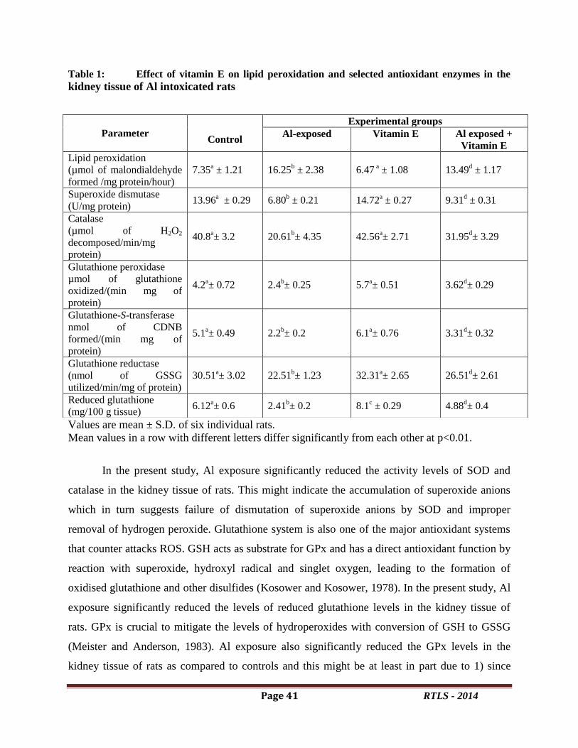
Page 41 RTLS - 2014
Table 1: Effect of vitamin E on lipid peroxidation and selected antioxidant enzymes in the
kidney tissue of Al intoxicated rats
Values are mean ± S.D. of six individual rats.
Mean values in a row with different letters differ significantly from each other at p<0.01.
In the present study, Al exposure significantly reduced the activity levels of SOD and
catalase in the kidney tissue of rats. This might indicate the accumulation of superoxide anions
which in turn suggests failure of dismutation of superoxide anions by SOD and improper
removal of hydrogen peroxide. Glutathione system is also one of the major antioxidant systems
that counter attacks ROS. GSH acts as substrate for GPx and has a direct antioxidant function by
reaction with superoxide, hydroxyl radical and singlet oxygen, leading to the formation of
oxidised glutathione and other disulfides (Kosower and Kosower, 1978). In the present study, Al
exposure significantly reduced the levels of reduced glutathione levels in the kidney tissue of
rats. GPx is crucial to mitigate the levels of hydroperoxides with conversion of GSH to GSSG
(Meister and Anderson, 1983). Al exposure also significantly reduced the GPx levels in the
kidney tissue of rats as compared to controls and this might be at least in part due to 1) since
Parameter
Control
Experimental groups
Al-exposed Vitamin E Al exposed +
Vitamin E
Lipid peroxidation
(µmol of malondialdehyde
formed /mg protein/hour)
7.35a ± 1.21 16.25
b ± 2.38 6.47
a ± 1.08 13.49
d ± 1.17
Superoxide dismutase
(U/mg protein) 13.96
a ± 0.29 6.80
b ± 0.21 14.72
a ± 0.27 9.31
d ± 0.31
Catalase
(µmol of H2O2
decomposed/min/mg
protein)
40.8a± 3.2 20.61
b± 4.35 42.56
a± 2.71 31.95
d± 3.29
Glutathione peroxidase
µmol of glutathione
oxidized/(min mg of
protein)
4.2a± 0.72 2.4
b± 0.25 5.7
a± 0.51 3.62
d± 0.29
Glutathione-S-transferase
nmol of CDNB
formed/(min mg of
protein)
5.1a± 0.49 2.2
b± 0.2 6.1
a± 0.76 3.31
d± 0.32
Glutathione reductase
(nmol of GSSG
utilized/min/mg of protein)
30.51a± 3.02 22.51
b± 1.23 32.31
a± 2.65 26.51
d± 2.61
Reduced glutathione
(mg/100 g tissue) 6.12
a± 0.6 2.41
b± 0.2 8.1
c ± 0.29 4.88
d± 0.4
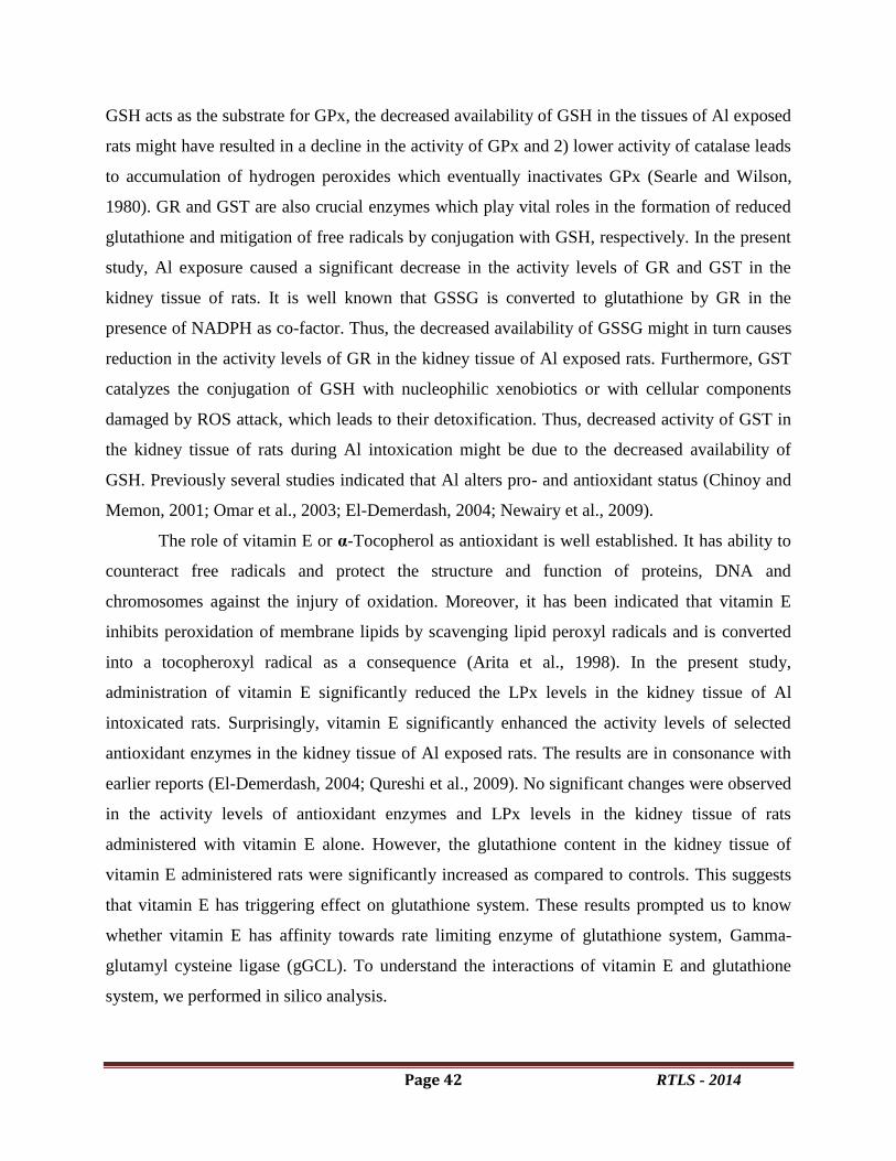
Page 42 RTLS - 2014
GSH acts as the substrate for GPx, the decreased availability of GSH in the tissues of Al exposed
rats might have resulted in a decline in the activity of GPx and 2) lower activity of catalase leads
to accumulation of hydrogen peroxides which eventually inactivates GPx (Searle and Wilson,
1980). GR and GST are also crucial enzymes which play vital roles in the formation of reduced
glutathione and mitigation of free radicals by conjugation with GSH, respectively. In the present
study, Al exposure caused a significant decrease in the activity levels of GR and GST in the
kidney tissue of rats. It is well known that GSSG is converted to glutathione by GR in the
presence of NADPH as co-factor. Thus, the decreased availability of GSSG might in turn causes
reduction in the activity levels of GR in the kidney tissue of Al exposed rats. Furthermore, GST
catalyzes the conjugation of GSH with nucleophilic xenobiotics or with cellular components
damaged by ROS attack, which leads to their detoxification. Thus, decreased activity of GST in
the kidney tissue of rats during Al intoxication might be due to the decreased availability of
GSH. Previously several studies indicated that Al alters pro- and antioxidant status (Chinoy and
Memon, 2001; Omar et al., 2003; El-Demerdash, 2004; Newairy et al., 2009).
The role of vitamin E or α-Tocopherol as antioxidant is well established. It has ability to
counteract free radicals and protect the structure and function of proteins, DNA and
chromosomes against the injury of oxidation. Moreover, it has been indicated that vitamin E
inhibits peroxidation of membrane lipids by scavenging lipid peroxyl radicals and is converted
into a tocopheroxyl radical as a consequence (Arita et al., 1998). In the present study,
administration of vitamin E significantly reduced the LPx levels in the kidney tissue of Al
intoxicated rats. Surprisingly, vitamin E significantly enhanced the activity levels of selected
antioxidant enzymes in the kidney tissue of Al exposed rats. The results are in consonance with
earlier reports (El-Demerdash, 2004; Qureshi et al., 2009). No significant changes were observed
in the activity levels of antioxidant enzymes and LPx levels in the kidney tissue of rats
administered with vitamin E alone. However, the glutathione content in the kidney tissue of
vitamin E administered rats were significantly increased as compared to controls. This suggests
that vitamin E has triggering effect on glutathione system. These results prompted us to know
whether vitamin E has affinity towards rate limiting enzyme of glutathione system, Gamma-
glutamyl cysteine ligase (gGCL). To understand the interactions of vitamin E and glutathione
system, we performed in silico analysis.
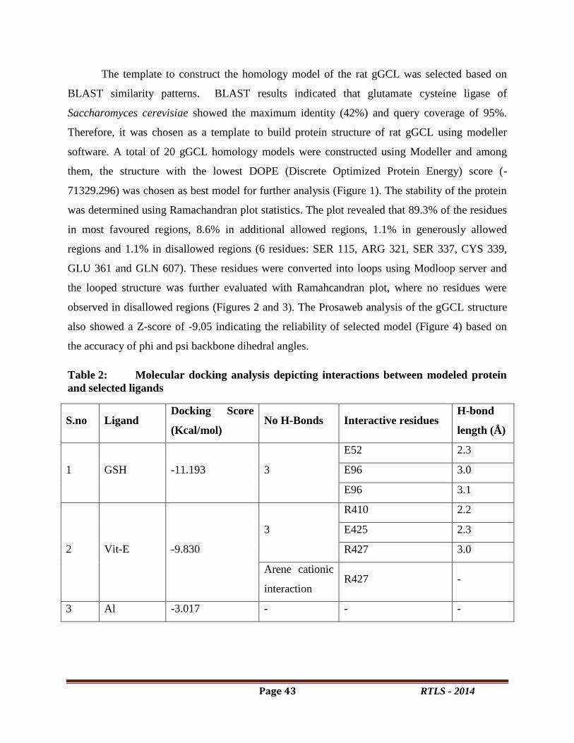
Page 43 RTLS - 2014
The template to construct the homology model of the rat gGCL was selected based on
BLAST similarity patterns. BLAST results indicated that glutamate cysteine ligase of
Saccharomyces cerevisiae showed the maximum identity (42%) and query coverage of 95%.
Therefore, it was chosen as a template to build protein structure of rat gGCL using modeller
software. A total of 20 gGCL homology models were constructed using Modeller and among
them, the structure with the lowest DOPE (Discrete Optimized Protein Energy) score (-
71329.296) was chosen as best model for further analysis (Figure 1). The stability of the protein
was determined using Ramachandran plot statistics. The plot revealed that 89.3% of the residues
in most favoured regions, 8.6% in additional allowed regions, 1.1% in generously allowed
regions and 1.1% in disallowed regions (6 residues: SER 115, ARG 321, SER 337, CYS 339,
GLU 361 and GLN 607). These residues were converted into loops using Modloop server and
the looped structure was further evaluated with Ramahcandran plot, where no residues were
observed in disallowed regions (Figures 2 and 3). The Prosaweb analysis of the gGCL structure
also showed a Z-score of -9.05 indicating the reliability of selected model (Figure 4) based on
the accuracy of phi and psi backbone dihedral angles.
Table 2: Molecular docking analysis depicting interactions between modeled protein
and selected ligands
S.no Ligand Docking Score
(Kcal/mol) No H-Bonds Interactive residues
H-bond
length (Å)
1 GSH -11.193 3
E52 2.3
E96 3.0
E96 3.1
2 Vit-E -9.830
3
R410 2.2
E425 2.3
R427 3.0
Arene cationic
interaction R427 -
3 Al -3.017 - - -

Page 44 RTLS - 2014
Figure 1: Homology model of gGCL constructed using Modeller.
Figure 2: Ramachandran plot showing the stereochemical quality of the model where no
residues were observed in the disallowed region.
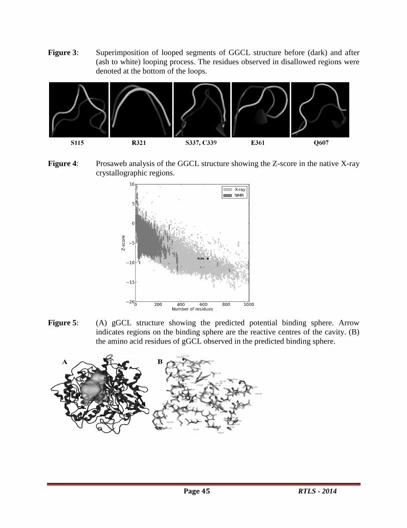
Page 45 RTLS - 2014
Figure 3: Superimposition of looped segments of GGCL structure before (dark) and after
(ash to white) looping process. The residues observed in disallowed regions were
denoted at the bottom of the loops.
Figure 4: Prosaweb analysis of the GGCL structure showing the Z-score in the native X-ray
crystallographic regions.
Figure 5: (A) gGCL structure showing the predicted potential binding sphere. Arrow
indicates regions on the binding sphere are the reactive centres of the cavity. (B)
the amino acid residues of gGCL observed in the predicted binding sphere.

Page 46 RTLS - 2014
Figure 6: Molecular docking of (A) GSH (B) Vit-E and (C) Al in the binding sphere of
gGCL. Hydrophobic, polar and solvent exposed binding regions of gGCL
involved in ligand affinity.
The final structure is energy minimised and refined in the MOE workspace and
proceeded for the next important task i.e the prediction of possible potential binding sites in
gGCL model. This is the critical task which forms the basis to predict and analyse the
interaction properties of the GGCL with small molecules. The hydrophobicity and
hydrophillicity are the main parameters along with the electrostatic potentials of the structure
that drives its molecular interactions. Small molecule ligands contain their own
hydrophilic/hydrophobic, electrostatic and steric properties and the same make their interactions
more specific in a defined binding sphere. By virtue of these qualities a potential binding sphere
was identified in the gGCL model where a highly accurate and specific interactions are expected
(Figure 5).
The molecular docking simulations of GSH, Vitamin E and Al in this binding site
encouraging the accuracy in the prediction of binding sphere where the ligands were observed to
be sit in the cavity in perfect orientations. The molecular interactions of the ligands are more
defined and specific with binding sphere. GSH is interacting with the E52 and E96 residues

Page 47 RTLS - 2014
forming three hydrogen bonds. The –COOH group present at one end of GSH is interacting with
the –COOH group of glutamate residue at 52nd
position. Another glutamate residue at 96th
position is also interacting in the same manner with its –COOH group but forming two hydrogen
bond interactions with two –NH groups of GSH. Whereas Vitamin E is interacting with gGCL
binding by means of R410, R427 and E425 residues forming three hydrogen bonds one with
each. Here the interaction is confined to a specific region of the Vitamin E molecule where the
ring structure is playing major role in forming the interactions. The –OH group of the aromatic
ring alone is interacting with all the three residues of the binding site and one more interesting
point is this aromatic ring is also forming one arene cationic interaction with R427 residue. All
these interaction make them to orient in the cavity in a specific way. Here Al is not forming any
interactions with the binding site but showing a sort of affinity for gGCL structure which is
defined by its docking energy i.e. -3.017 Kcal/mol. Whereas GSH and Vitamin E are showing
the docking energies of -11.193 and -9.830 Kcal/mol respectively indicating that GSH is
showing more affinity than Vit-E for GGCL (Figure 6 and Table 2).
5. Conclusion
The present study demonstrated that vitamin E administration in combination with
aluminium minimized its hazards. In addition, vitamin E proved to be beneficial in decreasing
the levels of free radicals and triggering the activity levels of SOD, CAT and glutathione system.
Consequently, exposure to aluminium should be minimized and caution should be paid towards
the sources of aluminium in foods, water and personal-care products. Furthermore, using diets
rich in vitamin E could be beneficial in mitigating aluminium toxicity.
Acknowledgement
The authors thank the Head, Department of Biotechnology, VSU, Nellore, AP, India for
extending facilities available at the department. Some of the results presented here was a part of
M.Sc thesis of P. Harish, IV semester, Dept. of Biotechnology, VSU, Nellore-524 001.
References
Al-Attar, A. M., 2011. Antioxidant effect of vitamin E treatment on some heavy metals-induced
renal and testicular injuries in
male mice. Saudi J Biol Sci.18, 63–72.
Arita, M., Sato, Y., Arai, H., Inoue, K., 1998.Binding of alphatocopherylquinone, an oxidized
form of alpha-tocopherol, to glutathione S-transferase in the liver cytosol. FEBS Lett
436:424-426.

Page 48 RTLS - 2014
Chinoy, N.J., Memon M.R., 2001.Beneficial effects of some vitamins and calcium on fluoride
and aluminium toxicity on gastrocnemius muscle and liver of male mice. Fluoride 34:21-
33.
Dulber, R.E., Anderson, B.M., 1981. Simultaneous inactivation of the catalytic activities of yeast
glutathione reductase by N-alkyl maleimides.Biochim.Biophys.Acta. 659, 70–85.
El-Demerdash. 2004. Antioxidant effect of vitamin E and selenium on lipid peroxidation,
enzyme activities and biochemical parameters in rats exposed to aluminium. J Trace
Elements Med Biol. 18, 113-121.
Habig, W.H., Pabst, M.J., Jakoby, W.B., 1974. Glutathione S-transferase.The first enzymatic
step in mercapturic acid formation.J.Biol.Chem. 249, 7130–7139.
Jalili, Sh., Ilkhanipour, M., Heydari, R., et al. 2007. The effects of vitamin E on endosulfan
induced oxidative stress in rat heart.
Pak J Nutr.6, 375–380.
Kosower, N.S., Kosower, E.M., 1978.The glutathione status of cells. Int. Rev. Cytol. 54, 109–
157.
Krewski, D., Yokel, R.A., Nieboer, E., et al. 2007. Human health risk assessment for aluminium,
aluminium oxide, and aluminium hydroxide.J Toxicol Environ Health B Crit Rev.1, 1–
269.
Kutlubay, R., Oguz, E.O., Guven, C., et al. 2007. Histological and ultrastructural evidence for
protective effects on aluminum-induced kidney damage by intraperitoneal administration
of alpha-tocopherol.Int J Toxicol.26, 95–101.
Lowry, O.H., Rosebrough, N.J., Farr, A.L., Randall, R.J. 1951.Protein measurement with the
Folin Phenol Reagent. J Biol Chem. 193:269-275.
Mahieu, S.T., Gionotti, M., Millen, N., Elías, M.M.,2003. Effect of chronic accumulation of
aluminum on renal function, cortical renal oxidative stress and cortical renal organic
anion transport in rats.Arch Toxicol. 77, 605-612.
Meister, A., Anderson, M.E., 1983.Glutathione.Annu. Rev. Biochem. 52, 711–760.
Misra, H.P., Fridovich, I., 1972.The role of superoxide anion in the autoxidation of epinephrine
and a simple assay for superoxide dismutase.J.Biol.Chem. 247, 3170–3175.
Newairy, A-S a, Salama, A.F., Hussien, H.M., Yousef, M.I., 2009. Propolis alleviates aluminium
induced lipid peroxidation and biochemical parameters in male rats. Food Chem
Toxicol.47, 1093- 1098.
Okawa, H., Ohishi, N., Yagi, K., 1979. Assay of lipidperoxides in animalvtissues by
thiobarbituric acid reaction. Ann. Biochem., 351–358.
Omar, H.M., Hassan, K.A., Abd-Elghaffar, S.Kh., Ahmed, E.A., 2003. Aluminium toxicity in
rats: the role of tannic acid as antioxidant. Ass. Univ. Bull. Environ. Res. 6, 1-14.
Quershi, F., Mohammed, T., Waqas, S., 2009. Protective role of vitamin c and e against sodium
arsenate induced changes in developing kidney of albino mice. J Ayub Med Coll
Abbottabad 21, 63-69.Rotruck, J.T., Pope, A.L., Ganther, H.E., Swanson, A.B.,
Page 49 RTLS - 2014
Hafeman, D.G., Hoekstra, W.G., 1973. Selenium: biochemical roles as a component of
glutathione peroxidase. Science 197, 588–590.
Roy A.K., Talukder, G., Sharma, A., 1991.Similar effects in vivo of two aluminum salts on the
liver, kidney, bone, and brain of Rattus norvegicus. Bull. Environ. Contam.Toxicol. 47,
288-295.
Sargazi, M., Shenkin, A., Roberts, N.B., 2006. Aluminium-induced injury to kidney proximal
tubular cells: Effects on markers of oxidative damage. J Trace Elem Med Biol. 19, 267-
273.
Searle, A.J., Wilson, R.L., 1980. Glutathione peroxidase: effect of superoxide, hydroxyl and
bromine free radicals on enzymatic activity. Int.J. Rad. Biol. 34, 125–129.
Sedlak, J., Lindsay, R.H., 1968. Estimation of total, protein-bound, and non protein sulfhydryl
groups in tissue with Ellman’s reagent. Anal.Biochem. 1, 192–205.
Takahara, S., Hamilton, B.H., Nell, J.V., Kobra, T.Y., Ogura, Y., Nishimura, E.T.,
1960.Hypocatalasemia, a new genetic carrier state. J. Clin. Invest. 29, 610–619.
Traber, M.G., Jeffrey, A.J., 2007. Vitamin E, antioxidant and nothing more.Free Radic Biol
Med.43, 4–15.

Page 50 RTLS - 2014
Deterioration of physicochemical and antioxidant properties of Malaysian Tualang honeys
on long term storage
R. Krishna Kishorea, c
*, S. Abdul Nasara, Manisha Mahender Kumar
a, Chin Keong Lim,
a, b, and
Ahmad Sukari Halimc
a Department of Biochemistry, CMR Institute of Management Studies, Kalyana Nagar,
Bangalore. 560043. Karnataka. INDIA. bCentre of Studies for Preclinical Sciences, Faculty of Dentistry, Universiti Teknologi MARA,
40450 Shah Alam, Selangor, Malaysia. c Reconstructive Sciences Unit, School of Medical Sciences, Universiti Sains Malaysia, Health
Campus, Kubang Kerian-16150. Kelantan, Malaysia..
*Corresponding Author:
Dr R.Krishna Kishore,
Assistant Professor,
PG Department of Biochemistry,
CMR Institute of Management Studies,
No 5, Bhuvanagiri, OMBR Layout,
Banaswadi, Bangalore. Karnataka.
Ph: +919480607761
Abstract Honey is a rich source of natural nutrients. Its production is a slow, natural process, the pace of
which varies with seasonally. However, based on recent reports, we hypothesize that the long-
term storage of processed honey, even under the most appropriate storage conditions, results in a
deterioration of its quality. To test our hypothesis, we collected Tualang honey samples
harvested during the years 2005, 2008, 2009 and 2010 and tested various parameters including
physicochemical properties and also performed comparative analyses of antioxidant capacities to
assess its medicinal values. Our results indicate that, upon long-term storage, the quality of
honey samples deteriorates, as observed in our TH 2008 and TH 2005 year honey samples,
which showed unacceptable quality based on the recommended criteria of free acidity
(71.34±1.31 meq/kg), moisture (27.72%), diastase activity (3.38±0.34 Goth scale) and HMF
(449.89±3.23 mg/kg) by Codex and European Commission Regulation. A significant (p<0.05)
decrease in antioxidant properties were also observed. In the present study, we show that, even
after appropriate processing, most of the quality parameters of honey decrease, which suggest
that these parameters could otherwise be used as markers to assess the age of the honey.
Keywords: Honey, Physicochemical properties, HMF, Antioxidant capacity
1. Introduction
Oxidative stress occurs in a system when free radicals and reactive oxygen species (ROS)
overwhelm its endogenous and exogenous antioxidant defenses beyond the ability of the system
to neutralize and eliminate them(Ortial et al., 2006). However, increases in exogenous
antioxidants are believed to alleviate any oxidative damage and help balance cellular free

Page 51 RTLS - 2014
radicals and ROS. Honey has been studied extensively because of its physicochemical,
antimicrobial, antioxidant, antiviral, antitumor and anti-inflammatory properties (Viuda-Martos
et al., 2008; Yolanda et al., 2011). Recent investigations of several natural compounds have
revealed that honey is one of the most nutritious products that exists in the form of a traditional
remedy containing monosaccharides, amino acids, proteins (including enzymes), organic acids,
vitamins, minerals, various phytochemicals and many biologically active entities (Ahmed et al.,
2007).
Honey is obtained from a variety of sources all across the world, and each variety varies
in its composition and hence, in its beneficial properties. Recently, we performed comparative
analyses using honey collected from various sources and have successfully shown that Malaysian
tualang honey is among the best honey available based on its high antioxidant and free radical-
scavenging ability (Kishore et al., 2011). However, in addition to the source, the
physicochemical and the antioxidant capacities of honey samples are highly dependent on the
storage conditions during climatic changes (Bath & Singh, 2000). Therefore, a lack of
knowledge about proper storage conditions may ultimately reduce the physicochemical
properties of honey and subsequently reduce the medicinal properties and the quality of this
foodproduct. Apparently, although Malaysian tualang honey is among the best honey, the
extreme temperatures in Malaysia (temperatures of up to 40°C) may have drastic effects on the
quality of honey during storage prior to consumption.
To the best of our knowledge, there has only been one study on the effect of storage on
the physicochemical and antioxidant properties of honey samples (Nombre et al., 2010). The
current study specifically addresses the effect of long-term storage on processed honey by
evaluating medicinal properties of Malaysian tualang honey that was harvested over several
years (collected during 2005, 2008, 2009 and 2010). The findings of the present study will help
to provide appropriate guidelines for processing and storage of Malaysian tualang honey and for
identifying markers to assess the age of honey.
2. Methods and materials
2.1 Chemicals and instruments
All chemicals, reagents and solvents used were of analytical grade and were purchased
from Merck (Germany). Standards used in the antioxidant assays were L-ascorbic acid, gallic

Page 52 RTLS - 2014
acid, quercetin, 2,2-diphenyl-1-picrylhydrazyl (DPPH) and Folin-Ciocalteu’s phenol reagent
(Sigma; St. Louis, MO, USA).
2.2 Honey samples
Malaysian Tualang honey samples harvested from the nectar of Apis dorsata bee’s nectar
during 2005, 2008, 2009 and 2010 from Tualang trees in the rain forest of Kedah (peninsular
Malaysia) were included in this study. The honey was intitially filtered to remove solid particles
by Federal Agricultural Marketing Authority (FAMA) of Malaysia and was subjected to gamma
irradiation at 25 kGy at Sterilgamma (M) Sdn. Bhd. prior to being submitted to us for analyses.
2.3. Physicochemical analyses
Moisture, color, pH, free acidity, electrical conductivity, ash content, reducing sugars and
sucrose content were analyzed in all honey samples using respective AOAC (Association of
Analytical Communities) methods(AOAC, 2000).
2.3.1.Diastase activity
Diastase activity, which is a measure of the hydrolytic activity of the enzymes in honey,
was determined using the Horwitz et al., method (1980).
2.3.2. Hydroxymethylfurfural
HPLC method
The HPLC method used was a modification of the basic method published by the International
Honey Commission in 1999, as described by Khalil et al., (2010). The hydroxymethylfurfural
(HMF) content of a sample was calculated by comparing the corresponding peak areas of the
sample with those of the standard solutions of HMF (Sigma–Aldrich, USA) after correcting for
the honey dilution. There was a linear relationship (R2 = 0.9998; Fig. 1) between the
concentration and the area of the HMF peak, and the results are expressed in mg/kg.
2.4. Total phenolic and biochemical analyses
2.4.1. Determination of total phenolic content
The total phenolic content of honey samples was determined using the Folin-Ciocalteau
method (Singleton et al., 1999). The total phenolic content is expressed in mg of gallic acid
equivalents (GAE)/100 g of honey.
2.4.2. Determination of total flavonoid content
Page 53 RTLS - 2014
The total flavonoid content of the honey samples was determined using the method
described by Woisky and Salatino (1998). Total flavonoids were calculated as mg quercetin
equivalents (QE)/100 g of honey.
2.4.3. Evaluation of total antioxidant capacity using the phosphomolybdenum method
The total antioxidant activity of tualang honey samples was evaluated using the green
phosphomolybdenum complex method (Prieto et al., 1999). The total antioxidant capacity was
calculated as ascorbic acid equivalents (mg AAE/ g honey).
2.4.4. Determination of ascorbic acid content
The standard 2, 4-dinitrophenylhidrazine method was used to determine the ascorbic acid
content of the honey samples (Omaye et al., 1979).
2.4.5. Determination of protein content
The protein content of honey samples was measured using the Bradford assay (Bradford,
1976).
2.5 Analyses of antioxidant activities
2.5.1. Free Radical scavenging activity of DPPH
The DPPH (2, 2-diphenyl-1-picrylhydrazyl) radical-scavenging effect of honey samples
was measured using the method described by Hatano et al., (1988)and the radical-scavenging
activity is expressed as IC50 (mg/mL).
2.5.2. Ferric reducing/antioxidant power assay (FRAP)
The antioxidant capacity of tualang honey samples was estimated according to the
procedure described by Benzie and Strain (1996). Ferrous sulfate was used for the calibration
curve (0-500 μM), and the results, which were obtained from triplicate analyses, are expressed in
μM of ferrous sulfate per 100 g of honey (μmol Fe (II)/100 g of honey).
2.5.3. Superoxide anion radical-scavenging activity
Superoxide radicals generated by the xanthine-xanthine oxidase system were determined
spectrophotometrically by monitoring the product of the reaction with nitroblue tetrazolium salt
(NBT) (Robak & Gryglewski, 1988). The results are expressed as the concentration of the test
sample required for a 50% reduction in the absorbance of the control in mg/mL (IC50).
2.5.4. Peroxynitrite radical-scavenging activity
Peroxynitrite-scavenging activity was determined according to methods described by
Koppenol et al., (1996). The IC50 value was determined and expressed as mg/mL.

Page 54 RTLS - 2014
2.6. Statistical Analyses
Three different harvests for each year were analyzed for each parameter, and the data are
expressed as the mean ± standard deviation (SD) (n=6). Each sample was analyzed in duplicate,
and the statistical differences represented by letters were obtained through a one-way analysis of
variance (ANOVA). Differences between the means at the p<0.05 confidence level were
considered statistically significant. Correlations were obtained using Pearson’s correlation
coefficient (r) in bivariate linear correlations using the SPSS statistical program.
3. Results and Discussion
3.1.1. Physicochemical properties
Physicochemical parameters that have been previously studied were divided based on origin and
their involvement with conductivity, coloration, sugar and quality control, which include
moisture, HMF and diastase activity. The obtained data are presented in Table 1.
3.1.2. Physicochemical parameters based on origin
Honey pH
Regardless of geographical origin, honey is acidic in nature, and its acidity influences its texture
and stability as well as its storage conditions (Williams et al.,2009; Saxena et al., 2010). The pH
of honey is also a useful indicator of any possible microbial contamination. As shown in Table 1,
the pH was statistically different (p<0.05) for samples collected during different years and for
samples that were stored for different periods of time, even though the pH range obtained
remained within the limits specified by the EU Council (2002) and Codex Alimentarius (2001).
The pH of Tualang honey samples varied from 3.02 to 4.14 with gradual differences observed
over extended periods of storage.
Color intensity
The color intensities of Tualang honey significantly (p<0.05) increased during storage. The most
recent honey samples, which were harvested in 2010 and 2009, had an intensity varying from
214.17±3.05 to 281.50±4.23 mAU. For the 2008 and 2005 samples, the extremely dark
intensities (412±2.97 and 679.83 mAU) from the Millard reaction were perhaps due to the
formation of colored pigments and higher colloidal content. Similar observations have been
made in previous studies involving Italian, Slovenian and Malaysian honeys (Beretta et al., 2005;
Bertoncelj et al., 2007; Khalil, et al., 2011).
Electrical conductivity
Page 55 RTLS - 2014
An electrical conductivity of <0.8 mS/cm is a good indicator of the botanical origin of honey as
well as its mineral content and increases upon storage. Accordingly, in the present study, the
electrical conductivity (mS/cm) values increased significantly from 0.30 for the freshest harvest
(2010) to 1.13 for the oldest harvest (2005). The high conductivity was perhaps due to an
increase in the organic ionizable fraction of the honey. This is further explained by a high acidity
(Persano Oddo et al., 2008). Previous studies have indicated that electrical conductivity does not
depend exclusively on mineral content but increases with an increase in ash, organic acids,
proteins, and some complex sugars content as well (Terraberint et al., 2003).
Free Acidity
The maximum free acidity permitted by Codex Alimentarius honey standards is 50meq/kg. In the
present study, honey harvests from 2010 and 2009 yielded free acidity values between 17.82 and
23.30 meq/kg, which are well within international regulations and thus, indicate the absence of
an undesirable fermentation. However, the other two older harvests from 2008 and 2005 had a
free acid content of 56.76 and 71.34 meq/kg, respectively, and thus, they exceed the acceptable
range proposed by Codex Alimentarius (2001). Higher levels may results from a possible
microbial alteration as a consequence of higher temperatures during prolonged storage (Kaur-
Bath & Narpinder, 2000). In the present study, the values indicate that the sugars present in the
samples have undergone fermentation in the presence of yeasts, and it is well known that during
fermentation, glucose and fructose are converted into carbon dioxide and alcohol. In the presence
of oxygen, the alcohol is further hydrolyzed and converted into acetic acid, which contributes to
the free acid content of the honey (Ouchemoukh et al., 2007). These findings, along with the
results from the current study, clearly indicate that the moisture content in Tualang honey
samples is influenced by the botanical source, geography, climatic conditions, season and
storage.
Ash content
Ash represents the direct measure of the inorganic residues after honey carbonization and
indicates the possible botanical origin of honey (Malika et al., 2005). The ash content in all
Tualang honey samples studied varied widely between the 2010, 2009, 2008 and 2005 samples
and measured 0.35, 0.38, 0.51 and 0.68 g%, respectively. The 2008 and 2005 samples had the
highest ash contents,where as 2005 sample is above the 0.6% limit (Codex Alimentarius
Commission, 2001). Honey normally has low ash content and, in the current study, the high

Page 56 RTLS - 2014
variability observed in the honey’s ash content may indicate that the harvest processes and/or the
beekeeping techniques used by the producers were non-uniform. Nevertheless, it has also been
proposed that the ash content of honey depends on the floral origin of the material collected by
the bees during foraging on the flora (Ojeda De Rodriguez, Sulbaran De Ferrer, Ferrer, &
Rodriguez, 2004).
Sugar content
Monosaccharides, such as glucose and fructose form the major carbohydrate content (60%) of
honey (Mendes, Brojo Proenca, Ferreira & Ferreira, 1998). Although the ratio of the two
monosaccharides mainly depends on the source of the nectar, it varies from 0.9 to 1.4
(fructose/glucose) and is a good indicator for explaining the structure and crystallization of
honey. The concentration of sucrose should be less than 5%. In the present study, this ratio fell
within the Codex Alimentarius (2001) limits for all honey samples, which suggested no variation
over the period of storage. Any observed changes in the concentrations are due to the hydrolysis
of sucrose by the enzyme invertase or sucrase, which release the monosaccharides fructose and
glucose. Similar results were observed by Cantarelli et al., (2008).
3.1.3. Physicochemical parameters based on quality control
Moisture content
The moisture content of honey significantly affects its quality during storage as it slows
fermentation and granulation to provide a longer shelf life. However, moisture content also
depends on various factors, such as the harvesting season, degree of maturity reached in the hive,
and climatic factors (Finola, Lasagno & Marioli, 2007). Two of the four Tualang honey samples,
especially from 2010 and 2009, yielded moisture contents between 10.29% and 16.25%, which
indicate a proper degree of maturity and that the beekeepers had used the optimal time for
extraction. The values were within the allowed limit of 21% moisture content permitted by the
EU Council (2002) and Codex (2001). Nevertheless, two of the samples, from 2008 and 2005,
had moisture contents of 22.77 and 27.72%, respectively, which are slightly above 20% and were
probably due to a prolonged storage period and prior extraction of honey from hives.
Diastase activity
Diastase activity has been found to be mainly influenced by honey storage and heat. It facilitates
the conversion of starch to maltose and is added by bees during honey production. Although
there is a large natural discrepancy of this parameter in honey, a minimum value of 8 on the
Page 57 RTLS - 2014
Gothe scale is expected of a high quality honey (Bogdanov et al., 1999). In the current study,
diastase activities were determined to be 19.53, 14.80, 5.78 and 3.38 (Goethe unit) for the 2010,
2009, 2008 and 2005 honey samples, respectively (p<0.05). The diastase activity decreased after
storage for at least 24 months with respect to fresh samples, which has been observed in previous
studies (Sahinler, 2007). This further substantiates that a measure of diastase activity in honey
can be an effective indicator of the storage time/freshness of the honey.
HMF Content
The HMF content in fresh honey is generally very low; however, honey with a HMF content up
to 80 mg/kg is still considered to be fresh (Codex Alimentarius, 2001; EU council, 2002).Higher
HMF values suggest probable exposure to heat and/or prolonged storage periods, which result in
a caramelization of carbohydrates, termed the Maillard reaction, as well as fructose
decomposition (Ruoff et al., 2007). Based on the HPLC method (Table 1), the HMF
concentrations in the tualang honey samples stored for 1 and 2 years (the 2010 and 2009 tualang
honey samples) exhibited initial HMF contents of 6.92±0.02 and 13.20±1.60 mg/kg,
respectively. However, Tualang honey samples stored for more than 2 years (2008 and 2005) had
the highest HMF values, 109.61±0.45 and 449.89±3.23 mg/kg, respectively, (p<0.05). It is
obvious that heating is not the only factor influencing HMF formation in honey, i.e., the honey’s
composition, pH and floral source also contribute. Our results are consistent with Khalil et al.,
(2010), who found that the amount of HMF in Tualang honey stored for more than one year was
significantly higher compared to newly harvested honey.
3.1.2. Total antioxidant capacity
The influence of storage on total antioxidant capacity is shown in the table 2. The total
antioxidant capacity assay is mainly based on the reduction of Mo (VI) to Mo (V) by the
extraction and subsequent formation of a green phosphate/Mo (V) complex. A high absorbance
value for a sample indicates a strong antioxidant activity. During the present study, the fresh
Tualang honey samples (2010 & 2009) were observed to function more effectively as
antioxidants and had significant (p<0.05) total antioxidant capacities. The results of the total
antioxidant capacity assays indicate that during storage the antioxidant capacities of the samples
decreased considerably with age. A good correlation between total phenolics and total
antioxidant capacity was observed (r = 0.987) (Kishore et al., 2011), and this may be the reason
for the greater total antioxidant capacity displayed by the fresh honey samples.

Page 58 RTLS - 2014
Table 1: Comparative physico-chemical properties of tualang honey samples on storage
Physico-chemical
analysis
Tualang honey
2010
Tualang honey
2009
Tualang honey
2008
Tualang honey
2005
Moisture (%) 10.29 16.25 22.77 27.72
Colour (mAU) 214.17±2.05a 281.50±4.42
b 412.00±3.45
c 679.83±4.58
d
pH 4.15±0.12 3.84±0.14 3.52±0.12 3.06±0.19
Electrical
conductivity (mS/cm)
0.307±0.006 0.529±0.009 0.802±0.011 1.134±0.043
Free acidity
(meq/kg)
17.82±0.98a 23.30±0.63
b 56.76±0.83
c 71.34±1.31
d
Ash (%) 0.35±0.03 0.38±0.03 0.51±0.04 0.68±0.04
Fructose (%) 46.13 39.90 34.72 29.70
Glucose (%) 41.92 36.83 32.50 26.60
F+G 88.05 76.73 67.22 56.30
Sucrose (%) 3.68 3.10 1.45 0.22
Diastase activity
(Gothe scale)
19.53±0.93a
14.80±0.49b
5.78±0.56c
3.38±0.34d
HMF (mg/kg) 6.92±0.02a 13.20±1.60
b 109.61±0.45c 449.89±3.23
d
According to the European Codex Honey Standards (Bogdanov et al., 1999), a well processed and ready
to be consumed honey must contain the following characteristics: maximum moisture content of 20–21
g/100 g of honey, electrical conductivity < 0.8 mS/cm, reducing sugars content >65 g/100 g, sucrose
content <5 g/100 g, free acidity <50 milliequivalents/kg, diastase number >8G, 5-HMF content < 80
mg/kg of honey. Significantly different values are represented by different letters.
3.2. Total phenolic and biochemical activities
3.2.1. Total phenolic and flavonoid content
Phenolics and flavonoids are the most widely detected secondary metabolites distributed in
honey samples, and it has been reported that the antioxidant activity of phenolics is mainly due
to their redox properties and abilities to serve as hydrogen donors and singlet oxygen quenchers
(Chuanphongpanich et al., 2006). According to the results shown in Table 2, the fresh honey
samples, which had strong antioxidant activities, also had high total phenolic and flavonoid
contents. However, in the older honey samples, these values deteriorated during storage and
varied significantly (p<0.05) between samples. The average content of total phenolics and
Page 59 RTLS - 2014
flavonoids obtained for our honey samples is similar to those of other honey samples from
various floral sources, which have been reported in the literature (Meda et al., 2005).
Table 2: A compilation of data from tualang honey samples: Total phenolic, flavonoid, ascorbic
acid content, antioxidant and free-radical scavenging activity of tualang honey samples.
Biochemical
parameter
Tualang honey
2010
Tualang honey
2009
Tualang honey
2008
Tualang honey
2005
Total Phenolic
content
(mg GAE/100 g)
85.52±2.69a 76.72±1.28
b 53.03±1.63
c 23.13±0.66
d
Total Flavonoid
content
(mg QE/100 g)
51.92±1.31a 43.91±1.97
b 27.37±1.07
c 12.52±0.89
d
Total Antioxidant
capacity
(mg AAE/g honey)
54.27±1.24a 50.37±0.97
a 33.32±0.85
b 14.75±0.65
c
Ascorbic acid
content
(mg/100 g of honey)
37.04±0.86a 31.15±0.70
b 24.45±0.60
c 10.02±1.12
d
Protein content
(mg BSA/100 g of
honey)
79.16±0.99a 76.42±1.68
a 50.57±0.82
b 37.41±1.22
c
DPPH radical-
scavenging activity
(IC50 values
mg/mL)
6.16±0.43a 6.69±0.36
a 9.71±0.36
b 15.02±0.45
c
FRAP
(μmol Fe(II)/100 g
of honey)
124.04±2.93a 112.17±2.49
b 74.80±2.68
c 38.79±1.42
c
Data are expressed as means±SD. GAE, gallic acid equivalents; QE, quercetin equivalents; AAE,
ascorbic acid equivalents; BSA, bovine serum albumin equivalents; IC50 , 50% inhibitory concentration;
FRAP, Ferric reducing/antioxidant power; SD, standard deviation. Significant P values are presented
(p<0.05). Significantly different values are represented by different letters.
3.1.3. Ascorbic acid and Total Protein content
Honey also contains ascorbic acid, which is an important water-soluble antioxidant and a
cofactor for several enzymes. The obtained values for ascorbic acid and total protein content
levels for the Tualang honey samples are presented in Table 2. The ascorbic acid levels (mg/100
g of honey) of the Tualang honey samples varied from 37.04 (year 2010) to 14.75 (year 2005). A
reduction in ascorbic acid content was observed in older honey samples, and this reduction was
mainly due to the sensitive nature of ascorbic acid towards water, heat and oxygen content. In
addition to these factors, different processing and storage techniques and tropical conditions may
have also led to the higher losses because of a higher oxidation rate of ascorbic acid (Padayatty
et al., 2003).

Page 60 RTLS - 2014
Fig.1. Linear relationship between the concentration of HMF and peak area.
Fig.2. Superoxide anion radical and Peroxy nitrite radical scavenging activities of tualang
honey samples.
Data are expressed as mean ± SD. IC50represents the mg/mL concentration providing 50%
inhibition of superoxide anion radical and peroxynitrite radical scavenging. Ascorbic acid (IC50,
2.3 mg/mL) was used as areference antioxidant for comparison. Significant P values are
presented (P<0.05).
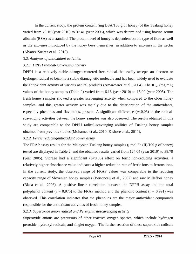
Page 61 RTLS - 2014
In the current study, the protein content (mg BSA/100 g of honey) of the Tualang honey
varied from 79.16 (year 2010) to 37.41 (year 2005), which was determined using bovine serum
albumin (BSA) as a standard. The protein level of honey is dependent on the type of flora as well
as the enzymes introduced by the honey bees themselves, in addition to enzymes in the nectar
(Alvarez-Suarez et al., 2010).
3.2. Analyses of antioxidant activities
3.2.1. DPPH radical-scavenging activity
DPPH is a relatively stable nitrogen-centered free radical that easily accepts an electron or
hydrogen radical to become a stable diamagnetic molecule and has been widely used to evaluate
the antioxidant activity of various natural products (Amarowicz et al., 2004). The IC50 (mg/mL)
values of the honey samples (Table 2) varied from 6.16 (year 2010) to 15.02 (year 2005). The
fresh honey samples showed a greater scavenging activity when compared to the older honey
samples, and this greater activity was mainly due to the deterioration of the antioxidants,
especially phenolics and flavonoids, present. A significant difference (p<0.05) in the radical-
scavenging activities between the honey samples was also observed. The results obtained in this
study are comparable to the DPPH radical-scavenging abilities of Tualang honey samples
obtained from previous studies (Mohamed et al., 2010; Kishore et al., 2011).
3.2.2. Ferric reducingantioxidant power assay
The FRAP assay results for the Malaysian Tualang honey samples (μmol Fe (II)/100 g of honey)
tested are displayed in Table 2, and the obtained results varied from 124.04 (year 2010) to 38.79
(year 2005). Storage had a significant (p<0.05) effect on ferric ion-reducing activities, a
relatively higher absorbance value indicates a higher reduction rate of ferric ions to ferrous ions.
In the current study, the observed range of FRAP values was comparable to the reducing
capacity range of Slovenian honey samples (Bertoncelj et al., 2007) and raw Milleflori honey
(Blasa et al., 2006). A positive linear correlation between the DPPH assay and the total
polyphenol content (r = 0.975) to the FRAP method and the phenolic content (r = 0.991) was
observed. This correlation indicates that the phenolics are the major antioxidant compounds
responsible for the antioxidant activities of fresh honey samples.
3.2.3. Superoxide anion radical and Peroxynitritescavenging activity
Superoxide anions are precursors of other reactive oxygen species, which include hydrogen
peroxide, hydroxyl radicals, and singlet oxygen. The further reaction of these superoxide radicals

Page 62 RTLS - 2014
with nitric oxide results in the formation of cytotoxic peroxynitrites, which oxidize cellular
components, such as proteins, lipids, and nucleic acids (Balavoine and Geletti, 1999).
Peroxynitrites are relatively stable but once protonated, they form highly reactive peroxynitrous
acid. The superoxide anion and peroxynitritescavenging activities of the Tualang honey samples
were investigated, and the results were compared with those of reference antioxidants. A higher
extract concentration required to scavenge radicals indicates a lower antioxidant activity. Based
on the results (Fig. 2), it is evident that the fresh honey samples (years 2010 & 2009) showed
significant superoxide anion and peroxynitritescavenging activities when compared to the older
stored honey samples.
Conclusions
Both the physicochemical properties and the phenolic compound composition of honey
are responsible for its quality and antioxidant activity, thus rendering it a healthy food and source
of antioxidants. Our findings suggest that the long-term storage of honey, even under the
appropriate temperatures and storage conditions, decreases the quality in a variety of aspects and
parameters and ultimately leads to the loss of its major nutritive and medicinal values. Based on
our findings, we also suggest that highly sensitive parameters, such as moisture, free acidity,
diastase activity, HMF content, total phenolics and antioxidant activity, can be used as markers
for approximating the shelf life and age of honey. However, further extensive studies need to be
performed to determine the most sensitive markers and to specify any guidelines for the
assessment of the age and quality of the honey.
Acknowledgments
The authors would like to thank the Universiti Sains Malaysia RU Grant
1001/PPSP/8120215 for providing financial support, and the beekeepers, the state of
Kedah, and the Federal Agricultural Marketing Authority (FAMA; Kedah, Malaysia) for
supplying the year wise Tualang honey samples for this study.
References
Ahmed J, Prabhu ST, Raghavan, GSV and Ngadi M (2007) Physico-chemical, rheological,
calorimetric and dielectric behavior of selected Indian honeys. Journal of Food
Engineering.79, 1207–1213.
Alvarez-Suarez JM, Tulipani S, Romandini S, Bertoli E, and Battino M (2010) Contribution of
honey in nutrition and human health: a review. Mediterranean Journal of Nutrition and
Metabolism, 3, 15–23.
Page 63 RTLS - 2014
Amarowicz R, Pegg RB, Rahimi-Moghaddam P, Barl B, and Weil JA (2004) Free-radical
scavenging capacity and antioxidant activity of selected plant species from the Canadian
prairies. Food Chemistry, 84, 551–562.
Anchling F (2007) La couleur du miel. L’Abeille de France et l’apiculteur, 942, 495-504.
AOAC (2000) Sugars and sugar products. In: Official Methods of Analysis of The Association
of Official Analytical Chemists (edited by W. Horowitz). Pp. 22–33. Gaithersburg, MD:
AOAC International.
Balavoine GG, Geletti YV (1999) Peroxynitrite scavenging by different antioxidants. Part 1:
convenient study. Nitric oxide, 3, 40-54.
Bath PK, and Singh N (2000) Chemical changes in Helianthus annus and Eucalyptus
lanceolatus honey during storage. Journal of Food Quality, 23, 443–451.
Benzie I F F, and Strain J J (1999) Ferric reducing/antioxidant power assay: direct measure of
total antioxidant activity of biological fluids and modified version for simultaneous
measurement of total antioxidant power and ascorbic acid concentration. Methods in
Enzymology, 299, 15–27.
Beretta G, Granata P, Ferrero M, Orioli M, and Facino RM (2005) Standardization of
antioxidant properties of honey by a combination of spectrophotometric/fluorimetric
assays and chemometrics.Analytica Chimica Acta.533,185–191.
Bertoncelj J, Dobersek U, Jamnik M, and Golob T (2007) Evaluation of the phenolic content,
antioxidant activity and colour of Slovenian honey.Food Chemistry, 105, 822–828.
Blasa M, Candiracci M, Accorsi A, Piacentini MP, Albertini MC, and Piatti E (2006) Raw
Millefiori honey is packed full of antioxidants. Food Chemistry, 97, 217–222.
Bogdanov S, Lullman C, Martin P, Von der Ohe, W, Russman H, Vorwohl G (1999) Honey
quality and international regulatory standards: Review by the International Honey
Commission. Mitteilungen aus Lebensmitteluntersuchung und Hygiene 90,108-125.
Bradford MM (1976) Rapid and sensitive method for quantification of microgram quantities of
protein utilizing the principle dye binding.Analytical Bichemistry, 72, 248–254.
Cantarelli MA, Pellerano RG, Marchevsky EJ, and Camina JM (2008) Quality of honey from
Argentina: Study of chemical composition and trace elements. The Journal of the
Argentine Chemical Society, 96, 33–41.
Chirife J, Zamora M C, and Motto A (2006) The correlation between water activity and %
moisture in honey: Fundamental aspects and application to Argentine honeys. Journal of
Food Engineering, 72, 287-292.
Chuanphongpanich S, Suttajit M, Phanichphant S, Buddhasukh D, and Sirithunyalug B (2006)
Antioxidant Capacity of Broccoli Seeds Grown in Thailand. Chiang Mai Journal of
Science, 33, 117-122.
Codex Alimentarius Commission (2001). Revised Codex Standard for Honey, Codex STAN12-
1981, Rev.1 (1987), Rev.2 (2001).

Page 64 RTLS - 2014
Costa L, Albuquerque M, Trugo L, Quinteiro L, Barth O, Ribeiro M, and De Maria C (1999)
Determination of non-volatile compounds of different botanical origin Brazilian
honeys.Food Chemistry, 65, 347–352.
EU Council (2002) Council Directive 2001 ⁄ 110 ⁄ EC of 20 Decem-Ziziphus 2001 relating to
honey.Official Journal of the European Community, 10, 47–52.
Finola MS, Lasagno MC, and Marioli JM (2007) Microbiological and chemical characterization
of honeys from central Argentina, Food Chemistry,100,1649–1653.
Hatano T, Kagawa H, Yashura T, and Okuda T (1988) Two new flavonoids and other
constituents in licorice root: Their relative astringency and radical scavenging effects.
Chemical Pharmaceutical Bulletin, 36, 2090–2097.
Horwitz W (Ed.) (1980) Official methods of analysis of the association of official analytical
chemists (13th ed.). Washington, DC: AOAC.
Juszczak L, and Fortuna, T (2006) Rheology of selected Polish honeys. Journal of Food
Engineering.75, 43–49.
Kaur-Bath P, and Narpinder S (2000) Chemical changes in Helianthus annuus and Eucalyptus
lanceolatus honey during storage. Journal of Food Quality,23, 443–451.
Khalil MI, SulaimanSH, and Gan (2010) High 5-hydroxymethylfurfural concentrations are
found in Malaysian honey samples stored for more than one year. Food and Chemical
Toxicology, 48, 2388–2392.
Kishore RK, Halim AS, Syazana MS, and Sirajudeen KN (2011) Tualang honey has higher
phenolic content and greater radical scavenging activity compared with other honey
sources. Nutrition Research, 31, 322-325.
Koppenol, W H, Kissne, R, and Beckman JS (1996) Synthesis of peroxynitrite: to go with the
flow or on solid grounds. Methods in Enzymology, 269, 296–302.
Malika N, Mohamed F, and Adlouni EL (2005) Microbiological and Physico-chemical
properties of Moroccan honey. International Journal of Agriculture and Biology, 5, 773–
776.
Marghitas L, Dezmirean D, Moise A, Bobis O, Laslo L, and Bogdanov S (2009) Physico-
chemical and bioactive properties of different floral origin honeys from Romania.Food
Chemistry, 112, 863-867.
Md Ibrahim Khalil, Mohamed Mahaneem, Syed Mohsin Sahil Jamalullail, Nadia Alam and Siti
Amrah Sulaiman (2011) Evaluation of Radical Scavenging Activity and Colour Intensity
of Nine Malaysian Honeys of Different Origin. Journal of ApiProduct and ApiMedical
Science, 3, 04–11.
Meda A, Lamien CE, Romito M, Millogo J, and Nacoulma OG (2005) Determination of the
total phenolic, flavonoid and proline contents in Burkina Fasan honey, as well as their
radical scavenging activity, Food Chemistry,91, 571–577.
Mendes E, Brojo Proenc E, Ferreira O, and Ferreira MA (1998) Quality evaluation of
Portuguese honey.Carbohydrate Polymers, 37, 219–223.
Page 65 RTLS - 2014
Mohamed M, Sirajudeen KNS, Swamy M, Yaacob NS, Sulaiman SA (2010) Studies on the
antioxidant properties of tualang honey of Malaysia. African Journal of Traditional,
Complementary and Alternative medicines, 7, 59-63.
Nombre I, Schweitzer P, Issaka Boussim J, and Jeanne Millogo Rasolodimby (2010) Impacts of
storage conditions on physicochemical characteristics of honey samples from Burkina
Faso.African Journal of Food Science, 4, 458-463.
Ojeda De Rodriguez G, Sulbaran De Ferrer B, Ferrer A, and Rodriguez B (2004)
Characterization of honey produced in Venezuela. Food Chemistry, 84, 499–502.
Okuda T, Kimura Y, Yoshida T, Hatano T, Okuda H, Arichi S (1983) Studies on the activity
and related compounds from medicinal plants and drugs. Inhibitory effects on lipid
peroxidation on mitochondria and microsomes of liver.Chemical and Phamaceutical
Bulletin,31, 1625-1631.
Omaye S T, Turnbull TO and Sauberlich HE (1979) Selected methods for the determination of
ascorbic acid in cells, tissues and fluids.Methods of Enzymology, 62, 3-11.
Ortial S, Durand G, Poeggeler B, Polidori A, Papolla MA, and Boker J (2006) Fluorinated
amphiphilic amino acid derivatives as antioxidant carriers: a new class of protective
agents. Journal of Medicinal Chemistry, 49, 2812–20.
Ouchemoukh S, Louaileche H, and Schweitzer P (2007) Physicochemical characteristics and
pollen spectrum of some Algerian honeys. Food Control, 18: 52-58.
Padayatty SJ, Katz A, Wang Y, Eck P, and Kwon O (2003) Vitamin C as an antioxidant:
Evaluation of Its role in disease prevention. Journal of American college of Nutrition, 22,
18-35.
Persano LO, Tim AH, Rodriguez-Malaver A, Ana Perez R, Fernandez-Muino, Maria Teresa
Sancho M, Giulio S, Lorenzo L, and Patricia V (2008) Composition and Antioxidant
Activity of Trigona carbonaria Honey from Australia. Journal of Medicinal Food, 11,
789–794.
Prieto P, Pineda M, and Aguilar M (1999) Spectophotometric quantitative of antioxidant
capacity through the formation of a phosphomolybdenum complex: Specific application
to the determination of vitamin E. Analytical Biochemistry, 269, 337–341.
Robak J, and Gryglewski RJ (1988) Flavonoids are scavengers of superoxide anions.
Biochemical Pharmacology, 37, 837–841.
Rodriguez GO, Ferrer, BS, Ferrer A, and Rodriguez B (2004) Characterization of honey
produced in Venezuela. Food Chemistry, 84, 499–502.
Ruoff K, Luginbu hl W, and Bagdanov S (2007) Quantitative determination of physical and
chemical measurands in honey by near-infrared spectrometry.European Food Research
and Technology, 225, 415–423.
Sahinler N (2007) Effects of heating and storage on hydroxymethylfurfural and diastase activity
of different Turkish honeys.Journal of Apiculture Research,46, 34–39.
Saxena S, Gautam S, and Sharma A (2010) Physical, biochemical and antioxidant properties of
some Indian honeys.Food Chemistry, 118, 391–397.

Page 66 RTLS - 2014
Singleton VL, Orthofer R, and Lamuela-Raventos RM (1999) Analysis of total phenols and
other oxidation substrates and antioxidants by means of Folin–Ciocalteu reagent.Methods
in Enzymology, 299, 152–178.
Terraberint A, Diez MJ, and Heredia FJ (2003) Palynological, physicochemical and colour
characterization of Moroccan honeys. I. River and gum (Eucalyptus camaldulensis
Dehnh) honey. Journal of Food Science and Technology,,38, 379–386.
Viuda-Martos M, Ruiz-Navajas Y, Fern ndez-L pez J, and Prez-lvarez JA (2008) Functional
Properties of Honey, Propolis and Royal Jelly. Journal of Food Science.73, 117-124.
Williams ET, Jeffrey J, Barminas JT and Toma I (2009) Studies on the effects of the honey of
two floral types on organism associated with burn wound infections. African Journal of
Pure and Applied Chemistry, 3, 9.
Woisky R, and Salatino A (1998) Analysis of propolis: some parameters and procedures for
chemical quality control. Journal of Apicultural Research, 37, 99-105.
Yilmaz H, and Kufrevioglu I (2000) Composition of honeys collected from eastem Anatolia and
effect of storage on hydroxymethylfurfural content and diastase activity. Turkish Journal
of Agriculture and Forestry,33, 347–349.
Yolanda R N, Manuel VM, Juana Fernandez L, Juan Manuel Z Cruz, Victor K, and Jose
Angel, P. A. (2011) Antioxidant Activity of Artisanal Honey from Tabasco, Mexico.
International Journal of Food Properties, 14, 459 – 470.

Page 67 RTLS - 2014
Therapeutic potential of ginger on cardiac antioxidant defense system and blood
glucose levels in experimental diabetic rats
Chilakala Ramakrishna 1, Kondeti Ramudu Shanmugam
1 2, Ganjikunta Venkata
Subbaiah3, Korivi Mallikarjuna
3,Kesireddy Sathyavelu Reddy
1 *
1 Division of Molecular Biology and Exercise Physiology,
Department of Zoology,
Sri Venkateswara University, Tirupati – 517 502, A. P, India 2 Department of Zoology, T.R.R.Government Degree,
Kandukur, Prakasam District, Andhra Pradesh, India 523 105
ABSTRACT
Antioxidant defense system plays a key role in diabetic complications. The effects of
ginger ethanolic extract on ruined antioxidant system in heart tissue remains unclear in diabetic
rats. In this study, we investigated the impact of oral administration of ginger ethanolic extract
on antioxidant parameters in experimental diabetic rats. Thirty rats were equally divided into five
groups, including normal control (NC), diabetic control (DC), ginger treatment (Gt), diabetics
treated with ginger (D+Gt) and diabetics treated with glibenclamide (D+Glb). The parameters
like XOD, AA, blood glucose and body weight levels. XOD, AA are significantly decreased in
heart of diabetic rats, which indicates impaired antioxidant status. However, we found that the
administration of ginger to diabetic rats restored the altered antioxidant status that is almost
similar to ginger treated group. Further, evidences from our histopathological studies also prove
that ginger protects the heart and liver tissues from oxidative stress in diabetic rats.
Keywords: Diabetes, antioxidant enzymes, heart, rats.
Introduction:
Diabetes is one of the most pressing global health problems and it is estimated that the
prevalence of diabetes will be more than 300 million in 2025 (Ojewole, 2006). Increased
oxidative stress and changes in antioxidant capacity was observed in both clinical and
experimental diabetes and thought to be the etiology of diabetic complications (Baynes, 1991).
Management of diabetes with agents devoid of any side effects is still a challenge to the medical
system. This has led to an increase in the demand for natural products with antihyperglycemic
activity and fewer side effects. Plants may act on blood glucose through different mechanisms,
some of them may have insulin-like substances and some may inhibit insulinase activity
(Bopanna et al., 1997). Some plants are involved in the stimulation of cells to produce more
insulin and others may increase cells in the pancreas by activating regeneration of pancreatic
cells.

Page 68 RTLS - 2014
Traditionally, medicinal plants are extensively used in India due to their low cost, easy
accessibility to everyone and perceived fewer side effects and their medicinal use has been
gradually increasing in developed countries. In many respects, the mechanism of action of the
herbal drugs differs from that of the synthetic drugs (or) pure compounds. Zingiber officinale
(Roscoe), commonly known as ginger is one of the commonly used spices in India and around
the world. Ginger has been used to treat a number of diseased conditions including headache,
cold, arthritis, postoperative nausea and vomiting, motion sickness, and reduces symptoms in
patients with nausea of pregnancy. Ginger has hypoglycemic, insulinotropic, hypolipidemic and
antioxidant properties. Ginger is used to treat alcoholism, bronchitis, cancer, ulcer and dyspepsia
(Shanmugam et al., 2010). A preliminary study has reported that ginger contains a large number
of bioactive compounds like polyphenols, alkaloids, saponins and tannins. Some of the chemical
constituents isolated from ginger include (6) - gingerol, α-zingiberene, phenolic compounds,
essential oilsand oleoresin resins (Van Wyk and Wink, 2003). We had recently reported the renal
protective effect of ginger in alcoholic subjects (Shanmugam et al., 2010).
So for there was limited work on the cardiac protective effect of ginger in diabetic rats.
Hence, the present study was carried out to know the impact of ginger on cardiac antioxidant
defense system parameters against STZ induced diabetic rats.
Materials and Methods:
Animals
Male albino Wistar rats, body weight of 180±200 grams, were used in this study. The rats were
housed in clean polypropylene cages having 6 rats per cage under hygienic conditions and
maintained under temperature controlled room (27±20C) with photoperiod of 12 hours light and
12 hours dark cycle. The rats were maintained in accordance with the guidelines of the National
Institute of Nutrition, Indian Council of Medical Research, Hyderabad, India, and approved by
the Institutional Animal Ethical Committee No: 09(iv)/a/CPCSCA/IAEC/07-08/SVU/Zool/KSR-
CHRK/ dated: 26/6/08. S.V.University, Tirupati, Andhra Pradesh. The rats were allowed
standard rat pellet diet (Lipton India Ltd., Mumbai, India) and water ad-libitum for the duration
of the experiment.
Chemicals
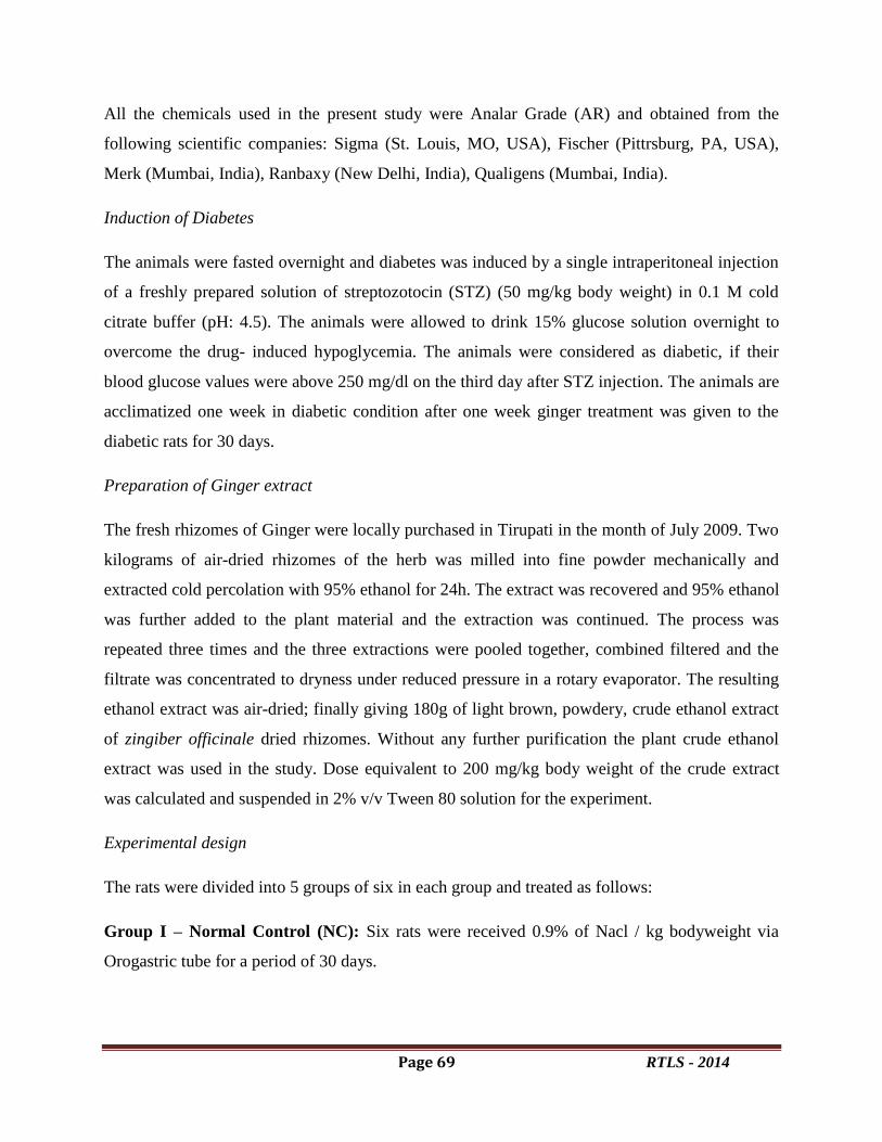
Page 69 RTLS - 2014
All the chemicals used in the present study were Analar Grade (AR) and obtained from the
following scientific companies: Sigma (St. Louis, MO, USA), Fischer (Pittrsburg, PA, USA),
Merk (Mumbai, India), Ranbaxy (New Delhi, India), Qualigens (Mumbai, India).
Induction of Diabetes
The animals were fasted overnight and diabetes was induced by a single intraperitoneal injection
of a freshly prepared solution of streptozotocin (STZ) (50 mg/kg body weight) in 0.1 M cold
citrate buffer (pH: 4.5). The animals were allowed to drink 15% glucose solution overnight to
overcome the drug- induced hypoglycemia. The animals were considered as diabetic, if their
blood glucose values were above 250 mg/dl on the third day after STZ injection. The animals are
acclimatized one week in diabetic condition after one week ginger treatment was given to the
diabetic rats for 30 days.
Preparation of Ginger extract
The fresh rhizomes of Ginger were locally purchased in Tirupati in the month of July 2009. Two
kilograms of air-dried rhizomes of the herb was milled into fine powder mechanically and
extracted cold percolation with 95% ethanol for 24h. The extract was recovered and 95% ethanol
was further added to the plant material and the extraction was continued. The process was
repeated three times and the three extractions were pooled together, combined filtered and the
filtrate was concentrated to dryness under reduced pressure in a rotary evaporator. The resulting
ethanol extract was air-dried; finally giving 180g of light brown, powdery, crude ethanol extract
of zingiber officinale dried rhizomes. Without any further purification the plant crude ethanol
extract was used in the study. Dose equivalent to 200 mg/kg body weight of the crude extract
was calculated and suspended in 2% v/v Tween 80 solution for the experiment.
Experimental design
The rats were divided into 5 groups of six in each group and treated as follows:
Group I – Normal Control (NC): Six rats were received 0.9% of Nacl / kg bodyweight via
Orogastric tube for a period of 30 days.

Page 70 RTLS - 2014
Group II – Diabetic Control (DC): Sixrats were used as diabetic control by the induction of
STZ intraperitonial injection of 50mg/kg body weight after fasting.
Group III – Ginger treatment (Gt): Six rats were received the ethanolic extract of ginger,
200mg/kg bodyweight via orogastric tube for a period of 30 days.
Group IV – Diabetic + Ginger treatment (D+Gt): Six diabetic rats were received the ethanolic
extract of ginger, 200mg/kg bodyweight via orogastric tube for a period of 30 days.
Group V - Diabetic + Glibenclamide treatment (D+Glb): Diabetic rats treated with
glibenclamide (600 µg/kg body weight) in aqueous solution for a period of 30 days.
The animals were sacrificed after 24 hours of the last treatment by cervical dislocation
and the heart and liver tissue was excised at 4°C. The tissues was washed with ice-cold saline,
immediately immersed in liquid nitrogen and stored in the deep freezer at -80˚C for further
biochemical analysis and enzymatic assays. Before assay, the tissues were homogenized under
ice-cold conditions. Selected parameters were estimated by employing standard protocols.
Biochemical parameters estimation
Cardiac Xanthine Oxidase and Ascorbic acid levels are estimated by by Omaye et al. (1971) &
Srikanthan & Krishnamurthy (1955).
All of the enzyme activities were expressed as per mg of protein and the tissue protein
was estimated according to the method of Lowry et al. (1951) using bovine serum albumin
(BSA) as a standard. The blood glucose levels were measured by using an Accucheck
glucometer (Roche – Germany).
Histopathological examinations of Heart and Liver
The heart and liver tissues were isolated immediately after sacrificing the animal and washed
with ice-cold saline andthey were then fixed in 10% formalin solution. Sections of 3µm
thickness were stained with haematoxylin and eosin (H&E) for histopathological examination.
Statistical analysis

Page 71 RTLS - 2014
The data are expressed as Mean values with their SD. Readings of the five different
groups were compared using one-way ANOVA analysis with Dennett’s multiple comparision
test. Statistical analysis was performed using SPSS (Version 13.5; SPSS Inc., Chicago, IL,
USA). Using M.S. Office, Excel Software the data has been analyzed for the significance of the
main effects (factors), and treatments along with their interactions. Differences were considered
significant at (*p < 0.001), (Ψp < 0.01).
Results
Blood glucose and Body weight changes
Blood glucose levels were found to be significantly increased after STZ-induction and the
administration of ginger, decreased the blood glucose levels significantly (Ψp<0.01) in diabetic
rats than that of diabetic control group (Fig-1). These decreasing levels of blood glucose may be
due to the regeneration of β - cells of the pancreas, which were destroyed by STZ.
A significant decrease in body weight in the diabetic control rats was observed than normal
control rats. Whereas with ginger treatment, the bodyweight was significantly (Ψp<0.01)
increased in diabetic rats (Fig-2).
Results of XOD, AA in the Heart tissue of all Experimental groups
The ethanolic extract of ginger was selected for antioxidant activity in diabetic rats. We observed
significant (*p < 0.001) decrease in the levels of XOD, AA in heart tissue of diabetic rats when
compared to normal control (NC). After administration of ginger to diabetic rats the XOD, AA
levels are significantly (Ψp<0.01) increased. The results have shown in (Fig-3).
Histological study
Histopathological investigation of heart tissue that also provided an essential evidence for
the biochemical analysis. The heart of normal control (NC) and ginger treated (Gt) rats shows
striations along the length of the normal muscle fibres (NMF) and the normal nuclei (N) of the
cardiac muscle cells lie in the middle of the cells, several cardiac fibres are seen branching
(CFBr) and a prominent perinuclear region is seen around several nuclei. Where as in the heart

Page 72 RTLS - 2014
tissue of diabetic control rats (DC), we observed, hyalinization of muscle fibres, with focal
cellular infiltration or necrosis of muscle fibres (NMF), congestion (C), slight infiltration (SI)
and slight vacuolation (SV). However in ginger treated diabetic rats (D+Gt), the heart tissue
showing normal nucleus (N), and we also observed mild swelling of muscle cells (MSMC) and
rearrangement of focal cardiac muscle fibres (RCMF) this also reveals normal architecture of the
myocardium with intact muscle fibres. Thus ginger has some protective effect on the
myocardium against STZ-induced diabetis. Where as in Diabetic rats treated with glibenclamide
(D+Glb), the heart tissue showing normal nucleus (N) and normal cardiac fibers (NCF) and
normal nuclei (N) of the cardiac muscle cells lie in the middle of the cells.
Table 1:- Effect of ginger and glibenclamide on XOD levels and AA levels in diabetic rats
Xanthine Oxidase (XOD)Ascorbic acid (AA)
(µ moles of uric acid/gram) (µ moles of ascorbic acid/gram)
Group I (NC) 0.81± 0.0141 2.15±0.166
Group II (Gt ) 0.981± 0.083 2.19±0.142*
Group III (DC) 1.53±0.033* 1.56±0.163
Group IV 1.028±0.019* 1.84 ±0.176*
(D+Gt )
Group V 1.20±0.012 1.98±0.207
(D+Gli)
All the values are mean, ± SD of six individual observations,
*significant at p< 0.001
Discussion
The present study investigates the effects of ginger antioxidant property in STZ- induced
diabetic rats. Oxidative stress is suggested to be a potential contributor to the development of

Page 73 RTLS - 2014
complications in diabetes. Increased free radical production or reduced antioxidant defense
responses, both of which occur in the diabetic state may give rise to increased oxidative stress
(Halliwell and Gutteridge, 1990). Consequences of oxidative stress are cell injury, i.e. damage
lipids, antioxidant enzyme system, disruption in cellular homeostasis and accumulation of
damaged molecules (Jakus, 2000).
In Streptozotocin-induced diabetic rats, we observed increased blood glucose levels. The
elevation of glucose in STZ-treated rats was due to an oxidative stress produced in the pancreas,
due to a single strand break in pancreatic islets DNA (Yamamoto et al., 1981). Where as with
ginger treatment in diabetic rats, we observed that blood glucose levels are decreased. This may
be due to the free radical scavenging and hypoglycemic activity of ginger. Ginger inhibits lipid
peroxidation and prevents streptozotocin-induced oxidative stress and protects β-cells from
diabetic shock.
Figure 1: The effect of ginger on Blood glucose levels and Body weight changes in diabetic
rats. The values are significant compared to the following: control (*p < 0.001), diabetic
(Ψ
p< 0.01)
We have registered a decrease in body weight in STZ diabetic rats. The characteristic loss
of body weight associated with STZ-induced diabetes is due to increased muscle wasting in
diabetes (Ravi et al., 2004). This indicates polyphagic condition and loss of weight due to
excessive break-down of tissue proteins in diabetic rats (Chatterjea and Shinde, 2002). Hakim et
al., (1997) have stated that decreased body weight in diabetic rats could be due to dehydration
and catabolism of fats and proteins. Increased catabolic reactions leading to muscle wasting
might also be the cause for the reduced weight gain by diabetic rats (Rajkumar et al., 1991).
When ginger was administered to diabetic rats, the body weights seemed to be increased, as was
Blood Glucose
0
50
100
150
200
250
300
Period of Experiment
Blo
od
glu
co
se l
evels
Mg
/DL
NC
DC
Gt
DC+Gt
DC+Glb
0-Day 30-Day
* *
*
*
Ψ
*Ψ
*
Body weight
0
50
100
150
200
250
Period of Experiment
Bo
dy w
eig
ht
in g
ram
s
NC
DC
Gt
DC+Gt
DC+Glb
0-Day 30-Day0-Day 30-Day
* Ψ
*
Ψ
Page 74 RTLS - 2014
the ability to reduce hyperglycaemia. However, it could not normalize the body weight
completely. The treatment with ginger showed a significant increase in body weight in diabetic
rats. The administration of ginger to STZ diabetic rats reduced blood glucose levels, in
accordance with earlier reports (Al-Amin et al., 2006).
XOD serves as a sensitive marker of oxidative stress and it plays an important role in
maintaining the integrity of the cell system. XODis involved in several reactions in the body and
is one of the most prominent non-enzymatic antioxidants. In the current study, A XOD levels
was decreased in heart tissues of diabetic rats. Depletion of tissue XOD levels enhances cellular
damage caused by oxidative stress. Significant depletion of XOD & AA (*p<0.001) in diabetic
rats suggests its increased utilisation against reactive oxygen species. However, ginger treatment
in diabetic rats, reversed the XOD & AA to normal levels, this shows that ginger has an
antioxidant property. Reports are available on the antioxidant effect of ginger by decreasing lipid
peroxidation, increasing XOD & AA level and maintaining normal levels of antioxidant
enzymes. A number of researchers have reported that 6-gingerol, tannins, polyphenolic
compounds (coumarins), flavonoids and triterpenoids of ginger have antioxidant properties in
various experimental models (Shanmugam et al., 2010).
The diabetogenic potential of STZ is responsible for a progressive development of the
hepatic and cardiac tissues lesions. In the present study the protective effects of ginger in
experimental groups of rats were established by histological study. Figure-5 illustrates
histopathological investigation that also provided an essential evidence for the biochemical
analysis. In the normal control rats the heart, shows striations along the length of the normal
muscle fibres (NMF), the normal nuclei (N) of the cardiac muscle cells lie in the middle of the
cells, several cardiac fibres are seen branching (CFBr) and a prominent perinuclear region is seen
around several nuclei. Where as in the heart tissue of diabetic rats hyalinization of muscle fibres,
with focal cellular infiltration or necrosis of muscle fibres (NMF), congestion (C), slight
infiltration (SI) and slight vacuolation (SV). However in ginger treated diabetic rats the heart
tissue showing normal nucleus (N), and we also observed mild swelling of muscle cells (MSMC)
and rearrangement of focal cardiac muscle fibres (RCMF). This reveals normal architecture of

Page 75 RTLS - 2014
the myocardium with intact muscle fibres thus ginger has some protective effect on the
myocardium against STZ-induced diabetes. Where as in D+Glb treated rats the heart tissue
showing normal nucleus (N) and normal cardiac fibers (NCF) and normal nuclei (N) of the
cardiac muscle cells lie in the middle of the cells.
In conclusion, our results demonstrated that STZ is capable of causing marked oxidative
stress in addition to deplete the antioxidants and inhibiting the activities of antioxidant
parameters. Finally, the results of our work reveals that ginger would be a valuable source of
anti-diabetic and antioxidant agents, with a potential use in pharmaceutical preparations. In all,
we suggest that the ginger diets have health promoting effects especially in metabolic disorders.
Moreover further studies of ginger extracts and in-vivo studies of inhibition are warranted.
References:
Al-Amin, Z.M., Thomson, M., Al-Qattan, K.K., Peltonen Shalaby, R., Ali, M., 2006.Anti-
diabetic and hypolipidemic properties of ginger (Zingiber officinale) in Streptozotocin-
induced diabetic rats.British Journal of Nutrition. 96, 660–666.
Baynes, J.W., 1991. Role of oxidative stress in the development of complications in
diabetes.Diabetes.40, 405–412.
Bopanna, K.N., Kannan, J., Sushma, G., Balaraman, R., Rathod, S.P., 1997. Antidiabetic and
antihyperlipidemic effects of neem seed kernel powder on alloxan diabetic rabbits. Indian
Journal of Pharmacology. 29, 162-167.
Chatterjea, M.N., Shinde, R., 2002. Textbook of medical biochemistry.Jaypee Brothers, Medical
Publishers Pvt. Ltd. New Delhi, p. 317.
Hakim, Z. S., Patel, B. K., & Goyal, R. K. (1997).Effects of chronic ramipril treatment in
streptozotocin-induced diabetic rats.Indian Journal of Physiology and Pharmacology, 41,
353–360.

Page 76 RTLS - 2014
Halliwell, B., & Gutteridge, J. M. (1990).Role of free radicals and catalytic metal ions in human
disease, an overview.Methods in Enzymology, 186, 1–85.
Jakus, V. (2000).The role of free radicals, oxidative stress and antioxidant systems in diabetic
vascular disease.Bratisl Lek Listy, 101(10), 541–551.
Lowry, O. H., Rosebrough, N. J., Farr, A. L., & Randall, R. J. (1951). “Protein measurement
with the Folin Phenol Reagent”.The Journal of Biological Chemistry, 193, 265-275.
Ojewole, J. A. O. (2006).Antinociceptive, anti-inflammatory and antidiabetic properties
ofHypoxis hemerocallideaFisch & C.A. Mey.(Hypoxidaceae) corm ['African Potato']
aqueous extract in mice and rats.Journal of Ethnopharmacology, 103, 126-134.
Rajkumar, L., Srinivasan, N., Balasubramanian, K.,& Govindarajulu, P. (1991).Increased
degradation of dermal collagen in diabetic rats.Indian Journal of Experimental Biology,
29, 1081–1083.
Ravi, K., Ramachandran, B., & Subramanian, S. (2004). Effect of Eugenia jambolana seed
kernel on antioxidant defense system in streptozotocin-induced diabetes in rats.Life
Science, 75(22): 2717-2731.
Shanmugam, K. R., Ramakrishna, C. H., Mallikarjuna, K., & Sathyavelu Reddy, K.
(2010).Protective effect of ginger against alcohol-induced renal damage and antioxidant
enzymes in male albino rats.Indian Journal of Experimental Biology, 48, 143–149.
Van Wyk, B. E., & Wink, M. (2003). Medicinal plants of the world. 1st edition.(Brizqa
Publication Pretoria: South Africa), 349.
Yamamoto, H., Uchigata, Y., & Okamoto, H. (1981). DNA strand breaks in pancreatic islets by
in vivo administration of alloxan streptozotocin. Biochemical and Biophysical Research
Communications, 103, 1014-1020.

Page 77 RTLS - 2014
Bioaccumulation of heavy metals in fish (Rastrelliger ) collected fromRoyapuram and
Kovalam waters of Chennai, India
Ramesh Babu, Y., Anitha, H.R., Ratna Prabha, D.,
Tara Government College, Sangareddy, Medak(Dt), Telangana, India
ABSTRACT
A study was done to know the concentration of heavy metals in the edible muscle of fishes
(Rastrelliger) collected from the coastal water of Royapuram and Kovalam of Chennai area.
Royapuram coast is associated with industries. Kovalam is located far away from Royapuram
area and is an unpolluted with reference to industries. The fishes were collected. The tissues are
dried and digested and the samples are prepared. The sample solutions were tested to find the
concentration of heavy metals using Atomic Absorption Sphectrophotometer. The concentration
of heavy metals followed the following ranking in the fishes collected from Royapuram and
Kovalam regions.
Royapuram: Fe > Zn > Cu >Mn. Kovalam: Fe > Cu > Zn >Mn .The concentration of heavy
metals was found to be more in the fishes collected from Royapuram than Kovalam. Nickel and
Lead was found to be below detectable limits from the fishes collected from both the areas.
KEY WORDS: Heavy metals; Rastrelliger; Royapuram; Kovalam; Chennai; Bioaccumulation
INTRODUCTION
According to the Food and Organization, the percapita availability of food in India, in terms
of calories is low. Indian diet is basically cereal based. Thirty seven percent of the population in
India live below the poverty line and majority of them suffer from malnutrition. Most
malnutrition in India is protein energy malnutrition, resulting from lack of energy food. Food
production from agricultural sources cannot keep pace with the ever increasing population. The
sea is known for its productivity, with rich and diversity of organisms. The sea is exploited for
different edible sources of food like fishes, Lobsters, Crabs, Oysters, Cephalopods, Holothurians,
Sea weed (Rao,1995)1 etc for their nutritive values and for other purposes.
Among all the marine resources, fish and crustaceans rank the majority. Fish serves as food
for mankind. Fish food is highly significant in that it helps to build up the health of the public.
The importance of fish as food lies not merely in its rich protein content, but also in its quality.
Protein of fish origin contains many essential amino acids such as arginine, histidine, isoleucine,
lysine, methionine, phenylalinine, threonine, tryptophan and valine(Shanmugam, 1992)2. In
shallow coastal and shelf waters the amunt of organic matter is high( Eastherson and
Mahadevan,1980)3.
Page 78 RTLS - 2014
The consumption of food can improve the health of the people. At the same time they can
accommodate the pollutants in their body because of the pollution of their habitat. The ocean has
often referred to as the ultimate sink. The Indian coastal waters indicates that petroleum
hydrocarbons constitute one of the major pollutants followed by heavy metals and chlorinated
hydrocarbons( James,1990)4. Heavy metals finally reaches the fishes through the food chain and
accumulate in the tissues. The major toxic effects of heavy metals include the blocking of the
functional groups and molecules of biological importance like enzymes, polynucleotides or
transport systems for essential nutrients and ions.
Now a days the marine region particularly the coastal area has been affected by the pollution
from different sources. In the marine environment, the near shore regions are of concern due to
the fact that they are the sites of the worlds most critically polluted areas(Ramachandran,1990)5.
The data and researches on the accumulation of heavy metals in pelagic fishes in the coastal
waters of Chennai is still short.
Rastrlliger kanagurta Cuvier is commonly called Indian Mackerel belongs to the family
Scombridae. It is widely spread in the Indo-pacific region. It is pelagic form and its is a
planktonic feeder. On average this fish accounts for about eight percent of the country’s marine
fish landings.
MATERIALS AND METHODS
Sample collection and preparation
The fish samples were collected from the coastal waters of Royapuram and Kovalam. The
same species of fishes were collected from the Royapuram and kovalam areas which are of same
size and length(18-20cms). The collected fishes were then placed in an icebox. Then they were
transported to the laboratory and preserved at 4C for further analysis. Removal of the muscles
from the species were done within twenty four hours of the collection. The fish samples were
brought to the room temperatures. Then the samples were rinsed with distilled water to remove
sands and debris. The muscle from the fish samples were removed using sterile scissors, knife
and forceps. The petridishes were labeled before dissection process. The muscle tissue is

Page 79 RTLS - 2014
removed and kept in the labeled petridish and were dried in the hot air oven at 110 C for 24
hours.
Heavy metal Analysis
Digestion Method
One gram of the dried sample tissue was taken, to which concentrated sulphuric acid and
concentrated perchloric acid were added in the ratio of 3:1. Then the mixture was exposed to
slight temperature. After this 0.05 N HNO3 was added and made up to a constant volume.
Analysis
The prepared solutions were aspirated in a Instrumental Laboratory(Vido-12) Atomic absorption
spectrophotometer to find out the concentration of heavy metals Copper, Iron, Manganese,
Nickel, Lead and Zinc. Blanks and standards were run simultaneously along with the sample
solutions.
RESULTS AND DISCUSSION
The mean concentrations of the heavy metals Copper, Iron, Manganese and Zinc in the
samples collected from Royapuram were 0.635 ppm, 4.23 ppm, 0.251 ppm and 0.796 ppm. The
heavy metals lead and Nickel were found to be below detectable limits.
The mean concentrations of the heavy metals Copper, Iron, Manganese and Zinc in the samples
collected from Kovalam were 0.461 ppm, 3.52 ppm, 0.132 ppm and 0.393 ppm. The heavy
metals lead and Nickel were found to be below detectable limits.
The S.D values of the heavy metals Copper, Iron, Manganese and Zinc in Rastrelliger
kanagurta from Royapuram coastal area were + 0.359; + 1.222; +0.244; +0.963 respectively
where as the same species from the Kovalam area showed + 0.099; +2.775; +0.047; +0.112
respectively.
All the values between the concentrations of the same heavy metals from the species between
Royapuram and Kovalam areas showed that the values were significant at 0.05% confidence
level.
Page 80 RTLS - 2014
In the fishes Rastrelliger kanagurta the heavy metal Iron concentration was found to be higher
when compared to that of other heavy metals. The heavy metals Lead and Nickel were found to
be below detectable limits. The heavy metals Cu, Fe, Mn and Zn are generally required for
normal physiological function, but needed only in traces.
The present findings are similar to the studies by Maheswari Nair et al.,where the content of
Iron and Zinc is more followed by Copper and Manganese. The concentration of heavy metals in
the samples from Royapuram were higher than the specimens collected Kovalam area. This can
be due to the fact that Royapuram is nearer to Ennore area which is highly polluted with
industries. Even Royapuram area has industrial complex which release their wastage in to the
sea.
The findings of Jaffar and Ashraf (1988)6 showed similarity with the present findings where the
concentration of Iron is more followed by Zinc, Copper, Manganese etc. The entry of heavy
metals into the fishes may also be through phytoplanktons and zoo planktons. Marine diatoms
showed the concentration of heavy metals where Iron content is higher than the other heavy
metals (Umamaheswara Rao and Mohanchand 1990)7.
George and Kurieshy (1979)8 reported the concentration of Iron more than Zinc followed by
other metals in Zooplankton from Bay of Bengal.
The concentrations of the heavy metal could have been high if the experiments could have
done in monsoon season because during monsoon season the pH and the salinity decreases so it
may facilitate the dissolution of precipitated form of the metal( hydroxide, carbonate or chloro
complexes) and increase the amount of ionic species in the solution. The most broadly accepted
paradigm is the bio availability of metal in the form of free metal iron(Sunda and Thoutte 1978)9.
During the summer season the concentration of the heavy metals is low. It may be due to the low
bio availability of the metal ions in the sea water due to high salinity and pH. The concentration
of manganese in the organisms were very low that they cannot even be predicted. It may be due
to the fact that marine animals, have very low tendency to accumulate manganese. The muscles
of fishes seem to accumulate less amount of heavy metals than the liver, kidney( Jaffar and
Ashraf 1988)6.

Page 81 RTLS - 2014
The research show that Royapuram area is polluted than the Kovalam area due to
Industrialization and urban activities.
The chart below shows the concentration of heavy metals Copper, Iron, Manganese and Zinc in
Rastrelliger kanagurta from Royapuram and Kovalam areas.
CONCLUSION
The comparison and analysis revealed the fact that the accumulation of heavy metals in
muscle tissues of Rastrelliger kanagurta collected from Royapuram and Kovalama areas are low.
The results reveal, that the amount of heavy metal accumulated in the muscle tissues were
comparatively less which may not affect the consumer at higher levels of food chain. It can be
concluded that it is safe for consumption.
The study also reveals that the level of pollution is more at Royapuram which is surrounded by
Industries when compared to Kovalam coastal waters which is free from Industries.
REFERENCES
1.Rao, V.P., 1995. Present Status of Mariculture in India.In: Shrimp Feeds (Eds) R. Jayaram,
R. Santhanam, V. Sundararaj, K.Venkataramanujam and G. Sanjeevaraj : 77-82.
0
0.5
1
1.5
2
2.5
3
3.5
4
4.5
Concentration of Heavy metals in
ppm
Royapuram ( metal Conc in ppm)
Kovalam( metal conc in ppm)
Page 82 RTLS - 2014
2. Shanmugam, K., 1992. Fishery Biology and AquacultureIst edition, Leo
Pathippagam, Madras, pp:6
3. Eastherson., and Mahadevan, 1980 Review of open sea environmental conditions along
Indian CoastC.M.F.R.I.Bull., 29: 17-21
4. James,P.S.B.R., 1990. Marine pollution in the coastal waters of India and its possible
effects on the fisheries ResourcesIn : Marine Pollution and Toxicology(Eds) V.K.
Venugopalan and T.Balasubramaniam: 185-192
5. Ramachandran, S., 1990. Marine Pollution in Tamilnadu- A status ReportIn : Marine
Pollution and Toxicology(Eds) V.K. Venugopalan and T.Balasubramaniam: 143-160
6.Jaffar, M., and M. Ashraff, 1988. Selected Trace metal concentrations in different
tissues of fish from coastal waters of Pakistan ( Arabia Sea). Indian. J.Mar. Sci., 17: 231-
234
7. George, M.D., and T.Q. Kureishy, 1979. Trace Metals in Zooplankton from the Bay of
BengalIndian. J. Mar.Sci.,8 : 190-193.
8. Sunda, W.G., D.W.Engle and R.M. Thuotte, 1978. Effects of Chemical speciation on the
toxicity of cadmium to the grass shrimp Palaemonetes pugio : importance of free cadmium ion
Environ. Sci. Technol., 12: 409 – 413.
Page 83 RTLS - 2014
A Survey of Medicinally Important Plants in around Venkatapur mandal ofBhadrachalam area; Khammam District; (A.P)
Dr.S.Rama mohana Rao (Department Of Botany, SR&BGNR GDC Khammam) G.Ravi (Research Assistant)Ethno botanical laboratory, SR&BGNR GDC Khammam
Email.Id. [email protected]
Abstract A survey on Ethnomedicinal plants was conducted in Venkatapuram- -mandal of
Bhadrachalam area in the month of June-2014. The tribal people of thatarea revealed 27 medicinal plants and their utility. One among them is Dioscorea-hispida, apart from ethnomedicinal value, it is fatal, when it is taken in the raw form.The chemical nature of the tuber is evaluated in the ethnobotanical laboratory of SR&BGNR G.D.C Khammam. The documented medicinal plants were tabulated alphabetically(family wise).
Key Words:-Ethnomedicinal plants, Dioscorea hispida(Tuber).
Introduction:- Khammam District in Andhrapradesh is one of the state, rich with forest wealth.
The tribal people like; Koyas, Lambadas and Konda Reddys are living in this forest by adopting
podu cultivation.The forest is predominated by Dry-deciduous, Moist-deciduous, riparian, Scrub
and grass land vegetation. Whenever the tribal people gets diseases, they may approach the
(Vejjolu) or ( Health healers) . They used to give herbal medicine for immediate cure. Instead of
relying on modern medicine, still, the tribes are depending on the age old traditional medicine.
Some of the medicinal plants may also used for edible purpose. At the time of cooking of
the above foods, if enough precautionary measures are not taken, it becomes fatal.
Material & Methods:- An ethnobotanical survey was conducted by a team of
SR&BGNRGovtCollege, Khammam to unearth the traditional knowledge of tribal people at
Venkatapuram mandal of Bhadrachalam area in the month of June -2014 by using questioniers ,
oral interviews, the team members collected 27 ethnomedicinal plants information. The
collected plants were pressed and make it in the form of herbarium and kept in the
ethnobotanical laboratory of SR&BGNRGovtCollege. For every specimen, proper voucher
number was given. survey of the literature shows that, ethnomedicinal information of
kondareddis is poor. A few persons like Koppula.HemadriV.S.Raju, Sateesh Sutari,
C.S.Reddyetc are concentrated on ethnobotanical studies. The collected plant specimens were
identified by using the flora of Bombay& The flora of Andhrapradesh by T.Pullaiah.
Results and Discussion:- The present work reported a total of 27 plants belonging to various
families are to be used for medicinal purpose. During survey, The research team found that one

Page 84 RTLS - 2014
of the tuberous plant Dioscorea hispida was eaten by the local tribes and subjected to death. The
tuber of the plant was collected and examined in the ethno –botanical laboratory of SR&BGNR
GDC Khammam, and came to know that an AlkoloidDioscorine-a, 4-epidioscorine causes
neuromuscular blockage. This result the death of the patient.
Dioscoria hispida:- It is a twining vine, arising from tuberous roots, and reaching a length of
several meters. Stems are covered with spines. Leaves are 3-foliolated. Panicle is axillary,
slender, hairy,12 to 20 cm long. Flowers are small, unisexual, male flowers with 6-stamens,
female flowers similar to males, 3-winged, 3-celled ovules, 2 in each cell.Fruit is a capsule,
oblong and about 5cm long. Flesh and sap of tubers are yellowish. The tuber is growing
luxuriantly in the Venkatapuram mandal of Khammam District.
Constituents:- Tubers yield alkaloid dioscorine-a and 4-epidioscorine and neuromuscular
blocking agent. A saponin glycoside, diosgenin is also reported. Study of the mineral content
reports the tubers are a good source of phosphorus, Calcium& Iron.A mature gadung tuber
weighs up to 15kg, each 100 gm of tuber (wet bases)yields 20gms of carbohydrates,78gm of
water, 1.81gm of protein, 0.16 gm fat,0.93 gm of fiber & 0.69 ash.
Properties:- Yellow flesh tubers reported to have better taste than white flesh tubers.Flesh and
sap of the tubers are yellowish; studies suggest that the tuber is aantihelmintic, antioxidant, anti-
inflamatory, analgesic and anti-tumor activities.
Toxin removal:- Without removing the toxin, if the fresh tubers are eaten, it becames, fatal, by
affecting on the nerves & muscles. It should be cut into pieces and thoroughly washed before
eating. Is better to soak the tuber in the salt water for one or two days for removing the toxins.
Ethnomedicinal importance:- It is used to cure Arthritis, Rheumatic pains & Sprains.
1) Used as poultice of freshly pounded material or decoction as external wash.
2) It is also used to killed the worms in whitlow, sores, boils. etc.
3) The decoction of the tuber is used as alternative and diuretic in chronic rheumatism.
4) Tendril of yam have been used as de-worming medicine.
5) Yellow juice from the flesh and sap of tubers is used for bleaching clothes &
abaca fibers.

Page 85 RTLS - 2014
ETHNOMEDICINAL PLANTS AND THEIR USES
S.No Botanical name Family Vernacular name Part used Ethnic use
1 Abrus precatorius Fabaceae Gurija Seed Abortifacient
2 Acalypha indica Euphorbiaceae Penta puti Whole plant Skin diseases
3 Alangium
salvifolium
Alangiaceae ooduga Bark Snake bite
4 Anogeissus
Acuminate
Combretaceae Pasi Leaf Cough
5 Argemone Mexicana Papavaraceae Mulu pucc ha Whole plant Skin diseases
6 Atlantia monophylla Rutaceae Adavi nimma Root Paralysis
7 Blumea bifoliata Asteraceae Kukka pogaku Root Cough
8 Boerhavia diffusa Nyctaginaceae Atika mamidi Whole plant Fever, tonic
9 Boswellia serrata Burseraceae Anduga Bark Fever, tonic
10 Casearia nigrescens Salicaceae Kanmeswaram Bark Fish poison
11 Cissus vitiginea Vitaceae Adavi draksha Fruit Healing power
12 Crotalaria verrucosa Fabaceae Tella janumu Root Snake bite
13 Curculigo orchioides Hypoxidaceae Nela thadi Twiner aphrodisiac
14 Dioscorea hispida Dioscoreaceae Chenna gadda Tuber Arthritis, Fever
15 Gmelina arborea Lamiaceae Gummadi teku Bark Anti-inflammatory ,
veterinary
16 Indigoferacassioides Fabaceae Karu kandi Root Scorpion sting
17 Millingtonia
hortensis
Bignoniaceae Boda malle Bark Bronchitis
18 Morinda pubescens Rubiaceae Togara mogili Bark Appetite, stimulant,
anthrax
19 Mucuna pruriens Fabaceae Dula gondi Seed Abortifacient,
aphrodisiac
20 Phyllanthus amarus Phyllanthaceae Nela usiri Whole plant Jaundice
21 Pueraria tuberose Fabaceae Nela gummadi Twiner All diseases
22 Sida cordata Malvaceae Gaya paku Root Tonic
23 Soymida febrifuga Meliaceae Somi Bark Tonic,Diuretic
24 Strychnos nux-
vomica
Loganiaceae Visha mushti Bark Snake bite,anti-septic
25 Strychnos potatorum Loganiaceae Chilla Seed Eye diseases
26 Trichodesma indicum Boraginaceae Gabba Leaf Wounds
27 Waltheria indica Malvaceae Nalla benda Leaf Insect bite

Page 86 RTLS - 2014
Figure:- Tribal man explaining the ethno- Figure-2. Interaction with tribal people.
-medicinal importance of the tuber
Figure-3.Dioscorea hispida (Tuber) Figure-4. Natural habitat of Dioscorea hispida.
Conclusion:- Traditional knowledge of plants in many tribal communities is changing because
of rapid socio-economic and cultural changes. This is particularly true in Koya &Konda Reddy
tribes in Khammam District of A.P. Documentation of this knowledge is valuable for the
communities and their future generations, and for scientific consideration of wider uses of
traditional knowledge. The wealth of this tribal knowledge of medicinal plants points to a great
potential for research and the discovery of new drugs to fight diseases, obtaining foods and other
uses. So further scientific assessment of these medicines for phytochemical, biological & clinical
studies is however greatly needed.
Acknowledgements:- The authors are grateful to the principal SR&BGNR Govt Degree
College Khammam for providing fecilities. Thanks are also due to UGC New Delhi for
providing research grant.

Page 87 RTLS - 2014
References:-
Koppula Hemadri, C.Rajarajeswari Sarma, 1987.Medicinal plant wealth of
AndhraPradesh.Ancient Science Of Life. 6(3) 167-186.
K.N.Reddy, C.S.Reddy and V.S.Raju 2008 Ethno-Botanical observations among the
Kondareddis of Khammam District, Andhrapradesh India.Ethnobotanical leaflets (12)
916-926.
Raju V.S. & Reddy K.N 2005 Ethnobotanic medicine for dysentery and diarrhea from
Khammam district of Andhrapradesh, India, Indian Journal Of Traditional Knowledge (4)
443-447.
Sateesh.Suthari, Sreeramulu & V.S.Raju 2014 Intracultural cognizance of medicinal plants of
Warangal North forest division, Northern Telangana, India, Ethnobotany journal.org(12)
1547-3465-12-1911.
Page 88 RTLS - 2014
Exercise physiology – Effects of cardiovascular functions
M. NIRMALA DEVI M.sc., M Phil
Lecturer in Zoology,
Govt college for women, Guntur A.P
Mail: [email protected]
Ph no: 9491185783
Abstract
Physiology is the study of mechanical, physical and biochemical functions of the body. Exercise
physiology is a branch of physiology, about the study of the cells and tissues of the body
functioning and how exercise alters the structure and function of the body. Regular exercise is
now used as a therapy during rehabilitation from various injuries or illness and it is used as a
preventive strategy to delay the onset and progression of atherosclerotic cardiovascular disease,
cancer, type2 diabetes and back pain.
The study of the cardiovascular system refers to the heart and all the vessels through which the
blood flows. Knowledge of exercise physiology is essential for implementing strategies to
develop optimal physical performance among individuals. Regular exercise makes the more
efficient at pumping blood and delivering oxygen to the exercise muscles. Releases of adrenaline
and lactic acid into the blood result in an increase of the heart rate (HR). Cardiovascular
physiology examines how oxygen and other nutrients are transported by cardiovascular system
and used by the muscles during exercise. The primary purpose of the system is to deliver
nutrients to and remove metabolic waste products from tissues.
Physical activity has been shown to have beneficial effects on glucose metabolism, skeletal
muscle function, ventilator muscle strength, bone stability, locomotor coordination,
psychological well-being, and other organ functions. It also reduces surgical risks and
strengthening the immune system. However, in the context of this review, we will focus entirely
on the cardiovascular system. The aim of this review is on about the physiological and
biochemical mechanisms involved in mediating exercise-induced cardiovascular effects.
Keywords: physiology, exercise physiology, cardiovascular
INTRODUCTION
Exercise Physiology :Exercise physiology is a branch of physiology, which studies how
exercise alters the structure and function of the body. Exercise is now used as a therapy during

Page 89 RTLS - 2014
rehabilitation from various injuries or illness and it is used as a preventive strategy to delay the
onset and progression of atherosclerotic cardiovascular disease. Exercise testing using a treadmill
or bicycle ergo meter is commonly used in the cardiology department to assess the exercise
tolerance and diagnosis of ischemic heart disease. However, exercise tolerance and capacity
depends on multiple factors such as age, sex, physical/ mental conditioning, medications, disease
status, etc.
During exercise, multiple physiological changes are encountered. In general cardiac output (CO)
increase is due to a larger increase in heart rate (HR) and a smaller increase in stroke volume
(SV). During exercise, the CO may increase to a maximum value of 35 L/min (baseline 5
L/min). Most of the increased cardiac output goes to the exercising muscle and part of it goes to
the skin (to dissipate heat) and heart. The increase in flow to these organs is due to vasodilatation
and the flow to the gastrointestinal organs and kidneys decreases (secondary to increased
sympathetic activity).
The total peripheral resistance to blood flow is reduced due to the arteriolar vasodilatation in the
skeletal muscle, skin, and cardiac muscle. The net result is a decrease in total peripheral
resistance (TPR). In addition, there is increased venous return because of increased muscular
activity, which in turn further increases the cardiac output. The cardiovascular and muscular
changes in blood flow with exercise is also controlled by various factors including but not
limited to exercise centres in the brain, local chemical changes in the muscle, mechanoreceptors
and chemo receptors in muscle, arterial baro receptors but not limited.
Cardiovascular changes with isometric exercise
Cardiovascular changes during isometric exercise differ from those during dynamic exercise.
Static exercise causes compression of the blood vessels in the contracting muscles, leading to a
reduction in the blood flow in them. Therefore, total peripheral resistance, which normally falls
during dynamic exercise, does not fall and may, in fact, increase, especially if several large
groups of muscles are involved in the exercise. The activation of the sympathetic system with
exercise thus leads to an increase in HR, cardiac output, and BP.
Because the total peripheral resistance does not decrease, the increase in HR and cardiac output
is less and an increase in the systolic, diastolic, and mean arterial pressure is more compared with

Page 90 RTLS - 2014
those seen with dynamic exercise. Because BP is a major determinant of after load, the left
ventricular wall stress, and thus the cardiac workload, is significantly higher during static
exercise compared with the cardiac workload achieved during dynamic exercise.
Exercise places an increased demand on the cardiovascular system. Oxygen demand by the
muscles increases sharply. Metabolic processes speed up and more waste is created. More
nutrients are used and body temperature rises. To perform as efficiently as possible the
cardiovascular system must regulate these changes and meet the body’s increasing demands
Below we will examine the acute or immediate response to exercise and also the long-term
adaptations that take place in the cardiovascular system with repeated exercise. The most
important aspects of the cardiovascular system to examine include:
Heart rate:Resting heart rate averages 60 to 80 beats per min in healthy adults. In sedentary,
middle aged individuals it may be as high as 100 beats per min heart rate increases rapidly with
the onset of activity, providing exercise intensity remains constant, heart rate will level off.
This is known as steady-state heart rate where the demands of the active tissues can be
adequately met by the cardiovascular system.
Page 91 RTLS - 2014
Stroke volume: Stroke volume is the amount of blood ejected per beat from left ventricle and
measured in ml/beat. Stroke volume increases proportionally with exercise intensity. In
untrained individuals stroke volume at rest it averages 50-70ml/beat increasing up to 110-
130ml/beat beat during intense, physical activity. In elite athletes resting stroke volume
averages 90-110ml/beat increasing to as much as 150-220ml/beat
Cardiac output: Cardiac output is the amount of blood pumped by the heart in 1 minute
measured in L/min. It is a product of stroke volume and heart rate (SV x HR). If either heartrate
or stroke volume increase, or both, cardiac output increases also. Cardiac output increases
proportionally with exercise intensity - which is predictable from understanding the response of
heart rate and stroke volume to activity. At rest the cardiac output is about 5L/min. During
intense exercise this can increase to 20-40L/min.
Blood Flow: The vascular system can redistribute blood to those tissues with the greatest
immediate demand and away from areas that have less demand for oxygen. Because at rest 15-
20% of circulating blood supplies skeletal muscle. During vigorous exercise this increases to
80-85% of cardiac output. Blood is shunted away from major organs such as the kidneys, liver,
stomach and intestines. It is then redirected to the skin to promote heat loss.
Blood pressure: At rest, a typical systolic blood pressure in a healthy individual ranges from
110-140mmHg and 60-90mmHg for diastolic blood pressure. During exercise systolic pressure,
the pressure during contraction of the heart (known as systole) can increase to over 200mmHg
and levels as high as 250mmHg.
Blood: During resting conditions the oxygen content of blood varies from about 20ml of oxygen
per 100ml of arterial blood to 14ml of oxygen per 100ml of venous blood (2). The difference in
oxygen content of arterial and venous blood is known as a-vO2 difference.
As exercise intensity increase the a-vO2 difference increase also and at maximal exertion the
difference between arterial and venous blood oxygen concentration can be three times that at a
resting level. Although no extra red blood cells have been produced, the greater concentration of
haemoglobin per unit of blood significantly increases the bloods oxygen carrying capacity.

Page 92 RTLS - 2014
The short & long term effects of exercise on the cardiovascular system
The cardiovascular system undergoes drastic changes during and immediately after intense
exercise. Even more importantly, the cardiovascular system makes long term and beneficial
adaptations to the demands of a regular exercise regimen.
During exercise, the cardiovascular system is called upon to meet the increased needs of the body
in many ways. The cardiovascular system rushes oxygen to hard working muscles, returns used
blood to the lungs to be re-oxygenated, and delivers fuel to the active tissues of the body.
Short-Term Effects
1. One of the short-term effects of exercise is an increase in hart rate. Actually heart rate will
begin to rise before the start to exercise. As brain realizes you are going to work out and releases
adrenaline to speed up our heart in preparation for the upcoming exertion. This is called
“anticipatory response”. Heart rate will continue to rise in direct proportion to the intensity of
exercise until maximum heart rate is achieved.
2. Stroke volume- the amount of blood pumped out of the left ventricle by each beat increases by
up to 80 ml per beat.
3. Cardiac output- the volume of blood the heart pumps in a period of one minute-increases from
the typical 5 liters per minute, to up to 40 liters per minute, during strenuous exercise.
4. Shot-term changes in blood flow. At rest, the muscles require only about 15 to 20 percent of
the total amount of blood circulating through the body. During exercise the hardworking muscles
demand more oxygen from the cardiovascular system, up to 80 percent. In response, blood is
shunted away from the digestive organs, kidney and liver and redirected to the skeletal muscles.
Blood flow to the skin also increases. The blood vessels serving the skin dilate to allow more
blood to the surface of the body. This helps to cool the body during exercise.
5. Blood pH-the level of acidity in our blood– becomes more acidic. Body uses by-products of
carbon dioxide to buffer the hydrogen ions in your bloodstream. Hydrogen ions are electrically
charged particles in your body. Because the greater the number of hydrogen ions the higher the
acidity. Because breathe faster during cardiovascular exercise, expel carbon dioxide faster than
you would normally. This gives the hydrogen ions time to accumulate.

Page 93 RTLS - 2014
Long-Term Effects
1. Decrease in resting heart rate. Because the rigors of regular exercise require so much work
from the cardiovascular system, sedentary periods become even easier for the heart by
comparison. The heart eventually becomes more efficient, and no longer needs to beat as quickly
to supply the body with blood while at rest.
2. Stroke volume increases at rest. Resting heart rate is able to slow down because the heart is
now trained to pump a larger quantity of blood with every beat.
3. Improved circulation. In response to the need to supply the muscles with more oxygen during
exercise, the body increases its number of capillaries, the smallest blood vessels in the body.
Existing capillaries also open wider.
4. Blood pressure decreases by up to 10 mmHg. An mmHg is a unit used for measuring pressure
levels.
5. Blood volume increases. The body produces a greater number of red blood cells in order to
keep the muscles supplied with oxygen during heavy exercise.
Conclusion
The study is all about the exercise physiology and its effects on cardiovascular system advances
on the understanding of the influence of physical activity as non-pharmacological treatment in the
prevention and control of cardiovascular disease, in particular to hypertension. Thus, regular
physical exercise is a good recommendation because it presents real gains in reducing blood
pressure in hypertensive patients, particularly when combined with pharmacological treatment
and even healthy living life.
References
http://www.ncbi.nlm.nih.gov/pmc/articles/PMC3371347/
http://emedicine.medscape.com/article/88484-overview#showall
http://www.philipmelillo.com/cardiovascularsystem_ep.htm
http://circ.ahajournals.org/content/122/12/1221.full
http://www.rehab.research.va.gov/mono/sci/glaser.pdf
http://exerciserx.cheu.gov.hk/files/DoctorsHanbook_ch8.pdf
http://physther.net/content/61/9/1260.full.pdf

Page 94 RTLS - 2014
EFFECT OF ANTIJUVNILE HORMONE PRECOCENE-II ON THE OVARION
DEVELOPMENT OF CHILO PARTELLUS
H.R. Anitha, D.RathnaPrabha, Y.RameshBabu,
Tara Govt.College, Sanga Reddy, Dist. Medak, Telangana State
ABSTRCT
The length and width of the oocyte of normal pupae and adults of Chilopartellus
increased gradually, where as in the precocene – II treated pupae and adults there was prominent
decrease in the length and width of the oocyte.
KEY WORDS:Chilopartellus, Precocene-II, Juvenile hormone (JH), ovaries.
INTRODUCTION:
In insects deposition of yolk or vitellogenesis is under hormonal control. Depending on
the species, JH and/or ecdysteroids may play important role in the control of
vitellogeninbiosynthesis.
Vitellogenin is synthesized extraovarially and then become the major egg yolk protein
i.e,vitellin. Vitellogenesis is the period of rapid oocyte growth during which yolk is deposited.
Vitellogenin is synthesized in the fat body of females prior to the onset of yolk deposition in the
oocytes. These female specific proteins are taken up by the developing oocytes deposited, in a
slightly modified form, as vitellins, the major yolk proteins.
MATERIALS AND METHODS:
Insect culture:
The stem borer Chilo partellus is the most destructive pest of Jowar, one of the main
millet crops grown in arid zones. For experimental purpose the above insect was reared in the
laboratory on artificial diet of sorghum leaf powder and chick pea flour at a temperature of 27+10
C and RH 65+5%.
The insect passes through five larval instars and then pupates. Freshly pupated pupae were
treated with 5 micro grams/ 2 micro liters of precocene-II on its abdominal region. Parallel
controls were treated each time and the experiments were triplicated. Ovaries of the precocene-II

Page 95 RTLS - 2014
treated, 2nd
, 3rd
and 4th
instar, resultant precocious adults and the resultant adults from the treated
pupae, dissected out to observe the gross morphology of oocytes.
RESULTS &DISCUSSION:
CONTROL:
PUPAE:
There was a gradual increase in the ovary and oocyte length and width during the
pupalperiod.On the 1stday the length and width of the oocyte was 170.5×155.0µm.On the 2
nd day
of the pupal period the oocyte was 201.5×170.5µm. On the 3rd
day of the pupal period the length
and width of the oocyte was 232.5×170.5µm. There is further increase in the length and width of
the oocyte. It was 263.5×186.0µm. On the 5th
day of the pupal period the length and width of the
oocyte was 294.5×263.5µm.
ADULT:
In the freshly emerged adult the length and width of the Oocyte was 325.5×325.5µm
respectively. On the 2nd
day of the adult life the length and width of the oocyte was
346.5×341.0µm.
TREATED:
EFFECT ON OVARIES:
In the resultant adults following Precocene-II treatment of 2nd
,3rd
,4th
instar larvae, the
ovaries remained immature and the ovaries appeared thin and filamentous ,vitellogenesis was
disturbed.
TREATMENT OF PUPAE:
The length and width of the oocyte was less, when compared with the controls. In the
freshly pupated pupa the length and width of the oocyte was 116.3×170.5µm. On the 2nd
day of
the treated pupae the length and width of the oocyte was 108.0×155µm. On the 3rd
day the length
and width of the oocyte was 93.0×139.5µm. There was a further decrease in the length and width

Page 96 RTLS - 2014
of the oocyte was 77.5×124µm. On the 5th
day of the pupal period the ovaries were thin and
filamentous, the length and width of the oocyte decreased to 62.0×108.5µm.
ADULT:
The ovaries were filamentous and the oocyte was 46.0×77.5µm. On the 2nd
day of the
adult life the length and width further decreased to 31.0×62.0µm.
DISCUSSION:
Precocious adults eclosed following Precocene-II treatment of 2nd,
, 3rd
and 4th
instar
larvae , possessed thin filamentous ovaries , that remained essentially undeveloped throughout
their short lives. This result gave conclusive proof, that the corpora allata were inactive, since the
corpora allata are known to be the source of gonadotropic (juvenile) hormone.
All these findings point towards a lowering of the reproductive potentiality of females as
a result of Precocine-II treatment.
TABLE-I
Age of
Pupae
and
moths(h)
No of
larvae
treated
with
acetone
No. of
Pupae
servived
Length and Width
of the Oocyte
24 hours 20 20 170.5 x 155.0 µm
48 hours 20 20 201.5 x 170.5 µm
72 hours 20 20 232.5 x 170.5 µm
96h ours 20 20 263.5 x 186.0 µm
120 hours 20 19 294.5 x 263.5 µm
24 hours 20 19 325.5 x 325.5 µm
48 hours 20 20 346.5 x 345.0 µm
Pupae
Adults
Oocyte development in control Pupae and adults
of Chilo partellus
TABLE-II

Page 97 RTLS - 2014
Age of
Pupae
and
moths(h)
No of larvae
treated with
Precocene-II
No. of
Pupae
servived
Length and Width
of the Oocyte
24 hours 20 16 116.3 x 170.5 µm
48 hours 20 17 108.0 x 155.0 µm
72 hours 20 18 93.0 x 138.5 µm
96h ours 20 18 77.5 x 124.0 µm
120 hours 20 19 62.0 x 108.5 µm
24 hours 20 16 46.0 x 77.5 µm
48 hours 20 18 31.0 x 62.0 µm
Pupae
Adults
Inhibition of Oocyte development by Precocene-II in
Chilo partellus (Pupae & Adults)
REFERENCES:
1.Chen.T.T; Couble .P.Abu-Hakima.R.and Wyatt.G.R.1979. Juvenile hormone controlled
vitellogenin synthesis in Locustamigratoria fat body. Devl. Biol.69: 59-72.
2.King. R.C. and Aggarwal.S.K. 1965.Oogenesis in Hyalophoracecropia.Growth. 29: 17-84.
3.Lanzrein. B. 1974. Influence of a juvenile hormone analogue on vitellogenin synthesis and
oogenesis in larvae of Nauphoetacineria. J. Insect physiol.20: 1871-1885.
4. Pener. M.P; Orshan.L.and De Wilde. J.1978. Precocine-II causes atrophy of Corpora allata in
Locustamigratoria. Nature. 272: 350-353.
5.Woodard, H.D. and Rankin, M.A. 1980. Effect of Precocine-II on circadian rhythms of feeding
and mating behavior in the milkweed bug, Oncopeltusfasciatus.Experientia. 36:554-555.
Page 98 RTLS - 2014
Seri culture –a preferred rural employment and enterprise
UpputuriVenkateswarlu,E.Ramaraju
Govt Degree College for women, Guntur.
Email: [email protected]
What Is Silk?
Man is always inquisitive for silk products. SILK - The Queen of Textiles, spells luxury,
elegance, class and comfort. Mankind has always loved this shimmering fibre of unparalleled
grandeur from the moment Chinese Empress Shiling Ti discovered it in her tea cup. It withstood
many a daunting challenges from other natural and artificial fibres and yet, remained the
undisputed Queen of Textiles since centuries. Exquisite qualities like the natural sheen, inherent
affinity for dyes and vibrant colours, high absorbance, light weight, resilience and excellent
drape etc. have made silk, the irresistible and inevitable companion of the eve, all over the world.
What is Sericulture?
Sericulture is an agro-based industry. It involves rearing of silkworms for the production of raw
silk. Chemically speaking, silk is made of proteins secreted in the fluid state by a caterpillar,
popularly known as 'silkworm'. These silkworms feed on the selected food plants and spin
cocoons as a 'protective shell' to perpetuate the life. Silkworm has four stages in its life
cycle viz., egg, caterpillar, pupa and moth. Man interferes this life cycle at the cocoon stage to
obtain the silk, a continuous filament of commercial importance, used in weaving of the dream
fabric.
Why Seri Culture?
High employment potential
Provides vibrancy to village economies.
Low gestation, High returns.
Women friendly occupation.
Ideal programme for weaker sections of the society.
Eco friendly activity.
Satisfies equity concerns.

Page 99 RTLS - 2014
Types of Silk;
There are five major types of silk of commercial importance, obtained from different species of
silkworms which in turn feed on a number of food plants. These are:
Mulberry
Tropical Tasar
Oak Tasar
Eri
Muga
Mulberry silk
The bulk of the commercial silk produced in the world comes from this variety and often silk
generally refers to mulberry silk. Mulberry silk comes from the silkworm, Bombyxmori
L. which solely feeds on the leaves of mulberry plant. These silkworms are completely
domesticated and reared indoors. In India, the major mulberry silk producing states are
Karnataka, Andhra Pradesh, West Bengal, Tamil Nadu and Jammu & Kashmir which together
accounts for 92 % of country's total mulberry raw silk production.
Tropical Tasar:
Tasar (Tussah) is copperishcolour, coarse silk mainly used for furnishings and interiors. It is less
lustrous than mulberry silk, but has its own feel and appeal. Tasar silk is generated by the
silkworm,Antheraeamylitta which mainly thrive on the food plants Asan and Arjun. The
rearings are conducted in nature on the trees in the open. In India, tasar silk is mainly produced
in the states of Jharkhand, Chattisgarh and Orissa, besides Maharashtra, West Bengal and
Andhra Pradesh. Tasar culture is the main stay for many a tribal community in India.
Oak Tasar:
It is a finer variety of tasar generated by the silkworm,Antheraeaproyeli J. in India which feed
on natural food plants of oak, found in abundance in the sub-Himalayan belt of India covering
the states of Manipur, Himachal Pradesh, Uttar Pradesh, Assam, Meghalaya and Jammu
&Kashmir. China is the major producer of oak tasar in the world and this comes from another
silkworm which is known as Antheraeapernyi.
Page 100 RTLS - 2014
Eri Silk
Also known as Endi or Errandi, Eri is a multivoltine silk spun from open-ended cocoons, unlike
other varieties of silk. Eri silk is the product of the domesticated silkworm, Philosamiaricini that
feeds mainly on castor leaves. Ericulture is a household activity practiced mainly for protein rich
pupae, a delicacy for the tribal. Resultantly, the eri cocoons are open-mouthed and are spun. The
silk is used indigenously for preparation of chaddars (wraps) for own use by these tribals. In
India, this culture is practiced mainly in the north-eastern states and Assam. It is also found in
Bihar, West Bengal and Orissa.
Muga Silk
This golden yellow colour silk is prerogative of India and the pride of Assam state. It is obtained
from semi-domesticated multivoltinesilkworm,Antheraeaassamensis. These silkworms feed on
the aromatic leaves of Som and Soalu plants and are reared on trees similar to that of tasar. Muga
culture is specific to the state of Assam and an integral part of the tradition and culture of that
state. The muga silk, an high value product is used in products like sarees, mekhalas, chaddars,
etc.
Silk Manufacturing Process
There are many steps involved in silk manufacturing.
Cultivation of Cocoons ( Sericulture)
Filature Operations
Manufacturing of Silk Yarns
Finishing of Silk Fabrics
Manufacturing Process for Silk Yarns
Silk is a fine translucent fiber produced from the silkworm. The manufacturing process of silk
starts with the rearing of cocoons. There are many varieties of the silkworm from which the silk
fiber can be obtained. However, it is found that the fiber obtained from the larva of Bombyxmori
is of commercial value. The process of obtaining silk fiber from Bombyxmori requires careful
nourishment of the cocoons which is put through the spinning process.

Page 101 RTLS - 2014
Cultivation of Cocoons:
The process of cultivating the silkworm for the production of raw silk is called as sericulture.
Silk fiber is a continuous filament fiber consisting of the fibroin, which is connected together
with the silk gum, sericin. This natural protein or fibroin is secreted from two salivary glands.
There are four stages in the life cycle of the moth which are as follows:
1. The egg, which develops into a larva, or caterpillar – the silkworm
2. The silkworm, which spins its cocoon for protection, to permit development into the pupa
3. The pupa emerges from the cocoon as the moth.
4. Female moth lays eggs, so continuing the life cycle.
They may be hatched three times a year. The female moth lays around 350 to 400 eggs and the
moths die soon after. As they are subject to hereditary infection, the eggs from infected moths
are destroyed which results into production of fine silk. Larvae of about 3mm are hatched from
the eggs. For about 20 to 30 days, they are carefully nurtured and are fed five times a day on
chopped mulberry leaves. Ideal temperature 23c-27c, humidity is 65%-80%.The silk moth lays
eggs,

Page 102 RTLS - 2014
Mating
The silk-moths cannot fly because their bodies are too heavy in relation to their wings.
Male’s silk-moths die soon after mating while one single female silk-moth lies from 300 to
500 tiny eggs before she dies. The eggs hatch into the silkworms in spring time and the
complete cycle takes approximately about two months.
In 11th
day, just after emergence mating with male and female by both are coupling.
Mating for about one day every 30 minutes ejaculate sperms.
Decoupling
After 1 ½ hours. Because need three ejaculations.
Egg laying
Silk moths lay eggs on specially prepared paper by using paper card method.
Chemical treatment of eggs
I) Formaldehyde treatment (2%)-When immersing eggs are disinfected.
II) Acid treatment (1.1 5g Hcl)-eggs are immersed for 5 minuets
III) Washing in running water
IV) Air drying-keeping under the fan
Incubation
I) 1st day to 7th
keep the eggs rapped in a tissue paper.
II) 8th day-black boxing, eggs are rapped with black paper or put in to black box to prevent
irregular hatching.
Hatching
11th
day morning all the eggs are gray in color with dark spot.
Expose eggs to light and then eggs are hatched around an hour.
Silkworm are white but this stage black in ant stage
.Which hatch into an ant called as larva about 1/8 inches (3mm) in length.
This can be done at breeding station or rearing station.

Page 103 RTLS - 2014
Brushing
Keep the eggs cards are rearing trays and removal of warms from egg shells/cards.
Then giving 1stfeed (1cm strips of lender mulberry leaves).warms are very attractive
to mulberry leaves because it contain citral.
Warm rearing station
I) feeding
II) Cleaning
III) Increasing space
Feeding the larva
The larva at this stage has voracious appetite and requires careful nourishment. They are
fed 5 times a day on chopped mulberry leaves.
After four changes of skin or molting the worm reaches full growth of about 3 ½ inches
(9cm) long.
At this stage the interest in the food ceases and is ready to spin its cocoon.
The silkworm begins to secrete a protein like substance through a small opening under
the caterpillar’s jaws which is called as the spinneret.
The silk solidifies when it comes in contact with the air.
Pupation-On 27th
day they leave the leaves and look for a corner to build a cocoon.
Within twenty – four hours and in three days the cocoon is complete to a size and shape
of a peanut shell.
The filament is in the form of a double strand of fibroin, which is held together by a
gummy substance called sericin, or silk gum.
As this cutting through damages the cocoon, the filament cannot be unwound in one long
thread. The life cycle is terminated at this point by a process known as stoving, or
stifling.
If left undisturbed, the pupae inside the cocoon develop into a moth within two weeks. To
emerge, the moth breaks open the cocoon by secreting an alkaline liquid that dissolves
the filament.
Processing & grading
The silk yarn production begins with selecting for their quality.

Page 104 RTLS - 2014
The cocoons will be sorted into normal and abnormal cocoons.
The fresh cocoons normal are reelable to produce raw silk but the abnormal cocoons are
unreelable.
The abnormal cocoons are double cocoons, perforated cocoons, internally and externally
soiled cocoons, thin-end cocoons, thin-middle cocoons, malformed cocoons, etc.
After the first selection of cocoons,
They have to be dried.
The first goal of cocoon drying is the protection of cocoon quality, to preserve conditions
for reeling cocoons and prevent damage that might be caused by long periods of storage.
Drying kills the pupa and evaporates moisture that would otherwise ruin cocoons.
Dried cocoon storage conditions are designed to keep the raw material for long periods
without any damage from moulds and pests.
Cocoon storage should preferably be built with double walls. Cloth or polyethylene bags
are recommended as containers for dried cocoon storage.
In most modern factories which aim at producing high-grade raw silk, the cocoons are
graded on visual inspection or by mechanical tests are actually mixed as in the required
proportions. This is called cocoon mixing or blending and is done to ensure speed and
uniformity of reeling as well as to obtain the desired effect in raw silk.
The reelable cocoons have then to undergo the reeling processing, after which they are
either processed further to raw silk or to thrown silk. The unreelable silk is processed
further to spun silk.
Page 105 RTLS - 2014
INDIAN SCENARIO
In India, sericulture is not only a tradition but also a living culture. It is a farm-based, labour
intensive and commercially attractive economic activity falling under the cottage and small-scale
sector. It particularly suits rural-based farmers, entrepreneurs and artisans, as it requires low
investment but, with potential for relatively higher returns. It provides income and employment
to the rural poor especially farmers with small land-holdings and the marginalized and weaker
sections of the society. Several socio-economic studies have affirmed that the benefit-cost ratio
in sericulture is highest among comparable agricultural crops.
Item
Mulberry
sericulture Sugarcane Turmeric
Total input costs 48,659 30,575 29,610
Gross returns 96,132 60,200 55,317
Net returns 47,476 29,625 25,707
CB ratio 1:1.98 1:1.97 1:1.02
Crop period 1 year 1 year 4 – 5 months
In India, mulberry silk is produced mainly in the states of Karnataka, Andhra Pradesh, Tamil
Nadu, Jammu & Kashmir and West Bengal, while the non-mulberry silks are produced in
Jharkhand, Chattisgarh, Orissa and north-eastern states. India has the unique distinction of being
the only country producing all the five known commercial silks, namely, mulberry, tropical tasar,
oak tasar, eri and muga, of which muga with its golden yellow glitter is unique and prerogative
of India. China leads the world with silk production of 1,26,000 MT or 82.41% of the produce.
India is the second largest producer of silk in the world with silk production of 23,679MT and
has 15.49 % share in global raw silk production. All the countries except China and India have
been witnessing a declining trend in raw silk production in the last two decades.
ANDHRAPRADESH SCENARIO
Andhra Pra1desh has many traditionally rich silk Handloom Weaving Centres like
Dharmavaram, Pochampally, Peddapuram, Gadwal, Narayanpet, Cuddapah, etc., Thetraditional,
Page 106 RTLS - 2014
heavy, broad bordered rich with buttasarees of Dharamavaram have worldwide popularity.
Similarly the tie & dye Jamadhani fabrics & silk. Sarees produced inPochampally are World
renowned and are earning lot of Foreign exchange. There are35,000 Nos. of Silk Handlooms in
Andhra Pradesh. The highest concentration is inDharmavaram (Ananthapur) Cuddapah and
Pochampally in Nalgonda District.Sericulture is an agro based labour intensive industry
providing gainful employment mostly to rural people. Andhra Pradesh occupies 2ndposition in
the country in production of silk.
Page 107 RTLS - 2014
HERBAL IMMUNOSTIMULANTS FOR SUSTAINABLE AQUACULTURE
N.Ankamma,
Lecturer in Zoology
Government Degree College for Women, Guntur. A.P, India
Aquaculture is a viable and legitimate sub-sector in India’s agriculture industry that
produces food for human consumption and stock enhancement, generating substantial
employment and economic prosperity inRural and coastal communities. Recently aquaculture is
beset by disease and environmental problems. Disease is a major constraint in cultured species
around the world. Production losses due to diseases outbreaks are a major threat to the
sustainability of this sector The diseases are caused mainly by virus, bacteria, fungi and
parasites. The world’s demands for high quality aquaculture products make control of diseases
increasingly important. Protecting the fish from diseases can be done by two ways .one is by
strengthening the self immune power of the organism and the second is through medication .But
the use antibiotics and chemotherapy has been criticized because their use has created with
resistance bacteria, and toxicity both in fish and environment. Another alternate for sustainable
aquaculture is herbal immunostimulants .
In recent years there has been a significant interest in the preparation, experimental and
clinical characteristics of biological active substances namely immunostimulants.
Immunostimulants are substances which enhance the humoral and cellular immune response
both in specific and non specific way. These are the products from natural or synthetic origin
having different mechanism of action immunostimulants activate bodies natural resistance in
order to cope with various pathogenic ,bacterial and viral disease some herb al extracts have
potential application in fish culture primly because they can be easily obtained , not expensive
and act against broad spectrum of pathogen .
some medicinal plant extracts or products have been used to control fish and shellfish
diseases. In this regard the medicinal plant extracts and their products act as immunostimulants
modulating the immune response to prevent and control fish and shellfish diseases. The
immunostimulants mainly facilitate the function of phagocytic cells, increase their bactericidal
activities, and stimulate the natural killer cells, complement, lysozyme activity, and antibody
responses in fish and shellfish which confer enhanced protection from infectious diseases.

Page 108 RTLS - 2014
Currently increased consumer demand for perfection in fish and shellfish farms has put new
dimensions to the quality, safety, elimination of concomitant pollutants, antibiotics, and
carcinogens during the production process. In this context plants or their byproducts are
preferred since they contain several phenolic, polyphenolic, alkaloid, quinone, terpenoid, lectine,
and polypeptide compounds many of which have been shown to be very effective alternatives to
antibiotics, chemicals, vaccines, and other synthetic compounds. In aquaculture the herbal
medicines are also known to exhibit anti-microbial activity, facilitate growth, and maturation of
cultured species; besides under intensive farming the anti-stress characteristics of herbs will be
of immense use without posing any environmental hazard. Administration of herbal extracts or
their products at various concentrations through oral (diet) or injection route enhance the innate
and adaptive immune response of different freshwater and marine fish and shellfish against
bacterial, viral, and parasitic diseases. Even an overdose of immunostimulants may induce
immunosuppression without side effects but helps to reduce the losses caused by disease in
aquaculture..
Characteristics of an ideal immunostimulant:
1. It should be non-toxic even at a high dose rate.
2.It should stimulate a wide range of non-specific immune responses against
bacteria,fungi,virus,protozoa and helminthes.
3.It should be capable of amplifying primary and secondary immune responses to infectious
agents.
4.Breakdown products of compound concerned should be either inactive or readily
biodegradable in the environment.
5.It should be having defined chemical composition or biological activity.
6.It should be active by oral route and should be stable both in its native state and after
incorporating into food and water.
7. It should be inexpensive
Main objectives of Immunostimulation:
1.Promoting a greater and more effective sustained immune response to tho se infectious agents
producing subclinical disease without risks of toxicity,carcinogenicity or tissue residues.
2.Hastening the maturation of non-specific and specific immunity in young susceptible animals.

Page 109 RTLS - 2014
3.Enhancing the level of duration of specific immune response, both cell mediated and
humoral,following vaccination.
4.Overcoming of immunosuppressive effects of stress and of those infectious agents that damage
or interface with the functioning of cells of immune system.
5. Selectively stimulating the relevant components of the immune system or non-specific
immune mechanism that preferentially confer protection against micro-organisms.
SOME PLANT ORIGINATED IMMUNOSTIMULANTS
Natural plant product promote various activities such as Antistress, Growth promotion, Appetite
stimulation,
Immunostimulation, Aphrodisiac and Antimicrobial properties Due to the active principles such
as alkaloids,
flavanoids pigments, phenolics, terpenoids, steroids and essential oils.
1.Ocimum sanctum (Tulsi) : Leaves of Ocimum sanctum contain water-soluble phenolic
compounds and various other constituents, such as eugenol, methyl eugenol and caryophylllene
that may act as an immunostimulant. Leaf extract of Ocimum sanctum affected both specific and
non-specific immune responses and disease resistance against Aeromonas hydrophila. It
stimulated both antibody response and neutrophil activity
2. Phyllanthus emblica (Amla) :Phyllanthus emblica has antioxidant activity, anti-fungal
activity, antimicrobial activity and anti-inflammatory activity. Amla fruit pulp contains large
proportion of vitamin C, which has also been identified as animmunostimulant.
3. Azadirachta indica (Neem) :Azadirachta indica is a highly esteemed ‘‘wonder’’ tree of India
that is widely dispersed throughout the country. Biomedical research has revealed that neem
possesses anti-human immunodeficiency virus, anti-tumor andantimicrobial activities.
Azadirachtin, a triterpenoid derived from A. indica, enhanced respiratory burst activities, the
leukocyte count
4. Solanum trilobatum (Purple Fruited Pea Eggplant) :The herbal extract of Solanum
trilobatum contains compound like Sobatum, b-solamarine, solaine, solasodine, glycoalkaloid,
diosogenin and tomatidineSolanum trilobatum possesses a broad spectrum of antibiotic,
antibacterial and anticancer activity. A study aimed at assessing the effects of the water- and

Page 110 RTLS - 2014
hexane-soluble fractions of S. trilobatum on the nonspecific immune mechanisms and disease
resistance
5. Eclipta alba (Bhringraj) :Eclipta alba a herb belonging to Asteraceae, is widely available and
distributed throughout India. This plant has been reported to possess several medicinal
properties. The methanol extracts of the whole plant of Eclipta albasignificantly increased the
phagocytic index, antibody titer and WBC count .
6. Zingiber officinale (Ginger) :Roots and the obtained extracts of Zingiber officinale contain
polyphenol compounds (6-gingerol and its derivatives), which have a high antioxidant activity.
7. Echinacea (purple coneflowers) and Allium sativum (garlic)
Echinacea and Allium sativum improve the gain in body weight, survival rate and resistance
against challenge infection of Aeromonas hydrophila. Both compounds showed extended effects
after withdrawal and improved resistance to cold stress during the winter season
8. Camellia sinensis (Green tea) :Green tea (GT) extracts contain a unique set of catechins that
possess biologic activity in antioxidant, antiangiogenesis, and antiproliferative assays that are
potentially relevant to the prevention and treatment of various forms of cancer
9. Aloe vera :Oral administration of aloe vera in common carp can enhance some of specific and
non specific immune responses. This appears to be achieved primarily by increasing lysozyme
activity, serum bactericidal power and the total protein and IgM levels.
10. Nyctanthes arbortristis (Night-flowering Jasmine) :Nyctanthes arbortristis (L) is widely
used plant in the traditional medicinal systems of India. It possesses hepatoprotective,
antileishmanial, antiviral and antifungal activities. extract-supplemented diet appears to be the
optimal regimen for maximal disease resistance .
11. Fermented vegetable product (FVP) :The phagocytic activities and superoxide generation
of peritoneal induced leukocytes were significantly higher in fish
12. Other plant extract :Plant extract of Cyanodon dactylon, Aegle marmelos, Tinospora
cordifolia, Picrorhiza kurooa and Eclipta alba were used to increase the immunity
Advantages of Herbal Immunostimmulents.
These are available in plenty and cheap
Their action is effective
No adverse effect on natural ecosystem.
They act as the substitution for feed,
They act as growth promoter.

Page 111 RTLS - 2014
They acts as anti bacterial substances.
They acts as anti fungal agents.
Conclusion : Herbal immunostimulants have the ability to reduse the stress effects and have a
potential application in fish culture, primarly because they can be easily obtained,not expensive
and acts against broad spectrum of pathogens.Most of the herbal extracts can be given orally,
which is the most convenient method of immunostimulation.It will be best if we use herbal
immunostimulants instead of chemicals.UsingS of chemicals to control diseases leads to effluent
remittance in the fish muscle which may cause side effects to the consumer.As this biological
and eco-friendly approach is not known to many of the farmers, awareness programs should be
done to popularize these herbal immunostimulants.It is better to conduct result demonstration to
disseminate this herbal approach rapidly among the farmers to control the diseases.A better
disease control management remains key for the further development of aquaculture in general,
and specifically a more sustainable industry SO USE of herbal immunostimulants for
sustainable aquaculture is the need of the hour.

Page 112 RTLS - 2014
Effect of 2-Methyl furan on sperm head morphology of mouse
Dr.Rathna Prabha, Y.Ramesh Babu and Dr.H.R.Anitha
Department of Zoology, Tara Government Degree College Sangaredy, Medak (Dist), Telangana.
Abstract
2- methyl furan is a dietary furan present in several vegetarian and non-vegetarian foods.
The experiments were designed to evaluate the mutagenic potential of 2-methyl furan in
inducing point mutations by scoring various types of sperm head abnormalities since sperm head
morphology is controlled by both autosomal and sex chromosomal genes.
8-10 week old Swiss albino mice were orally administered with 3 doses (1,000,2,000and
4,000 ppm)of 2-methyl furan. Control animals received the same volume of sterile double
distilled water. Animals of both series were maintained under identical conditions.
Treated animals were dissected after 24hr, and 1,2,3,4 and 5 weeks at weekly intervals
following administration to cover one spermatogenetic cycle. Slides were prepared from
epidydimis by the standard technique(Warbeck and Bruse,1978).1000 sperms were scored from
the slides for assessing various sperm head abnormalities, which could be classified, into
amorphous,hookless,double headed,double tailed, banana shaped, micro and mega head types.
The results showed that amorphous and hook less were predominant compared toother types.
However not much of variation could be seen in sperm head abnormalities in theexperimental set
compared to the controls.
Application of appropriate statistical tests showed that the values are insignificant. This
emphasises that 2-methyl furan lacks the ability to induce point mutations in autosomal and sex
chromosomal genes which control the morphology of sperm in mice.
INTRODUCTION
Food, air and water constitute the most minimal and essential requirements of all living
organisms.With an increasing knowledge in the fields of science and technology many
compounds have been introduced to serve the human requirements for a comfortable life. Nearly
one thousand new ones are added annually to this ever increasing list (1). They include drugs,
chemicals, cosmetics, food additives, pesticides, insecticides, fungicides and other environmental
pollutants. These add a new burden to the already existing imbalance in the ecosystem. This list
is increasing every day. In order to meet the food requirements of an ever increasing population,
the farmer uses many fertilizers to get higher and better yields. These contaminate air, water and
food. Moreover they get accumulated in the body tissues.An accumulation of these or their
biodegraded products in tissues may induce serious genetic damages to the precious genetic
apparatus. Further such a hazard may get transmitted to future generations also.
It is known that diet plays an important role in the etiology of some human cancers (2-6).It is
reported that diet is responsible for causing 1-5% of human cancers (5,6,and 7-9).
Page 113 RTLS - 2014
In view of this there is a dire need to evaluate the genetic damage induced by various
components of our daily foods. In the context of this information, the present study has great
relevance and significance to safeguard the genetic health of the teeming millions.
2-Methyl furan is present in both vegetarian and non-vegetarian foods like milk, meat, poultry
products, canned beef, coffee, tea, cocoa, various fruits and nuts(10). 2-Methyl furan caused
centrilobular necrosis of the liver and bronchial injury in male rats.Stich et al (1981) reported the
mutagenic action of dietary furans employing Chinese hamster ovary cells in culture. They found
them to be toxic and induced high frequency of chromatid breaks and exchanges in the absence
of s9 mixture.
Materials and methods:
Among the available battery of tests in vivo cytogenetic protocols are regarded as reliable
assays(1 to 6).Experiments were conducted in both single and cumulative dose series .In single
dose series 8-10 week old Swiss albino mice were orally administered with 3 doses
(1,000,2,000and 4,000 ppm) of 2-Methyl furan only once. Where as in cumulative dose series
they were orally fed for three days at 24 hr. Intervals. Control animals received the same
volume of sterile double distilled water. Animals of both series were maintained under identical
conditions.
Treated animals were dissected after 24hr, and 1,2,3,4 and 5weeks at weekly intervals following
administration to cover one spermatogenetic cycle. Slides were prepared from epidydimis by the
standard technique(11).1000 sperms were scored from the slides for assessing various sperm
head abnormalities, which could be classified,into amorphous, hookless, double headed, double
tailed, banana shaped, micro and mega head types.The data presented in table was analysed
statistically by the 2x2 contingency test of Pillai and Sinha (12)to see the statistical significance
of the quantitative data on sperm head abnormalities induced by 2-Methylfuran.
Results:
The quantitative data on sperm head abnormalities scored and computed from control and
treated animals at various stages of meiotic cycle after single and cumulative dose treatments
with 2-Methylfuran are tabulated in table. Among the sperm head abnormalities only hookless
and amorphous type of sperms were recorded in control as well as in treated animals in both
series of experiments(Figure-I & Figure-II). Since they were also observed in controls, the
significance was calculated over the respective control values.
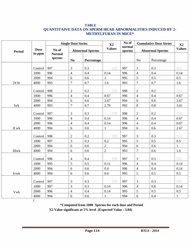
Page 114 RTLS - 2014
TABLE
QUANTITAIVE DATA ON SPERM HEAD ABNORMALITIES INDUCED BY 2-
METHYLFURAN IN MICE*
Period Dose
in ppm
Single Dose Series X2
Values
No of
normal
sperms
Cumulative Dose Series X2
Values No of
Normal
sperms
Abnormal Sperms Abnormal Sperms
No Percentage No Percentage
24 hr
Control 997 3 0.3 997 3 0.3
1000 996 4 0.4 0.14 996 4 0.4 0.14
2000 994 6 0.6 1 995 5 0.5 0.5
4000 993 7 0.7 1.6 993 7 0.7 1.6
Iwk
Control 998 2 0.2 998 2 0.2
1000 996 4 0.4 0.67 996 4 0.4 0.67
2000 994 6 0.6 2.67 994 6 0.6 2.67
4000 993 7 0.7 2.79 992 8 0.8 3.61
II wk
Control 997 3 0.3 998 2 0.2
1000 996 4 0.4 0.14 996 4 0.4 0.67
2000 996 4 0.4 0.14 996 4 0.4 0.67
4000 994 6 0.6 1 994 6 0.6 2.67
IIIwk
Control 998 2 0.2 997 3 0.3
1000 997 3 0.3 0.2 995 5 0.5 0.5
2000 994 6 0.6 2 994 6 0.6 1
4000 994 6 0.6 2 993 7 0.6 1.6
Ivwk
Control 996 4 0.4 997 3 0.3
1000 995 5 0.5 0.11 996 4 0.4 0.14
2000 994 6 0.6 0.4 996 4 0.4 0.14
4000 994 6 0.6 0.4 995 5 0.5 0.5
Vwk
Control 997 3 0.3 997 3 0.3
1000 997 3 0.3 0.14 996 4 0.6 0.14
2000 996 4 0.4 0.14 995 5 0.5 0.5
4000 994 6 0.6 1 994 6 0.4 1
(
*Computed from 1000 Sperms for each dose and Period
X2 Value significant at 5% level (Expected Value : 3.84)

Page 115 RTLS - 2014
PHOTOS
Discussion:
A close look at Table reveals that none of the quantitative data on the sperm head abnormality
assay following single and cumulative dose administration of 2-Methylfuran was significant .An
absence of cognisable difference between two sets of data indicates that 2-Methylfuran does not
induce any point mutations. In the context of the observations of Cohen et al ( 13), that “negative
chromosome findings with drugs that enjoy wide spread use should be published”(pp2425-2426).
Thus the present results have got great relevance and significance.
References:
1.Fishbein.L.(1978). Potential industrial carcinogens and mutagens (pub. 560/5-77-005-U.S.
Environmental protection Agency, Washington, 1977. T.M.Maugh, II) Science.199:162.
2.Wynder, E.L.(1975).Introductory remarks in nutrition in the causation of Cancer,Res.35:3328.
3.Wynder, E.L. and G.B. Gori. (1977). Contribution of the environment to cancer incidence: An
epidemiologic exercise, J. Natl. Cancer Inst; 58:825-832.
4.Gori G.B. (1979) Food as a factor in the etiology of certain human cancers, Food Technol. ,
December : 48-56.
5.Wieshburger , J.H , Reddy , B.S. , Hill , P. , Cohen , L.A. , Wynder E.L. and Spingarn , N.E.
(1980). Nutrition and Cancer – on the mechanism barring on causes of cancer of the colon ,
breast , prostrate and stomach . Bull. N. Y. Acad . Med. 56: 674-790.
6.Doll , R. and Peto, R. (1981). The causes of cancer, Quantitative estimates of avoidable risks of
cancer in the U.S today. J. Natl . Cancer Inst. , 66:1192-1305.

Page 116 RTLS - 2014
7.Higginson, J.(1979). Perspective and future development in research on environmental
carcinogenesis. In “Carcinogens:Identification and mechanism of action” (Eds.) Griffin , A. And
Shaw R. , Raven Press , New York , 187-208.
8.Higginson, J.(1980).Multiplicity of factors involved in cancer patterns and trends . J. Environ.
Pathol .Toxicol.3 : 113-125.
9.Higginson, J. And Muir,C.S. (1981).Epidemology of Cancer. In: “Cancer Medicine”, (Eds.)
Holland, J.F. and Frei, E., Lea and Febiger,Philadelphia.
10.Maga , J.A.(1979) . Furans in food.CRC Crit . Ref .Food .Sci . Nutr .11 : 355-400.
11.Wyrobek, A.J. and Bruce, W.R (1978). The induction of sperm shape abnormalities in mice
and humans. In : “Chemical mutagens principles and methods for their detection”.(Eds.) A.
Hollaender and F.J. de Series , Vol . 5: Plenum Press, New York and London, 257-285.
12.Pillai, S.K. and Sinha, H.C. (1968). Statistical methods for biological workers.Ram Prasad
and Sons, Agra, 85-87.
13.Cohen, M.M.,Hirschhorn, K and Frosch, W.A. (1969).Cytogenetic effects of tranquillizing
drugs invivo and invitro. Jour. Amer. Med. Assoc. 207: 2425-2426.

Page 117 RTLS - 2014
Phytochemical analysis of Bacopa monnieri
Dr. G. Swathi
Lecturer, Zoology Department, Government Degree College for Women,
Srikalahasthi-517644
ABSTRACT
Bacopa monnieri, an important medicinal plant belonging to the family of
Scrophulariaceae, which occurs naturally in India and has a long history of being used in the
traditional Ayurvedic medicine in the treatment of a number of disorders, particularly those
involving anxiety, intellect and poor memory. Traditionally, it was used as a brain tonic to
enhance memory development, learning, and concentration was extracted with ethanol solvent
for the purpose of preliminary screening .The different qualitative chemical tests were performed
on the extract to detect the various phyto constituents or antioxidants present in them. The
photochemical screening reveals the presence of many therapeutically important compounds
such as glycosides, alkaloids, saponins, phenols, proteins and carbohydrates. Since the phenolic
compounds have remarkable antioxidant activities, our present work aims at evaluating the
antioxidant activities by the three in vitro models such as DPPH free radical scavenging and
Superoxide Anion Radical scavenging Activity. From the present investigative phytochemical
analysis of Bacopa monnieri plant extract it is revealed that the antioxidant activity of the plant
material is due to the presence of phenolic compounds.
Key words: Bacopa monnieri, glycosides, alkaloids, saponins, phenols.
Introduction
Traditional knowledge of medicinal plants has always guided the search for new cures. In
spite of the advent of modern high throughput drug discovery and screening techniques,
traditional knowledge systems have given clues to the discovery of valuable drugs (Buenz et al.,
2004). Traditional medicinal plants are often cheaper, locally available and easily consumable,
raw or as simple medicinal preparations. Nowadays, traditional medicinal practices form an
integral part of complementary or alternative medicine. A member of the Scrophulariaccae
family, this small, creeping herb is much-branched, with small leaves and light purple flowers.
Bacopa’s soft, sessile leaves are succulent, reniform and spatulate, measuring about 2.5 mm in
length. The plant’s stem has soft, ascending branches, and is about 10-30 cm long and 1-2 mm
thick. Brahmi’s flowers are blue or white in color, and grow on peduncles that are usually longer
than the leaves. The fruits are ovoid, with acute capsules included in a persistent calyx.

Page 118 RTLS - 2014
Chemical composition
The active constituents of Brahmi include alkaloids,
saponins, and sterols. Many of these principles,
including the alkaloids brahmine and herpestin, and the
sapponins d-mannitol and harsaponin, acid A, and
monnierin, were isolated in India long ago. Other active
constituents have since been identified, including betulic
acid, stigmastarol, beta-sitosterol, as well as numerous
bacosides and bacopasaponins. The constituents
believed to be responsible for Brahmi’s cognitive effects
are bacosides A and B. (Chakravarty et al., 2003)
Material and Methods
Collection of the plant material
Bacopa monnieri (BM) plant was collected from Tirumala hills and indentified by a
botanist, Department of Botany, S.V.University, Tirupati. A voucher specimen was deposited in
the herbarium of the Department of Botany, S.V.University, Tirupati (Voucher no. 1688).
Preparation of Plant Extract
The whole plant was shade-dried and powdered. The plant material was percolated with
circulating 95% ethanol (200 ml) for three rounds. The residue was extracted twice using the
same procedure. The extract was filtered and concentrated under reduced pressure in the Buchi
rotavapour yielding a greenish-black sticky residue. Finally the extract was freeze - dried and
was used for further studies.
Determination of in vitro antioxidant activity
Determination of Antioxidant Activity by DPPH Radical Scavenging Method
The extract solution for the DPPH test (Fargare, 1995) was prepared by re-dissolving 0.2 g of
each dried extract in 10 ml methanol. The concentration of DPPH solution was 0.025 g in 1000
ml of methanol. Two ml of the DPPH solution was mixed with 10, 20 and 40 μl of the plant
extract/ethanol solution and transferred to a cuvette. The reaction solution was monitored at 515
nm, after an incubation period of 30 minutes at room temperature, using a UV-Visible
spectrophotometer. The inhibition percentage of the absorbance of DPPH solution was calculated
using the following equation:
Inhibition%= (Abst=0 min -Abst=30 min)/ Abst=0 min ×100
Where Abst=0 min was the absorbance of DPPH at zero time and Abst=30 min was the
absorbance of DPPH after 30 minutes of incubation. Ascorbic acid (0.5 mM) was dissolved in
methanol and used as a standard to convert the inhibition capability of plant extract solution to
Page 119 RTLS - 2014
the Ascorbic acid equivalent. IC50 is the concentration of the sample required to scavenge 50%
of DPPH free radicals.
Determination of Superoxide Anion Radical Scavenging Activity
Superoxide Anion Radical scavenging Activity was measured (Duan, 2007) with some
modifications. The various fractions of plants extracts were mixed with 3 ml of reaction buffer
solution (pH 7.4)containing 1.3 μM riboflavin, 0.02 M methionine and 5.1 μM NBT. The
reaction solution was illuminated by exposure to 30W fluorescent lamps for 20 minutes and the
absorbance was measured at 560 nm using UV-VIS spectrophotometer. Ascorbic acid (0.5 mM)
was dissolved in methanol and used as a standard to convert the inhibition capability of plant
extract solution to the Ascorbic acid equivalent. The reaction mixture without any sample was
used as negative control. The Superoxide anion radical scavenging activity (%) was calculated
as:
Where, Ao = absorbance of positive control
As = absorbance of sample
PHYTOCHEMICAL STUDY
Preliminary phytochemical screening was performed [Harborne1988]. The presenceof
phytoconstituentssuch as Tannins,Saponins, Phenolic, Terpenoids, Steroids, Phytosterol
Anthraquinone, Glycosides Flavonoid’swere confirmed by the following procedure.
1. TEST FOR TANNINS
About 2ml of filtered extract was taken in a test tube and 2ml of ferric chloride added. The
presence of blue-black coloured precipitate indicates the presence of tannins.
2. TEST FOR SAPONINS
To 0.5 ml of extract was added 5ml of distilled water in a test tube. The solution was shaken
vigorously and observed for stable persistent forth.
3. TEST FOR TERPENOIDS (salkowski test)
To 0.5 ml each of the extract was added 2ml of chloroform. Concentrated Sulphuric acid 3ml
was carefully added to form a layer. A reddish brown colouration of the interface indicates the
presence of terpenoids.
4. TEST FOR CARDIAC GLYCOSIDES
To 2ml of extract 1ml of glacial acetic acid containing one drop of ferric chloride solution. This
was underlayed with 1ml of sulphuric acid. A brown ring at the interface indicated the presence
of a deoxysugar characteristic of cardenolides.A violet ring may appear below the brown ring,

Page 120 RTLS - 2014
while in theacetic acid layer a greenish ring may form just above the brown ring and gradually
spread throughout this layer.
5. TEST FOR ANTHRAQUINONE
0.5ml of the extract was boiled with 10ml of sulphuric acid and filtered while hot. The filtrate
was shaken with 5ml of chloroform. The chloroform layer was pipette in to another test tube
observed for colour changes.
6. TEST FOR FLAVONOIDS
Dilute ammonia 5ml was added to the extract. Concentration sulphuric acid 1ml was added. A
yellow colouration that disappears on standing indicates the presence.
7. TEST FOR STEROIDS
To 1ml extract 10ml of chloroform was added.10ml of concentrated sulphuric acid was added
carefully to form coloured layer. Upper layer turns red.sulpuric acid layer forms yellow with
green fluorescence, indicates the presence of steroids.
8. TEST FOR PHYTOSTEROL
1ml of extract was dissolved in 10ml of chloroform and 10ml concentrated sulphuric acid along
the side of the test tube. Brown ring indicates presence of phytosterol.
9. TEST FOR PHENOLIC
2ml of extract 1ml ferric chloride was added, a blue or green colour indicates presence of
phenolic.
STATISTICAL ANALYSIS
Values of the measured parameters were expressed as mean ± SEM. One way-
ANOVA (F value) was used to test the significance of difference among more than two
arithmetic means, followed by Post – hoc test (Scheffe multiple comparison) to test the
difference between each two means. The significance was considered at p values <0.05. All the
statistical analyses were processed using Statistical Program of Social Sciences (SPSS) for
windows, version 11.5.
RESULT:
Determination of Antioxidant Activity by DPPH Radical Scavenging Method
DPPH Radical scavenging activity was determined of ethanolic extract. The experiments were
performed in triplicates and mean values of Antioxidant activity of each of the fraction of the
plant extract were determined. The extract of Bacopa monnieri (whole plant) tested for

Page 121 RTLS - 2014
antioxidant activity using DPPH radical scavenging was determined, the ethanolic extract
showed the maximum antioxidant activity with IC50 values of 49.12μg/ml as shown in and
Table 1. Superoxide anion radical scavenging was determined for ethanolic extract. The
experiments were performed in triplicates and mean values of antioxidant activity of each of the
fraction of the plant extract were determined. The values of antioxidant activity determination by
Superoxide Anion Radical Scavenging method follow the same order as that of DPPH assay.
Bacopa monnieri (whole plant) tested for antioxidant activity using Superoxide Anion radical
scavenging method, the extract showed the maximum antioxidant activity with 65.68 % and
62.34 % inhibition as shown in Table 1.
Table:1 Antioxidant Activity by DPPH Radical Scavenging Method and Superoxide
Anion Radical Scavenging Method
Method Method (IC50); μg/ ml
/%
Plant DPPH-Radical Scavenging 49.12
Superoxide Anion Radical
Scavenging Method (%)
62.4%
Table:2 Preliminary phytochemical screening of callus extraction of Bacopa monnieri.
In the present study preliminary phytochemical tests on the ethanolic extract revealed the
presence or absence ofTannins, Saponins Phenolic, Terpenoids, Steroids,
Phytosterol,Anthraquinone, Glycosides Flavonoid’s(given tab-1)
Sl No. Phytochemical(Test) Ethanol Aqueous
1. Tannins
+ +
2. Saponins + +
3. Terpenoids + +
4. Glycosides _ +
5. Anthroquione _ _
6. Flavanoids + _
7. Steroids + +
8. Phytosterol + +
9. Phenolic
_ _
Page 122 RTLS - 2014
DISCUSSION
A free radical can be defined as any molecular species capable of independent existence
that contains an unpaired electron in an atomic orbital. The presence of an unpaired electron
results in certain common properties that are shared by most radicals. Many radicals are unstable
and highly reactive. They can either donate an electron to or accept an electron from other
molecules, therefore behaving as oxidants or reductants (Cheeseman, 1993). The most important
oxygen-containing free radicals in many disease states are hydroxyl radical, superoxide anion
radical, hydrogen peroxide, oxygen singlet, hypochlorite, nitric oxide radical, and peroxynitrite
radical. These are highly reactive species, capable in the nucleus, and in the membranes of cells
of damaging biologically relevant molecules such as DNA, proteins, carbohydrates, and lipids
(Young, 2001).Free radicals attack important macromolecules leading to cell damage and
homeostatic disruption. Targets of free radicals include all kinds of molecules in the body.
Among them, lipids, nucleic acids, and proteins are the major targets causing oxidative
stress.Cells are protected against oxidative stress by an interacting network of antioxidants (Sies,
1997). Antioxidants were believed to be a cure for many disorders. The investigations on Bacopa
monnieri plant extract were found to yield substantial positive data pointing towards the
evidence of antioxidant activity. The data obtained from DPPH Radical Scavenging and
Superoxide Anion Radical Scavenging assay clearly suggested that the antioxidant activity of
Bacopa monnieri. It can also be noted that the extract of Bacopa monnieri was found to scavenge
the free radicals such as peroxides, superoxides and hydroxyl radicals. From the various test it
was concluded that Bacopa monnieri is found to possess greater antioxidant potential.
Antioxidants are intimately involved in the prevention of cellular damage -- the common
pathway for cancer, aging, and a variety of diseases.
The phytochemical screening of ethanolic extract of Bacopa monnieri showed the
presence of many bioactive chemicalconstituents including Tannins, Saponins, Terpenoids,
Steroids, Phytosterol, Anthraquinone, Glycosides Flavonoid’s etc which are considered to be
responsible for antioxidant activity. Thus, the saponins in the extract may be suspected to possess
the activity that may be attributed to their protective action on lipid peroxidation and at the same
time the enhancing effects on cellular antioxidant defense contributing to the protection against
oxidative damage.Therefore Bacopa monnieri was considered to possess antioxidant activity and
therefore it can be used as an antioxidant along with the other suggested and proven to have
therapeutic remedies.
CONCLUSIONS
Free radicals damage contributes to the etiology of many chronic health problems such as
cardiovascular and inflammatory disease, cataract, and cancer. Antioxidants prevent free radical
induced tissue damage by preventing the formation of radicals, scavenging them, or by
promoting their decomposition. Synthetic antioxidants are recently reported to be dangerous to
human health.These experimental evidences of Bacopa monnieri suggest that because of its

Page 123 RTLS - 2014
antioxidant activity, it may be useful in the treatment of human pathologies in which free radical
production plays a key role. The antioxidant property may be attributed to its phytochemical
constituents.
REFERENCES:
1. Buenz, I.F.F; Schenepple, D.J; and Motley, T.J. (2004) Technique: bio prospecting
historical herbal texts by hunting for new leads in old tomes. Trends in pharmacological
Sciences 25, 494-498.
2. T Fargare. Eur. Poly. J., 1995, 31, 489-497.
3. X Duan et al., 2007. Molecules, 2007, 12 (4), 759-771.
4. Harborne JB: Phytochemical methods, Third Edition, Chapman and Hall, London, 1988,
p 117.
5. Cheeseman KH, Slater TF. An introduction to free radicals chemistry. Br Med Bull.
1993;49:481–93. [PubMed]
6. Young IS, Woodside JV. Antioxidants in health and disease. J Clin Pathol. 2001;54:176–
86.
7. Sies H. Oxidative stress: Oxidants and antioxidants. Exp Physiol. 1997;82:291–5.
[PubMed]
Page 124 RTLS - 2014
Oxidative stress induced renal damage: A pharmacological appraisal of Annona squamosa
leaves extract under STZ induced diabetic rats
Mohan.PVenkata Subbaiah.G, Krishna Reddy.K, Chandra Kala.P, Sathyavelu Reddy.K*
Department of Zoology, Division of Ethno Pharmacology, Sri Venkateswara University,
Tirupati, Andhra Pradesh
*Corresponding author: [email protected]
Abstract
Annona squamosa is a multipurpose tree with edible fruits and is a source of medicinal
value. The leaves of Annona squamosa were proved to have antioxidant activity. Oxidative stress
is a major factor in the pathogenesis of diabetic complications. In the recent time plant sources
are explored widely for medicinal value. The present study was made to investigate the
protective effect of ethanol extract of Annona squamosa leaves on altered blood glucose levels
and endogenous antioxidant enzymes in Streptozotocin induced diabetic rats ((STZ 50mg/kg
body weight). Elevated blood glucose levels and down regulation of antioxidant enzymes
activity including superoxide dismutase (SOD), Catalase (CAT), glutathione peroxidase (GPx)
were observed in diabetic group when compared to control. However, blood glucose levels
declined and SOD, CAT, GPx activities invigorated with Annona squamosa leaves extract
(300mg/kg body weight) treatment to diabetic rats.Hence, I concluded that ethanol extract of
Annona squamosa leaves potentially reduce the diabetic complication by reducing the glucose
levels and improve the endogenous antioxidants activity.
Keywords:Annona squamosa, Antioxidant enzymes, Diabetes, Oxidative stress
Introduction
The alarming scalationintheglobalincidenceandprevalenceof
diabetesmellitusoverthepast20years,toitscurrentestimates ofalmost300million,isprojectedto
further increaseto440million individualsbytheyear2030 (Shawet al.,2010).Type 2 diabetes is
aconsequence of an imbalance between insulin responsiveness andinsulin production. One of the
first clinical signs is glucose intolerance,due to insulin resistance of peripheral organs, including
liver, fat andmuscle, and is initially compensated by increased pancreatic β-cellinsulin secretion.
Hyperglycemia-inducedimpairmentsinredoxbalanceareconsidereda
keytriggerofdiabeticcomplications,throughup regulated generationofreactiveoxygenspecies(ROS)
(Sourris et al., 2012; Haidaraet al., 2006), togetherwithan
impairedabilityoftheendogenousantioxidantdefensesystemto removethem (Woldet al.,2005)
which lead to damage of cellular organelles andenzymes, increased lipid peroxidation, and
development of insulinresistance (Maritim et al., 2003). These consequences of oxidative stress
can promote the development of complications of diabetesmellitus.The chronic hyperglycemia of
diabetes is associatedwith damage, dysfunction and failure of various organs, e.g.kidneys, retina,
heart, liver, peripheral and central nervous system,over the long term (Fajans, Cloutier,
Page 125 RTLS - 2014
&Crowther, 1997).High intracellular glucose concentration has been suggested to be a
prerequisite for the development of functionaland structural changes in the kidney typical of
diabetic nephropathy.
Herbal medicines have been used for many years by differentcultures around the world,
both for the prevention and managementof diabetes (Marles and Farnsworth, 1995). Annona
squamosa (A. squamosa), commonly known as custard apple, is cultivated throughout India,
mainly for its edible fruit. The plant is attributed with medicinal properties, which include
antifertility and antitumor activities in rats(Asolkar et al., 1992). The young leaves of A.
squamosa along with five grains of black pepper are used extensively for its antidiabetic activity
by tribal men (1985). Some other medicinal values includinganti-inflammatory, antioxidative
and antimicrobial potential (Yanget al., 2008; Baskar et al., 2007).Ethanol extract of the leaves
and stem is reported to have anti-cancer activity (Bhakuniet al., 1969). In the Ayurvedic system
of medicine, herbal extracts but not purified compounds have been used from centuries because
many constituents with more than one mechanism of action are considered to be beneficial.
No detailed study has been carried out on the effect of ethanolic extract of A.squamosa
leaves on nephro protective activity in STZ induced diabetic rats .Hence, the present study was
planned to evaluate the nephro protective effect of ethanolic extract of A.squamosa leaves on
blood glucose levels and antioxidant activity in diabetic rats.
2. Material and Methods
2.1Chemicals
All chemicals were Analar Grade (AR) and obtained from the following scientific
companies: Sigma (St. Louis, MO, USA), Fisher (Pitrsburg, PA, USA), and Merck (Mumbai,
India).
2.2Preparation of ginger ethanol extract
Annona leaves were collected from Seshachalam hills, Tirupati A.P. India. Four
kilogram of fresh leaves were cleaned, washed under running tap water, cut into small pieces, air
dried and powdered. 200g of this powder were macerated in 1000 ml of 99.9% ethanol for 48
hours at room temperature and were then filtered. The filtrate was concentrated to dryness under
reduced pressure in a rotary evaporator (Model: HS-2005V).
Page 126 RTLS - 2014
2.3Animals
Thirty Wister strain male albino rats, weighing 250 ± 20 g, were obtained from the Indian
Institute of Science, Bangalore, India. The animals were housed in plastic cages and kept in the
laboratory under constant temperature (27 ± 20C) with 12 hrs dark and 12 hrs light for
throughout treatment period. The rats were received a standard pellet diet (Sai Durga feed,
Bangalore, India) and water ad libitum. The experiments were carried out in accordance with
guidelines and protocol approved by the Institutional Animal Ethics Committee resolution
number (10 /i/a/CPCSEA/IAEC/SVU/KSR-GVS/dt 15/11/2010).
Induction of diabetes:
After fasting for 15 hrs, rats were injected intraperitonially with a single dose of
Streptozotocin of 50mg/kg body weight obtained from Sigma Chemical Co., St. Louis, MO,
USA, freshly dissolved in citrate buffer (pH 4.5). After injection, they had free access to food
and water and were given 15% glucose solution to drink overnight to counter hypoglycemic
shock. Diabetes in rats was identified by moderate polydipsia and marked polyurea. From the
second day onwards fasting blood samples were collected from the rats by tail vein and the blood
glucose was measured by Accuecheck Sensor comfort glucometer (Manufacturer-Roche
Germany) to know the induction of diabetes.
2.4Experimental Design
The rats were divided into five groups each of six rats and the treatment was given
everyday via orogastric tube for seven weeks.
Group I, Normal Control (NC): rats received 0.9% saline and feed with normal diet
Group II, Annona leaves extract (At): rats received 300mg/kg of ethanol extract of Annona
leaves extract orally for one month treatment
Group III, Diabetic control (DC):Streptozotocin50mg/kg b.w isgiven intraperitonially for the
induction of diabetes to this group
Group IV, Diabetic + Glibenclamide treatment (Di + Glb): diabetic rats treatedwith
glibenclamide (20 mg/kg body weight orally).
Group V, Diabetic + Annona treatment (Di+ At): Diabetic rats received Annona ethanolic
extract (300 mg/kg) for a period of 30 days.

Page 127 RTLS - 2014
After completion of one month treatment, the animals were sacrificed by cervical
dislocation. Blood was collected from heart puncture and kidney tissue was used for the
histopathological studies and biochemical assays.
Estimation of blood glucose levels
Estimation of blood glucose was carried out by using accuchek glucometer.
Estimation of antioxidant enzymes
SOD activity was assayed by the method of Misra and Fridovich (1972)at 480 nm for 4
min on a Hitachi U-2000 spectrophotometer.Activity was expressed as the amount of enzyme
that inhibits the oxidation of epinephrineby 50%, which is equal to 1 U per milligram of protein.
CAT activity wasdetermined at room temperature by using the modified version of Aebi
(1984)and absorbance of the sample was measured at 240 nm for 1 min in a UV-
spectrophotometer.Activity of GPx was determined by the method of Flohe and Gunzler(1984)
in the presence of NADPH and absorbance was measured at 340 nm usingcumene hydrogen
peroxide. GR enzyme activity was determined according to themethod of Carlberg and
Mannervik (1985).
Histopathology
Kidney tissues were isolated from the animal and washed with ice-cold saline. They were then
fixed in 10% formalin solution. Sections of 3 µm thickness were stained with haematoxylin and
eosin (H & E) for histopathological examination in the light microscope.
Statistical analysis
Statistical analysis of data was represented as mean ± SD. All the statistical analyses
were carried out by SPSS software. Dunnett’s multiple comparison tests and one-way analysis of
variance (ANOVA) were used to assess the differences, p<0.001was considered as highly
statistically significant.
Results
Blood glucose levels
The blood glucoselevels in STZ-injected diabetic rats were drasticallyincreased from the
baseline (Table 1). This increaseof blood glucose was almost three-fold higher evenafter 30 days
compared to the control rats. However,it was found that the elevated blood glucose levels

Page 128 RTLS - 2014
indiabetic rats dropped significantly (P < 0.001) after30-day Annona leaves extract
administration. Glibenclamide, whichhas been used as a standard diabetic drug to comparethe
beneficial effects of Annona leaves extract, showed significant(P < 0.001) decrease in blood
glucose levelsand this was almost equal to the normal control rats.
Table 1 : Effects of Annona leaves ethanolic extract and glibenclamide treatments on blood
glucose level in streptozotocin- induced diabetic rats. All the values are mean SD of six
individual observations
Groups Blood glucose levels
Initial Final
Group I (NC) 209 5.19 229 5.20
Group II (At) 226** 3.46 199** 3.63
Group III (DC) 216 4.86 164** 1.29
Group IV (Di +Glb) 226** 3.26 205* 4.30
Group V (Di+ At) 215 3.26 192** 3.43
Values in the parentheses denote per cent change over normal control Values are significant
compared to normal control at *** P<0.05
Antioxidant enzymes
Fig.2represents the activity of oxidative stress marker enzymesin the different
experimentalgroups. There was significant depletion in the SOD, CAT and GPx activities in the
kidney tissues of diabetic rats (p< 0.001) .However Annona treatment significantly reversesthe
SOD, CAT and GPx back to normal levels (p < 0.001).
Histopathology of kidney
Fig. 3 illustrates the pathological changes in thekidney of a diabetic rat including severe tubular
degeneration,degeneration of glomeruli, focal necrosisof tubules, cystic dilatation of tubules and
fatty infiltration.The above mentioned pathological symptomswere reduced in ginger-treated

Page 129 RTLS - 2014
diabetic rats. The histologicalpicture of Annona leaves treated diabetic rats showedrestoration of
glomeruli, tubules and renal cells.
Discussion:
Diabetes mellitus is a life-threatening metabolic disorder and itis estimated that its annual
incidence rate will continue to increasein the future, worldwide. Hyperglycaemia, the primary
clinicalmanifestation of diabetes mellitus, is associated with the developmentof micro- and
macro-vascular diabetic complications. Traditionalplant remedies have been used for centuries in
thetreatment of diabetes (Kesari, Gupta, & Watal, 2005), but only afew have been scientifically
evaluated. Therefore, we have investigatedthe effect of Annona leaves ethanolic extract on blood
glucose levels and biomarkers of oxidative stress in kidney tissues of STZ induced diabetic rats.
Streptozotocin injection resulted in diabetes mellitus, whichmay be due to destruction of
beta cells of Islets of Langerhans asproposed by others (Kavalali et al., 2002). Diabetes arises
from irreversibledestruction of pancreatic beta cells, causing degranulationand reduction of
insulin secretion (Zhang and Tan, 2000).STZ destroy the β-cell in the pancreas and increasethe
overproduction of glucose and gluconeogenesis.Gluconeogenesis and overproduction of the
glucose isthe prime factor of the hyperglycemia (Latner 1958). In our results, blood glucose
levels were increased in diabetic rats this may be due to decreased insulin levels and initiation of
gluconeogenesis in the liver resulting in diminish of pancreatic β-cell by STZ induced
intoxication. However, Annona leaves extract recovered the blood glucose was observed in
diabetic rats. It has been reported that using medicinal plant extract to treat STZ-induced diabetic
rats results in activation of ß-cells and insulinogenic effects(Padmini and Chakrabarti, 1982).
In the current study, SOD activity was measured in kidney tissue of all the experimental
groups such as Normal control, Annona treated, diabetic, Glibenclaimade treated diabetic and
diabetic rats with Annona treatment.
Oxidative stress is the imbalance between production and removalof reactive oxygen
species. Increased oxidative stress, whichcontributes substantially to the pathogenesis of diabetic
complications,is the consequences of either enhanced ROS production orattenuated ROS
scavenging capacity. Several reports have shownthe alterations in the anti-oxidant enzymes
during diabetic condition(Preet et al., 2005). SODhas been postulated as one of the most
importantenzymes in the enzymatic antioxidant defensesystem which catalyses the dismutation

Page 130 RTLS - 2014
ofsuperoxide radicals to produce H2O2 and molecularoxygen hence diminishing the toxic effects
causedby these radicals (Baynes, 1995).
Fig:1. Effect of NC,At, DC(STZ),Di+Glbt and Di+At on SOD, CAT in Se GPx activities in
kidney tissue of male albino rats. The Values marked with *(P<0.05) **(P<0.01).
In our result, reduced activity of SOD in kidney tissue was observed in diabetic rats.
Similar consequence were also ensued in Shanmugam et al (2011) studies which have been
demonstrated that SOD activity was decreased as a result of hyperglycemia provoked free
radicals may cause to inactivate the enzymes in diabetic kidney. SOD activity increased with
treatment of ethanolic extract of Annona leaves to diabetic rats. It has been reported that Annona
leaves extract have shown free radical scavenge activity that could be cause to enhance the SOD
activity in diabetic condition.
Catalase (CAT) is a hemeprotein which catalyses the reduction ofhydrogen peroxides and
protects the tissues fromhighly reactive hydroxyl radicals. The superoxideanion has been known
to inactivate CAT, which isinvolved in the detoxification of hydrogenperoxide.Reduced activity
0
0.2
0.4
0.6
µ m
ole
s o
f h
ydro
gen
pe
roxi
de
d
egr
ade
d/m
g …
Treatment
CAT
NC
At
DC
Di+Glbt
Di+At
0
0.5
1
µ m
ole
s o
f N
AD
PH
o
xid
ize
d/m
g p
rote
in/m
inu
te
Treatment
Se-GSH-Px
NC
At
DC
Di+Glbt
Di+At
0
2
4
6
8
sup
ero
xid
e a
nio
n r
ed
uce
d
/mg
pro
tein
/min
ute
Treatment
SOD
NC
At
DC
Di+Glbt
Di+At
Page 131 RTLS - 2014
of CAT in kidney tissues have been observed in diabetic rats and this activity may result in a
number of deleterious effects due to accumulation of hydrogen peroxide (Satheesh et al., 2004;
Huang et al., 2007).The activity of CAT in the kidney of diabetic rats was found to be decreased
there by leading to a possible sustained elevation of intracellur hydrogen peroxide levels due to
metabolic imbalances during diabetes accompanied by inhibition or down regulation of
antioxidant enzymes as CAT may be responsible for oxidative stress in the disease state of
hyperglycemia.
Basha and Subramanian (2011) have been reported that Annona increases plasma
antioxidant capacity and decreases free radical toxicity in diabetic rats.Ghadir et al., (2011)
Annona scavenges the free radicals involved in the process of oxidative stress thus enhancing the
concentration of antioxidant enzymes. In the present study similar consequences may occurred
CAT activity was significant increase with Annona leaves extract treated to diabetic rats
GPx plays a primary role in minimizingoxidative damage. It has been proposed that GPx
isresponsible for the detoxification of H2O2 in lowconcentration whereas Catalase comes into
playwhen GPx pathway is reaching saturation with thesubstrate. GPx catalyze the reduction of
hydrogenperoxide and hydro peroxides to non-toxicmetabolites. Depletion of GSH will render
the enzyme GPx inactivation and or less active (Mahendran and Shyamala Devi, 2001). The
reason for the reduced activity of GPx might lie in the decreased mitochondrial pool size of GSH
because of entry of cytosolic GSH in to mitochondria is impaired indeed it has been reported that
CAT and GPx can be inactivated by oxidative attacks (Fernadez et al., 199l;Kerem and Korean,
2003).
Glutathione peroxidase (GPx), an important antioxidant enzyme,was significantly
decreased in diabetic kidney tissues,which indicates impaired scavenging of H2O2 and
lipidhydroperoxides. This result is consistent with the studies of Friesen, Buchau, Schott-Ohly,
Lggssiar, and Gleichmann (2004). The decreasedGPx activity represents a compensatory
mechanism to degrade H2O2. However, GPx activity significantly upregulated with Annona
treatment in diabetic treated group. The increased GPx activity in Annona treated rats showing
the antioxidant property of Annona in removing the hydro peroxides from the kidney tissue. The
Phenolic compounds mentioned may involve in the antioxidant defense mechanism and protect
the tissues against the free radicals (Manju and Nalini, 2005).

Page 132 RTLS - 2014
4 Impact of ginger on kidney tissue during diabetic condition.
A) Photomicrograph of normal control (NC) kidney, showing Normal rat kidney architecture shows glomeruli (GM) and proximal convoluted tubules. BC-Bowman’s capsule; ET -Renal tubule; PN-Pycnotic nuclei
B) Plant extract control rat kidney architecture shows mild glomerular (GM) inflammation with lymphocytes and mild degenerative changes in renal tubules. BC-Bowman’s capsule; ET -Renal tubule; PN-Pycnotic nuclei
C) STZ Induced rat kidney shows Glomeruli architecture is damaged within folding of the Bowman’s capsules and mild necrotic changes in renal parenchyma. SCB-structural changes in bowman’s capsule; DET-Degenerative changes in renal tubules; PN-Pycnotic nuclei
D) Glebenclamide treated STZ Induced rat kidney shows Intact Glomerules architecture of cell is normal and mild inflammatory changes. RCET-Regenerative changes in elongated renal tubules; RBC-Regenerative bowmans capsule; PN-Pycnotic nuclei
E) Plant extrct treated STZ Induced rat kidney shows normal, healthy and functional Glomeruli and regenerative renal tubules with minimal inflammation is seen.No evidence of any necrotic changes.RCET-Regenerative changes in elongated renal tubules; RBC-Regenerative bowmans capsule
F) The histopathological studies of kidney revealed severe tubulardegeneration,
degeneration of glomeruli, focal necrosis of tubules,cystic dilatation of tubules and fatty
infiltration in diabetic controlrats, which might be associated with increased diuresis and
renalhypertrophy in diabetic rats. The collecting tubules showed thatdilation may be
under the pressure of increased urine flow. Theseobserved pathological changes were
decreased in diabetic ratswhen they were treated with Annona. The glomeruli appear to
A B
C D
E

Page 133 RTLS - 2014
be restored,tubules also appeared to be regenerated and less fatty infiltrationwas observed
in diabetic rats treated with Annona , whichmay be due to the protective effect of Annona
(Fig.4). Thus, in additionto the blood glucose-lowering effect, histopathological
observationsalso support the notion that Annona at 1% and 2% producedsignificant
increments of antioxidant enzymes and protected therenal tissues in diabetic rats.
G) In conclusion, our study suggests that Annona mayhave beneficial effects in diabetes that
hold the hope of a new generationof antidiabetogenic drugs. However, comprehensive
chemicaland pharmacological research is required to find out the exactmechanism of
Annona for its antidiabetogenic effect and to identifythe active constituent(s) responsible
for this effect.
References:
Aaishwarya B.Deshmukh and Jayvadan K. Patel.(2011 Nov-Dec).Aqueous extract of Annona
squamosa (L.) ameliorates renal failure induced by 5/6 nephrectomy in rat Indian J
Pharmacol 43(6): 718–721.
Aebi, H. (1984). Catalase. Methods in Enzymology, 105, 125–126.
Asolkar LV, Kakkar KK, Chakre OJ. (1992). Glossary of Indian Medicinal Plants with Active
Principle. New Delhi: Publication and Information Directorate: 72-3.
Atique A, Iqbal M, Ghouse AKM. (1985) Use of Annona squamosa and Piper nigrum against
diabetes. Fitoterapia.56:190-2.
Baskar. R.., Rajeswari. V., and. Kumar. T.S. (2007),,In vitro antioxidant studies in leaves of
Annona species, Ind J Exp Biol 45. 480–485.
Baynes JW. (1995). Reactive oxygen in the aetiology and complications of diabetes. Drug, diet
and disease, Mechanistic approach to diabetes. In: Ioannides C, Flatt PR editors.
Ellis Horwood Limited, Hertfordshire, Vol. 2, 2003-2231.
Bhakuni, D. S., Dhar, M. L., Dhar, M. M., Dhawan, B. N. and Mehrotra, B. B (1969).Screening
of Indian Plants for biological activity. Part II*. Indian J. Exp. Biol., ,7, 250–26
Cooper.M.T.,Nguyen.M.E., Ritchie.T.V., Forbes.R.H.,J.M.(2012). Ubiquinone(coenzymeQ10)
prevents RenalMitochondrial dysfunctioninan experimental model of type2
diabetes. Free Radic.Biol.Med. 52:716–723.
Flohe, L., & Gunzler, W. A. (1984).Glutathione peroxidase.Methods in Enzymology. 105, 115–
121.
Friesen, N. T., Buchau, A. S., Schott-Ohly, P., Lggssiar, A., & Gleichmann (2004).Generation of
hydrogen peroxide and failure of antioxidative responses in pancreatic islets of
male C57BL/6 mice are associated with diabetes induced by multiple low doses of
streptozotocin. Diabetologia, 47(4), 676–685.
Ghadir A. El-Chaghaby *, Abeer F. Ahmad, Eman S. (2011) Evaluation of the antioxidant and
antibacterial propertiesof various solvents extracts of Annona squamosa L. leaves
Ramis Peer review under responsibility of King Saud
University.doi:10.1016/j.arabjc06.019

Page 134 RTLS - 2014
Haidara,M.A.,Yaseen,H.Z.,Rateb,M.,Ammar,H.,Zorkani,M.A.(2006).Roleof
oxidativestressindevelopmentofcardiovascularcomplicationsindiabetesmellitus.
Curr.Vasc.Pharmacol. 4:215–227.
Hayath basha s. k. and subramanian. s.* subramanian & basha, (2011)biochemical evaluation of
antidiabetic and antioxidant potentials of annona squamosa leaves extracts
studied in stz induced diabetic rats ijpsr, vol. 2(3): 643-655.
Huang, H., Shan, J., Hong Pan, X., Ping Wang, H., BoQian, L., Xia, Q. (2007). Carvedilol
improved diabetic rat cardiac function depending on antioxidant ability. Diabetes
Research and Clinical Practice.75, 7-13.
Kesireddy Sathyavelu Reddy a,* (2011) Protective effect of dietary ginger on antioxidant
enzymes and oxidative damagein experimental diabetic rat tissuesFood Chemistry
124 1436–1442.
Kondeti Ramudu Shanmugam a, Korivi Mallikarjuna b, Kesireddy Nishanth c, Chia Hua Kuo b,
Kotkar HM, Mendki PS, Sadan SVGS, Jha SR, Upasani SM, maheshwari VL
(2002).Antimicrobial and pesticidal activity of partially purified flavonoids of Annona
squamosa.Pest Management Sciences. 58: 33-37
Latner A (1958) Clinical Biochemistry. Saunders, Philadelphia, pp 48–50.
Maritim, A.C., Sanders, R.A., Watkins, J.B., (2003). Diabetes, oxidative stress, and antioxidants:
a review. J. Biochem. Mol. Toxicol. 17, 24–38.
Misra, H. P., & Fridovich, I. (1972). The role of superoxide anion in the autooxidation of
epinephrine and a simple assay for superoxide dismutase. Journal of Biological
Chemistry, 247, 3170–3175.
Padmini K, Chakrabarti CH (1982). Effects of Bittergourd (Momordica charantia) seed and
glibenclamide in streptozotocin-induced diabetes mellitus. Indian J Exp Biol,
20:232-5.
Preet, A., Gupta, B.L., Yadava, P.K., Baquer, N.Z.,( 2005). Efficacy of lower doses of vanadium
in restoring altered glucose metabolism and antioxidant status in diabetic rat lenses.
J. Biosci. 30, 221–230.
Satheesh, M. A., Pari, L. (2004). Antioxidant effect ofBoerhavia diffusa L. in tissues of alloxan
induced diabetic rats. Indian. J. Exp. BioL, 42: 989-992.
Shaw,J.E.;Sicree,R.A.;Zimmet,P.Z. (2010). Global estimates of the prevalence of diabetes for
2010 and 2030. Diabetes Res.Clin.Pract. 87:4–14.
Sourris,K.C.;Harcourt,B.E.;Tang,P.H.;Morley,A.L.;Huynh,K.;Penfold,S.A.;Coughlan,
Wold,L.E.;Ceylan-Isik,A.F.;Ren,J.(2005).Oxidativestressandstresssignaling:menace
of diabetic cardiomyopathy. Acta Pharmacol.Sin. 26:908–917.
Yang. Y.L., Hua. K.F., Chuang. P.H., Wu. S.H., Wu. K.Y., Chang., F.R., and. Wu. Y.C.,
(2008).New cyclic peptides from the seeds of Annona squamosa L, and their anti-
inflammatory activities, J Agric Food Chem 56 386–392.

Page 135 RTLS - 2014
Sub-cloning and expression of nucleocapsid protein in E.coliDH5α
T. Madhavi,B. Mahitha, CH. N. Durga Maha Lakshmi, and N. John Sushmaa,*
aDepartment of Biotechnology, Sri Padmavati Mahila University, Tirupati-517502.
Abstract
Sub cloning is a technique used to move a particular gene of interest from a parent vector
to a destination vector in order to further study its functionality. N protein which encapsulates the
negative strand RNA genome and in association with the L protein P protein and subunits of the
viral RNA polymerase it forms the ribonucleoprotein core of the virus. The N protein plays
several important roles during the virus transcription and replication cycles. Restriction enzymes
are used to excise the gene of interest (N protein) from the clone it is purified and isolated
plasmid DNA which contain N protein this is digested with BamHI to check the orientation and
further cleaved restriction enzyme with EcoRI for 12 hrs to release the N gene and DNA
analysed on agarose gel. Ligation is carried out along with a vector (PBR322), ligation product is
transformed into competent E.coli DH5α. The transformation results the white coloured colonies
are indicative of positive clones while the blue colonies are indicates negative. The positive
clones were induced by 1M IPTG and protein expression was checked by SDS-PAGE. DNA
polymerase and primers are used to amplify specific target Sequence during multiple cycles of
invitro DNA synthesis.
Key words:E.coli DH5α , N protein, BamHI, PBR322, and RNA polymerase
INTRODUCTION
The N protein is a structural subunit of nucleocapsid and is the most abundant protein found
during infection. It is the key protein which encapsulates the negative strand RNA genome and in
association with the L protein, P protein and subunits of the viral RNA polymerase it forms the ribo
nucleoprotein core of the virusN protein is a highly charged, basic protein that can self associate to
form dimers (He et al., 2004; Surjit et al., 2004). N protein is thought to participate in the
replication and transcription of viral RNA and to interfere with cell-cycle processes of host cells
(Parker & Masters, 1990).
The three-dimensional structure of the N-terminal portionof the protein is similar to those of
other RNA-binding one of the most abundant viral proteins producedinside a coronavirus-infected
cell is the nucleocapsid(N) protein, a multifunctional phosphoprotein (Calvo et al.,2005; Chen et
al., 2005; Jayaram et al., 2005). During the replicative mode of the transcribing complex, the
nascent RNA is encapsulated by denovo synthesized N protein, resulting in the synthesis of the full
length plus strand N-RNA complex. This in turn serves as a template to synthesize and replicate the
minus strand N-RNA complex for its external morphogenesis and budding as viral particles.
In molecular biology, subcloning is a technique used to move a particular gene of interest from
Page 136 RTLS - 2014
a parent vector to a destination vector in order to further study its functionality. Subcloning is not to
be confused with molecular cloning, a related technique. Restriction enzymes are used to excise the
gene of interest (the insert) from the parent (mycoplasma). The idea behind using the same
restriction enzymes is to create complementarysticky ends, which will facilitate ligation later on.
The enzyme used to ligate DNA fragments is T4 DNA ligase, which originates from the T4
bacteriophage. CIAP is also added to prevent self-ligation of the destination vector insert should
become successfully incorporated into the parent plasmid.The plasmid is often transformed into a
bacterium like E.coli. Ideally when the bacterium divides the plasmid should also be replicated. In
order to ensure growth of only transformed bacteria (which carry the desired plasmids to be
harvested) ampicillin is used in the destination vector for selection. The full length N gene encoding
the nucleocapsid protein was sub cloned into the bacterial expression vector to facilitate the over
expression of the protein in E.coli. The full length N gene was excised out of parent plasmid and
blunt end ligated in to the vector, recombinant plasmid harboring the N gene was transformed in to
DH5α cells. Colonies harboring the gene in the proper orientation were subsequently implicated in
expression of the recombinant N protein.
MATERIALS AND METHODS
All the recombinant DNA protocols for the isolation of plasmid, ligation, restriction
analysis, transformation, screening for plasmids or inserts, agarose gel and acrylamide gel
electrophoresis etc., were performed as described in molecular cloning (Sambrook et.al., 1989)
and the promega applications guide.
Isolation of plasmid DNA from clone
Grow the clone culture in LB broth at 37°c for 12-24 hrs. Centrifuge 1.5ml of culture for
10mins at 4,000 rpm. Add 1ml of Distilled water to the pellet, resuspend it and centrifuge this at
4000rpm for 5mins. Re suspend the pellet in 100ul of solution-I. Add 200ul of Solution-II and
mix by gentle inversion. Incubate this at room temperature for 10mins. Add 300ul of solution-III
Mix it immediately and Incubate on ice for 15 mins Centrifuge this at 14,000rpm for 15mins.
Transfer the supernatant to a fresh tube and add equal volume of Phenol: Chloroform (1:1)
Example: For 500µl of supernatant add 250µl of Phenol and 250µl of Chloroform Add 500ul of
Page 137 RTLS - 2014
70% Ethanol to the pellet and centrifuge this at 14,000rpm for 5 mins. Discard the supernatant
and resuspend the pellet in 30ul of nuclease free water.
Agarose gel Electrophoresis
Cast a gel of 1% by dissolving 0.2gms Agarose in 20ml of 1X TAE, add 2µl of ethidium
bromide for visualization of DNA bands load the DNA sample i.e. 10µl of sample+5µl of gel
loading dye in to the wells, allow it to electrophores for some time and visualize the bands under
gel document system.
Purification of DNA
Add 1ul RNase to the DNA mixture and incubate the sample at 37˚C for one hour, add
volume of 3M Sodium acetate (pH-5.2) and 2.5 vol absolute alcohol (90%) keep it on ice for
10mins. Spin at 12,000rpm for 15-20 minutes and collect the pellet and wash it with 150µl of
70% ethyl alcohol. Keep it at room temp for 15-20minutes Airs dry the pellet and dissolves it in
appropriate volume of Nuclease free water, then separate the DNA fragments on agarose gel
Qualitative and quantitative estimation of DNA
Spectrophotometry follows the Principle of Beer-Lambert’s law: Take 2 ml distilled water
in a cuvette and calibrate the spectrophotometer at 260nm as well as 280nm, it acts as a blank. Add
10 l of each DNA sample to 1990l distilled water and mix well note the OD at 260nmand ODat
280nm values on spectrophotometer. Calculate the OD260/OD280 ratio
Concentration = OD260 X 50ug/ml X Dilution factor
Competent cells preparation
Day: 1
Prepare the LB medium and autoclaved. Inoculate a single colony of E.coli DH5α Cells
into 10ml LB medium, grow overnight with shaking at 37ºC.
Day: 2
Prepare 100mM Calcium chloride solution and Chill this solution for further use.
Inoculate 10ml of freshly prepared LB medium with 1ml of Overnight grown culture. Grow this
at 37ºC shaking at 150 rpm to an Optical Density of 0.375 at 590 nm, pour the culture into pre
Page 138 RTLS - 2014
chilled centrifuge tubes and chill this on ice for 10 mins and centrifuge at 3200rpm for 7 mins
and discard the supernatant and resuspend the pellet gently by swirling in 5ml of Chilled
Calcium chloride solution. Centrifuge this at 3200rpm for 5 mins. Collect the pellet and
resuspend it in 2.5ml of calcium chloride Solution. Leave the resuspended cells on ice for
30mins and centrifuge this at 3200rpm for 5min discard the supernatant and resuspend the pellet
in 200μl of calcium chloride solution, dispense the cells into pre chilled sterile 1.5ml tubes and
Freeze immediately and transfer this to -20ºc for storage.
Transformation
To 200µl of competent cells add 200ng of plasmid DNA gently mix this and place it on
ice for 30 mins. Heat shock in a 42ºC water bath for 90 sec followed by rapid chilling on ice for
2 min. 800μl of sterile LB broth was added to the transformation mix and the cells were allowed
to grow for 45 min at 37°c with vigorous shaking. About 200µl of transformation mixture was
plated on to a LB-amp (100μg/ml). Incubate these plates at 37°C for 12-16 hrs in incubator until
the transformed colonies appeared on the plate. After the incubation period check for the
presence of blue and white colonies. White colonies indicate the positive clones carrying the
insert in them.
Mini preparation of plasmid DNA from transformed cells:
Single transformed colony were incubated in a 3ml of LB broth with Ampicillin (100 ug/ml)
and incubate overnight at 37°c, the cells were harvested by centrifugation pellet was resuspended
in 100ul of solution-I and incubate at room temperature for 5 min. To this 200ul of freshly
prepared solution-II and 150ul of solution-III was added and the tubes were shaken till a curdy
white precipitate was observed. The tubes were stored on ice for 10 min. The precipitate was
removed by centrifugation and collect supernatant extracted with equal volumes of phenol:
chloroform: isoamylalcohol after centrifugation upper aqueous phase was extracted with equal
volumes of chloroform and collect the aqueous phase and the DNA was precipated by adding
2.5M ammonium acetate, 2 volumes of chilled ethanol and incubated at -20°c for 1½ hours. The
DNA pellet obtained after centrifugation was washed with 70% ethanol and the pellet was
resuspended in 40μl of TE buffer (pH 8.0). Separate the plasmid DNA on the agarose gel and if
any contamination is observed purification of DNA is to be done and contamination free plasmid
is separated on the agarose gel.

Page 139 RTLS - 2014
Digestion of plasmid DNA
The plasmid DNA was setup for restriction digestion overnight at 37°c with Plasmid DNA
(2.5μl), Bam HI (2μl), assay buffer(2μl) and sterile distilled(13.5μl) water and subsequently
analyzed on the 1% agarose gels.
Restriction digestion to release the N gene:
Restriction digestion with Eco R I was setup using Restricted DNA from Bam HI (2.5μl),
Eco R I (2 μl), assay buffer (2 μl) for 12 hours. The restricted DNA was subsequently analyzed
on the 1% agarose gel.
Gel elution of the n gene insert from agarose gel:
Slice the gel piece containing the DNA into small pieces add 2.5ml of sodium iodide solution
and incubate at 55°c for 10 min in a water bath and add30μl of silica glass beads solution mix it
and leave it at room temperature for 10 min and Centrifuge add 400μl of sterile distilled water to
pellet. Separate the DNA fragments on agarose gel.
Ligation:
Recovered DNA (1µl), 1X ligase assay buffer (2µl), T4 DNA ligase (1µl) and sterile
distilled H2O (16µl) incubate at 16ºC overnight. Separate the ligated DNA on the agarose gel and
visualize on UV transilluminater.
Competent cells preparation:
Mentioned as above
Transformation:
Aliquot100µl of competent cells into 1.5ml sterile eppendorffs add 7µl of ligation mix to the
tubes gently mix this and place it on ice for 15 mins. Heat shock in a 42ºC water bath for 2mins
and place back on ice for approximately 10 mins add 1ml of LB medium and incubate this at
37ºc with shaking for one hour to allow the expression of antibiotic resistance gene. Prepare a
LB agar plate and spread 100µl of ampicillin, 40µl of X-gal and 4 µl of IPTG onto a plate let this

Page 140 RTLS - 2014
dry for some time, to this add 100µl of transformed culture and spread it, other plate without
ampicillin is control. Spread100µl of competent cell onto the agar incubate these plates at 37°C
for 24hrs in incubator. After the incubation period check for the presence of blue and white
colonies. White colonies indicate the positive clones carrying the insert in them. The white
colonies are taken and are grown in the LB broth and the protein optimization is performed.
Expression optimization
Transfer transformed colonies to 3ml of freshly prepared LB broth with 3 µl of antibiotic
ampicillin and grow this at 37ºC for 3hrs, 1ml of this culture acts as un induced control. The
remaining 2ml of culture add 2µl of IPTG and grow this at different temperatures such as 37ºC,
30 ºC, 25 ºC. Pellet the culture and check for expression of protein by loading it onto denaturing
gel i.e., SDS-PAGE.
SDS-PAGE
Assemble two glass plates (one notched) with two side spacers, clamps, grease, etc. Stand
assembly upright using clamps as supports, on glass plate pour some pre-heated 1% agarose onto
glass plate, place assembly in pool of agarose this seals the bottom of the assembly prepare
Separating gel, ensuring no air bubbles form pour into glass plate assembly carefully. Prepare
stacking gel, then pour onto top of set resolving gel, insert comb, allow to set, Remove comb, fill
with electrophoresis buffer. Assemble top tank onto glass plate assembly. Fill with 1X
electrophoresis buffer. Load the samples into the wells created by the comb. Allow the samples to
electrophorese at 100Volts. After Electrophoresis remove the gel by separating the glass plates
and transfer the gel to Staining solution. Let it stand for 2-3hrs after this transfer the stained gel to
destaining solution to remove the excess of stain observe the gel for the formation of sharp thick
bands
Results and discussion
In association with the L&P proteins and subunits of the RNA polymerase, the N protein
forms the ribonucleo protein core of the virus. This RNP core is implicated in both transcription
of the viral RNA as well as replication of the RNA genome. N protein is composed of three
distinct regions containing RNA-binding motif(s), and appropriate signals for modulating cell
signaling (Jaehwan et al., 2005)

Page 141 RTLS - 2014
The basic and foremost step involved is the isolation of plasmid DNA from the clone.
The isolated DNA is depicted in the figure given below:
Figure: isolated DNA from clone
M = 1 Kb Marker
The Qualitative and quantitative estimation of the isolated DNA was done by
Spectrophotometry. The Absorbance was measured at 260nm and further at 280nm for the
quality and the following expression was used to calculate the quantity of DNA:
Concentration = OD260 X 50ug/ml X Dilution factor
= 0.236 X 50 X 2000/10
= 2360
Hence for 1µl the concentration is approximately. The quality of DNA is calculated by
taking the ratio absorbance at 260 and 280 pure DNA is in the range of 1.8-2.0. Hence the DNA
isolated is pure and free of any protein and RNA contamination.
Plasmid (PBR322) containing the full length N gene was digested with Bam HI to check
the orientation of the N gene. This was an essential step to identify the appropriate expression
vector into which the gene could be sub cloned. Bam HI was chosen as it has unique sites within
the N gene and on the plasmid carrying it. Restriction digestion with EcoRI for 12 hours was
done to release the N gene and the DNA sample was analysed on 1 % agarose gel. Gel elution of

Page 142 RTLS - 2014
the N gene insert from agarose gel was done by using silica glass bead method. In order to obtain
the fragment corresponding to the N gene insert, double digestion was carried out.
Double digestion with Restriction Enzymes: Bam HI.
The ligation reaction was set up using a vector: Insert ratio of 1:3. This was incubated at
16°c for 16hrs. The ATP-dependent DNA ligases catalyze the joining of single-stranded breaks
(nicks) in the phosphodiester backbone of double-stranded DNA in a three-step mechanism. The
first step in the ligation reaction is the formation of a covalent enzyme-AMP complex. The co-
factor ATP is cleaved to pyrophosphate and AMP, with the AMP being covalently joined to a
highly conserved lysine residue in the active site of the ligase. The activated AMP residue is then
transferred to the 5' phosphate of the nick, before the nick is sealed by phosphodiester-bond
formation and AMP elimination. The reaction catalyzed by the eubacterial NAD+-dependent
eubacterial ligases is essentially identical but for the initial formation of the enzyme-AMP
intermediate resulting in the breakdown of NAD+ and release of nicotinamide mononucleotide
(NMN) rather than pyrophosphate; although these two groups of enzymes belong to the same
family of nucleotidyl transferases , they share almost no protein sequence similarity outside the
catalytic core.
After the ligation reaction, the ligation product was transformed into competent E.coli
DH5α. The transformation resulted in the production of colonies with two differently coloured
colonies. The white coloured colonies are indicative of positive clones while the blue colonies
are indicative of clones into which the plasmid wouldn’t have transformed. The transformed

Page 143 RTLS - 2014
positive clones were induced by 1 M IPTG and protein expression was checked by 10% SDS-
PAGE.
Transformed clones
M Protein

Page 144 RTLS - 2014
M= Protein marker :14.4 Kda – 76 Kda
10% SDS-PAGE gels showing the protein bands (induced)
Applications of DNA subcloning are expanding rapidly in all fields of biology and medicine.
In medical genetics such applications range from the prenatal diagnosis of inherited human
diseases to the characterization of oncogenes and their roles in carcinogenesis. Pharmaceutical
applications include large-scale production from cloned human genes of biologic products with
therapeutic value, such as polypeptide hormones, interleukins, and enzymes. Applications in
public health and laboratory medicine include development of vaccines to prevent specific
infections and probes to diagnose specific infections by nucleic acid hybridization or polymerase
chain reaction (PCR).
SUMMARY AND CONCLUSSON
Subcloning is a technique used to move a particular gene of interest from a parent vector
to a destination vector in order to further study its functionality. N protein which encapsulates the
negative strand RNA genome and in association with the L protein, P protein and subunits of the
viral RNA polymerase it forms the ribonucleoprotein core of the virus. The N protein plays
several important roles during the virus transcription and replication cycles. Restriction enzymes
are used to excise the gene of interest (the insert) from the parent (mycoplsma), the insert is
purified with a common purification method is gel isolation. Isolation of plasmid DNA from the
clone and it is purified. Plasmid containing the full length N gene was digested with Bam HI to
check the orientation of the N gene. The digested product is further cleaved with Restriction
enzyme with Eco RI for 12 hours was done to release the N gene and the DNA sample was
analysed on 1 % agarose gel. Then the ligation reaction was set up using a vector: Insert ratio of
1:3. The ligation product was transformed into competent E.coli DH5α. The transformation
results, the white coloured colonies are indicative of positive clones while the blue colonies are
indicative of clones into which the plasmid wouldn’t have transformed. The transformed
positive clones were induced by 1 M IPTG and protein expression was checked by 10% SDS-
PAGE. DNA polymerase and Primers are used to amplify specific target DNA sequences during
multiple cycles of invitro DNA synthesis, making it possible to detect target DNA sequences.
Page 145 RTLS - 2014
References
1. Calvo, E., Escors, D., Lopez, J. A., Gonzalez, J. M., Alvarez, A., Arza, E. & Enjuanes,
L.(2005). Phosphorylation and subcellular localization of transmissible gastroenteritis virus
nucleocapsid protein in infected cells. J Gen Virol 86, 2255–2267.
2. Chen, H., Gill, A., Dove, B. K., Emmett, S. R., Kemp, F. C., Ritchie, M. A., Dee, M. &
Hiscox, J. A. (2005). Mass spectroscopic characterization of the coronavirus infectious
bronchitis virus nucleoprotein and elucidation of the role of phosphorylation in RNA
binding using surface plasmon resonance. J Virol 79, 1164–1179.
3. He, R., Leeson, A., Andonov, A. & 8 other authors (2003). Activation of AP-1 signal
transduction pathway by SARS coronavirus nucleocapsid protein. Biochem Biophys Res
Commun 311, 870–876.
4. Jaehwan You, Brian K. Dove, Luis Enjuanes, Marta L. De Diego, Enrique Alvarez, Gareth
Howell, Paul Heinen, Maria Zambon and Julian A (2005). Subcellular localization of the
severe acute respiratory syndrome coronavirus nucleocapsid protein. J of Gen Virol 86,
3303–3310.
5. Jayaram, J., Youn, S. & Collisson, E. W. (2005). The virion N protein of infectious
bronchitis virus is more phosphorylated than the N protein from infected cell lysates.
Virology 339, 127–135.
6. Parker, M. M. & Masters, P. S. (1990). Sequence comparison of the N genes of five strains
of the coronavirus mouse hepatitis-virus suggests a three domain-structure for the
nucleocapsid protein. Virology 179, 463–468.
7. Sambrook, J. Fritsch, E.F. and Maniatis, T. (1989). Molecular Cloning. A Laboratory
Manual, 2nd Edition (3 volumes). Cold Spring Harbor Laboratory Press, Cold Spring
Harbor, N.Y
8. Surjit, M., Liu, B., Jameel, S., Chow, V. T. & Lal, S. K. (2004). The SARS coronavirus
nucleocapsid protein induces actin reorganization and apoptosis in COS-1 cells in the
absence of growth factors. Biochem J 383, 13–18.
Page 146 RTLS - 2014
Curcuma longa in combination with Trigonella foenum graecum restored altered lipid
metabolic profiles in alloxan induced type 1 diabetic rat kidneys
M.Guru Sekhar1, S. Sadak Basha
2, S.Mannur Ismail
2, M. Bhaskar
2*
1SKBR Govt. Degree College, Macherla, Guntur-522426,A.P,India 2Dept. of Zoology, S. V. University, Tirupati-517502, A.P, India.
Email: [email protected]
ABSTRACT
Curcuma longa (CL) and Trigonella foenum graecum (TFG) are medicinal plants used in
Ayurveda for treating various diseases; one of it is diabetes mellitus. The present study was
undertaken to investigate the effects of ethanol extracts of CL and TFG on blood glucose levels,
tissue lipid profile and lipid peroxidation in alloxan induced diabetes on treatment for 30 days.
Albino rats each weighing 180 ± 30g were given a single dose of intra peritoneal injection of 120
mg of alloxan per kg body weight. The rats were divided into five groups; each group consists of
six rats. 1st group treated as control, 2
nd group (alloxan induced diabetic rats), 3
rd group (Diabetic
+ CL), 4th
group (Diabetic + TFG) and 5th
group (Diabetic + CL + TFG). Ethanolic extracts of
CL rhizome powder and TFG seed powder was administered orally 250 mg/kg b.w/day for 30
days. The impact of CL and TFG on blood glucose levels in experimental rats were studied and
the levels of lipid peroxides (TBARS) and tissue lipids (cholesterol, Triglycerides) were also
estimated in both control and experimental rats. Treatment withCL and TFG individually and
combinedly resulted in a significant reduction of blood glucose levels. CL and TFG also resulted
in a significant decrease in tissue lipids and lipid peroxidation. The analyzed results conclude
that a combination of herbal plants exerted a significant anti hyperlipidemic and anti
peroxidative effect in alloxan induced diabetic rats when compared to individual treatment.
Key words: Curcuma longa, Trigonella foenum graecum, antiperoxidation, lipids, alloxan
induced diabetes.
Introduction: Diabetes mellitus is a major public health problem in the world. There was
estimation that 143 million people worldwide are suffering in the form of diabetes and this
number would increase by almost five times by 2030. Diabetes mellitus is syndrome, initially
characterized by a loss of glucose homeostasis resulting from defects in insulin secretion, insulin
action both resulting impaired metabolism of glucose and other energy- was yielding fuels such
as lipids and protein (Scheenet al 1990). Insulin dependent diabetes mellitus (IDDM) or type 1
diabetes is an auto immune disorder caused by auto aggressive T-lymphocytes that infiltrate the
pancreas and destroy insulin producing -cells. This leads to hypoinsulinemia and thus a
hyperglycemic condition (Bachet al 1995), which over a period of time develops diabetic
complications such as nephropathy, retinopathy, neuropathy and cardiac problem (Arky et
al1982). Most of the metabolic complications associated with type 1 diabetes are due to insulin
deficiency and related glucose under utilization of the insulin dependent tissue such as liver, and

Page 147 RTLS - 2014
glucose over utilization in insulin dependent tissue, such as Kidney (Sochor et al 1985).The
insulin deficiency causes excessive break down of lipid in adipose depots, resulting in increased
level of free fatty acids (FAA) (Gupta et al 1999). Significant changes in lipid metabolism and
structure also occur in diabetes (Sochar et al 1985). In diabetic rats, increased lipidperoxidation
was also associated with hyperlipidemia (Morel and Chisolm et al1989).
Liver and kidney participates in the uptake, oxidation and metabolic conversion of free
fatty acids, synthesis of cholesterol, phospholipids, and triglycerides. During diabetes, a
profound alteration in the concentration and composition of lipids occurs. Several workers have
shown the presence of lipid deposits in the kidney of diabetic human and experimental animals
and they have proposed that these deposits may play an important role in the pathogenesis of
diabetic kidney disease (Guijarro et al 1995; Lee et al 1991). Base on the WHO
recommendations hypoglycemic agents of plant origin used in traditional medicine are
important. Many traditional plant treatments for diabetes are used throughout the world. Plant
drugs (Baily and Day et al 1989), and herbal formulation (Mitra et al 1996; Annapurna et
al,2001; Bhattacharya et al., 1997)are frequently considered to be less toxic and more free from
side effects than synthetic one. Hence treatment with herbal drugs has an effect on protecting β-
cells and smoothing out fluctuation in glucose levels (Gia, 2003, Elder et al 2004). Various
herbal formulations such as Curcuma longa and Trigonella foenum graecum are well known for
their antidiabetic effects.
Curcuma longa is a rhizomatousherbaceousperennial plant of the ginger family, Zingiberaceae.
Research on Curcuma longa has been focused on alzheimer's, arthritis, cancer and diabetes. In
India, Curcuma longa has been in use as a culinary ingredient since 3000 BC. As a medicine it is
used to treat a wide variety of ailments including stomach ache, skin problems, muscular
problems and arthritis. Practitioners of traditional Indian medicine believe that curcumin powder
is beneficial against many diseases including biliary disorders, diabetes, hepatic disorders,
rheumatism, sinusitis, cancer and Alzheimer disease. Some of the compounds such as ascorbic
acid, beta-carotene, caffeic acid, curcumin, eugenol, p-coumaric acid, p-methoxy-cinnamic acid,
protocatechuic acid, syringic acid and vanillic acid, which perform antioxidant activity are
present in Curcuma longa might be responsible in inducing antioxidant action on treatment of
diabetes.

Page 148 RTLS - 2014
Fenugreek (Trigonella foenum graecum) is an annual herb that belongs to the family
fabaceae. It has a wide range of medicinal applications, one of its constituent alkaloids, called
'trigonelline', has shown potential for use in cancer therapy. Many researches have shown that
the seeds can inhibit cancer of the liver, lower blood cholesterol levels. The seed and leaves are
anticholesterolemic, anti-inflammatory, antitumor, carminative, demulcent, deobstruent,
emollient, expectorant, febrifuge, galactogogue, hypoglycaemic, laxative, parasiticide,
restorative and uterine tonic. Fenugreek seeds are used as a traditional remedy for the treatment
of diabetes (Basch et al ,2003). Some of the compounds such as Alanine, Ascorbic-Acid, Beta-
Carotene, Beta-Sitosterol, Histidine, Isovitexin, which perform antioxidant activity are present in
Trigonella foenum graecum and might be responsible in inducing antioxidant action on treatment
to diabetic rats.
The present investigation was under taken to study the effect of CL and TFG
constituents on hyperglycemic, oxidative stress, lipid peroxidation(LPO) and tissue lipid profile
(TC, TG) in Alloxan induced diabetic rats.
Material and methods:
Animals: A total of 30 adult (3 months old) male Wistar stain rats weighing 180 ± 30 g
obtained from animal house of Bangalore were used for this study. They were housed six per
each polycarbonate cage under standard laboratory conditions at a room temperature of 24±20C,
humidity of 45-64% with 12h light/dark cycle. The animals were fed with standard diet obtained
from Hindustan Lever Limited, Mumbai and tap water was given adlibitum until treatment or
time of sacrifice. All institutional guide lines were adhered to in the care and treatment of the
animals used in the present study.
Induction of Diabetes:
Alloxan is used to induce Insulin dependent diabetes mellitus (IDDM) (Type 1). Single
injection of Alloxan at a dosage concentration of 120mg/Kg body weight is usually enough for
induction. So, after fasting for 18h, rats were injected intraperitoneally with a single dose of
120mg/Kg body weight. Alloxan freshly dissolved in saline. After injection, they were allowed
to have free access to food and water. 15% glucose solution was also provided overnight to
counter hypoglycemic shock. Diabetes in rats was identified by moderate polydipsia and marked

Page 149 RTLS - 2014
polyuria. From the second day onwards fasting blood samples were collected from the rats by tail
vein and the blood glucose was measured by Accu Chek Glucometer (Sensor Comfort) to
confirm the induction of diabetes. If the blood glucose levels were more than 300mg/dl, insulin
(IIU protamine Zinc insulin) is given to the diabetic rats and the rats are allowed to acclimatize
for diabetic condition for one week. After one week the rats with hyperglycemia (blood glucose
level 250 mg/dl) were selected and used for the end of experiment.
Preparation of plant extracts: The fine powder of Curcuma longa rhizomeand Trigonella
foenum graecum seeds powder were purchased (AGMARK symbol) in Tirupati. The powder is
extracted by cold percolation with 95% ethanol for 24h. The extract was recovered and 95%
ethanol was further added to the plant material and the extraction was continued. The process
was repeated three times. The three extractions were pooled together, combined, filtered and the
filtrate was concentrated to dryness under reduced pressure in rotary evaporator. The resulting
ethanol extract was air-dried. Finally yellow and light yellow powdery, crude ethanol extract of
Trigonella foenum graecum and Curcuma longa were obtained respectively. Without any further
purification the plant crude ethanol extract was used in the study.
Experimental design: The rats were divided into five groups of six each. Overnight fasted
animals were injected with Alloxan (120 mg/Kg b.w) dissolved in 5 units of saline)
intraperitoneally (i.p). The experimental period was 30 days. Group I: Control rats, Group II:
Diabetic rats, Group III: Diabetic rats were given Curcuma longa ethanol extract (250 mg/kg
b.w/day), Group IV: Diabetic rats were given Trigonella ethanol extract (250 mg/kg b.w/day),
Group: V Diabetic rats treated with both Curcuma and Trigonella ethanol extracts. At the end of
30 days the animals were deprived of food overnight and sacrificed cervical dislocation and the
kidney tissues were excised at -800C.
1. Estimation of Blood glucose:
Blood glucose was measured by using Accu Chek glucometer (Sensor Comfort) on 0 day,
7th
day and 30th
day of all 5 group rats.
2. [Lipid Peroxidation (LPO)]:
This assay is used to determine MDA levels as described by (Ohkawa et al., 1979). The Kidney
tissues were homogenized (5% - w/v) in 50 mM phosphate buffer (pH 7.0) containing 0.1 mM

Page 150 RTLS - 2014
EDTA, The homogenates were centrifuged at 10,000 rpm for 10 min at 0°C in cold centrifuge. The
separated supernatant part was used for the estimation. 200 µl of the tissue extract was added to
50 µl of 8.1% sodium dodecyl sulphate (SDS), vortexed and incubated for 10 min at room
temperature. 375 µl of 20% acetic acid (pH 3.5) and 375 µl of thiobarbituric acid (0.6%) were added
and placed in a boiling water bath for 60 min. The samples were allowed cool at room
temperature. A mixture of 1.25 ml of butanol: phyridine (15:1) was added, vortexed and
centrifuged at 1000 rpm for 5 min. The colored layer (500 µl) was measured at 532 nm using 1, 1, 3,
3-tetraethoxypropane as a standard. The values were expressed in µ moles of malondialdehyde
formed / gram wet weight of the tissue.
3. Triglycerides :
Triglycerides were estimated by the method of (Natelson et al1971) with slight modifications
as given below. Triglycerides were assayed by hydrolyzing them to glycerol and the liberated glycerol
was determined.
Tissue homogenates were prepared in 1N H2SO4 and to it 4 ml of chloroform was added.
0.5 ml of tissue homogenate was taken. To it 0.5 ml of 1N H2SO4 and 4 ml of chloroform were added.
The contents were centrifuged at 1000 rpm for 15 min 0.5 ml of chloroform layer was taken and to it 0.4
ml of methanol and 0.1 ml of alkaline barium solution were added and the contents were heated
for 30 min at 80°C, the total volume was made up to 1 ml with 2N H2SO4 and centrifuged for 10 min
at 1000 rpm. 0.5 ml of this supernatant was taken and to it 0.1 ml of sodium periodate was added
and shaken well for 1 min, 0.1 ml of sodium arsenate and 5 ml of chromotrophic acid reagent was
added and heated for 30 min and cooled. The samples were read at 575 nm in Spectrophotometer
against the reagent blank. The results were finally expressed in mg of triglycerides / gram wet weight of
the tissue.
4. Total Cholesterol: The total cholesterol content was estimated using Liebermann Burchard
reaction as described by Natelson, (1971). The Kidney tissues were homogenized in isopropanol.
The contents were centrifuged at l000 rpm for 15 min. 0.5 ml of supernatant was taken and to it 4 ml of
cholesterol reagent was added. Then the contents were heated at 90°C for 15 min. After cooling, the
samples were read at 560nm in spectrophotometer against the reagent blank. The results were finally
expressed in mg of total cholesterol/gram wet weight of the tissue.
Enzyme units:

Page 151 RTLS - 2014
Enzyme activities were expressed in standard unit’s i.e. moles of product formed or substrate
cleaved/mg protein/hour.
Statistical analysis: The data obtained was expressed as Mean value with their SD. Readings of the five
different groups were compared using one way ANOVA analysis with DUNNETTS multiple comparison
test. Stastical analysis was performed using SPSS. Using M.S -Office, Excel software the data has been
analyzed for the significance of the main effects and treatment s along with their interactions. The results were
presented with the F – value. F- Value was found to be significant with P0.001.
Results:
BLOOD GLUCOSE:
Glucose is the primarysource of energy for body's cells;fats and oils (i.e. lipids)being
primarily a compact energy store. Insulin, a hormone produced by the pancreas, helps maintain
normal blood sugar levels. Uncontrolled or high blood sugar levels can lead to health
complications such as blindness, heart disease, kidney disease. Increase in concentration of
glucose in the blood leads to a condition called "diabetic coma" or hyperglycaemia.
In the present study blood glucose levels were measured by using glucometer (Accu
Chek) in all groups of control, diabetic, diabetic treated with Curcuma, diabetic treated with
Trigonella and diabetic treated with both Curcuma + Trigonella on 0 day, 7th
day and 30th
day of
the experimental period. Blood glucose levels were expressed in units of mg/dL In case of group
II (diabetic rats) the blood glucose levels were significantly increased after induction of Alloxan.
In the groups III, IV and V where the rats were subjected to CL, TFG and CL+TFG respectively
have showed decreased blood glucose levels. However, the decrease is more significant in Group
V where the rats were subjected to combined extracts of CL+TFG. The decreased levels of blood
glucose in group V back to control levels after treatment with both Curcuma + Trigonella extract
treatment. The various blood glucose values of alterations are as shown in Table 1.1.
Lipid peroxidation:
Peroxidation is important in food deterioration and in the oxidative modification of biological
molecules particularly lipids. Inhibition of lipid peroxidation by any external agent is often used
to evaluate its antioxidant activity. In the present study the inhibition of lipid peroxidation
induced by H2O2 by the seed extract was carried out in rat kidney.

Page 152 RTLS - 2014
Table 1 represents the concentration of TBARS, Cholesterol and triglycerides in kidney of
control and experimental rats respectively. Kidney of diabetic rats showed significantly increased
levels of TBARS, Cholesterol and Triglycerides, when compared with normal rats. In rats treated
with Curcuma and Trigonella there was a significant decrease in the content ofTBARS,
Cholesterol and Triglycerides in both the tissues, when compared with diabetic control rats. The
combined treatment of CL+TFG was most effective in preventing alteration of lipid profile. The
maximum prevention in the alteration of the lipid profile was observed in the combined
treatment of CL and TFG by 30 days.
Table: 1.Showing Blood glucose levels in the control and experimental rats
Group/
Day Group I Group II Group III Group IV GroupV
0 Day
Mean
SD
105 6.3
107 5.6
106 6.4
102 5.3
104 9.4
7th
Day
Mean
SD
106 7.9 225 20.2
179 15.3
213 16.2
225 19.8
30th
Day
Mean
SD
108 7.2
290 26.7
101 10.9
102 12.2
81 6.3
Values are mean ± S.D. of 6 individual rats
Values in the parenthesis are % change from that of control Values are significantly different from control at P < 0.0001
DISCUSSION:
Most of the body cells use the sugar called glucose as their major source of energy.
Glucose molecules are broken down within cells in order to produce adenosine tri
phosphate (ATP) molecules, energy-rich molecules that power numerous cellular
processes. Glucose molecules are delivered to cells by the circulating blood and
therefore, to ensure a constant supply of glucose to cells, it is essential that blood glucose
levels be maintained at relatively constant levels.

Page 153 RTLS - 2014
Table: 2. Showing Lipid metabolic profile levels in Kidney tissues of control and
experimental rats
Parameter / Groups
Group I Group II Group III Group IV Group V
Lipid peroxidation
38.167.497
51.819.16
35.293.148
33.183.20
31.026.585
Triglycerides 1.8100.071
4.920.032
1.6200.059
1.4100.067
1.320.079
Total Cholesterol
63.861.559
89.022.149
59.131.636
58.322.185
53.011.442
Values are mean ± S.D. of 6 individual rats
VALUES in the parenthesis are % change from that of control Values are significantly different from control at P < 0.0001
Blood glucose concentration is maintained within the normal range of 70 to 110 mg/dl.
The levels of glucose in the blood are monitored by the cells in the pancreas. If the blood glucose
level falls to dangerous levels (as in very heavy exercise or lack of food for extended periods),
the Alpha cells of the pancreas release glucagon, a hormone which alerts the liver to increase
blood glucose levels and converts stored glycogen into glucose (Glycogenesis). Thus glucose is
released into the bloodstream, increasing blood sugar levels. There are several other causes for
an increase in blood sugar levels. Among them diabetic stress due to the accumulation of reactive
oxygen species is a major cause.
A significant increase in blood glucose levels found in alloxan treated rats could be due
to the destruction of pancreatic beta-cells by alloxan induced oxidative stress. The elevations of
glucose in alloxan treated rats (group II) were due to an oxidative stress produced in the pancreas, due
to a single strand break in pancreatic islets DNA (Omamto and Uchigata et al 1981). In
experimental diabetes, enzymes of glucose and fatty acid metabolism are markedly altered; hence blood
glucose levels were increased (Gottried and Rosenberg, 1973; Sochar et al., 1985).
Blood glucose levels were decreased in group III, where diabetic rats subjected to
Curcuma. Oral administration of Curcuma longa extract lowers blood glucose and attenuates
alloxan-induced hyperlipidemia in diabetic rabbits. (Sarah Nwozo, et al., 2009). Curcuma longa
rhizomes have been reported to possess active constituents showing blood glucose lowering
Page 154 RTLS - 2014
activity in alloxan induced diabetic rats (Shnkar et al., 1980). The administration of an aqueous
extract of turmeric and abromine powder resulted in a significant reduction in blood glucose and
an increase in total hemoglobin (Halim Eshrat Ali Hussain et al 2002).
Blood glucose levels were decreased in group IV, where diabetic rats subjected to
Trigonella. Seeds of fenugreek have been showed multiple benefits in patients with diabetes such
as reduction of blood sugar and its complications (Madar et al., 1988; Preet et al., 2006).
Whether using the whole seeds or extracts showed that fenugreek seeds decreased fasting blood
sugar levels in animals. The administration of Trigonella foenum graecum seed powder to
diabetic animals have showed lower blood glucose levels and partially restore the activities of
key enzymes of carbohydrate and lipid metabolism close to normal values in animal model
systems (Vats et al., 2003; Raju et al., 2001).
However decreased blood glucose levels were more significant in group V, which were
administered combined plant extracts. Chattopadhyay 1999 reported that Azadiracta indica leaf
extract was found to have the most potential activity as blood sugar lowering agent followed by
Catharanthus rooseusGymnemaq sylvestre and Oscimum sanctum. Thus, the present study
provides substantial evidence on the hypoglycemic action of CL, TFG ethanolic extracts in
alloxan induced diabetic rats. The significant antihyperglycemic effect may be due to the
potentiation of plasma insulin effect by increasing either the pancreatic secretion of insulin from
the existing β-cells or its release from the bound form, as evidenced by the significant increase in
the level of insulin by CL, TFG ethanol extracts in diabetic rats.
Diabetes is known to affect large number of metabolic pathways, including lipid
metabolism, by altering the activities of various enzymes involved in these pathways. Alloxan, a
beta cytotoxin, induces "chemical diabetes" (alloxan diabetes) in a wide variety of animal species
by damaging the insulin secreting pancreatic -cell, resulting in a decrease in endogenous insulin
release, which paves the ways for the decreased utilization of glucose by the tissues (Omamotoet
al1981). Renal glucose uptake is markedly increased during diabetes and is inversely correlated
with renal FFA uptake, which is reduced during diabetes (Meyer et al 1998). Thus the major
assumption that kidney lipid deposits, which plays an important role in the pathogenesis of
diabetic kidney disease (Guijarro et al 1995; Lee et al 1991), originate from increased levels of
Page 155 RTLS - 2014
serum lipids has to be further investigated with the possibility of an increase in renal lipid
synthesis resulting in renal lipid accumulation.
Apart from the regulation of carbohydrate metabolism, insulin also plays an important role in
the metabolism of lipids. Insulin is potent inhibitor of lipolysis. Since it inhibits the activity of
the hormone sensitive lipases in adipose tissue and suppresses the release of free fatty acids
(Lociet al1994). During diabetes, enhanced activity of this enzyme increases lipolysis and
releases more free fatty acids in to the circulation (Agardhet al1999). Increased fatty acids
concentration also increases the β-oxidation of fatty acids, producing more acetyl CoA and
cholesterol during diabetes. In normal condition, insulin increases the receptor-mediated removal
of LDL-cholesterol and decreased activity of insulin during diabetes causes
hypercholesterolemia. Hypercholesterolemia and hypertriglyceridemia have been reported to
occur in diabetic rats. The increased concentration of free fatty acids in kidney may be due to
lipid breakdown and this may cause increased generation of NADPH, which results in the
activation of NADPH dependent microsomal lipid peroxidation. The results show increased lipid
peroxidation in the tissues (liver and kidney) of diabetic group. Previous studies have reported
that there was an increased lipid peroxidation in liver, kidney and brain of diabetic rats (Latha et
al 2003; Ananthan et al 2004). Administration of CL, TFG and CL+TFG reduced the lipid
peroxidative markers in kidney tissue of diabetic rats. This indicates that CL, TFG inhibit
oxidative damage due to the antiperoxidative effect of ingredients present in CL, TFG.
Trigonella seed powder has shown many positive and encouraging results in the management of
diabetes. (Sharma et al, 1990). Scavenging activity of Curcumin administration also reduces
lipid peroxidation (Elder et al 2004). The use of CL+TFG in combination can be suggested as an
effective alternative for the amelioration of diabetes.
Triglycerides:-
Accumulation of triglycerides is one of the risk factors in Coronary Heart Disease
(CHD). The significant increase in the level of triglycerides in liver and kidney of diabetic
control rats may be due to the lack of insulin. Since under normal condition, insulin activates the
enzyme lipoprotein lipase and hydrolysis triglycerides (Frayn 1993). In the present study
triglyceride content was decreased in diabetic rats treated with CL plant extract .The significant
fall in serum triglyceride levels indicate antitriglyceride activity, antiglycemic, antilipidemic and

Page 156 RTLS - 2014
antioxidant properties of Curcuma. Triglyceride content was decreased in diabetic rats treated
with combination of Curcumin from Curcuma longa (L), and partially purified product from
Abroma augusta (L) in streptozotocin induced diabetes (Halim Eshrat and Ali Hussain et al.,
2002). Turmeric (Curcuma longa), as well as its active constituent Curcumin, inhibits lipid
peroxidation. Curcuma and Abroma augustaalso decreases serum cholesterol levels in
hyperlipidaemic rats (Halim Eshrat and Ali Hussain et al., 2002. The increase in kidney lipid
level during diabetes appears to be due to increased glucose flux and reducing equivalents
leading to enhanced over all biosynthetic pathways. Administrations of Trigonella in case of in
group IV, the TG levels were reduced in kidney tissues of diabetic rats. This indicates that TFG
inhibit oxidative damage due to the antiperoxidative effect of ingredients present in Trigonella.
The decreased total triglycerides were more pronounced in group V, whereas compared to
individual plant extract treated. The decreased lipid peroxides and tissue lipids clearly showed the
antihyperlipidemic and antiperoxidative effect of CL+TFG apart from its antidiabetic effect.
Cholesterol:-
Cholesterol is an amphipathic lipid and is present in tissues and in plasma lipoprotein
either as free cholesterol with a long chain fatty acid as cholesterol ester.
The increased levels of plasma cholesterol, LDL cholesterol and/ or the decreased levels of HDL
cholesterol are primary risk factors in the development of coronary heart diseases (Castelli et al1977;
lipid research clinics programme II, 1984). In the current study total cholesterol levels were increased
in diabetic rats (Group 2). The increase in total cholesterol was observed or reported in serum of
STZ induced diabetic rats (Nawanjoet al 2005). Pari and Saravanan, (2000) reported that higher
levels of cholesterol, triglycerides and free fatty acids inserum and tissues (Liver and Kidney) of
alloxan diabetic rats. (Li et al2007) demonstrated that some lipid peroxidation products such as
total cholesterol, MDA, HDL-Cholesterol etc in blood significantly diverges from their normal
levels in diabetic rats. These results confirm that there is a strong correlation between oxidative
stress and diabetes occurrence. The concentrations of lipids such as total cholesterol, LDL-C,
HDL-C were significantly higher in diabetic rats than in the control group. Serum total
cholesterol level increases significantly in diabetic group. These effects may be due to higher
activity of cholesterol biosynthesis enzymes and or high levels of liposysis (Mallick et al., 2006).
In CL plant extract treated diabetic rats (group III), the total cholesterol content in kidney
tissue was decreased. This was due to the hypocholestemic effects of plant extract by the
Page 157 RTLS - 2014
inhibition of cellular cholesterol synthesis.Total cholesterol levels were decreased in group IV,
where diabetic rats treated with Trigonella. Seeds of Trigonella have hypocholesterolemic and
hyperinsulinomic effects on type-I and type-II diabetes mellitus patients and experimental
diabetic animals (Puri et al2002; Stark and Madar et al1993; Dukeet al 1992). The combined
treatment of CL and TFG was found to be most effect in preventing the changes in total
cholesterol levels after 30 days treatment in group V. The decreased total cholesterol levels were
more pronounced in group V whereas compared to individual plant extract treatment. Lipid
peroxides and Total cholesterol levels clearly showed the antihyperlipidemic and
anticholesterolemic and antiperoxidative effect of CL+TFG apart from its antidiabetic effect.
The present study substantiates this new concept and suggests the effectiveness of
combined therapy of CL and TFG on the control of glucose homeostasis and lipid metabolism
during experimental diabetes and can be considered as a better alternative for further
investigation for the amelioration of diabetes.
References:-
Agardh CD, Bjorgell P, Nilson EP: The effect of tolbutamide on lipoproteins, and
lipoproteinlipase and hormone sensitive lipase. Diabetes Res Clin Pract 1999, 46:99-108.
Ajabnoor, M. A., & Tilmisany, A. K. (1988).Effect of Trigonella foenum graecum on blood
glucose levels in normal and alloxan diabetic mice.Journal of Ethnopharmacology, 22, 45–49.
Ananthan R, Latha M, Ramkumar KM, Pari L, Baskar C, Narmatha Bai V: Antidiabetic Effect of
Gymnema montanumLeaves: Effect on lipid peroxidation induced Oxidative stress in
experimental diabetes. Nutrition2004, 6:379-386.
Annapurna A, Kanaka , Mahalakshmi D, Murali Krishna K: Antidiabetic activity of a polyherbal
preparation (tincture of punchparna) in normal and diabetic rats. Indian J Exp Biol2001,
39:500-502.
Aragno M, Tamagno E, Gatto V, Brignardello E, Parola S, Danni O, Boccuzzi G:
Dehydroepiandrosterone protects tissues of streptozotocin-treated rats against oxidative
stress. Free Rad Biol Med 1999, 26:1467-1474.
Arky R A 1982 Clinical correlates of metabolic derangements of diabetes mellitus; in
Complications of diabetes mellitus (ed.) G P Kozak (Philadelphia: W B Saunders) pp 16-20.
Bach J F 1995 Insulin-dependent diabetes mellitus as a -cell targeted disease of immunoregulation.
J. Autoimrnun. 8. 439-463.
Bailey CJ, Day C: Traditional treatments for diabetes. Diabetes Care 1989, 12:553-564.

Page 158 RTLS - 2014
Basch, E., Ulbricht, C., Kuo, G., Szapary, P., & Smith, M. (2003).Therapeutic applications of
fenugreek. Alternative Medicinal Review,8(1), 20–27.
Baynes JW: Reactive oxygen in the aetiology and complications of diabetes. In Drug, Diet and
Disease, Mechanistic Approach to Diabetes. Volume 2. Edited by: Ioannides C, Flatt PR.
Hertfordshive: Ellis Horwood limited; 1995:230-31.
Bierman EL, Amaral JAP, Balknap BH: hyperlipidemia and diabetes mellitus. Diabetes 1975,
25:509-515.
Brichard S M, Ongemba E N. Girard J and Henquin J C 1994 Tissue-specific correction of
lipogenic enzyme gene expression in diabetic rats given vanadate.Diabetologia 37 1065-1072.
Elder C: Ayurveda for diabetes mellitus: a review of the biomedical literature.Altern TherHealth
Med 2004, 10:44-50.
Frayn KN: Insulin resistance and lipid metabolism.Curr Opin Lipidol 1993, 4:197-204.
Guijarro C, Kasiske B L, Kim Y, CTDonnell M P, Lee H S and Keane W F 1995 Early
glomerular changes in rats with dietary-induced hypercholesterolemia; Am. J. Kidney Dis.
26 152-161.
Gupta D, Raju J. Prakash J and Baquer N Z 1999 Change in the lipid profile, lipogenic and related
enzymes in the livers of experimental diabetic rats: effect of insulin and vanadate; Diabetes
Res. Clin. Pract.461-7.
Halim eshrat m. Ali hussain. Hypoglycemic, hypolipidemic and antioxidant properties of
combination of curcumin from curcuma longa, linn, and partially purified product from
abroma augusta, linn. In streptozotocin induced diabetes .Indian Journal of Clinical
Biochemistry, 2002, 17 (2) 33-43.
Hattacharya sk, satyan ks, chakrbarti a: effect of trasina, an ayurvedic herbal formulation, on
pancreatic islet superoxide dismutase activity in hyperglycaemic rats.Indian j exp biol 1997,
35:297-299.
Hussein, H.E.M.A., Jamil, K. and Rao, M (2001) Hypoglycemic, hypolipidemic and antioxidant
properties of tulsi (Ocimum sanctum Linn) on streptozotocin induced diabetes in rats. Ind. J.
clin.Biochem.16(2).190-194.
Jia W, Gao WY, Xiao PG: Antidaibetic drugs of plant origin used in China: Composition,
pharmacology and hypoglycemic mechanisms. Zhongguo Zhong Yao Za Zhi 2003, 28:108-
113.
Khosla P, Gupta D D and Nagpal R K 1995 Effect of 7V/-gonella foenum-graecum (Fenugreek)
on blood glucose in normal and diabetic rats; Indian J. Phvsiol. Pharmacol.39 173-174.
Khosla, P., Gupta, D. D., & Nagpal, R. K. (1995).Effect of Trigonella foenum graecum
(Fenugreek) on serum lipids in normal and diabetic rats. International Journal of
Pharmacolology, 27, 89–93.
Page 159 RTLS - 2014
Latha M, Pari L: Antihyperglycaemic effect of Cassia auriculatain experimental diabetes and its
effects on key metabolic enzymes involved in carbohydrate metabolism. Clin Exp Pharmacol
Physiol 2003, 30:38-43.
Latha M, Pari L: Preventive effects of Cassia auriculata L. flowers on brain lipid peroxidation in
rats treated with streptozotocin. Mol Cell Biochem 2003, 243:23-28.
Lee H S, Lee J S, Koh H I and Ko K W 1991 Intraglomerular lipid deposition in routine biopsies;
Clin. Nephrol.36 67-75.
Loci AS, Shaabha M, Khazraji AL, Husain A, Twaija A: Hypoglycemic effect of a valuable
extract of artemicisia herb Alba II. Effect of a valuable extract on some blood parameters in
diabetic animals.J Ethnopharmacol 1994, 43:167-171.
Melinda A., (1988) Non insulin dependent diabetes mellitus treatment with sulphonylureas in
Clinical Endocrinology and metabolism. Des natures, M. and Hale, P., Balliere- Tindall,
London. ; P, 443-453.
Meyer C, Stumvoll M, Nadkarni V, Dostou J, Mitrakou A and Gerich .1 1998 Abnormal renal
and hepatic glucose metabolism in type 2 diabetes mellitus; J. Clin. Invest. 102 619-624.
Mitra SK, Gopumadhavan S, Muralidhar TS, Anturlikar SD, Sujatha MB:Effect of a
herbomineral preparation D-400 in streptozotocin induced diabetic rats. J Ethnopharmacol
1996, 54:41-46.
Moorthy R, Prabhu K M and Murthy P S 1989 Studies on the isolation and effect of orally active
hypoglycemic principle from the seed of fenugreek (Trigonella Foenum-graecum); Diabetes
Bull. 9 69-72.
Morel DW, Chisolm GM: Antioxidant treatment of diabetic rats inhibits lipoprotein oxidation
and cytotoxicity. J Lipid Res 1989, 30:1827-1834.
Oberley LW: Free radicals and diabetes. Free Radic Biol Med 1988, 5:113-124.
Omamoto H, Ucgigata Y, Hiroskitckan :STZ and alloxan induces DNA strand breaks and poly
(ADPribose) synthatase in pancreatic islets. Nature 1981, 294:284-286.
Raju J, Gupta D, Rao A R, Yadava P K and Baquer N Z 2001 W^ Trigonella foenum-graecum
(fenugreek) seed powder improves glucose homeostasis in alloxan diabetic rat tissues by
reversing the altered glycolytic. gluconcogenic and lipogenic enzymes; Mol. Cell.
Biochem.224 45-51.
Ramalingam Saravanan and Leelavinothan Pari .Antihyperlipidemic and antiperoxidative effect of
Diasulin, a polyherbal formulation in alloxan induced hyperglycemic rats. BMC
Complementary and Alternative Medicine 2005, 5:14doi:10.1186/1472-6882-5-14.
Ramasarma T 1996 Vanadium complexes with insulin-mimetic actions - A second line of
protection against diabetes: Indian J. Clin. Biochem.11 92-107.

Page 160 RTLS - 2014
Scheen JA: Drug treatment of non- insulin dependent diabetes mellitus in the 1990s.
Achievements and future development.Drug 1997, 54:355-368.
Sekar N, Li .1 and Shechter Y 1996 Vanadium salts as insulin /^ substitute: Mechanism of
action, a scientific and therapeutic tool in diabetes mellitus research; Crit. Rev. Biochem.
Mol. Biol. 31 339-359.
Sharma R D, Raghuram T C and Rao N S 1990 Effect of fenu-^Xgreek seeds on blood glucose
and serum lipids in type I diabetes; Eur. J. Clin. Nutr.44 301-306.
Sheela C G and Augusti K T 1992 Antidiabetic effects of S-allyl cyteine sulphoxide isolated from
garlic, Allium sativum Linn; Indian J. Exp. Biol. 30 523-526.
Sochar M, Baquer NZ, Mclean P:Glucose under utilization in diabetes. Comparative studies on
the changes in the activities of enzymes of glucose metabolism in rat kidney and liver.Mol
Physiol1985, 7:51-68.
Sochor M. Baquer N Z and McLean P 1985 Glucose over and underutilization in diabetes:
Comparative studies on the changes in activities of enzymes of glucose metabolism in rat
kidney and liver; Mol. Physiol. 1 51-68.
Szkudelski T: The mechanism of alloxan and streptozotocin action in b cells of the rat pancreas.
Physiol Res 2001, 50:536-546.
The WHO Expert Committee on Diabetes Mellitus Technical Report Series 646, Geneva, and
World Health Organisation 1980.
Umesh C S Yadav, K Moorthy And Najma Z Baquer; Effects of sodium-orthovanadate and
Trigonella foenum graecum seeds on hepatic and renal lipogenic enzymes and lipid profile
during alloxan diabetes. J. Biosci. | Vol. 29 | No. 1 | March 2004 | 81–91.
Venkateswaran S, Pari L: Effect of Coccinia indicaon blood glucose, insulin and hepatic key
enzymes in experimental diabetes. Pharm Biol 2002, 40:165-170.

Page 161 RTLS - 2014
A Review on Cord Stem cell banking ,a present day Life saving technology
Dr.V.Gurumurthy,
Dept,of Microbiology,
NTR Govt. Degree college,Vamikipuram.Chittoor Dt.
Abstract
Stem cells are immature cells that have the ability to become other kind of cells that form
organs and tissues. They are powerful unique cells that are the building blocks of the body.Stem
cell banking(CSB) is playing a vital role in present day medical technology. After the baby is
born the umbilical cord has been clamped and cut , some blood remains in the blood vessels of
placenta and attached portion of the umbilical cord. This cord blood is rich in powerful
hematopoietic stem cells. As on today cord blood stem cells has been used in more than 3000
Transplants to generate healthy blood and immune system. Over the last 20 years cord blood
stem cells used in the treatment of more than 80 serious diseases and disorders. In addition to
saving lives through stem cell technology cord blood stem cells(CBSC)have properties that have
been shown in the laboratory to help the body repair itself. Its estimated that one in three people
in USA may benefit from the regenerative therapy in their life time. when stored properly new
born stem cells will not expire, they remain safe and ready for the future use. CBSC therapy is
being investigated in patients for regenerative medicine applications in clinical studies for
conditions such as Autism, Pediatric stroke, Cerebral palsy, Traumatic brain injury. So let’s save
our child from blood cancers, tumors, immunodeficiency disorders using CSB . In India ,Life
cell international is the first and largest stem cell bank and stem cell providers founded in
Chennai, India (2004). Recently in 2013 Ishwarya Rai Bachan become a pattern of life cell and
good will ambassador for stem cell banking and advocates stem cell banking as the best gift for a
child for the life time.

Page 162 RTLS - 2014
Some ascepects of applied statistical methods in genetics and zoology
P.Sudha
Department of Statistics, S.V.University, A.P
Abstract
To developing quantitative process skills demanded in modern biology. In science
courses and incorporating inter disciplinary(Maths, Biology) learning activities in genetics and
zoology courses in this article we describe to developed two courses measure the learning with
respect to biology and statistics in this paper we have easy to understanding maths and Statistical
concepts and improve their ability to solve a biological problems. Many biological processes or
described by mathematical equations, mathematical algorithms and statistical test to propose
hypothesis regarding the relationships of our biodiversity
Under graduate students majoring in biology often questions why they are required to take
statistics and mathematics. Maths and Statistics are incapable of applying the concepts to solve a
biological and genetically analysis of data problems. To improve Mathematical skills among
under graduate biology students. We describe the activities developed to attain assignment tools
improved learning in biology and statistics the development of new ways in which the fields of
maths and biology can enhance knowledge. To develop Statistical skills among biology students
is just right for several reasons.
The Statistical unit addressed concepts relevant to analysis of genetics data such as the basic loss
of probability, binomial, probability distribution, null hypothesis. We have to use in genetic
data’s expected genetic results out comes with the purpose of measuring.
The understanding of basic statisticalconcepts that students brought into the genetic courses.If
we go with projects requiring the application of some kind of skills to answer questions. In
statistical point of view that means we estimate the future results. And also we focus on the
integration of statistical methods we have also used here.
In any data analysis if students have to improvedmostly in skills the required interpretation of
graphs and correlation coefficients this out comes suggests the efforts of students very easy
patterns from calculate correlations.
This paper presents the importance of understanding of ability to apply statistical concepts of
correlation association between variables in biology.
Page 163 RTLS - 2014
A STUDY ON CAPSAICIN CONTENT AND SOME NUTRITIONAL COMPONENTS
OF MALE STERILE HYBRIDS AND THEIR PARENTS OF CAPSICUM
Dr. Ch. G. Gupta, Lecturer in Botany,
SR&BGNR Govt. Degree and P.G. College,
Khammam, Telangana, INDIA – 507202
Abstract
Chillies are rich in biochemical compounds. They are used extensively as a spice in food
industry and in medicine as a rubefacient and caraminative. The pungency of chillies is due to
alkaloid capsaicin, which is a potent analgesic, anti-inflammatory and anti-obesity agent. During
the present study 6 exotic and 5 indigenous pollinator parents are hybridized with a genetic male
sterile line resulting in 11 hybrids exhibiting high degree of variation in their heterosis. The
nutritional components sugars, proteins, ascorbic acid contents and the alkaloid capsaicin
contents were estimated both in parents and their corresponding hybrids. A great variation in the
contents of the nutritional components and the alkaloid capsaicin was observed. The heterosis
observed for these components in hybrids was analysed.

Page 164 RTLS - 2014
FLUORIDE INDUCED BIOCHEMICAL CHANGES IN ALBINO MICE
Dr. P. Ravi Sekhar and Dr. Y. Savithri2
Dept. of Zoology, Govt. Degree College (M), Anantapur, A.P, India.
Dept. of Zoology, Govt. Degree College (W), Kadapa, A.P, India.
Abstract
Fluoride is an essential trace element for human beings and animals. As fluoride is found
in small quantities in almost all foods, it enters the human body mainly through the oral route
along with food and water. It can be rapidly absorbed by passive diffusion through stomach,
small intestine, mouth, lungs and skin. Chronic exposure to fluoride above the permissible limits,
it causes a disease called “Fluorosis”. Fluorosis is an important clinical and public health
problem in several parts of the world. There are several million people in India exposed to
drinking water sources with high fluoride content. If fluoride is in excess of 1.5 mg / litre,
consuming such water can cause dental fluorosis and if it is too much in excess, the body faces
the problem of skeletal fluorosis. In the present investigation mice were treated with different
doses of fluoride through drinking water and the biochemical parameters like total proteins,
carbohydrates and glycogen contents were observed. The fall in glycogen concentration in
treated animals were observed this may be due to inhibition of many enzymes concerned with
glycolysis. Total Proteins and carbohydrate content showed decreased trend. During stress
condition, organisms needed more energy to detoxify the toxicants and to overcome stress,
chronic period of stress, proteins and carbohydrates are also a source of energy.
Key words: Fluoride, Carbohydrates, Proteins, Glycogen, Albino mice.

Page 165 RTLS - 2014
EFFECTS OF AFLOTOXICOSIS IN FRESH WATER FISH CYPRINUS CARPIO
Shaik Mannur Ismail, Sudheer Aluru, CH. Anil Krisha and Matcha Bhaskar
Division of Animal Biotechnology, Dept of Zoology, S.V University, Tirupati, AP, India
Abstract
Mycotoxins are potentially toxic molecules which are extremely difficult to remove when
they enter the food chain. The major concern of mycotoxicosis is with their entry as feed
contaminants of fish, fowl and other cattle. Fish has been cultured using various feed ingredients
by many farmers, who purchace feed grains directly from the feed mills or from arable farmers
to prepare compound diets for their fish, without paying considerable attention towards the
quality of the feed, which are possibly contaminated with fugal species capable of producing
mycotoxins. Mycotoxins are virtually ubiquitous in livestock diets and blood analysis reveals the
harmful effect of mycotoxicosis in fish. Stunted growth, weight loss, blood profile and
biochemical alterations were discovered in the serum of aflatoxin B1 exposed common carp
Cyprinus carpioover an experimental period of 30 days. The results shows -16.37% and -1.14%
changes were recorded in the body weight and body length of the fishes respectively, denoting
the considerable effect of aflotoxin B1. Similarly, -26.0% change in hemoglobin level, -16.7%
change in albumin, -24.2% change in total proteins, -22.5% change in globulins, -11.6% change
in cholesterol, -4.3% change in creatinine and -26.7% change in Triglycerides were recorded. In
contrast to all other parameters mentioned earlier, the total leukocyte counts (WBC) were
significantly elevated (8.75% change) on exposure to aflatoxins. Since, aflotoxin induction
causes severe sub-cellular, hepatic and muscular damage, which might lead to possible
immunological changes observed. Under such conditions the animal needs activated immune
mechanism which could be a possible reason for the elevated WBC count during aflotoxin
pathogenesis which in-turn lead to functional and structural alterations at sub cellular level. The
harmful effects of mycotoxins emphasizes the need for further research in the area to more
thoroughly evaluate the risk to the health of fish and their implications on the health of
consumers of this meat minimizing hazards to human health.
Key words:Cyprinus carpio, Aflotoxins, Aquaculture, Mycotoxins.

Page 166 RTLS - 2014
EFFECT OF CADMIUM ON ANTIOXIDANT METABOLIC MODULATIONSIN
HEART AND MUSCLE OF FEMALE RABBITS
K. Subba Rao, J.A.Pradeepkiran, V.Praveen chakravarthi, M.Bhaskar*
Division of Animal Biotechnology, Department of Zoology,
Sri Venkateswara University, Tirupati - 517502, AP, India.
*Corresponding Author Email: [email protected]
Abstract
Disturbance in pro-oxidant/anti-oxidant systems and oxidative challenges of cadmium
induced altering the antioxidant system in both heart and muscle tissue of female rabbits were
studied. The cadmium induced oxidative stress leads to the tissue destruction by the elevated free
radicals by suppression of antioxidant scavenger system. The imbalance between the free
radicals and scavenger antioxidants leads to the tissue damage caused by the free radicals and
peroxidants in rabbits. The present findings suggest that cadmium induction decreased
Superoxide dismutase (SOD) activity is observed in both tissues like myocardial heart and
muscle of rabbits over control rabbits. The decreased SOD activity on induction of cadmium
envisages reduced neutralization of superoxide anions which might leads to cause an increase in
superoxide radicals. The Glutathione-S-transferase (GST) activity was significantly inhibited in
both heart and muscle on induction of cadmium. The rate of inhibition was maximum in heart
myocardial tissue than in muscle when compared to control. Suppressed Reactive oxygen species
(ROS) formation in both heart and muscle tissues by inhibiting Xanthine oxidase (XOD) activity
on induction of cadmium. Decreased XOD can be envisaged that the XOD probably inter
converting into xanthine dehydrogenase enzyme either by reversible sulphydryl oxidation or by
irreversible photolytic modification. We concluded that the cadmium toxicity could have induced
oxidative damage in both heart and muscle by enhancing peroxidation of membrane lipids by
inducing inhibition of the antioxidant enzymes.
KEY WORDS :Cadmium, antioxidants, Reactive oxygen species (ROS)

Page 167 RTLS - 2014
CHLORPYRIFOS INDUCED TOXIC STRESS ON FREE AMINO ACIDS AND AAT,
ALAT ENZYME ACTIVITY LEVELS IN ALBINO RAT
Y. Savithri and P. Ravi Sekhar
Lecturer in Zoology, Govt. Degree College for Women, Kadapa.
Lecturer in Zoology, Govt. Degree College (Men), Anantapur.
Abstract
Every year nearly one billion pounds of pesticides, many of which are linked to cancer,
birth defects, neurological disorders and environmental impacts. Currently one of the most
important problems connected with the use of pesticides in most developing countries is the
incidence of intoxication, death and residue problems resulting mainly from the misuse of certain
pesticides. Pesticides play an eminent role in agriculture industry, so the pesticide pollution was
considered as a major and serious problem of today’s world. In the present investigation Albino
rats were taken for the experiment and divided into 4 groups, each group having 6 animals.
Group I was treated as control where as II, III and IV groups of animals were given single,
double and multiple doses of chlorpyrifos with an interval of 48 hours. After stipulated time
period of dosing, the animals were sacrificed by cervical dislocation. Liver and kidney tissues
were isolated for biochemical studies. Total proteins were found decreased while the free amino
acid level was elevated in different tissues under chlorpyrifos toxicity. Aminotransferase activities
(AST & AlAT) were increased by chlorpyrifos administration. Maximum elevation of AST &
AlAT activities were observed in multiple doses. The elevation in AST & AlAT activities
suggests enhanced feeding of amino acids in to the energy cycle.

Page 168 RTLS - 2014
Insilico Molecular Modelling and Docking Studies on Therapeutic Target Non
Structural Protein3 (NS3) of Dengue Virus1.
Komala S, Ravi S, UsnanTaj P, Shanmugam B, Venkata Subbaiah G, Sathyavelu Reddy Ka
aDivision of Molecular Biology and Ethanopharmacology, Department of Zoology,
Sri Venkateswara University, Tirupati – 517 502, India.
Abstract
Dengue virus (DENV) is a family member of flaviviruses, tranfer their pathogenecity to
humans by the vector mosquitoes and it is showing similar positive sense genomic RNA as such
flavivirus, encodes single polyprotein in order to synthesize structural and non structural
proteins. Non structural proteins of dengue virus play important role in replication and
development of virus. Non Structural protein3 (NS3) has serine protease and RNA helicase
activities. Ns3 combined with the NS2b activator to form the heterodimer for the cleavage at
intermolecular sites in polyprotein to generate other non-structural proteins. In the present study
we build three dimensional model structure of Non Structural protein 3(NS3) using modeller
9.10, molecular docking studies with common drugs available in market for dengue virus, yellow
fever virus and westinile virus and its analogs to find lead molecules in Auto Dock Vina in
PyRx. Docking studies with 440 analogues of mycophenolic acid from the ZINC database was
identified top five lead molecules such as ZINC 78034162, 62001571, 62001658, 03813466 and
34527414. These compound structures have least binding energy through hydrogen bond
interaction, bond lengths and bond angles with NS3 target protein comparatively by means of
reference drug mycophenolic acid. In conclusion the compounds which are showing high
binding affinity with NS3 target protein better than mycophenolic acid is suggesting therapeutic
lead molecule for inhibition of NS3 therapeutic target of dengue viurs1.
Keywords:Dengue virus, Molecular docking, Yellow fever virus, Westinile virus

Page 169 RTLS - 2014
Screening of Marine Bioactive Compounds as Anti Lung Cancer Agents with Reference to
Proline Rich Protein 11 (PRR11): An Approach using Molecular Modelling, Structure
Based Virtual Screening.
Ravi S, Shanmugam B, Venkata Subbaiah G, Sathyavelu Reddy Ka@
aDivision of Molecular Biology and Ethanopharmacology, Department of Zoology,
Sri Venkateswara University, Tirupati – 517 502, India. @
Corresponding address: [email protected]
Abstract
Background: Lung cancer is one of the most common fatal diseases to cause death among
cancers in human beings. Lung cancer cells stimulate rapid metabolic events and striking
expression of some proteins are answerable for survival. PRR11 is a Cytosolic protein over
expressed during cell division S phase and RNAi-mediated silencing of PRR11 caused S phase
arrest, suppressed cellular proliferation and colony formation ability in lung cancer cells. DNA
and protein level of PRR11 over expression observed in lung cancer indicates that it is a novel
protein involved in cell cycle regulation; hence it considered as therapeutic target for lung
cancer.
Materials and Methods: In the present study molecular modelling of PRR11 using
modeller 9.10 and identification of lead molecules by structured based virtual screening was
done in Auto Dock Vina with marine bioactive compounds. The drug property prediction was
done using Molinspiration and Osiris servers.
Results: The docking results showed that the ZINC 28863059, 28863044, 28863051,
79642438, 27766155have high binding affinity and low binding energy with PRR11. Met1,
Lys30, Ser40, Trp86, Cys90, Gln93 and Glu96 are key amino acids showing hydrogen bond
interactions with lead molecules. Drug property prediction for the lead molecules revealed that,
they follow all the drug properties and passed insilico adverse effect properties.
Conclusion: Identified lead molecules were may acts as inhibitor candidates from marine
sources against PRR11 involved in lung cancer.
Key words Modelling, Virtual screening, Auto dock vina, Binding affinity, Marine
bioactive compounds.

Page 170 RTLS - 2014
Effect of culture filtrate of Trichoderma species against important seed borne fungal
pathogens of paddy
Lalitha.V1, Kiran.B
2 and Raveesha.K.A
3
1.Department of Studies in Botany and Microbiology, Maharanis Science College for Women,
Palace Road, Bangalore-560001.
2.PG department of Biosciences, CMR Institute of Management Studies (Autonomous) , C.A.
#2, 3rd „C‟ Cross, 6th „A‟ Main, HRBR layout, 2nd Block, Kalyana, Nagar, Bangalore -
560043.
3.Department of Studies in Botany, Manasagangotri, University of Mysore, Mysore- 570 006.
Abstract
Culture filtrate of all the Trichoderma sps tested showed significant activity against test
pathogens of paddy. The percent inhibition of the test pathogens in T. viride treatment varied
between 13.7(F. oxysporum) to 82% (P. oryzae), In case of T. harzianum, it varied between 13
(F. oxysporum) to 82% (C. lunata). In case of T. virens, the inhibition percentage varied
between 24.44 (F. solani) to 87.36% (D. halodes). Similarly in T. koningii treatment, the percent
inhibition ranged between 69% (T. padwickii) to 100% inhibition (P. oryzae and F. oxysporum).
Among the treatments total inhibition was observed only in case ofT. koningii against P. oryzae
and F. oxysporum and none of the other treatments recorded total inhibition. Among the culture
filtrates of Trichoderma sps tested, it is observed that culture filtrates of T. koningii recorded
significantly higher percentage of inhibition against all the test pathogens followed by T. viride,
T. virens and T. harzianum. More than 80% inhibition of the test pathogens was observed in
case of T. koningii treatment except T. padwickii and D. tetramera.
Key words: Trichoderma, Paddy, seed borne fungi
Page 171 RTLS - 2014
Antimicrobial activity of aqueous and solvent extract of Seeds of Psoralea corylifolia L.
against some important biodegrading microorganisms of Maize and Sorghum
Kiran.B1, Lalitha.V
2 and Raveesha.K.A
3
1.PG Department of Biosciences, CMR Institute of Management Studies (Autonomous) , C.A.
#2, 3rd „C‟ Cross, 6th „A‟ Main, HRBR layout, 2nd Block, Kalyana, Nagar, Bangalore -
560043.
2.Department of Studies in Botany and Microbiology, Maharanis Science College for Women,
Palace Road, Bangalore-560001
3.Department of Studies in Botany, Manasagangotri, University of Mysore, Mysore- 570 006.
Abstract
Antifungal activity of aqueous and solvent extract showed a significant activity against
all the test microorganism. In aqueous extract, total inhibition of F. graminearum was observed
at 15% conc. And similarly total inhibition of F. equiseti , F. moniliforme, F. semitectum and F.
solani was observed at 40% conc and above. However total inhibition was not observed even at
50% conc in case of F. lateritium, F. proliferatum and F. oxysporum. Among the different
species tested, F. graminearum was highly susceptible. Species of Aspergillus were fairly
resistant. Total inhibition of A. niger, A. flavipes, A. terreus and A. fumigatus was observed at
45% concentration. While A. tamarii and A. columnaris were observed at 50% conc. Total
inhibiton of A. flavus, A. flavus oryzae and A. candidus was not observed even at 50%
concentration.
In solvent extract, total inhibition of F. graminearum was observed in Petroleum ether
extract more than 85% inhibition of F. monilforme, F. proliferatum, F. semitectum and F. solani
fungi was observed in Petroleum ether extract . Least inhibition (%) was observed in case of F.
lateritium.The inhibition percentage of Fusarium species varied between 60 and 77 in case of
benzene extract and maximum inhibition (77%) was observed in case of F. semitectum at 2%
conc. Least percentage of inhibition was observed in case of F. lateritium. Chloroform extract
recorded highest percent inhibition of 71.4% in case of F. solani at 2.0% conc. More than 50%
inhibition of all the test Fusarium species were observed at 0.5% conc. F. equiseti was least
susceptible (59.3%) even at 2% conc of the chloroform extract. In case of methanol extract
the susceptibility of Fusarium species was much lower than that of the other extract except
ethanol. The percent inhibition was lowest in case of F. equiseti (27.03%) and highest in case of
F. proliferatum (79.53%) at 0.5% conc. Maximum inhibition of F. solani (80.2%). F.
proliferatum (87.53%) and F. moniliforme (84.10%) was observed at 2% conc.
Page 172 RTLS - 2014
Key words:Psoralea corylifolia L, aqueous extract, solvent extract, Fusarium, Aspergillus.
Functions of Red Grape Extract on Nicotine Induced Oxidative Stress on Antioxidants
Defence Mechanism in the Heart Tissue of Male Albino Rat With Reference to Aging
M. Jayachandrudu, K.KhalindarBasha, R. Sivasankar,M. Subahan and
K.Chennaiah*
Department of Zoology, Sri VenkateswaraUniversity.Tirupati, Andhra Pradesh, India.
Abstract
A grape (Vitisvinifera) is commercial juice products from Concord grapes have been
applied in medical research studies, showing potential benefits against the diseases. Nicotine as
most biologically active chemical in tobacco smoke. Nicotine has been reported to induce
changes both in vivo and in vitro. Pathogen free, Wistar strain male albino rats were used in the
present study, rats were divided into 4 groups of six in each group i) Normal Control (NC)
(Control rats received 0.9% saline) ; ii) Nicotine treated (Nt) (at a dose of 0.6 mg/ kg body
weight by subcutaneous injection for a period of 2 months ); iii) Red Grape extract treated
(RGEt) (red grape extract 50mg/kg body weight (after the standardization) via orogastric tube
for a period of 2 months.); IV)) Nicotine + Red Grape (Nt+RGEt) (Rats were received the
nicotine with a dose as mentioned for Group II through subcutaneous injection and , red grape
extract as mentioned for Group III via orogastric tube for a period of 2 months). The animals
were sacrificed after 24 hrs after the last treatment by cervical dislocation and isolated the heart
tissuesuch as the activites of the levels of Superoxide dismutase(SOD), Catalase (CAT),
Glutathione (GSH) and Glutathioneperoxidase (GPx), were decreased in nicotine treated rats in
the heart tissueand increase was observed in the combination (Nt+RGEt),but at 50 mg/kg body
weight found to be more effective. This results stating that red grape extract treated rats are
beneficial, especially for the nicotine subjects to improve the antioxidants enzymes and thereby
to improve the health status and life span.
Key words: Nicotine, Red Grape extract, SOD, CAT, GSH, GPx, Heart and Male albino rat.

Page 173 RTLS - 2014
ASSURING SAFETY AND QUALITY OF MILK AND DAIRY FOODS
Rajkumar.Y, John David
Warner's School of Food and Dairy Technology, Sam Higginbottom Institute of Agriculture,
Technology & Sciences, Allahabad - 211007, U.P. E mail; [email protected]
Abstract
The traditional dairy products provide an excellent opportunity for “value-added” dairy foods
andrepresent an untapped potential for growth in domestic as well as international market.
Despite theirincreasing popularity, economic significance and potential economic significance,
traditional dairy foodsare largely produced by small –scale processors employing age- old
methods of processing, handling,storage and distribution, which limit the quality and shelf life of
the products. Milk and dairy foods aregood growth medium for many microorganisms. The
quality and safety of milk and dairy products largelydepend on controlling entry and growth of
microorganisms from cow to consumer.The main objective of this presentation is to review the
role of microorganisms in safety and quality ofmilk and dairy products and discuss strategies for
controlling the threat of pathogens and spoilageorganisms including microbial risk assessment
and the Hazard Analysis and Critical Control Points(HACCP) program.
Key words ; safety, Quality, value added Dairy Foods, traditional dairy foods, processing.

Page 174 RTLS - 2014
Invivo anticholinergic effect of Withania somnifera in non-transgenic Alzheimer’s disease
animal model
G. Visweswari*1,2
, Rita Christopher1 and W. Rajendra
2
1Department of Neurochemistry, National Institute of Mental Health and Neuro Sciences
1,
Bangalore, Karnataka, India 2Department of Zoology, Sri Venkateswara University
2, Tirupati, Andhra Pradesh, India
Abstract
Alzheimer's disease is the most common cause of dementia, worldwide about 27 million
people suffered from dementia of which about 4 million resides in India.There is no cure for
Alzheimer's disease, but drugs designed to slow disease progression are available. Some herbs
may help to improve brain function, but scientific evidence to prove that they can treat
Alzheimer's disease, is limited. Withania somnifera (Ashwagandha) is one such plant used to
treat many diseases including Alzheimer’s disease. Alzheimer’s disease (AD) is a progressive
neurodegenerative disorder with many cognitive and neuropsychiatric symptoms.As the dried
roots of Withania somnifera are widely used in the treatment of many disorders, the current
investigation aimed to assess the extraction and screening of active compounds in different
extracts of Withania somniferaand cholinergic inhibitory effect of aqueous extract of Withania
somnifera in scopolamine-induced alzheimer’s disease in rat. The rats were randomly divided
into 6 groups of 5 each: normal control rats treated with saline, scopolamine (2mg/Kg b.w. i.p.) –
induced Alzheimer’s rats treated with saline, scopolamine-induced Alzheimer’s rats post-treated
with aqueous extract of three different concentrations (100, 200 and 300mg/Kg b.w. oral) and
scopolamine-induced Alzheimer’s rat post-treated with donepezil, a reference control (5mg/Kg
b.w. oral) for 10 successive days.Increased acetylcholinesterase activity and decreased
Acetylcholine content in the tissues, cerebral cortex and hippocampus were recorded in
scopolamine-induced Alzheimer’s rat. Post-treatment with aqueous extract of W. somnifera
extract caused inhibition in the levels of acetylcholinesterase and recovery in acetylcholine
content in a dose dependent manner. Phytochemical screening of different extractions revealed
the presence of phenols, flavonoids, tannins, saponins, alkaloids, steroids, terpenoids, glyosides
and reducing sugars.
Key words: Ashwagandha, Withania somnifera, phytochemical screening,Alzheimer’s disease,
Cholinergic system, rat brain.
Page 175 RTLS - 2014
The Study of Neuro protective role of Chloroform and aqeous extracts of Bacopamonnieri
on Catecholamine metabolism during Pentylenetetrazole - induced Epilepsy in different
brain regions
E. Komali, W. Rajendra
Department of Zoology,Sri Venkateswara University,
Tirupati, A.P.
Abstract
Epilepsy is one of the most prevalent serious neurological disorders, affecting 0.5 to 1.0%
of the world’s population. Interestingly, the prevalence of epilepsy in developing countries is
generally higher than in developed countries. In this modern era, epilepsy is the most frequent
neurodegenerative disease after stroke. Bacopamonnieri (BM), belongs to Scrophulariaceae
family and commonly known as Brahmi, has been used for a long time in Ayurvedic medicine as
nerve tonic for promoting mental health and improving memory and has been tested for their
neuroprotective and antiepileptic activity. Bacopa’s soft, sessile leaves are succulent, Reni form
and spantulate, measuring about 2.5 mm in length. Brahmi’s leaves are used in tribal veterinary
medicine, particularly in the treatment of epilepsy. The present study mainly aims to investigate
the neuroprotective effect of Chloroform and Aqueous extracts of Bacopamonnieri on
catecholamine metabolism in brain during Pentylenetetrazole (PTZ) induced epilepsy. The rats
were divided into 4 groups 6 in each group. First group was treated with saline control, the
second group with Pentylenetetrazole, the third and fourth groups were pretreated with
Chloroform and Aqueous extract (AE) respectively before the Pentylenetetrazole injection.
Different monoamine levels (NE, EP, DA and 5-HT) were estimated in different brain regions of
rat such as cerebral cortex (CC) cerebellum (CB), pons medulla (PM) and hippocampus (HC).
All the catecholamine were significantly increased in all the brain regions during PTZ-induced
epilepsy. Pretreatment with Chloroform extract of BM decreased the monoamine contents in all
the regions significantly. Similarly AE of BM decreased all the catecholamine levels and all the
brain regions increased. The present study reveals that Chloroform extract of BM offers
antiepileptic effect and neuroprotective against Pentylenetetrazole induced epilepsy.
Key words: Epilepsy, Bacopamonnieri ,Pentylenetetrazole
Page 176 RTLS - 2014
Therapeutic approach on Functional Characterization of 3-Deoxy Glucosone by Molecular
Modeling and Virtual Screening.
Usnantaj P, Ravi S, Shanmugam B, Venkata Subbaiah G, Dora swamy G, Sathyavelu
Reddy Ka@
aDivision of Molecular Biology and Ethanopharmacology, Department of Zoology,
Sri Venkateswara University, Tirupati – 517 502, India. @
Corresponding address:[email protected]
Abstract
3-Deoxyglucosone (3DG) significantly increased the risk of diabetic nephropathy. An
increase in advanced glycation end products (AGEs) and oxidative stress have been associated
with diabetic nephropathy and cardiomyopathy. The glycation (millard reaction) of proteins by
glucose has been linked to the development of diabetic complications and other diseases. At this
early glycation the glyoxal, methylglyoxal and 3-deoxyglucosone are formed. 3DG is known as a
highly reactive intermediate of millard treaction and a precursor of AGEs. 3DG is accumulated
in uremic serum and uremic erythrocytes. In uremic patients, 3DG levels significantly increased
in plasma due to enhance in the synthesis of 3DG under glycemic conditions and it enhance the
oxidative stress resulting in down regulation of antioxidant enzyme activity. In the present study
we perform three dimensional structure of 3DG by using Modeller 9v10. The final model
reliability was assessed by procheck through Ramachandran plot calculations, verify 3D and
procheck programs. The virtual screening was performed with 500 similar structural compounds
of meglumin against 3DG by using Autodock vina. In PyRx virtual screening, ZINC35052362,
ZINC35052358, ZINC04566613, ZINC04566615, ZINC56960281, ZINC04566616,
ZINC03644703 and ZINC56960285 shows highest binding affinity. Meglumin similar
compounds have shown -6.0, -5.9, -5.9, -5-8, -5.8, -5.7, -5.6 and -5.6 k.cal/mol binding affinity
with 3DG.our results demonstrated that meglumin similar compounds have shown significant
binding affinity with 3DG when compared to meglumin. Based on our experiments we
hypothesized that meglumin similar compounds may show significant activity in diabetes than
meglumin.
Keywords: 3-deoxyglucosone, Diabetic nephropathy, molecular modeling, virtual screening,
meglumin.
Page 177 RTLS - 2014
GINGER EXTRACT ATTENUATES ETHANOL- INDUCED OXIDATIVE STRESS IN
RAT SMALL INTESTINE
Himabindhu J, Venkata Subbaiah G, Shanmugam B, Hari Prasad, Ravi S, Sathyavelu
Reddya@
Department of Zoology,Sri Venkateswara University, Tirupati – 517 502, India.
Abstract
Chronic alcohol consumption can exert deleterious effects on the structures and functions
of all parts of the Gastro intestinal tract (GIT). This study evaluates the protective effect of
ethanol extract of ginger on ethanol-induced oxidative stress in the small intestine of rats.
Oxidative stress in the intestine tissue was evaluated by estimation of the activities of superoxide
dismutase (SOD), catalase (CAT), glutathione S-transferase (GST), glutathione reductase (GR)
and glutathione peroxidation (Se-GSH-Px). In the present study GST activity was significantly
(p<0.001) increased whereas GR, Se-GSH-Px, SOD and CAT activities were significantly
(p<0.001) decreased in the intestine of rats treated with ethanol alone (6g/kg). Ethanol extract of
ginger (200mg/kg) administration exerts a significant (p<0.001) increase in GR, Se-GSH-Px,
SOD and CAT activities, a marked reduction in the GST. However, ginger administration
ameliorated the effects of ethanol, suggesting that ginger is a potential antioxidant against
ethanol-induced oxidative stress.
Key words: Gastro Intestinal tract, oxidative stress, intestine, antioxidants
Page 178 RTLS - 2014
Thin layer chromatographic enzyme inhibition method for the detection of lead compound.
G. Seethamma and N.V. Nanda kumar
Lecturer in Zoology, GDC(M), Kurnool.518002.
Dept. of Zoology, S. V. University, Tirupati.
ABSTRACT
Chromatographic technique for detection of lead compound by enzymatic method is
necessary as it is a simple and low cost method. This method is an alternative to chemical
method of detection. So many enzyme sources were tried for the detection of lead but chick brain
was selected as better source for detection of lead. Detection of lead compound with least
concentration was find out on thin layer chromatographic plates. Appearance of white inhibition
spots on a pink background on TLC plate indicates the presence of lead compound. The lead
compounds were separates from other compounds with the help of specific solvent system. This
enzyme method is sensitive for the detection of lead compounds from water sample.
Key words: TLC, Lead, Enzyme source.
Page 179 RTLS - 2014
Alcohol induced Exaggeration in plasma lipid profile and cardiac biomarkers repealed by
ginger treatment in myocardial infarction rats
Ganjikunta Venkata Subbaiah, Bhasha Shanmugam, Sahukari Ravi, Kesireddy Sathyavelu
Reddya@
a Division of Molecular Biology and Ethanopharmacology, Department of Zoology,
Sri Venkateswara University, Tirupati – 517 502, India.
Abstract
The present study was aimed to investigate the cardio protective effect of ethanol extract
of ginger (EG) on alcohol induced exaggeration in plasma lipid profiles and cardiac bio marker
enzymes in myocardial infarction rats. Plasma lipid profiles such as Free fatty acids (FFAs),
Triglycerides (TG), Total Cholesterol (TC), Phospholipids (PL), Low density lipoprotein (LDL)
and cardiac marker enzymes like, Lactate dehydrogenase (LDH), Asperatate aminotransferase
(AST), Creatine kinase -MB (CK-MB), Cardiac troponin –T(cTn-T), and troponin –I (cTn-
I)levels were significantly (p<0.001) increased in plasma of alcohol treated (6g/kg) rats, except
High density lipoprotein (HDL). However, with EG supplementation lipid profiles and cardiac
biomarkers are significantly (p <0.001) decreased in alcoholic rats while HDL was significantly
increased in plasma. The present findings revealed that EGcould potential prevent the ethanol
toxicity on cardiac tissue with reference to lipid profile and cardiac biomarkers. The
histopathological studies also supported that ginger protected the cardiac tissue from alcohol
toxicity.
Key words: Ethanol, Ginger, CK-MB, Plasma lipid profile, Myocardial infarction

Page 180 RTLS - 2014
A Survey of Diabetic complications in Kandukur town: a Study with reference to Socio-
Economic Status
P. Abgigna,D. Vana Priyanka,S. Madhu Babu,G.Manohar K.R.Shanmugam, K.Srinivas
Department of Zoology, T.R.R. Government Degree College, Kandukur
Abstract
In the present study, we undertake the complications of diabetic condition in kandukur
town. The peoples who suffer from diabetic condition is having the age of 55 to 65 years and
height of the diabetic patients are ranges from 5 to 5.5 feet. They are taking rice as the food. The
diabetic patients are visiting MBBS doctor or diabetic specialist. The diabetic patients are
spending more than 1500 Rs for month. Hence, from the above survey we conclude that the
diabetic patients are from 5 to 5.5 feet and they are taking rice as food.
Keywords : Diabetes, food consumption, etc

Page 181 RTLS - 2014
A Project Report on the medicinal plants to cure diseases in Oguru and Kandukur in 2014
U.Yedukondalu S. Varalakshmi, R. Revathi, M. Swathi, K.Srinivas, K.R.Shanmugam
Department of Zoology,
T.R.R. Government Degree College,
Kandukur – Prakasam District – 523105
Abstract
A survey was conducted on the usage of medicinal plants in Oguru and Kandukur places.
The peoples in oguru and kandukur used many plants to cure many disease. Of them Neem,
Phyllanthus, ginger, psidium, pomegranate etc.The plants like phyllanthus was used to cure liver
related disorders. Ginger was used to cure kidney and diabetes. Psidium was used to cure kidney
related disorders. Oimum was used to cure bacterial disease.
Keywords : Medicinal plants, diseases,

Page 182 RTLS - 2014
ABOUT EDITOR
Name : Dr.K.R.SHANMUGAM,
Designation : Lecturer in Zoology,
Address : Department of Zoology,
T.R.R. Government Degree College,
Kandukur – Prakasam District, A.P
India