PROCEEDINGS OF THE AUSTRALIAN …aups.org.au/Proceedings/32-1/Proceedings321.pdfProceedings of the...
222
Proceedings of the Australian Physiological and Pharmacological Society (2001) 32(1) i PROCEEDINGS OF THE AUSTRALIAN PHYSIOLOGICAL AND PHARMACOLOGICAL SOCIETY Volume 32 (1) June, 2001 Correspondence Correspondence concerning literary and technical aspects of the journal should be addressed to the Editor. Correspondence concerning subscriptions to the journal should be addressed to the Treasurer and that involving all other Society business should be addressed to the National Secretary.
Transcript of PROCEEDINGS OF THE AUSTRALIAN …aups.org.au/Proceedings/32-1/Proceedings321.pdfProceedings of the...
Proceedings of the Australian Physiological and Pharmacological Society (2001) 32(1) i
PROCEEDINGS OF THE AUSTRALIAN PHYSIOLOGICAL
AND PHARMACOLOGICAL SOCIETY
Volume 32 (1) June, 2001
Correspondence Correspondence concerning literary and technical aspects of the journal should be addressed to the Editor. Correspondence concerning subscriptions to the journal should be addressed to the Treasurer and that involving all other Society business should be addressed to the National Secretary.
Proceedings of the Australian Physiological and Pharmacological Society (2001) 32(1) ii
AUSTRALIAN PHYSIOLOGICAL AND PHARMACOLOGICAL
SOCIETY INC.
President: P.W. Gage (2000-2005) Department of Physiology JCSMR, ANU Canberra 2601
National Secretary: R.J. Lang (1999-2002) Department of Physiology Monash University Clayton, Victoria 3168
Treasurer: D.G.Allen (1999-2002) Department of Physiology University of Sydney Sydney, NSW 2006
Editor: I. McCance (1993-2002) P.O. Box 2371 Mount Waverley Victoria 3149
Elected Members of Council (Year of retirement in parentheses):
E. Lumbers (2001) University of NSW M.A. Hill (2002) RMIT University G.D. Lamb (2002) Latrobe University A. Allen (2003) Howard Florey Institute D. Laver (2003) Australian National University M. Roberts (2003) University of Adelaide
Co-opted Members of Council
D. Tracey (2001) University of NSW (FASTS rep) P. Cragg (2001) University of Otago (PSNZ Secretary) E. Badoer (2001) RMIT University A. Luff (2001) Monash University
2001 Australian Physiological and Pharmacological Society Inc.
Proceedings of the Australian Physiological and Pharmacological Society (2000) 31(1) iii
PROCEEDINGS OF THE AUSTRALIAN PHYSIOLOGICAL AND PHARMACOLOGICAL SOCIETY Volume 32, Number 1 June, 2001 CONTENTS APPS Invited Lecture
Central Mechanisms Underlying Short-Term and Long-Term Regulation of the Cardiovascular System R.A.L. Dampney, M.J. Coleman, M.A.P. Fontes, Y. Hirooka, J. Horiuchi, J.W. Polson, P.D. Potts and T. Tagawa .......................................................................................... 1 APPS Plenary Lecture
Endothelium-dependent Hyperpolarizing Factor: is there a Novel Chemical Mediator? Chris R. Triggle and Hong Ding................................................................................................... 13 APPS Symposia from RMIT, Melbourne:
I. New Frontiers in Muscle Research
'Current' Advances in Mechanically-Skinned Skeletal Muscle Fibres Giuseppe S. Posterino ................................................................................................................... 28
The power of single channel recording and analysis: its application to ryanodine receptors in lipid bilayers D.R. Laver..................................................................................................................................... 40
Gene Transfer: Manipulating and Monitoring Function in Cells and Tissues Rekha G. Panchal, David A. Williams, Peter D. Kitchener, Angela M. Reilly, Jameel Khan, David N. Bowser and Steven Petrou...................................................................... 60
Hybrid skeletal muscle fibres: a rare or common phenomenon? Gabriela M.M. Stephenson ........................................................................................................... 69
Does Ca2+ release from the sarcoplasmic reticulum influence the heart rate? Yue-Kun Ju & David G. Allen ..................................................................................................... 88
II Symposium honouring the contribution of Prof John Ludbrook to Physiology and Medical Research
Clinical Research: Introduction Judith A. Whitworth ................................................................................................................... 100
Proceedings of the Australian Physiological and Pharmacological Society (2001) 32(1) iv
Coronary- Bronchial Blood Flow And Airway Dimensions In Exercise Induced Syndromes S.W. White, K.F. Pitsillides, G.H. Parsons, S.G. Hayes, R.A. Gunther and D.B. Cottee .....................................................................................................107
Neural mechanisms in the cardiovascular responses to acute central hypovolaemia Roger G. Evans, Sabatino Ventura, Roger A.L. Dampney and John Ludbrook..........................119
Statistics in Physiology and Pharmacology: a Slow and Erratic Learning Curve John Ludbrook .............................................................................................................................136
III Integrative Physiology of Exercise
Exercise and Skeletal Muscle Gene Expression David Cameron-Smith .................................................................................................................146
Large Artery Stiffness: Implications for Exercise Capacity and Cardiovascular Risk Bronwyn A. Kingwell ..................................................................................................................156
Adaptations of Skeletal Muscle to Prolonged, Intense Endurance Training John A. Hawley............................................................................................................................162
IV Cellular and Mechanical Coupling in the Arteriolar Wall
Cellular Signalling in Arteriolar Myogenic Constriction: Involvement of Tyrosine-Phosphorylation Pathways Timothy V. Murphy, Brian E. Spurrell and Michael A. Hill .......................................................170
Heterogeneity in the Distribution of Vascular Gap Junctions and Connexins: Implications for Function C.E. Hill, N. Rummery, H. Hickey and S.L. Sandow..................................................................181
Cellular Coupling and Conducted Vasomotor Responses T.O. Neild and G.J. Crane ...........................................................................................................191
Electrical Coupling Between Smooth Muscle and Endothelial Cells in Arteries and Arterioles and its Implications for EDHF H.A. Coleman, Marianne Tare and Helena C. Parkington ..........................................................197
Instructions for Authors ................................................................................................................ 1P Guidelines for Statistical Notation................................................................................................ 2P Information on Symbols and Abbreviations in the Proceedings .................................................. 3P APPS Statement on Neuromuscular Blocking Agents ................................................................. 4P

Proceedings of the Australian Physiological and Pharmacological Society (2000) 31(1) v
AUSTRALIAN PHYSIOLOGICAL AND PHARMACOLOGICAL SOCIETY INC
SUSTAINING MEMBERS
ADInstruments Pty Ltd 6/4 Gladstone Rd CASTLE HILL NSW 2154 Phone: 02 9899 5455 FAX: 02 9899 5847 E-mail: [email protected] Ms Kellie Forrester Website: http//www.adinstruments.com
Ansell Healthcare Australian Pacific PO Box 4584 GLEN WAVERLEY Victoria 3150 Phone: 03 9264 0888 FAX: 03 9264 0910 E-mail: [email protected] Dr Steve Sarandopoulos Australian Laboratory Services Pty Ltd PO Box 328 SYDNEY MARKETS NSW 2129 Phone: 1800 252 286 FAX: 02 9764 3533 E-mail: [email protected] Website: http://www. austlabservices.com.au Ms May Chew BioScientific Pty Ltd PO Box 78 GYMEA NSW 2227 Phone: 02 9521 2177 FAX: 02 9524 3100 E-mail: [email protected] Website: http://www.biosci.com.au Toll-free ## Aust 1800 251 437 NZ 0800 444 157 Mr Alan Metti
Blackwell Science Pty Ltd PO Box 378 CARLTON SOUTH VIC 3053 Phone: 03 9347 0300 FAX: 03 9347 5001 E-mail: [email protected] Website: http://www.blacksci.co.uk Mr Mark Robertson
Coherent Scientific Pty Ltd 116 Burbridge Road HILTON SA 5033 Phone: 08 8150 5200 FAX: 08 8352 2020 E-mail: [email protected] Website: http://www.cohsci.com.au Ms Margaret Skipworth
Sapphire Bioscience Suite 205, 109 Alexander St CROW’S NEST NSW 2065 Phone: 02 9966 9662 FAX: 02 9966 9661 E-mail: [email protected] Website: http://www.sapphirebio.com.au Ms Kirrily Smith
SDR Clinical Technology 213 Eastern Valley Way MIDDLE COVE NSW 2068 Phone: 02 9958 2688 FAX: 02 9958 2655 Dr Peter Kenney Mr Roger Lainson
Taylor-Wharton (Australia) Pty Ltd Unit 1 882 Leslie Drive ALBURY NSW 2640 Phone: 02 6040 2533 (Toll free- 1800 804 037) FAX: 02 6040 2510 E-mail: 17 [email protected] au Mr William Smits (General Manager)

Proceedings of the Australian Physiological and Pharmacological Society (2001) 32(1) vi
Submission of Abstracts There will be no Meeting of the Society in 2001, when the IUPS Meeting is to be held in Christchurch from August 26th to September 1st (Congress Website http://www.iups2001.org.nz).
In 2002 the APPS Meeting will be part of the ASMR Health & Medical Research Congress, to be held in the Exhibition Centre in Melbourne. At present five societies will hold their major annual meeting and more than 13 other societies will hold symposia within the Congress.
Members should consult the APPS Website www.apps.org.au for further information.
The National Secretary’s email address is [email protected] .
The Editor’s email address is [email protected] .

Proceedings of the Australian Physiological and Pharmacological Society (1998) 29(2) vii
STUDENT PRIZES
At each of the Society's meetings, prizes are awarded for the most outstanding presentations by student members (including applicants for student membership). Normally, two awards are made in each of the categories "Oral" and "Poster". The presentations are judged by a panel of senior physiologists and pharmacologists. The Society acknowledges the generous support of the joint sponsors of these awards, Blackwell Scientific Publications, (publishers of Clinical and Experimental Pharmacology and Physiology), and SDR Clinical Technology. The winners of the awards at the Sixty-eighth Meeting in Melbourne in November 2000 are listed below. The P numbers refer to the abstracts in Issue 31(2) of the Proceedings.
ORAL PRESENTATION
CEPP Prize SDR Clinical Technology Prize Rebecca Starkie Robyn Murphy
Physiology Health Science Melbourne Deakin
78P 36P
POSTER PRESENTATION
SDR Clinical Technology Prize CEPP Prize Emad Abro Jong Sam Lee Physiology Human Biology & Human
Science Melbourne RMIT
84P 94P
Proceedings of the Australian Physiological and Pharmacological Society (2000) 31(1) viii
Details of the Lectures presented at the Meeting of the Society in November, 2000, at RMIT University, in Melbourne The article on pp. 1 - 12 is based on the APPS Invited Lecture, Central Mechanisms Underlying Short-Term and Long-Term Regulation of the Cardiovascular System, given by Prof R.A.L. Dampney on November 23rd. The lecture was chaired by Prof Peter Gage.
The article on pp. 13 - 26 is based on an APPS Plenary Lecture, Endothelium-dependent Hyperpolarizing Factor: is there a Novel Chemical Mediator?, given by Prof Chris Triggle on November 22nd. The lecture was chaired by Prof M.A. Hill
Both MSS are to appear in Clinical and Experimental Pharmacology and Pharmacology.
Proceedings of the Australian Physiological and Pharmacological Society (2001) 32(1) 1
333 CENTRAL MECHANISMS UNDERLYING SHORT-TERM AND LONG-TERM
REGULATION OF THE CARDIOVASCULAR SYSTEM R.A.L. Dampney, M.J. Coleman, M.A.P. Fontes, Y. Hirooka, J. Horiuchi, J.W. Polson, P.D. Potts
and T. Tagawa
Department of Physiology and Institute for Biomedical Research, University of Sydney, NSW 2006, Australia
Introduction
The blood flow to any region in the body depends on the perfusion pressure (which is essentially the arterial pressure) and the resistance to flow in that region. The arterial pressure is regulated by feedback control systems, operating in both the short term and long term, which rely on autonomic nerves and circulating hormones as their effector mechanisms. The vascular resistance in any particular region is influenced, to varying degrees depending on the region, by the activity of sympathetic vasomotor nerves, the level of circulating vasoactive hormones, and also by local factors, including metabolites and endothelial factors. Fundamentally, homeostasis depends upon the blood flow to all regions of the body being appropriate for the metabolic demands of each region. The metabolic activity may vary greatly, particularly in skeletal muscle or the heart, and under some circumstances (e.g. strenuous exercise) a large increase in cardiac output is required, if the metabolic demands of skeletal muscles and the heart are to be met by appropriate increases in blood flow to those regions. An increase in metabolic activity in these regions results in local vasodilation and thus increased blood flow, which depends upon the direct effect of metabolites and endothelial factors on vascular smooth muscle (Delp & Laughlin, 1998). This is a highly efficient means of matching local blood flow to local metabolic demands, provided that the perfusion pressure (arterial pressure) is maintained at an appropriate level. The optimal level of arterial pressure is presumably determined by a balance between the need to ensure an adequate perfusion pressure on the one hand, and on the other hand by the fact that, as the arterial pressure increases, the cardiac work and risk of structural damage to the heart and blood vessels also increases. The level around which arterial pressure is regulated, the "set point", varies under different conditions. For example, during dynamic exercise arterial pressure is increased by approximately 15-20% (Delp & Laughlin, 1998), and this increase in pressure has been shown to confer the benefit of an increased blood flow to exercising skeletal muscles and consequent reduction in muscle fatigue (Hobbs & McCloskey, 1987). Thus, natural selection appears to favour a control system that regulates the arterial pressure around a set point that varies according to the animal's behaviour. It is therefore not surprising that continuous measurements of arterial pressure in humans and other animals show large variations in arterial pressure over a 24-hour period, which are related to changes in the level of activity or arousal (Drayer et al., 1985). Apart from being the principal mechanism for regulating arterial pressure in the short term, the sympathetic nervous system also controls the distribution of cardiac output to different vascular beds. The distribution pattern also varies according to the external stimuli or stresses imposed upon an animal. For example, hypoxia (signalled by peripheral chemoreceptors) elicits a pattern of changes in the activity of sympathetic nerves innervating various vascular beds which is different to that evoked by hypotension (signalled by arterial baroreceptors) (Jänig & McLachlan, 1992). Thus, central mechanisms can produce differentiated patterns of sympathetic activity, according to the particular stimulus.
2 Proceedings of the Australian Physiological and Pharmacological Society (2001) 32(1)
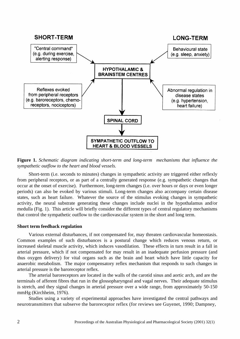
Figure 1. Schematic diagram indicating short-term and long-term mechanisms that influence the sympathetic outflow to the heart and blood vessels.
Short-term (i.e. seconds to minutes) changes in sympathetic activity are triggered either reflexly from peripheral receptors, or as part of a centrally generated response (e.g. sympathetic changes that occur at the onset of exercise). Furthermore, long-term changes (i.e. over hours or days or even longer periods) can also be evoked by various stimuli. Long-term changes also accompany certain disease states, such as heart failure. Whatever the source of the stimulus evoking changes in sympathetic activity, the neural substrate generating these changes include nuclei in the hypothalamus and/or medulla (Fig. 1). This article will briefly consider the different types of central regulatory mechanisms that control the sympathetic outflow to the cardiovascular system in the short and long term. Short term feedback regulation
Various external disturbances, if not compensated for, may threaten cardiovascular homeostasis. Common examples of such disturbances is a postural change which reduces venous return, or increased skeletal muscle activity, which induces vasodilation. These effects in turn result in a fall in arterial pressure, which if not compensated for may result in an inadequate perfusion pressure (and thus oxygen delivery) for vital organs such as the brain and heart which have little capacity for anaerobic metabolism. The major compensatory reflex mechanism that responds to such changes in arterial pressure is the baroreceptor reflex. The arterial baroreceptors are located in the walls of the carotid sinus and aortic arch, and are the terminals of afferent fibres that run in the glossopharyngeal and vagal nerves. Their adequate stimulus is stretch, and they signal changes in arterial pressure over a wide range, from approximately 50-150 mmHg (Kirchheim, 1976). Studies using a variety of experimental approaches have investigated the central pathways and neurotransmitters that subserve the baroreceptor reflex (for reviews see Guyenet, 1990; Dampney,
Proceedings of the Australian Physiological and Pharmacological Society (2001) 32(1) 3
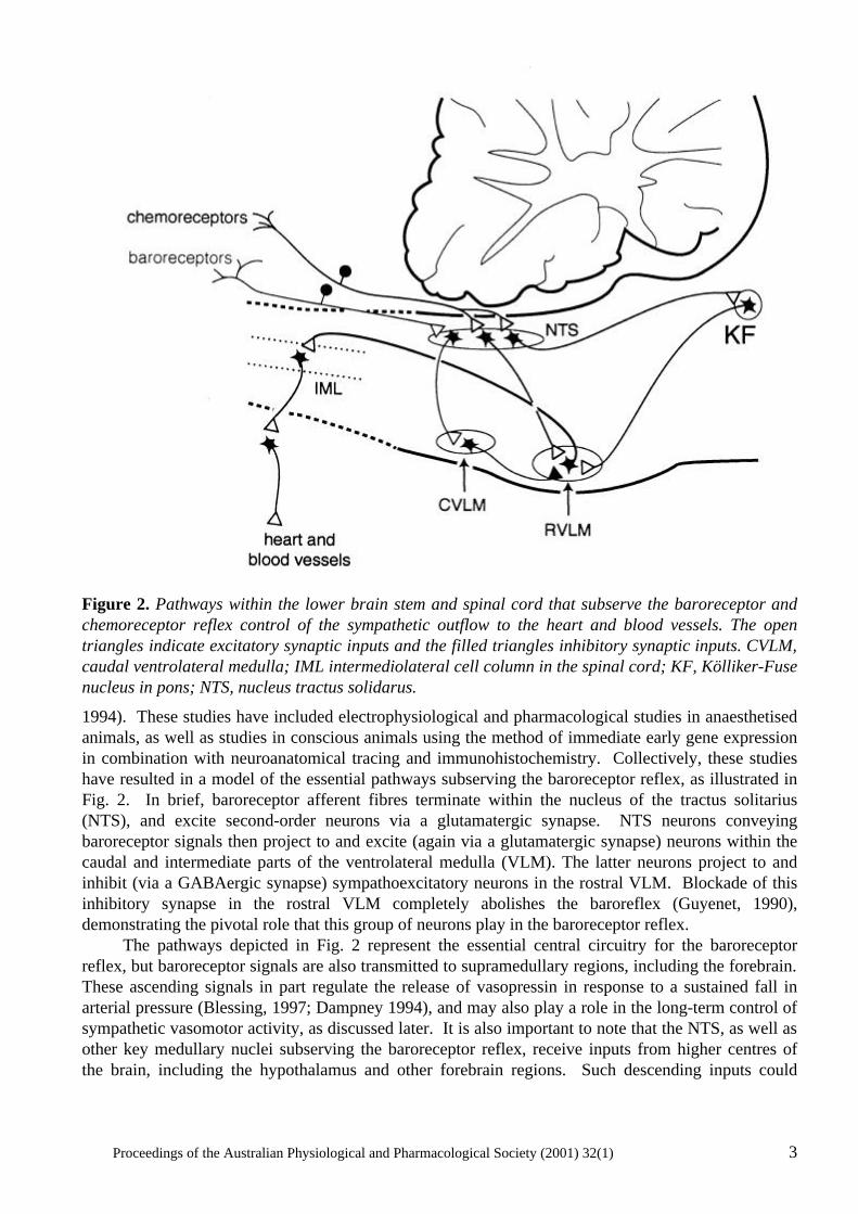
Figure 2. Pathways within the lower brain stem and spinal cord that subserve the baroreceptor and chemoreceptor reflex control of the sympathetic outflow to the heart and blood vessels. The open triangles indicate excitatory synaptic inputs and the filled triangles inhibitory synaptic inputs. CVLM, caudal ventrolateral medulla; IML intermediolateral cell column in the spinal cord; KF, Kölliker-Fuse nucleus in pons; NTS, nucleus tractus solidarus.
1994). These studies have included electrophysiological and pharmacological studies in anaesthetised animals, as well as studies in conscious animals using the method of immediate early gene expression in combination with neuroanatomical tracing and immunohistochemistry. Collectively, these studies have resulted in a model of the essential pathways subserving the baroreceptor reflex, as illustrated in Fig. 2. In brief, baroreceptor afferent fibres terminate within the nucleus of the tractus solitarius (NTS), and excite second-order neurons via a glutamatergic synapse. NTS neurons conveying baroreceptor signals then project to and excite (again via a glutamatergic synapse) neurons within the caudal and intermediate parts of the ventrolateral medulla (VLM). The latter neurons project to and inhibit (via a GABAergic synapse) sympathoexcitatory neurons in the rostral VLM. Blockade of this inhibitory synapse in the rostral VLM completely abolishes the baroreflex (Guyenet, 1990), demonstrating the pivotal role that this group of neurons play in the baroreceptor reflex. The pathways depicted in Fig. 2 represent the essential central circuitry for the baroreceptor reflex, but baroreceptor signals are also transmitted to supramedullary regions, including the forebrain. These ascending signals in part regulate the release of vasopressin in response to a sustained fall in arterial pressure (Blessing, 1997; Dampney 1994), and may also play a role in the long-term control of sympathetic vasomotor activity, as discussed later. It is also important to note that the NTS, as well as other key medullary nuclei subserving the baroreceptor reflex, receive inputs from higher centres of the brain, including the hypothalamus and other forebrain regions. Such descending inputs could
4 Proceedings of the Australian Physiological and Pharmacological Society (2001) 32(1)
modulate the operation of the baroreceptor reflex under particular conditions, as will also be discussed later with respect to the cardiovascular response to exercise or to alerting stimuli. The properties of the sympathoexcitatory neurons in the rostral VLM, which as mentioned above are powerfully influenced by baroreceptor signals, have been extensively investigated since the original discovery by Feldberg and co-workers that the rostral VLM contained a group of tonically active neurons that play an essential role in the maintenance of tonic sympathetic vasomotor activity and thus resting arterial pressure (for review see Dampney, 1994). Many physiological, pharmacological and anatomical studies have shown that the sympathoexcitatory neurons in the rostral VLM project directly to cardiac and vasomotor sympathetic preganglionic neurons in the thoracic and lumbar spinal cord, and therefore can be regarded as presympathetic neurons. Furthermore, they are a site of convergence of central pathways mediating cardiovascular responses evoked by stimulation of peripheral receptors as well as higher centres of the brain. The synaptic inputs to rostral VLM neurons are excitatory or inhibitory, and are generally mediated via glutamate or GABA receptors, respectively. In addition, however, the rostral VLM presympathetic neurons have receptors for other putative neurotransmitters or neuromodulators, such as angiotensin II (Ang II), enkephalin, or ATP (Dampney, 1994; Sun, 1996). The Ang II receptors, which are principally of the AT1 subtype, are particularly interesting because in the VLM they appear to be specifically associated with cardiovascular neurons (Dampney et al., 1996; Allen, 1998). The tonic activity of rostral VLM presympathetic neurons appears to be the major factor driving tonic activity in sympathetic preganglionic vasomotor neurons, at least in anaesthetised animals (for reviews see Dampney et al., 2000, Guyenet, 1990). Such tonic activity obviously also permits sympathetic vasomotor activity to be decreased as well as increased via inhibition and excitation, respectively, of the rostral VLM presympathetic neurons. The mechanisms generating tonic activity in these neurons has been a controversial subject for a long time. There is, however, clear evidence that these neurons receive tonic GABAergic inputs that are, at least in part, independent of peripheral baroreceptors (Dampney et al., 1988) and also some evidence that they receive tonic excitatory inputs (Dampney et al., 2000). The source of these tonic inputs, however, is unknown. A second example of short term feedback regulation of the cardiovascular system is the chemoreceptor reflex. The chemoreceptors are highly specialised receptors that are stimulated primarily by a decrease in the oxygen partial pressure of the arterial blood. They are located in the carotid and aortic bodies, and their afferent fibres, like baroreceptor afferent fibres, run in the glossopharyngeal and vagus nerves. Chemoreceptor stimulation reflexly evoked both an increase in ventilation and sympathetically mediated vasoconstriction in most vascular beds (excluding the brain and heart). The increase in ventilation will tend to increase oxygen uptake into the blood, while the sympathetic vasoconstriction will tend to reduce oxygen consumption by the tissues, and thus conserve the available oxygen. Like the baroreceptor reflex, studies in both anaesthetized and conscious animals have helped to define the essential pathways that mediate this reflex (Guyenet & Koshiya, 1995, Hirooka et al., 1997), and these are also shown in Fig. 2. Like baroreceptor primary afferent fibres, chemoreceptor primary afferent fibres terminate in the NTS. In contrast to the baroreflex pathways, however, chemoreceptor signals are transmitted to the rostral VLM presympathetic neurons via a direct excitatory glutamatergic synapse (Guyenet & Koshiya, 1995). Blockade of this glutamatergic synapse abolishes the sympathetic component of the chemoreceptor reflex (Guyenet & Koshiya, 1995), again illustrating the pivotal role of rostral VLM neurons in subserving fundamental cardiovascular reflexes. In addition, there is also evidence that a group of neurons in the pons (A5 cells) are also a component of central chemoreflex pathways (Koshiya & Guyenet, 1994).
Proceedings of the Australian Physiological and Pharmacological Society (2001) 32(1) 5
Short term feedforward regulation
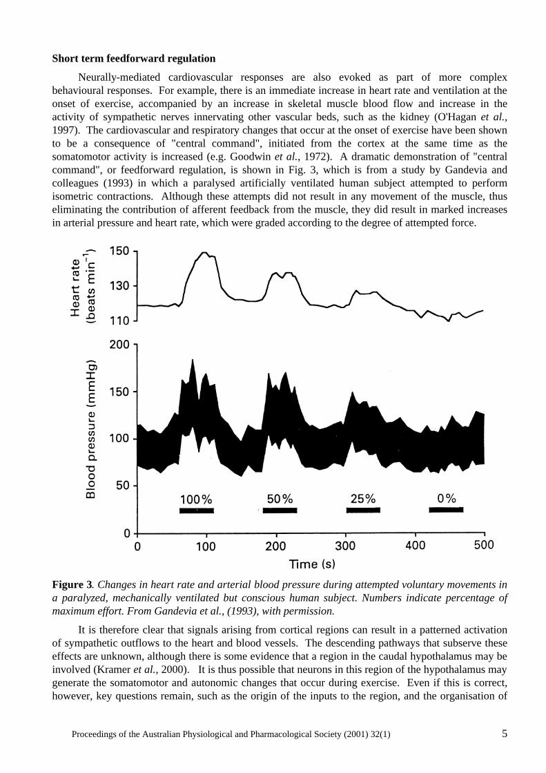
Neurally-mediated cardiovascular responses are also evoked as part of more complex behavioural responses. For example, there is an immediate increase in heart rate and ventilation at the onset of exercise, accompanied by an increase in skeletal muscle blood flow and increase in the activity of sympathetic nerves innervating other vascular beds, such as the kidney (O'Hagan et al., 1997). The cardiovascular and respiratory changes that occur at the onset of exercise have been shown to be a consequence of "central command", initiated from the cortex at the same time as the somatomotor activity is increased (e.g. Goodwin et al., 1972). A dramatic demonstration of "central command", or feedforward regulation, is shown in Fig. 3, which is from a study by Gandevia and colleagues (1993) in which a paralysed artificially ventilated human subject attempted to perform isometric contractions. Although these attempts did not result in any movement of the muscle, thus eliminating the contribution of afferent feedback from the muscle, they did result in marked increases in arterial pressure and heart rate, which were graded according to the degree of attempted force.
Figure 3. Changes in heart rate and arterial blood pressure during attempted voluntary movements in a paralyzed, mechanically ventilated but conscious human subject. Numbers indicate percentage of maximum effort. From Gandevia et al., (1993), with permission.
It is therefore clear that signals arising from cortical regions can result in a patterned activation of sympathetic outflows to the heart and blood vessels. The descending pathways that subserve these effects are unknown, although there is some evidence that a region in the caudal hypothalamus may be involved (Kramer et al., 2000). It is thus possible that neurons in this region of the hypothalamus may generate the somatomotor and autonomic changes that occur during exercise. Even if this is correct, however, key questions remain, such as the origin of the inputs to the region, and the organisation of
6 Proceedings of the Australian Physiological and Pharmacological Society (2001) 32(1)
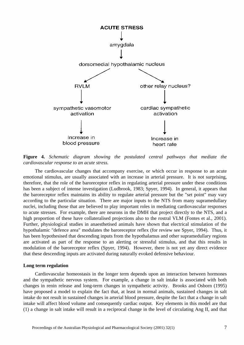
the descending pathways from this region to the spinal sympathetic outflow. A further question is whether there is a common set of "command neurons" within this region of the hypothalamus that trigger both the somatomotor and autonomic changes. It is well known that acute emotional or threatening stimuli can also elicit a marked cardiovascular response. For example, the classic "defence" or "alerting" response is characterised by an increase in arterial pressure, heart rate and skeletal muscle blood flow, accompanied by vasoconstriction in the splanchnic, renal and cutaneous vascular beds (Hilton, 1975). Such a response has been observed in conscious animals or humans subjected to an acute alerting stimulus such as air-jet stress or a loud noise (Davisson et al., 1994; Edwards et al., 1999; Schadt & Hasser, 1998). This patterned response has the effect of increasing cardiac output and re-distributing it preferentially towards the skeletal muscle beds, and is thus appropriate for an animal that may need to fight or flee from a threatening situation. Such a response is not part of a feed-back regulatory mechanism (Hilton, 1975) and can therefore be regarded as a feedforward response. It was first shown many years ago that electrical stimulation of a region in the hypothalamus, referred to as the "defence area", elicits a cardiovascular response very similar to that described above (Hilton, 1975). It is not clear, however, whether this response is due to activation of neuronal cell bodies within this hypothalamic region, or to fibres of passage that originate from higher centres, such as the amygdala. More recently, evidence has accumulated to suggest that the dorsomedial hypothalamic nucleus (DMH) plays a key role in integrating the cardiovascular response to acute stress. It is possible that this nucleus corresponds with the hypothalamic "defence area", although the boundaries of the latter region are not clearly defined. In any case, it is very interesting to note that activation of DMH neurons, by microinjection of either excitatory amino acids or GABA receptor antagonists results in a cardiovascular response that is very similar to the defence or alerting reaction, as well as neuroendocrine, gastrointestinal and behavioural changes very similar to that evoked by an acute emotional stress (DiMicco et al., 1996). Even more importantly, inhibition of neurons in the DMH greatly reduces the pressor and tachycardic response evoked by air stress in the conscious rat (Stotz-Potter et al, 1996). These observations indicate that the DMH may be a critical region integrating the cardiovascular as well as other autonomic and non-autonomic components of the response to an acute emotional stress or alerting stimulus. Consistent with this, the DMH receives inputs from several forebrain nuclei which are believed to play a role in mediating the response to stress, including the amygdala (DiMicco et al., 1991). In particular, activation of the basolateral nucleus of the amygdala generates a cardiovascular response very similar to that evoked by acute stress (Sanders & Shekhar, 1991), and this evoked response is dependent on synaptic transmission in the DMH (Soltis et al., 1998). Very recently, a study in our laboratory demonstrated that the vasomotor and cardiac responses that are evoked from the DMH are mediated by descending pathways that are dependent and independent, respectively, of synaptic transmission within the rostral VLM (Fontes et al., 2001). Taking all these different observations into account, Fig. 4 is a model of the key central connections mediating the cardiovascular response to an acute emotional stress. The classic "defence reaction" is not the only stereotyped response that is evoked by a threatening or alerting stimulus. For example, in the conscious rabbit, a stimulus such as a sound or touching the fur elicits cutaneous vasoconstriction, but unlike the defence reaction this is not accompanied by hindlimb vasodilation or an increase in heart rate (Yu & Blessing, 1997). At the same time, the amygdala appears to play a critical role in mediating this response (Yu & Blessing, 1999), as is the case with the stimuli that produce a classic "defence reaction". It therefore seems clear that different acute stressors can produce quite different patterns of cardiovascular responses, and that even though the same key nuclei may be involved in mediating these different responses, the relay neurons involved may be quite specific for the particular stimulus.
Proceedings of the Australian Physiological and Pharmacological Society (2001) 32(1) 7
Figure 4. Schematic diagram showing the postulated central pathways that mediate the cardiovascular response to an acute stress.
The cardiovascular changes that accompany exercise, or which occur in response to an acute emotional stimulus, are usually associated with an increase in arterial pressure. It is not surprising, therefore, that the role of the baroreceptor reflex in regulating arterial pressure under these conditions has been a subject of intense investigation (Ludbrook, 1983; Spyer, 1994). In general, it appears that the baroreceptor reflex maintains its ability to regulate arterial pressure but the "set point" may vary according to the particular situation. There are major inputs to the NTS from many supramedullary nuclei, including those that are believed to play important roles in mediating cardiovascular responses to acute stresses. For example, there are neurons in the DMH that project directly to the NTS, and a high proportion of these have collateralised projections also to the rostral VLM (Fontes et al., 2001). Further, physiological studies in anaesthetised animals have shown that electrical stimulation of the hypothalamic "defence area" modulates the baroreceptor reflex (for review see Spyer, 1994). Thus, it has been hypothesised that descending inputs from the hypothalamus and other supramedullary regions are activated as part of the response to an alerting or stressful stimulus, and that this results in modulation of the baroreceptor reflex (Spyer, 1994). However, there is not yet any direct evidence that these descending inputs are activated during naturally evoked defensive behaviour. Long term regulation
Cardiovascular homeostasis in the longer term depends upon an interaction between hormones and the sympathetic nervous system. For example, a change in salt intake is associated with both changes in renin release and long-term changes in sympathetic activity. Brooks and Osborn (1995) have proposed a model to explain the fact that, at least in normal animals, sustained changes in salt intake do not result in sustained changes in arterial blood pressure, despite the fact that a change in salt intake will affect blood volume and consequently cardiac output. Key elements in this model are that (1) a change in salt intake will result in a reciprocal change in the level of circulating Ang II, and that

8 Proceedings of the Australian Physiological and Pharmacological Society (2001) 32(1)
(2) a sustained change in the level of circulating Ang II will result in a sustained change (in the same direction) in the level of sympathetic nerve activity (Brooks & Osborn, 1995). For example, salt depletion leads to activation of the renin-angiotensin system and thus an increase in sympathetic nerve activity which helps to maintain arterial pressure despite the reduced salt intake. According to this model, therefore, Ang II (and probably other hormones as well) have a major influence in determining the long term level of sympathetic activity. This mechanism could also be a major factor contributing to the increase in sympathetic nerve activity in other conditions where the renin-angiotensin system is activated (such as renovascular hypertension or severe heart failure)(Goldsmith, 1999). Consistent with this, blockade of AT1 receptors has been shown to reduce sympathetic nerve activity in congestive heart failure (Liu et al., 1999). How can an increase in circulating Ang II lead to an increase in sympathetic nerve activity? It is possible that Ang II may act by enhancing neurotransmitter release at sympathetic nerve terminals, or else enhance synaptic transmission through sympathetic ganglia (Reid, 1992). Alternatively, although circulating Ang II does not cross the blood-brain barrier, there are abundant Ang II receptors in the circumventricular organs, particularly the subfornical organ and the area postrema. Activation of these receptors as a result of an increase in circulating Ang II leads to various brain-mediated effects, including the release of vasopressin from the posterior pituitary and also drinking behaviour. In addition, it has long been thought that circulating Ang II may also increase blood pressure via a centrally-evoked activation of sympathetic nerve activity, although the pathway responsible for this effect has not been defined. There are several lines of evidence to suggest that the hypothalamic paraventricular nucleus (PVN) could be a key component in the central pathways mediating sustained increases in sympathetic nerve activity in response to a raised level of circulating Ang II. First, the PVN receives direct and indirect inputs from Ang-sensitive neurons in the subfornical organ, and activation of this pathway has been shown to increase arterial pressure (Ferguson & Washburn, 1998). Secondly, PVN neurons appear to have a higher tonic activity in renal-wrapped hypertensive rats, in which Ang II levels are high (Martin & Haywood, 1998). Furthermore, there is also evidence that the PVN may contribute to sustained high levels of sympathetic activity in other models of hypertension, such as the spontaneously hypertensive rat (Allen, 2001), or the Dahl salt-sensitive hypertensive rat (Azar et al., 1981) as well as in heart failure (Patel & Zhang, 1996). Thus, the PVN could be a central site mediating sustained increases in sympathetic activity in response to inputs from a variety of sources. Consistent with this, the PVN receives inputs originating from higher centres and peripheral receptors, as well as from circumventricular organs (Dampney, 1994). Thus, it may be proposed that PVN sympathoexcitatory are tonically activated by inputs that in turn are activated by one or more of a variety of stimuli, such as increases in the level of circulating Ang II, chronic stress or anxiety, or peripheral receptors which may be tonically activated under certain conditions (e.g. chemosensitive cardiac receptors during heart failure (Zucker et al., 1995). Sympathoactivation evoked by disinhibition of the PVN is partly mediated by a descending pathway which includes a synapse in the rostral VLM, and partly via a pathway that is independent of the rostral VLM (Tagawa & Dampney, 1999). It is interesting to note that the activation of rostral VLM presympathetic neurons in response to disinhibition of the PVN is mediated by AT1 receptors (Tagawa & Dampney, 1999), just as the activation of PVN neurons by inputs from the subfornical organ is also mediated, at least in part, by AT1 receptors (Ferguson & Washburn, 1998). In addition, the PVN also has a major direct projection to the NTS (Dampney, 1994), and it is possible that activation of this pathway causes inhibition of the baroreceptor reflex, as also occurs in conditions where sympathetic activity is chronically increased, such as heart failure (Murakami et al., 1996). In this regard, it is interesting to note that AT1 receptors in the NTS mediate the inhibitory effect on the baroreceptor reflex that occurs in heart failure (Murakami et al., 1996). Thus, Ang II within the brain, quite apart from circulating Ang II, may play a key role in generating sustained high levels of
Proceedings of the Australian Physiological and Pharmacological Society (2001) 32(1) 9
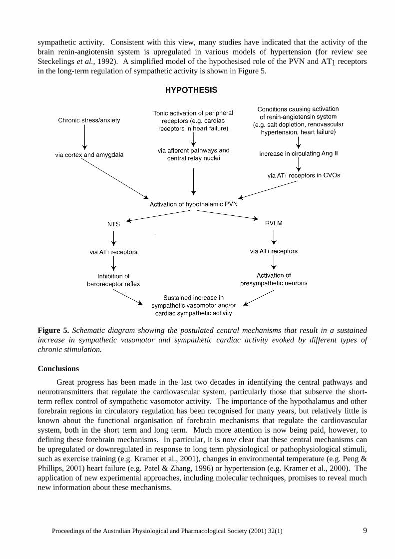
sympathetic activity. Consistent with this view, many studies have indicated that the activity of the brain renin-angiotensin system is upregulated in various models of hypertension (for review see Steckelings et al., 1992). A simplified model of the hypothesised role of the PVN and AT1 receptors in the long-term regulation of sympathetic activity is shown in Figure 5.
Figure 5. Schematic diagram showing the postulated central mechanisms that result in a sustained increase in sympathetic vasomotor and sympathetic cardiac activity evoked by different types of chronic stimulation. Conclusions
Great progress has been made in the last two decades in identifying the central pathways and neurotransmitters that regulate the cardiovascular system, particularly those that subserve the short-term reflex control of sympathetic vasomotor activity. The importance of the hypothalamus and other forebrain regions in circulatory regulation has been recognised for many years, but relatively little is known about the functional organisation of forebrain mechanisms that regulate the cardiovascular system, both in the short term and long term. Much more attention is now being paid, however, to defining these forebrain mechanisms. In particular, it is now clear that these central mechanisms can be upregulated or downregulated in response to long term physiological or pathophysiological stimuli, such as exercise training (e.g. Kramer et al., 2001), changes in environmental temperature (e.g. Peng & Phillips, 2001) heart failure (e.g. Patel & Zhang, 1996) or hypertension (e.g. Kramer et al., 2000). The application of new experimental approaches, including molecular techniques, promises to reveal much new information about these mechanisms.
10 Proceedings of the Australian Physiological and Pharmacological Society (2001) 32(1)
Acknowledgments
The work in our laboratory has been supported by the National Health and Medical Research Council of Australia and New Zealand, National Heart Foundation of Australia, and the Ramaciotti Foundations. The contributions of many collaborators and other colleagues to our research over many years is gratefully acknowledged. References
Allen, A.M., Moeller, I., Jenkins, T.A., Zhuo, J., Aldred, G.P. & Chai, S.Y. (1998) Angiotensin receptors in the nervous system. Brain Research Bulletin, 47, 17-28.
Allen, A.M. (2001) Inhibition of the hypothalamic paraventricular nucleus reduces sympathetic nerve discharge and blood pressure. Proceedings of the Australian Neuroscience Society, 29, 44.
Azar, S., Ernsberger, P., Livingston, S., Azar, P. & Iwai, J. (1981) Paraventricular--suprachiasmatic lesions prevent salt-induced hypertension in Dahl rats. Clinical Science, 61 Supp 7, 49s-51s.
Blessing, W.W. (1997) Arterial pressure and blood flow to the tissues. In: The Lower Brainstem and Bodily Homeostasis. pp. 165-268. New York: Oxford University Press.
Brooks, V.L & Osborn, J.W. (1995) Hormonal-sympathetic interactions in long-term regulation of arterial pressure: an hypothesis. American Journal of Physiology, 268, R1343-R1358.
Dampney, R.A.L. (1994) Functional organization of central pathways regulating the cardiovascular system Physiological Reviews, 74, 323-364.
Dampney, R.A.L., Blessing W.W. & Tan E. (1988) Origin of tonic GABAergic inputs to vasopressor neurons in the subretrofacial nucleus of the rabbit. Journal of the Autonomic Nervous System, 24, 227-239.
Dampney, R.A.L., Hirooka, Y., Potts, P.D. & Head, G.A. (1996) Functions of angiotensin peptides in the rostral ventrolateral medulla. Clinical and Experimental Pharmacology and Physiology, 23, S105-S111.
Dampney, R.A.L., Tagawa T., Horiuchi, J., Potts, P.D., Fontes, W. & Polson, J.W. (2000) What drives the tonic activity of presympathetic neurons in the rostral ventrolateral medulla? Clinical and Experimental Pharmacology and Physiology, 27, 1049-1053.
Davisson, R.L., Johnson, A.K. & Lewis, S.J. (1994) Nitrosyl factors mediate active neurogenic hindquarters vasodilation in the conscious rat. Hypertension, 23, 962-966.
Delp, M.D. & Laughlin, M.H. (1998) Regulation of skeletal muscle perfusion during exercise. Acta Physiologica Scandinavica, 162, 411-419.
DiMicco, J.A., Stotz-Potter, E.H., Monroe, A.J. & Morin, S.M. (1996) Role of the dorsomedial hypothalamus in the cardiovascular response to stress. Clinical and Experimental Pharmacology and Physiology, 23, 171-176.
DiMicco, J.A., Soltis, R.P., Anderson, J.J. & Wible, J.H. (1991) Hypothalamic mechanisms and the cardiovascular response to stress. In: Central Neural Mechanisms in Cardiovascular Regulation Volume 2, ed. Kunos, G. & Ciriello, J. pp. 52-79. Boston: Birkhaüser.
Drayer, J.I., Weber, M.A. & Nakamura D.K. (1985) Automated ambulatory blood pressure monitoring: a study in age-matched normotensive and hypertensive men. American Heart Journal, 109,1334-1338.
Edwards, C.M., Marshall, J.M. & Pugh, M. (1999) Cardiovascular responses evoked by mild cool stimuli in primary Raynaud's disease: the role of endothelin. Clinical Science, 96, 577-588.
Ferguson, A.V. & Washburn, D.L. (1998) Angiotensin II: a peptidergic neurotransmitter in central autonomic pathways. Progress in Neurobiology, 54, 169-192.
Proceedings of the Australian Physiological and Pharmacological Society (2001) 32(1) 11
Fontes, M.A.P., Tagawa, T., Polson, J.W., Cavanagh, S.-J. & Dampney, R.A.L. (2001) Descending pathways mediating cardiovascular response from the dorsomedial hypothalamic nucleus. American Journal of Physiology, 280, H2891-H2901.
Gandevia, S.C., Killian, K., McKenzie, D.K., Crawford, M., Allen, G.M., Gorman, R.B. & Hales J.P. (1993) Respiratory sensations, cardiovascular control, kinaesthesia and transcranial stimulation during paralysis in humans. Journal of Physiology, 470, 85-107.
Goldsmith, S.R. (1999) Angiotensin II and sympathoactivation in heart failure. Journal of Cardiac Failure, 5, 139-145.
Goodwin, G.M., McCloskey, D.I. & Mitchell, J.H. (1972) Cardiovascular and respiratory responses to changes in central command during isometric exercise at constant muscle tension. Journal of Physiology, 226, 173-190.
Guyenet, P.G. (1990) Role of the ventral medulla oblongata in blood pressure regulation. In: Central Regulation of Autonomic Functions, ed. Loewy, A.D. & Spyer, K.M. pp. 145-167. New York: Oxford University Press.
Guyenet, P.G. & Koshiya, N. (1995) Working model of the sympathetic chemoreflex in rats. Clinical and Experimental Hypertension, 17, 167-179.
Hilton, S.M. (1975) Ways of viewing the central nervous control of the circulation - old and new. Brain Research, 87, 213-219.
Hirooka, Y., Polson, J.W., Potts, P.D. & Dampney, R.A.L. (1997) Hypoxia-induced Fos expression in neurons projecting to the pressor region in the rostral ventrolateral medulla. Neuroscience, 80, 1209-1224.
Hobbs, S.F. & McCloskey, D.I. (1987) Effects of blood pressure on force production in cat and human muscle. Journal of Applied Physiology, 63, 834-839.
Jänig, W. & McLachlan, E.M. (1992) Specialized functional pathways are the building blocks of the autonomic nervous system. Journal of the Autonomic Nervous System, 41, 3-13.
Kirchheim, H.R. (1976) Systemic arterial baroreceptor reflexes. Physiological Reviews, 56, 100-176.
Koshiya, N. & Guyenet P.G. (1994) A5 noradrenergic neurons and the carotid sympathetic chemoreflex. American Journal of Physiology, 267, R519-R526.
Kramer, J.M., Plowey, E.D., Beatty, J.A., Little, H.R. & Waldrop, T.G. (2000) Hypothalamus, hypertension, and exercise. Brain Research Bulletin, 53, 77-85.
Liu, J.L & Zucker I.H. (1999) Regulation of sympathetic nerve activity in heart failure: a role for nitric oxide and angiotensin II. Circulation Research, 84, 417-423.
Ludbrook, J. (1983) Reflex control of blood pressure during exercise. Annual Review of Physiology, 45, 155-168.
Martin, D.S. & Haywood, J.R. (1998) Reduced GABA inhibition of sympathetic function in renal-wrapped hypertensive rats. American Journal of Physiology, 275, R1523-1529
Murakami, H., Liu, J.L. & Zucker, I.H. (1996) Blockade of AT1 receptors enhances baroreflex control of heart rate in conscious rabbits with heart failure. American Journal of Physiology, 271, R303-R309.
O'Hagan, K.P., Casey, S.M. & Clifford, P.S. (1997) Muscle chemoreflex increases renal sympathetic nerve activity during exercise Journal of Applied Physiology, 82, 1818-1825.
Patel, K.P. & Zhang, K. (1996) Neurohumoral activation in heart failure: role of paraventricular nucleus. Clinical and Experimental Pharmacology and Physiology, 23, 722-726.
12 Proceedings of the Australian Physiological and Pharmacological Society (2001) 32(1)
Peng, J.F. & Phillips, M.I. (2001) Opposite regulation of brain angiotensin type 1 and type 2 receptors in cold-induced hypertension. Regulatory Peptides, 97, 91-102.
Reid, I.A. (1992) Interactions between ANG II, sympathetic nervous system, and baroreceptor reflexes in regulation of blood pressure. American Journal of Physiology, 262, E763-E778.
Sanders, S.K. & Shekhar, A. (1991) Blockade of GABAA receptors in the region of the anterior basolateral amygdala of rats elicits increases in heart rate and blood pressure. Brain Research, 576, 101-110.
Schadt, J.C. & Hasser, E.M. (1998) Hemodynamic effects of acute stressors in the conscious rabbit. American Journal of Physiology, 274, R814-R821.
Soltis, R.P., Cook, J.C., Gregg, A.E., Stratton, J.M. & Flickinger, K.A. (1998) EAA receptors in the dorsomedial hypothalamic area mediate the cardiovascular response to activation of the amygdala. American Journal of Physiology, 275, R624-R631.
Spyer, K.M. (1994) Central nervous mechanisms contributing to cardiovascular control. Journal of Physiology, 474, 1-19.
Steckelings, U., Lebrun, C., Qadri, F., Veltmar, A. & Unger, T. (1992) Role of brain angiotensin in cardiovascular regulation. Journal of Cardiovascular Pharmacology, 19 Suppl. 6, S72-S79.
Stotz-Potter, E.H., Willis, L.R. & DiMicco, J.A. (1996) Muscimol acts in dorsomedial but not paraventricular hypothalamic nucleus to suppress cardiovascular effects of stress. Journal of Neuroscience, 16, 1173-1179.
Sun, M.K. (1996) Pharmacology of reticulospinal vasomotor neurons in cardiovascular regulation. Pharmacological Reviews, 48, 465-494.
Tagawa, T. & Dampney, R.A.L. (1999) AT1 receptors mediate excitatory inputs to RVLM pressor neurons from hypothalamus. Hypertension, 34, 1301-1307.
Yu, Y.H. & Blessing, W.W. (1999) Amygdala co-ordinates sudden falls in ear pinna blood flow in response to unconditioned salient stimuli in conscious rabbits. Neuroscience, 93, 135-141.
Yu, Y.H. & Blessing, W.W. (1997) Cutaneous vasoconstriction in conscious rabbits during alerting responses detected by hippocampal theta-rhythm. American Journal of Physiology, 272, R208-R216, 1997.
Zucker, I.H., Wang, W., Brandle, M., Schultz, H.D. & Patel, K.P. (1995) Neural regulation of sympathetic nerve activity in heart failure. Progress in Cardiovascular Diseases, 37, 397-414.
Proceedings of the Australian Physiological and Pharmacological Society (2001) 32(1) 13
ENDOTHELIUM-DEPENDENT HYPERPOLARIZING FACTOR: IS THERE A
NOVEL CHEMICAL MEDIATOR?
Chris R. Triggle and Hong Ding
Smooth Muscle Research Group and Department of Pharmacology and Therapeutics, Faculty of Medicine, University of Calgary, Calgary, Alberta, Canada, T2N 4N1.
Summary
Endothelium-dependent hyperpolarization (EDH) has been reported in many vessels and an extensive literature suggests that a novel, non-nitric oxide and non-prostanoid, endothelium-derived factor(s) may be synthesized in endothelial cells. The endothelium-dependent hyperpolarizing factor, or EDHF, is synthesized by the putative EDHF synthase and mediates its cellular effects by, directly or indirectly, opening K-channels on vascular smooth muscle cells. The question of the chemical identity of EDHF has received considerable attention, however, no consensus has been reached. Considerable tissue and species differences exist that may imply that there are multiple EDHFs. Leading candidate molecules for EDHF include an arachidonic acid product, possibly an epoxygenase product, or an endogenous cannabinoid, or simply an increase in extracellular K+. An increasing body of evidence suggests that endothelial-dependent hyperpolarization, notably in the resistance vasculature, may be mediated via electrical coupling through myoendothelial gap junctions negates the need to hypothesize the existence of a true endothelium-derived chemical mediator. In this presentation we review the evidence that supports and refutes the existence of a novel EDHF versus a hyperpolarization event mediated solely by myoendothelial gap junctions. Introduction
The endothelial-cell derived relaxing factor (EDRF), which was originally described by Furchgott and Zawadzki (1980), has been identified as nitric oxide (NO) and is now known to play an important role as a key paracrine regulator of vascular tone. However, in many vessels, and notably in the resistance vasculature, the pharmacological inhibition, or genetic “knockout,” of the synthesis of NO, (or inhibition, of the other identified endothelial-cell derived vasodilator factor, prostacyclin, PGI2) does not greatly affect the endothelium-dependent relaxation response to either chemical (i.e. acetylcholine, ACh; bradykinin, BK) or mechanical (shear stress) stimulation. There is considerable species and tissue variation in the contribution of an NO- and PGI2-independent vasodilatation and this could indicate heterogeneity in the nature of the putative mediator and/or, as will be discussed later, heterogeneity in the nature and contribution of gap junction proteins. Since the cellular action of this putative non-NO/PGI2 mediator has been associated with endothelium-dependent hyperpolarization (EDH) of the vascular smooth muscle cell (VSMC) the factor has been named the endothelium-derived hyperpolarizing factor or EDHF (see Triggle et al., 1999; Ding et al., 2000a) – see Figure 1A. A change in membrane potential of just a few millivolts (mV) can result in a substantial change in vessel diameter (Brayden & Nelson, 1992; Nelson & Quayle, 1995) and thus it can be predicted that the release of an EDHF, a putative opener of K+ channels, will make an important contribution to the regulation of vascular tone. Furthermore, hyperpolarization of the smooth muscle will, in comparison to cellular events mediated by second messengers, produce a rapid effect on blood flow. In addition, since the contribution of EDHF to endothelium-dependent vasodilatation is most apparent in resistance vessels, it might be anticipated that any intervention that leads to a diminution in the synthesis and/or
14 Proceedings of the Australian Physiological and Pharmacological Society (2001) 32(1)
Proceedings of the Australian Physiological and Pharmacological Society (2001) 32(1) 15
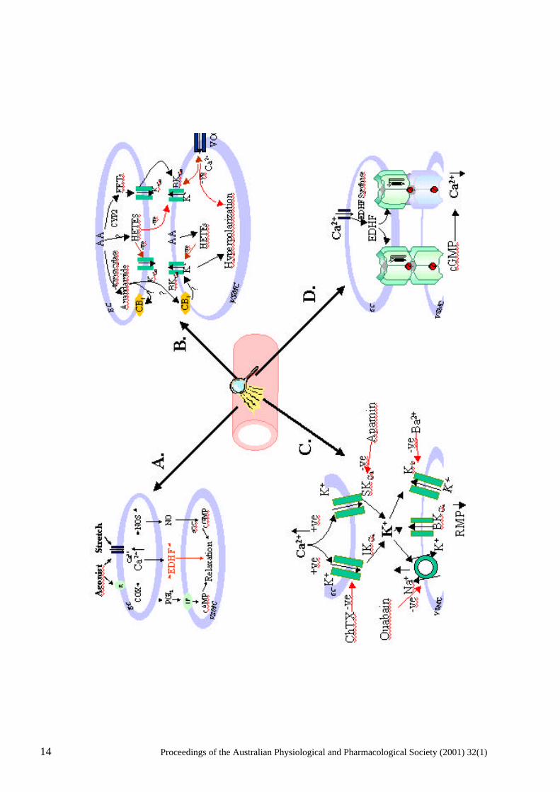
Figure 1. Endothelium-dependent vasodilators, such as acetylcholine, as well as shear stress (stretch) activate endothelial cell (EC) plasma membrane receptors (R) and open a non-selective cation channel(s) leading to the entry of extracellular calcium (Ca2+), as well as the release of intracellular Ca2+. The increase in intracellular Ca2+ leads to the activation of endothelial nitric oxide synthase (NOS), cyclooxygenase (COX), the putative endothelium-dependent hyperpolarization factor(s) EDHF synthase and the synthesis of nitric oxide (NO), prostacyclin (PGI2) and EDHF respectively. NO and PGI2 ,mediate relaxation of vascular smooth muscle cells (VSMC) via cyclic GMP and AMP-dependent mechanisms respectively and EDHF via, directly or indirectly, opening of a VSMC K-channel(s). Arachidonic acid (AA) can be metabolized via an epoxygenase (cytochrome P450 isozyme, CYP2) to produce epoxyeicosatrienoic acids (EETs) that, directly or indirectly, have been shown to increase the probability of opening of big conductance calcium-activated K-channels (BKCa). EETs may function as autocrine and/or paracrine mediators; in VSMC they hyperpolarize the cell and decrease the probability of opening of voltage-operated Ca2+ channels (VOCC). 20- and 19-hydroxyeicosatetraenoic acid (20-, 19-HETE), which are produced in VSMC and, possibly EC, contract VSMC putatively via an increase in the probability of opening of VOCC and/or closure of BKCa. The endogenous cannabinoid, anandamide, is also synthesized from AA via a transacylase. Anandamide activates cannabinoid receptors (CB1) in both EC and VSMC and has been reported to hyperpolarize VSMC. C/ An increase in intracellular Ca2+ in EC activates and increases the opening probability of opening of apamin-sensitive small conductance KCa (SKCa) and charybdotoxin-sensitive intermediate conductance (IKCa) channels in EC leading to the efflux of K+ from the EC and an increase in extracellular K+. A small increase in extracellular K+ leads to the hyperpolarization of VSMC via the activation of ouabain-sensitive Na/K- ATPase and an increase in the open probability of barium-sensitive Kir channels and a lowering of the resting membrane potential (RMP) of VSMC. D/ Myoendothelial gap junctions, depicted as six connexin subunits from each cell docking to form either a homomeric and heteromeric connexon, provide the means by which low molecular weight water soluble molecules, including cGMP, can pass between EC and VSMC and contribute to endothelium-dependent hyperpolarization.
release, of EDHF, could critically affect the regulation of organ blood flow thus contributing to pathophysiological states such as hypertension. There is also evidence that EDHF-mediated vasodilatation is negatively regulated by NO and this may reflect an inhibitory effect of NO on the hypothetical “EDHF synthase”. There have been a number of recent reviews on EDHF (Hecker et al., 1994; Mombouli & Vanhoutte, 1997; Quilley et al., 1997; Edwards & Weston, 1998; Félétou & Vanhoutte, 1999; Triggle et al., 1999; Waldron et al., 1999; Ding et al., 2000a). Nonetheless, the nature and, indeed, the existence of EDHF remains controversial. In this presentation we will discuss the evidence for and against EDH being mediated by: A/ Residual NO; B/ An arachidonic acid product; C/ A small increase in extracellular potassium; D/ Myoendothelial cell gap junctions. A/ NO can mediate EDH:
Cohen et al. (1997) raised the possibility that since NO itself can, in some vessels, directly or indirectly mediate hyperpolarization, EDHF may be NO. A number of other investigators have reached the same conclusion (Kemp & Cocks, 1999; Simonsen et al., 1999; Ge et al., 2000). Cohen et al.
16 Proceedings of the Australian Physiological and Pharmacological Society (2001) 32(1)
(1997) demonstrated that it was not possible to completely inhibit the synthesis of NO with just a single nitric oxide synthase (NOS) inhibitor thus challenging the interpretation of data from studies wherein it has been concluded that the NOS (and COX) inhibitor-insensitive component of an endothelium-dependent relaxation reflected a novel, NO and PGI2-independent, mechanism. Some blood vessels, such as the rabbit carotid, human coronary resistance arteries and rat superior mesenteric artery, may be able to generate NO, possibly from a non-L-arginine source (see Kemp & Cocks, 1999), and this NOS inhibitor–insensitive production of NO (“residual NO”) mediates the EDH (Vanheel & Van de Voorde, 2000). NO may also directly or indirectly activate K-channels in vascular smooth muscle cells (VSMC). Thus, Bolotina et al. ( 1994) and Mistry and Garland (1998) have reported that NO directly, via a soluble guanylyl cyclase (sGC)-independent mechanism, stimulates charybdotoxin (ChTX)-sensitive K+ channels in the rabbit aorta and rat mesenteric arterioles respectively. NO activates KATP channels channels in rat mesenteric arteries (Garland & McPherson, 1992) and guinea-pig coronary arteries (Parkington et al., 1995), an apamin-sensitive K+ channel (Murphy & Brayden, 1995) and BKCa in rabbit middle cerebral arteries (Dong et al., 1997). In the human umbilical artery NO mediates vascular relaxation via K-channel and sGC-independent mechanism(s) (Lovren & Triggle, 2000). It is, however, important to note that a number of studies have shown that the hyperpolarization mediated by NO requires a higher concentration than that required to mediate relaxation (40-fold higher in guinea-pig coronary arteries (Parkington et al., 1995)). To determine whether NO contributes to edh a number of studies have used NO-scavenging compounds such as carboxy-PTIO, hydroxocobalamin, oxyhaemoglobin, or free radical generating compounds such as xanthine-xanthine oxidase, or studied genetic “knockouts” of the endothelial cell (EC) nitric oxide synthase (Huang et al., 1995; Waldron et al., 1999a; Ding et al., 2000a). B. An arachidonic acid product as an EDHF:
A number of enzymes can metabolize arachidonic acid into products that affect the vasculature and a number of recent reviews have stressed the importance of metabolites of arachidonic acid generated by cytochrome P450 (CYP) enzyme activity as being key signaling modulators of vascular tone (McGiff et al., 1996; Campbell & Harder, 1999; Alonso-Galicia et al., 1999). Considerable evidence has accumulated in support of the hypothesis that an epoxygenase (CYP) product of arachidonic acid, notably 5,6-epoxyeicosatrienoic (5,6 EET), is an EDHF in at least some vascular beds. Another arachidonic acid product is the endogenous cannabinoid, or “endocannabinoid”, anandamide (N-arachidonylethanolamine), which is formed via the action of a transacylase enzyme. Randall et al. (1996) reported that in the isolated perfused mesenteric arteriole bed anandamide was a potent vasorelaxant. Furthermore, the NO-independent action of the endothelium-dependent vasodilator, bradykinin, was inhibited by a putatively selective cannabinoid receptor (CB1) antagonist, SR141716A as well as when vascular tone was elevated with high extracellular K+, suggesting that anandamide is an EDHF). However, this hypothesis has not received a great deal of support. Anandamide does not seem to have the same physiological and pharmacological properties as does EDHF (Plane et al., 1997; White & Hiley, 1997; Zygmunt et al., 1997; Chataigneau et al., 1998; White & Hiley, 1998). Of interest is that Mombouli et al. (1999) reported that anandamide mobilizes endothelial cell Ca2+ from a caffeine-sensitive store via a CB1 receptor-insensitive mechanism. Thus, anandamide may serve in an autocrine function as a regulator of endothelial cell calcium and may influence the production of EDHF but may not necessarily itself be an EDHF. Stronger evidence in support of a role for an arachidonic acid product in EDH has been provided by a study with porcine coronary arteries where a transferable “EDHF” could be detected by bioassay and its ability to hyperpolarize detector rat aortic smooth muscle cells (Popp et al., 1996). Popp et al.
(1996) also demonstrated that the effects of this putative factor were inhibited by CYP inhibitors, clotrimazole and 17-ODYA and that the CYP product 5,6-epoxyeicosatrienoic (5,6 EET), acid induced
Proceedings of the Australian Physiological and Pharmacological Society (2001) 32(1) 17
a hyperpolarization of smooth muscle cells; and the induction of CYP activity by beta-naphthoflavone significantly enhanced the EDH response. Other products of CYP (CYP4 isozyme) -mediated arachidonic acid metabolism, at least in smooth muscle, are 20- and 19-hydroxyeicosatetraenoic acids (20-OH-AA), and ω-2, ω-3, and ω–4-hydroxyeicosatetraenoic acids (ω-terminal hydroxylase reactions) (Capdevila et al., 2000). 20-OH-AA, and related compounds, cause vasoconstriction of cerebral and renal vessels (Harder et al., 1994; Imig et al., 1996) and inhibit big conductance calcium-activated K+ channels, BKCa, enhancing Ca2+ entry by depolarization of VSM (Zou et al., 1996). EETs can therefore be considered to be physiological antagonists of HETES. (Figure 1B) However, 20-HETE can also relax VSMC, possibly via metabolism by cyclooxygenase to PGI2 (Pratt et al., 1998). Fisslthaler et al. (Fisslthaler et al., 1999) demonstrated that the transfection of porcine coronary arteries with antisense oligonucleotides against CYP 2C8/34 attenuated EDHF-mediated coronary vasodilatation and this data is very suggestive that a CYP product is an EDHF. Similar data has been provided from studies with the gracilis muscle resistance vessels from the hamster (Bolz et al., 2000). Overall, the evidence in favour of an EET being EDHF is strongest in coronary and renal tissues (see
Komori & Vanhoutte, 1990; McGiff et al., 1996; Harder et al., 1995a,b). Furthermore, if an EET does serve as an EDHF and hyperpolarizes smooth muscle via opening BKCa
2+ channels, it would provide an endothelial cell-derived antagonist for the action of the vascular smooth muscle derived arachidonic acid product, 20-HETE, which has been hypothesized to be an inhibitor of BKCa
2+ channels (Zou et al., 1996). Nonetheless, the hypothesis that a CYP product functions as an EDHF has been challenged for several reasons. First of all, many of the CYP inhibitors used have considerable non-specific actions, notably on K-channels. Edwards et al. (1996) have reported that miconazole and other imidazoles are non-specific inhibitors of CYP and also block K-channels whereas the suicide substrate of CYP, 17-ODYA, in so far as it only inhibited hyperpolarization of VSMC, appeared to show specificity towards CYP. Somewhat similar data has also been provided by Vanheel et. al. (1999) and such data clearly indicates the need to verify the specificity of the pharmacological probes used in such studies. Furthermore, although the data presented by Fisslthaler et al. (!999) can be interpreted as supportive of a role for a CYP product being an EDHF the data could also be interpreted as reflecting an autocrine function of an endothelium-derived CYP product that enhances the synthesis/release of a non-arachidonic acid EHDF that mediates the hyperpolarization/vasodilatation of VSMC. Such a hypothesis has been advanced for EETs (Graier et al., 1995; Hoebel et al., 1997) and also anandamide (Mombouli et al., 1999). An additional problem in accepting that an EET may an EDHF is that although EETs can hyperpolarize VSMC they seem to do so via the activation of iberiotoxin-sensitive BKCa
2+ channels (Hu & Kim, 1993) whereas the hyperpolarization mediated by ACh is usually only significantly inhibited by a combination of charybdotoxin and apamin (Edwards et al.,1998). C/ Potassium as an EDHF:
Edwards et al. (1998) measured potassium, Ko, in the extracellular space between endothelial and vascular smooth muscle cells in rat hepatic artery with a K+- sensitive microelectrode and reported an ACh-mediated increase in Ko from 4.6 to 11.6mM. Additional evidence in support of the hypothesis that an increase in extracellular potassium can mimic the effect of EDHF was also presented by Edwards et al. (1998) and was based on the measurement of the membrane potential of both endothelial and vascular smooth muscle cells with glass microelectrodes. ACh was shown to hyperpolarize both vascular and endothelial cells and hyperpolarization of the endothelial cell was inhibited by a combination of apamin and ChTX and vascular hyperpolarization by a combination of ouabain and barium (30 µM). These data lead to the conclusion that apamin-sensitive small conductance calcium-activated K+ channels (SKCa) and charybdotoxin-sensitive intermediate conductance calcium-activated K+ channels (IKCa) on endothelial cells regulate the release of EDHF and the ouabain-sensitive electrogenic Na+,K+-ATPase and inward rectifying K+ channel (Kir) on the vascular smooth muscle mediate the vascular actions of EDHF (Edwards et al., 1998). See Figure 1C.
18 Proceedings of the Australian Physiological and Pharmacological Society (2001) 32(1)
The conclusion was that EDHF is endothelium-derived K+ that exits endothelial cells as a result of ACh-mediated opening of apamin/ChTX-sensitive K+ channels. The increase in extracellular K+ activates Na+, K+-ATPase and opens Kir on VSMCs. An increase in K+
o by 5 mM mimicked the effects of ACh, and comparable data was reported for the rat mesenteric artery preparation. An increase in K+
o was already known to cause vascular smooth muscle relaxation and a role as (an) EDHF is an attractive hypothesis that would place K+, together with NO, as a cell-signalling mediator that likely evolved as an early regulator of vascular function (Vanhoutte, 1998). Because of the similarity of K+-induced vasodilation to that mediated by EDHF in other vessels it was concluded that K+ might be the “universal EDHF”. There is a substantive literature that supports hypothesis that small changes in K+
o result in vasodilatation. Thus, the activation of VSMC Na+, K+-ATPase as the cellular basis for mediating the relaxant effects of ACh in canine femoral arteries has also been reported (De Mey & Vanhoutte, 1980) and ouabain has also been shown to inhibit the hyperpolarization, but not the relaxation, initiated by ACh in canine coronary arteries (Félétou & Vanhoutte, 1998). Furthermore, it has been established that the activation of Na+, K+- ATPase will lead to hyperpolarization of smooth muscle Haddy, 1978; Fleming, 1980; Haddy, 1983; Hermsmeyer, 1993) Somewhat higher increases in K+
o than are needed to activate the Na+, K+-ATPase also lead to a reduction in inward rectification allowing the Kir channel to carry more outward current (McCarron & Halpern, 1990). An increase in K+
o from 6 to 16 mM has been reported to result in a sustained dilation of pressurized coronary and cerebral arteries from the rat and these dilations were sensitive to block by concentrations of barium (IC50, 3-8 µM) Knot et al. (1996) (< 50 µM) that selectively block Kir channels (Quayle et al., 1993). Kir has, for instance, been demonstrated to be much greater in the smaller branches of guinea pig vessels (Quayle et al., 1996) and cerebral vessels from gene-targeted mice lacking the Kir2.1 fail to dilate to raising K+
o from 6 to15 mmol/L9Zaritsky et al., 2000). Overall these data are suggestive that K+ can function as a regulator of vascular tone and are supportive of the hypothesis that K+ may be an EDHF. However, the origin of the increase in K+
o is unknown. Given the comparatively small size of the endothelial cells it might well be argued that VSMC would more likely contribute to an increase in K+
o than would EC. Periods of high neuronal activity can also result in an increase in K+o and
increases of > 10 mmol/L have been reported in the cerebral spinal fluid (Sykova, 1983). Ischemia in the coronary circulation increases K+
o (Weiss et al., 1989), and raising K+o results in dilation in the
renal circulation Scott et al., 1959). Thus a modest increase in K+o results in an increase in blood
supply to areas of high metabolic activity. Data from a number of laboratories have challenged the hypothesis that “K+” is the universal EDHF. Thus, Ding et al. (2000a) reported that in saphenous arteries from both endothelial nitric oxide synthase expressing (eNOS +/+) and eNOS lacking (-/-) C57 mice relax to both ACh and K+ in phenylephrine pre-contracted vessels, however, ACh-mediated relaxations were insensitive to 30 µM barium and 10 µM ouabain but were inhibited by a combination of ChTX and apamin; K+-mediated relaxations were inhibited by a combination of barium and ouabain but were insensitive to a combination of apamin and ChTX. Data from the same laboratory had previously indicated an “upregulation”of EDHF in some vessels from mice lacking eNOS (-/-) (Waldron et al., 1999). The contribution of K+ to endothelium-dependent vasodilatation may be vessel dependent, which, in itself, is very interesting as it would suggest vessel heterogeneity with respect to the contribution of different EDHFs in different vascular beds. For instance, in first order mesenteric arterioles from C57 mice, barium alone partially blocked both ACh and K+ evoked relaxations, however, a combination of barium and ouabain totally blocked K+, but not ACh, evoked relaxations (Ding et al., 2000). In a study of guinea pig third order mesenteric artery and the middle cerebral artery Dong et al. (2000) provide contrasting data. In neither vessel would the addition of low concentrations of K+ evoke relaxation and although ouabain greatly attenuated EDHF-mediated relaxation in the mesenteric arteries it enhanced relaxation in the cerebral vessels. These data suggest that, whereas an increase in extracellular K+ may
Proceedings of the Australian Physiological and Pharmacological Society (2001) 32(1) 19
be a contributing factor to EDHF–mediated relaxation in some vascular beds, K+ is unlikely to be the primary mediator in all vessels.The ability of K+ to relax vessels may also depend on the level of contraction of the vessel (Dora & Garland, 2000), however, it has also been reported that, even under comparable levels of contraction, some vessels fail to relax to K+ (Dong et al., 2000; Ding & Triggle, 2000). Doughty et al. (1999) have also demonstrated in rat mesenteric small arteries that although both K+ and EDHF dilate the vessels their profile is quite different and that it is therefore unlikely that K+ is EDHF, at least in rat mesenteric small arteries. Nonetheless, Edwards et al. (1998), Ding et al. (2000) and Dong et al. (2000) found in the mesenteric vessels of rat, mouse, and guinea-pig evidence supportive of a role for K+ and/or Kir in, at least, contributing to the effects of EDHF and the study by Beny and Schaad (2000) provides support for the hypothesis that an increase in K+
o may serve as an EDHF in some blood vessels. D/ Myoendothelial cell gap junctions:
There is also increasing evidence that endothelium-dependent hyperpolarization (EDH) may be mediated by myoendothelial cell gap junctions (Chayter et al., 1998). Gap junctions, via intercellular hemi-channels, allow the passage of inorganic ions and of small water-soluble molecules (<1000 Da), including cAMP, cGMP, inositol trisphosphate, but not peptides/proteins, between cells. Connexins are the principal proteins that make up the gap junction with each connexin molecule possessing four transmembrane domains, six connexin subunits forming a connexon and the gap junction is established by the docking of the two connexons hemichannels supplied by the two interacting cells. Thirteen rodent connexins have been identified to date (see review by Kumar & Gilula, 1996). Connexin 43 has been described as the dominant gap junction protein present in both VSMC and EC (Christ et al., 1996; Christ & Brink, 1999). However, Van Kempen and Jongsma (1999) used immunohistochemical techniques to study the distribution of connexins 37, 40 and 43 in bovine, micropig and rat aorta and coronary veesels and concluded that connexin 40 is the constitutive connexin that was found between VSMC and EC with connexin 43 only between VSMC and connexin 37 between EC. Connexin 45 is expressed in intestinal smooth muscle Nakamura et al., 1998), connexin 45 has also been shown to play a role in the regulation of human uterine smooth muscle contractilty (Kilarski et al., 1998) and connexin 45 deficient mice show defects in the development of the vasculature Kruger et al., 2000) The role, however, of connexin 45 in the regulation of VSMC-EC communication has not yet been reported. Species and vessel differences in the distribution of connexins does exist and the co-localization of connexin 40 and 43 has also been reported in both EC and VSMC Valiunas et al., 2000). The conductance properties of heteromeric gap junction channels that are formed when more than one type of connexin forms the gap junction, are reported to be intermediate between those of the homomeric junction and, if expression of connexins varies between vascular beds, there is the potential for specialization of function exists within the circulation Little et al., 1995; Brink, 2000). Myoendothelial gap junctions occur in greater density in resistance compared to conduit arteries Daut et al., 1994) and this may explain the predominance of EDH in the resistance vasculature. Sandow and Hill (Sandow & Hill, 2000) have provided anatomical support for this hypothesis with a serial-section electron microscopic study of proximal versus distal rat mesenteric arteries and demonstrated a significantly greater density of myoendothelial gap junctions in the distal arteries. An elegant study by Emerson and Segal (2000) has illustrated the importance of the EC layer as the pathway for the EDH signal to VSMC. In the later study it was found that the conduction of the ACh-mediated hyperpolarization and vasodilation of the hamster retractor muscle feed artery was interrupted by damage to the EC, but not the smooth muscle cell layer. Segal and Duling (1986) have also reported bi-directional conductance of ACh-mediated vasodilation in microvessels. On the other hand, Welsh and Segal (2000) have demonstrated what appears to be an important role for a CYP
20 Proceedings of the Australian Physiological and Pharmacological Society (2001) 32(1)
metabolite as the most important mediator of the conducted vasodilation response to ACh in hamster cheek pouch arterioles. Despite the ultrastructural data presented by Sandow and Hill (2000) and the functional data from Emerson and Segal (2000), data using pharmacological probes remains controversial as many of the studies of the role of myoendothelial gap junctions have used gap junction uncouplers of questionable specificity. Agents such as heptanol are notoriously nonselective (Chaytor et al., 1997) and the lipophilic saponins derived from the licorice root Glycyrrhizia glabra, that have been reported to inhibit intercellular gap-junctional communication (Davidson et al., 1986; Davidson & Baumgarten, 1988; Yamamoto et al. 1999; Santicioli & Maggi, 2000), also have non-specific actions in a dose and tissue-dependent manner (Santicioli & Maggi, 2000; Taylor et al., 1998). A novel approach was taken by Griffith and his colleagues who designed an inhibitor based on the amino acid sequence of a portion of the second extracellular loop of the fourth transmembrane connexin segment of connexin 43 (Chaytor et al., 1997, 1998; Dora et al., 1998). The peptide, Gap 27, has 11 amino acids (SRPTEKTIFII) and when used at concentrations of 300 µM it perturbs channel integrity by, it is assumed, competing with the docking sites on the connexins and thereby preventing connexin-connexin interactions in pre-existing gap junctions (Chaytor et al., 1998). The specificity of action of Gap 27 is implied by two sets of data obtained with the rabbit thoracic aorta and superior mesenteric artery: 1/ Gap 27 did not modify force development initiated by phenyleprine nor relaxation mediated by NO or sodium nitroprusside. 2/ The "control" peptide, Gap 20, which possesses homology with a sequence of the intracellular loop of connexin 43, was inactive (Chaytor et al., 1998). Block of cell-cell transfer of Lucifer yellow, a small flourescent tracer (MW 457 Da), by Gap 27 has been reported by Dora et al. (1998) in a study with cultured COS-7 cells; a monkey fibroblast cell line that expresses a low level of connexin 43 (George et al., 1998). Lucifer yellow has been used as a tracer of junctions between EC but is a poor tracer for VSMC in hamster cheek pouch arterioles (Little et al., 1995b). The contribution of gap junctions to the mediation of EDH may depend on the mechanism whereby the EC is activated. Gap 27 inhibited ACh-, but not A23187, evoked hyperpolarization of rabbit superior mesenteric artery suggesting that A23187-mediated endothelium-dependent relaxation requires chemical transmission whereas relaxation to ACh involves gap junction communication (Hutcheson et al., 1999). The study by Sandow and Hill (2000) provided ultrastructural data indicating that there are few gap junctions between smooth muscle cells and this would seemingly provide additional data supporting the importance of myoendothelial gap junctions. Most of the studies with Gap 27 have been with conduit vessels, furthermore, and as pointed out by Fleming (2000), none of the pharmacological probes used to date, including Gap 27, can selectively inhibit myoendothelial cell communication without affecting communication between smooth muscle cells. Additional studies are clearly required before we can determine the contribution of myoendothelial cell gap junctions to EDH. The likelihood for heterogeneity between vessels is stressed by Edwards et al.(1999) who have shown that the role of myoendothelial cell junctions varies considerably from one vessel bed to another. Conclusions:
Considerable heterogeneity is apparent in the cellular mechanisms that mediate EDH and this may reflect vessel specialization. Although the evidence for myoendothelial cell gap junctions in mediating EDH is particularly strong in resistance vessels considerable indirect evidence also supports the contribution of a chemical mediator. An arachidonic acid metabolite and/or small changes in Ko are leading contenders for EDHF. However, myoendothelial gap junctions, Ko and arachidonic acid metabolites do not meet all of the criteria in all vessels thus indicating that other mechanisms and mediators need to be pursued. The question of whether different vascular beds have evolved unique endothelium-dependent vasodilatation mechanisms remains unanswered but, nonetheless, is an exciting area for further research.
Proceedings of the Australian Physiological and Pharmacological Society (2001) 32(1) 21
References:
Alonso-Galicia, M., Hudetz, A.G., Shen, H., Harder, D.R. & Roman, R.J. (1999) Contribution of 20-HETE to vasodilator actions of nitric oxide in the cerebral microcirculation. Stroke, 30, 2727-2734.
Beny, J.L. & Schaad, O. (2000) An evaluation of potassium ions as endothelium-derived hyperpolarizing factor in porcine coronary arteries. British Journal of Pharmacology, 131, 965-973.
Bolotina, V.M., Najibi, S., Palacino, J.J., Pagano, P.J. & Cohen, R.A. (1994) Nitric oxide directly activates calcium-dependent potassium channels in vascular smooth muscle. Nature, 368, 850-853.
Bolz, S.-S., Fisslthaler, B., Pieperhoff, S., de Wit, C., Fleming, I., Busse, R. & Pohl, U. (2000) Antisense oligonucleotides against cytochrome P450 2C8 attenuate EDHF-mediated Ca2+ changes and dilation in isolated resistance arteries. FASEB Journal, 14, 255-260.
Brayden, J.E. & Nelson, M.T. (1992) Regulation of arterial tone by activation of calcium-dependent potassium channels. Science, 256, 532-535.
Brink, P. (2000) Gap junction voltage dependence: A clear picture emerges. Journal of General Physiology, 116, 11-12.
Campbell, W.B. & Harder, D.R. (1999) Endothelium-derived hyperpolarizing factors and vascular cytochrome P450 metabolites of arachidonic acid in the regulation of tone. Circulation Research, 84, 484-488.
Capdevila, J.H., Falck, J.R. & Harris, R.C. (1996) Cytochrome P450 and arachidonic acid bioactivation. Molecular and functional properties of the arachidonate monooxygenase. Journal of Lipid Research, 2000; 41, 163-181.
Chataigneau, T., Félétou, M., Thollon, C., Villeneuve, N., Vilaine, J.-P., Duhault, J. & Vanhoutte, P.M. (1998) Cannabinoid CB1 receptor and endothelium-dependent hyperpolarization in guinea-pig carotid, rat mesenteric and porcine coronary arteries. British Journal of Pharmacology, 123, 968-974.
Chaytor, A.T., Evans, W.H. & Griffith, T.M. (1997) Peptides homologous to extracellular loop motifs of connexin 43 reversibly abolish rhythmic contractile activity in rabbit arteries. Journal of Physiology, 503, 99-110.
Chaytor, A.T., Evans, W.H. & Griffith, T.M. (1998) Central role of heterocellular gap junctional communication in endothelium-dependent relaxations of rabbit arteries. Journal of Physiology, 508, 561-573.
Christ, G.J. & Brink, P.R. (1999) Analysis of the presence and physiological relevance of subconducting states of connexin 43-derived gap junction channels in cultured human corporal vascular smooth muscle cells. Circulation Research, 84, 797-803.
Christ, G.J., Spray, D.C., EL-Sabban, M., Moore, L.K. & Brink, P.R. (1996) Gap junctions in vascular tissues. Evaluating the role of intercellular communications in the modulation of vasomotor tone. Circulation Research, 79, 631-646.
Cohen, R.A., Plane, F., Najibi, S., Huk, I., Malinski, T. & Garland, C.J. (1997) Nitric oxide is the mediator of both endothelium-dependent relaxation and hyperpolarization of the rabbit carotid artery. Proceedings of the National Academy of Sciences USA, 94, 4193-4198.
Daut, J., Standen, N.B. &Nelson, M.T. (1994) The role of the membrane potential of endothelial and smooth muscle cells in the regulation of coronary blood flow. Journal of Cardiovascular Electrophysiology, 5, 154-181.
22 Proceedings of the Australian Physiological and Pharmacological Society (2001) 32(1)
Davidson, J.S. & Baumgarten, I.M. (1988) Glycyrrhetinic acid derivatives: A novel class of inhibitiors of gap-junctional intercellular communication. Structure-Activity relationships. Journal of Pharmacology and Experimental Therapeutics, 246, 1104-1107.
Davidson, J.S., Baumgarten, I.M. & Harley, E.H. (1986) Reversible inhibition of intercellular junctional communication by glycyrrhetinic acid. Biochemical and Biophysical Research Communications, 134, 29-36.
De Mey, J. & Vanhoutte, .P.M. (1980) Interaction between Na+, K+ exchanges and the direct inhibitory effects of acetylcholine on canine femoral arteries. Circulation Research, 46, 826-836.
Ding, H. & Triggle, C.R. (2000) Novel endothelium-derived relaxing factors: identification of factors and cellular targets. Journal of Pharmacological and Toxicological Methods, (in press).
Ding, H., Kubes, P. & Triggle, C.R. (2000b) Potassium- and acetylcholine-induced vasorelaxation in mice lacking endothelial nitric oxide synthase. British Journal of Pharmacology; 129, 1194-1200.
Ding, H., McGuire, J. & Triggle, C.R. (2000a) The other endothelium-derived relaxing factor: A review of recent findings concerning the nature and cellular actions of endothelium-derived hyperpolarizing factor (EDHF). Biomedical Research, 11, 119-129.
Dong, H., Jiang, Y., Cole, W.C. & Triggle, C.R. (2000) Comparison of the pharmacological properties of EDHF-mediated vasorelaxation in guinea-pig cerebral and mesenteric resistance vessels. British Journal of Pharmacology, 130, 1983-1991.
Dong, H., Waldron, G.J., Galipeau, D., Cole, W.C. & Triggle, C.R. (1997) NO/PGI2-independent vasorelaxation and the cytochrome P450 pathway in rabbit carotid artery. British Journal of Pharmacology, 120, 695-701.
Dora, K.A. & Garland, C.J. (2000) A crucial influence of precontraction on potassium-induced relaxation in the rat isolated mesentery artery. British Journal of Pharmacology, 131, 27P.
Dora, K.A., Martin, P.E.M., Chaytor, A.T., Evans, W.H., Garland, C.J. & Griffith, T.M. (1998) Role of heterocellular gap junctional communication in endothelium-dependent smooth muscle hyperpolarization: Inhibition by a connexin-mimetic peptide. Biochemical and Biophysical Research Communications. 254, 27-31.
Doughty, J.M., Plane, F. & Langton, P.D. (1999) Charybdotoxin and apamin block EDHF in rat mesenteric artery if selectively applied to the endothelium. American Journal of Physiology, 276, H1107-H1112.
Edwards, G. & Weston, A.H. (1998) Endothelium-derived hyperpolarizing factor - a critical appraisal. Progress in Drug Research, 60, 109-132.
Edwards, G., Dora, K.A., Gardener, M.J., Garland, C.J. & Weston, A.H. (1998) K+ is an endothelium-derived hyperpolarizing factor in rat arteries. Nature, 396, 269-272.
Edwards, G., Félétou, M., Gardiner, M.J., Thollon, C., Vanhoutte, P.M. & Weston, A.H. (1999) Role of gap junctions in the responses to EDHF in rat and guinea-pig small arteries. British Journal of Pharmacology, 128, 1788-1794.
Edwards, G., Zygmunt, P.M., Högestätt, E.D. & Weston, A.H. (1996) Effects of cytochrome P450 inhibitors on potassium currents and mechanical activity in rat portal vein. British Journal of Pharmacology, 119, 691-701.
Emerson, G.G. & Segal, S.S. (2000) Endothelial Cell Pathway for conduction of hyperpolarization and vasodilation along hamster feed artery. Circulation Research, 86, 94-100.
Proceedings of the Australian Physiological and Pharmacological Society (2001) 32(1) 23
Félétou, M. & Vanhoutte, P.M. (1988) Endothelium-dependent hyperpolarization of canine coronary smooth muscle. British Journal of Pharmacology, 93, 515-524.
Félétou, M. & Vanhoutte, P.M. (1999) The alternative: EDHF. Journal of Molecular and Cellular Cardiology, 31, 15-22.
Fisslthaler, B., Popp, R., Kiss, L., Potente, M., Harder, D.R., Fleming, I. & Busse, R. (1999) Cytochrome P450 2C is an EDHF synthase in coronary arteries. Nature, 401, 493-496.
Fleming, I. (2000) Myoendothelial gap junctions. The gap is there, but does EDHF go through it? Circulation Research, 86, 249-250.
Fleming, W.W. (1980) The electrogenic Na+, K+ pump in smooth muscle. Physiologic and pharmacologic significance. Annual Review of Pharmacology and Toxicology, 20, 129-149.
Furchgott, R.F. & Zawadzki, J.V. (1980) The obligatory role of endothelial cells in the relaxation of arterial smooth muscle by acetylcholine. Nature, 288, 373-376.
Garland, C.J. & McPherson, G.A. (1992) Evidence that nitric oxide does not mediate the hyperpolarization and relaxation to acetylcholine in the rat small mesenteric artery. British Journal of Pharmacology, 105, 429-435.
Ge, Z.D., Zhang, X.H., Fung, P.C.W. & He, G.W. (2000) Endothelium-dependent hyperpolarization and relaxation resistant to NG-nitro-L-arginine and indomethacin in coronary circulation. Cardiovascular Research, 46, 547-556.
George, C.H., Kendall, J.M., Campbell, A.K. Evans, W.H. (1998) Connexin-aequorin chimerae report cytoplasmic calcium environment along trafficking pathway leading to gap junction biosynthesis in living COS-7 cells. Journal of Biological Chemistry, 273, 29822-29829.
Graier, W.F., Simecek, S. & Sturek, M. (1995) Cytochrome P450 mono-oxygenase-regulated signalling of Ca2+ entry in human and bovine endothelial cells Journal of Physiology, 482, 259-274.
Haddy, F.J. (1978) The mechanism of potassium vasodilation. In: Mechanisms of vasodilatation. Ed. Vanhoutte, P.M. & Leusen, I. pp. 200-205. Basel: Karger.
Haddy, F.J. (1983) Potassium effects on contraction in arterial smooth muscle mediated by Na+, K+-ATPase. Federation Proceedings, 42, 239-245.
Harder, D.R., Campbell, W.B. & Roman, R.J. (1995b) Role of cytochrome P-450 enzymes and metabolites of arachidonic acid in the control of vascular tone. Journal of Vascular Research, 32, 79-92.
Harder, D.R., Gebremedhin, D., Narayanan, J., Jefcoat, C., Falck, J.R., Campbell, W.B. & Roman, R.J. (1994) Formation and action of a P-450 4A metabolite of arachidonic acid in cat cerebral microvessels. American Journal of Physiology, 266, H2098-2107.
Harder, D.R., Narayanan, J., Gebremedhin, D., Roman, R.J. (1995a) Transduction of physical force by the vascular wall. Role of phospholipase C and cytochrome P450 metabolites of arachidonic acid. Trends in Cardiovascular Medicine, 5, 7-14.
Hecker, M., Bara, A.T., Bauersachs, J. & Busse, R. (1994) Characterization of endothelium-derived hyperpolarizing factor as a cytochrome P450-derived arachidonic acid metabolite in mammals. Journal of Physiology, 481, 407-414.
Hermsmeyer, K. (1983) Sodium pump hyperpolarization relaxation in rat caudal artery. Federation Proceedings, 42, 246-252.
24 Proceedings of the Australian Physiological and Pharmacological Society (2001) 32(1)
Hoebel, B.G., Kostner, G.M. & Graier, W.F. (1997) Activation of microsomal cytochrome P450 mono-oxygenase by Ca2+ store depletion and its contribution to Ca2+ entry in porcine aortic endothelial cells. British Journal of Pharmacology, 121, 1579-1588.
Hu, S. & Kim, H.S. (1993) Activation of K+ channel in vascular smooth muscles by cytochrome P450 metabolites of arachidonic acid. European Journal of Pharmacology, 230, 215-221.
Huang, P.L., Huang, Z., Mashimo, H., Bloch, K.D., Moskowitz, M.A., Bevan, J.A. & Fishman, M.C. (1995) Hypertension in mice lacking the gene for endothelial nitric oxide synthase. Nature, 377, 239-242.
Hutcheson, I.R., Chaytor, A.T., Evans, W.H. & Griffith, T.M. (1999) Nitric oxide – independent relaxations to acetylcholine and A23187 involve different routes of heterocellular communication. Role of gap junctions and phospholipase A2. Circulation Research, 84, 53-63.
Imig, J.D., Zou, A.P., Stec, D.E., Harder, D.R., Falck, J.R. & Roman, R.J. (1996) Formation and actions of 20-hydroxyeicosatetraenoic acid in rat renal arterioles. American Journal of Physiology, 270, R217–R227.
Kemp, B.K. & Cocks, T.M. (1999) Adenosine mediates relaxation of human small resistance-like coronary arteries via A2B receptors. British Journal of Pharmacology, 126, 1796-1800.
Kilarski, W.M., Dupont, E., Coppen, S., Yeh, H.I., Vozzi, C., Gourdie, R.G., Rezapour, M., Ulmsten, U., Roomans, G.M. & Severs, N.J. (1998) Identification of two further gap-junctional proteins, connexin40 and connexin45, in human myometrial smooth muscle cells at term. European Journal of Cell Biology, 75, 1-8.
Knot, H.J., Zimmermann, P.A. & Nelson, M.T. (1996) Extracellular K+-induced hyperpolarization and dilations of rat coronary cerebral arteries involve inward rectifier channels. Journal of Physiology, 492, 419-430.
Komori, K. & Vanhoutte, P.M. (1990) Endothelium-derived hyperpolarizing factor. Blood Vessels, 27, 238-245.
Kruger, O., Plum, A., Kim, J., Winterhager, E., Maxeiner, S., Hallas, G., Kirchhoff, S., Traub, O., Lamers, W.H. & Willecke, K. (2000) Defective vascular development in connexin 45-deficient mice. Development, 127, 179-193.
Kumar, N.M. & Gilula, N.B. (1996) The gap junction communication channel. Cell, 84(3), 381-388.
Little, T.L., Beyer, E.C. & Duling, B.R. (1995a) Connexin 43 and connexin 40 gap junctional proteins are present in arteriolar smooth muscle and endothelium in vivo. American Journal of Physiology, 268, H729-H739.
Little, T.L., Xia, J. & Duling, B.R. (1995b) Dye tracers define differential endothelial and smooth muscle coupling patterns within the arteriolar wall. Circulation Research, 76, 498-504.
Lovren, F. & Triggle, C.R. (2000) Nitric oxide and sodium nitroprusside-induced relaxation of the human umbilical artery. British Journal of Pharmacology, 131, 521-529.
McCarron, J.G. & Halpern, W. (1990) Potassium dilates rat cerebral arteries by two independent mechanisms. American Journal of Physiology, 259, H902-H908.
McGiff, J.C., Steinberg, M. & Quilley, J. (1996) Missing links: Cytochrome P450 arachidonate products. A new class of lipid mediators. Trends in Cardiovascular Medicine, 6, 4-10.
Mistry, D.K. & Garland, C.J. (1998) Nitric oxide (NO.-induced activation of large conductance Ca2+-dependent K+ channels (BKCa. in smooth muscle cells isolated from the rat mesentery artery. British Journal of Pharmacology, 124, 1131-1140.
Mombouli, J.-V. & Vanhoutte. P/M. (1997) Endothelium-derived hyperpolarizing factor(s): updating the unknown. Trends in Pharmacological Sciences, 18, 252-256.
Proceedings of the Australian Physiological and Pharmacological Society (2001) 32(1) 25
Mombouli, J.-V., Schaeffer, G., Holzmann, S., Kostner, G.M. & Graier. W.F. (1999) Anandamide-induced mobilization of cytosolic Ca2+ in endothelial cells. British Journal of Pharmacology, 126, 1593-1600.
Murphy, M.E. & Brayden. J.E. (1995) Nitric oxide hyperpolarizes rabbit mesenteric arteries via ATP-sensitive potassium channels. Journal of Physiology, 486, 47-58.
Nakamura, K., Kuraoka, A., Kawabuchi, M. & Shibata, Y. (1998) Specific localization of gap junction protein, connexin45, in the deep muscular plexus of dog and rat small intestine. Cell and Tissue Research, 292, 87-94.
Nelson, M.T. & Quayle, J.M. (1995) Physiological roles and properties of potassium channels in arterial smooth muscle. American Journal of Physiology, 268, 799-822.
Parkington, H.C., Tonta, M.A., Coleman, H.A.& Tare, M. (1995) Role of membrane potential in endothelium-dependent relaxation of guinea-pig coronary arterial smooth muscle. Journal of Physiology, 484, 469-480.
Plane, F., Holland, M., Waldron, G.J., Garland, C.J. & Boyle, J.P. (1997) Evidence that anandamide and EDHF act via different mechanisms in rat isolated mesenteric arteries. British Journal of Pharmacology, 121, 1509-1511.
Popp, R., Bauersachs, J., Hecker, M., Fleming. I. & Busse, R. (1996) A transferable, β-naphthoflavone-inducible, hyperpolarizing factor is synthesized by native and cultured porcine coronary endothelial cells. Journal of Physiology, 497, 699-709.
Pratt, P.F., Falck, J.R., Reddy, K.M., Kurian, J.B. & Campbell, W.B. (1998) 20-HETE relaxes bovine coronary arteries through the release of prostacyclin. Hypertension, 31, 237-241.
Quayle, J.M., Dart, C. & Standen, N.B. (1996) The properties and distribution of inward rectifier potassium currents in pig coronary smooth muscle. Journal of Physiology, 494, 715-726.
Quayle, J.M., McCarron, J.G., Brayden, J.E. & Nelson, M.T. (1993) Inward rectifier K+ currents in smooth muscle cells from rat resistance sized cerebral arteries. American Journal of Physiology, 265, C1363-1370.
Quilley, J., Fulton, D. & McGiff, J.C. (1997) Hyperpolarizing factors. Biochemical Pharmacology, 54, 1059-1070.
Randall, M.D., Alexander, S.P.H., Bennett, T., Boyd, E.A., Fry, J.R., Gardiner, S.M., Kemp, P.A., McCulloch, A.I. & Kendall, D.A. (1996) An endogenous cannabinoid as an endothelium-derived vasorelaxant. Biochemical and Biophysical Research Communications, 229, 114-120.
Sandow, S.L. & Hill, C.E. (2000) Incidence of myoendothelial gap junctions in the proximal and distal mersenteric arteries of the rat is suggestive of a role in endothelium-derived hyperpolarizing factor-mediated responses. Circulation Research, 86, 341-346.
Santicioli, P. & Maggi., C.A. (2000) Effect of 18β-glycyrrhetinic acid on electrochemical coupling in the guinea-pig renal pelvis and ureter. British Journal of Pharmacology, 129, 163-169.
Scott, J., Emanuel, D. & Haddy, F. (1959) Effect of potassium on renal vascular resistance and urine blood flow rate. American Journal of Physiology, 197, 305-308.
Segal, S,S. & Duling, B.R. (1986) Flow control among microvessels co-ordinated by intercellular conduction. Science, 234, 868-870.
Simonsen, U., Wadsworth, R.M., Buus, N.H. & Mulvaney, M.J. (1999) In vitro simultaneous measurements of relaxation and nitric oxide concentrations in rat superior mesenteric artery. Journal of Physiology, 516, 271-282.
26 Proceedings of the Australian Physiological and Pharmacological Society (2001) 32(1)
Sykova, E. (1983) Extracellular K+ accumulation in the central nervous system. Progress in Biophysics and Molecular Biology, 42, 135-189.
Taylor, H.J., Chaytor, A.T., Evans, W.H. & Griffith, T.M. (1998) Inhibition of the gap junctional component of endothelium-dependent relaxations in rabbit iliac artery by 18-β glycyrrhetinic acid. British Journal of Pharmacology, 125, 1-3.
Triggle, C.R., Dong, H., Waldron, G.J. & Cole, W.C. (1999) Endothelium-derived hyperpolarizing factor(s): Species and tissue heterogenity. Clinical and Experimental Pharmacology and Physiology, 26, 176-179.
Valiunas, V., Weingart, R. & Brink, P.R. (2000) Formation of heterotypic gap junction channels by connexins 40 and 43. Circulation Research, 86, 42-49.
van Kempen, M.J. & Jongsma, H.J. (1999) Distribution of connexin37, connexin40 and connexin43 in the aorta and coronary artery of several mammals. Histochemistry and Cell Biology, 112, 479-486.
Vanheel, B. & Van de Voorde, J. (2000) EDHF and residual NO: different factors. Cardiovascular Research, 46, 370-375.
Vanheel, B., Calders, P., Van den Bossche, I. & Van de Voorde, J. (1999) Influence of some phospholipase A2 and cytochrome p450 inhibitors on rat arterial smooth muscle K+ currents. Canadian Journal of Physiology and Pharmacology, 77, 481-489.
Vanhoutte, P.M. (1998) Old timer makes a comeback. Nature, 396, 213-215.
Waldron, G.J., Ding, H., Lovren, F., Kubes, P. & Triggle, C.R. (1999) Acetylcholine-induced relaxation of peripheral arteries isolated from mice lacking endothelial nitric oxide synthase. British Journal of Pharmacology, 128, 653-658.
Waldron, G.J., Ding, H., Lovren, F., Kubes, P. & Triggle, C.R. (1999a) Acetylcholine-induced relaxation of peripheral arteries isolated from mice lacking endothelial nitric oxide synthase. British Journal of Pharmacology, 128, 653-658.
Weiss, J.N., Lamp, S.T. & Shine, K.I. (1989) Cellular K+ loss and anion efflux during myocardial ischemia and metabolic inhibition. American Journal of Physiology, 256, H1165-H1175.
Welsh, D.G. & Segal, S.S. (2000) Role of EDHF in conduction of vasodilation along hamster cheek pouch arterioles in vivo. American Journal of Physiology, 278, H1832-H1839.
White, R. & Hiley, C.R. (1997) A comparison of EDHF-mediated and anandamide-induced relaxations in the rat isolated mesenteric artery British Journal of Pharmacology, 122: 1573-1584.
White, R. & Hiley, C.R. (1998) The actions of some cannabinoid receptor ligands in the rat isolated mesenteric artery. British Journal of Pharmacology, 125, 533-541.
Yamamoto, Y., Imaeda, K. & Suzuki, H. (1999) Endothelium-dependent hyperpolarization and intercellular electrical coupling in guinea pig mesenteric arterioles. Journal of Physiology, 514, 505-513.
Zaritsky, J.J., Eckman, D.M., Wellman, G.C., Nelson, M.T. & Schwartz, T.L. (2000) Targeted disruption of Kir2.1 and Kir2.2 genes reveals the essential role of the inwardly rectifying K+ current in K+-mediated vasodilation. Circulation Research, 87, 160-166.
Zou, A.P., Fleming, J.R., Falck, J.R., Jacobs, E.R., Gerbremedhin, D., Harder, D.R. & Roman, R.J. (1996) 20-HETE is an endogenous inhibitor of the large-conductance Ca2+ K+ channel in renal arterioles. American Journal of Physiology, 270: (Regulatory Integrative Comp. Physiol. 39): R228-R237.
Zygmunt, P.M., Högestätt, E.D., Waldeck, K., Edwards, G., Kirkup, A.J. & Weston, A.H. (1997) Studies on the effects of anandamide in rat hepatic artery. British Journal of Pharmacology, 122, 1679-1686.
Proceedings of the Australian Physiological and Pharmacological Society (2001) 32(1) 27
.
NEW FRONTIERS IN MUSCLE RESEARCH A Symposium New Frontiers in Muscle Research took place on Wednesday November 22nd, 2000, during the Meeting of the Society at RMIT University in Melbourne. The Symposium contained five papers, reproduced here in pp. 28 – 98. These papers are to appear also in Clinical and Experimental Pharmacology and Physiooogy. The Symposium was chaired by Dr Graham Lamb and Dr David Williams.
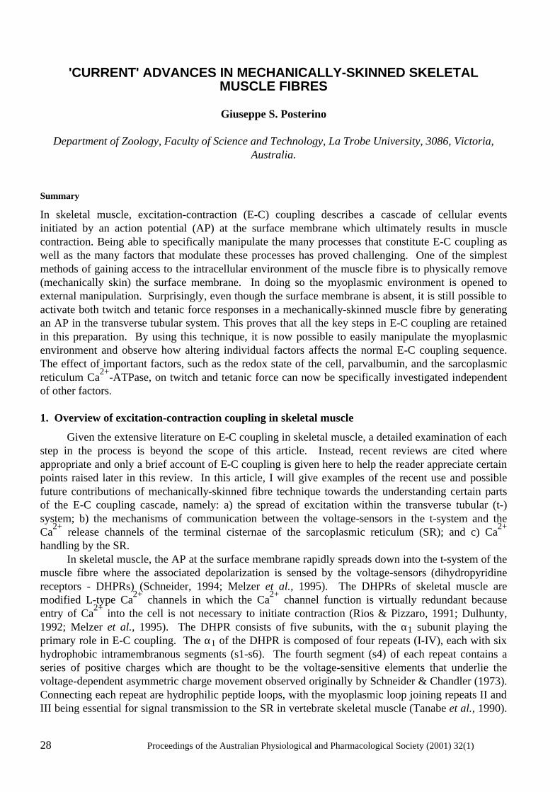
28 Proceedings of the Australian Physiological and Pharmacological Society (2001) 32(1)
'CURRENT' ADVANCES IN MECHANICALLY-SKINNED SKELETAL
MUSCLE FIBRES
Giuseppe S. Posterino
Department of Zoology, Faculty of Science and Technology, La Trobe University, 3086, Victoria, Australia.
Summary
In skeletal muscle, excitation-contraction (E-C) coupling describes a cascade of cellular events initiated by an action potential (AP) at the surface membrane which ultimately results in muscle contraction. Being able to specifically manipulate the many processes that constitute E-C coupling as well as the many factors that modulate these processes has proved challenging. One of the simplest methods of gaining access to the intracellular environment of the muscle fibre is to physically remove (mechanically skin) the surface membrane. In doing so the myoplasmic environment is opened to external manipulation. Surprisingly, even though the surface membrane is absent, it is still possible to activate both twitch and tetanic force responses in a mechanically-skinned muscle fibre by generating an AP in the transverse tubular system. This proves that all the key steps in E-C coupling are retained in this preparation. By using this technique, it is now possible to easily manipulate the myoplasmic environment and observe how altering individual factors affects the normal E-C coupling sequence. The effect of important factors, such as the redox state of the cell, parvalbumin, and the sarcoplasmic reticulum Ca2+-ATPase, on twitch and tetanic force can now be specifically investigated independent of other factors. 1. Overview of excitation-contraction coupling in skeletal muscle
Given the extensive literature on E-C coupling in skeletal muscle, a detailed examination of each step in the process is beyond the scope of this article. Instead, recent reviews are cited where appropriate and only a brief account of E-C coupling is given here to help the reader appreciate certain points raised later in this review. In this article, I will give examples of the recent use and possible future contributions of mechanically-skinned fibre technique towards the understanding certain parts of the E-C coupling cascade, namely: a) the spread of excitation within the transverse tubular (t-) system; b) the mechanisms of communication between the voltage-sensors in the t-system and the Ca2+ release channels of the terminal cisternae of the sarcoplasmic reticulum (SR); and c) Ca2+ handling by the SR. In skeletal muscle, the AP at the surface membrane rapidly spreads down into the t-system of the muscle fibre where the associated depolarization is sensed by the voltage-sensors (dihydropyridine receptors - DHPRs) (Schneider, 1994; Melzer et al., 1995). The DHPRs of skeletal muscle are modified L-type Ca2+ channels in which the Ca2+ channel function is virtually redundant because entry of Ca2+ into the cell is not necessary to initiate contraction (Rios & Pizzaro, 1991; Dulhunty, 1992; Melzer et al., 1995). The DHPR consists of five subunits, with the α1 subunit playing the primary role in E-C coupling. The α1 of the DHPR is composed of four repeats (I-IV), each with six hydrophobic intramembranous segments (s1-s6). The fourth segment (s4) of each repeat contains a series of positive charges which are thought to be the voltage-sensitive elements that underlie the voltage-dependent asymmetric charge movement observed originally by Schneider & Chandler (1973). Connecting each repeat are hydrophilic peptide loops, with the myoplasmic loop joining repeats II and III being essential for signal transmission to the SR in vertebrate skeletal muscle (Tanabe et al., 1990).
Proceedings of the Australian Physiological and Pharmacological Society (2001) 32(1) 29
The DHPRs co-localize in arrangements of four, termed tetrads (Block et al., 1988), and are located immediately adjacent to alternate Ca2+-release channels (ryanodine receptors - RYRs) in the adjacent SR. Activation of the DHPRs subsequently leads to the activation of the RYRs by a mechanism that is not fully understood. The RYRs are large homotetrameric Ca2+ channels which tightly bind the plant alkaloid ryanodine. The RYRs specific to skeletal muscle are termed RYR1 and are arranged in closely packed arrays in vivo (Block et al., 1988). In mammalian muscle, every RYR1 appears to be functionally identical at the biochemical level (Ogawa, 1994), although the properties of DHPR coupled versus DHPR uncoupled RYR1s in vivo may differ. In amphibian muscle, the RYR1s are composed of two isoforms, α and β, of which the properties of the β-isoform may or may not be differ from amphibian α and mammalian RYR1 (Ogawa, 1994; Franzini-Armstrong & Protasi, 1997; Ogawa et al., 1999). A distinct feature of the various RYR1s of both amphibian and mammalian skeletal muscle is the strong inhibition of channel activity by physiological levels of Mg2+ (~1 mM), millimolar concentrations of Ca2+, and the ability of ATP to stimulate channel activity even in the absence of Ca2+ (Lamb, 2000). These features are essential for the type of E-C coupling observed in skeletal muscle as opposed to cardiac and smooth muscle cells. Precisely how the DHPR and the RYR1 interact has not been established, although a direct interaction between these two channels is thought to occur (Melzer et al., 1995; Meissner & Lu, 1995; Franzini-Armstrong & Protasi, 1997). Activation of the RYR allows Ca2+ stored in the SR to enter the myoplasm where it binds to the contractile apparatus to initiate force production (Melzer et al., 1995). The release of Ca2+ is tightly controlled by the DHPRs (Rios & Pizzaro, 1991; Melzer et al., 1995). The cessation of Ca2+ release upon deactivation of the DHPRs leads to relaxation of force as the Ca2+ initially released is resequestered back into the SR through the activity of the SR Ca2+-ATPases and in fast-twitch fibres, relaxation may be aided by the binding of Ca2+ to parvalbumin (Rall, 1996). 2. Techniques for investigating E-C coupling in skeletal muscle
Many techniques have been used in the study of E-C coupling and all have both advantages and disadvantages. Whole intact cell preparations have the advantage that they retain normal physiological function. However, the usefulness of these preparations is to some extent limited by difficulties in controlling and measuring intracellular processes. One way around this problem is to use molecular biology techniques, such as the knockout of a specific gene. In this way the influence of a specific protein can be removed and the effect of this studied in the intact system. However, given the complex interaction between many cellular constituents, the removal of a specific component may cause some unintentional change in the function of other components. In contrast, the biochemical approach involves the study of key components in well controlled artificial environments, such as the isolation of a single channel in an artificial lipid bilayer. In this way the basic function of a particular component can be determined in isolation, although the effect of complex interactions with other cellular components that may normally occur in vivo is lost. Bridging the gap between intact fibre and biochemical techniques are the skinned muscle fibre preparations. The key advantage of these preparations is that the myoplasmic environment can be easily manipulated whilst in certain skinned fibre preparations (see below), all the essential elements in the E-C coupling cascade also remains intact. 3. Types of skinned fibre preparations
There are a two main ways to permeablise a skeletal muscle fibre – chemically and mechanically. However, these differ in the consequences they have on the various structures of the muscle fibre.
30 Proceedings of the Australian Physiological and Pharmacological Society (2001) 32(1)
Figure 1. Intact and mechanically-skinned portion of a skeletal muscle fibre. Schematic diagram showing the skinning of a single skeletal muscle fibre by rolling back the surface membrane (sarcolemma - S) with a pair of forceps (F), forming a 'cuff'. The transverse tubular system (T) seals off to form a closed compartment after skinning. SR, sarcoplasmic reticulum; Z, Z-line; C, contractile apparatus. (Modified from Posterino et al., 2000).
a) Chemical skinning
This involves the use of a number of chemical reagents that permeablise the various membranes of the fibre – some are more selective than others. Commonly used reagents are saponin, β-escin, glycerol and triton-X 100. The more selective permeablising agents (e.g. saponin and β-escin) are thought to act primarily on the surface membranes (sarcolemma) and t-tubules by binding cholesterol which is largely absent from the SR. However, this selectivity is not as precise as first thought and significant effects on the SR have been observed (Launikonis & Stephenson, 1997, 1999). Other non-specific reagents (such as triton-X 100 and glycerol) destroy all the membrane structures and leave only the contractile machinery intact. The type of permeablising reagent that is most appropriate depends on the particular cellular process the investigator wishes to examine. For examination of the properties of the contractile apparatus, it is often best to remove all membrane structures to ensure that they do not interfere with the measurements. For example, the presence of a functional SR can greatly affect the Ca2+ gradients within the fibre and this may significantly affect the force-[Ca2+] relationship unless the [Ca2+] was buffered very strongly. If on the other hand the purpose is to investigate the Ca2+-handling properties of the SR, one needs to use the more selective reagents that leave the SR intact and functional - and this may not be possible with even the more selective reagents used (cf. Launikonis & Stephenson, 1997, 1999). Thus, to avoid any undesired effects of permeablising reagents it is perhaps best to use mechanically-skinned fibres. b) Mechanical-skinning
The mechanically-skinned fibre technique (originally termed Natori-type fibres) was first developed by Natori in 1954. He showed that it was possible to gain access to intracellular environment by physically rolling back the surface membrane of a single muscle fibre with a pair of fine needles under paraffin oil (see Fig. 1.). Unlike chemically-skinned fibres, in which the surface
Proceedings of the Australian Physiological and Pharmacological Society (2001) 32(1) 31
membrane, t-system and SR are perforated by various chemical agents, in mechanically-skinned fibres the t-system seals off to form an intact, fully functional compartment (see later). The SR also remains intact and fully functional. There are other variations of this technique, such as splitting the fibre, however, the t-system in this preparation does not seal off which limits its experimental application. The functional integrity of mechanically-skinned fibres is one of the key advantages of this preparation and this can be seen by its use in the examination of many aspects of E-C coupling. Examination of the contractile apparatus: One of the earliest uses of mechanically-skinned fibres was in the study of the various properties of the contractile apparatus in which a number of models derived from earlier biochemical studies could be tested in a more physiological preparation (Gordon et al., 2000). These fibres were often also treated with membrane permeablising reagents, such as triton X- 100, and are thus better termed chemically-skinned fibres. Nevertheless, the specific role of Ca2+, Mg2+, ATP and other compounds in the regulating contraction has been studied extensively by exposing the myofilaments of both mechanically-skinned and chemically-skinned fibres to various buffered solutions (Stephenson, 1981; Gordon et al., 2000). Mechanically-skinned fibres are useful in understanding the properties of the contractile apparatus as it has been shown that the intrinsic contractile properties of intact fibres can be observed in mechanically-skinned fibres provided that the buffering of various factors in solution is firmly controlled (Moisescu, 1976). More recently, a study comparing the Ca2+-activation properties (i.e. the Ca2+-sensitivity and Hill coefficient) of both intact and mechanically-skinned fibres further demonstrate that contractile function remains unchanged following skinning (Konishi & Watanabe, 1998). Examination of SR properties: Biochemical assays have provided enormous insight into the function of the SR and its key molecules. However, during preparation of these assays, the structure of the SR and associated proteins may be compromised and no longer representative of their state in vivo. Furthermore, the normal constraints and relationships between RYR1s and other key molecules present in vivo are typically lost (Favero, 1999). The importance of such constraints and relationships between molecules for their normal function is becoming clearer. As mentioned earlier, RYR1s are arranged in a closely packed array in vivo and it has been shown recently that RYRs will spontaneously form these arrangements in solution (Yin & Lai, 2000). Furthermore, RYRs in bilayers have been observed to open and close in synchrony (termed coupled gating, Marx et al., 1998). This phenomena suggested that there is physical cooperativity between such channels and this could well be important for normal Ca2+ release. There is also a close association between RYR1 and DHP receptors of the t-system (Block et al., 1988) and recent studies have suggested that RYR1 can bind DHP receptors in vitro (Murray & Ohlendiek, 1997). The close proximity of the DHP receptors and RYR1 appears to directly influence their individual functions (Nakai et al., 1996). Associated proteins of the RYR1, such as FKBP-12, also help to link and control neighbouring channels (Marx et al., 1998) whilst other associated molecules, such as calmodulin and calsequestrin, appear to regulate channel activity at an individual level (Franzini-Armstrong & Protasi, 1997). Mechanically-skinned fibres retain these relationships and structural constraints and are thus ideal for the examination of the activity of the RYR1 and the Ca2+-ATPase in their native state. The endogenous Ca2+ content of the SR, which is reported to regulate the activity of the RYR1 (Sitsapesan & Williams, 1997), can also be assayed and controlled in this preparation. By using a number of RYR agonists and antagonists, such as caffeine and ryanodine, as well as antagonists of the Ca2+-ATPase, such as 2,5-di(tert-butyl)-1,4-benzohydroquinone (TBQ), the properties of the SR and related molecules can be examined (see later). Examination of voltage-dependent Ca2+ release: Considering the apparent importance of maintaining normal structural integrity, how do we really know that mechanically-skinned fibres accurately describe events that occur in vivo given that the surface membrane is removed in this preparation?
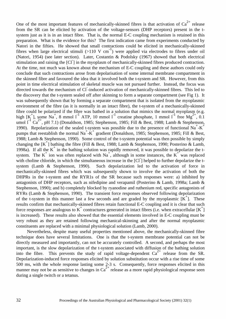
32 Proceedings of the Australian Physiological and Pharmacological Society (2001) 32(1)
One of the most important features of mechanically-skinned fibres is that activation of Ca2+ release from the SR can be elicited by activation of the voltage-sensors (DHP receptors) present in the t-system just as it is in an intact fibre. That is, the normal E-C coupling mechanism is retained in this preparation. What is the evidence for this? The first indication came from experiments conducted by Natori in the fifties. He showed that small contractions could be elicited in mechanically-skinned fibres when large electrical stimuli (>110 V cm-1) were applied via electrodes to fibres under oil (Natori, 1954) (see later section). Later, Costantin & Podolsky (1967) showed that both electrical stimulation and raising the [Cl-] in the myoplasm of mechanically-skinned fibres produced contraction. At the time, not much was known about the mechanism of E-C coupling and these authors could only conclude that such contractions arose from depolarisation of some internal membrane compartment in the skinned fibre and favoured the idea that it involved both the t-system and SR. However, from this point in time electrical stimulation of skeletal muscle was not pursued further. Instead, the focus was directed towards the mechanism of Cl--induced activation of mechanically-skinned fibres. This led to the discovery that the t-system sealed off after skinning to form a separate compartment (see Fig 1). It was subsequently shown that by forming a separate compartment that is isolated from the myoplasmic environment of the fibre (as it is normally in an intact fibre), the t-system of a mechanically-skinned fibre could be polarized if the fibre was bathed in a solution that mimics the normal myoplasm (e.g. high [K+], some Na+, 8 mmol l-1 ATP, 10 mmol l-1 creatine phosphate, 1 mmol l-1 free Mg2+, 0.1 umol l-1 Ca2+, pH 7.1) (Donaldson, 1985; Stephenson, 1985; Fill & Best, 1988; Lamb & Stephenson, 1990). Repolarization of the sealed t-system was possible due to the presence of functional Na+-K+ pumps that reestablish the normal Na+-K+ gradient (Donaldson, 1985; Stephenson, 1985; Fill & Best, 1988; Lamb & Stephenson, 1990). Some control of the t-system potential was then possible by simply changing the [K+] bathing the fibre (Fill & Best, 1988; Lamb & Stephenson, 1990; Posterino & Lamb, 1998a). If all the K+ in the bathing solution was rapidly removed, it was possible to depolarize the t-system. The K+ ion was often replaced with Na+, although in some instances, the K+ was replaced with choline chloride, in which the simultaneous increase in the [Cl-] helped to further depolarize the t-system (Lamb & Stephenson, 1990). Such depolarization led to the activation of force in mechanically-skinned fibres which was subsequently shown to involve the activation of both the DHPRs in the t-system and the RYR1s of the SR because such responses were: a) inhibited by antagonists of DHP receptors, such as nifedipine and verapamil (Posterino & Lamb, 1998a; Lamb & Stephenson, 1990); and b) completely blocked by ryanodine and ruthenium red, specific antagonists of RYRs (Lamb & Stephenson, 1990). The transient force responses observed following depolarization of the t-system in this manner last a few seconds and are graded by the myoplasmic [K+]. These results confirm that mechanically-skinned fibres retain functional E-C coupling and it is clear that such force responses are analogous to K+ contractures generated in intact fibres (i.e. when extracellular [K+] is increased). These results also showed that the essential elements involved in E-C coupling must be very robust as they are retained following mechanical-skinning and after the normal myoplasmic constituents are replaced with a minimal physiological solution (Lamb, 2000). Nevertheless, despite many useful properties mentioned above, the mechanically-skinned fibre technique does have several limitations. One is that the t-system membrane potential can not be directly measured and importantly, can not be accurately controlled. A second, and perhaps the most important, is the slow depolarization of the t-system associated with diffusion of the bathing solution into the fibre. This prevents the study of rapid voltage-dependent Ca2+ release from the SR. Depolarization-induced force responses elicited by solution substitution occur with a rise time of some 500 ms, with the whole response lasting some 2-3 s. Consequently, force responses elicited in this manner may not be as sensitive to changes in Ca2+ release as a more rapid physiological response seen during a single twitch or a tetanus.
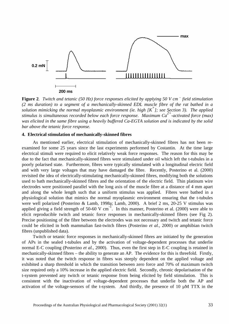
Proceedings of the Australian Physiological and Pharmacological Society (2001) 32(1) 33
Figure 2. Twitch and tetanic (50 Hz) force responses elicited by applying 50 V cm-1 field stimulation (2 ms duration) to a segment of a mechanically-skinned EDL muscle fibre of the rat bathed in a solution mimicking the normal myoplasmic environment (ie. high [K+]; see Section 3). The applied stimulus is simultaneous recorded below each force response. Maximum Ca2+-activated force (max) was elicited in the same fibre using a heavily buffered Ca-EGTA solution and is indicated by the solid bar above the tetanic force response.
4. Electrical stimulation of mechanically-skinned fibres
As mentioned earlier, electrical stimulation of mechanically-skinned fibres has not been re-examined for some 25 years since the last experiments performed by Costantin. At the time large electrical stimuli were required to elicit relatively weak force responses. The reason for this may be due to the fact that mechanically-skinned fibres were stimulated under oil which left the t-tubules in a poorly polarised state. Furthermore, fibres were typically stimulated with a longitudinal electric field and with very large voltages that may have damaged the fibre. Recently, Posterino et al. (2000) revisited the idea of electrically-stimulating mechanically-skinned fibres, modifying both the solutions used to bath mechanically-skinned fibres and the orientation of the electric field. Thin platinum wire electrodes were positioned parallel with the long axis of the muscle fibre at a distance of 4 mm apart and along the whole length such that a uniform stimulus was applied. Fibres were bathed in a physiological solution that mimics the normal myoplasmic environment ensuring that the t-tubules were well polarized (Posterino & Lamb, 1998a; Lamb, 2000). A brief 2 ms, 20-25 V stimulus was applied giving a field strength of 50-60 V cm-1. In this manner, Posterino et al. (2000) were able to elicit reproducible twitch and tetanic force responses in mechanically-skinned fibres (see Fig 2). Precise positioning of the fibre between the electrodes was not necessary and twitch and tetanic force could be elicited in both mammalian fast-twitch fibres (Posterino et al., 2000) or amphibian twitch fibres (unpublished data). Twitch or tetanic force responses in mechanically-skinned fibres are initiated by the generation of APs in the sealed t-tubules and by the activation of voltage-dependent processes that underlie normal E-C coupling (Posterino et al., 2000). Thus, even the first step in E-C coupling is retained in mechanically-skinned fibres – the ability to generate an AP. The evidence for this is threefold. Firstly, it was noted that the twitch response in fibres was steeply dependent on the applied voltage and exhibited a sharp threshold in which the transition between zero force and 70% of maximum twitch size required only a 10% increase in the applied electric field. Secondly, chronic depolarisation of the t-system prevented any twitch or tetanic response from being elicited by field stimulation. This is consistent with the inactivation of voltage-dependent processes that underlie both the AP and activation of the voltage-sensors of the t-system. And thirdly, the presence of 10 µM TTX in the
max
0.2 mN
200 ms
34 Proceedings of the Australian Physiological and Pharmacological Society (2001) 32(1)
sealed t-tubules strongly inhibited such responses proving that activation of Na+ channels (and the generation of an AP) in the t-system is essential in triggering further steps in the cascade. The characteristics of both twitch and tetanic force responses elicited in mechanically-skinned fibres closely resemble the responses observed in intact fibres (Fryer & Neering, 1988; Schwaller et al., 1999). The peak amplitude of the twitch response in fast-twitch mammalian and twitch amphibian muscle is between 30% and 60% of maximum Ca2+-activated force. Tetani (50 Hz) elicits force responses between 80-100% of maximum Ca2+-activated force. The twitch-tetanus ratio in mammalian fast-twitch skinned fibres ranges between 0.40 and 0.60 which is larger than that observed in intact fibres (~0.30) (Schwaller et al., 1999). The larger twitch-tetanus ratio and the ability to achieve near maximal force during a tetanus, is possibly due at least in part to the loss of parvalbumin in mechanically-skinned fibre preparations (Stephenson et al., 1999). The clear functional similarities between intact and mechanically-skinned fibres highlight the potential of the mechanically-skinned fibre technique in the study of skeletal muscle physiology. Nevertheless, there are a few differences in the responses observed between mechanically-skinned fibres and intact fibres. One such difference is that tetanic force in fast-twitch mechanically-skinned fibres declines much more rapidly during high frequency stimuli than in intact fibres, with force fading after ~200 ms of stimulation (termed fade; see Fig. 2). The cause of this phenomenon is not certain. Posterino et al. (2000) attributed this to a gradual build up of K+ in the sealed t-system with repeated APs leading to depolarization; this would not normally occur in an intact fibre where the t-system is open to the extracellular environment. However, we have recently observed that this phenomena appears to be related to the fibre length, as fade was largely eliminated in fibres that were stretched from between 120% to 140% of their resting length (unpublished results). This is currently being further examined. Another difference between mechanically-skinned fibres and intact fibres is that the absence of a surface membrane means the intracellular Ca2+ content of the fibre can vary from the endogenous level. The comparatively large volume of solution bathing the mechanically-skinned fibre means that a substantial amount of Ca2+ can be gained from (or lost to) the bulk solution. In order to maintain the normal endogenous level of SR Ca2+, both over time and with repeated responses, it is necessary ensure that the free [Ca2+] of the bathing solution is buffered to the normal resting myoplasmic concentration (pCa 7.0). This limits the amount of Ca2+ loading. It is noteworthy that little variability is observed between successive twitch responses in mechanically-skinned fibres which suggests that the SR Ca2+ content remains relatively stable under the conditions used. However, a more precise determination of changes in the SR Ca2+ content during repeated stimulation is still needed as well as a better way of clamping the SR Ca2+ content. 5. Recent contributions and future directions
The ability to electrically stimulate mechanically-skinned fibres helps bridge the gap between biochemical and whole cell studies. Some recent findings illustrate the current and future potential of this technique towards the understanding of E-C coupling in skeletal muscle. a) Mechanisms and pathways involved in the initial spread of excitation
The first step in E-C coupling involves the initiation and spread of the AP throughout the muscle fibre. It is generally accepted that the spread of excitation into the t-system in amphibian skeletal muscle involves an AP (Costantin, 1970; Bezanilla et al., 1972; Nakajima & Gilal, 1980). In mammalian skeletal muscle, it was not known if the spread was either passive or active, although it was assumed to involve an AP. As indicated earlier, the ability to generate an AP in mechanically-skinned fibres by electrical stimulation now provides direct evidence that excitation also spreads down the t-tubules via an active process in mammalian muscle (Posterino et al., 2000). This observation is unambiguous as there is no surface membrane present. Furthermore, it is apparent that other
Proceedings of the Australian Physiological and Pharmacological Society (2001) 32(1) 35
characteristics of the AP in the t-system can also be determined using this preparation. By using the twitch response as an indirect measure of the AP, it is possible to estimate the relative refractory period of the AP. If two single stimuli are elicited in close succession (less than 6 ms) no summation of the twitch force response was observed. However, if the second stimulus is applied 6 ms (or later) after the first, the twitch response is potentiated. This indicates that the refractory period of the AP is at most 6 ms long (unpublished data). Refinement of this measure could be achieved by examining the Ca2+ transient rather than force. Experiments with electrically-stimulated mechanically-skinned fibres have also revealed the role of a previously identified structure in skeletal muscle. The observation that contractions (either spontaneous or elicited electrically) have the ability to propagate over hundreds of sarcomeres in mechanically-skinned fibres revealed a mechanism that allows excitation to spread throughout a skeletal muscle fibre independent of the surface membrane (Natori, 1954; Costantin & Podolsky, 1967; Posterino et al., 2000). It was found that an AP(s) could propagate along the entire length of skinned fibre segment (without the presence of a surface membrane) and could cause relatively synchronous activation of a large proportion of the fibre (>70%) travelling with an estimated velocity of some 13 mm s-1
(Posterino et al., 2000). It was suggested that the structure involved in the spread of the AP must be the longitudinal tubular system (LTS) which has been observed with electron microscopy (EM) (Franzini-Armstrong & Jorgenson, 1988; Stephenson & Lamb, 1992) and by confocal imaging of mechanically-skinned fibres in which a fluophore was trapped in the t-system (Peachey, L.D., 1965). These findings in mechanically-skinned fibres revealed a fundamental property that is likely to be important in the spread of the AP throughout a fibre, in fatigue and during myogenesis. b) Internal transmission of excitation and control of Ca2+ release
Data obtained from mechanically-skinned fibres in which functional E-C coupling is retained have also provided strong evidence for and against a number of ideas regarding the mechanism by which the DHPRs and the RYR1s communicate. It is clear from some experiments in mechanically-skinned fibres that the link between these two channels does not involve a diffusible second messenger such as inositol 1,4,5-trisphosphate (Walker et al., 1987; Posterino et al., 1998b) or Ca2+(Endo, 1985; Meissner et al., 1986; Lamb & Stephenson, 1991; Owen et al., 1997; Lamb & Laver, 1998). The link was also previously thought to involve the transient formation of disulphide bonds between the DHPRs and the RYRs (Salama et al., 1992). However, recent experiments in mechanically-skinned fibres have showed that strong sulphydryl reducing reagents do not interfere with normal E-C coupling(Posterino & Lamb, 1996). Nevertheless, various sulphydryl oxidants have been shown to modulate the RYR1 function (Dulhunty et al., 1996) indicating that the cellular redox state may effect E-C coupling in a more complex manner that is dependent on the precise ratio of endogenous redox reagents (ie. ratio of glutathione to reduced glutathione) both within the myoplasm and the lumen of the SR Feng et al., 2000). Here again we can see the potential advantage of mechanically-skinned fibres in addressing this question which can not readily be observed in an intact fibre preparation. c) Ca2+ handling
As mentioned earlier, mechanically-skinned fibres rapidly loose their parvalbumin after skinning and this may account for some of the dynamic differences between twitch responses observed between mechanically-skinned fibres and intact fibres. The absence of parvalbumin in mechanically-skinned fibres allows the examination of the functional role of this protein in SR Ca2+ handling. Most previous studies examining the role of parvalbumin have been limited by the inability to remove the effects of parvalbumin completely. Only one recent study using mice in which the parvalbumin gene has been knocked out has been able to examine the precise contribution of parvalbumin on twitch and tetanic
36 Proceedings of the Australian Physiological and Pharmacological Society (2001) 32(1)
characteristics (Fryer & Neering, 1988). This study showed that twitch-tetanus ratio was greater in fibres from parvalbumin knockout mice (PKM) than from wildtype mice. Interestingly, the characteristics of the twitch response from PKM greatly resemble those from mechanically-skinned fibres. Nevertheless, it is possible that the absence of parvalbumin in PKM may have effected the other constituents important in E-C coupling and Ca2+ handling. The unique properties of mechanically-skinned fibres allows the examination of parvalbumin more precisely without the problem of non-specific effects that may arise from gene knockout. Parvalbumin can be simply added to and removed from the bathing solution of mechanically-skinned fibres and the effects on the twitch and tetanus observed in the same fibre. Apart from parvalbumin, mechanically-skinned fibres can allow the precise examination of the role of the SR Ca2+ ATPase in contraction and relaxation. In intact fibre studies, the role of the SR Ca2+ ATPase is often examined by using specific inhibitors such as TBQ and thapsigargin (Westerblad & Allen, 1994; Caputo et al., 1999). However, it is difficult to be sure that complete block of the pump has taken place and that there are no complicating effects of increased resting Ca2+. Similarly, the intact fibre studies must also take into account any effects of parvalbumin. An advantage of mechanically-skinned fibres is that distinct qualities of the SR Ca2+ ATPase can be examined in isolation of parvalbumin and without changes to resting myoplasmic [Ca2+]. Conclusion
To date, mechanically-skinned fibres have been a useful tool in the study of many aspects of E-C coupling in skeletal muscle. The controlled nature of the myoplasmic environment of skinned fibres, the presence of functional E-C coupling that can be now be activated in the same manner and with a similar time course as an intact fibre, and the fact that the key structures involved in E-C coupling obviously remain as they were in vivo, demonstrate the potential of this technique in further aiding our understanding of E-C coupling in skeletal muscle. Acknowledgments
I wish to thank Professor George Stephenson and Associate Professor Graham Lamb for their helpful advice and comments on the manuscript. This work was supported by the National Health and Medical Research Council of Australia. References
Bezanilla, F., Caputo, C., Gonzalez-Serratos, H. & Venosa, R.A. (1972) Sodium dependence of the inward spread of activation in isolated twitch muscle fibres of the frog. Journal of Physiology, 223, 507-523.
Block, B.A., Imagawa, T., Campbell, K.P. & Franzini-Armstrong, C. (1988) Structural evidence for direct interaction between the molecular components of the transverse tubule/sarcoplasmic reticulum junction in skeletal muscle. Journal of Cell Biology, 107(6/2), 2587-2600.
Caputo, C., Bolanos, P. & Escobar, A.L. (1999) Fast calcium removal during single twitches in amphibian skeletal muscle fibres. Journal of Muscle Research & Cell Motility, 20, 555-567.
Costantin, L.L. & Podolsky, R.J. (1967) Depolarization of the internal membrane system in the activation of frog skeletal muscle. Journal of General Physiology, 50, 1101-1124.
Costantin, L.L. (1970) The role of sodium current in the radial spread of contraction in frog muscle fibres. Journal of General Physiology, 55, 703-715.
Donaldson, S.K.B. (1985) Peeled mammalian skeletal muscle fibres. Possible stimulation of Ca2+ release via a transverse tubule-sarcoplasmic reticulum mechanism. Journal of General Physiology, 86, 501-525.
Proceedings of the Australian Physiological and Pharmacological Society (2001) 32(1) 37
Dulhunty, A.F. (1992) The voltage-activation of contraction in skeletal muscle. Progress in Biophysics and Physiology, 53, 1-16.
Dulhunty, A.F., Junankar, P.R., Eager, K.R., Ahern, G.P. & Laver, D.R. (1996) Ion channels in the sarcoplasmic reticulum of striated muscle. Acta Physiologica Scandanavica, 156, 375-385.
Endo, M. (1985) Calcium release from the sarcoplasmic reticulum. Current Topics in Membranes and Transport, 25, 181-230.
Favero, T.G. (1999) Sarcoplasmic reticulum Ca2+ release and muscle fatigue. Journal of Applied Physiology, 87(2), 471-483.
Feng, W., Guohua, L., Allen, P.D. & Pessah, I.N. (2000) Transmembrane redox sensor of ryanodine receptor complex. Journal of Biological Chemistry, 275, 35902-35907.
Fill, M.D. & Best, P.M. (1988) Contractile activation and recovery in skinned frog muscle stimulated by ionic substitution. American Journal of Physiology, 254, C107-114.
Franzini-Armstrong, C. & Jorgenson, A.O. (1988) Discrimination between fast and slow twitch fibres of guinea pig skeletal muscle using the relative surface density of junctional transverse tubule membrane. Journal of Muscle Research and Cellular Communication, 9, 403-413.
Franzini-Armstrong, C. & Protasi, F. (1997) Ryanodine receptors of striated muscles: a complex channel capable of multiple interactions. Physiological Reviews, 77, 699-729.
Fryer, M.F. & Neering, I.R. (1988) Actions of caffeine on fast- and slow-twitch muscles of the rat. Journal of Physiology, 416, 435-454.
Gordon, A.M., Homsher, E. & Regnier, M. (2000) Regulation of contraction in striated muscle. Physiological Reviews, 80(2), 853-924.
Konishi, M. & Watanabe, M. (1998) Steady state relation between cytoplasmic free Ca2+ concentration and force in intact frog skeletal muscle fibres. Journal of General Physiology, 111, 505-519.
Lamb, G.D. & Laver, D.R. (1998) Adaption, inactivation and inhibition in ryanodine receptors. In: The Structure and Function of Ryanodine Receptors. Ed. Sitsapesan, R. & Williams, A.J. Ch. 14. London: Imperial College Press.
Lamb, G.D. & Stephenson, D.G. (1990) Calcium release in skinned muscle fibres of the toad by transverse tubule depolarization or by direct stimulation. Journal of Physiology, 423, 495-517.
Lamb, G.D. & Stephenson, D.G. (1991) Effect of Mg2+ on the control of Ca2+ release in skeletal muscle fibres of the toad. Journal of Physiology, 434, 507-528.
Lamb, G.D. (2000) Excitation-contraction coupling in skeletal muscle: comparisons with cardiac muscle. Clinical and Experimental Physiology and Pharmacology, 27, 216-224.
Launikonis, B.S. & Stephenson, D.G. (1997) Effect of saponin treatment on the sarcoplasmic reticulum of rat, cane toad and crustacean (yabby) skeletal muscle. Journal of Physiology, 504.2, 425-437.
Launikonis, B.S. & Stephenson, D.G. (1999) Effects of β-escin and saponin on the transverse-tubular system and the sarcoplasmic reticulum membranes of rat and toad skeletal muscle. Pflügers Archiv, 437, 955-965.
Marx, S.O., Ondrias, K. & Marks, A.R. (1998) Coupled gating between individual skeletal muscle Ca2+ release channels (ryanodine receptors). Science, 281, 818-821.
Meissner, G. & Lu, X. (1995) Dihyropyridine receptor-ryanodine receptor interactions in skeletal muscle excitation-contraction coupling. Bioscience Reports, 15(5), 399-408.
38 Proceedings of the Australian Physiological and Pharmacological Society (2001) 32(1)
Meissner, G., Darling, E. & Eveleth, J. (1986) Kinetics of rapid Ca2+ release by sarcoplasmic reticulum. Effects of Ca2+, Mg2+ and adenine nucleotides. Biochemistry, 25, 236-244.
Melzer, W., Herrmann-Frank, A. & Lüttgau, H.Ch. (1995) The role of Ca2+ ions in excitation-contraction coupling in skeletal muscle fibres. Biochimica et Biophysica Acta, 1241, 59-116.
Moisescu, D.G. (1976) Kinetics of reaction of calcium-activated skinned frog muscle fibres. Nature, 262, 610-613.
Murray, B.E. & Ohlendiek, K. (1997) Cross-linking analysis of the ryanodine receptor and α1-dihydropyridine receptor in rabbit skeletal muscle triads. Biochemical Journal, 324, 689-696.
Nakai, J., Dirksen, R.T., Nguyen, H.T., Pessah, I.N., Beam, K.G. & Allen, P.D. (1996) Enhanced dihydropyridine receptor channel activity in the presence of ryanodine receptor. Nature, 380, 72-75.
Nakajima, S. & Gilal, A. (1980) Radial propagation of muscle action potentials along the tubular system examined by potential-sensitive dyes. Journal of General Physiology, 76, 751-762.
Natori, R. (1954) The property and contraction process of isolated myofibrils. Jikei-Kai Medical Journal, 1, 119-126.
Ogawa, Y. (1994) Role of ryanodine receptors. Critical Reviews in Biochemistry and Molecular Biology, 29(4), 229-274.
Ogawa, Y., Murayama, T. & Kurebayashi, N. (1999) Comparison of properties of Ca2+ release channels between rabbit and frog skeletal muscles. Molecular and Cellular Biochemistry, 190, 191-201.
Owen, V.J., Lamb, G.D., Stephenson, D.G. & Fryer, M.F. (1997) Relationship between depolarization-induced force responsese and Ca2+ content in skeletal muscle fibres of rat and toad. Journal of Physiology, 498, 571-586.
Peachey, L.D. (1965) The sarcoplasmic reticulum and the transverse tubules of the frog's sartorius. Journal of Cell Biology, 25, 209-231.
Posterino, G.S. & Lamb, G.D. (1996) Effects of reducing agents and oxidants on excitation-contraction coupling in skeletal muscle fibres of rat and toad. Journal of Physiology, 496.3, 809-825.
Posterino, G.S. & Lamb, G.D. (1998a) Effect of nifedipine on depolarization-induced force responses in skinned skeletal muscle fibres of rat and toad. Journal of Muscle Research & Cell Motility, 19, 53-65.
Posterino, G.S. & Lamb, G.D. (1998b) Investigation of the effect of inositol trisphosphate in skinned skeletal muscle fibres with functional excitation-contraction coupling. Journal of Muscle Research & Cell Motility, 19, 67-74.
Posterino, G.S., Lamb, G.D. & Stephenson, D.G. (2000) Twitch and tetanic force responses and longitudinal propagation of action potentials in skinned skeletal muscle. Journal of Physiology, 527.1, 131-137.
Rall, J. (1996) The role of parvalbumin in skeletal muscle relaxtion. News in Physiological Sciences, 11, 249-255.
Rios, E. & Pizzaro, G. (1991) Voltage-sensor of excitation-contraction coupling in skeletal muscle. Physiological Reviews, 71, 849-908.
Salama, G., Abramson, J.J. & Pike, G.K. (1992) Sulphydryl reagents triggers Ca2+ release from the sarcoplasmic reticulum of skinned rabbit psoas fibres. Journal of Physiology, 454, 389-420.
Schneider, M.F. & Chandler, W.K. (1973) Voltage-dependent charge movement in skeletal muscle: a possible step in excitation-contraction coupling. Nature, 242, 244-246.
Proceedings of the Australian Physiological and Pharmacological Society (2001) 32(1) 39
Schneider, M.F. (1994) Control of caclium release in functioning muscle fibres. Annual Review of Physiology, 56, 463-484.
Schwaller, B., Dick, J., Dhoot, G., Carroll, S., Vrbova, G., Nicotera, P., Pette, D., Wyss, A., Bluethmann, H., Hunziker, W. & Celio, M.R. (1999) Prolonged contraction relaxation cycle of fast-twitch muscles in parvalbumin knockout mice. American Journal of Physiology, 276, C395-403.
Sitsapesan, R. & Williams, A.J. (1997) Regulation of current flow through ryanodine receptors by luminal Ca2+. Journal of Membrane Biology, 159, 179-185.
Stephenson, D.G. & Lamb, G.D. (1992) Confocal imaging of key intracellular compartments in muscle fibres. Proceedings of the Australian Physiological and Pharmacological Society, 23, 244P.
Stephenson, D.G., Nguyen, L.T. & Stephenson, G.M.M. (1999) Glycogen content and excitation-contraction coupling in mechanically skinned muscle fibres of the cane toad. Journal of Physiology, 519(1),177-187.
Stephenson, E.W. (1981) Activation of fast skeletal muscle: contributions of studies on skinned fibres. American Journal of Physiology, 240, C1-C19.
Stephenson, E.W. (1985) Excitation of skinned muscle fibres by imposed ion gradients. I. Stimulation of 45Ca efflux at constant [K][Cl] product. Journal of General Physiology, 86, 813-832.
Tanabe, T., Beam, K.G., Adams, B.A., Nicodome, T. & Numa, S. (1990) Regions of skeletal muscle dihydropyridine receptor critical fro excitation-contraction coupling. Nature, 346, 567-569.
Walker, J.W., Somlyo, A.V., Goldman, Y.E., Somlyo, A.P. & Trentham, D.R. (1987) Kinetics of smooth and skeletal muscle activation by laser pulse photolysis of caged inositol 1,4,5-trisphosphate. Nature, 327, 249-252.
Westerblad, H. & Allen, D.G. (1994) The role of sarcoplasmic reticulum in relaxation of mouse muscle; effects of 2,5-di(tert-butyl)-1,4-benzohydroquinone. Journal of Physiology, 474.2, 201-301.
Yin, C.C. & Lai, F.A. (2000) Intrinsic lattice formation by the ryanodine receptor calcium-release channel. Nature Cell Biology, 2, 669-671.
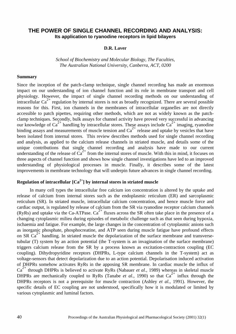
40 Proceedings of the Australian Physiological and Pharmacological Society (2001) 32(1)
THE POWER OF SINGLE CHANNEL RECORDING AND ANALYSIS:
Its application to ryanodine receptors in lipid bilayers
D.R. Laver
School of Biochemistry and Molecular Biology, The Faculties, The Australian National University, Canberra, ACT, 0200
Summary
Since the inception of the patch-clamp technique, single channel recording has made an enormous impact on our understanding of ion channel function and its role in membrane transport and cell physiology. However, the impact of single channel recording methods on our understanding of intracellular Ca2+ regulation by internal stores is not as broadly recognized. There are several possible reasons for this. First, ion channels in the membranes of intracellular organelles are not directly accessible to patch pipettes, requiring other methods, which are not as widely known as the patch-clamp techniques. Secondly, bulk assays for channel activity have proved very successful in advancing our knowledge of Ca2+ handling by intracellular stores. These assays include Ca2+ imaging, ryanodine binding assays and measurements of muscle tension and Ca2+ release and uptake by vesicles that have been isolated from internal stores. This review describes methods used for single channel recording and analysis, as applied to the calcium release channels in striated muscle, and details some of the unique contributions that single channel recording and analysis have made to our current understanding of the release of Ca2+ from the internal stores of muscle. With this in mind, it focuses on three aspects of channel function and shows how single channel investigations have led to an improved understanding of physiological processes in muscle. Finally, it describes some of the latest improvements in membrane technology that will underpin future advances in single channel recording. Regulation of intracellular [Ca2+] by internal stores in striated muscle
In many cell types the intracellular free calcium ion concentration is altered by the uptake and release of calcium from internal stores such as the endoplasmic reticulum (ER) and sarcoplasmic reticulum (SR). In striated muscle, intracellular calcium concentration, and hence muscle force and cardiac output, is regulated by release of calcium from the SR via ryanodine receptor calcium channels (RyRs) and uptake via the Ca-ATPase. Ca2+ fluxes across the SR often take place in the presence of a changing cytoplasmic milieu during episodes of metabolic challenge such as that seen during hypoxia, ischaemia and fatigue. For example, the large changes in the concentration of cytoplasmic anions such as inorganic phosphate, phosphocreatine, and ATP seen during muscle fatigue have profound effects on SR Ca2+ handling. In striated muscle the depolarization of the surface membrane and transverse-tubular (T) system by an action potential (the T-system is an invagination of the surface membrane) triggers calcium release from the SR by a process known as excitation-contraction coupling (EC coupling). Dihydropyridine receptors (DHPRs, L-type calcium channels in the T-system) act as voltage-sensors that detect depolarization due to an action potential. Depolarisation induced activation of DHPRs somehow activates RyRs in the apposing SR membrane. In cardiac muscle the influx of Ca2+ through DHPRs is believed to activate RyRs (Nabauer et al., 1989) whereas in skeletal muscle DHPRs are mechanically coupled to RyRs (Tanabe et al., 1990) so that Ca2+ influx through the DHPRs receptors is not a prerequisite for muscle contraction (Ashley et al., 1991). However, the specific details of EC coupling are not understood, specifically how it is modulated or limited by various cytoplasmic and luminal factors.
Proceedings of the Australian Physiological and Pharmacological Society (2001) 32(1) 41
Figure 1. Formation of lipid bilayers and incorporation of RyRs. (A) A photograph of a lipid bilayer (bottom half of aperture) during its formation from a thick lipid film (top half). The lipid film was spread across an aperture (~100µm diameter) in a delrin septum. The thick lipid film strongly reflects the incident light whereas the bilayer, which shows the black background, is totally transparent. The bilayer portion of the film spreads across the entire aperture in a few seconds leaving a region of thick film at the periphery. (B) A schematic diagram of the process of lipid bilayer formation showing the lipid monolayers that the two oil-water interfaces. The lengths of the arrows, which are shown in each aqueous phase, indicate the relative strengths of the Van der Waals compressive forces between the adjacent water phases. This compressive force squeezes the oil (n-alkane in this case) out from between the monolayers. (C) The procedure for incorporating ion channels form the SR (RyRs in this case) into lipid bilayers. Vesicles of SR membrane ( ~0.1 µm diameter) are isolated from muscle tissues using differential centrifugation methods. SR vesicle containing ion channels are added to the bath near the lipid bilayer. Fusion of the vesicles with the bilayer carries ion channels into the bilayer membrane.
The RyR is a homotetramer of ~560 kDa subunits containing ~5035 amino acids. Electron microscope image reconstruction shows RyRs to have four-fold symmetry with a large cytoplasmic domain (the foot region) and a relatively small transmembrane region that forms the Ca2+ pore (Orlova et al., 1996). The trans membrane pore is comprised of the ~1000 C-terminal amino acids (aa 4000-5000) and the remaining amino acids form the foot region. In mammals, three isoforms of RyRs have been cloned and sequenced: namely, ryr-1 found in skeletal muscle and brain, ryr-2 most abundantly found in brain and cardiac muscle and ryr-3, though originally found in the brain, is the major isoform in smooth muscle (Ogawa, 1994).
42 Proceedings of the Australian Physiological and Pharmacological Society (2001) 32(1)
The Bilayer method
At the same time that Neher and Sakmann were developing the patch-clamp technique (Neher and Sakmann, 1976), Miller and Racker (1976) discovered that SR vesicles isolated from muscle could be fused with artificial lipid bilayers and so incorporate ion channels from muscle membranes into artificial membranes. Artificially produced, planar, bimolecular lipid membranes (bilayers) were originally used as model systems for studying cell membrane structure. Their large area (they can be produced with diameters ranging from 1 µm to 1 cm) and planar geometry made them particularly convenient membrane models for electrical and mechanical measurements. For the purposes of studying ion channels, bilayers are usually formed using a modification of the film drainage method developed by (Mueller et al., 1962). A solution of lipids in a hydrophobic solvent (usually n-decane) is smeared across a hole in a plastic septum (eg. Delrin, polycarbonate or Teflon) to produce a thick lipid film separating two baths (Fig. 1A). The bilayer forms spontaneously from this thick lipid film. During bilayer formation the surface-active lipids aggregate into monolayers at the oil-water interfaces on each side of the thick film. The solvent drains away from between the two monolayers thus allowing their apposition and formation of the bilayer structure (Fig. 1B). Incorporation of ion channels into bilayers is usually simply done by adding ion channel protein to one of the baths and stirring. Ion channel incorporations occur spontaneously and can be detected by conductance changes in the bilayer membrane. For studying the SR ion channels, the bilayers are produced with a diameter of ~100 µm. SR vesicles are added to a final concentration of 1-10 µg/ml and the bath is stirred until channel activity indicates vesicle fusion with the bilayer. The side of the bilayer to which the vesicles are added is usually defined as the cis side (Fig. 1C). Conditions that promote vesicle fusion are: 1) a gradient in osmotic potential across the membrane (cis high), 2) cis [Ca2+] at mM concentrations and 3) vigorous stirring of the cis bath. The cytoplasmic side of the SR membrane, when fused with the bilayer, faces the cis bath and the luminal side faces the trans bath. Stirring of the cis and trans (cytoplasmic and luminal) chambers is usually done using magnetic stirrers. Cesium methanesulfonate (CsMS) is commonly used as the principal salt in the bathing solutions. This is to prevent current signals from other ion channels from interfering with RyR recordings: the RyR is quite permeable to Cs+ whereas other ion channels in the SR do not conduct MS- and Cs+. Experiments also use a [Cs+] gradient across the bilayer (250 mM cis and 50 mM trans) to promote vesicle fusion (see above). When a vesicle has fused with a bilayer the ion channels embedded in the vesicle membrane become incorporated into the bilayer. Once this happens it is possible to determine the ionic conductance of a single channel and to monitor it’s opening and closing (gating) by measuring the current through the membrane in response to an applied electrochemical gradient. The bilayer technique allows considerable flexibility in manipulating the experimental conditions. One can examine the response of channels to a variety of substances in the cytosolic and luminal baths as well as changes in the composition of the bilayer itself. One can also measure channel function under steady-state conditions or when solutions are rapidly and transiently altered. With this experimental technique, like with the patch-clamp technique, it is possible to obtained very detailed information about mechanisms determining channel conductance and gating. Single channel analysis.
This section is devoted to examining the most commonly used methods for describing the properties of ion channel signals and inferring their mechanisms of function. Single channel signals generally appear as a series of stochastic current jumps between stationary current levels (e.g. see Fig. 2A). For most channel types the jumps are mainly between the closed state and a maximum level (the
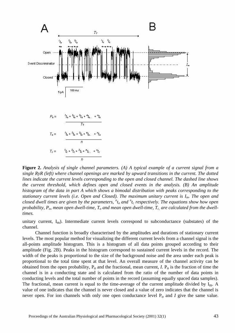
Proceedings of the Australian Physiological and Pharmacological Society (2001) 32(1) 43
Figure 2. Analysis of single channel parameters. (A) A typical example of a current signal from a single RyR (left) where channel openings are marked by upward transitions in the current. The dotted lines indicate the current levels corresponding to the open and closed channel. The dashed line shows the current threshold, which defines open and closed events in the analysis. (B) An amplitude histogram of the data in part A which shows a bimodal distribution with peaks corresponding to the stationary current levels (i.e. Open and Closed). The maximum unitary current is Im. The open and closed dwell times are given by the parameters, nto and ntc respectively. The equations show how open probability, Po, mean open dwell-time, To and mean open dwell-time, Tc, are calculated from the dwell-times.
unitary current, Im). Intermediate current levels correspond to subconductance (substates) of the channel. Channel function is broadly characterised by the amplitudes and durations of stationary current levels. The most popular method for visualizing the different current levels from a channel signal is the all-points amplitude histogram. This is a histogram of all data points grouped according to their amplitude (Fig. 2B). Peaks in the histogram correspond to sustained current levels in the record. The width of the peaks is proportional to the size of the background noise and the area under each peak is proportional to the total time spent at that level. An overall measure of the channel activity can be obtained from the open probability, Po and the fractional, mean current, I. Po is the fraction of time the channel is in a conducting state and is calculated from the ratio of the number of data points in conducting levels and the total number of points in the record (assuming equally spaced data samples). The fractional, mean current is equal to the time-average of the current amplitude divided by Im. A value of one indicates that the channel is never closed and a value of zero indicates that the channel is never open. For ion channels with only one open conductance level Po and I give the same value.
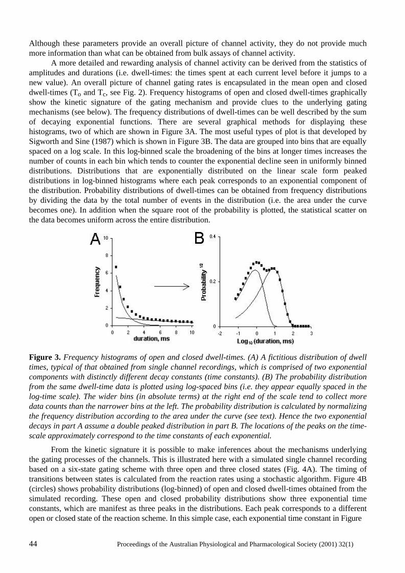
44 Proceedings of the Australian Physiological and Pharmacological Society (2001) 32(1)
Although these parameters provide an overall picture of channel activity, they do not provide much more information than what can be obtained from bulk assays of channel activity. A more detailed and rewarding analysis of channel activity can be derived from the statistics of amplitudes and durations (i.e. dwell-times: the times spent at each current level before it jumps to a new value). An overall picture of channel gating rates is encapsulated in the mean open and closed dwell-times (To and Tc, see Fig. 2). Frequency histograms of open and closed dwell-times graphically show the kinetic signature of the gating mechanism and provide clues to the underlying gating mechanisms (see below). The frequency distributions of dwell-times can be well described by the sum of decaying exponential functions. There are several graphical methods for displaying these histograms, two of which are shown in Figure 3A. The most useful types of plot is that developed by Sigworth and Sine (1987) which is shown in Figure 3B. The data are grouped into bins that are equally spaced on a log scale. In this log-binned scale the broadening of the bins at longer times increases the number of counts in each bin which tends to counter the exponential decline seen in uniformly binned distributions. Distributions that are exponentially distributed on the linear scale form peaked distributions in log-binned histograms where each peak corresponds to an exponential component of the distribution. Probability distributions of dwell-times can be obtained from frequency distributions by dividing the data by the total number of events in the distribution (i.e. the area under the curve becomes one). In addition when the square root of the probability is plotted, the statistical scatter on the data becomes uniform across the entire distribution.
Figure 3. Frequency histograms of open and closed dwell-times. (A) A fictitious distribution of dwell times, typical of that obtained from single channel recordings, which is comprised of two exponential components with distinctly different decay constants (time constants). (B) The probability distribution from the same dwell-time data is plotted using log-spaced bins (i.e. they appear equally spaced in the log-time scale). The wider bins (in absolute terms) at the right end of the scale tend to collect more data counts than the narrower bins at the left. The probability distribution is calculated by normalizing the frequency distribution according to the area under the curve (see text). Hence the two exponential decays in part A assume a double peaked distribution in part B. The locations of the peaks on the time-scale approximately correspond to the time constants of each exponential.
From the kinetic signature it is possible to make inferences about the mechanisms underlying the gating processes of the channels. This is illustrated here with a simulated single channel recording based on a six-state gating scheme with three open and three closed states (Fig. 4A). The timing of transitions between states is calculated from the reaction rates using a stochastic algorithm. Figure 4B (circles) shows probability distributions (log-binned) of open and closed dwell-times obtained from the simulated recording. These open and closed probability distributions show three exponential time constants, which are manifest as three peaks in the distributions. Each peak corresponds to a different open or closed state of the reaction scheme. In this simple case, each exponential time constant in Figure
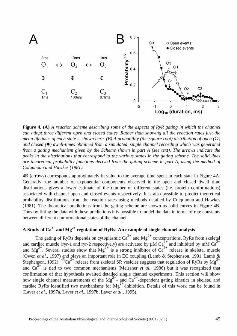
Proceedings of the Australian Physiological and Pharmacological Society (2001) 32(1) 45
Figure 4. (A) A reaction scheme describing some of the aspects of RyR gating in which the channel can adopt three different open and closed states. Rather than showing all the reaction rates just the mean lifetimes of each state is shown here. (B) A probability (the square root) distribution of open (¡) and closed (l) dwell-times obtained from a simulated, single channel recording which was generated from a gating mechanism given by the Scheme shown in part A (see text). The arrows indicate the peaks in the distributions that correspond to the various states in the gating scheme. The solid lines are theoretical probability functions derived from the gating scheme in part A, using the method of Colquhoun and Hawkes (1981).
4B (arrows) corresponds approximately in value to the average time spent in each state in Figure 4A. Generally, the number of exponential components observed in the open and closed dwell time distributions gives a lower estimate of the number of different states (i.e. protein conformations) associated with channel open and closed events respectively. It is also possible to predict theoretical probability distributions from the reaction rates using methods detailed by Colquhoun and Hawkes (1981). The theoretical predictions from the gating scheme are shown as solid curves in Figure 4B. Thus by fitting the data with these predictions it is possible to model the data in terms of rate constants between different conformational states of the channel. A Study of Ca2+ and Mg2+ regulation of RyRs: An example of single channel analysis
The gating of RyRs depends on cytoplasmic Ca2+ and Mg2+ concentrations. RyRs from skeletal and cardiac muscle (ryr-1 and ryr-2 respectively) are activated by µM Ca2+ and inhibited by mM Ca2+ and Mg2+. Several studies show that Mg2+ is a strong inhibitor of Ca2+ release in skeletal muscle (Owen et al., 1997) and plays an important role in EC coupling (Lamb & Stephenson, 1991, Lamb & Stephenson, 1992). 45Ca2+ release from skeletal SR vesicles suggests that regulation of RyRs by Mg2+ and Ca2+ is tied to two common mechanisms (Meissner et al., 1986) but it was recognized that confirmation of that hypothesis awaited detailed single channel experiments. This section will show how single channel measurements of the Mg2+- and Ca2+-dependent gating kinetics in skeletal and cardiac RyRs identified two mechanisms for Mg2+-inhibition. Details of this work can be found in (Laver et al., 1997a, Laver et al., 1997b, Laver et al., 1995).
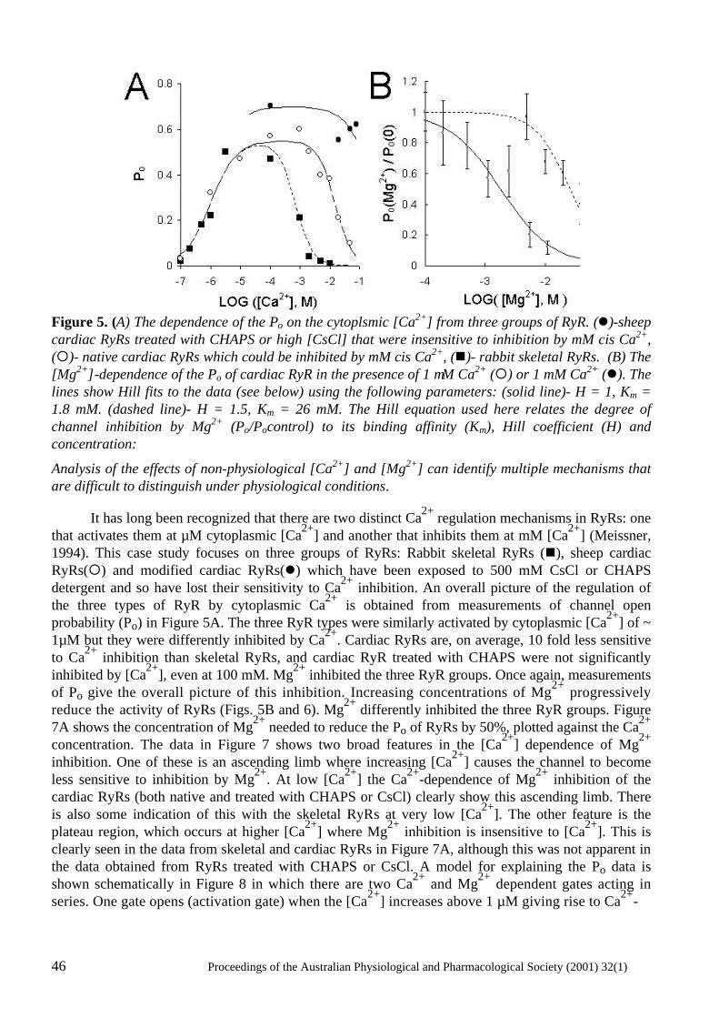
46 Proceedings of the Australian Physiological and Pharmacological Society (2001) 32(1)
Figure 5. (A) The dependence of the Po on the cytoplsmic [Ca2+] from three groups of RyR. (l)-sheep cardiac RyRs treated with CHAPS or high [CsCl] that were insensitive to inhibition by mM cis Ca2+, (¡)- native cardiac RyRs which could be inhibited by mM cis Ca2+, (n)- rabbit skeletal RyRs. (B) The [Mg2+]-dependence of the Po of cardiac RyR in the presence of 1 µM Ca2+ (¡) or 1 mM Ca2+ (l). The lines show Hill fits to the data (see below) using the following parameters: (solid line)- H = 1, Km = 1.8 mM. (dashed line)- H = 1.5, Km = 26 mM. The Hill equation used here relates the degree of channel inhibition by Mg2+ (Po/Pocontrol) to its binding affinity (Km), Hill coefficient (H) and concentration:
Analysis of the effects of non-physiological [Ca2+] and [Mg2+] can identify multiple mechanisms that are difficult to distinguish under physiological conditions.
It has long been recognized that there are two distinct Ca2+ regulation mechanisms in RyRs: one that activates them at µM cytoplasmic [Ca2+] and another that inhibits them at mM [Ca2+] (Meissner, 1994). This case study focuses on three groups of RyRs: Rabbit skeletal RyRs (n), sheep cardiac RyRs(¡) and modified cardiac RyRs(l) which have been exposed to 500 mM CsCl or CHAPS detergent and so have lost their sensitivity to Ca2+ inhibition. An overall picture of the regulation of the three types of RyR by cytoplasmic Ca2+ is obtained from measurements of channel open probability (Po) in Figure 5A. The three RyR types were similarly activated by cytoplasmic [Ca2+] of ~ 1µM but they were differently inhibited by Ca2+. Cardiac RyRs are, on average, 10 fold less sensitive to Ca2+ inhibition than skeletal RyRs, and cardiac RyR treated with CHAPS were not significantly inhibited by [Ca2+], even at 100 mM. Mg2+ inhibited the three RyR groups. Once again, measurements of Po give the overall picture of this inhibition. Increasing concentrations of Mg2+ progressively reduce the activity of RyRs (Figs. 5B and 6). Mg2+ differently inhibited the three RyR groups. Figure 7A shows the concentration of Mg2+ needed to reduce the Po of RyRs by 50%, plotted against the Ca2+ concentration. The data in Figure 7 shows two broad features in the [Ca2+] dependence of Mg2+ inhibition. One of these is an ascending limb where increasing [Ca2+] causes the channel to become less sensitive to inhibition by Mg2+. At low [Ca2+] the Ca2+-dependence of Mg2+ inhibition of the cardiac RyRs (both native and treated with CHAPS or CsCl) clearly show this ascending limb. There is also some indication of this with the skeletal RyRs at very low [Ca2+]. The other feature is the plateau region, which occurs at higher [Ca2+] where Mg2+ inhibition is insensitive to [Ca2+]. This is clearly seen in the data from skeletal and cardiac RyRs in Figure 7A, although this was not apparent in the data obtained from RyRs treated with CHAPS or CsCl. A model for explaining the Po data is shown schematically in Figure 8 in which there are two Ca2+ and Mg2+ dependent gates acting in series. One gate opens (activation gate) when the [Ca2+] increases above 1 µM giving rise to Ca2+-
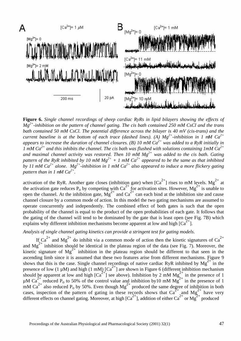
Proceedings of the Australian Physiological and Pharmacological Society (2001) 32(1) 47
Figure 6. Single channel recordings of sheep cardiac RyRs in lipid bilayers showing the effects of Mg2+-inhibition on the pattern of channel gating. The cis bath contained 250 mM CsCl and the trans bath contained 50 mM CsCl. The potential difference across the bilayer is 40 mV (cis-trans) and the current baseline is at the bottom of each trace (dashed lines). (A) Mg2+-inhibition in 1 µM Ca2+ appears to increase the duration of channel closures. (B) 10 mM Ca2+ was added to a RyR initially in 1 mM Ca2+ and this inhibits the channel. The cis bath was flushed with solutions containing 1mM Ca2+ and maximal channel activity was restored. Then 10 mM Mg2+ was added to the cis bath. Gating pattern of the RyR inhibited by 10 mM Mg2+ + 1 mM Ca2+ appeared to be the same as that inhibited by 11 mM Ca2+ alone. Mg2+-inhibition in 1 mM Ca2+ also appeared to induce a more flickery gating pattern than in 1 µM Ca2+.
activation of the RyR. Another gate closes (inhibition gate) when [Ca2+] rises to mM levels. Mg2+ at the activation gate reduces Po by competing with Ca2+ for activation sites. However, Mg2+ is unable to open the channel. At the inhibition gate, Mg2+ and Ca2+ can each bind at the inhibition site and cause channel closure by a common mode of action. In this model the two gating mechanisms are assumed to operate concurrently and independently. The combined effect of both gates is such that the open probability of the channel is equal to the product of the open probabilities of each gate. It follows that the gating of the channel will tend to be dominated by the gate that is least open (see Fig. 7B) which explains why different inhibition mechanisms become apparent at low and high [Ca2+].
Analysis of single channel gating kinetics can provide a stringent test for gating models.
If Ca2+ and Mg2+ do inhibit via a common mode of action then the kinetic signatures of Ca2+ and Mg2+ inhibition should be identical in the plateau region of the data (see Fig. 7). Moreover, the kinetic signature of Mg2+ inhibition in the plateau region should be different to that seen in the ascending limb since it is assumed that these two features arise from different mechanisms. Figure 9 shows that this is the case. Single channel recordings of native cardiac RyR inhibited by Mg2+ in the presence of low (1 µM) and high (1 mM) [Ca2+] are shown in Figure 6 (different inhibition mechanism should be apparent at low and high [Ca2+] see above). Inhibition by 2 mM Mg2+ in the presence of 1 µM Ca2+ reduced Po to 50% of the control value and inhibition by10 mM Mg2+ in the presence of 1 mM Ca2+ also reduced Po by 50%. Even though Mg2+ produced the same degree of inhibition in both cases, inspection of the pattern of gating in these records shows that Ca2+ and Mg2+ have very different effects on channel gating. Moreover, at high [Ca2+], addition of either Ca2+ or Mg2+ produced
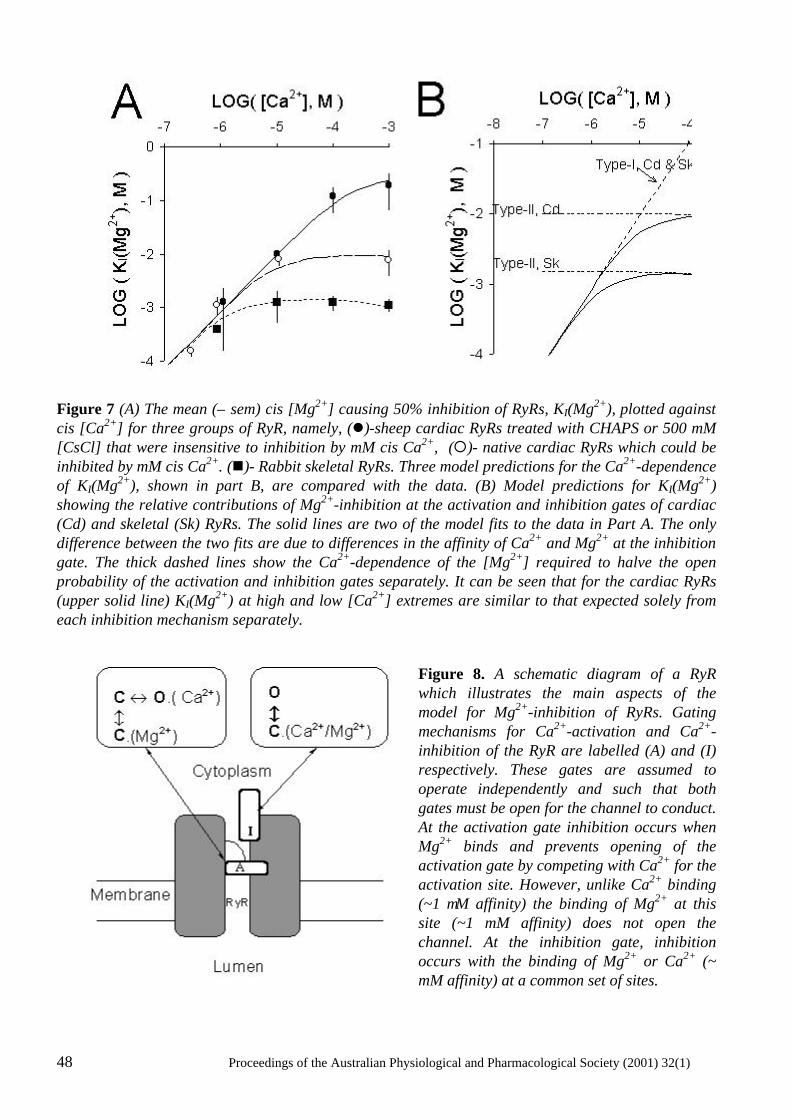
48 Proceedings of the Australian Physiological and Pharmacological Society (2001) 32(1)
Figure 7 (A) The mean (± sem) cis [Mg2+] causing 50% inhibition of RyRs, KI(Mg2+), plotted against cis [Ca2+] for three groups of RyR, namely, (l)-sheep cardiac RyRs treated with CHAPS or 500 mM [CsCl] that were insensitive to inhibition by mM cis Ca2+, (¡)- native cardiac RyRs which could be inhibited by mM cis Ca2+. (n)- Rabbit skeletal RyRs. Three model predictions for the Ca2+-dependence of KI(Mg2+), shown in part B, are compared with the data. (B) Model predictions for KI(Mg2+) showing the relative contributions of Mg2+-inhibition at the activation and inhibition gates of cardiac (Cd) and skeletal (Sk) RyRs. The solid lines are two of the model fits to the data in Part A. The only difference between the two fits are due to differences in the affinity of Ca2+ and Mg2+ at the inhibition gate. The thick dashed lines show the Ca2+-dependence of the [Mg2+] required to halve the open probability of the activation and inhibition gates separately. It can be seen that for the cardiac RyRs (upper solid line) KI(Mg2+) at high and low [Ca2+] extremes are similar to that expected solely from each inhibition mechanism separately.
Figure 8. A schematic diagram of a RyR which illustrates the main aspects of the model for Mg2+-inhibition of RyRs. Gating mechanisms for Ca2+-activation and Ca2+-inhibition of the RyR are labelled (A) and (I) respectively. These gates are assumed to operate independently and such that both gates must be open for the channel to conduct. At the activation gate inhibition occurs when Mg2+ binds and prevents opening of the activation gate by competing with Ca2+ for the activation site. However, unlike Ca2+ binding (~1 µM affinity) the binding of Mg2+ at this site (~1 mM affinity) does not open the channel. At the inhibition gate, inhibition occurs with the binding of Mg2+ or Ca2+ (~ mM affinity) at a common set of sites.
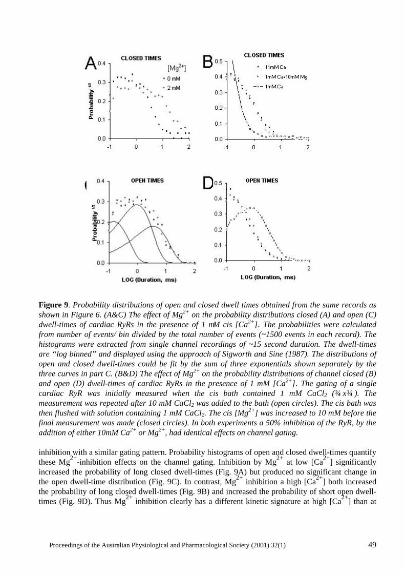
Proceedings of the Australian Physiological and Pharmacological Society (2001) 32(1) 49
Figure 9. Probability distributions of open and closed dwell times obtained from the same records as shown in Figure 6. (A&C) The effect of Mg2+ on the probability distributions closed (A) and open (C) dwell-times of cardiac RyRs in the presence of 1 µM cis [Ca2+]. The probabilities were calculated from number of events/ bin divided by the total number of events (~1500 events in each record). The histograms were extracted from single channel recordings of ~15 second duration. The dwell-times are “log binned” and displayed using the approach of Sigworth and Sine (1987). The distributions of open and closed dwell-times could be fit by the sum of three exponentials shown separately by the three curves in part C. (B&D) The effect of Mg2+ on the probability distributions of channel closed (B) and open (D) dwell-times of cardiac RyRs in the presence of 1 mM [Ca2+]. The gating of a single cardiac RyR was initially measured when the cis bath contained 1 mM CaCl2 (x). The measurement was repeated after 10 mM CaCl2 was added to the bath (open circles). The cis bath was then flushed with solution containing 1 mM CaCl2. The cis [Mg2+] was increased to 10 mM before the final measurement was made (closed circles). In both experiments a 50% inhibition of the RyR, by the addition of either 10mM Ca2+ or Mg2+, had identical effects on channel gating. inhibition with a similar gating pattern. Probability histograms of open and closed dwell-times quantify these Mg2+-inhibition effects on the channel gating. Inhibition by Mg2+ at low [Ca2+] significantly increased the probability of long closed dwell-times (Fig. 9A) but produced no significant change in the open dwell-time distribution (Fig. 9C). In contrast, Mg2+ inhibition a high [Ca2+] both increased the probability of long closed dwell-times (Fig. 9B) and increased the probability of short open dwell-times (Fig. 9D). Thus Mg2+ inhibition clearly has a different kinetic signature at high [Ca2+] than at
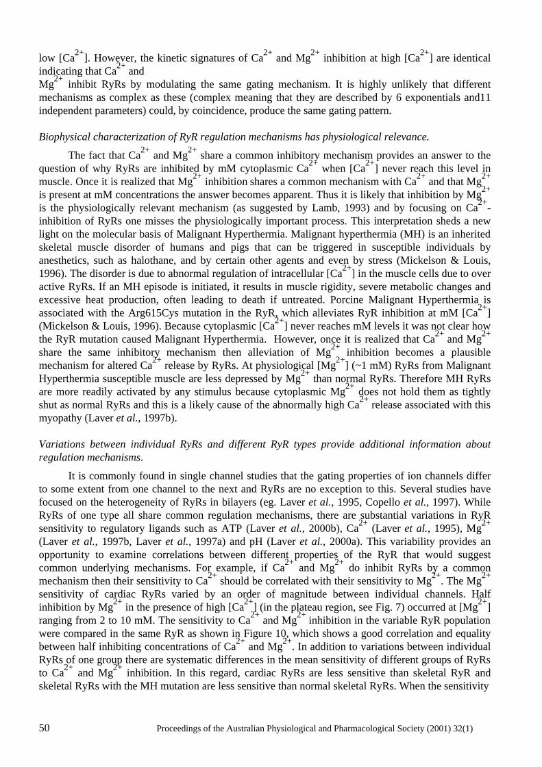
50 Proceedings of the Australian Physiological and Pharmacological Society (2001) 32(1)
low [Ca2+]. However, the kinetic signatures of Ca2+ and Mg2+ inhibition at high [Ca2+] are identical indicating that Ca2+ and Mg2+ inhibit RyRs by modulating the same gating mechanism. It is highly unlikely that different mechanisms as complex as these (complex meaning that they are described by 6 exponentials and11 independent parameters) could, by coincidence, produce the same gating pattern. Biophysical characterization of RyR regulation mechanisms has physiological relevance.
The fact that Ca2+ and Mg2+ share a common inhibitory mechanism provides an answer to the question of why RyRs are inhibited by mM cytoplasmic Ca2+ when [Ca2+] never reach this level in muscle. Once it is realized that Mg2+ inhibition shares a common mechanism with Ca2+ and that Mg2+ is present at mM concentrations the answer becomes apparent. Thus it is likely that inhibition by Mg2+ is the physiologically relevant mechanism (as suggested by Lamb, 1993) and by focusing on Ca2+-inhibition of RyRs one misses the physiologically important process. This interpretation sheds a new light on the molecular basis of Malignant Hyperthermia. Malignant hyperthermia (MH) is an inherited skeletal muscle disorder of humans and pigs that can be triggered in susceptible individuals by anesthetics, such as halothane, and by certain other agents and even by stress (Mickelson & Louis, 1996). The disorder is due to abnormal regulation of intracellular [Ca2+] in the muscle cells due to over active RyRs. If an MH episode is initiated, it results in muscle rigidity, severe metabolic changes and excessive heat production, often leading to death if untreated. Porcine Malignant Hyperthermia is associated with the Arg615Cys mutation in the RyR, which alleviates RyR inhibition at mM [Ca2+] (Mickelson & Louis, 1996). Because cytoplasmic [Ca2+] never reaches mM levels it was not clear how the RyR mutation caused Malignant Hyperthermia. However, once it is realized that Ca2+ and Mg2+
share the same inhibitory mechanism then alleviation of Mg2+ inhibition becomes a plausible mechanism for altered Ca2+ release by RyRs. At physiological [Mg2+] (~1 mM) RyRs from Malignant Hyperthermia susceptible muscle are less depressed by Mg2+ than normal RyRs. Therefore MH RyRs are more readily activated by any stimulus because cytoplasmic Mg2+ does not hold them as tightly shut as normal RyRs and this is a likely cause of the abnormally high Ca2+ release associated with this myopathy (Laver et al., 1997b). Variations between individual RyRs and different RyR types provide additional information about regulation mechanisms.
It is commonly found in single channel studies that the gating properties of ion channels differ to some extent from one channel to the next and RyRs are no exception to this. Several studies have focused on the heterogeneity of RyRs in bilayers (eg. Laver et al., 1995, Copello et al., 1997). While RyRs of one type all share common regulation mechanisms, there are substantial variations in RyR sensitivity to regulatory ligands such as ATP (Laver et al., 2000b), Ca2+ (Laver et al., 1995), Mg2+ (Laver et al., 1997b, Laver et al., 1997a) and pH (Laver et al., 2000a). This variability provides an opportunity to examine correlations between different properties of the RyR that would suggest common underlying mechanisms. For example, if Ca2+ and Mg2+ do inhibit RyRs by a common mechanism then their sensitivity to Ca2+ should be correlated with their sensitivity to Mg2+. The Mg2+ sensitivity of cardiac RyRs varied by an order of magnitude between individual channels. Half inhibition by Mg2+ in the presence of high [Ca2+] (in the plateau region, see Fig. 7) occurred at [Mg2+] ranging from 2 to 10 mM. The sensitivity to Ca2+ and Mg2+ inhibition in the variable RyR population were compared in the same RyR as shown in Figure 10, which shows a good correlation and equality between half inhibiting concentrations of Ca2+ and Mg2+. In addition to variations between individual RyRs of one group there are systematic differences in the mean sensitivity of different groups of RyRs to Ca2+ and Mg2+ inhibition. In this regard, cardiac RyRs are less sensitive than skeletal RyR and skeletal RyRs with the MH mutation are less sensitive than normal skeletal RyRs. When the sensitivity
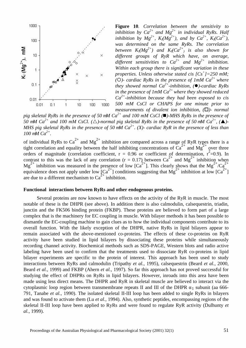
Proceedings of the Australian Physiological and Pharmacological Society (2001) 32(1) 51
Figure 10. Correlation between the sensitivity to inhibition by Ca2+ and Mg2+ in individual RyRs. Half inhibition by Mg2+, KI(Mg2+), and by Ca2+, KI(Ca2+), was determined on the same RyRs. The correlation between KI(Mg2+) and KI(Ca2+
) is also shown for different groups of RyR which have, on average, different sensitivities to Ca2+ and Mg2+ inhibition. Within each group there is significant variation in these properties. Unless otherwise stated cis [Cs+]=250 mM; (¡)- cardiac RyRs in the presence of 1mM Ca2+ where they showed normal Ca2+-inhibition, (l)-cardiac RyRs in the presence of 1mM Ca2+ where they showed reduced Ca2+-inhibition because they had been exposed to cis 500 mM CsCl or CHAPS for one minute prior to measurements of divalent ion inhibition, (o)- normal
pig skeletal RyRs in the presence of 50 µM Ca2+ and 100 mM CsCl (n)-MHS RyRs in the presence of 50 µM Ca2+ and 100 mM CsCl. (r)-normal pig skeletal RyRs in the presence of 50 µM Ca2+, (p)-MHS pig skeletal RyRs in the presence of 50 µM Ca2+. (X)- cardiac RyR in the presence of less than 100 µM Ca2+.
of individual RyRs to Ca2+ and Mg2+ inhibition are compared across a range of RyR types there is a tight correlation and equality between the half inhibiting concentrations of Ca2+ and Mg2+ over three orders of magnitude (correlation coefficient, r = 0.96 or coefficient of determination, r2=0.9). In contrast to this was the lack of any correlation (r = 0.17) between Ca2+ and Mg2+ inhibition when Mg2+ inhibition was measured in the presence of low [Ca2+]. This clearly shows that the Mg2+/Ca2+ equivalence does not apply under low [Ca2+] conditions suggesting that Mg2+ inhibition at low [Ca2+] are due to a different mechanism to Ca2+ inhibition. Functional interactions between RyRs and other endogenous proteins.
Several proteins are now known to have effects on the activity of the RyR in muscle. The most notable of these is the DHPR (see above). In addition there is also calmodulin, calsequestrin, triadin, junctin and the FK506 binding protein (FKBP). These proteins are believed to form part of a large complex that is the machinery for EC coupling in muscle. With bilayer methods it has been possible to dismantle the EC-coupling machine to gain clues as to how the individual components contribute to its overall function. With the likely exception of the DHPR, native RyRs in lipid bilayers appear to remain associated with the above-mentioned co-proteins. The effects of these co-proteins on RyR activity have been studied in lipid bilayers by dissociating these proteins while simultaneously recording channel activity. Biochemical methods such as SDS-PAGE, Western blots and radio active labeling have been used to confirm that the treatments used to dissociate RyR co-proteins in lipid bilayer experiments are specific to the protein of interest. This approach has been used to study interactions between RyRs and calmodulin (Tripathy et al., 1995), calsequestrin (Beard et al., 2000, Beard et al., 1999) and FKBP (Ahern et al., 1997). So far this approach has not proved successful for studying the effect of DHPRs on RyRs in lipid bilayers. However, inroads into this area have been made using less direct means. The DHPR and RyR in skeletal muscle are believed to interact via the cytoplasmic loop region between transmembrane repeats II and III of the DHPR α1 subunit (aa 666-791, Tanabe et al., 1990). The isolated skeletal II-III loop has been added to single RyRs in bilayers and was found to activate them (Lu et al., 1994). Also, synthetic peptides, encompassing regions of the skeletal II-III loop have been applied to RyRs and were found to regulate RyR activity (Dulhunty et al., 1999).
52 Proceedings of the Australian Physiological and Pharmacological Society (2001) 32(1)
An example of how single channel experiments have been used to study the functional interactions between RyRs and other proteins is the investigation of calsequestrin. Calsequestrin is a protein found in the lumen of the SR, which acts as a moderate affinity (KD=1 x 10⁻⁵ M) Ca2+ binding protein that buffers the luminal free [Ca2+] (MacLennan & Wong, 1971). Two other proteins, triadin and junctin, span the SR membrane and these proteins bind to both calsequestrin and the RyR (Knudson et al., 1993, Kanno & Takishima, 1990, Zhang et al., 1997, Jones et al., 1995, Guo et al., 1996). SDS-PAGE and Western Blotting techniques have shown that calsequestrin can be dissociated from the RyR complex by exposing SR membranes to raised ionic strength (ie 500 mM as apposed to 250 mM), or by exposing them to higher than usual [Ca2+] (13 mM as apposed to 1 mM). By applying similar solutions changes to RyRs in bilayers it was possible to see the effects of a calsequestrin dissociation event during recordings of RyR activity. To do this the ionic strength or [Ca2+] of the luminal bath in bilayer experiments was increased like that in the SDS-PAGE experiments. This caused an increase in RyR activity that could only be reversed by addition of purified calsequestrin to the luminal bath. The presence or absence of calsquestrin on the RyR in bilayers was confirmed using an anti-calsequestrin antibody that inhibits channel activity when it binds to the calsequestrin-RyR complex. The data were consistent with an overall picture in which calsequestrin dissociation enhanced native RyR activity and calsequestrin binding suppressed channel opening. The next step would be to assess the combined effects of triadin, junctin and calsequestrin on RyR activity. Junctin and triadin can be dissociated from the RyR by solublising the SR membranes with CHAPS detergent and purifying the RyR. Thus by applying triadin, junctin and calsequestrin, in various combinations, to purified RyRs in bilayer experiments it will be possible to systematically piece together these components of the EC coupling machinery and measure their effects on RyR activity. RyR function and its relationship to its tetrameric structure
Electron microscope image reconstruction shows RyRs to have four-fold symmetry (Orlova et al., 1996). This four-fold symmetry means that each of the four subunits must somehow contribute equally to ligand binding and channel gating. Therefore the mechanisms regulating channel gating are likely to be complex. Single channel studies are now starting to give an insight into how homotetrameric structures like the RyR control channel activity. Four-fold structure inferred from channel conductance.
Telltale signs of the contribution of four subunits to channel conductance appeared not long after the discovery of the RyR (Ma et al., 1988, Smith et al., 1988). RyRs displayed multiple conductance levels with subconductances near 25%, 50% and 75% of the maximum (ie equally spaced levels) conductance level. These were interpreted a current flow through pores formed by the activation of different numbers of subunits; 25% corresponding to one, 50% to two subunits etc. Very clear and sustained substate activity at approximately equally spaced levels has been observed in RyRs that were modified either by the binding of ryanodine (Ma & Zhao, 1994) or the stripping of the FKBP, a co protein to the RyR (Ahern et al., 1997). Amplitude histograms of channel activity revealed that the spacing of substates was not exactly equal. Nonetheless, these studies interpreted the substates as the opening of four separate conducting pathways in the channel. These must gate cooperatively and simultaneously in order to produce the observed gating of the RyR complex between closed and maximum conductance levels in a single transition. More recently mutations in the RyR have been discovered that alter channel conductance and experiments with hybrid RyRs containing these mutations have shed more light on subunit contributions to channel conductance. A highly conserved 10 amino acid segment within the pore-forming region about aa4824 was found to be the determinant of channel conductance (Zhao et al., 1999). Single channel studies showed that the glycine to alanine substitution, G4824A, reduced channel conductance by 97%. Co-expression of mutant and wild-type
Proceedings of the Australian Physiological and Pharmacological Society (2001) 32(1) 53
subunits produced a range of hybrid channel types with six different maximal conductance levels that lie between those of the homozygous mutant and wild-type RyRs. The number of different conductance levels corresponds to the number of subunit combinations that are possible with two types of subunits in a tetramer. The fact that six conductance levels could be observed suggest that the four subunits somehow contribute to a single conduction pathway through the RyR as apposed to the idea that each subunit possesses a separate pathway. Four-fold structure inferred from steady state channel gating.
The gating of RyRs has exhibited phenomena that suggest mechanisms stemming from a tetrameric structure. The Arg615Cys mutation (MH mutation in pigs, see above) in the RyR alleviates RyR inhibition at mM [Ca2+] and [Mg2+]. Shomer et al. (1995) found that RyRs expressed in heterozygous pigs gave rise to hybrid RyRs with gating properties intermediate to those of the wild type and mutant. This indicated that each subunit in the RyR tetramer has an influence over the Ca2+ sensitivity of the channel. More recently, Chen et al. (1998) discovered an alanine to glutamate substitution, E3885A, which decreased the sensitivity of RyR to Ca2+ activation by four orders of magnitude such that mM levels of cytoplasmic Ca2+ were required to activate the channel. Co-expression of subunits containing altered sites for Ca2+-activation with wild-type subunits produced five or six types of RyRs with sensitivities to Ca2+ activation ranging between those seen for the mutant and wild type homotetramers. Chen et al. (1998) suggested that all four subunits contribute to each Ca2+ activation site. A more detailed interpretation of these experiments awaits the development of methods for linking the stoichiometry of the hybrid channels with the observed channel function. Four-fold structure inferred from non-steady state channel gating.
Aspects of the tetrametric structure of RyRs may also show up in the response of RyRs to rapid changes in the concentration of regulatory ligands. It is recognized that during muscle contraction the RyRs respond to rapid (~ms) step increases in [Ca2+] resulting from Ca2+ flow through nearby dihydropyridine and ryanodine receptors. Consequently there has been considerable interest in the effects of rapid [Ca2+] transients on the activity of RyRs in bilayers (see perspectives by Sitsapesan & Williams, 2000, Lamb et al., 2000, Fill et al., 2000). Now there is also a growing interest in the effects of rapid application of other regulatory ligands such a protons (Laver et al., 2000a). The effect of steady cytoplasmic pH on RyRs has been addressed in several studies (Rousseau & Pinkos, 1990, Shomer et al., 1994, Ma et al., 1988), which show acid pH inhibits RyRs; with half inhibition at pH6.5 and with near total inhibition below pH6. However, rapid changes in pH reveal new characteristics of RyR gating not apparent in steady state recordings. The pH was increased from ~5.5 (inhibiting pH) to above 7 (activating pH) over a 500 ms period by squirting solutions from a perfusion tube placed in the vicinity of the bilayer. Rather than activating in a graded manner reflecting the continuous change of pH over this period, the RyRs activate in a stepwise manner. As the RyRs activate their open probability RyRs increases in what appears to be different gating modes with ascending Po (see Fig. 11A). Amplitude histograms of RyR Po (Fig. 11B) show four of these gating modes as four distinct peaks. The four gating modes correspond in number to what would be expected if RyR subunits activated separately in response to proton dissociation. Thus the first, lowest Po mode would be when one subunit is active and higher Po modes would occur as more of the four subunits activated. If this hypothesis were correct then the stability of ligand mediated channel openings would depend on the number of subunits that have bound ligand molecules. This phenomenon has been seen in the cyclic nucleotide gated channel (Ruiz & Karpen, 1997).
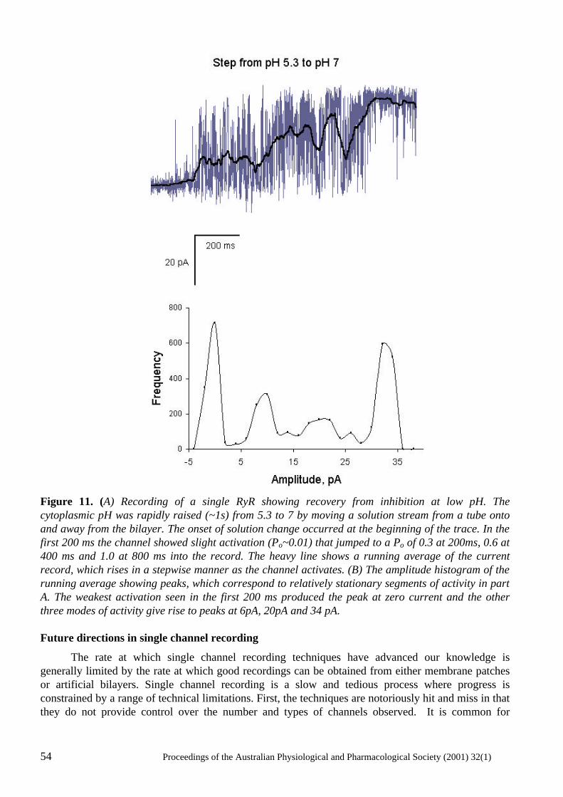
54 Proceedings of the Australian Physiological and Pharmacological Society (2001) 32(1)
Figure 11. (A) Recording of a single RyR showing recovery from inhibition at low pH. The cytoplasmic pH was rapidly raised (~1s) from 5.3 to 7 by moving a solution stream from a tube onto and away from the bilayer. The onset of solution change occurred at the beginning of the trace. In the first 200 ms the channel showed slight activation (Po~0.01) that jumped to a Po of 0.3 at 200ms, 0.6 at 400 ms and 1.0 at 800 ms into the record. The heavy line shows a running average of the current record, which rises in a stepwise manner as the channel activates. (B) The amplitude histogram of the running average showing peaks, which correspond to relatively stationary segments of activity in part A. The weakest activation seen in the first 200 ms produced the peak at zero current and the other three modes of activity give rise to peaks at 6pA, 20pA and 34 pA. Future directions in single channel recording
The rate at which single channel recording techniques have advanced our knowledge is generally limited by the rate at which good recordings can be obtained from either membrane patches or artificial bilayers. Single channel recording is a slow and tedious process where progress is constrained by a range of technical limitations. First, the techniques are notoriously hit and miss in that they do not provide control over the number and types of channels observed. It is common for
Proceedings of the Australian Physiological and Pharmacological Society (2001) 32(1) 55
experiments to be confounded by the appearance of more than a single channel, the appearance of the wrong channel type or the non-appearance of any channel. Secondly, these membrane systems are extremely fragile and rupture of membrane patches and bilayers terminate experiments within minutes. This severely restricts the range of experimental protocols that can be used for single channel recordings. Any way of overcoming these problems would provide a major advance in single channel recording techniques. This section describes a number of new methods for producing robust, long-lived membrane systems that could be applied to studying individual ion channels. As described above, the most commonly used method of making planar bilayers is the film drainage method, which was developed by (Mueller et al., 1962). Tien, one of the authors on the original bilayer paper, has published several new methods for forming robust lipid bilayers using solid supports (Ottova & Tien, 1997). As before, bilayers are formed from solutions of lipids in hydrophobic solvents such as n-decane. However, instead of producing lipid films that are suspended across an aperture, bilayer membranes are formed from lipid films spread across solid or gel surfaces. The viscous support offered by a nearby solid surface boosts enormously the stability of the bilayer. Doping these stable membranes with various substances has been shown to endow these membranes with a range of useful properties such as ion selectivity (by incorporation of ionophors such as gramicidin), immunologic reactivity (with antibodies), electron transporters (with C60 fullerenes) and photosensitivity (with Zn-phthalocyanine) (Ottova & Tien, 1997, Tien et al., 1991). However, the advantages of supported bilayer systems have not yet been realised in single channel studies. If one could incorporate RyRs, for example, into bilayers on agar supports then conventional electrophysiology experiments could be carried out on a single channel over extended periods of time (hours). Moreover, because the supported bilayers are robust and usually made on the end of electrodes, they are remarkably manoeuvrable. Hence, these bilayers could be aligned within complex measuring apparatus such as those using confocal microscopy or rapid perfusion. The rate and quality of data acquired this way would be far superior to that obtained using conventional bilayer methods. More recently solid substrate bilayer methods were further improved by developing bilayer structures that could be chemically anchored to a gold surface (Cornell et al., 1997). These were developed for the purpose of making a new generation of biosensors. These systems are remarkably robust: being able to be stored dehydrated for periods of many months. In principle, ion channels could be embedded in these membranes though no successful attempts to do this have been published. Future developments of this system could revolutionise bilayer-based methods of single channel recording. In the future one might purchase bilayers off the shelf and perform weeks of experiments on a single ion channel. Moreover, the hit and miss aspect of single channel methods (see above) may soon be a thing of the past. It is recognised in the biosensor industry that biosensors based on arrays of detectors can offer vastly superior combination of detection sensitivity and speed. Once these bilayer array platforms are produced then the scope for single channel recording is truly enormous. Incorporation of ion channels onto arrays of 10,000 or more electrically isolated bilayers will allow an experimenter to select bilayers in the array that contain the desired number and type of ion channels. One could easily work with mixtures of ion channels as found, for example, in SR membranes because one could choose particular bilayers that contained only the ion channels of interest. It will also be possible to get information from individual ion channels at the same time as obtaining measurements of the average response of many channels by averaging signals from many bilayers. In fact, a highly parallel solid-state electrode array has been developed for patch-clamping that makes possible multiple, simultaneous, single-cell electrical recordings (Axon instruments press release). Thus by layering cells onto these arrays one can simultaneously obtain single-channel and whole-cell currents. So it appears that even now the former limits to data acquisition are giving way to new frontiers in which the rate-limiting factor will be the rate at which data can be acquired with massively parallel detection systems.
56 Proceedings of the Australian Physiological and Pharmacological Society (2001) 32(1)
Concluding remarks
The regulation of RyR channels by intracellular metabolites such as ATP, Ca2+, Mg2+ and pH is a complex interplay of several regulation processes. Overlayed on this is the fact that these mechanisms are modulated by oxidation and phosphorylation mediated modifications of the RyR and by RyR interactions with a variety of co-proteins. Consequently, in order to understand how RyRs are regulated by normal metabolism or during muscle fatigue and myocardial ischemia it is necessary to deal with the very complex problem of understanding RyR function. Single channel recording and analysis methods are powerful enough to examine RyRs in sufficient detail to dissect the complex regulation mechanisms operating in RyRs. The enormous scope with the bilayer method for manipulating the quaternary structure of the EC-coupling machinery have allowed us to probe the effects of DHPRs, calmodulin, calsequestrin, triadin and the FKBP on RyR function. Three examples of forays into these areas by single channel studies have been described here. First, the analysis of [Ca2+] and [Mg2+] regulation of RyRs showed how it is possible to identify and characterise multiple mechanisms by which a single ligand can regulate RyRs and how this leads to an improved understudying of physiological processes in muscle. Secondly, experiments with RyRs in which co-proteins such as calsequestrin and triadin were systematically removed and replaced show how it is possible to understand the functional importance interactions between RyRs and co-proteins. Finally, investigations of the relationship between structure and function of native and mutated RyRs showed several aspects of RyR that reflects the four-fold symmetry of the RyRs structure. However, this area is still at an early stage and it is not yet clear how the four identical subunits of the RyR cooperate in regulating RyR gating. In spite of the power of single channel recording the bilayer method has an Achilles heel. The fact that RyRs are studied in isolation means that it is not possible to examine their function in the physiological context. Hence one cannot directly apply RyR phenomena in bilayers to the physiological situation. However, the underlying mechanisms for RyR regulation identified from bilayer measurements are likely to apply to the in vivo situation and so contribute to our understanding of EC-coupling. On the other hand, studies of more intact muscle preparations such as suspensions of SR vesicles and mechanically skinned fibres are well suited for addressing the physiological situation. However, these systems are complex and it is difficult to identifying underlying mechanisms from the experimental data. Parallel experiments on lipid bilayers and muscle preparations in which the machinery for EC coupling is still intact have proved to be a powerful tool for elucidating mechanisms of Ca2+ regulation in muscle. In this collaboration of techniques the bilayer studies identify the basic mechanisms of channel function and the experiments on intact systems show the outworking of these mechanisms in the physiological situation. Acknowledgements
We wish to thank Dr. Graham Lamb for his helpful comments. This work was supported by the National Health & Medical Research Council of Australia (Grant #9936486). References
Ahern, G.P., Junankar, P.R. & Dulhunty, A.F. (1997) Subconductance states in single channel activity of skeletal muscle ryanodine receptors after removal of FKBP12. Biophysical Journal, 72, 146-162.
Ashley, C.C., Mulligan, I.P. & Lea, T.J. (1991) Ca2+ and activation mechanisms in skeletal muscle Quarterly Reviews in Biophysics, 24, 1-73.
Beard, N.A., Dulhunty, A.F. & Laver, D.R. (2000) The effect of increasing luminal calcium on skeletal muscle Proceedings of the Australian Physiological and Pharmacological Society, 31, 22P.
Proceedings of the Australian Physiological and Pharmacological Society (2001) 32(1) 57
Beard, N.A., Laver, D.R. & Dulhunty, A.F. (1999) Regulation of skeletal muscle ryanodine receptors by calsequestrin Proceedings of the Australian Physiological and Pharmacological Society, 30, 43P.
Chen, S.R., Ebisawa, K., Li, X. & Zhang, L. (1998) Molecular identification of the ryanodine receptor Ca2+ sensor. Journal of Biological Chemistry, 273, 14675-14678.
Colquhoun, D. & Hawkes, A.G. (1981) On the stochastic properties of single ion channels Proceedings Royal Society London Biol, 211, 205-235.
Copello, J.A., Barg, S., Onoue, H. & Fleischer, S. (1997) . Heterogeneity of Ca2+ gating of skeletal muscle and cardiac ryanodine receptors. Biophysical Journal, 73, 141-156.
Cornell, B.A., Braach-Maksvytis, V.L., King, L.G., Osman, P.D., Raguse, B., Wieczorek, L. & Pace, R.J. (1997) A biosensor that uses ion-channel switches. Nature, 387, 580-583.
Dulhunty, A.F., Laver, D.R., Gallant, E.M., Casarotto, M.G., Pace, S.M. & Curtis, S. (1999) Activation and inhibition of skeletal RyR channels by a part of the skeletal DHPR II-III loop: effects of DHPR Ser687 and FKBP12 Biophysical Journal, 77, 189-203.
Fill, M., Zahradnikova, A., Villalba-Galea, C.A., Zahradnik, I., Escobar, A.L. & Gyorke, S. (2000) . Ryanodine receptor adaptation. Journal of General Physiology, 116, 873-82.
Guo, W., Jorgensen, A.O. & Campbell, K.P. (1996) Triadin, a linker for calsequestrin and the ryanodine receptor. Society of General Physiologists Series, 51, 19-28.
Jones, L. R., Zhang, L., Sanborn, K., Jorgensen, A.O. & Kelley, J. (1995) Purification, primary structure, and immunological characterization of the 26-kDa calsequestrin binding protein (junctin) from cardiac junctional sarcoplasmic reticulum. Journal of Biological Chemistry, 270, 30787-96.
Kanno, T. & Takishima, T. (1990) Chloride and potassium channels in U937 human monocytes. Journal of Membrane Biology, 116, 149-161.
Knudson, C. M., Stang, K. K., Jorgensen, A. O. & Campbell, K. P. Biochemical characterization and ultrastructural localization of a major junctional sarcoplasmic reticulum glycoprotein (Triadin). Journal of Biological Chemistry, (1993) 268, 12637-12645.
Lamb, G.D. (1993) Ca2+ inactivation, Mg2+ inhibition and malignant hyperthermia [news]. Journal of Muscle Research and Cell Motility, 14, 554-556.
Lamb, G.D., Laver, D. R. & Stephenson, D.G. (2000) Questions about Adaptation in Ryanodine Receptors. Journal of General Physiology, 116, 883-890.
Lamb, G.D. & Stephenson, D.G. (1991) Effect of Mg2+ on the control of Ca2+ release in skeletal muscle fibres of the toad. Journal of Physiology, 434, 507-528.
Lamb, G.D. & Stephenson, D.G. (1992) Importance of Mg2+ in excitation-contraction coupling in skeletal muscle. News in Physiological Sciences, 7, 270-274.
Laver, D.R., Baynes, T.M. & Dulhunty, A.F. (1997a) Journal of Membrane Biology, 156, 213-229.
Laver, D.R., Eager, K.R., Taoube, L. & Lamb, G.D. (2000a) Biophysical Journal, 78, 1835-1851.
Laver, D.R., Lenz, G.K.E. & Lamb, G.D. (2000b) Proceedings of the Australian Physiological and Pharmacological Society, 31, 7P.
Laver, D.R., Owen, V.J., Junankar, P.R., Taske, N.L., Dulhunty, A.F. & Lamb, G.D. (1997b) Reduced inhibitory effect of Mg2+ on ryanodine receptor-Ca2+ release channels in malignant hyperthermia. Biophysical Journal, 73, 1913-1924.
58 Proceedings of the Australian Physiological and Pharmacological Society (2001) 32(1)
Laver, D.R., Roden, L.D., Ahern, G.P., Eager, K.R., Junankar, P.R. & Dulhunty, A.F. (1995) Cytoplasmic Ca2+ inhibits the ryanodine receptor from cardiac muscle. Journal of Membrane Biology, 147, 7-22.
Lu, X., Xu, L. & Meissner, G. (1994) Activation of the skeletal muscle calcium release channel by a cytoplasmic loop of the dihydropyridine receptor. Journal of Biological Chemistry, 269, 6511-6.
Ma, J., Fill, M., Knudson, C. M., Campbell, K. P. & Coronado, R. (1988) Ryanodine receptor of skeletal muscle is a gap junction-type channel. Science, 242, 99-102.
Ma, J. & Zhao, J. (1994) . Highly cooperative and hysteretic response of the skeletal muscle ryanodine receptor to changes in proton concentrations. Biophysical Journal, 67, 626-633.
MacLennan, D. H. & Wong, P. T. (1971) Isolation of a calcium-sequestering protein from sarcoplasmic reticulum. Proceedings of the National Academy of Sciences USA, 68, 1231-1235.
Meissner, G. (1994) Ryanodine receptor/Ca2+ release channels and their regulation by endogenous effectors. Annual Review of Physiology, 56, 485-508.
Meissner, G., Darling, E. & Eveleth, J. (1986) Kinetics of rapid Ca2+ release by sarcoplasmic reticulum. Effects of Ca2+, Mg2+, and adenine nucleotides. Biochemistry, 25, 236-244.
Mickelson, J. R. & Louis, C. F. (1996) Malignant hyperthermia: Excitation-contraction coupling, Ca2+ release channel, and cell Ca2+ regulation defects. Physiological Reviews, 76, 537-592.
Miller, C. & Racker, E. (1976) Ca++-induced fusion of fragmented sarcoplasmic reticulum with artificial planar bilayers. Cell, 9, 283-300.
Mueller, P., Rudin, D. O., Tien, H. T. & Westcott, W. C. (1962) . Reconstitution of cell membrane structure in vitro and its transformation into an excitable system. Nature, 194, 979-981.
Nabauer, M., Callewaert, G., Cleemann, L. & Morad, M. (1989) Regulation of calcium release is gated by calcium current, not gating charge, in cardiac myocytes. Science, 244, 800-803.
Neher, E. & Sakmann, B. (1976) Single-channel currents recorded from membrane of denervated frog muscle fibres. Nature, 260, 799-802.
Ogawa, Y. (1994) Role of ryanodine receptors. Critical Reviews of Biochemistry and Molecular Biology, 29, 229-74.
Orlova, E. V., Serysheva, I. I., Van Heel, M., Hamilton, S. L. & Chiu, W. (1996) Two structural configurations of the skeletal muscle calcium release channel. Nature Structural Biology, 3, 547-552.
Ottova, A. L. & Tien, H. T. (1997) Self-assembled bilayer lipid membranes: from mimicking biomembranes to practical applications. Bioelectrochemistry and Bioenergetics, 42, 141-152.
Owen, V. J., Taske, N. L. & Lamb, G. D. (1997) Reduced inhibitory effect of Mg2+ on Ca2+ release in porcine muscle fibers with ryanopdine receptor mutation for malignant hyperthermia American Journal of Physiology, 272, C203-211.
Rousseau, E. & Pinkos, J. (1990) pH modulates conducting and gating behaviour of single calcium release channels. Pflugers Archiv, 415, 645-647.
Ruiz, M. L. & Karpen, J. W. (1997) Single cyclic nucleotide-gated channels locked in different ligand- bound states. Nature, 389, 389-392.
Shomer, N. H., Mickelson, J. R. & Louis, C. F. (1994) Caffeine stimulation of malignant hyperthermia-susceptible sarcoplasmic reticulum Ca2+ release channel. American Journal of Physiology, 267, C1253-C1261.
Proceedings of the Australian Physiological and Pharmacological Society (2001) 32(1) 59
Shomer, N. H., Mickelson, J. R. & Louis, C. F. (1995) Ca2+ release channels of pigs heterozygous for malignant hyperthermia. Muscle Nerve, 18, 1167-1176.
Sigworth, F. J. & Sine, S. M. (1987) Data transformations for improved display and fitting of single- channel dwell time histograms. Biophysical Journal, 52, 1047-1054.
Sitsapesan, R. & Williams, A. J. (2000) Do inactivation mechanisms rather than adaptation hold the key to understanding ryanodine receptor channel gating?Journal of General Physiology, 116, 867-72.
Smith, J. S., Imagawa, T., Ma, J., Fill, M., Campbell, K. P. & Coronado, R. (1988) Purified ryanodine receptor from rabbit skeletal muscle is the calcium-release channel of sarcoplasmic reticulum. Journal of General Physiology, 92, 1-26.
Tanabe, T., Beam, K. G., Adams, B. A., Niidome, T. & Numa, S. (1990) . Regions of the skeletal muscle dihydropyridine receptor critical for excitation-contraction coupling. Nature, 346, 567-569.
Tien, H. T., Salamon, Z. & Ottova, A. (1991) . Lipid bilayer-based sensors and biomolecular electronics. CriticalReviews in Biomedical Engineering, 18, 323-340.
Tripathy, A., Xu, L., Mann, G. & Meissner, G. (1995) Calmodulin activation and inhibition of skeletal muscle Ca2+ release channel (ryanodine receptor). Biophysical Journal, 69, 106-119.
Zhang, L., Kelley, J., Schmeisser, G., Kobayashi, Y. M. & Jones, L. R. (1997) . Complex formation between junctin, triadin, calsequestrin, and the ryanodine receptor. Proteins of the cardiac junctional sarcoplasmic reticulum membrane. Journal of Biological Chemistry, 272, 23389-23397.
Zhao, M., Li, P., Li, X., Zhang, L., Winkfein, R. J. & Chen, S. R. (1999) . Molecular identification of the ryanodine receptor pore-forming segment. Journal of Biological Chemistry, 274, 25971-25974.
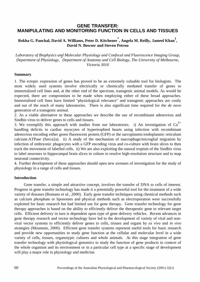
60 Proceedings of the Australian Physiological and Pharmacological Society (2001) 32(1)
GENE TRANSFER:
MANIPULATING AND MONITORING FUNCTION IN CELLS AND TISSUES
Rekha G. Panchal, David A. Williams, Peter D. Kitchener*, Angela M. Reilly, Jameel Khan1, David N. Bowser and Steven Petrou
Laboratory of Biophysics and Molecular Physiology and Confocal and Fluorescence Imaging Group, Department of Physiology, *Department of Anatomy and Cell Biology, The University of Melbourne,
Victoria 3010 Summary
1. The ectopic expression of genes has proved to be an extremely valuable tool for biologists. The most widely used systems involve electrically or chemically mediated transfer of genes to immortalized cell lines and, at the other end of the spectrum, transgenic animal models. As would be expected, there are compromises to be made when employing either of these broad approaches. Immortalised cell lines have limited "physiological relevance" and transgenic approaches are costly and out of the reach of many laboratories. There is also significant time required for the de novo generation of a transgenic animal. 2. As a viable alternative to these approaches we describe the use of recombinant adenovirus and Sindbis virus to deliver genes to cells and tissues. 3. We exemplify this approach with studies from our laboratories. i) An investigation of Ca2+ handling deficits in cardiac myocytes of hypertrophied hearts using infection with recombinant adenovirus encoding either green fluorescent protein (GFP) or the sarcoplasmic/endoplasmic reticulum calcium-ATPase (Serca2a). ii) A study of the mechanism of macrophage/microglial migration by infection of embryonic phagocytes with a GFP encoding virus and co-culture with brain slices to then track the movement of labelled cells. ii) We are also exploiting the natural tropism of the Sindbis virus to label neurones in hippocampal brain slices in culture to resolve high-resolution structure and to map neuronal connectivity. 4. Further development of these approaches should open new avenues of investigation for the study of physiology in a range of cells and tissues. Introduction
Gene transfer, a simple and attractive concept, involves the transfer of DNA to cells of interest. Progress in gene transfer technology has made it a potentially powerful tool for the treatment of a wide variety of diseases (Romano et al., 2000). Early gene transfer techniques using chemical methods such as calcium phosphate or liposomes and physical methods such as electroporation were successfully exploited for basic research but had limited use for gene therapy. Gene transfer technology for gene therapy approaches is based on the ability to efficiently deliver the therapeutic gene to relevant target cells. Efficient delivery in turn is dependent upon type of gene delivery vehicles. Recent advances in gene therapy research and vector technology have led to the development of variety of viral and non-viral vector systems to efficiently deliver genes to cells, tissues and organs by ex vivo and in vivo strategies (Mountain, 2000). Efficient gene transfer systems represent useful tools for basic research and provide new opportunities to study gene function at the cellular and molecular level in a wide variety of cells, tissues, organotypic cultures and whole animals. At this stage integration of gene transfer technology with physiological genomics to study the function of gene products in context of the whole organism and its environment or in a particular cell type at a specific stage of development will play a major role in physiology and medicine.
Proceedings of the Australian Physiological and Pharmacological Society (2001) 32(1) 61
Gene Transfer Systems
An important challenge to gene transfer technology is the development of a single gene delivery system that can adequately satisfy all of the following criteria: 1) Efficient and targeted cell-specific delivery 2) High levels and long term expression of the transgene 3) Low toxicity for in vivo delivery with minimal side effects 4) Non-immunogenicity. Although existing viral and non-viral vector systems can fulfil some of these criteria, none can single-handedly provide all of the necessary functions. Control of gene expression and targeting to specific cells or tissues is currently an intensive area of gene transfer research. Some of the genetic elements that are being incorporated into the design of new and improved vectors include promoters that are cell-type specific, cell-cycle regulated or tumor selective promoters as well as promoters that respond to radiation, chemotherapy or are heat induced (Nettelbeck et al., 2000). Non Viral Delivery Systems
Non-viral vectors using mechanical or chemical approaches can efficiently transfect cells in vitro. Mechanical methods involve direct injection or the use of “gene gun technology” to introduce the plasmid DNA (Yang et al., 1996). Low levels of gene expression and inability to use these methods for systemic administration due to the presence of serum nucleases has limited their applications to tissues that are easily accessible such as skin and muscle cells. Electroporation using electrical mediated disruption of cell membranes to effect transfection is used mainly for in vitro applications. Chemical methods are divided into two classes: different formulations of cationic liposomes and cationic polymers such as polylysine, protamine, DEAE dextran or polyethyleneimine (PEI). Classical liposomes (positively charged) have been used to deliver encapsulated drugs and transfer genes into cells in culture (Gao & Huang, 1995). Problems with encapsulation of DNA have led to the development of different formulations of cationic liposomes that are able to interact spontaneously with negatively charged DNA. The transfection efficiency of the liposome/DNA complexes in vivo is very low and can sometimes be cytotoxic in vitro. New and improved formulations of cationic lipids that have enhanced the cellular internalization and transfection efficiency are being used for human gene therapy. The success of non-viral delivery will be greatly dependent on the ability to design systems that can transfect cells with high efficiency, increased stability in presence of serum proteins and reduced toxicity to cells both in vitro and in vivo. One advantage of this system is they have no constraints on size of the gene that can be delivered. Viral Delivery Systems
Viruses are naturally evolved vehicles that efficiently transfer their genes into host cells. This ability has made them attractive as tools for gene delivery purposes. Viral vectors that have been extensively studied and genetically manipulated for safety concerns in laboratory research and for in vivo gene transfer protocols include retroviruses, adenoviruses, herpes simplex viruses, lentiviruses, adeno associated viruses and Sindbis viruses. Each of the viral vectors has their own individual advantages, problems, and specific applications. Choice of viral vectors is dependent on gene transfer efficiency, capacity to carry foreign genes, tropism, toxicity, stability, immune responses towards viral antigens and potential viral recombination. For functional studies of different transgenes our laboratory is using recombinant adeno and Sindbis viruses for gene transfer purposes and this paper will discuss and review literature related to only these viral gene delivery systems.
62 Proceedings of the Australian Physiological and Pharmacological Society (2001) 32(1)
Adenoviral Mediated Gene Transfer
Adenoviral genome consists of double stranded linear DNA of ~36 kb in length (Graham & Prevec, 1995). Adenoviral vectors can infect a wide variety of cells and tissues. They can transfer genes to both proliferating and quiescent cells and express the transgene at very high levels. Transgene expression is transient, as these viruses do not integrate into the host genome. To generate replication defective viruses early genes such as E1A, E1B, E2, E3 and E4 involved in adenoviral gene transcription, DNA replication and host cell immune suppression can be deleted. The first-generation recombinant adenoviruses were constructed using a two-plasmid system. The “shuttle” plasmid contains part of the viral genome where the E1A and E1B genes are replaced by a transgene driven by its own regulatory system. The “helper” plasmid contains a complete but unpackageable viral genome. On cotransfection of the two plasmids, homologous recombination takes place to generate a recombinant adenovirus encoding the transgene. Infectious viral particles can be generated in permissive host cells such as HEK293 in which E1A proteins are provided in trans. First generation recombinant adenoviral vectors have several drawbacks. Transgenes of only 4-5 kb can be packaged. They cause significant cytotoxicity in infected cells and immunological responses in the infected animal. Low efficiency of homologous recombination in HEK293 cells made the process of generating recombinant adenovirus very tedious. To increase the packaging capacity of the foreign gene (~ 7 kb), viral vectors in which E1A and E3 or E4 are deleted were developed (Bett et al., 1995; Gao et al., 1996). To reduce the cytotoxicity in vivo and prolong the transgene expression, a second generation of adenoviral vector with E1 and E2 deletions/mutations was developed (Engelhardt et al., 1994a,b). To simplify the process of generating recombinant viruses, Vogelstein and group (He et al., 1998) developed the AdEasy method that has the significant advantage in that homologous recombination is carried out in E. coli bacterial cells. This made the process of screening for recombinants less cumbersome. Successful recombinants are isolated and then transfected into HEK293 cells to generate replication deficient infectious viral particles. These recent advances in adenoviral technology have made them attractive tools for physiological studies, functional genomics and gene therapy purposes (Wang & Huang, 2000).
Figure 1. Laser scanning confocal microscopy image of AdSERCA2a-EGFP infected myocyte from rat right ventricle. Expression of EGFP is apparent throughout the cytosol of the cell which exhibits a longitudinal banding pattern reflecting exclusion of expressed protein from the mitochondria, sarcoplasmic reticulum and T-tubules (cell length 120 mm). Ca2+ Handling In Cardiac Hypertrophy
In our laboratory recombinant adenoviruses were generated using the AdEasy system. Within 24 hrs of infection with a recombinant adenovirus carrying GFP, high levels of GFP expression (as evidenced by specific fluorescence emission), was observed in majority (95%) of adult rat cardiac myocytes possessing the rod-shaped morphology characteristic of healthy Ca2+-tolerant cardiac myocytes (Fig. 1). This gene transfer method was then used to manipulate the Ca2+ handling system
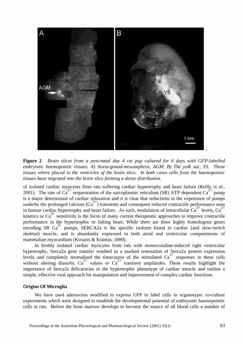
Proceedings of the Australian Physiological and Pharmacological Society (2001) 32(1) 63
Figure 2. Brain slices from a post-natal day 4 rat pup cultured for 6 days with GFP-labelled embryonic haemopoietic tissues. A) Aorta-gonad-mesonephros, AGM. B) The yolk sac, YS. These tissues where placed in the ventricles of the brain slice. In both cases cells from the haemopoietic tissues have migrated into the brain slice forming a dense distribution.
of isolated cardiac myocytes from rats suffering cardiac hypertrophy and heart failure (Reilly et al., 2001). The rate of Ca2+ sequestration of the sarcoplasmic reticulum (SR) ATP dependent Ca2+ pump is a major determinant of cardiac relaxation and it is clear that reductions in the expression of pumps underlie the prolonged calcium (Ca2+) transients and consequent reduced contractile performance seen in human cardiac hypertrophy and heart failure. As such, modulation of intracellular Ca2+ levels, Ca2+ kinetics or Ca2+ sensitivity is the focus of many current therapeutic approaches to improve contractile performance in the hypertrophic or failing heart. While there are three highly homologous genes encoding SR Ca2+ pumps, SERCA2a is the specific isoform found in cardiac (and slow-twitch skeletal) muscle, and is abundantly expressed in both atrial and ventricular compartments of mammalian myocardium (Kiriazis & Kranias, 2000). In freshly isolated cardiac myocytes from rats with monocrotaline-induced right ventricular hypertrophy, Serca2a gene transfer resulted in a marked restoration of Serca2a protein expression levels and completely normalised the timecourse of the stimulated Ca2+ responses in these cells without altering diastolic Ca2+ values or Ca2+ transient amplitudes. These results highlight the importance of Serca2a deficiencies in the hypertrophic phenotype of cardiac muscle and outline a simple, effective viral approach for manipulation and improvement of complex cardiac functions. Origins Of Microglia
We have used adenovirus modified to express GFP to label cells in organotypic co-culture experiments which were designed to establish the developmental potential of embryonic haemopoietic cells in rats. Before the bone marrow develops to become the source of all blood cells a number of
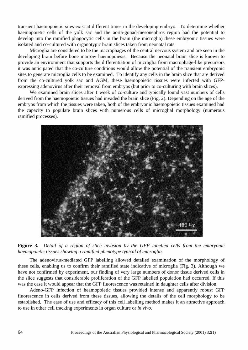
64 Proceedings of the Australian Physiological and Pharmacological Society (2001) 32(1)
transient haemopoietic sites exist at different times in the developing embryo. To determine whether haemopoietic cells of the yolk sac and the aorta-gonad-mesonephros region had the potential to develop into the ramified phagocytic cells in the brain (the microglia) these embryonic tissues were isolated and co-cultured with organotypic brain slices taken from neonatal rats. Microglia are considered to be the macrophages of the central nervous system and are seen in the developing brain before bone marrow haemopoiesis. Because the neonatal brain slice is known to provide an environment that supports the differentiation of microglia from macrophage-like precursors it was anticipated that the co-culture conditions would allow the potential of the transient embryonic sites to generate microglia cells to be examined. To identify any cells in the brain slice that are derived from the co-cultured yolk sac and AGM, these haemopoietic tissues were infected with GFP-expressing adenovirus after their removal from embryos (but prior to co-culturing with brain slices). We examined brain slices after 1 week of co-culture and typically found vast numbers of cells derived from the haemopoietic tissues had invaded the brain slice (Fig. 2). Depending on the age of the embryos from which the tissues were taken, both of the embryonic haemopoietic tissues examined had the capacity to populate brain slices with numerous cells of microglial morphology (numerous ramified processes).
Figure 3. Detail of a region of slice invasion by the GFP labelled cells from the embryonic haemopoietic tissues showing a ramified phenotype typical of microglia.
The adenovirus-mediated GFP labelling allowed detailed examination of the morphology of these cells, enabling us to confirm their ramified state indicative of microglia (Fig. 3). Although we have not confirmed by experiment, our finding of very large numbers of donor tissue derived cells in the slice suggests that considerable proliferation of the GFP labelled population had occurred. If this was the case it would appear that the GFP fluorescence was retained in daughter cells after division. Adeno-GFP infection of heamopoietic tissues provided intense and apparently robust GFP fluorescence in cells derived from these tissues, allowing the details of the cell morphology to be established. The ease of use and efficacy of this cell labelling method makes it an attractive approach to use in other cell tracking experiments in organ culture or in vivo.
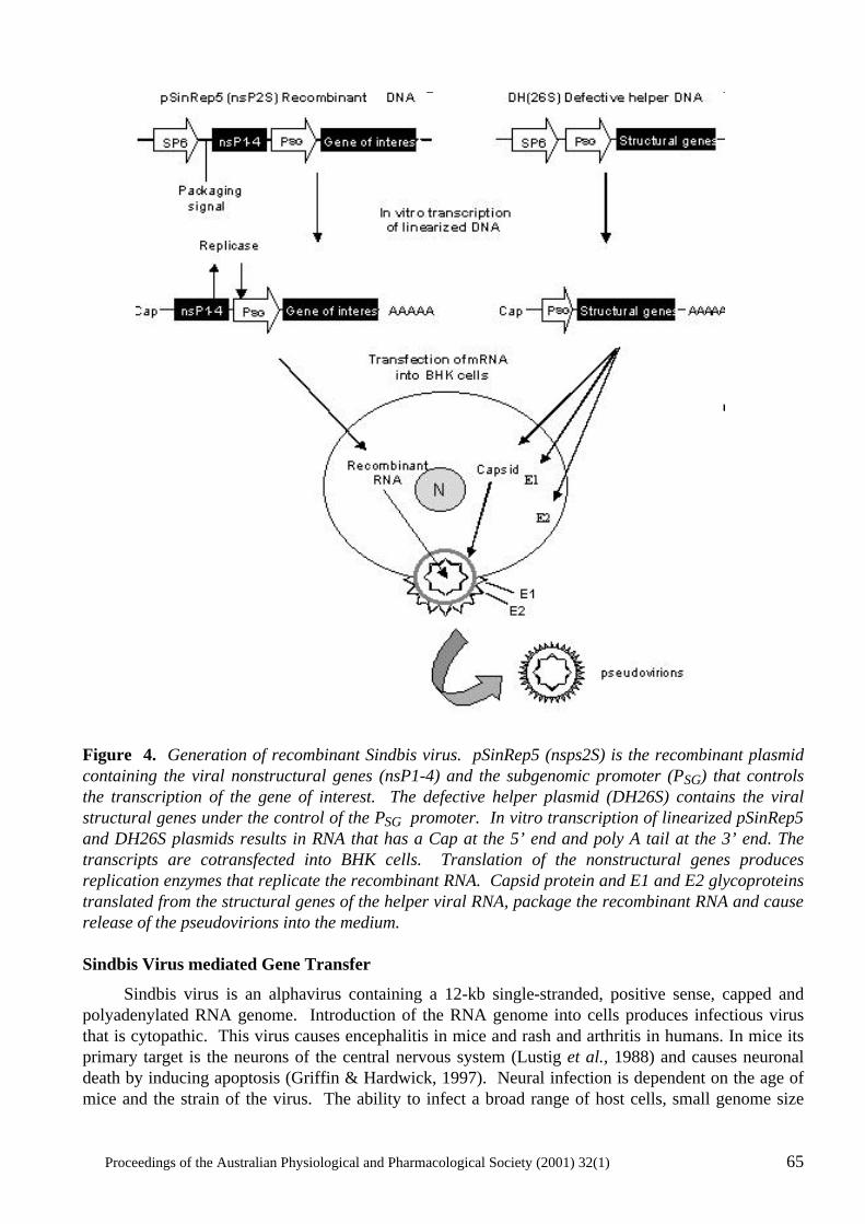
Proceedings of the Australian Physiological and Pharmacological Society (2001) 32(1) 65
Figure 4. Generation of recombinant Sindbis virus. pSinRep5 (nsps2S) is the recombinant plasmid containing the viral nonstructural genes (nsP1-4) and the subgenomic promoter (PSG) that controls the transcription of the gene of interest. The defective helper plasmid (DH26S) contains the viral structural genes under the control of the PSG promoter. In vitro transcription of linearized pSinRep5 and DH26S plasmids results in RNA that has a Cap at the 5’ end and poly A tail at the 3’ end. The transcripts are cotransfected into BHK cells. Translation of the nonstructural genes produces replication enzymes that replicate the recombinant RNA. Capsid protein and E1 and E2 glycoproteins translated from the structural genes of the helper viral RNA, package the recombinant RNA and cause release of the pseudovirions into the medium. Sindbis Virus mediated Gene Transfer
Sindbis virus is an alphavirus containing a 12-kb single-stranded, positive sense, capped and polyadenylated RNA genome. Introduction of the RNA genome into cells produces infectious virus that is cytopathic. This virus causes encephalitis in mice and rash and arthritis in humans. In mice its primary target is the neurons of the central nervous system (Lustig et al., 1988) and causes neuronal death by inducing apoptosis (Griffin & Hardwick, 1997). Neural infection is dependent on the age of mice and the strain of the virus. The ability to infect a broad range of host cells, small genome size
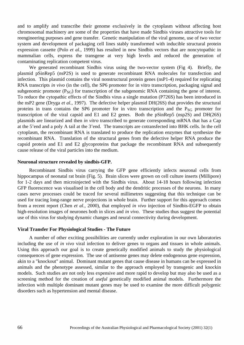
66 Proceedings of the Australian Physiological and Pharmacological Society (2001) 32(1)
and to amplify and transcribe their genome exclusively in the cytoplasm without affecting host chromosomal machinery are some of the properties that have made Sindbis viruses attractive tools for reengineering purposes and gene transfer. Genetic manipulation of the viral genome, use of two vector system and development of packaging cell lines stably transformed with inducible structural protein expression cassette (Polo et al., 1999) has resulted in new Sindbis vectors that are noncytopathic in mammalian cells, express the transgene at very high levels and reduced the generation of contaminating replication competent virus. We generated recombinant Sindbis virus using the two-vector system (Fig 4). Briefly, the plasmid pSinRep5 (nsP2S) is used to generate recombinant RNA molecules for transfection and infection. This plasmid contains the viral nonstructural protein genes (nsP1-4) required for replicating RNA transcripts in vivo (in the cell), the SP6 promoter for in vitro transcription, packaging signal and subgenomic promoter (PSG) for transcription of the subgenomic RNA containing the gene of interest. To reduce the cytopathic effects of the Sindbis virus a single mutation (P726S) has been introduced in the nsP2 gene (Dryga et al., 1997). The defective helper plasmid DH(26S) that provides the structural proteins in trans contains the SP6 promoter for in vitro transcription and the PSG promoter for transcription of the viral capsid and E1 and E2 genes. Both the pSinRep5 (nsp2S) and DH(26S) plasmids are linearized and then in vitro transcribed to generate corresponding mRNA that has a Cap at the 5’end and a poly A tail at the 3’end. The transcripts are cotransfected into BHK cells. In the cell cytoplasm, the recombinant RNA is translated to produce the replication enzymes that synthesize the recombinant RNA. Translation of the structural genes from the defective helper RNA produce the capsid protein and E1 and E2 glycoproteins that package the recombinant RNA and subsequently cause release of the viral particles into the medium. Neuronal structure revealed by sindbis-GFP.
Recombinant Sindbis virus carrying the GFP gene efficiently infects neuronal cells from hippocampus of neonatal rat brain (Fig. 5). Brain slices were grown on cell culture inserts (Millipore) for 1-2 days and then microinjected with the Sindbis virus. About 14-18 hours following infection GFP fluorescence was visualised in the cell body and the dendritic processes of the neurons. In many cases nerve processes could be traced for several millimetres suggesting that this technique can be used for tracing long-range nerve projections in whole brain. Further support for this approach comes from a recent report (Chen et al., 2000), that employed in vivo injection of Sindbis-EGFP to obtain high-resolution images of neurones both in slices and in vivo. These studies thus suggest the potential use of this virus for studying dynamic changes and neural connectivity during development. Viral Transfer For Physiological Studies - The Future
A number of other exciting possibilities are currently under exploration in our own laboratories including the use of in vivo viral infection to deliver genes to organs and tissues in whole animals. Using this approach our goal is to create genetically modified animals to study the physiological consequences of gene expression. The use of antisense genes may delete endogenous gene expression, akin to a "knockout" animal. Dominant mutant genes that cause disease in humans can be expressed in animals and the phenotype assessed, similar to the approach employed by transgenic and knockin models. Such studies are not only less expensive and more rapid to develop but may also be used as a screening method for the creation of useful genetically modified animal models. Furthermore the infection with multiple dominant mutant genes may be used to examine the more difficult polygenic disorders such as hypertension and mental disease.
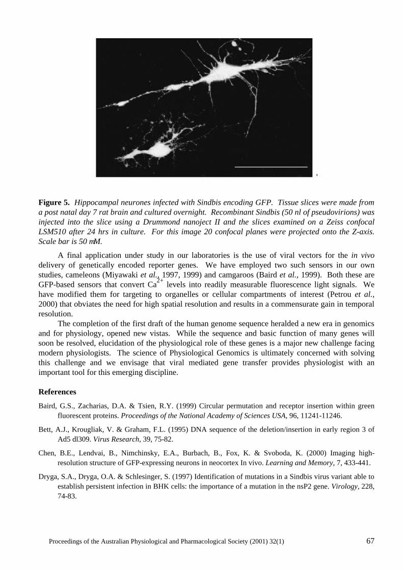
Proceedings of the Australian Physiological and Pharmacological Society (2001) 32(1) 67
Figure 5. Hippocampal neurones infected with Sindbis encoding GFP. Tissue slices were made from a post natal day 7 rat brain and cultured overnight. Recombinant Sindbis (50 nl of pseudovirions) was injected into the slice using a Drummond nanoject II and the slices examined on a Zeiss confocal LSM510 after 24 hrs in culture. For this image 20 confocal planes were projected onto the Z-axis. Scale bar is 50 µM.
A final application under study in our laboratories is the use of viral vectors for the in vivo delivery of genetically encoded reporter genes. We have employed two such sensors in our own studies, cameleons (Miyawaki et al., 1997, 1999) and camgaroos (Baird et al., 1999). Both these are GFP-based sensors that convert Ca2+ levels into readily measurable fluorescence light signals. We have modified them for targeting to organelles or cellular compartments of interest (Petrou et al., 2000) that obviates the need for high spatial resolution and results in a commensurate gain in temporal resolution. The completion of the first draft of the human genome sequence heralded a new era in genomics and for physiology, opened new vistas. While the sequence and basic function of many genes will soon be resolved, elucidation of the physiological role of these genes is a major new challenge facing modern physiologists. The science of Physiological Genomics is ultimately concerned with solving this challenge and we envisage that viral mediated gene transfer provides physiologist with an important tool for this emerging discipline. References
Baird, G.S., Zacharias, D.A. & Tsien, R.Y. (1999) Circular permutation and receptor insertion within green fluorescent proteins. Proceedings of the National Academy of Sciences USA, 96, 11241-11246.
Bett, A.J., Krougliak, V. & Graham, F.L. (1995) DNA sequence of the deletion/insertion in early region 3 of Ad5 dl309. Virus Research, 39, 75-82.
Chen, B.E., Lendvai, B., Nimchinsky, E.A., Burbach, B., Fox, K. & Svoboda, K. (2000) Imaging high-resolution structure of GFP-expressing neurons in neocortex In vivo. Learning and Memory, 7, 433-441.
Dryga, S.A., Dryga, O.A. & Schlesinger, S. (1997) Identification of mutations in a Sindbis virus variant able to establish persistent infection in BHK cells: the importance of a mutation in the nsP2 gene. Virology, 228, 74-83.
68 Proceedings of the Australian Physiological and Pharmacological Society (2001) 32(1)
Engelhardt, J.F., Litzky, L. & Wilson, J.M. (1994a) Prolonged transgene expression in cotton rat lung with recombinant adenoviruses defective in E2a. Human Gene Therapy, 5, 1217-1229.
Engelhardt, J.F., Ye, X., Doranz, B. & Wilson, J.M. (1994b) Ablation of E2A in recombinant adenoviruses improves transgene persistence and decreases inflammatory response in mouse liver. Proceedings of the National Academy of Sciences USA, 91, 6196-6200.
Gao, G.P., Yang, Y. & Wilson, J.M. (1996) Biology of adenovirus vectors with E1 and E4 deletions for liver-directed gene therapy. Journal of Virology, 70, 8934-8943.
Gao, X. & Huang, L. (1995) Cationic liposome-mediated gene transfer. Gene Therapy, 2, 710-722.
Graham, F.L. & Prevec, L. (1995) Methods for construction of adenovirus vectors. Molecular Biotechnology, 3, 207-220.
Griffin, D.E. & Hardwick, J.M. (1997) Regulators of apoptosis on the road to persistent alphavirus infection. Annual Review of Microbiology, 51, 565-592.
He, T.C., Zhou, S., da Costa, L.T., Yu, J., Kinzler, K.W. & Vogelstein, B. (1998) A simplified system for generating recombinant adenoviruses. Proceedings of the National Academy of Sciences USA, 95, 2509-2514.
Kiriazis, H. & Kranias, E.G. (2000) Genetically engineered models with alterations in cardiac membrane calcium-handling proteins. Annual. Review of Physiology, 62, 321-351.
Lustig, S., Jackson, A.C., Hahn, C.S., Griffin, D.E., Strauss, E.G. & Strauss, J.H. (1988) Molecular basis of Sindbis virus neurovirulence in mice. Journal of Virology, 62, 2329-2336.
Miyawaki, A., Griesbeck, O., Heim, R. & Tsien, R.Y. (1999) Dynamic and quantitative Ca2+ measurements using improved cameleons. Proceedings of the National Academy of Sciences USA, 96, 2135-2140.
Miyawaki, A., Llopis, J., Heim, R., McCaffery, J.M., Adams, J.A., Ikura, M. & Tsien, R.Y. (1997) Fluorescent indicators for Ca2+ based on green fluorescent proteins and calmodulin. Nature, 388, 882-887.
Mountain, A. (2000) Gene therapy: the first decade. Trends in Biotechnology, 18, 119-128.
Nettelbeck, D.M., Jerome, V. & Muller, R. (2000) Gene therapy: designer promoters for tumour targeting. Trends in Genetics, 16, 174-181.
Petrou, S., Bowser, D.N., Nicholls, R.A., Panchal, R.G., Smart, M.L., Reilly, A.M. & Williams, D.A. (2000) Genetically targeted calcium sensors enhance the study of organelle function in living cells. Clinical and Experimental Pharmacology and Physiology, 27, 738-744.
Polo, J.M., Belli, B.A., Driver, D.A., Frolov, I., Sherrill, S., Hariharan, M.J., Townsend, K., Perri, S., Mento, S.J., Jolly, D.J. et al. (1999) Stable alphavirus packaging cell lines for Sindbis virus and Semliki Forest virus-derived vectors. Proceedings of the National Academy of Sciences USA, 96, 4598-4603.
Reilly, A.M., Petrou, S., Panchal, R.G. & Williams, D.A. (2001) Restoration of calcium handling properties of adult cardiac myocytes from hypertrophied hearts (submitted).
Romano, G., Michell, P., Pacilio, C. & Giordano, A. (2000) Latest developments in gene transfer technology: achievements, perspectives, and controversies over therapeutic applications. Stem Cells, 18, 19-39.
Wang, I. & Huang, I. (2000) Adenovirus technology for gene manipulation and functional studies. Drug Discovery Today, 5, 10-16.
Yang, N.S., Sun, W.H. & McCabe, D. (1996) Developing particle-mediated gene-transfer technology for research into gene therapy of cancer. Molecular Medicine Today, 2, 476-481.
Proceedings of the Australian Physiological and Pharmacological Society (2001) 32(1) 69
HYBRID SKELETAL MUSCLE FIBRES: A RARE OR COMMON
PHENOMENON?
Gabriela M.M. Stephenson
Muscle Cell Biochemistry Laboratory, School of Life Sciences and Technology, Victoria University, PO Box 14428, MCMC, Melbourne, Victoria 8001
Summary
1. The main aim of this review is to raise awareness of the molecular complexity of single skeletal muscle fibres from ‘normal’ and ‘transforming’ muscles, in recognition of the many types of hybrids that have been observed in vertebrate skeletal muscle. The data used to illustrate various points made in the review were taken from studies on mammalian (mostly rat) and amphibian muscles. 2. The review provides a brief overview of the pattern and extent of molecular heterogeneity in hybrid muscle fibres and of the methodological problems encountered when attempting to identify and characterise such fibres. Particular attention is given to four types of skeletal muscle hybrids: myosin heavy chain (MHC) hybrids, mismatched MHC-myosin light chains (MLC) hybrids, mismatched MHC-regulatory protein hybrids and hybrids containing mismatched MHC-sarcoplasmic reticulum protein isoforms. 3. Some of the current ideas regarding the functional significance, origin and cognitive value of hybrid fibres are critically examined. Introduction
Skeletal muscle contraction is the net result of a series of cellular events known collectively as the excitation-contraction-relaxation (E-C-R) cycle. The major events in the E-C-R cycle were summarised by Stephenson et al (1998). Major events in the E-C-R cycle in the vertebrate skeletal muscle include: (i) initiation and propagation of an action potential along the sarcolemma and transverse (T)-tubular system, (ii) transmission of the T-system depolarisation signal from the T-tubule to the sarcoplasmic reticulum (SR) membrane, (iii) release of calcium ions (Ca2+) from the SR, (iv) transient rise of myoplasmic [Ca2+], (v) binding of Ca2+ to the regulatory protein troponin C (Tn C), (vi) transient activation of the regulatory system and contractile apparatus, (vi) dissociation of Ca2+ from Tn C and (vii) Ca2+ reuptake by SR mediated by SERCA. The key roles in these events are played by Ca2+ (the activation ion) and by a large number of proteins/protein complexes located in several subcellular compartments. It is now widely accepted that many of the proteins involved in events of the E-C-R cycle exist as multiple forms (isoforms), which can be distinguished and identified by biochemical methods such as gel electrophoresis and immunochemistry (Moss et al., 1995). Polymorphous skeletal muscle proteins of the E-C-R cycle include: the α-subunit of the dyhydropyridine receptor (DHPR), ryanodine receptor/SR calcium release channel (RyR), sarco (endoplasmic) reticulum Ca2+-ATPase (SERCA), calsequestrin, myosin heavy chain (MHC), myosin light chain (MLC) and the regulatory proteins troponin C (TnC), troponin I (TnI), troponin T (TnT) and tropomyosin (Tm) (Pette & Staron, 2000, 1997, 1990). It is important to point out that the list of isoforms of skeletal muscle proteins (particularly myofibrillar proteins) has been increasing in parallel with the development/refinement of protein separation/identification techniques and with the application of these techniques to a wider range of muscles and animal species. Updated versions of this list can be found in reviews produced regularly by major contributors
70 Proceedings of the Australian Physiological and Pharmacological Society (2001) 32(1)
to the field ( Pette & Staron, 2000; Schiaffino & Salviati, 1998; Pette & Staron, 1997; Schiaffino & Reggiani, 1996; Pette & Staron, 1990; Swynghedauw, 1986). It is noteworthy that the number of isoforms varies markedly between various skeletal muscle proteins. For example, according to a recent count, mammalian skeletal muscle expresses as many as ten MHC isoforms, but only two TnC isoforms [one typical of fast-twitch muscle (TnC-f), the other of slow-twitch muscle (TnC-s) (Pette & Staron, 2000; 1997). The ten MHC isoforms* include four isoforms present in adult mammalian muscle (slow-twitch isoform MHCI or MHCβ/slow and fast-twitch isoforms MHCIIa, MHCIId/x and MHCIIb), two isoforms present in developing and regenerating muscles (MHC-emb, MHC-neo), and four isoforms present in some highly specialized muscles [extraocular and jaw closing muscles (MHC-exoc). Currently it is not known whether there is any relationship between the number of isoforms of a muscle protein, its cellular function and/or the molecular mechanisms responsible for its molecular diversity. So far, the terms ‘hybrid muscle fibres†’, ‘polymorphic fibres’, or ‘MHC hybrids’ have been used interchangeably to define fibres that co-express more than one MHC isoform. However, there is now compelling evidence to suggest that this meaning of the term ‘hybrid fibres’ is highly inadequate because it does not apply to fibres displaying patterns of molecular heterogeneity with respect to other proteins. A population of such fibres, expressing only one MHC isoform (MHCIIa) and both fast- and slow-twitch isoforms of the MLC subunits has been detected in rat soleus (SOL) in an early study by Mizusawa et al. (1982) and in a very recent study by Bortolotto et al. (2000a). Based only on MHC composition, these fibres would be classified as ‘pure’, but such classification would be incorrect because it would not provide information on the molecular heterogeneity of the fibres with respect to MLC composition. Two other major groups of hybrids not covered by the traditional meaning of the term ‘hybrid fibres’ include fibres in which two or several proteins are expressed as isoforms (e.g. fibres containing several isoforms of MLC and several isoforms of tropomyosin) and fibres in which one isoform is expressed as a protein and another as a mRNA species. It is important to note that the sets of isoforms detected so far in fibres co-expressing isoforms of two or more muscle proteins have been found to be either of the same type (matched) or of different types (mismatched). Matched sets of MHC and MLC isoforms would comprise, for example, fast-twitch MHC isoforms MHCIIa and MLCIIb and fast-twitch MLC isoforms MLC1f, MLC2f and MLC3, while mismatched sets would comprise fast-twitch MHC isoforms MHCIIa and MHCIIb, slow-twitch MLC isoform MLC1s and fast-twitch MLC isoforms MLC1f, MLC2f and MLC3. In recognition of the many kinds of hybrids that have been observed so far in vertebrate skeletal muscles, the hybrid fibres discussed in this review will be described by terms that indicate both the muscle protein(s) whose isoforms are being considered (e.g. MHC-MLC ) and the relationship (matched or mismatched) between the sets of isoforms co-expressed in the fibre. It is worth pointing out that hybrid fibres were once regarded as a rare phenomenon and often discarded from studies concerned with the functional characteristics of single fibre preparations (Danieli-Betto et al., 1990). More recently, however, hybrid muscle fibres have started to attract considerable interest from a broad range of cell biologists. This can be explained, in part, by the finding that MHC hybrids represent the dominant biochemical phenotype even in skeletal muscles that were previously thought to be ‘pure’ in terms of fibre type composition (Bortolotto et al., 2000a). *In this review a fibre type is identified by a roman numeral and a capital letter (e.g. IIA), while the MHC isoform expressed in the fibre is identified by a roman numeral and the corresponding lower case letter (i.e. MHCIIa). This nomenclature, which has been introduced by Pette’s laboratory (e.g. Hämäläinen & Pette, 1995), has not been adopted consistently by other laboratories working in the field, causing a certain degree of confusion among readers of articles and reviews on MHC isoforms and MHC-based fibre types. †Term coined by Pette’s laboratory about a decade ago to describe fibres expressing more than one MHC
isoform and to distinguish them from fibres expressing only one MHC isoform (‘pure fibres’).
Proceedings of the Australian Physiological and Pharmacological Society (2001) 32(1) 71
Moreover, hybrid muscle fibres are now seen as valuable experimental tools for gaining further insights into two major areas of research: (i) the physiological role of muscle protein isoforms and (ii) the regulation of gene expression in multinucleated cells (see last section). This is the first review to focus on skeletal muscle hybrid fibres. Its main aim is to raise awareness of the molecular complexity of single muscle fibres, particularly among physiologists concerned with basic and applied aspects of skeletal muscle function. The data used to illustrate various points made in the review were taken from studies of mammalian (mostly rat) and amphibian muscle. The general background sections provide a brief overview of the pattern and extent of molecular heterogeneity in hybrid muscle fibres from ‘normal’ and ‘transforming’ muscles and of the methodological problems encountered when attempting to identify and characterise such fibres. Particular attention is given to four types of skeletal muscle hybrids: MHC hybrids, mismatched MHC-MLC hybrids, mismatched MHC-regulatory protein hybrids and mismatched MHC-SR protein hybrids. The last section of the review comprises a critical examination of some of the current ideas regarding the functional significance, origin and cognitive value of hybrid fibres. MHC Hybrids
Methods used for the detection and characterisation of MHC hybrids.
It is now quite clear that neither of the three methods traditionally employed to distinguish fibre types in skeletal muscles [light microscopy, myosin/myofibrillar ATPase (mATPase)-based or metabolic enzyme-based histochemistry] can be used effectively for the identification and characterisation of MHC hybrids (Hämäläinen & Pette, 1995; Schiaffino & Reggiani, 1996). The most suitable methods for detecting MHC polymorphism in individual muscle fibres, at protein or mRNA level, include MHC-based immunohistochemistry (MHC- IHChem), single fibre polyacrylamide gel microelectrophoresis under denaturing conditions (SDS-PAGE sf), pyrophosphate gel electrophoresis of myosin isoenzymes in single fibre segments, reverse transcription-polymerase chain reaction (RT-PCR) and in situ hybridisation (for review see Pette & Staron, 2000; Pette et al., 1999; Schiaffino & Salviati, 1998; Hämäläinen & Pette, 1995). This methodological point is well illustrated by the bar graph shown in Figure 1, which allows a quick comparison of the fibre type composition of rat extensor digitorum longus (EDL) (A,B) and SOL (C,D) muscles reported by Armstrong and Phelps (1984), on the basis of mATPase-based histochemistry (A, C) and that found in more recent studies using SDS-PAGEsf only (EDL; Bortolotto et al., 2000a) (B) or a combination of SDS-PAGEsf and MHC-IHChem (SOL, Bottinelli et al., 1994a) (D). A significant conclusion emerging from these data is that the largest proportion (~70%) of fibres in rat EDL are hybrid IIB+IID fibres, rather than IIA or IIB fibres, as previously thought. The newly discovered heterogeneity of rat EDL muscle and muscle fibres has important theoretical and methodological implications because rat EDL has been for many years the preferred experimental model in physiological investigations of mammalian fast-twitch muscle contractility. It is important to note that even methods such as MHC-based IHChem and SDS-PAGEsf have intrinsic limitations when used for the detection and characterisation of MHC hybrids. Some of these limitations have been pointed out by Pette et al.(1999) and Schiaffino & Salviati (1998). For example, MHC-based IHChem is limited by the availability and specificity of anti-MHC isoform antibodies and it does not always detect fibres containing both MHCIId and MHCIIb isoforms (IID + IIB hybrids; Rivero et al., 1998). Furthermore, while MHC-based IHChem can provide considerable information about the proportion and intramuscular distribution of hybrid fibres and about the intracellular distribution of the co-expressed MHC isoforms (Dix & Eisenberg, 1988), it does not allow for the quantification of MHC isoforms co-expressed in individual hybrid fibres. By comparison, SDS-PAGEsf combined with scanning densitometry, enables the researcher to separate and quantify the relative proportion of MHC isoforms co-expressed in a single fibre, and to relate these results to other
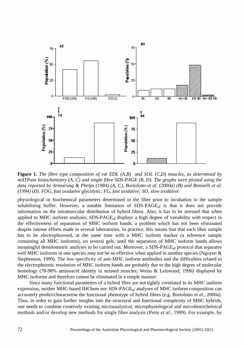
72 Proceedings of the Australian Physiological and Pharmacological Society (2001) 32(1)
Figure 1. The fibre type composition of rat EDL (A,B) and SOL (C,D) muscles, as determined by mATPase histochemistry (A, C) and single fibre SDS-PAGE (B, D). The graphs were plotted using the data reported by Armstrong & Phelps (1984) (A, C), Bortolotto et al. (2000a) (B) and Botinelli et al. (1994) (D). FOG, fast oxidative glycolytic; FG, fast oxidative; SO, slow oxidative.
physiological or biochemical parameters determined in the fibre prior to incubation in the sample solubilising buffer. However, a notable limitation of SDS-PAGEsf is that it does not provide information on the intramuscular distribution of hybrid fibres. Also, it has to be stressed that when applied to MHC isoform analyses, SDS-PAGEsf displays a high degree of variability with respect to the effectiveness of separation of MHC isoform bands, a problem which has not been eliminated despite intense efforts made in several laboratories. In practice, this means that that each fibre sample has to be electrophoresed, at the same time with a MHC isoform marker (a reference sample containing all MHC isoforms), on several gels, until the separation of MHC isoform bands allows meaningful densitometric analyses to be carried out. Moreover, a SDS-PAGEsf protocol that separates well MHC isoforms in one species may not be so effective when applied to another species (Nguyen & Stephenson, 1999). The low specificity of anti-MHC isoform antibodies and the difficulties related to the electrophoretic resolution of MHC isoform bands are probably due to the high degree of molecular homology (78-98% aminoacid identity in striated muscles; Weiss & Leinwand, 1996) displayed by MHC isoforms and therefore cannot be eliminated in a simple manner. Since many functional parameters of a hybrid fibre are not tightly correlated to its MHC isoform expression, neither MHC-based IHChem nor SDS-PAGEsf analyses of MHC isoform composition can accurately predict/characterise the functional phenotype of hybrid fibres (e.g. Bortolotto et al., 2000a). Thus, in order to gain further insights into the structural and functional complexity of MHC hybrids, one needs to combine creatively existing microanalytical, microphysiological and microhistochemical methods and/or develop new methods for single fibre analysis (Pette et al., 1999). For example, by
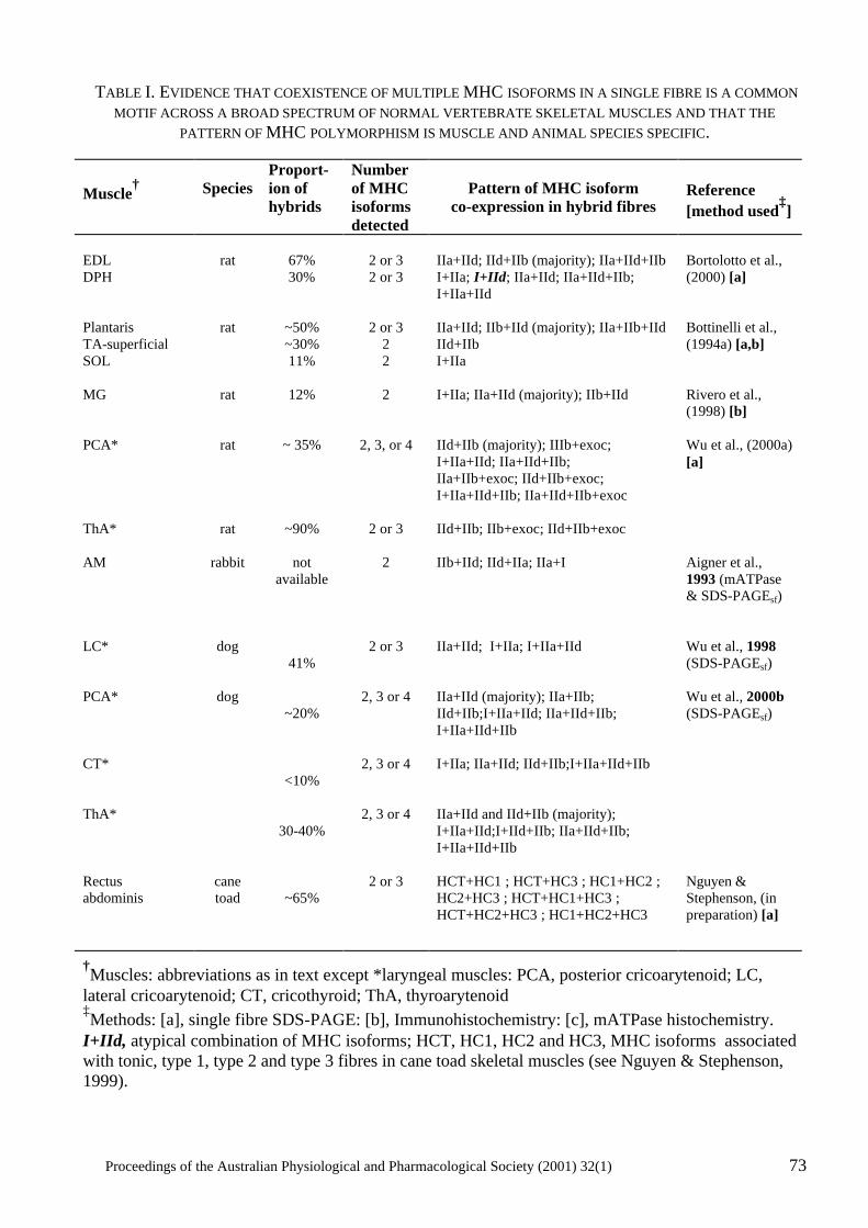
Proceedings of the Australian Physiological and Pharmacological Society (2001) 32(1) 73
TABLE I. EVIDENCE THAT COEXISTENCE OF MULTIPLE MHC ISOFORMS IN A SINGLE FIBRE IS A COMMON MOTIF ACROSS A BROAD SPECTRUM OF NORMAL VERTEBRATE SKELETAL MUSCLES AND THAT THE
PATTERN OF MHC POLYMORPHISM IS MUSCLE AND ANIMAL SPECIES SPECIFIC. Muscle
†
Species
Proport-ion of hybrids
Number of MHC isoforms detected
Pattern of MHC isoform
co-expression in hybrid fibres
Reference [method used
‡]
EDL DPH Plantaris TA-superficial SOL MG PCA* ThA* AM LC* PCA* CT* ThA* Rectus abdominis
rat
rat
rat
rat
rat
rabbit
dog
dog
cane toad
67% 30%
~50% ~30% 11%
12%
~ 35%
~90%
not available
41%
~20%
<10%
30-40%
~65%
2 or 3 2 or 3
2 or 3 2 2
2
2, 3, or 4
2 or 3
2
2 or 3
2, 3 or 4
2, 3 or 4
2, 3 or 4
2 or 3
IIa+IId; IId+IIb (majority); IIa+IId+IIb I+IIa; I+IId; IIa+IId; IIa+IId+IIb; I+IIa+IId IIa+IId; IIb+IId (majority); IIa+IIb+IId IId+IIb I+IIa I+IIa; IIa+IId (majority); IIb+IId IId+IIb (majority); IIIb+exoc; I+IIa+IId; IIa+IId+IIb; IIa+IIb+exoc; IId+IIb+exoc; I+IIa+IId+IIb; IIa+IId+IIb+exoc IId+IIb; IIb+exoc; IId+IIb+exoc IIb+IId; IId+IIa; IIa+I IIa+IId; I+IIa; I+IIa+IId IIa+IId (majority); IIa+IIb; IId+IIb;I+IIa+IId; IIa+IId+IIb; I+IIa+IId+IIb I+IIa; IIa+IId; IId+IIb;I+IIa+IId+IIb IIa+IId and IId+IIb (majority); I+IIa+IId;I+IId+IIb; IIa+IId+IIb; I+IIa+IId+IIb HCT+HC1 ; HCT+HC3 ; HC1+HC2 ; HC2+HC3 ; HCT+HC1+HC3 ; HCT+HC2+HC3 ; HC1+HC2+HC3
Bortolotto et al., (2000) [a] Bottinelli et al., (1994a) [a,b] Rivero et al., (1998) [b] Wu et al., (2000a) [a] Aigner et al., 1993 (mATPase & SDS-PAGEsf) Wu et al., 1998 (SDS-PAGEsf) Wu et al., 2000b (SDS-PAGEsf) Nguyen & Stephenson, (in preparation) [a]
†Muscles: abbreviations as in text except *laryngeal muscles: PCA, posterior cricoarytenoid; LC, lateral cricoarytenoid; CT, cricothyroid; ThA, thyroarytenoid ‡Methods: [a], single fibre SDS-PAGE: [b], Immunohistochemistry: [c], mATPase histochemistry. I+IId, atypical combination of MHC isoforms; HCT, HC1, HC2 and HC3, MHC isoforms associated with tonic, type 1, type 2 and type 3 fibres in cane toad skeletal muscles (see Nguyen & Stephenson, 1999).
74 Proceedings of the Australian Physiological and Pharmacological Society (2001) 32(1)
applying three different biochemical methods (in situ hybridization, MHC-based IHChem and mATPase histochemistry) to serial cryosections from human vastus lateralis muscles (prior and post-37 day period of bed rest), Andersen et al. (1999) discovered a novel population of MHC hybrids, in which the protein of one MHC isoform (MHCI) coexisted with the mRNA of another (MHCIId). The discovery of MHC hybrids with mismatched protein-mRNA species adds a new meaning to the concept of skeletal muscle cell heterogeneity and prompts the obvious question whether vertebrate skeletal muscles contain any fibres that are genuinely ‘pure’. MHC hybrids detected in ‘normal’* muscles
Fibres co-expressing the slow-twitch and fast-twitch MHC isoforms MHCI and MHCIIa (I+IIA or I/IIA fibres) are the earliest examples of MHC hybrids reported to occur in normal mammalian skeletal muscle. These fibres, previously referred to as IC or IIC fibres on the basis of the most abundant MHC isoform expressed (MHCI or MHCIIa, respectively) (Pierobon-Bormioli et al., 1981), are still the easiest to identify and characterise both by MHC-based IHChem and SDS-PAGEsf. This is because the currently available monoclonal antibodies (Mabs) against MHCI and MHCIIa have a high degree of specificity and the electrophoretic bands corresponding to the MHCI and MHCIIa proteins are separated quite effectively and reproducibly by all SDS-PAGEsf protocols developed and/or used in different laboratories. As described in detail in a recent study by Bortolotto et al. (2000a), type I+IIA hybrid fibres can also be identified by the physiological fibre typing method of Fink et al. (1986), because they produce characteristic staircase-like force-pSr curves (‘composite’ curves). A large number of data generated over the last two decades by MHC-based IHChem and/or SDS-PAGEsf, strongly suggest that normal muscles from mammals and amphibians contain a sizeable proportion of MHC hybrids, which co-express, at the protein level, two, three and even four MHC isoforms. A small sample of these data (Table I) shows that the pattern of MHC polymorphism (as indicated by the proportion of hybrids, the number of isoforms co-expressed and the combination of MHC isoforms detected in individual fibres) is muscle and animal species specific. For example in adult rat, soleus muscle (SOL) was found to contain about 11% MHC hybrids, all of which co-expressed two MHC isoforms (I and IIa), while laryngeal thyroarytenoid (ThA) muscle was found to contain about 90% MHC hybrids, some of which co-expressed combinations of two or even three MHC isoforms. In the dog, however, ThA muscle was found to contain a smaller proportion (30-40%) of MHC hybrids and many of these hybrids co-expressed all 4 major MHC isoforms commonly found in mammalian muscle (I, IIa, IId and IIb). MHCexoc, a tissue specific isoform co-expressed with MHCIId and MHCIIb in rat ThA muscle was not detected in dog ThA muscle. MHC hybrids detected in muscles in transformation
It is now widely accepted that the proportion of MHC hybrids and their molecular complexity (as judged by the number of MHC isoforms co-expressed and the pattern of co-expression) is higher in muscles undergoing molecular and functional transformation than in normal muscles (see review by Pette et al., 1999). As seen in Table II, soleus muscles of rats subjected to four week unloading by hindlimb suspension (a strategy inducing a slow to fast transition in muscle phenotype) contained 4 times more hybrid fibres than the controls (Oishi et al., 1998). Moreover, most hybrid fibres in the transforming soleus co-expressed three MHC isoforms (I, IIa and IId) , while all hybrids in the control soleus co-expressed two MHC isoforms only (I and IIa). Some MHC isoform combinations, such as I+ IId, which are seen only rarely in fibres from normal muscles, have been found to occur fairly * The term ‘normal’ muscles is used to indicate muscles from animals free of disease, muscles from adult animals or muscles that had not been subjected to ‘transforming conditions’ (e.g. changes in the neural impulse pattern or hormone level).
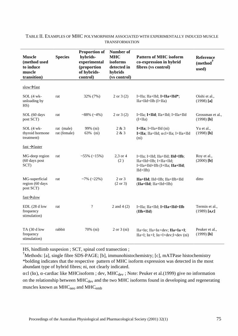
Proceedings of the Australian Physiological and Pharmacological Society (2001) 32(1) 75
TABLE II. EXAMPLES OF MHC POLYMORPHISM ASSOCIATED WITH EXPERIMENTALLY INDUCED MUSCLE TRANSFORMATION
Muscle (method used to induce muscle transition)
Species
Proportion of hybrids-experimental (proportion of hybrids-control)
Number of MHC isoforms detected in hybrids (vs control)
Pattern of MHC isoform co-expression in hybrid fibres (vs control)
Reference (method† used)
slowàfast SOL (4 wk-unloading by HS) SOL (60 days post SCT) SOL (4 wk- thyroid hormone treatment) fast àfaster MG-deep region (60 days post SCT) MG-superficial region (60 days post SCT) fastàslow EDL (28 d low frequency stimulation) TA (30 d low frequency stimulation)
rat rat rat (male) rat (female) rat rat rat rabbit
32% (7%)
~88% (~4%)
99% (ni) 63% (ni)
~55% (~15%)
~7% (~22%) ?
70% (ni)
2 or 3 (2)
2 or 3 (2)
2 & 3 2 & 3
2,3 or 4 (2 )
2 or 3 (2 or 3)
2 and 4 (2)
2 or 3 (ni)
I+IIa; IIa+IId; I+IIa+IId*; IIa+IId+IIb (I+IIa) I+IIa; I+IId; IIa+IId; I+IIa+IId (I+IIa) I+IIa; I+IIa+IId (ni) I+IIa; IIa+IId; αcl+IIa; I+IIa+IId (ni) I+IIa; I+IId; IIa+IId; IId+IIb; IIa+IId+IIb; I+IIa+IId; I+IIa+IId+IIb (I+IIa; IIa+IId; IId+IIb) IIa+IId; IId+IIb; IIa+IIb+IId (IIa+IId; IIa+IId+IIb) I+IIa; IIa+IId; I+IIa+IId+IIb (IIb+IId) IIa+Iα; IIa+Iα+dev; IIa+Iα+I; IIa+I; Iα+I; Iα+I+dev;I+dev (ni)
Oishi et al., (1998) [a] Grossman et al., (1998) [b] Yu et al., (1998) [b] Roy et al., (2000) [b] ditto Termin et al., (1989) [a,c] Peuker et al., (1999) [b]
HS, hindlimb suspesion ; SCT, spinal cord transection ; †Methods: [a], single fibre SDS-PAGE; [b], immunohistochemistry; [c], mATPase histochemistry *bolding indicates that the respective pattern of MHC isoform expression was detected in the most abundant type of hybrid fibres; ni, not clearly indicated. αcl (Iα), α-cardiac like MHCisoform ; dev, MHCdev ; Note: Peuker et al.(1999) give no information on the relationship between MHCdev and the two MHC isoforms found in developing and regenerating muscles known as MHCneo and MHCemb
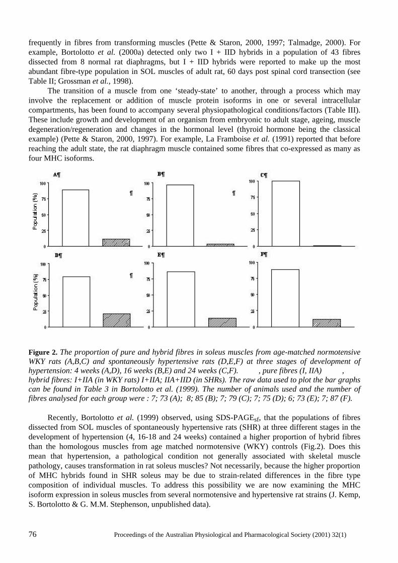
76 Proceedings of the Australian Physiological and Pharmacological Society (2001) 32(1)
frequently in fibres from transforming muscles (Pette & Staron, 2000, 1997; Talmadge, 2000). For example, Bortolotto et al. (2000a) detected only two I + IID hybrids in a population of 43 fibres dissected from 8 normal rat diaphragms, but I + IID hybrids were reported to make up the most abundant fibre-type population in SOL muscles of adult rat, 60 days post spinal cord transection (see Table II; Grossman et al., 1998). The transition of a muscle from one ‘steady-state’ to another, through a process which may involve the replacement or addition of muscle protein isoforms in one or several intracellular compartments, has been found to accompany several physiopathological conditions/factors (Table III). These include growth and development of an organism from embryonic to adult stage, ageing, muscle degeneration/regeneration and changes in the hormonal level (thyroid hormone being the classical example) (Pette & Staron, 2000, 1997). For example, La Framboise et al. (1991) reported that before reaching the adult state, the rat diaphragm muscle contained some fibres that co-expressed as many as four MHC isoforms.
Figure 2. The proportion of pure and hybrid fibres in soleus muscles from age-matched normotensive WKY rats (A,B,C) and spontaneously hypertensive rats (D,E,F) at three stages of development of hypertension: 4 weeks (A,D), 16 weeks (B,E) and 24 weeks (C,F). , pure fibres (I, IIA) , hybrid fibres: I+IIA (in WKY rats) I+IIA; IIA+IID (in SHRs). The raw data used to plot the bar graphs can be found in Table 3 in Bortolotto et al. (1999). The number of animals used and the number of fibres analysed for each group were : 7; 73 (A); 8; 85 (B); 7; 79 (C); 7; 75 (D); 6; 73 (E); 7; 87 (F). Recently, Bortolotto et al. (1999) observed, using SDS-PAGEsf, that the populations of fibres dissected from SOL muscles of spontaneously hypertensive rats (SHR) at three different stages in the development of hypertension (4, 16-18 and 24 weeks) contained a higher proportion of hybrid fibres than the homologous muscles from age matched normotensive (WKY) controls (Fig.2). Does this mean that hypertension, a pathological condition not generally associated with skeletal muscle pathology, causes transformation in rat soleus muscles? Not necessarily, because the higher proportion of MHC hybrids found in SHR soleus may be due to strain-related differences in the fibre type composition of individual muscles. To address this possibility we are now examining the MHC isoform expression in soleus muscles from several normotensive and hypertensive rat strains (J. Kemp, S. Bortolotto & G. M.M. Stephenson, unpublished data).
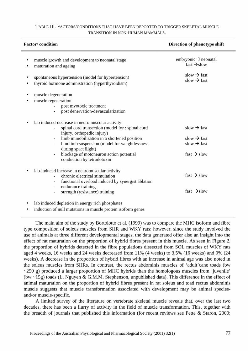
Proceedings of the Australian Physiological and Pharmacological Society (2001) 32(1) 77
TABLE III. FACTORS/CONDITIONS THAT HAVE BEEN REPORTED TO TRIGGER SKELETAL MUSCLE TRANSITION IN NON-HUMAN MAMMALS.
Factor/ condition
Direction of phenotype shift
• muscle growth and development to neonatal stage • maturation and ageing
• spontaneous hypertension (model for hypertension) • thyroid hormone administration (hyperthyroidism)
• muscle degeneration • muscle regeneration
- post myotoxic treatment - post denervation-devascularization
• lab induced-decrease in neuromuscular activity
- spinal cord transection (model for : spinal cord injury, orthopedic injury)
- limb immobilization in a shortened position - hindlimb suspension (model for weightlessness
during spaceflight) - blockage of motoneuron action potential
conduction by tetrodotoxin
• lab-induced increase in neuromuscular activity - chronic electrical stimulation - functional overload induced by synergist ablation - endurance training - strength (resistance) training
• lab induced depletion in energy rich phosphates • induction of null mutations in muscle protein isoform genes
embryonic àneonatal
fast àslow
slow à fast slow à fast
slow à fast
slow à fast slow à fast
fast à slow
fast à slow
fast àslow
The main aim of the study by Bortolotto et al. (1999) was to compare the MHC isoform and fibre type composition of soleus muscles from SHR and WKY rats; however, since the study involved the use of animals at three different developmental stages, the data generated offer also an insight into the effect of rat maturation on the proportion of hybrid fibres present in this muscle. As seen in Figure 2, the proportion of hybrids detected in the fibre populations dissected from SOL muscles of WKY rats aged 4 weeks, 16 weeks and 24 weeks decreased from 11% (4 weeks) to 3.5% (16 weeks) and 0% (24 weeks). A decrease in the proportion of hybrid fibres with an increase in animal age was also noted in the soleus muscles from SHRs. In contrast, the rectus abdominis muscles of ‘adult’cane toads (bw ~250 g) produced a larger proportion of MHC hybrids than the homologous muscles from ‘juvenile’ (bw ~15g) toads (L. Nguyen & G.M.M. Stephenson, unpublished data). This difference in the effect of animal maturation on the proportion of hybrid fibres present in rat soleus and toad rectus abdominis muscle suggests that muscle transformation associated with development may be animal species- and/or muscle-specific. A limited survey of the literature on vertebrate skeletal muscle reveals that, over the last two decades, there has been a flurry of activity in the field of muscle transformation. This, together with the breadth of journals that published this information (for recent reviews see Pette & Staron, 2000;
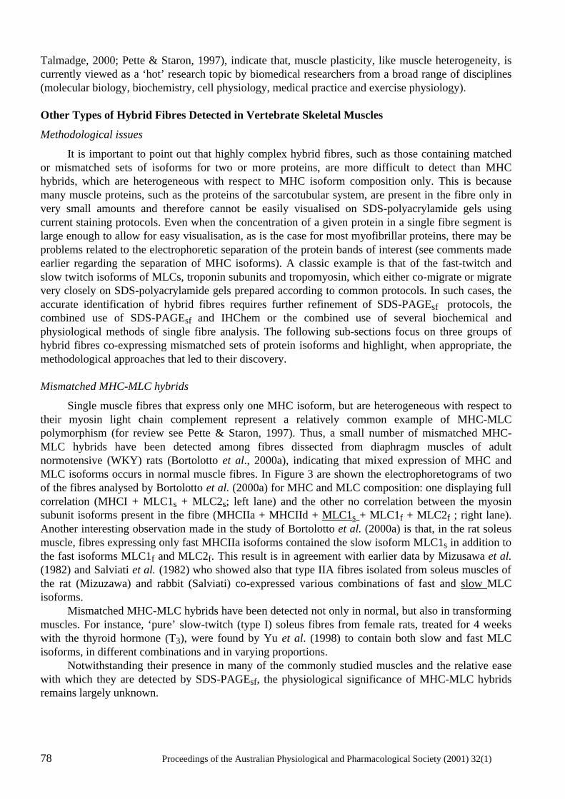
78 Proceedings of the Australian Physiological and Pharmacological Society (2001) 32(1)
Talmadge, 2000; Pette & Staron, 1997), indicate that, muscle plasticity, like muscle heterogeneity, is currently viewed as a ‘hot’ research topic by biomedical researchers from a broad range of disciplines (molecular biology, biochemistry, cell physiology, medical practice and exercise physiology). Other Types of Hybrid Fibres Detected in Vertebrate Skeletal Muscles
Methodological issues
It is important to point out that highly complex hybrid fibres, such as those containing matched or mismatched sets of isoforms for two or more proteins, are more difficult to detect than MHC hybrids, which are heterogeneous with respect to MHC isoform composition only. This is because many muscle proteins, such as the proteins of the sarcotubular system, are present in the fibre only in very small amounts and therefore cannot be easily visualised on SDS-polyacrylamide gels using current staining protocols. Even when the concentration of a given protein in a single fibre segment is large enough to allow for easy visualisation, as is the case for most myofibrillar proteins, there may be problems related to the electrophoretic separation of the protein bands of interest (see comments made earlier regarding the separation of MHC isoforms). A classic example is that of the fast-twitch and slow twitch isoforms of MLCs, troponin subunits and tropomyosin, which either co-migrate or migrate very closely on SDS-polyacrylamide gels prepared according to common protocols. In such cases, the accurate identification of hybrid fibres requires further refinement of SDS-PAGEsf protocols, the combined use of SDS-PAGEsf and IHChem or the combined use of several biochemical and physiological methods of single fibre analysis. The following sub-sections focus on three groups of hybrid fibres co-expressing mismatched sets of protein isoforms and highlight, when appropriate, the methodological approaches that led to their discovery. Mismatched MHC-MLC hybrids
Single muscle fibres that express only one MHC isoform, but are heterogeneous with respect to their myosin light chain complement represent a relatively common example of MHC-MLC polymorphism (for review see Pette & Staron, 1997). Thus, a small number of mismatched MHC-MLC hybrids have been detected among fibres dissected from diaphragm muscles of adult normotensive (WKY) rats (Bortolotto et al., 2000a), indicating that mixed expression of MHC and MLC isoforms occurs in normal muscle fibres. In Figure 3 are shown the electrophoretograms of two of the fibres analysed by Bortolotto et al. (2000a) for MHC and MLC composition: one displaying full correlation (MHCI + MLC1s + MLC2s; left lane) and the other no correlation between the myosin subunit isoforms present in the fibre (MHCIIa + MHCIId + MLC1s + MLC1f + MLC2f ; right lane). Another interesting observation made in the study of Bortolotto et al. (2000a) is that, in the rat soleus muscle, fibres expressing only fast MHCIIa isoforms contained the slow isoform MLC1s in addition to the fast isoforms MLC1f and MLC2f. This result is in agreement with earlier data by Mizusawa et al. (1982) and Salviati et al. (1982) who showed also that type IIA fibres isolated from soleus muscles of the rat (Mizuzawa) and rabbit (Salviati) co-expressed various combinations of fast and slow MLC isoforms. Mismatched MHC-MLC hybrids have been detected not only in normal, but also in transforming muscles. For instance, ‘pure’ slow-twitch (type I) soleus fibres from female rats, treated for 4 weeks with the thyroid hormone (T3), were found by Yu et al. (1998) to contain both slow and fast MLC isoforms, in different combinations and in varying proportions. Notwithstanding their presence in many of the commonly studied muscles and the relative ease with which they are detected by SDS-PAGEsf, the physiological significance of MHC-MLC hybrids remains largely unknown.
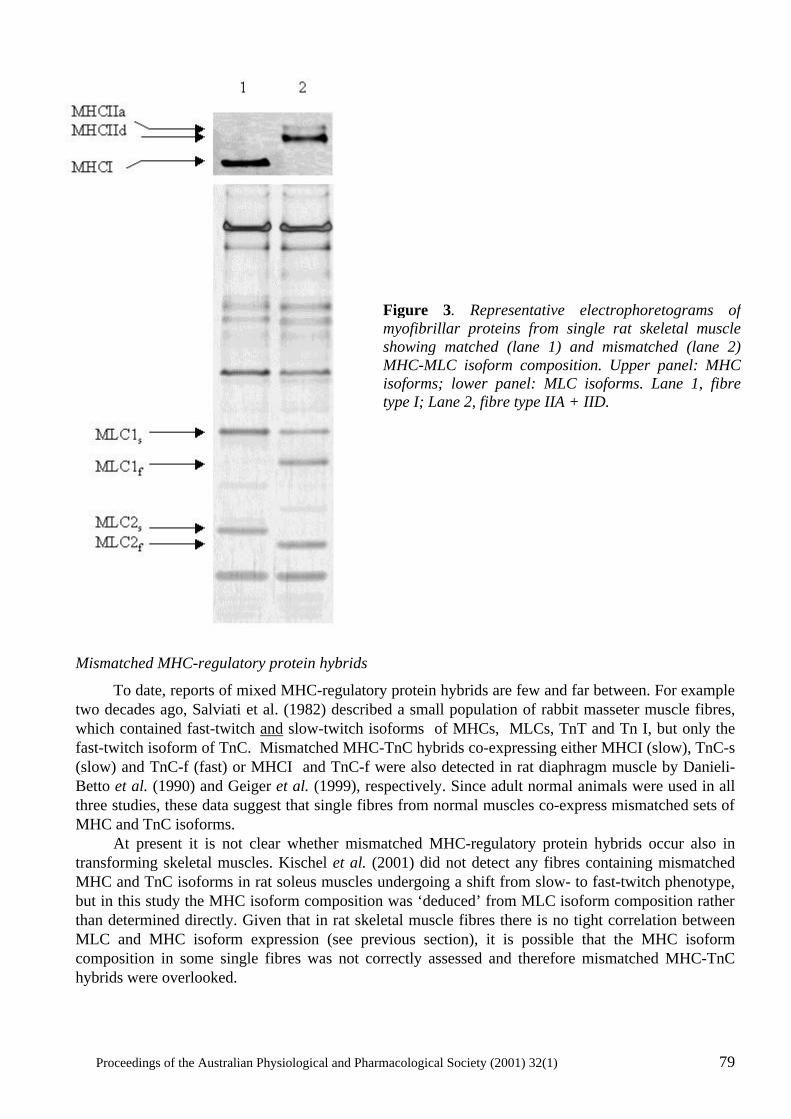
Proceedings of the Australian Physiological and Pharmacological Society (2001) 32(1) 79
Mismatched MHC-regulatory protein hybrids
To date, reports of mixed MHC-regulatory protein hybrids are few and far between. For example two decades ago, Salviati et al. (1982) described a small population of rabbit masseter muscle fibres, which contained fast-twitch and slow-twitch isoforms of MHCs, MLCs, TnT and Tn I, but only the fast-twitch isoform of TnC. Mismatched MHC-TnC hybrids co-expressing either MHCI (slow), TnC-s (slow) and TnC-f (fast) or MHCI and TnC-f were also detected in rat diaphragm muscle by Danieli-Betto et al. (1990) and Geiger et al. (1999), respectively. Since adult normal animals were used in all three studies, these data suggest that single fibres from normal muscles co-express mismatched sets of MHC and TnC isoforms. At present it is not clear whether mismatched MHC-regulatory protein hybrids occur also in transforming skeletal muscles. Kischel et al. (2001) did not detect any fibres containing mismatched MHC and TnC isoforms in rat soleus muscles undergoing a shift from slow- to fast-twitch phenotype, but in this study the MHC isoform composition was ‘deduced’ from MLC isoform composition rather than determined directly. Given that in rat skeletal muscle fibres there is no tight correlation between MLC and MHC isoform expression (see previous section), it is possible that the MHC isoform composition in some single fibres was not correctly assessed and therefore mismatched MHC-TnC hybrids were overlooked.
Figure 3. Representative electrophoretograms of myofibrillar proteins from single rat skeletal muscle showing matched (lane 1) and mismatched (lane 2) MHC-MLC isoform composition. Upper panel: MHC isoforms; lower panel: MLC isoforms. Lane 1, fibre type I; Lane 2, fibre type IIA + IID.
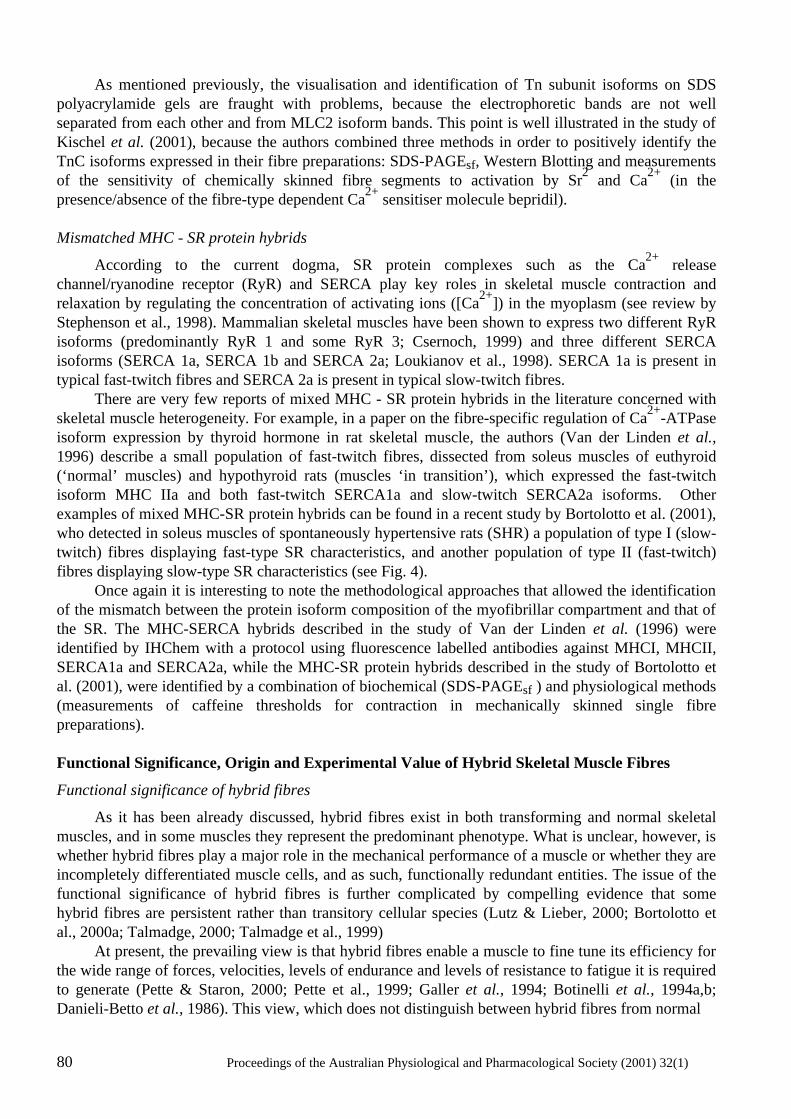
80 Proceedings of the Australian Physiological and Pharmacological Society (2001) 32(1)
As mentioned previously, the visualisation and identification of Tn subunit isoforms on SDS polyacrylamide gels are fraught with problems, because the electrophoretic bands are not well separated from each other and from MLC2 isoform bands. This point is well illustrated in the study of Kischel et al. (2001), because the authors combined three methods in order to positively identify the TnC isoforms expressed in their fibre preparations: SDS-PAGEsf, Western Blotting and measurements of the sensitivity of chemically skinned fibre segments to activation by Sr2 and Ca2+ (in the presence/absence of the fibre-type dependent Ca2+ sensitiser molecule bepridil). Mismatched MHC - SR protein hybrids
According to the current dogma, SR protein complexes such as the Ca2+ release channel/ryanodine receptor (RyR) and SERCA play key roles in skeletal muscle contraction and relaxation by regulating the concentration of activating ions ([Ca2+]) in the myoplasm (see review by Stephenson et al., 1998). Mammalian skeletal muscles have been shown to express two different RyR isoforms (predominantly RyR 1 and some RyR 3; Csernoch, 1999) and three different SERCA isoforms (SERCA 1a, SERCA 1b and SERCA 2a; Loukianov et al., 1998). SERCA 1a is present in typical fast-twitch fibres and SERCA 2a is present in typical slow-twitch fibres. There are very few reports of mixed MHC - SR protein hybrids in the literature concerned with skeletal muscle heterogeneity. For example, in a paper on the fibre-specific regulation of Ca2+-ATPase isoform expression by thyroid hormone in rat skeletal muscle, the authors (Van der Linden et al., 1996) describe a small population of fast-twitch fibres, dissected from soleus muscles of euthyroid (‘normal’ muscles) and hypothyroid rats (muscles ‘in transition’), which expressed the fast-twitch isoform MHC IIa and both fast-twitch SERCA1a and slow-twitch SERCA2a isoforms. Other examples of mixed MHC-SR protein hybrids can be found in a recent study by Bortolotto et al. (2001), who detected in soleus muscles of spontaneously hypertensive rats (SHR) a population of type I (slow-twitch) fibres displaying fast-type SR characteristics, and another population of type II (fast-twitch) fibres displaying slow-type SR characteristics (see Fig. 4). Once again it is interesting to note the methodological approaches that allowed the identification of the mismatch between the protein isoform composition of the myofibrillar compartment and that of the SR. The MHC-SERCA hybrids described in the study of Van der Linden et al. (1996) were identified by IHChem with a protocol using fluorescence labelled antibodies against MHCI, MHCII, SERCA1a and SERCA2a, while the MHC-SR protein hybrids described in the study of Bortolotto et al. (2001), were identified by a combination of biochemical (SDS-PAGEsf ) and physiological methods (measurements of caffeine thresholds for contraction in mechanically skinned single fibre preparations). Functional Significance, Origin and Experimental Value of Hybrid Skeletal Muscle Fibres
Functional significance of hybrid fibres
As it has been already discussed, hybrid fibres exist in both transforming and normal skeletal muscles, and in some muscles they represent the predominant phenotype. What is unclear, however, is whether hybrid fibres play a major role in the mechanical performance of a muscle or whether they are incompletely differentiated muscle cells, and as such, functionally redundant entities. The issue of the functional significance of hybrid fibres is further complicated by compelling evidence that some hybrid fibres are persistent rather than transitory cellular species (Lutz & Lieber, 2000; Bortolotto et al., 2000a; Talmadge, 2000; Talmadge et al., 1999) At present, the prevailing view is that hybrid fibres enable a muscle to fine tune its efficiency for the wide range of forces, velocities, levels of endurance and levels of resistance to fatigue it is required to generate (Pette & Staron, 2000; Pette et al., 1999; Galler et al., 1994; Botinelli et al., 1994a,b; Danieli-Betto et al., 1986). This view, which does not distinguish between hybrid fibres from normal
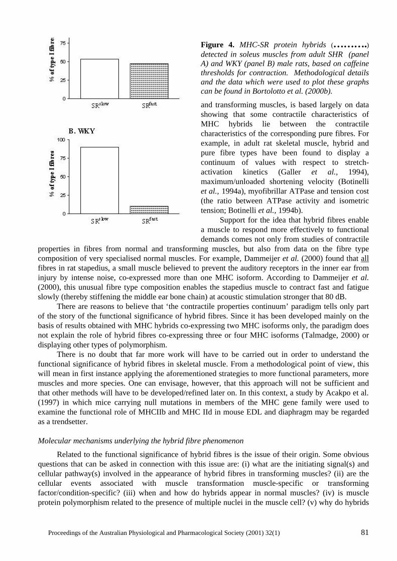
Proceedings of the Australian Physiological and Pharmacological Society (2001) 32(1) 81
Figure 4. MHC-SR protein hybrids (……….) detected in soleus muscles from adult SHR (panel A) and WKY (panel B) male rats, based on caffeine thresholds for contraction. Methodological details and the data which were used to plot these graphs can be found in Bortolotto et al. (2000b).
and transforming muscles, is based largely on data showing that some contractile characteristics of MHC hybrids lie between the contractile characteristics of the corresponding pure fibres. For example, in adult rat skeletal muscle, hybrid and pure fibre types have been found to display a continuum of values with respect to stretch-activation kinetics (Galler et al., 1994), maximum/unloaded shortening velocity (Botinelli et al., 1994a), myofibrillar ATPase and tension cost (the ratio between ATPase activity and isometric tension; Botinelli et al., 1994b). Support for the idea that hybrid fibres enable a muscle to respond more effectively to functional demands comes not only from studies of contractile
properties in fibres from normal and transforming muscles, but also from data on the fibre type composition of very specialised normal muscles. For example, Dammeijer et al. (2000) found that all fibres in rat stapedius, a small muscle believed to prevent the auditory receptors in the inner ear from injury by intense noise, co-expressed more than one MHC isoform. According to Dammeijer et al. (2000), this unusual fibre type composition enables the stapedius muscle to contract fast and fatigue slowly (thereby stiffening the middle ear bone chain) at acoustic stimulation stronger that 80 dB. There are reasons to believe that ‘the contractile properties continuum’ paradigm tells only part of the story of the functional significance of hybrid fibres. Since it has been developed mainly on the basis of results obtained with MHC hybrids co-expressing two MHC isoforms only, the paradigm does not explain the role of hybrid fibres co-expressing three or four MHC isoforms (Talmadge, 2000) or displaying other types of polymorphism. There is no doubt that far more work will have to be carried out in order to understand the functional significance of hybrid fibres in skeletal muscle. From a methodological point of view, this will mean in first instance applying the aforementioned strategies to more functional parameters, more muscles and more species. One can envisage, however, that this approach will not be sufficient and that other methods will have to be developed/refined later on. In this context, a study by Acakpo et al. (1997) in which mice carrying null mutations in members of the MHC gene family were used to examine the functional role of MHCIIb and MHC IId in mouse EDL and diaphragm may be regarded as a trendsetter. Molecular mechanisms underlying the hybrid fibre phenomenon
Related to the functional significance of hybrid fibres is the issue of their origin. Some obvious questions that can be asked in connection with this issue are: (i) what are the initiating signal(s) and cellular pathway(s) involved in the appearance of hybrid fibres in transforming muscles? (ii) are the cellular events associated with muscle transformation muscle-specific or transforming factor/condition-specific? (iii) when and how do hybrids appear in normal muscles? (iv) is muscle protein polymorphism related to the presence of multiple nuclei in the muscle cell? (v) why do hybrids
82 Proceedings of the Australian Physiological and Pharmacological Society (2001) 32(1)
from transforming muscles have a more complex pattern of MHC isoform co-expression than hybrids from normal muscles? (vi) is the muscle protein complement expressed in a hybrid fibre predetermined or does it result from various inductive influences exerted on a naive cell? Once again, it is important to stress that our current understanding of the origin of hybrid fibres (particularly of those present in normal muscles) is very limited. According to currently available data, most proteins involved in the E-C-R cycle are encoded by unique genes and their isoforms are generated by alternative splicing of the primary RNA transcript through a process mediated by muscle-specific factors. The most frequently cited examples of muscle protein isoforms produced by alternative splicing are the two fast-twitch isoforms of myosin light chains, MLC1f and MLC3 (Wade & Kedes, 1989). Some muscle proteins (such as MHCs) are encoded, however, by multiple genes (referred to as a ‘multi-gene family’or ‘isogenes’) and their isoforms results from the differential expression of the gene-family members. It is interesting to note that isogenes can be located on the same chromosome (in tandem or as a cluster), but also on different chromosomes, and that their expression is regulated by muscle-specific cis-acting sequences (such as the E-box elements) and trans-acting factors (Talmadge 2000; Weiss & Leinwand, 1996). Based on this insight into the molecular mechanisms underlying muscle protein polymorphism, it is reasonable to suggest that, in transforming muscles, the hybrid fibre phenotype is the product of a series of coordinated or independent transcriptional events initiated by transforming factors/conditions. In a recent review, Talmadge (2000) raised also the possibility that MHC hybrid fibres observed in skeletal muscle after alterations in electrical activity result from the differential responsiveness of individual myonuclei in a muscle fibre to regulators of MHC isoform gene expression. The idea of nuclei of transforming fibres not working in synchrony, which has been canvassed earlier by Staron & Pette (1987), provides an interesting perspective when inquiring into the origin of hybrid fibres in muscles in transition. It has been suggested that the molecular heterogeneity of hybrid fibres can derive not only from pre-translational , but also from translational and even post-translational events. In the former case, the resulting hybrid fibres may contain one isoform expressed as a protein, and another expressed as a matched or mismatched mRNA species (Andersen et al., 1999; Barton & Buckingham, 1985). In the latter case, the presence of certain isoforms in a fibre may be the result of relatively slow rates of degradation. Thus, Staron & Pette (1993) argued that the MHCIIb isoform detected in hybrid fibres from rat fast-twitch muscles undergoing fast-to-slow transition is not a newly expressed protein, but a transient species with a half life of about 14.7 days. If the rates of degradation of muscle proteins are slow and if they are isoform-specific it is quite clear that after a sudden change in certain conditions there would be an isoform-specific lag between the time when the synthesis of a protein isoform stops and the time when the protein disappears completely from the cell. In this case, however, one would have to wonder about the functional status of the isoform that is in the process of being replaced by a newly synthesized one. Experimental value of hybrid fibres.
In this review it has been argued that identifying and characterising hybrid fibres is not an easy task. Indeed, each of the methods used so far for this purpose has been found to be fraught with technical difficulties and, under certain conditions, to be of limited effectiveness. So, why study hybrid fibres? The reasons for studying hybrid fibres become clearer if one considers a small sample of research questions that have already benefited or are likely to benefit in the future from studies of hybrid skeletal muscle fibres (Table IV). These questions, whose scope may at times overlap, belong loosely to two major fields of inquiry: one concerned with the relationship between the structure of
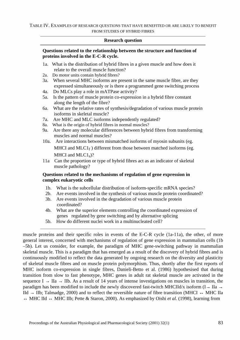
Proceedings of the Australian Physiological and Pharmacological Society (2001) 32(1) 83
TABLE IV. EXAMPLES OF RESEARCH QUESTIONS THAT HAVE BENEFITED OR ARE LIKELY TO BENEFIT FROM STUDIES OF HYBRID FIBRES
Research question
Questions related to the relationship between the structure and function of proteins involved in the E-C-R cycle.
1a. What is the distribution of hybrid fibres in a given muscle and how does it relate to the overall muscle function?
2a. Do motor units contain hybrid fibres? 3a. When several MHC isoforms are present in the same muscle fibre, are they
expressed simultaneously or is there a programmed gene switching process 4a. Do MLCs play a role in mATPase activity? 5a. Is the pattern of muscle protein co-expression in a hybrid fibre constant
along the length of the fibre? 6a. What are the relative rates of synthesis/degradation of various muscle protein
isoforms in skeletal muscle? 7a. Are MHC and MLC isoforms independently regulated? 8a. What is the origin of hybrid fibres in normal muscles? 9a. Are there any molecular differences between hybrid fibres from transforming
muscles and normal muscles? 10a. Are interactions between mismatched isoforms of myosin subunits (eg.
MHCI and MLC1f ) different from those between matched isoforms (eg. MHCI and MLC1s)?
11a Can the proportion or type of hybrid fibres act as an indicator of skeletal muscle pathology?
Questions related to the mechanisms of regulation of gene expression in complex eukaryotic cells
1b. What is the subcellular distribution of isoform-specific mRNA species? 2b. Are events involved in the synthesis of various muscle protein coordinated? 3b. Are events involved in the degradation of various muscle protein
coordinated? 4b. What are the superior elements controlling the coordinated expression of
genes regulated by gene switching and by alternative splicing 5b. How do different nuclei work in a multinucleated cell?
muscle proteins and their specific roles in events of the E-C-R cycle (1a-11a), the other, of more general interest, concerned with mechanisms of regulation of gene expression in mammalian cells (1b –5b). Let us consider, for example, the paradigm of MHC gene-switching pathway in mammalian skeletal muscle. This is a paradigm that has emerged as a result of the discovery of hybrid fibres and is continuously modified to reflect the data generated by ongoing research on the diversity and plasticity of skeletal muscle fibres and on muscle protein polymorphism. Thus, shortly after the first reports of MHC isoform co-expression in single fibres, Danieli-Betto et al. (1986) hypothesised that during transition from slow to fast phenotype, MHC genes in adult rat skeletal muscle are activated in the sequence I → IIa → IIb. As a result of 14 years of intense investigations on muscles in transition, the paradigm has been modified to include the newly discovered fast-twitch MHCIId/x isoform (I→ IIa → IId → IIb; Talmadge, 2000) and to reflect the reversible nature of fibre transition (MHCI ↔ MHC IIa ↔ MHC IId ↔ MHC IIb; Pette & Staron, 2000). As emphasized by Oishi et al. (1998), learning from
84 Proceedings of the Australian Physiological and Pharmacological Society (2001) 32(1)
hybrid fibres the MHC composition in a fibre, at the start of the transforming process, is essential when defining the specific steps in the MHC activation sequence. There is little doubt that, in terms of attention received from muscle researchers, the ‘hybrid’ fibre (regardless of its type) has reached the big time. This is not surprising, for as Pette et al. (1999) stated recently, these fibres ‘offer unique opportunities for relating molecular patterns of protein expression to functional properties and for elucidating mechanisms controlling gene expression in muscle’. Conclusions
The major points made in this review can be summarized as follows: • Hybrid fibres are present in both normal and transforming skeletal muscle, but their proportion and molecular complexity is higher in the latter. The frequency of their occurrence in various muscles and species suggests that hybrid fibres are not a rare phenomenon. • To date, the phrase ‘hybrid fibres’ has been used to describe only fibres co-expressing several MHC isoforms. MHC hybrids represent, however, only one of the many types of hybrid fibres that exist in vertebrate skeletal muscles. Therefore, in order to facilitate progress in the area concerned with muscle fibre diversity and plasticity, a new set of terms is required. This new terminology should be able to include all the hybrid fibre types discovered so far, as well as those that are likely to be discovered in the future. • To detect and characterize hybrid muscle fibres one needs to combine creatively existing microanalytical, microphysiological and microhistochemical methods and to develop new methods for single fibre analysis. • Regardless of their type, hybrid fibres have the potential to become valuable tools for the pursuit of knowledge related to events of the E-C-R cycle and to the regulation of gene expression in multinucleated cells. Acknowledgements
The support provided by NH&MRC, ARC and Vera Ramaciotti Foundation to the Muscle Cell Biochemistry Laboratory at Victoria Institute of Technology, for work on hybrid fibres, is gratefully acknowledged. References
Acakpo-Satchivi, L.J.R., Edelmann, W., Sartorius, C., Lu, B.D., Wahr, P.A., Watkins, S.C., Metzger, J., Leinwand, L & Kucherlapati, R. (1997) Growth and muscle defects in mice lacking adult myosin heavy chain genes. Journal of Cell Biology, 139, 1219-1229.
Aigner, S., Golsch, B., Hämäläinen, N., Staron, R.S., Uber, A., Wehrle, U. & Pette, D. (1993) Fast myosin heavy chain diversity in skeletal muscles of the rabit: heavy chain IId, not IIb predominates. European Journal of Biochemistry, 211, 367-372.
Andersen, J.L., Gruschi-Knudsen, T., Sandri, C., Larsson, L. & Schiaffino, S. (1999) Bed rest increases the amount of mismatched fibres in human skeletal muscles. Journal of Applied Physiology, 86, 455-460.
Armstrong, R.B. & Phelps, R.O. (1984) Muscle fibre type composition of the rat hindlimb. American Journal of Anatomy, 171, 259-272.
Proceedings of the Australian Physiological and Pharmacological Society (2001) 32(1) 85
Barton, P.J. & Buckingham, M.E. (1985) The myosin light chain proteins and their genes. Biochemical Journal, 231, 249-261.
Bortolotto, S.K., Cellini, M., Stephenson, D.G.& Stephenson, G.M.M. (2000a) MHC isoform composition and Ca2+- or Sr2+-activation properties of rat skeletal muscle fibres. American Journal of Physiology, 279, 1564-1577
Bortolotto, S.K., Stephenson, D.G.& Stephenson, G.M.M. (2000b) Caffeine thresholds for contraction in electrophoretically typed, mechanically skinned muscle fibres from SHR and WKY rat. Pflügers Archiv (in print).
Bortolotto, S.K., Stephenson, D.G.& Stephenson, G.M.M. (1999) Fibre type populations and Ca2+- activation properties of single fibres in soleus muscles from SHR and WKY rats. American Journal of Physiology, 276, C628-C637.
Bottinelli, R., Betto, R., Schiaffino, S & Reggiani, C. (1994a) Maximum shortening velocity and coexistence of myosin heavy chain isoforms in single skinned fast fibres of rat skeletal muscle. Journal of Muscle Research and Cell Motility, 15, 413-419.
Bottinelli, R., Canepari, M., Reggiani, C. & Stienen, G.J.M. (1994b) Myofibrillar ATP ase activity during isometric contraction and isomyosin composition in rat single skinned muscle fibres. Journal of Physiology, 481.3, 663-675.
Csernoch, L. (1999) Regulation of the ryanodine receptor calcium release channel of the sarcoplasmic reticulum in skeletal muscle. Acta Physiologica Hungarica, 86, 77-97.
Dammeijer, P.F.M., Van Mameren, H., Van Dijk, P., Moorman, A.F.M., Habets, P., Manni, J.J. & Drukker, J. (2000) Stapedius muscle fibre composition in the rat. Hearing Research, 141, 169-179.
Danieli-Betto, D., Betto, R. & Midrio, M. (1990) Calcium sensitivity and myofibrillar protein isoforms of rat skinned skeletal muscle fibres. Pflügers Archiv, 417, 303-308.
Danieli-Betto, D., Zerbato, E. & Betto, R. (1986) Type I, 2A, and 2B myosin heavy chain electrophoretic analysis of rat muscle fibres. Biochemical and Biophysical Research Communications, 138, 981-987.
Dix, D.J. & Eisenberg, B.R. (1988) In situ hybridization and immunohistochemistry in serial sections of rabbit skeletal muscle to detect myosin expression. Journal of Histochemistry and Cytochemistry, 36, 1519-1526.
Fink, R.H., Stephenson, D.G. and Williams, D.A. (1986) Calcium and strontium activation of single skinned muscle fibres of normal and dysstrophic mice. Journal of Physiology, 373, 513-525.
Galler, S., Schmitt, T.L. & Pette, D. (1994) Stretch activation, unloaded shortening velocity, and myosin heavy chain isoforms of rat skeletal muscle fibres. Journal of Physiology, 478, 513-521.
Geiger, C.G., Cody, M.J. & Sieck, G.C. (1999) Force-calcium relationship depends on myosin and troponin isoforms in rat diaphragm muscle fibres. Journal of Applied Physiology, 87, 1894-1900.
Grossman, E.J., Roy, R.R., Talmadge, R.J., Zhong, Hui. & Edgerton, V.R. (1998) Effects of inactivity on myosin heavy chain composition and size of rat soleus fibres. Muscle & Nerve, 21, 375-389.
Hämäläinen, N. & Pette, D. (1995) Patterns of myosin isoforms in mammalian skeletal muscle fibres. Microscopy Research and Technique, 30, 381-389.
Kischel, P., Bastide, B., Stevens, L. & Mounier, Y. (2001) Expression and functional behaviour of troponin C in soleus muscle fibres of rat after hindlimb unloading. American Journal of Applied Physiology, (In press).
86 Proceedings of the Australian Physiological and Pharmacological Society (2001) 32(1)
La Framboise, W.A., Daood, M.J., Guthrie, R.D., Schiaffino, S., Moretti, P., Brozanski, B., Ontell, M.P., Butler-Browne, G.S., Whalen, R.G & Ontell, M. (1991) Emergence of the mature myosin phenotype in the rat diaphragm muscle. Developmental Biology, 144, 1-15.
Loukianov, E., Ji, Y., Baker, D.L., Reed, T., Babu, J., Loukianova, T., Greene, A., Shull, G & Periasamy, M. (1998) Sarco(endo)plasmic reticulum Ca2+ ATPase isoforms and their role in muscle physiology and pathology. Annals of the New York Academy of Sciences, 853, 251-259.
Lutz, G.J. & Lieber, R.L. (2000) Myosin isoforms in anuran skeletal muscle: their influence on contractile properties and in vivo muscle function. Microscopy Research and Technique, 50, 443-457.
Mizusawa, H., Takagi, A., Sugita, H. & Toyokura, Y. (1982) Coexistence of fast and slow types of myosin light chains in a single fiber of rat soleus muscle. Journal of Biochemistry (Tokyo), 91, 423-425.
Moss, R.L., Diffee, G.M. & Greaser, M.L. (1995) Contractile properties of skeletal muscle fibres in relation to myofibrillar protein isoforms. Reviews in Physiology, Biochemistry and Pharmacology, 126, 2-63.
Nguyen, L.T. & Stephenson, G.M.M. (1999) An electrophoretic study of myosin heavy chain expression in skeletal muscles of the toad Bufo marinus. Journal of Muscle Research and Cell Motility, 20, 687-95.
Oishi, Y., Ishihara, A., Yamamoto, H. & Miyamoto, E. (1998) Hindlimb suspension induces the expression of multiple myosin heavy chain isoforms in single fibres of the rat soleus muscle. Acta Physiologica Scandinavica, 162, 127-134.
Pette, D., Peuker, H. & Staron, R.S. (1999) The impact of biochemical methods for single muscle fibre analysis. Acta Physiologica Scandinavica, 166, 261-277.
Pette, D. & Staron, R.S. (2000) Myosin isoforms, muscle fibre types and transitions. Microscopy Research and Technique, 50, 500-509.
Pette, D. & Staron, R.S. (1997) Mammalian skeletal muscle fibre type transitions. International Review of Cytology, 170, 143-223.
Pette, D. & Staron, R.S. (1990) Cellular and molecular diversities of mammalian skeletal muscle fibres. Reviews of Physiology, Biochemistry and Pharmacology, 116, 2-76.
Peuker, H., Conjard, A., Putman, C.T. & Pette, D. (1999) Journal of Muscle Research and Cell Motility, 20, 147-154.
Pierobon-Bormioli, S., Sartore, S., Libera, L.D., Vitadello, M. & Schiaffino, S. (1981)"Fast" isomyosins and fiber types in mammalian skeletal muscle. Journal of Histochemistry and Cytochemistry, 29, 1179-1188.
Rivero, J.L., Talmadge, R.J. & Edgerton, V.R. (1998) Fibre size and metabolic properties of myosin heavy chain-based fibre types in rat skeletal muscle. Journal of Muscle Research and Cell Motility,19, 733-742.
Roy, R.R., Kim, J.A., Grossman, E.J., Bekmezian, A., Talmadge, R.J., Zhong, H. & Edgerton, V.R. (2000) Persistence of myosin heavy chain-based fiber types in innervated but silenced rat fast muscle. Muscle & Nerve, 23, 735-747.
Salviati, G., Betto, R.& Danieli-Betto, D. (1982) Polymorphism of myofibrillar proteins of rabbit skeletal muscle fibres. Biochemical Journal, 207, 261-272.
Schiaffino, S. & Salviatti, G. (1998) Molecular diversity of myofibrillar proteins: isoforms analysis at the protein and mRNA level. Methods in Cell Biology, 52, 349-369
Schiaffino, S. & Reggiani, C. (1996) Molecular diversity of myofibrillar proteins: gene regulation and functional significance. Physiological Reviews, 76, 371-423.
Staron, R.C. & Pette, D. (1987) Nonuniform myosin expression along single fibres of chronically stimulated and contralateral rabbit tibialis anterior muscles. Pflügers Archiv, 409, 67-73.
Proceedings of the Australian Physiological and Pharmacological Society (2001) 32(1) 87
Staron, R.C. & Pette, D. (1993) The continuum of pure and hybrid myosin heavy chain based fibre types in rat skeletal muscles. Histochemistry, 100, 149-153.
Stephenson, D.G., Lamb, G.D. & Stephenson, G.M.M. (1998) Events of the excitation-contraction-relaxation cycle in fast-and slow-twitch mammalian muscle fibres relevant to muscle fatigue. Acta Physiologica Scandinavica, 162, 229-245.
Swynghedauw, B. (1986) Developmental and functional adaptation of contractile proteins in cardiac and skeletal muscles. Physiological Reviews, 66, 710-771.
Talmadge, R.J. (2000) Myosin heavy chain expression following reduced neuromuscular activity: potential regulatory mechanisms. Muscle & Nerve, 23, 661-679.
Talmadge, R.J., Roy R.R. & Edgerton, V.R. (1999) Persistence of hybrid fibres in rat soleus after spinal cord transection. Anatomical Records, 255, 188-201.
Termin, A., Staron, R.S. & Pette, D. (1989) Changes in myosin heavy chain isoforms during chronic low-frequency stimulation of rat fast hindlimb muscles. European Journal of Biochemistry, 186, 749-754.
Van der Linden, G.C., Simonides, W.S., Muller, A., Van der Laarse, W.J., Vermeulen, j.L., Zuidvijk, M.J., moorman, A.F. & van Hardeveld, C. (1996) Fibre-specific regulation of Ca2+ - ATPase isoform expression by thyroid hormone in rat skeletal muscle. American Journal of Physiology, 271, C1908-C1919.
Wade, R. & Kedes, L. (1989) Developmental regulation of contractile protein genes. Annual Review of Physiology, 51, 179-188.
Weiss, A. & Leinwand, L.A. (1996) The mammalian myosin heavy chain gene family. Annual. Review of Cellular and Developmental Biology, 12, 417-39.
Wu, Y.Z., Baker, M.J., Crumley, R.L., Blanks, R.H. & Caiozzo, V.J. (1998) A new concept in laryngeal muscle: multiple myosin isoform typesin single muscle fibers of the lateral cricoarytenoid. Otolaryngology of the Head Neck Surgery, 118, 86-94.
Wu, Z.Y., Baker, M.J., Crumley, R.L. & Caiozzo, V.J. (2000a) Single-fiber myosin heavy chain isoform composition of rodent laryngeal muscle. Modulation by thyroid hormone. Archives of Otolaryngological Head and neck Surgery, 126, 874-880.
Wu, Y.Z., Crumley, R.L. & Caiozzo, V.J. (2000b) Are hybrid fibers a common motif of canine laryngeal muscles? Archives of Otolaryngological Head and Neck Surgery, 126, 865-873.
Yu, F., Degens, H., Li, X & Larsson, L. (1998) Gender- and age-related differences in the regulatory influence of thyroid hormone on the contractility and myosin composition of single rat soleus muscle fibres. Pflügers Archiv, 437, 21-30.
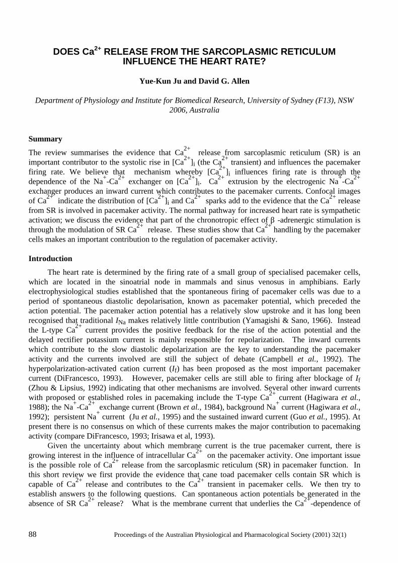
88 Proceedings of the Australian Physiological and Pharmacological Society (2001) 32(1)
DOES Ca2+ RELEASE FROM THE SARCOPLASMIC RETICULUM
INFLUENCE THE HEART RATE?
Yue-Kun Ju and David G. Allen
Department of Physiology and Institute for Biomedical Research, University of Sydney (F13), NSW 2006, Australia
Summary
The review summarises the evidence that Ca2+ release from sarcoplasmic reticulum (SR) is an important contributor to the systolic rise in [Ca2+]i (the Ca2+ transient) and influences the pacemaker firing rate. We believe that mechanism whereby [Ca2+]i influences firing rate is through the dependence of the Na+-Ca2+ exchanger on [Ca2+]i. Ca2+ extrusion by the electrogenic Na+-Ca2+ exchanger produces an inward current which contributes to the pacemaker currents. Confocal images of Ca2+ indicate the distribution of [Ca2+]i and Ca2+ sparks add to the evidence that the Ca2+ release from SR is involved in pacemaker activity. The normal pathway for increased heart rate is sympathetic activation; we discuss the evidence that part of the chronotropic effect of β -adrenergic stimulation is through the modulation of SR Ca2+ release. These studies show that Ca2+ handling by the pacemaker cells makes an important contribution to the regulation of pacemaker activity. Introduction
The heart rate is determined by the firing rate of a small group of specialised pacemaker cells, which are located in the sinoatrial node in mammals and sinus venosus in amphibians. Early electrophysiological studies established that the spontaneous firing of pacemaker cells was due to a period of spontaneous diastolic depolarisation, known as pacemaker potential, which preceded the action potential. The pacemaker action potential has a relatively slow upstroke and it has long been recognised that traditional INa makes relatively little contribution (Yamagishi & Sano, 1966). Instead the L-type Ca2+ current provides the positive feedback for the rise of the action potential and the delayed rectifier potassium current is mainly responsible for repolarization. The inward currents which contribute to the slow diastolic depolarization are the key to understanding the pacemaker activity and the currents involved are still the subject of debate (Campbell et al., 1992). The hyperpolarization-activated cation current (If) has been proposed as the most important pacemaker current (DiFrancesco, 1993). However, pacemaker cells are still able to firing after blockage of If (Zhou & Lipsius, 1992) indicating that other mechanisms are involved. Several other inward currents with proposed or established roles in pacemaking include the T-type Ca2+ current (Hagiwara et al., 1988); the Na+-Ca2+ exchange current (Brown et al., 1984), background Na+ current (Hagiwara et al., 1992); persistent Na+ current (Ju et al., 1995) and the sustained inward current (Guo et al., 1995). At present there is no consensus on which of these currents makes the major contribution to pacemaking activity (compare DiFrancesco, 1993; Irisawa et al, 1993). Given the uncertainty about which membrane current is the true pacemaker current, there is growing interest in the influence of intracellular Ca2+ on the pacemaker activity. One important issue is the possible role of Ca2+ release from the sarcoplasmic reticulum (SR) in pacemaker function. In this short review we first provide the evidence that cane toad pacemaker cells contain SR which is capable of Ca2+ release and contributes to the Ca2+ transient in pacemaker cells. We then try to establish answers to the following questions. Can spontaneous action potentials be generated in the absence of SR Ca2+ release? What is the membrane current that underlies the Ca2+-dependence of
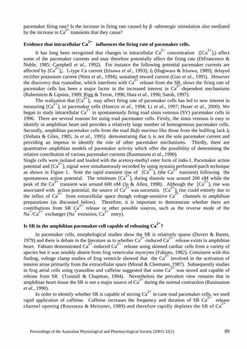
Proceedings of the Australian Physiological and Pharmacological Society (2001) 32(1) 89
pacemaker firing rate? Is the increase in firing rate caused by β -adrenergic stimulation also mediated by the increase in Ca2+ transients that they cause? Evidence that intracellular Ca2+ influences the firing rate of pacemaker cells.
It has long been recognised that changes in intracellular Ca2+ concentration ([Ca2+]i) affect some of the pacemaker currents and may therefore potentially affect the firing rate (DiFrancesco & Noble, 1985; Campbell et al., 1992). For instance the following potential pacemaker currents are affected by [Ca2+]i; L-type Ca current (Irisawa et al., 1993); If (Hagiwara & Irisawa, 1989); delayed rectifier potassium current (Nitta et al., 1994); sustained inward current (Guo et al., 1995). However the discovery that ryanodine, which interferes with Ca2+ release from the SR, slows the firing rate of pacemaker cells has been a major factor in the increased interest in Ca2+-dependent mechanisms (Rubenstein & Lipsius, 1989; Rigg & Terrar, 1996; Hata et al., 1996; Satoh, 1997). The realisation that [Ca2+]i may affect firing rate of pacemaker cells has led to new interest in measuring [Ca2+]i in pacemaker cells (Hancox et al., 1994; Li et al., 1997; Huser et al., 2000). We began to study intracellular Ca2+ in spontaneously firing toad sinus venosus (SV) pacemaker cells in 1996. There are several reasons for using toad pacemaker cells. Firstly, the sinus venosus is easy to identify in amphibian heart and provides a relatively large number of homogeneous pacemaker cells. Secondly, amphibian pacemaker cells from the toad Bufo marinus like those from the bullfrog lack If (Shibata & Giles, 1985; Ju et al., 1995) demonstrating that If is not the sole pacemaker current and providing an impetus to identify the role of other pacemaker mechanisms. Thirdly, there are quantitative amphibian models of pacemaker activity which offer the possibility of determining the relative contribution of various pacemaker currents (Rasmusson et al., 1990). Single cells were isolated and loaded with the acetoxy-methyl ester form of indo-1. Pacemaker action potential and [Ca2+]i signal were simultaneously recorded by using nystatin perforated-patch technique as shown in Figure 1. Note the rapid transient rise of [Ca2+]i (the Ca2+ transient) following the spontaneous action potential. The minimum [Ca2+]i during diastole was around 200 nM while the peak of the Ca2+ transient was around 600 nM (Ju & Allen, 1998). Although the [Ca2+]i rise was associated with action potential, the source of Ca2+ was uncertain. [Ca2+]i rise could entirely due to the influx of Ca2+ from extracellular space though voltage-sensitive Ca2+ channels in amphibian preparations (as discussed below). Therefore, it is important to demonstrate whether there are contributions from SR Ca2+ release or other possible sources, such as the reverse mode of the Na+/Ca2+ exchanger (Na+ extrusion, Ca2+ entry). Is SR in the amphibian pacemaker cell capable of releasing Ca2+?
In pacemaker cells, morphological studies show the SR is relatively sparse (Duvert & Barets, 1979) and there is debate in the literature as to whether Ca2+-induced Ca2+ release exists in amphibian heart. Fabiato demonstrated Ca2+-induced Ca2+ release using skinned cardiac cells from a variety of species but it was notably absent from frog ventricular myocytes (Fabiato, 1982). Consistent with this finding, voltage clamp studies of frog ventricle showed that the Ca2+ involved in the activation of tension arose primarily from the extracellular space (Morad & Cleemann, 1987). Subsequently studies in frog atrial cells using ryanodine and caffeine suggested that some Ca2+ was stored and capable of release from SR (Tunstall & Chapman, 1994). Nevertheless the prevalent view remains that in amphibian heart tissue the SR is not a major source of Ca2+ during the normal contraction (Rasmusson et al., 1990). In order to identify whether SR is capable of storing Ca2+ in cane toad pacemaker cells, we used rapid application of caffeine. Caffeine increases the frequency and duration of SR Ca2+ release channel opening (Rousseau & Meissner, 1989) and therefore rapidly depletes the SR of Ca2+
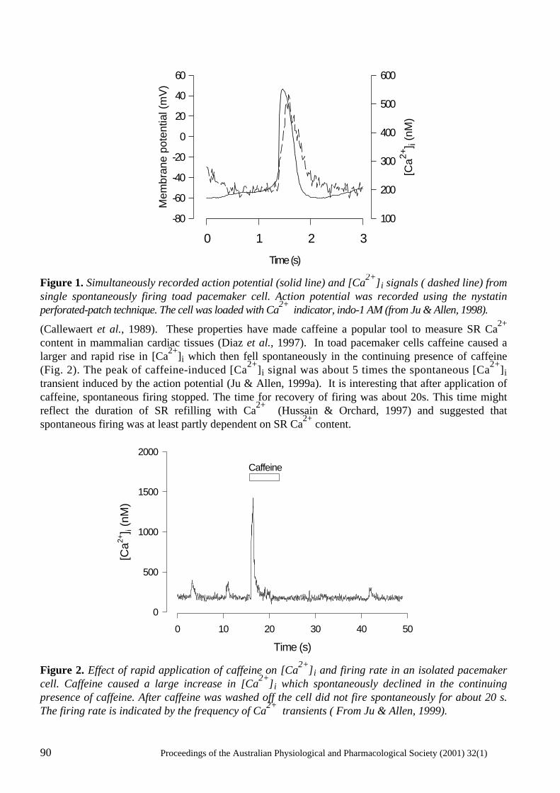
90 Proceedings of the Australian Physiological and Pharmacological Society (2001) 32(1)
Figure 1. Simultaneously recorded action potential (solid line) and [Ca2+]i signals ( dashed line) from single spontaneously firing toad pacemaker cell. Action potential was recorded using the nystatin perforated-patch technique. The cell was loaded with Ca2+ indicator, indo-1 AM (from Ju & Allen, 1998).
(Callewaert et al., 1989). These properties have made caffeine a popular tool to measure SR Ca2+ content in mammalian cardiac tissues (Diaz et al., 1997). In toad pacemaker cells caffeine caused a larger and rapid rise in [Ca2+]i which then fell spontaneously in the continuing presence of caffeine (Fig. 2). The peak of caffeine-induced [Ca2+]i signal was about 5 times the spontaneous [Ca2+]i transient induced by the action potential (Ju & Allen, 1999a). It is interesting that after application of caffeine, spontaneous firing stopped. The time for recovery of firing was about 20s. This time might reflect the duration of SR refilling with Ca2+ (Hussain & Orchard, 1997) and suggested that spontaneous firing was at least partly dependent on SR Ca2+ content. Figure 2. Effect of rapid application of caffeine on [Ca2+]i and firing rate in an isolated pacemaker cell. Caffeine caused a large increase in [Ca2+]i which spontaneously declined in the continuing presence of caffeine. After caffeine was washed off the cell did not fire spontaneously for about 20 s. The firing rate is indicated by the frequency of Ca2+ transients ( From Ju & Allen, 1999).
Time (s)
0 10 20 30 40 50
[Ca2+
] i (n
M)
0
500
1000
1500
2000
Caffeine
Time (s)
0 1 2 3
Mem
bran
e po
tent
ial (
mV
)
-80
-60
-40
-20
0
20
40
60
[Ca2+
] i (n
M)
100
200
300
400
500
600
Proceedings of the Australian Physiological and Pharmacological Society (2001) 32(1) 91
Is SR Ca2+ release involved in pacemaker activity?
Although the experiments with caffeine above demonstrate that SR of toad pacemaker cells are capable of releasing Ca2+, they do not identify whether release of Ca2+ from the SR occurs during the normal action potential. To test this possibility, we used ryanodine which is an SR Ca2+ release channel blocker (Fleischer & Inui, 1989). We found that 5 min after application of 10 µM ryanodine, the peak of the Ca2+ transient decreased to 50% of control level (Fig. 3). Cells were still able to firing at this stage though at a reduced frequency. After 30 min exposure to ryanodine, spontaneous firing ceased. This effect of ryanodine on pacemaker activity is consistent with the idea that the Ca2+ transients consist of a component of Ca2+ release from SR. Decreasing SR Ca2+ release slows the heart rate. The caffeine experiments show that when the SR is emptied of Ca2+ firing temporarily ceases while the ryanodine experiments show that preventing SR Ca2+ release also slows pacemaker firing. Thus normal SR Ca2+ release seems to be needed for regular firing of the pacemaker cells. Furthermore, the argument for involvement of the SR is strengthened by recent observations of single Ca2+ release events (Ca2+ sparks) during pacemaker action potential (Huser et al., 2000; Ju & Allen, 2000a) (as described below) .
[Ca2+
] i (nM
)
0
1 0 0
2 0 0
3 0 0
4 0 0
5 0 0
Mem
bran
e po
tent
ail (
mV
)
- 60
-40
-20
0
2 0
4 0
6 0
5 s
AC o n t ro l R y a n o d ine
5 m in 3 0 m in
B
Figure 3. The effects of 10 µM ryanodine on [Ca2+]i and the spontaneous action potential. A, [Ca2+]i transient recorded under control and after 5, and 30 min exposure to ryanodine (10 µM). B, the effects of ryanodine on spontaneous action potential were recorded from a non-indo loaded cell to avoid the possible effect of indo-1 AM loading on pacemaker activity (from Ju & Allen, 1998). The role of Na+ -Ca2+ exchanger in pacemaker activity
We have established that SR Ca2+ release occurs in pacemaker cells and that when it is prevented firing rate slows. However, the nature of the link between Ca2+ release from SR and diastolic depolarisation needs to be established. How does Ca2+ release from SR generate an inward current during the pacemaker potential? It is known that Na+-Ca2+ exchanger exist in most cardiac cells. It is also known that the Na+-Ca2+ exchanger generates a electrogenic current, since the coupling
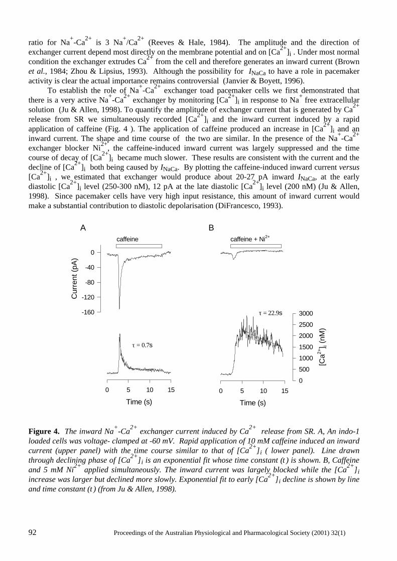
92 Proceedings of the Australian Physiological and Pharmacological Society (2001) 32(1)
ratio for Na+-Ca2+ is 3 Na+/Ca2+ (Reeves & Hale, 1984). The amplitude and the direction of exchanger current depend most directly on the membrane potential and on [Ca2+]i . Under most normal condition the exchanger extrudes Ca2+ from the cell and therefore generates an inward current (Brown et al., 1984; Zhou & Lipsius, 1993). Although the possibility for INaCa to have a role in pacemaker activity is clear the actual importance remains controversial (Janvier & Boyett, 1996). To establish the role of Na+-Ca2+ exchanger toad pacemaker cells we first demonstrated that there is a very active Na+-Ca2+ exchanger by monitoring [Ca2+]i in response to Na+ free extracellular solution (Ju & Allen, 1998). To quantify the amplitude of exchanger current that is generated by Ca2+ release from SR we simultaneously recorded [Ca2+]i and the inward current induced by a rapid application of caffeine (Fig. 4 ). The application of caffeine produced an increase in [Ca2+]i and an inward current. The shape and time course of the two are similar. In the presence of the Na+-Ca2+ exchanger blocker Ni2+, the caffeine-induced inward current was largely suppressed and the time course of decay of [Ca2+]i became much slower. These results are consistent with the current and the decline of [Ca2+]i both being caused by INaCa. By plotting the caffeine-induced inward current versus [Ca2+]i , we estimated that exchanger would produce about 20-27 pA inward INaCa, at the early diastolic [Ca2+]i level (250-300 nM), 12 pA at the late diastolic [Ca2+]i level (200 nM) (Ju & Allen, 1998). Since pacemaker cells have very high input resistance, this amount of inward current would make a substantial contribution to diastolic depolarisation (DiFrancesco, 1993).
Figure 4. The inward Na+-Ca2+ exchanger current induced by Ca2+ release from SR. A, An indo-1 loaded cells was voltage- clamped at -60 mV. Rapid application of 10 mM caffeine induced an inward current (upper panel) with the time course similar to that of [Ca2+]i ( lower panel). Line drawn through declining phase of [Ca2+]i is an exponential fit whose time constant (τ) is shown. B, Caffeine and 5 mM Ni2+ applied simultaneously. The inward current was largely blocked while the [Ca2+]i increase was larger but declined more slowly. Exponential fit to early [Ca2+]i decline is shown by line and time constant (τ) (from Ju & Allen, 1998).
Cur
rent
(pA
)
-160
-120
-80
-40
0
Time (s)
0 5 10 15
Time (s)
0 5 10 15
[Ca2+
] i (n
M)
0
500
1000
1500
2000
2500
3000
A Bcaffeine caffeine + Ni2+
τ = 0.7s
τ = 22.9s
Proceedings of the Australian Physiological and Pharmacological Society (2001) 32(1) 93
Distribution of [Ca2+]i during pacemaking
Given that SR Ca2+ release contributes pacemaker function at least in part through stimulating the Na+-Ca2+ exchanger, it becomes of interest to know the distribution of [Ca2+]i during the action potential. This is because the Na+-Ca2+ exchanger is situated in the surface membrane and is sensitive only to the near membrane [Ca2+]i. This issue was examined using confocal microscopy during spontaneous firing of isolated pacemaker cells. For these studies, pacemaker cells were loaded with fluo-3. Surprisingly, given that the pacemaker cells have no T-tubules we found that the distribution of Ca2+ release during an action potential was uniform (Ju & Allen, 2000a). This is surprising because one would expect the Ca2+ distribution resulting from L-type Ca channels to be localised around the edges of the cell. In fact Ca2+ reached a similar peak in the centre of the cell as at the edge and there was no detectable delay in the rise of Ca in the middle of the cell compared to the edge. One explanation for these findings is that SR is uniformly distributed across the cell and the triggering mechanism is so fast that no detectable decay occurs between edge and centre of these small 4 µm diameter cells. Confocal studies of [Ca2+]i are also capable of localised, spontaneous Ca2+ release from SR release channels (Ca2+ sparks) which provide further information about Ca2+ release from the SR. Ca2+ sparks were detected in cane toad cells and become smaller in magnitude and longer in duration in the presence of 250 nM of ryanodine (Ju & Allen, 2000a). This finding is consistent with the ability of low concentration of ryanodine to cause the SR Ca2+ channels to enter an intermediate conductance state with long openings (Rousseau et al., 1987). A novel finding was that the frequency of Ca2+ sparks increased just before an action potential. A recent study in mammalian pacemaker cells has confirmed this finding and suggested that the mechanism involved is that T-type Ca2+ current triggers Ca2+ sparks from SR close to the membrane (Huser et al., 2000). We do not believe this is the only mechanism involved because in our experiments the increased frequency of sparks was also observed in the middle of the cell. What is a trigger for SR Ca2+ release in pacemaker cells?
In order to study the mechanism underlying SR Ca2+ release in pacemaker cells, we simultaneously voltage-clamped the cells and measured [Ca2+]i. In the present of SR Ca2+ pump inhibitor 2,5-di(tert-butyl)-1,4-hydroquinone (TBQ), which would be expected to deplete the SR of Ca2+, Ca2+ transients were reduced to 34% while there was no significant effect on the peak inward current. This result suggests that about 66% of Ca2+ contributing to the Ca2+ transient is released from SR, which is consistent with previous observation in spontaneous firing cells with ryanodine. In response to a series of membrane depolarisations we found that the amplitude of the Ca2+ transient is not simply related to the size of inward current (Ju & Allen, 2000b). Ca2+ transients increased continuously as membrane potential was increased whereas the current-voltage relationship of the inward current was bell-shaped. By using various channel blockers we found that not only L-type Ca2+ current but also reversal mode Na+ -Ca2+ exchanger current could trigger Ca2+ SR release in pacemaker cells (Ju & Allen, 2000b). The results pose the question whether reversal model Na+-Ca2+ exchanger induces Ca2+ induced Ca2+ release during the spontaneous pacemaker action potentials. However, lack a specific Na+-Ca2+ exchanger blocker prevents us addressing this issue directly at precent. Is the increase heart rate by adrenaline related to the change of SR Ca2+ release?
It is generally thought that the increase in the heart rate after β-adrenergic stimulation is caused by modulation of ionic current, such as L-type Ca2+ current (Noma et al., 1980) and If (DiFrancesco, 1981). It is also known that β-adrenergic stimulation increase the amplitude of Ca2+ transients in cardiac myocytes (Allen & Blinks, 1978; Hussain & Orchard, 1997; Hancox et al., 1994). We have
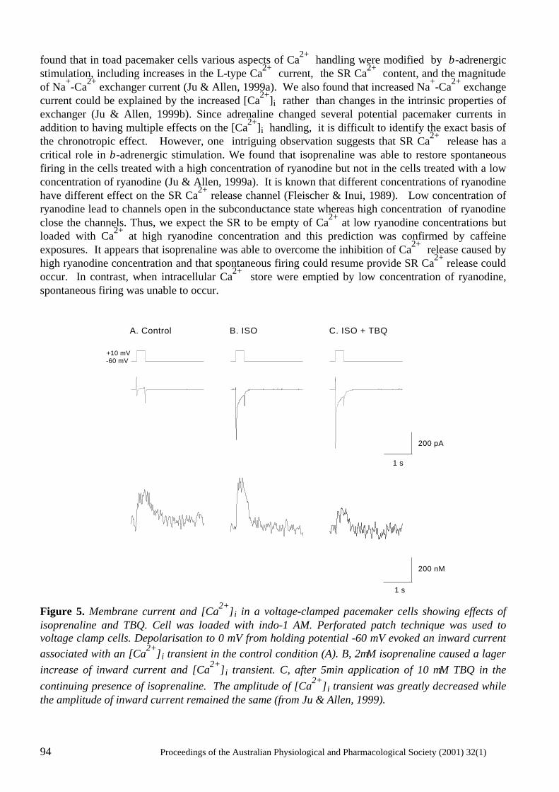
94 Proceedings of the Australian Physiological and Pharmacological Society (2001) 32(1)
found that in toad pacemaker cells various aspects of Ca2+ handling were modified by β-adrenergic stimulation, including increases in the L-type Ca2+ current, the SR Ca2+ content, and the magnitude of Na+-Ca2+ exchanger current (Ju & Allen, 1999a). We also found that increased Na+-Ca2+ exchange current could be explained by the increased [Ca2+]i rather than changes in the intrinsic properties of exchanger (Ju & Allen, 1999b). Since adrenaline changed several potential pacemaker currents in addition to having multiple effects on the [Ca2+]i handling, it is difficult to identify the exact basis of the chronotropic effect. However, one intriguing observation suggests that SR Ca2+ release has a critical role in β-adrenergic stimulation. We found that isoprenaline was able to restore spontaneous firing in the cells treated with a high concentration of ryanodine but not in the cells treated with a low concentration of ryanodine (Ju & Allen, 1999a). It is known that different concentrations of ryanodine have different effect on the SR Ca2+ release channel (Fleischer & Inui, 1989). Low concentration of ryanodine lead to channels open in the subconductance state whereas high concentration of ryanodine close the channels. Thus, we expect the SR to be empty of Ca2+ at low ryanodine concentrations but loaded with Ca2+ at high ryanodine concentration and this prediction was confirmed by caffeine exposures. It appears that isoprenaline was able to overcome the inhibition of Ca2+ release caused by high ryanodine concentration and that spontaneous firing could resume provide SR Ca2+ release could occur. In contrast, when intracellular Ca2+ store were emptied by low concentration of ryanodine, spontaneous firing was unable to occur.
200 pA
200 nM
1 s
1 s
A. Control B. ISO C. ISO + TBQ
-60 mV+10 mV
Figure 5. Membrane current and [Ca2+]i in a voltage-clamped pacemaker cells showing effects of isoprenaline and TBQ. Cell was loaded with indo-1 AM. Perforated patch technique was used to voltage clamp cells. Depolarisation to 0 mV from holding potential -60 mV evoked an inward current associated with an [Ca2+]i transient in the control condition (A). B, 2µM isoprenaline caused a lager increase of inward current and [Ca2+]i transient. C, after 5min application of 10 µM TBQ in the continuing presence of isoprenaline. The amplitude of [Ca2+]i transient was greatly decreased while the amplitude of inward current remained the same (from Ju & Allen, 1999).
Proceedings of the Australian Physiological and Pharmacological Society (2001) 32(1) 95
The above experiments suggest that SR Ca2+ release plays a specific role in response to β-stimulation. In order to separate the effects of β-stimulation on Ca2+ influx from that due to SR Ca2+ release we simultaneously recorded Ca2+ current and [Ca2+]i . We found that in the present of isoprenaline both Ca2+ current and [Ca2+]i transients were increased (Fig. 5). TBQ was used to reveal the SR contribution. We found that application of TBQ had no significant effect on Ca2+ current enhanced by isoprenaline. However, [Ca2+]i transient was greatly decreased. The similar result was obtained by using low concentrations of ryanodine. Such experiments suggest that SR Ca2+ release contributes about 50% of the Ca2+ transient both in the absence and presence of β- adrenergic stimulation (Ju & Allen, 1999a). Therefore the increase of [Ca2+] transient by β-stimulation is partly caused by increased SR Ca2+ release. In order to maintain the homeostasis of [Ca2+]i , the Na+-Ca2+ exchanger would produce more inward current by extruding more Ca2+. Thus increased inward current during the diastolic potential would accelerated the diastolic depolarisation, therefore increasing the heart rate. Conclusion
The evidence is clear that [Ca2+]i and SR Ca2+ release are in some way related to the firing rate of cane toad pacemaker cells. It is very likely that INaCa is at least part of the intermediary process which links the Ca2+ to the pacemaker current. However many other details are less clear; does [Ca2+]i affect other pacemaker currents which have significant effects? Are Ca2+ sparks important in the pacemaker process and is the mechanism proposed by Huser et al. (2000) correct and applicable in other cell types? Does SR Ca2+ release have some special role over and above its contribution to the Ca2+ transients? The ryanodine experiments suggest that it may and studies by Cousins & Bramich (1998) also suggest there may be a class of Ca2+ store which is modulated only by neuronally-released adrenaline. Cellular studies of pacemaker cells have been impeded by the small numbers of these cells and the difficulties in isolating them. There is increasing evidence that pacemaker function declines in the elderly and those with ischaemic heart disease (Benditt et al., 1995) and understanding and treatment of these problems is dependent on increasing understanding of pacemaker function at a cellular and molecular level. Acknowledgements.
We are grateful for support from the National Health and Medical Research Council of Australia. References
Allen, D.G. & Blinks, J.R. (1978) Calcium transients in aequorin-injected frog cardiac muscle. Nature, 273, 509-513.
Benditt, D.G., Sakaguchi, S., Golstein, M.A., Lurie, K.G., Gornick, C.C. & Adler, S.W. (1995) Sinus Node Dysfunction:Pathophysiology, clinical feature, evaluation, and treatment. In: Cardiac electrophysiology: from cell to bedside.
Brown, H.F., Kimura, J., Noble, D., Noble, S.J. & Taupignon, A. (1984) The slow inward current, isi, in the rabbit sino-atrial node investigated by voltage clamp and computer simulation. Proceedings of the Royal Society of London.Series B: Biological Sciences, 222, 305-328.
Callewaert, G., Cleemann, L. & Morad, M. (1989) Caffeine-induced Ca2+ release activates Ca2+ extrusion via Na+-Ca2+ exchanger in cardiac myocytes. American Journal of Physiology, 257, C147-52.
96 Proceedings of the Australian Physiological and Pharmacological Society (2001) 32(1)
Campbell, D.L., Rasmusson, R.L. & Strauss, H.C. (1992) Ionic current mechanisms generating vertebrate primary cardiac pacemaker activity at the single cell level: an integrative view. Annual Review of Physiology, 54, 279-302.
Cousins, H.M. & Bramich, N.J. (1998) Effects of sympathetic nerve stimulation on membrane potential, [Ca2+]i and force in the arrested sinus venosus of the toad, Bufo marinus. Journal of Physiology, 505, 513-527.
Diaz, M.E., Trafford, A.W., O'Neill, S.C. & Eisner, D.A. (1997) Measurement of sarcoplasmic reticulum Ca2+ content and sarcolemmal Ca2+ fluxes in isolated rat ventricular myocytes during spontaneous Ca2+release. Journal of Physiology, 501 ( Pt 1), 3-16.
DiFrancesco, D. (1981) A study of the ionic nature of the pace-maker current in calf Purkinje fibres. Journal of Physiology, 314, 377-393.
DiFrancesco, D. (1993) Pacemaker mechanisms in cardiac tissue. Annual Review of Physiology, 55, 455-472.
DiFrancesco, D. & Noble, D. (1985) A model of cardiac electrical activity incorporating ionic pumps and concentration changes. Philosophical Transactions of the Royal Society of London - Series B: Biological Sciences, 307, 353-398.
Duvert, M. & Barets, A.L. (1979) Fine structure and organization of the sarcoplasmic reticulum in the sino-atrial fibres of the frog heart. Zeitschrift fur Naturfurschung, 34, 865-875.
Fabiato, A. (1982) Calcium release in skinned cardiac cells: variations with species, tissues, and development. Federation Proceedings, 41, 2238-2244.
Fleischer, S. & Inui, M. (1989) Biochemistry and biophysics of excitation-contraction coupling. Annual Review of Biophysics & Biophysical Chemistry, 18, 333-364.
Guo, J., Ono, K. & Noma, A. (1995) A sustained inward current activated at the diastolic potential range in rabbit sino-atrial node cells. Journal of Physiology, 483, 1-13.
Hagiwara, N. & Irisawa, H. (1989) Modulation by intracellular Ca2+ of the hyperpolarization- activated inward current in rabbit single sino-atrial node cells. Journal of Physiology, 409, 121-141.
Hagiwara, N., Irisawa, H. & Kameyama, M. (1988) Contribution of two type of calcium currents to the pacemaker potentials of rabbit sinoatrial node cells. Journal of Physiology, 395, 233-253.
Hagiwara, N., Irisawa, H., Kasanuki, H. & Hosoda, S. (1992) Background current in sino-atrial node cells of the rabbit heart. Journal of Physiology, 448, 53-72.
Hancox, J.C., Levi, A.J. & Brooksby, P. (1994) Intracellular calcium transients recorded with Fura-2 in spontaneously active myocytes isolated from the atrioventricular node of the rabbit heart. Proceedings of the Royal Society of London - Series B: Biological Sciences, 255, 99-105.
Hata, T., Noda, T., Nishimura, M. & Watanabe, Y. (1996) The role of Ca2+ release from sarcoplasmic reticulum in the regulation of sinoatrial node automaticity. Heart and Vessels, 11, 234-241.
Huser, J., Blatter, L. A. & Lipsius, S.L. (2000) Intracellular Ca2+ release contributes to automaticity in cat atrial pacemaker cells [see comments]. Journal of Physiology, 524 Pt 2, 415-422.
Hussain, M. & Orchard, C.H. (1997) Sarcoplasmic reticulum Ca2+ content, L-type Ca2+ current and the Ca2+ transient in rat myocytes during β-adrenergic stimulation. Journal of Physiology, 505, 385-402.
Irisawa, H., Brown, H.F. & Giles, W. (1993) Cardiac pacemaking in the sinoatrial node. Physiological Reviews, 73, 197-227.
Janvier, N.C. & Boyett, M.R. (1996) The role of Na-Ca exchange current in the cardiac action potential. CardiovascularResearch, 32, 69-84.
Proceedings of the Australian Physiological and Pharmacological Society (2001) 32(1) 97
Ju, Y.K. & Allen, D.G. (1998) Intracellular calcium and Na+-Ca2+ exchange current in isolated toad pacemaker cells. Journal of Physiology, 508, 153-166.
Ju, Y. K. & Allen, D.G. (1999b) Does adrenaline modulate the Na+/Ca2+ exchanger in isolated toad pacemaker cells. Pflügers Archiv European Journal of Physiology, 438, 338-343.
Ju, Y.K. & Allen, D.G. (1999a) How does β-adrenergic stimulation increase heart rate? The role of intracellular Ca2+ release in amphibian pacemaker cells. Journal of Physiology, 516, 793-804.
Ju, Y.K. & Allen, D.G. (2000a) The distribution of calcium in toad cardiac pacemaker cells during spontaneous firing. Pflügers Archiv European Journal of Physiology, 441, 219-227.
Ju, Y.K. & Allen, D.G. (2000b) The mechanisms of sarcoplasmic reticulum Ca2+ release in toad pacemaker cells. Journal of Physiology, 525 Pt 3, 695-705.
Ju, Y.K., Saint, D.A., Hirst, G.D. & Gage, P.W. (1995) Sodium currents in toad cardiac pacemaker cells. Journal of Membrane Biology, 145, 119-128.
Li, J., Qu, J. & Nathan, R.D. (1997) Ionic basis of ryanodine's negative chronotropic effect on pacemaker cells isolated from the sinoatrial node. American Journal of Physiology, 273, H2481-H2489.
Morad, M. & Cleemann, L. (1987) Role of Ca2+ channel in development of tension in heart muscle. Journal of Molecular and Cellular Cardiology, 19, 527-553.
Nitta, J., Furukawa, T., Marumo, F., Sawanobori, T. & Hiraoka, M. (1994) Subcellular mechanism for Ca(2+)-dependent enhancement of delayed rectifier K+ current in isolated membrane patches of guinea pig ventricular myocytes. Circulation Research, 74, 96-104.
Noma, A., Kotake, H. & Irisawa, H. (1980) Slow inward current and its role mediating the chronotropic effect of epinephrine in the rabbit sinoatrial node. Pflügers Archiv European Journal of Physiology, 388, 1-9.
Rasmusson, R.L., Clark, J.W., Giles, W.R., Shibata, E.F. & Campbell, D.L. (1990) A mathematical model of a bullfrog cardiac pacemaker cell. American Journal of Physiology, 259, H352-H369.
Reeves, J.P. & Hale, C.C. (1984) The stoichiometry of the cardiac sodium-calcium exchange system. Journal of Biological Chemistry, 259, 7733-7739.
Rigg, L. & Terrar, D.A. (1996) Possible role of calcium release from the sarcoplasmic reticulum in pacemaking in guinea-pig sino-atrial node. Experimental Physiology, 81, 877-880.
Rousseau, E. & Meissner, G. (1989) Single cardiac sarcoplasmic reticulum Ca2+-release channel: activation by caffeine. American Journal of Physiology, 256, H328-H333.
Rousseau, E., Smith, J.S. & Meissner, G. (1987) Ryanodine modifies conductance and gating behavior of single Ca2+ release channel. American Journal of Physiology, 253, C364-C368.
Rubenstein, D.S. & Lipsius, S.L. (1989) Mechanisms of automaticity in subsidiary pacemakers from cat right atrium. Circulation Research, 64, 648-657.
Satoh, H. (1997) Electrophysiological actions of ryanodine on single rabbit sinoatrial nodal cells. General Pharmacology, 28, 31-38.
Shibata, E.F. & Giles, W.R. (1985) Ionic currents that generate the spontaneous diastolic depolarization in individual cardiac pacemaker cells. Proceedings of the National Academy of Sciences USA, 82, 7796-7800.
Tunstall, J. & Chapman, R.A. (1994) The effect of ryanodine on the contraction of isolated frog atrial trabeculae is triggered by caffeine. ExperimentalPhysiology, 79, 435-444.
98 Proceedings of the Australian Physiological and Pharmacological Society (2001) 32(1)
Yamagishi, S. & Sano, T. (1966) Effect of tetrodotoxin on the pacemaker action potential of sinus node. Proceedings of the Japan Academy, 42, 1194-1196.
Zhou, Z. & Lipsius, S.L. (1992) Properties of the pacemaker current (If) in latent pacemaker cells isolated from cat right atrium. Journal of Physiology, 453, 503-523.
Zhou, Z. & Lipsius, S.L. (1993) Na+-Ca2+ exchange current in latent pacemaker cells isolated from cat right atrium. Journal of Physiology, 466, 263-285.
Proceedings of the Australian Physiological and Pharmacological Society (2001) 32(1) 99
PROFESSOR JOHN LUDBROOK: A SYMPOSIUM HONOURING HIS CONTRIBUTION TO PHYSIOLOGY AND MEDICAL RESEARCH
This Symposium was held on Thursday, November 23rd, 2000, at the RMIT University during the Society’s 68th Meeting in Melbourne. The Symposium was made up of 4 sections:
Clinical Research, reproduced here in the paper and abstracts in pp. 100-106. Basic Cardiovascular Research I, reproduced here in the paper and abstracts in pp. 107-118 Basic Cardiovascular Research II, reproduced here in a paper combining the presentations of Drs Evans and Ventura and Prof Dampney pp. 119-135. Biostatistics, reproduced here in a paper by Prof Ludbrook pp. 136-143.
Concluding remarks from Prof W. Anderson appear on p. 144. The Symposium was chaired by Dr R. Evans and Prof W. Anderson. These papers have been published in Clinical and Experimental Pharmacology and Physiology, Vol. 28, May/June 2001, from p. 470. .
100 Proceedings of the Australian Physiological and Pharmacological Society (2001) 32(1)
CLINICAL RESEARCH: INTRODUCTION
Judith A. Whitworth
John Curtin School of Medical Research, Canberra
John Ludbrook’s distinguished research career has been multi-faceted, from surgery to cardiovascular physiology to biostatistics, and characterised by methodological rigor, whether in the operating theatre, the laboratory or at the computer. Some of his many contributions will be highlighted and illustrated.
The APPS Symposium to honour John Ludbrook is multi-faceted, covering as it does some aspects of the career of a Renaissance man. There are sessions on clinical research, both surgical and cardiovascular; basic cardiovascular research including exercise, pharmacology, cardiac afferents and opioids, neurotransmitters, and barorceptors; and finally biostatistics, loosely mirroring the progression of John’s very distinguished career. There could equally well have been sessions on research in Italy, (Mancia et al., 1976, 1977; Ludbrook et al., 1976, 1977) surgical education, (Marshall & Ludbrook, 1972; Cox et al., 1973) trauma, (Annetts et al., 1970; Ludbrook, 1986a), AIDS (Ludbrook, 1986b), morbid obesity (ludbrook & Jamieson, 1978) and even the effects of tobacco smoking where John examined whether intravenous nicotine had any advantages over the conventional route (Ludbrook et al., 1974a) and even, but only once, compared sham smoking with the real thing (Ludbrook et al., 1974b). There could have been sessions on academic leadership, research policy, career mentoring, or undergraduate or post-graduate teaching in all of which he has been a giant of Australian and international surgery and of research. John has brought rigor and an iconoclastic view to all his scientific interests (Ludbrook, 1977). His clinical research has been distinguished by his methodological precision and from his early days he has been concerned with numbers. Some 25 years ago he wrote on probabilistic application of plasma carcinoembryonic antigen in cancer patients in the BMJ (Ashman et al., 1975). More recently he has been interested in evidence and the Cochrane collaboration (Ludbrook, 1991; Clunie et al., 1995). In the best (or at least what was regarded as the best in a different ethical climate) clinical experimental tradition he was frequently his own first subject, and still bears the scars of cannulating his own leg veins, a procedure which in itself has thrown some light on the etiology of varicosities (Ludbrook, 1996). This symposium honours John’s research career. But we could equally have celebrated his many other contributions. His contribution to surgery has been immense, particularly the development of vascular and transplantation surgery in Australia (Ludbrook & Westcott, 1968; Rao et al., 1972), his role in surgical education, both undergraduate (Ludbrook, 1975) and post-graduate, including the definitive texts Introduction to Surgery, Guide to House-Surgeons, and Clinical Science for Surgeons, and his service to the RACS Council. John was active in the College over many years and became Vice President, having been instrumental in development of the College’s Research Foundation (Ludbrook, 1990), and promoting basic and clinical science in its education policies (Ludbrook, 1983). But over all perhaps his biggest contribution to surgery has been his promotion of surgical science (Ludbrook, 1988a, b), through the RACS, the Surgical Research Society (Ludbrook, 1988c), and his many contributions to research administration. He served on a range of research funding bodies, including the Medical Research Advisory Committee of NHMRC, and the Ramaciotti Foundation, which he Chaired for many years.
Proceedings of the Australian Physiological and Pharmacological Society (2001) 32(1) 101
John has always been a champion of good scientific conduct (Ludbrook, 1986; Ludbrook, 1987; Ludbrook & Clunie, 1993; Ludbrook, 1996) and methodologically rigourous, both in experimentation and its analysis, and not one to suffer fools gladly. But he has always been more demanding of himself than others, and his former students and fellows and staff, many of whom are speaking in this Symposium, have moved on to distinguished research and academic positions around the country. John has played a major role in shaping medicine in Australia, not only through his own work but through his roles on numerous committees looking at surgical services, and most particularly his role as Chairman of the Red Cross National Blood Transfusion Committee in the eighties when very difficult decisions had to be made to preserve the integrity of the blood supply. John was born in Auckland, his family having been in New Zealand for many generations, and his father was a well-known pediatrician. There is a Ludbrook Road in the North Island. One of John’s forebears, the Rev. Henry Williams (who signed the treaty of Waitangi) acquired a great deal of land around the Bay of Islands, but the Ludbrook family managed to dispose of this over a number of years for next to nothing, and the road sign is all that remains. John was a boxing champion as a schoolboy, an exponent of the game played in Heaven – the latter not the former responsible for his nasal contour - and excelled academically. He has always been interested in both words and numbers, (Ludbrook & Vincent, 1974; Ludbrook, 1989) and topped his year in NZ in French. English (and later Italian), language and grammar have been a life long interest. John trained in Otago, then NZ’s only medical school. For a time he was resident in Selwyn College until an incident with the Bishop of Chichester and a water bomb, of which John swears he was entirely innocent, a view not shared by the College authorities. After house jobs and surgical training he went to St. Mary’s Hospital in London as Leverhulme Research Fellow, where he worked with Charles Robb and, shared an office with a young physician named Stan Peart. He returned to NZ to the Department of Surgery in Otago, but after a few years made the move to Australia and at the age of 34 became Professor of Surgery at UNSW. It is characteristic of John that having made the decision to move, he embraced Australia whole-heartedly, and became a fervent Wallaby supporter and an Australian citizen in short order. From Sydney he moved to the Chair in Surgery at the Royal Adelaide Hospital, and then left surgical practice to become an NHMRC Senior Principal Research Fellow, first at the Baker Institute and then in the Department of Surgery at the Royal Melbourne Hospital. He has been an ornament to Australian and international surgery and to research. References
Annetts, D.L., Harris, J.D., Jepson, R.P., Ludbrook, J., Miller, J.H. & Tracy, G.D. (1970) Arterial injuries in civilian practice. Australian and New Zealand Journal of Surgery, 39,: 340-345.
Ashman, L.K., Ludbrook, J. & Marshall. V.R. (1975) Probabilistic application of plasma carcinoembryonic antigen assay in cancer patients. British Medical Journal, 28, 721-724.
Clunie, G., Ludbrook, J. & Faris, I. (1995) The reporting of randomized controlled clinical trials and the Cochrane Collaboration. Australian and New Zealand Journal of Surgery, 65, 461-462.
Cox, K.R., Ludbrook, J., McCarthy, W.H. & Dunstan, M. (1973) National comparisons in a trial examination in surgery. British Journal of Medical Education, 7, 21-24.
Ludbrook, J. & Clunie, G. (1993) Problems with the design of observations and their statistical analysis. Australian and New Zealand Journal of Surgery, 63, 671-672.
Ludbrook, J. & Jamieson, G.G. (1978) Surgical treatment of morbid obesity. Medical Journal of Australia, 2, 480-481, 483.
102 Proceedings of the Australian Physiological and Pharmacological Society (2001) 32(1)
Ludbrook, J. & Vincent, A.H. (1974) The effects of mental arithmetic on hand blood flow. Australian Journal of Experimental Biology and Medical Science, 52, 679-686.
Ludbrook, J. & Westcott, E.J. (1968) Venous outflow obstruction of the lower limb following plication of the inferior vena cava. Surgery, Gynecology and Obstetrics, 127, 1017-1022.
Ludbrook, J. (1975) Medical schools of the year 2000. Trade schools, or centers of academic excellence? New Zealand Medical Journal, 81, 133-134.
Ludbrook, J. (1977) Is surgery a scientific discipline? Australian and New Zealand Journal of Surgery, 47, 732-737.
Ludbrook, J. (1986) Plagiarism and fraud. Australian and New Zealand Journal of Surgery, 56, 741-742.
Ludbrook, J. (1986a) Road accident research. Medical Journal of Australia, 145, 430-431.
Ludbrook, J. (1986b) AIDS and surgery. Australian and New Zealand Journal of Surgery, 56, 97-98.
Ludbrook, J. (1987) Animals in surgical research. Australian and New Zealand Journal of Surgery, 57, 795-796.
Ludbrook, J. (1988a) More on surgery and the new biology. Australian and New Zealand Journal of Surgery, 58: 841.
Ludbrook, J. (1988b) Academic surgery and the bachelor of medical science degree. Australian and New Zealand Journal of Surgery, 59, 907-908.
Ludbrook, J. (1988c) The surgical research society of Australasia – a silver jubilee. Australian and New Zealand Journal of Surgery, 58, 173-174.
Ludbrook, J. (1989) Words on numbers: statistics in medical journals. Australian and New Zealand Journal of Surgery, 59, 443-444.
Ludbrook, J. (1990) The RACS Foundation: a decade of self-help for continuing education and research. Australian and New Zealand Journal of Surgery, 60, 325-326.
Ludbrook, J. (1991) How to find the surgical articles you are looking for. Australian and New Zealand Journal of Surgery, 61, 251-255.
Ludbrook, J. (1996) The musculovenous pumps of the human lower limb. American Heart Journal, 71: 635-641.
Ludbrook, J. (1996) The Wilcoxon-Mann-Whitney test condemned. British Journal of Surgery, 83, 136-137.
Ludbrook, J. .(1983) Research and the college. Australian and New Zealand Journal of Surgery, 53, 393-394.
Ludbrook, J., Mancia, G., Ferrari, A. & Zanchetti A. (1977) The variable-pressure neck-chamber method for studying the carotid baroreflex in man. Clinical Science and Molecular Medicine, 53, 165-171.
Ludbrook, J., Mancia, G., Ferrari, A., Gregorini, L., Valentini, R. & Zanchetti, A. (1976) The pneumatic collar in the study of the human carotid baroreceptor reflex. Bolletino della Societa Italiana Cardiologia, 21, 1509-1514 (Italian).
Ludbrook, J., Vincent, A.H. & Walsh, J.A. (1974b) The effects of sham smoking and tobacco smoking on the response of hand blood flow to distant ice application. Australian Journal of Experimental Biology and Medical Science, 52, 285-290.
Proceedings of the Australian Physiological and Pharmacological Society (2001) 32(1) 103
Ludbrook, J., Walsh, J. & Vincent, A.H. (1974a) The effects of tobacco smoking, intravenous nicotine and other procedures on the response of hand blood flow to distant ice application. Australian Journal of Experimental Biology and Medical Science, 52, 291-300.
Mancia, G., Ferrari, A., Gregorini, L., Valentini, R., Ludbrook, J. & Zanchetti, A. (1977) Circulatory reflexes from carotid and extracarotid baroreceptor areas in man. Circulation Research, 41, 309-315.
Mancia, G., Ludbrook, J., Ferrari, A., Gregorini, L. & Zanchetti, A. (1976) Carotid baroreceptor reflex in normotensive subjects and hypertensive patients. Bolletino della Societa Italiana Cardiologia; 21, 2043-2047. (Italian).
Marshall, V.R. & Ludbrook, J. (1972) The relative importance of patient and examiner variability in a test of clinical skills. British Journal of Medical Education, 6, 212-217.
Rao, M.M., Lawrence, J.R., Barratt, L.J., Miller, C.D., Knight, P.R., Higgins, B.A., Proudman, W.D., Slavotinek, A.H., Harris, J.D., Elmslie, R.G. & Ludbrook, J. (1972) Renal transplantation in South Australia. Medical Journal of Australia, 1, 209-214.
104 Proceedings of the Australian Physiological and Pharmacological Society (2001) 32(1)
RESEARCH: THE PURSUIT OF THE NEW AND THE TESTING OF THE OLD Glyn G. Jamieson, Department of Surgery, University of Adelaide and Royal Adelaide Hospital, Adelaide, South Australia, 5000, Australia. My research career commenced when I was a surgical registrar with John Ludbrook, who was then Professor of Surgery in Adelaide. I was involved with two studies: a) A physiological study in normal controls and patients of the influence of acute sympathetic stimulation on hand blood flow using venous occlusion plethysmography, and b) A clinical study of gastric polyps. I learned several important things from these studies. First, that the physiological study in which we were attempting to produce new and (we hoped) useful knowledge, was what most people think of when we talk of research. Second, that retrospective clinical studies are only really useful to the person undertaking the study, and that they are unlikely to produce new knowledge. And third, and most importantly from my perspective, was that attention to detail, and getting it right, are perhaps the most important attributes a researcher can have. In other words, quality is everything. The importance of obsessive attention to detail, if quality is to be produced, was reinforced for me when I returned as a Senior Lecturer with John Ludbrook several years later, and joined his program studying carotid baroreceptors in conscious rabbits. With his departure from the Department of Surgery, my research strayed every more widely from its vascular beginnings. My interests began to focus on oesophageal and gastric surgery, and in particular, gastric emptying, which we investigated at both a physiological level in an animal model, and at a clinical level. Disappointingly, because of lack of funding, the animal model project was halted. However, the development of laparoscopic surgery in the early nineties provided us with an opportunity to test the old methods of surgery prospectively. And evidence based medicine has taught us that research is not only the pursuit of the new, but also the vigorous testing of beliefs already held. And, as always, the principle of attention to detail, quality always uppermost, has stood me in good stead as our department has embraced the era of evidence based medicine. THIS PRE-ABSTRACT OF DR JAMIESON’S PAPER APPEARED IN THE PROCEEDINGS, ISSUE 32(1), AS 120P.
Proceedings of the Australian Physiological and Pharmacological Society (2001) 32(1) 105
EXERCISE IS GOOD FOR THE ARTERIES – TESTING THE DULL HYPOTHESIS Garry Jennings, Cardiovascular Medicine Services, The Alfred Hospital and Baker Medical Research Institute. Doing acute exercise is exquisite control of the circulation with local metabolic, reflex and later hormonal contributions which allow matching of the needs of working skeletal muscle with the maintenance of homeostasis of the remainder of the body. John Ludbrook has made great contributions in this field bringing an integrative approach, novel techniques and analysis. My colleagues and I have been stimulated by this work in pursuing an overlapping series of projects designed to examine whether the long term effects of regular exercise are a result of the acute changes and how they might be beneficial to cardiovascular health. Much of the conventional wisdom on the benefits of exercise arises from epidemiology which shows that fitter members of the community are less likely to develop hypertension and have lower all cause mortality rates. However these shed little light on the mechanisms involved and it is difficult if not impossible to tease out the direct effects of regular exercise from other associated lifestyle factors. In well controlled intervention studies regular aerobic exercise, lowered blood pressure, increased insulin sensitivity, improved lipid profile, reduced sympathetic activity and increased arterial compliance. There appears to be a window of optimum frequency and intensity of exercise to achieve these benefits but this differs for different physiological variables. Compared to the sedentary state trained subjects were found to have enhancement of the sympathetic component of the baroreceptor heart rate reflex. They also had reduced overall noradrenaline spillover and the majority of this was accounted for by reduction in the renal bed. Examination of the time course showed that the autonomic changes occurred some weeks after an initial fall in blood pressure and at a time when increased cardiopulmonary volumes and ventricular dilatation could be seen. Arterial compliance increased with regular exercise both in the short and longer term. There was a close relationship between the change in arterial compliance both within and between subjects and altered gain in the baroreceptor heart rate reflex. This led to an hypothesis that the reflex responses to exercise training were related to altered afferent input from arterial baroreceptors and cardiopulmonary receptors following structural remodelling. Mathematical analysis of the influence of altered carotid artery compliance on baroreceptor firing rates was consistent with this hypothesis. Although the reflex changes associated with regular exercise are important the time course and other associations suggest that they are secondary to earlier changes at the vascular level. In resistance vessels there is increased nitric oxide mediated vasodilatation after training. Once again this has a complex time course with alterations in basal NO mediated vasodilatation following a different time course to those in stimulated release by acetylcholine and other agonists. In exercising skeletal muscle nitric oxide has metabolic effects that are at least as important as those on haemodynamics. The biological responses to regular exercise are multiple, complex, and explicable to a large degree by changes in the major control mechanisms and interactions between them. THIS PRE-ABSTRACT OF DR JENNINGS’S PAPER APPEARED IN THE PROCEEDINGS, ISSUE 32(1), AS 135P.
106 Proceedings of the Australian Physiological and Pharmacological Society (2001) 32(1)
NEW APPROACHES TO THE UNDERSTANDING OF BARORECEPTOR REFLEXES G.A. Head, Baker Medical Research Institute, Commercial Road, Prahran, Victoria, 3181, Australia. It is appropriate in this symposium reflecting the contributions of Professor John Ludbrook, to acknowledge the impact he has made to our understanding of the role of the baroreceptor reflex in the short term regulation of blood pressure and blood flow. Driven by a need to apply more rigorous and critical approaches to assessing afferent inputs to the central nervous system, John has over the years, helped clarify our understanding of arterial and non-arterial baroreceptor reflex mechanisms. In 1984, he wrote “The development of new methods for describing the capacity of reflexes to control blood pressure in human or experimental hypertension poses less of a problem than the recognition that existing methods have grave deficiencies” (Ludbrook, 1984). His contributions to remedy this situation have been many and include a sigmoidal curve fitting computer program, development of the neck chamber approach and its animal counterpart, the carotid capsule for rabbits, to name a few. Much of this work has profoundly influenced the thinking and direction of the newer participants in this field such as myself. I would therefore like to continue his theme and to briefly review some of the newer twists to the more traditional methodology and an assessment of the increasingly popular “non invasive” approaches. In assessing baroreceptor reflex gain, the most common approach is to determine baroreflex slope using a linear model. However if the sigmoidal model is used then there is additional information such as the range of the reflex which is an indication of the maximum capacity of the reflex and also the curvature which reflects the amount of blood pressure excursion required to reach these maxima. These two parameters are independently influenced by various physiological events. For example partial de-afferentation leads to reductions in both parameters, while de-efferentation leads to a reduction in only the range parameter. Changes to the central integration of the reflex alters the curvature. A recent development has been the five parameter logistic equation, which has two independent estimates of the curvature, thus allowing for non-symmetrical fitting procedures which significantly improve the curve fit. Newer dynamic methods of baroreflex assessment such as power spectral analysis and the sequence method offer a non-invasive estimate of baroreflex gain simply by measuring blood pressure fluctuations. Studies show that these techniques can be used to accurately estimate the gain under a number of states including hypertension but they cannot be used to calculate either range or curvature. Thus the prediction by John Ludbrook concerning the development of new methods for baroreflex assessment outpacing our ability to fully understand existing methods appears to have been quite true, and I suspect will continue to be so for some time. Ludbrook, J. (1984) Concern about gain: is this the best measure of performance of cardiovascular reflexes? Clinical and Experimental Pharmacology and Physiology, 11, 385-390. THIS PRE-ABSTRACT OF DR HEAD’S PAPER APPEARED IN THE PROCEEDINGS, ISSUE 32(1), AS 121P.
Proceedings of the Australian Physiological and Pharmacological Society (2001) 32(1) 107
CORONARY- BRONCHIAL BLOOD FLOW AND AIRWAY DIMENSIONS IN
EXERCISE INDUCED SYNDROMES
S.W. White†, K.F. Pitsillides‡, G.H. Parsons§, S.G. Hayes‡, R.A. Gunther#and D.B. Cottee†
†Disciplines of Human Physiology and Surgical Science, Faculty of Medicine and Health Sciences,
University of Newcastle, 2308, NSW, Australia, and Divisions of §Pulmonary and Critical Care Medicine, ‡Cardiovascular Medicine and #Surgery, University of California Davis, California, 95616,
USA. Summary 1. We have an incomplete understanding of integrative cardiopulmonary control during exercise, and
particularly during the post-exercise period, when symptoms and signs of myocardial ischaemia and exercise–induced asthma not present during exercise may appear. 2. The hypothesis is advanced that baroreflex de-resetting during exercise recovery is normally
associated with a (i) dominant sympathetic vasoconstrictor effect in the coronary circulation, which, when associated with obstructive coronary disease, may initiate a potentially positive-feedback cardio-cardiac sympathetic reflex. Variable myocardial ischaemia with symptoms and signs result; (ii) dominant parasympathetic bronchoconstrictor effect in the presence of bronchovascular dilatation, which, when associated with raised mediator release in the bronchial wall, reinforces the tendency for airway obstruction. Variable dyspnoea results. 3. There is a need for new techniques to examine hypotheses concerning autonomic control, during
and after exercise, of the coronary and bronchial circulations, and the dimensions of airways. Accordingly, a new ultrasonic instrument has been designed named Airways Internal Diameter Assessment (AIDA) Sonomicrometer. It combines pulsed Doppler flowmetry with transit-time sonomicrometry of airway circumference and single-crystal sonomicrometry of airway wall thickness. Initial evaluation suggests it is relatively easy to apply during thoracotomy in recovery animals. The component devices are linear and will measure target variables with excellent accuracy. 4. In anaesthetized sheep, intubated with controlled ventilation, intravenous isoproterenol causes
large increases in bronchial blood flow, a fall in arterial pressure, and a reduction in airway circumference. This may reflect the dominant action of reflex vagal activity over direct β-adrenoceptor inhibition of bronchial smooth muscle, the reflex source being baroreflex secondary to the fall in arterial pressure reinforced possibly by arterial chemoreceptor excitation by circulating isoproterenol. These findings provide insight into the integrative mechanisms underlying the paradoxical negative effects sometimes observed when β-adrenoceptor agonists are used in asthma. Introduction
A frontier of integrative systems research concerns the control of cardiopulmonary function in exercise. Despite our incomplete understanding of the field, exercise testing to exhaustion continues to be an important diagnostic tool in defining the limitations of cardiopulmonary performance in man and animals. This paper considers briefly autonomic regulation of the coronary and bronchial circulations, and of the dimensions of airways, during and following exercise. Specific reference is made to the lack of data concerning the mechanism of post-exercise myocardial ischaemia, and of exercise induced asthma, in man. A new technique is outlined which may provide insight into the mechanisms underlying these clinical syndromes.
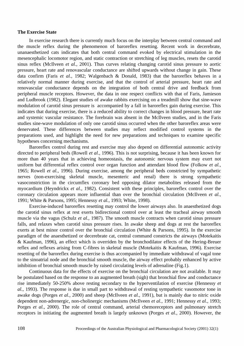
108 Proceedings of the Australian Physiological and Pharmacological Society (2001) 32(1)
The Exercise State
In exercise research there is currently much focus on the interplay between central command and the muscle reflex during the phenomenon of baroreflex resetting. Recent work in decerebrate, unanaesthetized cats indicates that both central command evoked by electrical stimulation in the mesencephalic locomotor region, and static contraction or stretching of leg muscles, resets the carotid sinus reflex (McIlveen et al., 2001). Thus curves relating changing carotid sinus pressure to aortic pressure, heart rate and renovascular conductance are shifted upwards without change in gain. These data confirm (Faris et al., 1982; Walgenbach & Donald, 1983) that the baroreflex behaves in a relatively normal manner during exercise, and that the control of arterial pressure, heart rate and renovascular conductance depends on the integration of both central drive and feedback from peripheral muscle receptors. However, the data in one respect conflicts with that of Faris, Jamieson and Ludbrook (1982). Elegant studies of awake rabbits exercising on a treadmill show that sine-wave modulation of carotid sinus pressure is accompanied by a fall in baroreflex gain during exercise. This indicates that during exercise, there is a reduced ability to correct changes in blood pressure, heart rate and systemic vascular resistance. The forebrain was absent in the McIlveen studies, and in the Faris studies sine-wave modulation of only one carotid sinus occurred when the other baroreflex areas were denervated. These differences between studies may reflect modified control systems in the preparations used, and highlight the need for new preparations and techniques to examine specific hypotheses concerning mechanisms. Baroreflex control during rest and exercise may also depend on differential autonomic activity directed to peripheral beds (Rowell et al., 1996). This is not surprising, because it has been known for more than 40 years that in achieving homeostasis, the autonomic nervous system may exert not uniform but differential reflex control over organ function and attendant blood flow (Folkow et al., 1965; Rowell et al., 1996). During exercise, among the peripheral beds constricted by sympathetic nerves (non-exercising skeletal muscle, mesenteric and renal) there is strong sympathetic vasoconstriction in the circumflex coronary bed opposing dilator metabolites released from the myocardium (Heyndrickx et al., 1982). Consistent with these principles, baroreflex control over the coronary circulation appears more influential than over the bronchial circulation (McIlveen et al., 1991; White & Parsons, 1995; Hennessy et al., 1993; White, 1998). Exercise-induced baroreflex resetting may control the lower airways also. In anaesthetized dogs the carotid sinus reflex at rest exerts bidirectional control over at least the tracheal airway smooth muscle via the vagus (Schulz et al., 1987). The smooth muscle contracts when carotid sinus pressure falls, and relaxes when carotid sinus pressure rises. In awake sheep and dogs at rest the baroreflex exerts at best minor control over the bronchial circulation (White & Parsons, 1995). In the exercise paradigm of the anaesthetized or decerebrate cat, central command constricts the airways (Motekaitis & Kaufman, 1996), an effect which is overriden by the bronchodilator effects of the Hering-Breuer reflex and reflexes arising from C-fibres in skeletal muscle (Motekaitis & Kaufman, 1996). Exercise resetting of the baroreflex during exercise is thus accompanied by immediate withdrawal of vagal tone to the sinoatrial node and the bronchial smooth muscle, the airway effect probably enhanced by active inhibition of bronchial smooth muscle by raised circulating levels of adrenaline (Fig.1). Continuous data for the effects of exercise on the bronchial circulation are not available. It may be postulated based on the response to an augmented breath (sigh) that bronchial flow and conductance rise immediately 50-250% above resting secondary to the hyperventilation of exercise (Hennessy et al., 1993). The response is due in small part to withdrawal of resting sympathetic vasomotor tone in awake dogs (Porges et al., 2000) and sheep (McIlveen et al., 1991), but is mainly due to nitric oxide dependent non-adrenergic, non-cholinergic mechanisms (McIlveen et al., 1991; Hennessy et al., 1993; Porges et al., 2000). The role of central command, arterial chemoreceptors and pulmonary stretch receptors in initiating the augmented breath is largely unknown (Porges et al., 2000). However, the
Proceedings of the Australian Physiological and Pharmacological Society (2001) 32(1) 109
bronchovascular response may have as its origin local parasympathetic reflexes. One mechanism invokes sudden changes in transmural pressure across bronchial vessels secondary to augmented breathing (Hennessy et al., 1993).
Figure 1. Modified Figure showing change in peak expiratory flow rate (PEFR) during and after exercise in a subject with exercise induced asthma (from Anderson (1984), and the period (post-exercise) over which “recovery only” ST-segment depression occurs (heavy horizontal bar, from Lachterman et al., 1990). During exercise (Work), there is a rise in PEFR, suggesting an increase in airway conductance occurs early in exercise followed by a fall toward baseline as exercise progresses. Following cessation of exercise PEFR continues to fall to reach a nadir at about 5 min post-exercise. This is the time in general when maximal recovery only ST-segment depression occurs.
In 1993 (Hennessy et al., 1993), we noted that in some dogs a sudden rise in bronchial perfusion pressure evoked by lower thoracic intra-aortic balloon inflation caused a sterotyped bronchovascular dilatation. The response could not be modified by combined α-, β-adrenoceptor and cholinoceptor blockade (Hennessy et al., 1993). We therefore proposed that the sudden rise in transmural pressure across bronchial vessels coincident with the sudden fall in intrapleural pressure at the onset of augmented breathing excites investing stretch-sensitive C-fibres of the axon-reflex kind. An associated mechanism could be the stimulus of high velocity air movement in the airways (McIlveen et al., 1991). In the awake sheep respiratory behaviours such as coughing, sneezing and sighing all involve inhalation and negative spikes in intrapleural pressure and large increases in bronchial flow and conductance (McIlveen et al., 1991). By contrast, others such as bleating vocalisation do not involve negative intrapleural pressure spikes, and there is no change in bronchial flow and conductance. Eructation of cud on the other hand is associated with a negative intrapleural pressure spike when inspiration is attempted against a closed glottis. In this case no movement of air occurs in the lower airways and there is no change in bronchial flow and conductance. These data suggest that high velocity of airflow may also stimulate C-fibre nerve endings in the airway mucosa as part of a local reflex. There is a substantial anatomical and neurophysological substrate (Myers et al., 1996) supporting a local reflex role for intrinsic parasympathetic ganglia and sensory-motor nerve endings investing bronchial vessels and mucosal structures. On stimulation they release the vasodilator
110 Proceedings of the Australian Physiological and Pharmacological Society (2001) 32(1)
peptides substance P, calcitonin gene-related peptide and neurokinin A (Hennessey et al., 1993; Myers et al., 1996). The hypothesis was put that these local reflexes protect the airway from drying and cooling (Hennessey et al., 1993). An alternative hypothesis is that bronchial flow will be reduced during exercise by sympathetic vasoconstriction, as occurs in other non-exercising beds in exercise (Rowell et al., 1996). There is thus a need for on-line and continuous information from running animals and man about individual and net autonomic outputs and effects evoked by the integration of central command and peripheral reflexes driving the blood flow and dimensions of airways. The Post-Exercise State
The post-exercise state is of considerable significance to clinicians. They are aware that in exercise stress clinics some 10% of patients who have proven obstructive coronary artery disease, but who do not meet ECG criteria for myocardial ischaemia during exercise, do meet the criteria 3-8 minutes following exercise (Lachterman et al., 1990). This period (Fig. 1) is marked by downsloping ST-segment depression equal to or greater than 1 mm, and in some cases is accompanied by chest pain and a variety of arrhythmias. These patients have a less favorable prognosis for myocardial events in the ensuing 5 years. In pulmonary clinics this period coincides with the appearance of exercise induced asthma (Fig. 1). While we would agree that, as in coronary artery disease, pathology ultimately determines the symptoms and signs, we have little or no understanding of the neural/local interactions between mechanisms that normally determine changes in airway smooth muscle tone, or in bronchial blood flow conductance. Thus, in neither of these two anatomically small but clinically important vascular beds do we understand autonomic mechanisms underlying the post-exercise state. It is difficult to predict the autonomic events during this period of baroreflex readjustment toward the resting state. Major interactions are likely to be present between neural and metabolic factors, particularly in the heart, but also in the lung. Changes in airway dimensions implied by clinical tests during or after exercise (Fig. 1) are likely to be determined at least in part by interplay between central command and feed-back reflexes from the periphery affecting both bronchial smooth muscle and the bronchovascular bed. The mechanics of airway narrowing have been long thought to depend in part on events and their mechanisms in the bronchial circulation (McFadden & Gilbert, 1999). Knowledge of autonomic events may be of considerable importance, because they may facilitate or oppose the effects of pathology, and may adapt with exercise training. Thus pharmacological and non-pharmacological therapeutic /rehabilitation initiatives may help or hinder patients. Post-exercise arterial pressure in patients exercising to exhaustion varies but may remain considerably higher than normal for some minutes before falling below pre-exercise levels at about 20 min. This is a time when the vagal withdrawal of baroreflex re-setting in exercise is reversed, and vagal activity increases to lower heart rate toward resting values. The increased sympathetic activity to the sino-atrial node, and increased sympathetic vasoconstriction in the coronary circulation during exercise (Heyndrickx et al., 1982), presumably is also reversed. We do not know whether the reversal of these separate neural events following exercise occurs simultaneously, or whether reversal occurs earlier in the vagus nerves than in the sympathetic nerves. But any delay in the sympathetic reversal will be coincident with the rapid metabolism of local dilator factors in the myocardium and the restitution of the powerful vasoconstrictor myogenic tone of resting coronary resistance vessels (White, 1998). If for these reasons there is normally enhanced coronary vasoconstriction post-exercise, this would be countered by an encroachment on the oxygen reserve in the coronary circulation. However, the positive ECG signs for ischaemia and the cardiovascular symptoms and arrhythmias in patients post-exercise may be explained by the recent studies of Dilaveris and colleagues (1998). Heart rate variability analysis in patients and aged-matched controls subjected to maximal treadmill exercise and recovery reveals that vagal tone withdraws during exercise and increases during recovery, and that sympathetic activity predominates during recovery in patients, and controls, but especially in patients
Proceedings of the Australian Physiological and Pharmacological Society (2001) 32(1) 111
with coronary artery disease and myocardial ischaemia. These findings support the hypothesis that sympathetic vasoconstrictor activity may normally dominate vagal vasodilator (White, 1998) activity in the coronary circulation post-exercise. Sympathetic vasoconstriction in the microcirculation under these circumstances will conflict with vasodilator metabolites distal to coronary obstruction. The authors raise the possibility that in obstructive coronary disease the ischaemic outcomes post-exercise are sufficient to trigger cardio-cardiac sympathetic excitatory reflexes (Malliani, 1986), resulting in potential positive feedback vasoconstriction in the coronary circulation with attendant signs and symptoms. These data are supported by findings from patients with effort angina during balloon angioplasty at rest (Joho et al., 1999). Heart rate and left ventricular pressure variability was analysed, and showed that induced ECG signs of myocardial ischaemia were associated with indices of a strong symatho-excitatory response. The post-exercise effects on the airways are likely to be more complex. Normally, at rest, there is vagal bronchoconstrictor tone in the lower airways. Post-exercise, while adrenaline effects may persist for some minutes, the degree to which airway conductance falls toward resting will depend on the time-course and intensity of the bronchoconstrictor effects mediated by returning vagal tone. As with the presence of pathology characterizing the coronary circulation, it has been suggested by Gandevia (Gandevia et al., 2000) that there may be excessive C-fibre stimulation during stressful exercise. There may also be bronchial hyperaemia following airway cooling (McFadden & Gilbert, 1999), or an hyperosmotic interstitium following airway drying (Anderson, 1984: Anderson & Daviskas, 1999). If these singly or together are associated with mediator release (Bauer & Razin, 2000) and inflammatory responses, the restitution of vagal tone and lower airways constriction would be enhanced and lead to exercise induced asthma. Airways Internal Diameter Assessment (AIDA) Sonomicrometer
We have an imprecise understanding of the time-course of events and mechanisms during and after exercise in the heart and airways. This particularly applies to bronchial blood flow and to the dimensions of the airways. We have therefore designed and evaluated an instrument that can directly measure airway dimensions and bronchial blood flow simultaneously, using ultrasonic techniques. This we have named the Airways Internal Diameter Assessment Sonomicrometer (AIDA) (White et al., 1999). It is designed for awake animal use (large animals in the first instance, viz. dogs, sheep, cat) at rest and during exercise. The instrument is composed of 4 parts, 1. 20 MHz pulsed-Doppler flow probes (of lightweight design) custom-made in Newcastle for application to e.g. the fragile right bronchial arteries in the dog (Hennessey et al., 1993), are used with a Triton flowmeter (System 6-200), for measurement of bronchial blood flow. The flowmeter has been modified to cope with aliasing errors inherent in other flowmeters of this type when measuring high velocity flows in very small vessels (i.d. ~ 1-3 mm) (Hennessey et al., 1993; Quail et al., 1993). This transducer/flowmeter system is currently preferred to the Transonic flowmeter system because of utility, relatively low-cost, its miniaturized transducer system, and capacity for multichannel synchronization, e.g. for adding coronary flow to the measurements, 2. A 2-crystal transit time (2.3 MHz) sonomicrometer (TTS, modified from Pitsillides et al., 1992) for absolute measures of bronchial hemicircumference, and 3. A 20 MHz single-crystal sonomicrometer for absolute measures of bronchial wall thickness (SCS, modified from Pitsillides and Longhurst, 1995). 4. An integrator module to eliminate cross talk during recording. The components have been evaluated in vitro and in vivo for accuracy, linearity (r = 0.99) and frequency response when applied to end-systolic and diastolic dimensions and wall thickness of beating pig and rat hearts (HR > 450 beats/min) (Pitsillides et al., 1992; Pitsillides & Longhurst, 1995). Changes in bronchial dimensions are less demanding on frequency response (less than 20 Hz). Initial signals showed new high frequency information in bronchial circumference consistent with
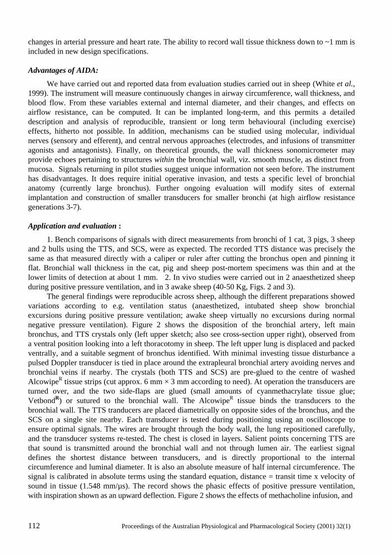
112 Proceedings of the Australian Physiological and Pharmacological Society (2001) 32(1)
changes in arterial pressure and heart rate. The ability to record wall tissue thickness down to ~1 mm is included in new design specifications. Advantages of AIDA:
We have carried out and reported data from evaluation studies carried out in sheep (White et al., 1999). The instrument will measure continuously changes in airway circumference, wall thickness, and blood flow. From these variables external and internal diameter, and their changes, and effects on airflow resistance, can be computed. It can be implanted long-term, and this permits a detailed description and analysis of reproducible, transient or long term behavioural (including exercise) effects, hitherto not possible. In addition, mechanisms can be studied using molecular, individual nerves (sensory and efferent), and central nervous approaches (electrodes, and infusions of transmitter agonists and antagonists). Finally, on theoretical grounds, the wall thickness sonomicrometer may provide echoes pertaining to structures within the bronchial wall, viz. smooth muscle, as distinct from mucosa. Signals returning in pilot studies suggest unique information not seen before. The instrument has disadvantages. It does require initial operative invasion, and tests a specific level of bronchial anatomy (currently large bronchus). Further ongoing evaluation will modify sites of external implantation and construction of smaller transducers for smaller bronchi (at high airflow resistance generations 3-7). Application and evaluation :
1. Bench comparisons of signals with direct measurements from bronchi of 1 cat, 3 pigs, 3 sheep and 2 bulls using the TTS, and SCS, were as expected. The recorded TTS distance was precisely the same as that measured directly with a caliper or ruler after cutting the bronchus open and pinning it flat. Bronchial wall thickness in the cat, pig and sheep post-mortem specimens was thin and at the lower limits of detection at about 1 mm. 2. In vivo studies were carried out in 2 anaesthetized sheep during positive pressure ventilation, and in 3 awake sheep (40-50 Kg, Figs. 2 and 3). The general findings were reproducible across sheep, although the different preparations showed variations according to e.g. ventilation status (anaesthetized, intubated sheep show bronchial excursions during positive pressure ventilation; awake sheep virtually no excursions during normal negative pressure ventilation). Figure 2 shows the disposition of the bronchial artery, left main bronchus, and TTS crystals only (left upper sketch; also see cross-section upper right), observed from a ventral position looking into a left thoracotomy in sheep. The left upper lung is displaced and packed ventrally, and a suitable segment of bronchus identified. With minimal investing tissue disturbance a pulsed Doppler transducer is tied in place around the extrapleural bronchial artery avoiding nerves and bronchial veins if nearby. The crystals (both TTS and SCS) are pre-glued to the centre of washed AlcowipeR tissue strips (cut approx. 6 mm × 3 mm according to need). At operation the transducers are turned over, and the two side-flaps are glued (small amounts of cyanmethacrylate tissue glue; VetbondR) or sutured to the bronchial wall. The AlcowipeR tissue binds the transducers to the bronchial wall. The TTS tranducers are placed diametrically on opposite sides of the bronchus, and the SCS on a single site nearby. Each transducer is tested during positioning using an oscilloscope to ensure optimal signals. The wires are brought through the body wall, the lung repositioned carefully, and the transducer systems re-tested. The chest is closed in layers. Salient points concerning TTS are that sound is transmitted around the bronchial wall and not through lumen air. The earliest signal defines the shortest distance between transducers, and is directly proportional to the internal circumference and luminal diameter. It is also an absolute measure of half internal circumference. The signal is calibrated in absolute terms using the standard equation, distance = transit time x velocity of sound in tissue (1.548 mm/µs). The record shows the phasic effects of positive pressure ventilation, with inspiration shown as an upward deflection. Figure 2 shows the effects of methacholine infusion, and
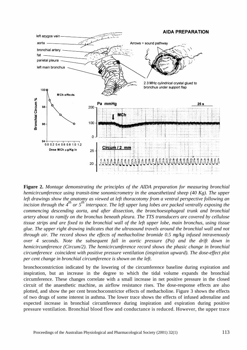
Proceedings of the Australian Physiological and Pharmacological Society (2001) 32(1) 113
Figure 2. Montage demonstrating the principles of the AIDA preparation for measuring bronchial hemicircumference using transit-time sonomicrometry in the anaesthetized sheep (40 Kg). The upper left drawings show the anatomy as viewed at left thoracotomy from a ventral perspective following an incision through the 4th or 5th interspace. The left upper lung lobes are packed ventrally exposing the commencing descending aorta, and after dissection, the bronchoesophageal trunk and bronchial artery about to ramify on the bronchus beneath pleura. The TTS transducers are covered by cellulose tissue strips and are fixed to the bronchial wall of the left upper lobe, main bronchus, using tissue glue. The upper right drawing indicates that the ultrasound travels around the bronchial wall and not through air. The record shows the effects of methacholine bromide 0.5 µg/kg infused intravenously over 4 seconds. Note the subsequent fall in aortic pressure (Pa) and the drift down in hemicircumference (Circum/2). The hemicircumference record shows the phasic change in bronchial circumference coincident with positive pressure ventilation (inspiration upward). The dose-effect plot per cent change in bronchial circumference is shown on the left.
bronchoconstriction indicated by the lowering of the circumference baseline during expiration and inspiration, but an increase in the degree to which the tidal volume expands the bronchial circumference. These changes correlate with a small increase in net positive pressure in the closed circuit of the anaesthetic machine, as airflow resistance rises. The dose-response effects are also plotted, and show the per cent bronchoconstrictor effects of methacholine. Figure 3 shows the effects of two drugs of some interest in asthma. The lower trace shows the effects of infused adrenaline and expected increase in bronchial circumference during inspiration and expiration during positive pressure ventilation. Bronchial blood flow and conductance is reduced. However, the upper trace
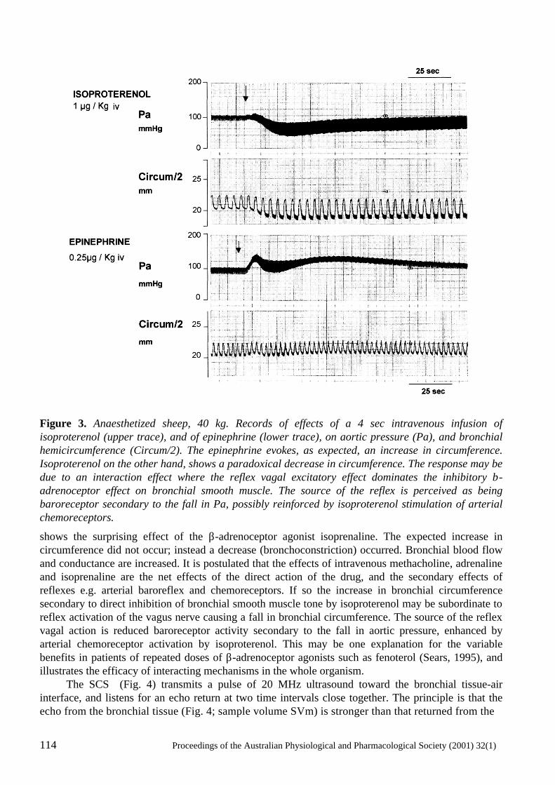
114 Proceedings of the Australian Physiological and Pharmacological Society (2001) 32(1)
Figure 3. Anaesthetized sheep, 40 kg. Records of effects of a 4 sec intravenous infusion of isoproterenol (upper trace), and of epinephrine (lower trace), on aortic pressure (Pa), and bronchial hemicircumference (Circum/2). The epinephrine evokes, as expected, an increase in circumference. Isoproterenol on the other hand, shows a paradoxical decrease in circumference. The response may be due to an interaction effect where the reflex vagal excitatory effect dominates the inhibitory β-adrenoceptor effect on bronchial smooth muscle. The source of the reflex is perceived as being baroreceptor secondary to the fall in Pa, possibly reinforced by isoproterenol stimulation of arterial chemoreceptors.
shows the surprising effect of the β-adrenoceptor agonist isoprenaline. The expected increase in circumference did not occur; instead a decrease (bronchoconstriction) occurred. Bronchial blood flow and conductance are increased. It is postulated that the effects of intravenous methacholine, adrenaline and isoprenaline are the net effects of the direct action of the drug, and the secondary effects of reflexes e.g. arterial baroreflex and chemoreceptors. If so the increase in bronchial circumference secondary to direct inhibition of bronchial smooth muscle tone by isoproterenol may be subordinate to reflex activation of the vagus nerve causing a fall in bronchial circumference. The source of the reflex vagal action is reduced baroreceptor activity secondary to the fall in aortic pressure, enhanced by arterial chemoreceptor activation by isoproterenol. This may be one explanation for the variable benefits in patients of repeated doses of β-adrenoceptor agonists such as fenoterol (Sears, 1995), and illustrates the efficacy of interacting mechanisms in the whole organism. The SCS (Fig. 4) transmits a pulse of 20 MHz ultrasound toward the bronchial tissue-air interface, and listens for an echo return at two time intervals close together. The principle is that the echo from the bronchial tissue (Fig. 4; sample volume SVm) is stronger than that returned from the
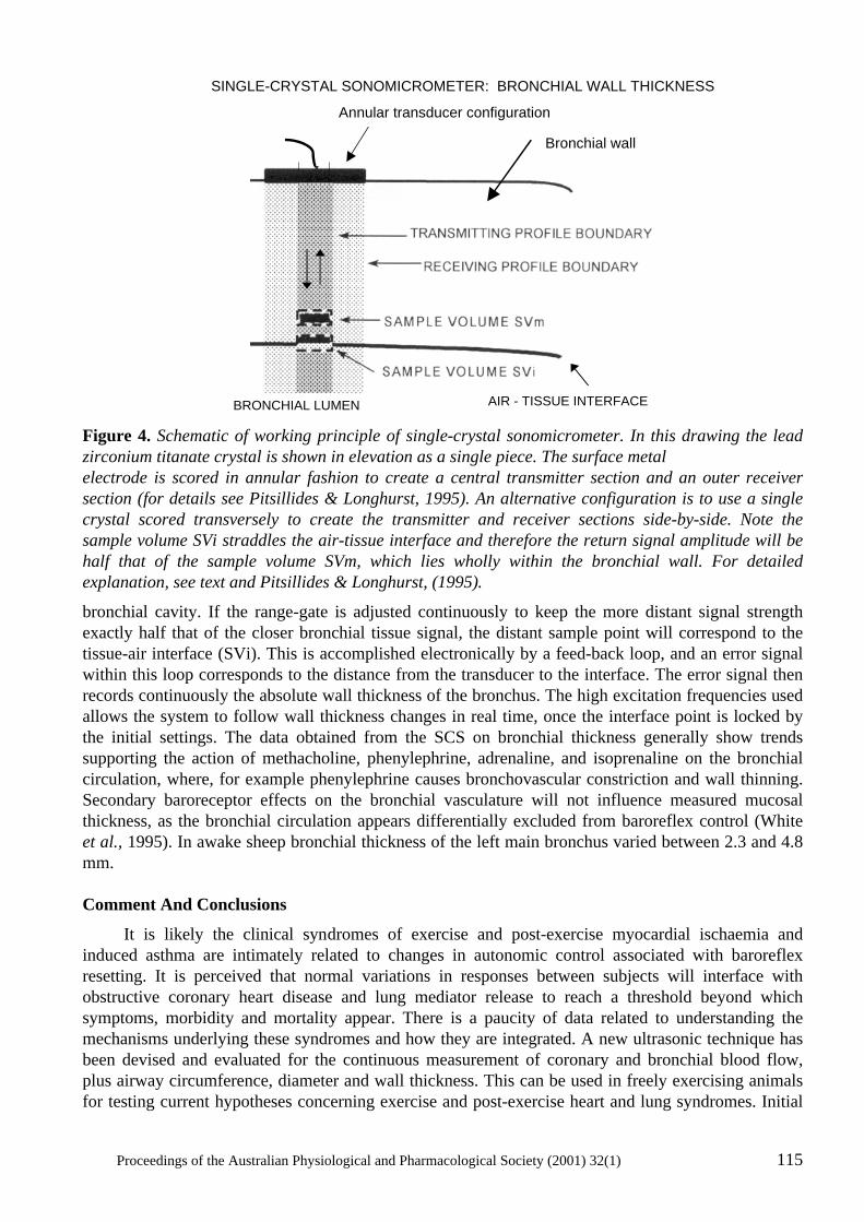
Proceedings of the Australian Physiological and Pharmacological Society (2001) 32(1) 115
Figure 4. Schematic of working principle of single-crystal sonomicrometer. In this drawing the lead zirconium titanate crystal is shown in elevation as a single piece. The surface metal electrode is scored in annular fashion to create a central transmitter section and an outer receiver section (for details see Pitsillides & Longhurst, 1995). An alternative configuration is to use a single crystal scored transversely to create the transmitter and receiver sections side-by-side. Note the sample volume SVi straddles the air-tissue interface and therefore the return signal amplitude will be half that of the sample volume SVm, which lies wholly within the bronchial wall. For detailed explanation, see text and Pitsillides & Longhurst, (1995).
bronchial cavity. If the range-gate is adjusted continuously to keep the more distant signal strength exactly half that of the closer bronchial tissue signal, the distant sample point will correspond to the tissue-air interface (SVi). This is accomplished electronically by a feed-back loop, and an error signal within this loop corresponds to the distance from the transducer to the interface. The error signal then records continuously the absolute wall thickness of the bronchus. The high excitation frequencies used allows the system to follow wall thickness changes in real time, once the interface point is locked by the initial settings. The data obtained from the SCS on bronchial thickness generally show trends supporting the action of methacholine, phenylephrine, adrenaline, and isoprenaline on the bronchial circulation, where, for example phenylephrine causes bronchovascular constriction and wall thinning. Secondary baroreceptor effects on the bronchial vasculature will not influence measured mucosal thickness, as the bronchial circulation appears differentially excluded from baroreflex control (White et al., 1995). In awake sheep bronchial thickness of the left main bronchus varied between 2.3 and 4.8 mm. Comment And Conclusions
It is likely the clinical syndromes of exercise and post-exercise myocardial ischaemia and induced asthma are intimately related to changes in autonomic control associated with baroreflex resetting. It is perceived that normal variations in responses between subjects will interface with obstructive coronary heart disease and lung mediator release to reach a threshold beyond which symptoms, morbidity and mortality appear. There is a paucity of data related to understanding the mechanisms underlying these syndromes and how they are integrated. A new ultrasonic technique has been devised and evaluated for the continuous measurement of coronary and bronchial blood flow, plus airway circumference, diameter and wall thickness. This can be used in freely exercising animals for testing current hypotheses concerning exercise and post-exercise heart and lung syndromes. Initial
SINGLE-CRYSTAL SONOMICROMETER: BRONCHIAL WALL THICKNESS
Annular transducer configuration
Bronchial wall
AIR - TISSUE INTERFACEBRONCHIAL LUMEN
116 Proceedings of the Australian Physiological and Pharmacological Society (2001) 32(1)
data show that airway narrowing can occur with large physiological doses of β-adrenoceptor agonists. The hypothesis is put that the expected bronchodilator effects may be subordinate to simultaneous baroreflex and arterial chemoreceptor activation of the vagus causing bronchoconstriction secondary to a fall in arterial pressure, and activation of the arterial chemoreceptors by isoprenaline. This effect would be made worse by concomitant bronchovascular dilatation and an increase in wall thickness. Acknowledgements
The excellent technical assistance of Linda Talken and Jessica Davis at the Animal Research Service at University of California, Davis, is gratefully acknowledged. This work was supported in part by the National Health and Medical Research Council of Australia, and The Ramaciotti Foundation of Australia. References
Anderson, S.D. & Daviskas, E. (1999) Airway drying and exercise induced asthma. In: Exercise Induced Asthma-Lung Biology in Health and Disease. ed. McFadden, E.R. pp. 77-113. New York: Marcel Dekker.
Anderson, S.D. (1984) Is there a unifying hypothesis for exercise-induced asthma? Journal of Allergy and Clinical Immunology, 73, 660-665.
Bauer, O. & Razin, E. (2000) Mast cell-nerve interactions. News in Physiological Sciences, 15, 213-218.
Dilaveris, P.E., Zervopoulos, G.A., Michaelides, A.P., Sideris, S.K., Psomadaki, Z.D., Gialafos, E.J., Gialafos, J.E. & Toutouzas, P.K. (1998) Ischaemia-induced reflex sympathoexcitation during the recovery period after maximal treadmill exercise testing. Clinical Cardiology, 21, 585-590.
Faris, I.B., Jamieson, G.G. & Ludbrook, J. (1982) Effect of exercise on gain of the carotid-sinus reflex in rabbits. Clinical Science, 63, 115-119.
Folkow, B., Heymans, C. & Neil, E. (1965) Integrated aspects of cardiovascular regulation. In: Handbook of Physiology, Circulation. Sect 2, Vol III. pp. 1787-1823. ed. Hamilton, W.F. Washington DC: American Physiological Society.
Gandevia, S., Taylor, J. & Butler, J. (2000) Stopping exercise: role of pulmonary c fibers and inhibition of motoneurons. News in Physiological Sciences, 15, 241-245.
Hennessy, E., White, S.W., Van Der Touw, T., Quail, A., Porges, W. & Glenfield, P. (1993) Control of resting bronchial hemodynamics in the awake dog. American Journal of Physiology. 265, H649-H660.
Heyndrickx, G.R., Muylaert, P. & Pannier, J.L. (1982) Alpha-adrenergic control of oxygen delivery to myocardium during exercise in conscious dogs. American Journal of Physiology, 242, H805-H809.
Joho, S., Asanoi, H., Remah, H.A., Igawa, A., Kameyama, T., Nozawa, T., Umeno, K. & Inoue, H. (1999) Time-varying spectral analysis of heart rate and left ventricular pressure variability during balloon coronary occlusion in humans: a sympathoexcitatory response to myocardial ischemia. Journal of the American College of Cardiology, 34, 1924-1931.
Lachterman, B., Lehmann, K.G., Abrahamson, D. & Froelicher, V.F. (1990) "Recovery only" ST-segment depression and the predictive accuracy of the exercise test. Annals of Internal Medicine, 113, 333-334.
Malliani, A. (1986) Homeostasis and instability: the hypothesis of tonic interaction in the cardiovascular regulation of negative and positive feedback mechanisms. In: Neural Mechanisms and Cardiovascular Disease. ed. Lown, B., Malliani, A. & Prosdocimi, M. pp. 1-9. Padova: Liviana Press.
Proceedings of the Australian Physiological and Pharmacological Society (2001) 32(1) 117
McFadden, E.R. & Gilbert, I.A. (1999) Exercise-induced asthma as a vascular phenomenon. In: Exercise Induced Asthma-Lung Biology in Health and Disease. ed. McFadden, E.R. pp. 115-135. New York: Marcel Dekker..
McIlveen, S.A., Hayes, S.G. & Kaufman, M.P. (2001) Both central command and the exercise pressor reflex reset the carotid sinus baroreflex. American Journal of Physiology, (In press).
McIlveen, S.A., White, S.W. & Parsons, G. (1991) Autonomic control of bronchial circulation in awake sheep during rest and behaviour. Clinical and Experimental Pharmacology and Physiology, 24, 940-947.
Motekaitis, A.M. & Kaufman, M..P. (1996) Stimulation of the mesencephalic locomotor region constricts the airways of cats. Respiration Physiology, 106, 263-271.
Myers, A., Undem, B. & Kummer, W. (1996) Anatomical and electrophysiological comparison of the sensory innervation of bronchial and tracheal parasympathetic ganglion neurons. Journal of the Autonomic Nervous System, 61, 162-168.
Pitsillides, K.F. & Longhurst, J.C. (1995) An ultrasonic system for measurement of absolute myocardial thickness using a single transducer. American Journal of Physiology, 268, H1358-H1367.
Pitsillides, K.F., Symons, J.D. & Longhurst, J.C. (1992) Biotelemetry of cardiovascular hemodynamic measurements in miniswine. IEEE Transactions on Biomedical Engineering, 39, 982-986.
Porges, W.L., Hennessy, E.J., Quail, A.W., Cottee, D.B., Moore, P.G., McIlveen, S.A., Parsons, G.H. & White, S.W. (2000) Heart-Lung Interactions: the sigh and autonomic control in the bronchial and coronary circulations. Clinical and Experimental Pharmacology and Physiology, 27, 1022-1027.
Quail, A.W., Cottee, D.B.F. & White, S.W. (1993) Limitations of a Doppler velocimeter for blood flow measurement in small vessels. Journal of Applied Physiology, 75, 2745-2754.
Rowell, L.B., O’Leary, D.S. & Kellog, D.L. (1996) Integration of cardiovascular control systems in dynamic exercise. In: Handbook of Physiology, Exercise: Regulation and Integration of Multiple Systems. Section 12. ed. Rowell, L.B. & Shepherd, J.T. Ch. 17, pp 781-786. Oxford: American Physiological Society.
Schultz, H.D., Pisarri, T.E., Coleridge, H.M. & Coleridge, J.C.G. (1987) Carotid sinus baroreceptors modulate tracheal smooth muscle tension in dogs. Circulation Research, 60, 337-345.
Sears, M.R. (1995) Changing patterns in asthma morbidity and mortality. Journal of Investigational Allergology and Clinical Immunology, 5, 66-72.
Walgenbach, S.C. & Donald, D.E. (1983) Inhibition by carotid baroreflex of exercise-induced increases in arterial pressure. Circulation Research, 52, 253-262.
White, S.W. & Parsons, G.H. (1995) Baroreflexes and the bronchial circulation in awake sheep; absence of control through cholinoceptor and adrenoceptor autonomic mechanisms. American Journal of Respiration and Critical Care Medicine, 151, A522.
White, S.W. (1998) Integrated baroreflex control of regional coronary blood flow. Cardiologia, 43, 559-570.
White, S.W., Pitsillides, K. & Parsons, G. (1999) Airways Internal Diameter Assessment (AIDA) Sonomicrometer: a novel instrument for the continuous measurement of airways dimensions and blood flow in awake animals. Proceedings of the Australian Physiological and Pharmacological Society, 30, 67P.
118 Proceedings of the Australian Physiological and Pharmacological Society (2001) 32(1)
NO DEBATE? ENHANCED TOTAL PERIPHERAL VASCULAR RESPONSIVENESS IN HYPERTENSION ACCORDS WITH THE AMPLIFIER HYPOTHESIS James A. Angus, Christine E. Wright and Paul I. Korner, Department of Pharmacology, The University of Melbourne, Vic 3010, Australia. Since the early work of Folkow (1982) and his colleagues with the perfused SHR and WKY rat hind limb preparation, it is considered that changes in vascular resistance to constrictor drugs are amplified in SHR because of structural remodelling of the vasculature. A reduced internal radius (ri) and increased wall thickness (ω) to lumen radius ratio (ω/ri) can theoretically explain this increase in reactivity in hypertension. We have published work from the conscious rabbit with bilateral renal cellophane wrap experimental hypertension, confirming that amplified resistance increases to constrictor drugs, and amplified falls in resistance to dilator drugs, are a feature of the hindquarter vasculature (Wright & Angus, 1986). Recently, we extended the observations to the ‘total’ peripheral circulation in the renal hypertensive rabbit (Wright & Angus, 1999). The more sophisticated technology used in this study such as the Transonic™ flow probe chronically implanted on the aortic root and the chronic indwelling left atrial catheter, owes much to Professor John Ludbrook’s research at the Baker Medical Research Institute and Department of Surgery, University of Melbourne. In our study we showed that total peripheral resistance and conductance changes to left atrial infusions of methoxamine and adenosine do indeed accord with the amplifier hypothesis. Moreover, total pharmacological antonomic blockade, neurohumoral block or control conditions gave insights into the role of cardiac and vascular homeostatic mechanisms. There is some debate as to whether the phenomenon of the vascular resistance amplifier does occur in the total circulation and in all types of hypertension (Izzard et al., 1999; Korner et al., 2000). In this presentation, we will show how integrated whole animal pharmacology and physiology can benefit the analysis of an important fundamental problem in hypertension research. Folkow, B. (1982) Physiological Reviews, 62, 347-504. Izzard, A.S., Heagerty, A.M. & Leenen, F.H.H. (1999) Journal of Hypertension, 17, 1667-1669. Korner, P.L., Angus, J.A. & Wright, C.E. (2000) Journal of Hypertension, 18, 235-239. Wright, C.E., Angus, J.A. & Korner, P.I. (1987) Hypertension, 9, 122-131. Wright, C.E. & Angus, J.A. (1999) Journal of Hypertension, 17, 1-11.
THIS PRE-ABSTRACT OF PROF ANGUS’S PAPER APPEARED IN THE PROCEEDINGS, ISSUE 32(1), AS 122P.
Proceedings of the Australian Physiological and Pharmacological Society (2001) 32(1) 119
NEURAL MECHANISMS IN THE CARDIOVASCULAR RESPONSES
TO ACUTE CENTRAL HYPOVOLAEMIA
Roger G. Evans*, Sabatino Ventura†, Roger A. L. Dampney‡ and John Ludbrook¥
Departments of *Physiology and †Pharmacology, Monash University, ‡Department of Physiology,
University of Sydney and ¥Department of Surgery, University of Melbourne, Australia. Summary
The haemodynamic response to acute central hypovolaemia consists of two phases. During Phase I, arterial pressure is well maintained in the face of falling cardiac output, by baroreceptor-mediated reflex vasoconstriction and cardio-acceleration. Phase II commences once cardiac output has fallen to a critical level of 50-60% of its resting value, equivalent to loss of about 30% of blood volume. During Phase II, sympathetic vasoconstrictor and cardiac drive fall abruptly, and cardiac vagal drive increases. In man, this response is invariably associated with fainting, and has been termed vasovagal syncope. In both experimental animals and man, the responses to acute central hypovolaemia are greatly affected by anaesthetic agents, in that the compensatory responses during Phase I (e.g., halothane), or their failure during Phase II (e.g., alfentanil) are blunted or abolished. Therefore, our present knowledge of the neurochemical basis of the response to hypovolaemia depends chiefly on the results of experiments in conscious animals. Use of techniques for simulating haemorrhage has greatly enhanced this research effort, by allowing the effects of multiple treatments on the response to acute central hypovolaemia to be tested in the same animal. The results of such experiments indicate that Phase II of the response to hypovolaemia is triggered, at least in part, by a signal from cardiac vagal afferents. There is also strong evidence that Phase II depends on brainstem δ1-opioid receptor and nitrergic mechanisms, and can potentially be modulated by circulating or neuronally-released adrenocorticotropic hormone, brainstem serotonergic pathways operating through 5-HT1A-receptors, and opioids acting through µ- and κ-receptors in the brainstem. Phase II also appears to require input from supramedullary brain centres. Future studies should determine how these neurotransmitter systems interact, and their precise neuroanatomical arrangements. Introduction
The first documented direct measurements of arterial pressure were made by Stephen Hales in conscious horses, and reported in his monograph of 1733 (Hales, 1964). Hales can also be credited with making the first observations of the response of arterial blood pressure to haemorrhage, and the state of haemorrhagic shock. ‘We may observe.....that the decrease in the force of the blood in the arteries, was not proportioned to the several quantities of blood which were evacuated........When between fourteen and fifteen quarts of blood had been evacuated, and thereby the force of that which remained in the vessels greatly decreased, then the mare fell into cold clammy sweats, such as frequently attend dying persons; which shows how low a state the vital force of the blood is at that time reduced: Whence we see, that these faint sweats are not occasioned by a greater protrusive force of the blood at that time, but rather by a general relaxation of the pores, as well as of all other parts of the body.’ We can see from this quote that, based on the results of his crude but effective experimental approach, Hales may have had some inkling of the complex nature of the haemodynamic response to haemorrhage. The major watershed in our knowledge of the physiology of haemorrhage had to await development of methods for directly monitoring cardiac output. Hence, during World War II, research
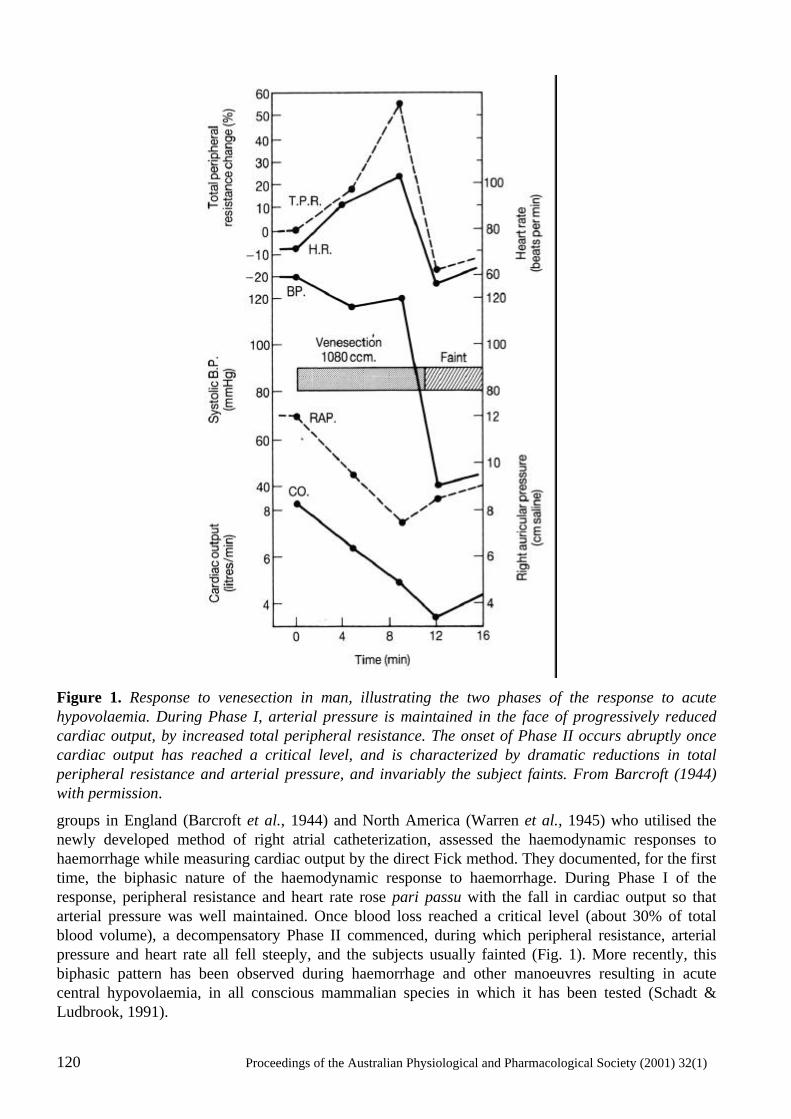
120 Proceedings of the Australian Physiological and Pharmacological Society (2001) 32(1)
Figure 1. Response to venesection in man, illustrating the two phases of the response to acute hypovolaemia. During Phase I, arterial pressure is maintained in the face of progressively reduced cardiac output, by increased total peripheral resistance. The onset of Phase II occurs abruptly once cardiac output has reached a critical level, and is characterized by dramatic reductions in total peripheral resistance and arterial pressure, and invariably the subject faints. From Barcroft (1944) with permission.
groups in England (Barcroft et al., 1944) and North America (Warren et al., 1945) who utilised the newly developed method of right atrial catheterization, assessed the haemodynamic responses to haemorrhage while measuring cardiac output by the direct Fick method. They documented, for the first time, the biphasic nature of the haemodynamic response to haemorrhage. During Phase I of the response, peripheral resistance and heart rate rose pari passu with the fall in cardiac output so that arterial pressure was well maintained. Once blood loss reached a critical level (about 30% of total blood volume), a decompensatory Phase II commenced, during which peripheral resistance, arterial pressure and heart rate all fell steeply, and the subjects usually fainted (Fig. 1). More recently, this biphasic pattern has been observed during haemorrhage and other manoeuvres resulting in acute central hypovolaemia, in all conscious mammalian species in which it has been tested (Schadt & Ludbrook, 1991).
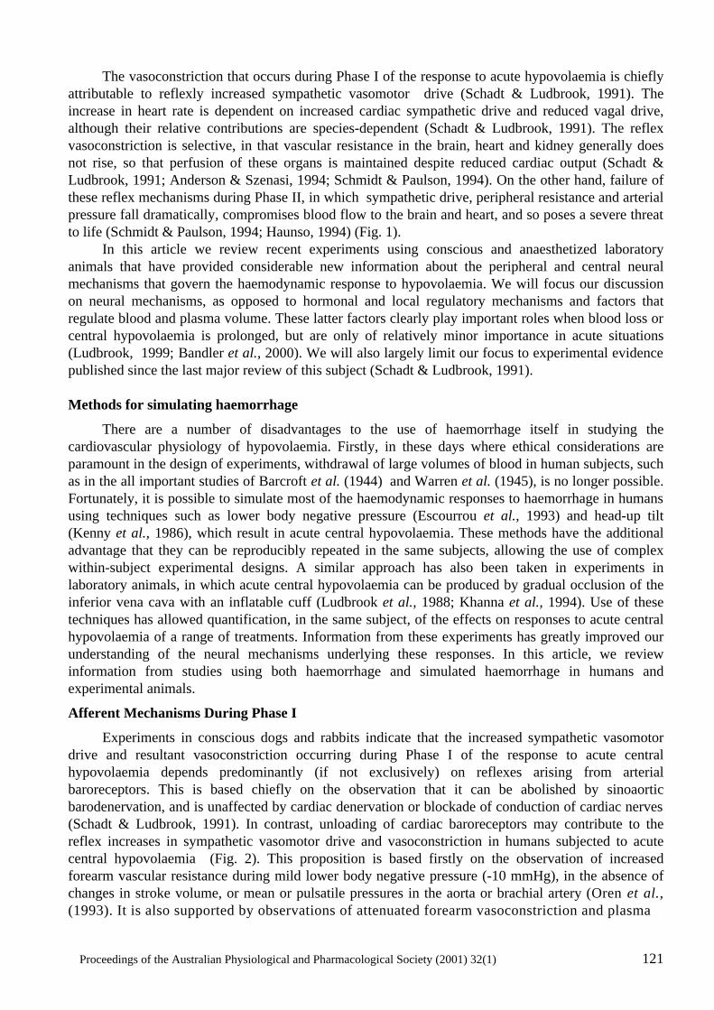
Proceedings of the Australian Physiological and Pharmacological Society (2001) 32(1) 121
The vasoconstriction that occurs during Phase I of the response to acute hypovolaemia is chiefly attributable to reflexly increased sympathetic vasomotor drive (Schadt & Ludbrook, 1991). The increase in heart rate is dependent on increased cardiac sympathetic drive and reduced vagal drive, although their relative contributions are species-dependent (Schadt & Ludbrook, 1991). The reflex vasoconstriction is selective, in that vascular resistance in the brain, heart and kidney generally does not rise, so that perfusion of these organs is maintained despite reduced cardiac output (Schadt & Ludbrook, 1991; Anderson & Szenasi, 1994; Schmidt & Paulson, 1994). On the other hand, failure of these reflex mechanisms during Phase II, in which sympathetic drive, peripheral resistance and arterial pressure fall dramatically, compromises blood flow to the brain and heart, and so poses a severe threat to life (Schmidt & Paulson, 1994; Haunso, 1994) (Fig. 1). In this article we review recent experiments using conscious and anaesthetized laboratory animals that have provided considerable new information about the peripheral and central neural mechanisms that govern the haemodynamic response to hypovolaemia. We will focus our discussion on neural mechanisms, as opposed to hormonal and local regulatory mechanisms and factors that regulate blood and plasma volume. These latter factors clearly play important roles when blood loss or central hypovolaemia is prolonged, but are only of relatively minor importance in acute situations (Ludbrook, 1999; Bandler et al., 2000). We will also largely limit our focus to experimental evidence published since the last major review of this subject (Schadt & Ludbrook, 1991). Methods for simulating haemorrhage
There are a number of disadvantages to the use of haemorrhage itself in studying the cardiovascular physiology of hypovolaemia. Firstly, in these days where ethical considerations are paramount in the design of experiments, withdrawal of large volumes of blood in human subjects, such as in the all important studies of Barcroft et al. (1944) and Warren et al. (1945), is no longer possible. Fortunately, it is possible to simulate most of the haemodynamic responses to haemorrhage in humans using techniques such as lower body negative pressure (Escourrou et al., 1993) and head-up tilt (Kenny et al., 1986), which result in acute central hypovolaemia. These methods have the additional advantage that they can be reproducibly repeated in the same subjects, allowing the use of complex within-subject experimental designs. A similar approach has also been taken in experiments in laboratory animals, in which acute central hypovolaemia can be produced by gradual occlusion of the inferior vena cava with an inflatable cuff (Ludbrook et al., 1988; Khanna et al., 1994). Use of these techniques has allowed quantification, in the same subject, of the effects on responses to acute central hypovolaemia of a range of treatments. Information from these experiments has greatly improved our understanding of the neural mechanisms underlying these responses. In this article, we review information from studies using both haemorrhage and simulated haemorrhage in humans and experimental animals.
Afferent Mechanisms During Phase I
Experiments in conscious dogs and rabbits indicate that the increased sympathetic vasomotor drive and resultant vasoconstriction occurring during Phase I of the response to acute central hypovolaemia depends predominantly (if not exclusively) on reflexes arising from arterial baroreceptors. This is based chiefly on the observation that it can be abolished by sinoaortic barodenervation, and is unaffected by cardiac denervation or blockade of conduction of cardiac nerves (Schadt & Ludbrook, 1991). In contrast, unloading of cardiac baroreceptors may contribute to the reflex increases in sympathetic vasomotor drive and vasoconstriction in humans subjected to acute central hypovolaemia (Fig. 2). This proposition is based firstly on the observation of increased forearm vascular resistance during mild lower body negative pressure (-10 mmHg), in the absence of changes in stroke volume, or mean or pulsatile pressures in the aorta or brachial artery (Oren et al., (1993). It is also supported by observations of attenuated forearm vasoconstriction and plasma
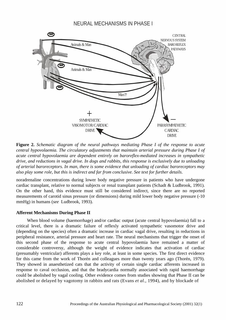
122 Proceedings of the Australian Physiological and Pharmacological Society (2001) 32(1)
Figure 2. Schematic diagram of the neural pathways mediating Phase I of the response to acute central hypovolaemia. The circulatory adjustments that maintain arterial pressure during Phase I of acute central hypovolaemia are dependent entirely on baroreflex-mediated increases in sympathetic drive, and reductions in vagal drive. In dogs and rabbits, this response is exclusively due to unloading of arterial baroreceptors. In man, there is some evidence that unloading of cardiac baroreceptors may also play some role, but this is indirect and far from conclusive. See text for further details.
noradrenaline concentrations during lower body negative pressure in patients who have undergone cardiac transplant, relative to normal subjects or renal transplant patients (Schadt & Ludbrook, 1991). On the other hand, this evidence must still be considered indirect, since there are no reported measurements of carotid sinus pressure (or dimensions) during mild lower body negative pressure (-10 mmHg) in humans (see Ludbrook, 1993). Afferent Mechanisms During Phase II
When blood volume (haemorrhage) and/or cardiac output (acute central hypovolaemia) fall to a critical level, there is a dramatic failure of reflexly activated sympathetic vasomotor drive and (depending on the species) often a dramatic increase in cardiac vagal drive, resulting in reductions in peripheral resistance, arterial pressure and heart rate. The neural mechanisms that trigger the onset of this second phase of the response to acute central hypovolaemia have remained a matter of considerable controversy, although the weight of evidence indicates that activation of cardiac (presumably ventricular) afferents plays a key role, at least in some species. The first direct evidence for this came from the work of Thorén and colleagues more than twenty years ago (Thorén, 1979). They showed in anaesthetized cats that the activity of certain single cardiac afferents increased in response to caval occlusion, and that the bradycardia normally associated with rapid haemorrhage could be abolished by vagal cooling. Other evidence comes from studies showing that Phase II can be abolished or delayed by vagotomy in rabbits and rats (Evans et al., 1994), and by blockade of
NEURAL MECHANISMS IN PHASE I
Animals & Man
Animals & Man
Man??
--
+SYMPATHETIC
VASOMOTOR/CARDIACDRIVE
PARASYMPATHETICCARDIAC
DRIVE
CENTRALNERVOUS SYSTEM
BAROREFLEXPATHWAYS
-
_
Proceedings of the Australian Physiological and Pharmacological Society (2001) 32(1) 123
Figure 3. Schematic diagram of the possible neural mechanisms mediating Phase II of the response to acute central hypovolaemia. The precise nature of the stimuli initiating Phase II remain unknown, but there is good evidence for contributions of (paradoxical) increased firing of cardiac (persumably left ventricular) afferents, and signals from higher brain centres. There is good evidence that Phase II depends on stimulation of both δ1-opioidergic and nitrergic mechanisms in the brainstem. One possibility that is supported by experimental evidence (see text) is that activity in nitrergic neurones activates enkephalinergic neurones, which in turn leads to stimulation of δ1-opioid receptors. Phase II is triggered by activation of brainstem δ1-opioid receptors, but inhibited by adrenocorticotropic hormone. Other neurotransmitter systems may also be involved in the brainstem pathways mediating Phase II, including opioids acting at µ- and κ-receptors, serotonin acting at 5-HT1A-receptors, and catecholamines acting at α2-adrenoceptors. The anatomical arrangement of these pathways remains unknown, but the nucleus tractus solitarius, rostral ventrolateral medulla and caudal mid-line medulla are candidate sites (see text for details).
cardiac nerves by intrapericardial infusion of local anaesthetics in rabbits (Ludbrook & Ventura, 1996). On the other hand, Phase II still occurs after vagotomy (Morita & Vatner, 1985) or cardiac denervation (Shen et al., 1990) in conscious dogs, and in humans a Phase II-like response to acute central hypovolaemia occurs in heart transplant recipients (Fitzpatrick et al., 1993; Lightfoot et al., 1993). Thus, sympathoinhibitory inputs from noncardiac, nonvagal afferents may also play an important role, particularly in humans and dogs (Fig. 3).
NEURAL MECHANISMS IN PHASE II
SYMPATHETICDRIVE
VAGALDRIVE
OtherSignals?
NO
BRAINSTEM NUCLEIRESPONSIBLE FOR
PHASE II
Possible Neuromodulatory Pathways:Opioids ( )5-HT ( )
Catecholamines ( -adrenoceptor)
µ & κ
α1A
2
5-HT
+
+
_
+_
Opioid ( )/
ACTH
δ1
124 Proceedings of the Australian Physiological and Pharmacological Society (2001) 32(1)
Central Nervous System Pathways
It is clear that arterial and cardiac baroreceptors are the chief players in mediating the reflex responses to acute hypovolaemia. These afferents converge in the central nervous system, synapsing within the nucleus tractus solitarius. On the output side, vagal preganglionic neurones innervating cardiac ganglia arise in and around the nucleus ambiguus, and to a lesser extent within the dorsal motor nucleus of the vagus (Dampney, 1994). Sympathetic pre-ganglionic neurones arise in the intermediolateral cell column of the spinal cord, and to a lesser extent in some surrounding spinal cord regions. The ‘pre-sympathetic’ neurones that project to these spinal cord pre-ganglionic neurones arise from a number of brainstem and mid-brain nuclei, the most important of which (and most well studied) are those in the rostral ventrolateral medulla (Dampney, 1994). Information from recent studies combining immunohistochemistry and autoradiography to identify the anatomical localisation of neurotransmitters and their receptors, cellular markers of neuronal activation, micro-injection and microdialysis techniques, and retrograde and trans-synaptic tracer techniques, has greatly improved the understanding of the neuronal pathways linking baroreceptor input and sympathetic vasomotor drive. This is particularly true for the arterial baroreflex, which at least in rabbits and dogs, appears to be the predominant cardiovascular reflex mediating the increased sympathetic vasomotor drive and vasoconstriction during Phase I of the response to hypovolaemia (see Afferent Mechanisms During Phase I). Although there is some disagreement on this matter, the general consensus (Dampney, 1994) is that unloading of arterial baroreceptors, and so inhibition of neurotransmitter (chiefly glutamate and/or other excitatory amino acids (EAA)) release in the nucleus tractus solitarius, leads to reduced firing of EAA neurones that project from the nucleus tractus solitarius to the caudal ventrolateral medulla. Inhibitory interneurones (containing the inhibitory neurotransmitter gamma amino butyric acid (GABA)) arise from the caudal ventrolateral medulla and project to the rostral ventrolateral medulla. Reduced firing of these inhibitory neurones will increase firing of tonically active pre-sympathetic neurones in the rostral ventrolateral medulla, so increasing sympathetic vasomotor drive (Dampney, 1994). Arterial baroreceptor unloading reduces activity in the vagal preganglionic neurones in the nucleus ambiguus, through monosynaptic EAA inputs from the nucleus tractus solitarius, and probably also through pathways involving other brainstem nuclei, including the caudal ventrolateral medulla (Dampney, 1994; Henderson et al., 2000). Other central autonomic nuclei also receive inputs arising from arterial baroreceptors, but their role in the arterial baroreceptor reflex remains to be determined (Dampney, 1994). Furthermore, spinally projecting neurones from supramedullary regions, such as the supraoptic and paraventricular nuclei in the hypothalamus, are also activated during haemorrhage in rats (Badoer et al., 1993), yet their role in the haemodynamic responses to hypovolaemia remain to be determined. It should also be pointed out that, although arterial baroreceptors predominate in the reflex increase in sympathetic vasomotor drive during Phase I of hypovolaemia in rabbits and dogs, arterial baroreceptor denervation makes little difference to the pattern of neuronal activation (as shown by Fos staining) during Phase I in conscious rabbits (Potts et al., 1999). This may reflect activation of central nervous system pathways by unloading of cardiopulmonary baroreceptors, although this is difficult to reconcile with the absence of evidence for a functional role of these afferents in Phase I in this species (see Afferent Mechanisms During Phase I). A possible explanation for this observation is that Fos expression is only evoked in central baroreflex pathways when the change is arterial pressure is approximately 15 mmHg or more (Li & Dampney, 1994). During the Phase I response, the fall in arterial pressure is typically less than this threshold, so that even though central baroreflex pathways are activated, the baroreceptor signal alone is not sufficient to evoke Fos expression in the neurons that constitute this pathway. Thus, the Fos expression that is evoked in response to hypovolaemia is a consequence of other factors, such as signals arising from cardiopulmonary receptors. In contrast to Fos expression evoked by hypovolaemia, the Fos expression that is evoked in several brainstem and
Proceedings of the Australian Physiological and Pharmacological Society (2001) 32(1) 125
hypothalamic cell groups in response to a sustained hypotension of approximately 20 mmHg (Li & Dampney, 1994) is abolished by arterial baroreceptor denervation (Potts et al., 1997). Despite the fact that there is now considerable information about the central neurotransmitters that mediate Phase II of the response to hypovolaemia (see below), there is little information about the neuroanatomy of this presumptive cardiac reflex. This is largely attributable to the difficulties associated with studying this reflex in anaesthetized animals (see Effects of Anaesthesia). Nevertheless, there are some studies that have addressed this matter. Increased Fos staining (a marker of neuronal activation) following hypotensive haemorrhage has been observed in a number of mid-brain (e.g., paraventricular and supraoptic nuclei, ventrolateral column of the periaqueductal grey region) and brainstem (eg nucleus tractus solitarius, rostral ventrolateral medulla) nuclei, some of which (in the rostral ventrolateral medulla and paraventricular nucleus) project to the spinal cord (Badoer et al., 1993; Bandler et al., 2000). Hypotensive haemorrhage also increases Fos staining in some neurons projecting from the paraventricular nucleus to the rostral ventrolateral medulla (Badoer & Merolli, 1998). The possibility that supra-pontine centres play an important role in the initiation of Phase II is supported by the observation that Phase II is abolished by high mesencephalic decerebration in unanaesthetized rabbits (Evans et al., 1991). Furthermore, the fact that Phase II is characterised by simultaneous withdrawal of sympathetic drive and increased vagal drive also suggests that it is either initiated at brainstem sites distant from the chief pre-sympathetic (rostral ventrolateral medulla) and vagal pre-ganglionic (nucleus ambiguus) nuclei, or else there is considerable cross-talk between these nuclei. One brainstem nucleus that may be an important mediator of Phase II of the response to haemorrhage is the depressor area of the caudal midline medulla. Microinjection of the local anaesthetic lignocaine, or of cobalt chloride (a synaptic blocker) into this region can delay or even abolish the hypotensive phase of haemorrhage in anaesthetized rats (Henderson et al., 2000). Furthermore, the depressor area of the caudal midline medulla also receives a strong projection from the ventrolateral periaqueductal grey region of the caudal midbrain, excitation of which results in hypotension and bradycardia, and which is activated by haemorrhage (see Henderson et al., 1998, 2000). The ventrolateral periaqueductal grey region is also excited by deep somatic or visceral pain, and its activation is associated with behavioural effects characteristic of passive emotional coping (eg, quiescence, immobility and decreased responsiveness to the environment). It is possible, therefore, that this mid-brain region contributes to syncopal reactions to painful and even emotional stimuli, as well as those resulting from acute central hypovolaemia (Bandler et al., 2000). Effects of anaesthesia
It is widely assumed that general anaesthetic agents depress cardiovascular reflexes, and so blunt Phase I of the response to acute central hypovolaemia. This is only partly true, since in reality general anaesthetic agents fall broadly into two categories with regard to their effects on the haemodynamic response to acute central hypovolaemia. Many agents used in clinical anaesthetic practice, such as halothane, ketamine, propofol and dexmedetomidine do attenuate the vasoconstriction of Phase I in man (Ebert et al., 1985) and rabbits (Van Leeuwen et al., 1990; Blake et al., 2000). However, the (µ) opioid agents fentanyl and alfentanil do not affect Phase I (Ebert et al., 1988; Van Leeuwen et al., 1990), and can prevent the occurrence of Phase II (Van Leeuwen et al., 1990). Anaesthesia by a mixture of chloralose and urethane also falls into this second category (Evans et al., 1991) (Fig. 4). Because general anaesthesia distorts the haemodynamic responses to hypovolaemia, experiments aimed at elucidating the central neurotransmitter systems subserving Phase II of the response have mainly been confined to conscious animals. This has largely precluded the use of microinjection techniques, which have been so important in characterisation of neurotransmitter systems mediating the arterial baroreflex (see Central nervous system pathways). On the other hand, the technique of
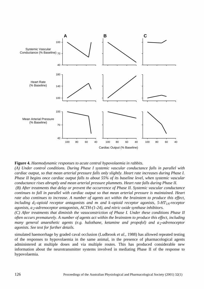
126 Proceedings of the Australian Physiological and Pharmacological Society (2001) 32(1)
Figure 4. Haemodynamic responses to acute central hypovolaemia in rabbits. (A) Under control conditions. During Phase I systemic vascular conductance falls in parallel with cardiac output, so that mean arterial pressure falls only slightly. Heart rate increases during Phase I. Phase II begins once cardiac output falls to about 55% of its baseline level, when systemic vascular conductance rises abruptly and mean arterial pressure plummets. Heart rate falls during Phase II. (B) After treatments that delay or prevent the occurrence of Phase II. Systemic vascular conductance continues to fall in parallel with cardiac output so that mean arterial pressure is maintained. Heart rate also continues to increase. A number of agents act within the brainstem to produce this effect, including δ1-opioid receptor antagonists and µ- and κ-opioid receptor agonists, 5-HT1A-receptor agonists, α2-adrenoceptor antagonists, ACTH-(1-24), and nitric oxide synthase inhibitors. (C) After treatments that diminish the vasoconstriction of Phase I. Under these conditions Phase II often occurs prematurely. A number of agents act within the brainstem to produce this effect, including many general anaesthetic agents (e.g. halothane, ketamine and propofol) and α2-adrenoceptor agonists. See text for further details.
simulated haemorrhage by graded caval occlusion (Ludbrook et al., 1988) has allowed repeated testing of the responses to hypovolaemia in the same animal, in the presence of pharmacological agents administered at multiple doses and via multiple routes. This has produced considerable new information about the neurotransmitter systems involved in mediating Phase II of the response to hypovolaemia.
Systemic VascularConductance (% Baseline)
100
140
180
40608010040
70
100
406080100 406080100
40
70
100
Heart Rate(% Baseline)
Mean Arterial Pressure(% Baseline)
Cardiac Output (% Baseline)
A B C
Proceedings of the Australian Physiological and Pharmacological Society (2001) 32(1) 127
Opioidergic Mechanisms
The work of Holaday and his colleagues in the late 1970s and early 1980s provided the first evidence for the involvement of endogenous opioids in the hypotension of severe haemorrhage (Holaday, 1983). They showed, in anaesthetized rats, that the opioid antagonist naloxone increased arterial pressure in rats in haemorrhagic shock, but not in normovolaemic rats. Subsequent studies by Holaday’s group, and by others in a range of animal species, showed that the pressor effect of naloxone in hypovolaemic animals was mediated chiefly by increased sympathoadrenal activity (Schadt & Ludbrook, 1991). In 1988, Ludbrook and Rutter (1988) and Burke and Dorward (1988) demonstrated that naloxone had a very specific effect. It prevented the failure of sympathetic vasomotor drive and peripheral vascular resistance that occurs once blood loss reaches the critical level for initiation of Phase II of the response to haemorrhage (Fig. 4). Subsequent studies demonstrated that the effect of naloxone was due to blockade of δ1-opioid receptors in the brainstem (Evans et al., 1989a,b; Ludbrook & Ventura, 1994). Thus, a brainstem δ1-opioid receptor mechanism appears to be critical for triggering Phase II of the response to acute hypovolaemia (Fig. 3). Recent evidence suggests that central opioid mechanisms might have a more general role in mediating responses to activation of cardiac mechanoreceptors, since naloxone can inhibit responses to a range of stimuli thought to increase cardiac mechanoreceptor activity, including the sympathoinhibitory response to cardiac tamponade in conscious rabbits (Hagiike et al., 1999), the systemic vasodilator and natriuretic responses to plasma volume expansion in conscious rabbits (Shweta et al., 1999), and increased effective renal blood flow during head-out water immersion in humans (van Tilborg et al., 1995), . On the other hand, endogenous opioids do not appear to play a role in mediating responses to activation of cardiac chemoreceptors (Evans & Ludbrook, 1991), or arterial baroreceptors (Evans et al., 1989a,b). The precise neuroanatomical site of the opioidergic mechanism mediating Phase II of the response to hypovolaemia remains to be determined. In rabbits, δ-opioid receptors have been identified in the nucleus tractus solitarius (May et al., 1989). Considering the proximity of this nucleus to the tip of 4th ventricular catheters used for studying central opioidergic mechanisms in this species (Evans et al., 1989a,b), and its role as a relay station for arterial and cardiopulmonary baroreceptor input (see Central nervous system pathways), it must be considered a major candidate . On the other hand, pre-synaptic δ-opioid receptors have also been identified in various brainstem regions in the rat, and in particular on bulbospinal C1 neurones in the rostral ventrolateral medulla, which in turn synapse with sympathetic preganglionic neurons (Stasinopoulos et al., 2000). There also appears to be a role for spinal cord δ-opioid receptors in mediating Phase II in rats (Ang et al., 1999). Other central opioid receptor subtypes might also play some role in Phase II of the response to hypovolaemia, since 4th ventricular administration of µ- (Evans & Ludbrook, 1990) and κ-opioid agonists (Evans et al., 1989b) prevents phase II in conscious rabbits. Species differences are also likely, since blockade of µ-receptors prevents Phase II in conscious rats (Ang et al., 1999) but not rabbits (Evans & Ludbrook, 1990) (Figs 3 & 4). Monoaminergic mechanisms
A central nervous system serotonergic mechanism was proposed as contributing to Phase II of the response to hypovolaemia, based on the observation that the 5-hydroxytryptamine (5-HT) mixed agonist/antagonist methsergide prevented Phase II of the response to simulated haemorrhage in conscious rabbits (Evans et al., 1992b) (Fig. 4). It was concluded that this effect of methysergide was mediated in the brainstem, based on its differential potency when administered intravenously, and into the 4th ventricle, the pontomedullary cistern, the lateral ventricle, and the spinal subarachnoid space (Evans et al., 1992b). However, the effects of methysergide were not mimicked by the serotonergic neurotoxins p-chlorophenylalanine methyl ester or 5,7-dihydroxytryptamine (Evans et al., 1992b).
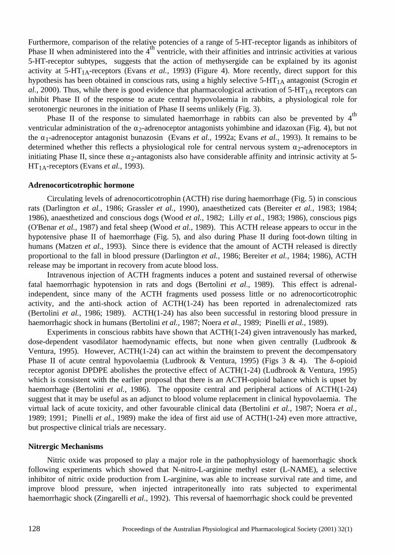
128 Proceedings of the Australian Physiological and Pharmacological Society (2001) 32(1)
Furthermore, comparison of the relative potencies of a range of 5-HT-receptor ligands as inhibitors of Phase II when administered into the 4th ventricle, with their affinities and intrinsic activities at various 5-HT-receptor subtypes, suggests that the action of methysergide can be explained by its agonist activity at 5-HT1A-receptors (Evans et al., 1993) (Figure 4). More recently, direct support for this hypothesis has been obtained in conscious rats, using a highly selective 5-HT1A antagonist (Scrogin et al., 2000). Thus, while there is good evidence that pharmacological activation of 5-HT1A receptors can inhibit Phase II of the response to acute central hypovolaemia in rabbits, a physiological role for serotonergic neurones in the initiation of Phase II seems unlikely (Fig. 3). Phase II of the response to simulated haemorrhage in rabbits can also be prevented by 4th ventricular administration of the α2-adrenoceptor antagonists yohimbine and idazoxan (Fig. 4), but not the α1-adrenoceptor antagonist bunazosin (Evans et al., 1992a; Evans et al., 1993). It remains to be determined whether this reflects a physiological role for central nervous system α2-adrenoceptors in initiating Phase II, since these α2-antagonists also have considerable affinity and intrinsic activity at 5-HT1A-receptors (Evans et al., 1993). Adrenocorticotrophic hormone
Circulating levels of adrenocorticotrophin (ACTH) rise during haemorrhage (Fig. 5) in conscious rats (Darlington et al., 1986; Grassler et al., 1990), anaesthetized cats (Bereiter et al., 1983; 1984; 1986), anaesthetized and conscious dogs (Wood et al., 1982; Lilly et al., 1983; 1986), conscious pigs (O'Benar et al., 1987) and fetal sheep (Wood et al., 1989). This ACTH release appears to occur in the hypotensive phase II of haemorrhage (Fig. 5), and also during Phase II during foot-down tilting in humans (Matzen et al., 1993). Since there is evidence that the amount of ACTH released is directly proportional to the fall in blood pressure (Darlington et al., 1986; Bereiter et al., 1984; 1986), ACTH release may be important in recovery from acute blood loss. Intravenous injection of ACTH fragments induces a potent and sustained reversal of otherwise fatal haemorrhagic hypotension in rats and dogs (Bertolini et al., 1989). This effect is adrenal- independent, since many of the ACTH fragments used possess little or no adrenocorticotrophic activity, and the anti-shock action of ACTH(1-24) has been reported in adrenalectomized rats (Bertolini et al., 1986; 1989). ACTH(1-24) has also been successful in restoring blood pressure in haemorrhagic shock in humans (Bertolini et al., 1987; Noera et al., 1989; Pinelli et al., 1989). Experiments in conscious rabbits have shown that ACTH(1-24) given intravenously has marked, dose-dependent vasodilator haemodynamic effects, but none when given centrally (Ludbrook & Ventura, 1995). However, ACTH(1-24) can act within the brainstem to prevent the decompensatory Phase II of acute central hypovolaemia (Ludbrook & Ventura, 1995) (Figs 3 & 4). The δ-opioid receptor agonist DPDPE abolishes the protective effect of ACTH(1-24) (Ludbrook & Ventura, 1995) which is consistent with the earlier proposal that there is an ACTH-opioid balance which is upset by haemorrhage (Bertolini et al., 1986). The opposite central and peripheral actions of ACTH(1-24) suggest that it may be useful as an adjunct to blood volume replacement in clinical hypovolaemia. The virtual lack of acute toxicity, and other favourable clinical data (Bertolini et al., 1987; Noera et al., 1989; 1991; Pinelli et al., 1989) make the idea of first aid use of ACTH(1-24) even more attractive, but prospective clinical trials are necessary. Nitrergic Mechanisms
Nitric oxide was proposed to play a major role in the pathophysiology of haemorrhagic shock following experiments which showed that N-nitro-L-arginine methyl ester (L-NAME), a selective inhibitor of nitric oxide production from L-arginine, was able to increase survival rate and time, and improve blood pressure, when injected intraperitoneally into rats subjected to experimental haemorrhagic shock (Zingarelli et al., 1992). This reversal of haemorrhagic shock could be prevented
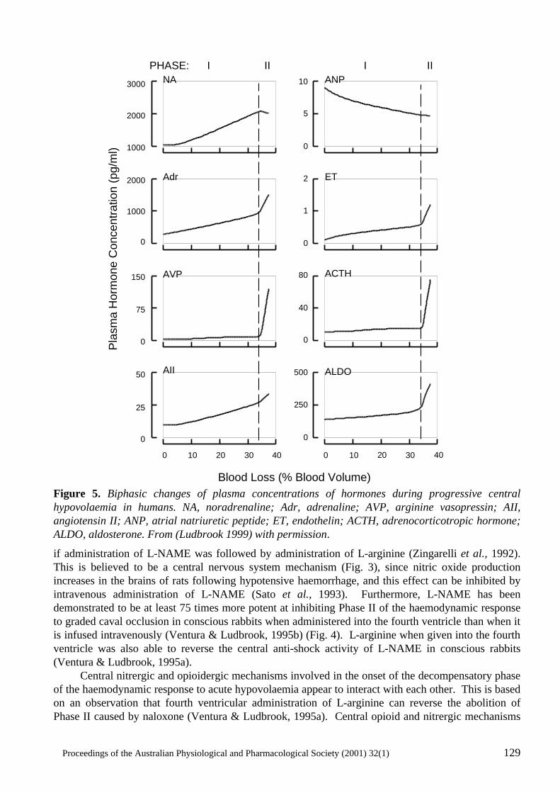
Proceedings of the Australian Physiological and Pharmacological Society (2001) 32(1) 129
Figure 5. Biphasic changes of plasma concentrations of hormones during progressive central hypovolaemia in humans. NA, noradrenaline; Adr, adrenaline; AVP, arginine vasopressin; AII, angiotensin II; ANP, atrial natriuretic peptide; ET, endothelin; ACTH, adrenocorticotropic hormone; ALDO, aldosterone. From (Ludbrook 1999) with permission.
if administration of L-NAME was followed by administration of L-arginine (Zingarelli et al., 1992). This is believed to be a central nervous system mechanism (Fig. 3), since nitric oxide production increases in the brains of rats following hypotensive haemorrhage, and this effect can be inhibited by intravenous administration of L-NAME (Sato et al., 1993). Furthermore, L-NAME has been demonstrated to be at least 75 times more potent at inhibiting Phase II of the haemodynamic response to graded caval occlusion in conscious rabbits when administered into the fourth ventricle than when it is infused intravenously (Ventura & Ludbrook, 1995b) (Fig. 4). L-arginine when given into the fourth ventricle was also able to reverse the central anti-shock activity of L-NAME in conscious rabbits (Ventura & Ludbrook, 1995a). Central nitrergic and opioidergic mechanisms involved in the onset of the decompensatory phase of the haemodynamic response to acute hypovolaemia appear to interact with each other. This is based on an observation that fourth ventricular administration of L-arginine can reverse the abolition of Phase II caused by naloxone (Ventura & Ludbrook, 1995a). Central opioid and nitrergic mechanisms
Pla
sma
Hor
mon
e C
once
ntra
tion
(pg/
ml)
PHASE: I II I II
AII
AVP
Adr
NA ANP
ET
ALDO
Blood Loss (% Blood Volume)
0
5
10
0
1
2
0
40
80 ACTH
0
250
500
403020100 403020100
0
0
3000
1000
2000
1000
2000
75
150
0
25
50
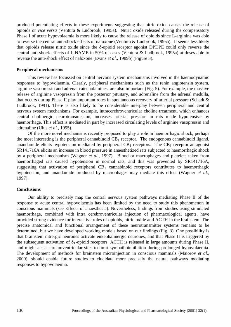
130 Proceedings of the Australian Physiological and Pharmacological Society (2001) 32(1)
produced potentiating effects in these experiments suggesting that nitric oxide causes the release of opioids or vice versa (Ventura & Ludbrook, 1995a). Nitric oxide released during the compensatory Phase I of acute hypovolaemia is more likely to cause the release of opioids since L-arginine was able to reverse the central anti-shock effects of naloxone (Ventura & Ludbrook, 1995a). It seems less likely that opioids release nitric oxide since the δ-opioid receptor agonist DPDPE could only reverse the central anti-shock effects of L-NAME in 50% of cases (Ventura & Ludbrook, 1995a) at doses able to reverse the anti-shock effect of naloxone (Evans et al., 1989b) (Figure 3). Peripheral mechanisms
This review has focussed on central nervous system mechanisms involved in the haemodynamic responses to hypovolaemia. Clearly, peripheral mechanisms such as the renin angiotensin system, arginine vasopressin and adrenal catecholamines, are also important (Fig. 5). For example, the massive release of arginine vasopressin from the posterior pituitary, and adrenaline from the adrenal medulla, that occurs during Phase II play important roles in spontaneous recovery of arterial pressure (Schadt & Ludbrook, 1991). There is also likely to be considerable interplay between peripheral and central nervous system mechanisms. For example, intracerebroventricular choline treatment, which enhances central cholinergic neurotransmission, increases arterial pressure in rats made hypotensive by haemorrhage. This effect is mediated in part by increased circulating levels of arginine vasopressin and adrenaline (Ulus et al., 1995). Of the more novel mechanisms recently proposed to play a role in haemorrhagic shock, perhaps the most interesting is the peripheral cannabinoid CB1 receptor. The endogenous cannabinoid ligand, anandamide elicits hypotension mediated by peripheral CB1 receptors. The CB1 receptor antagonist SR141716A elicits an increase in blood pressure in anaesthetized rats subjected to haemorrhagic shock by a peripheral mechanism (Wagner et al., 1997). Blood or macrophages and platelets taken from haemorrhaged rats caused hypotension in normal rats, and this was prevented by SR141716A, suggesting that activation of peripheral CB1 cannabinoid receptors contributes to haemorrhagic hypotension, and anandamide produced by macrophages may mediate this effect (Wagner et al., 1997). Conclusions
Our ability to precisely map the central nervous system pathways mediating Phase II of the response to acute central hypovolaemia has been limited by the need to study this phenomenon in conscious mammals (see Effects of anaesthesia). Nevertheless, findings from studies using simulated haemorrhage, combined with intra cerebroventricular injection of pharmacological agents, have provided strong evidence for interactive roles of opioids, nitric oxide and ACTH in the brainstem. The precise anatomical and functional arrangement of these neurotransmitter systems remains to be determined, but we have developed working models based on our findings (Fig. 3). One possibility is that brainstem nitrergic neurones activate enkephalinergic neurones, and that Phase II is triggered by the subsequent activation of δ1-opioid receptors. ACTH is released in large amounts during Phase II, and might act at circumventricular sites to limit sympathoinhibition during prolonged hypovolaemia. The development of methods for brainstem microinjection in conscious mammals (Maiorov et al., 2000), should enable future studies to elucidate more precisely the neural pathways mediating responses to hypovolaemia.
Proceedings of the Australian Physiological and Pharmacological Society (2001) 32(1) 131
References
Anderson, W.P. & Szenasi, G. (1994) Regional blood flow - renal. In: Blood loss and shock. ed. Secher, N., Pawelczyk, J. & Ludbrook, J. Ch. 10, pp. 121-131. .London: Edward Arnold.
Ang, K.K., McRitchie, R.J., Minson, J.B., Llewellyn-Smith, I.J., Pilowsky, P.M., Chalmers, J.P. & Arnolda, L.F. (1999) Activation of spinal opioid receptors contributes to hypotension after hemorrhage in conscious rats. American Journal of Physiology, 276, H1552-H1558.
Badoer, E., McKinley, M.J., Oldfield, B.J. & McAllen, R.M. (1993) A comparison of hypotensive and non-hypotensive hemorrhage on Fos expression in spinally projecting neurons of the paraventricular nucleus and rostral ventrolateral medulla. Brain Research, 610, 216-223.
Badoer, E. & Merolli, J. (1998) Neurons in the hypothalamic paraventricular nucleus that project to the rostral ventrolateral medulla are activated by haemorrhage. Brain Research, 791, 317-320.
Bandler, R., Price, J.L. & Keay, K.A. (2000) Brain mediation of active and passive emotional coping. Progress in Brain Research, 122, 333-349.
Barcroft, H., McMichael, J., Edholm, O.G. & Sharpey-Schafer, E.P. (1944) Posthaemorrhagic fainting: study by cardiac output and forearm flow. Lancet, 1, 489-491.
Bereiter, D.A, Plotsky, P.M. & Gann, D.S. (1983) Selective opiate modulation of the physiological responses to hemorrhage in the cat. Endocrinology, 113, 1439-1446.
Bereiter, D.A., Zaid, A.M. & Gann, D.S. (1984) Adrenocorticotropin response to graded blood loss in the cat. American Journal of Physiology, 247, E398-E404.
Bereiter, D.A., Zaid, A.M. & Gann, D.S. (1986) Effect of rate of hemorrhage on release of ACTH in cats. American Journal of Physiology, 250, E76-E81.
Bertolini, A., Ferrari, W. & Guarini, S. (1989) The adrenocorticotropic hormone (ACTH)-induced reversal of hemorrhagic shock. Resuscitation, 18, 253-267.
Bertolini, A., Guarini, S. & Ferrari, W. (1986). Adrenal-independent, anti-shock effect of ACTH-(1-24) in rats. European Journal of Pharmacology, 122, 387-388.
Bertolini, A., Guarini, S., Ferrari, W., Noera, G., Massini, C. & Di Tizio, S. (1987) ACTH-(1-24) restores blood pressure in acute hypovolaemia and haemorrhagic shock in humans. European Journal of Clinical Pharmacology, 32, 537-538.
Bertolini, A., Guarini, S., Ferrari, W. & Rompianesi, E. (1986) Adrenocorticotropin reversal of experimental hemorrhagic shock is antagonized by morphine. Life Sciences, 39, 1271-1280.
Blake, D.W., Ludbrook, J. & Van Leeuwen, A.F. (2000) Dexmedetomidine and haemodynamic responses to acute central hypovolaemia in conscious rabbits. Clinical and Experimental Pharmacology and Physiology, 27, 801-809.
Burke, S. L. & Dorward, P.K. (1988) Influence of endogenous opiates and cardiac afferents on renal nerve activity during haemorrhage in conscious rabbits. Journal of Physiology, 402, 9-27.
Dampney, R.A.L. (1994) Organization of central cardiovascular pathways. Physiological Reviews, 74, 323-364.
Darlington, D.N., Shinsako, J. & Dallman, M.F. (1986) Responses of ACTH, epinephrine, norepinephrine, and cardiovascular system to hemorrhage. American Journal of Physiology, 251, H612-H618.
Ebert, T.J., Kotrly, K.J., Vucins, E.J., Pattison, C.Z. & Kampine, J.P. (1985) Halothane anesthesia attenuates cardiopulmonary baroreflex control of peripheral resistance in humans. Anesthesiology, 63, 668-674.
132 Proceedings of the Australian Physiological and Pharmacological Society (2001) 32(1)
Ebert, T.J., Kotrly, K.J., Madsen, K.E., Bernstein, J.S. & Kampine, J.P. (1988) Fentanyl-diazepam anesthesia with or without N2O does not attenuate cardiopulmonary baroreflex-mediated vasoconstrictor responses to controlled hypovolemia in humans. Anesthesia and Analgesia, 67, 548-554.
Escourrou, P., Raffestin, B., Papelier, Y., Pussard, E. & Rowell, L.B. (1993) Cardiopulmonary and carotid baroreflex control of splanchnic and forearm circulations. American Journal of Physiology, 264, H777-H782.
Evans, R.G., Hayes, I.P. & Ludbrook, J. (1992a) Effects of α-adrenoceptor antagonists and clonidine on the haemodynamic response to acute hypovolaemia in conscious rabbits. European Journal of Pharmacology, 216, 265-272.
Evans, R.G., Haynes, J.M. & Ludbrook, J. (1993) Effects of 5-HT-receptor and α2-adrenoceptor ligands on the haemodynamic response to acute central hypovolaemia in conscious rabbits. British Journal of Pharmacology, 109, 37-47.
Evans, R.G., Kapoor, V. & Ludbrook, J. (1992b). A CNS serotonergic mechanism in acute central hypovolemia in conscious rabbits. Journal of Cardiovascular Pharmacology, 19, 1009-1017.
Evans, R. G. & Ludbrook, J. (1990) Effects of µ-opioid receptor agonists on circulatory responses to simulated haemorrhage in conscious rabbits. British Journal of Pharmacology, 100, 421-426.
Evans, R.G. & Ludbrook, J. (1991) Chemosensitive cardiopulmonary afferents and the haemodynamic response to simulated haemorrhage in conscious rabbits. British Journal of Pharmacology, 102, 533-539.
Evans, R.G. Ludbrook, J. & Potocnik, S.J. (1989a) Intracisternal naloxone and cardiac nerve blockade prevent vasodilatation during simulated haemorrhage in awake rabbits. Journal of Physiology, 409, 1-14.
Evans, R.G., Ludbrook, J. & Van Leeuwen, A.F. (1989b) Role of central opiate receptor subtypes in the circulatory responses of awake rabbits to graded caval occlusions. Journal of Physiology, 419, 15-31.
Evans, R.G., Ludbrook, J., Woods, R.L. & Casley, D. (1991) Influence of higher brain centres and vasopressin on the haemodynamic response to acute central hypovolaemia in rabbits. Journal of the Autonomic Nervous System, 35, 1-14.
Evans, R.G., Ludbrook. J. & Ventura, S. (1994) Role of vagal afferents in the haemodynamic response to acute central hypovolaemia in unanaesthetized rabbits. Journal of the Autonomic Nervous System, 46, 251-260.
Fitzpatrick, A.P., Banner, N., Cheng, A., Yacoub, M. & Sutton, R. (1993) Vasovagal reactions may occur after orthotopic heart transplantation. Journal of the American College of Cardiology, 21, 1132-1137.
Grassler, J., Jezova, D., Kvetnasky, R. & Scheuch, D.W. (1990) Hormonal responses to hemorrhage and their relationship to individual hemorrhagic shock susceptibility. Endocrinologia Experimentalis, 24, 105-116.
Hagiike, M., Maeta, H., Murakami, H., Okada, K. & Morita, H. (1999) Mechanism of biphasic response of renal nerve activity during cardiac tamponade in conscious rabbits. American Journal of Physiology, 276, R1232-R1240.
Hales, S. (1964) Statical essays: containing haemastaticks. New York: Hafner Publishing Company.
Haunso, S. (1994). Coronary blood flow. In: Blood Loss and Shock. ed. Secher, N., Pawelczyk, J. & Ludbrook, J. Ch. 9, pp. 109-120. London: Edward Arnold.
Henderson, L.A., Keay, K.A. & Bandler, R. (1998) The ventrolateral periaqueductal grey projects to caudal brainstem depressor regions: a functional-anatomical and physiological study. Neuroscience, 82, 201-221.
Henderson, L.A., Keay, K.A. & Bandler, R. (2000) Caudal midline medulla mediates behaviourally-coupled but not baroreceptor-mediated vasodepression. Neuroscience, 98, 779-792.
Proceedings of the Australian Physiological and Pharmacological Society (2001) 32(1) 133
Holaday, J.W. (1983) Cardiovascular effects of endogenous opiate systems. Annual Review of Pharmacology and Toxicology, 23, 541-594.
Kenny, R.A., Ingram, A., Bayliss, J. & Sutton, R. (1986) Head-up tilt: a useful test for investigating unexplained symcope. Lancet, 1, 1352-1355.
Khanna, S., Sibbald, J.R., Smith, D.W. & Day, T.A. (1994) Initiation of rat vasopressin cell responses to simulated hypotensive hemorrhage. American Journal of Physiology, 267, R1142-R1149.
Li, Y.-W. & Damney, R.A.L. (1994) Expression of fos-like protein in brain following sustained hypertension and hypotension in conscious rabbits. Neuroscience, 61, 613-634.
Lightfoot, J.T., Rowe, S.A. & Fortney, S.M. (1993) Occurrence of presyncope in subjects without ventricular innervation. Clinical Science, 85, 695-700.
Lilly, M.P., Engeland, W.C. & Gann, D.S. (1983) Responses of cortisol secretion to repeated hemorrhage in anaesthetized dog. Endocrinology, 112, 681-688.
Lilly, M.P., Engeland, W.C. & Gann, D.S. (1986) Pituitary-adrenal responses to repeated small hemorrhage in conscious dogs. American Journal of Physiology, 251, R1200-R1207.
Ludbrook, J. (1993) Haemorrhage and Shock. In: Cardiovascular Reflex Control in Health and Disease. eds Hainsworth, R. & Mark, A.L. Ch. 18, pp. 463-490. London: W.B. Saunders.
Ludbrook, J. (1999) Shock and its management. In: Autonomic Failure 4th edn. ed. Mathias, C.J., Bannister, R.G. Ch. 47, pp. 461-467. Oxford: Oxford University Press.
Ludbrook, J., Potocnik, S.J. & Woods, R.L. (1988) Simulation of acute haemorrhage in unanaesthetized rabbits. Clinical and Experimental Pharmacology and Physiology, 15, 575-584.
Ludbrook, J. & Rutter, P.C. (1988) Effect of naloxone on haemodynamic responses to acute blood loss in unanaesthetized rabbits. Journal of Physiology, 400, 1-15.
Ludbrook, J. Ventura, S. (1994) The decompensatory phase of acute hypovolaemia in rabbits involves a central δ1-opioid receptor. European Journal of Pharmacology, 252, 113-116.
Ludbrook, J. & Ventura, S. (1995) ACTH-(1-24) blocks the decompensatory phase of the haemodynamic response to acute hypovolaemia in conscious rabbits. European Journal of Pharmacology, 275, 267-275.
Ludbrook, J. & Ventura, S. (1996) Roles of carotid baroreceptor and cardiac afferents in hemodynamic responses to acute central hypovolemia. American Journal of Physiology, 270, H1538-H1548.
Maiorov, D.N., Malpas, S.C. & Head, G.A. (2000) Influence of pontine A5 region on renal sympathetic nerve activity in conscious rabbits. American Journal of Physiology, 278, R311-R319.
Matzen, S., Secher, N.H., Knigge, U., Pawelczyk, J., Perko, G., Iversen, H., Bach, F.W. & Warberg, J. (1993) Effect of serotonin receptor blockade on endocrine and cardiovascular responses to head-up tilt in humans. Acta Physiologica Scandinavica, 149, 163-176.
May, C.N., Dashwood, M.R., Whitehead, C.J. & Mathias, C.J. (1989) Differential cardiovascular and respiratory responses to central administration of selective opioid agonists in conscious rabbits: correlation with receptor distribution. British Journal of Pharmacology, 98, 903-913.
Morita, H. & Vatner, S.F. (1985) Effect of hemorrhage on renal nerve activity in conscious dogs. Circulation Research, 57, 788-793.
Noera, G., Angiello, L., Biagi, B. & Pensa, P. (1991) Haemorrhagic shock in cardiac surgery. Pharmacological treatment with ACTH-(1-24). Resuscitation, 22, 123-127.
134 Proceedings of the Australian Physiological and Pharmacological Society (2001) 32(1)
Noera, G., Pensa, P., Guelfi, P., Biagi, B., Curcio, C. & Bentini, B. (1989) ACTH-(1-24) and hemorrhagic shock: preliminary clinical results. Resuscitation, 18, 145-147.
O'Benar, J.D., Hannon, J.P., Peterson, J.L. & Bossone, C.A. (1987) Beta-endorphin, ACTH, and cortisol response to hemorrhage in conscious pigs. American Journal of Physiology, 252, R953-R958.
Oren, R.M., Schobel, H.P., Weiss, R.M., Stanford, W. & Ferguson, D.W. (1993) Importance of left atrial baroreceptors in the cardiopulmonary baroreflex of normal humans. Journal of Applied Physiology, 74, 2672-2680.
Pinelli, G., Chesi, G., Di Donato, C., Reverzano, A. Marani, L. (1989) Preliminary data on the use of ACTH-(1-24) in human shock conditions. Resuscitation, 18, 149-150.
Potts, P.D., Ludbrook, J., Gillman-Gaspari, T.A., Horiuchi, J. & Dampney, R.A.L. (1999) Activation of brain neurons following central hypervolaemia and hypovolaemia: contribution of baroreceptor and non-baroreceptor inputs. Neuroscience, 95, 499-511.
Potts, P.D., Polson, J.W., Hirooka, Y. & Dampney, R.A.L. (1997) Effects of sinoaortic denervation on Fos expression evoked by hypertension and hypotension in conscious rabbits. Neuroscience, 77, 503-520.
Sato, S., Tominaga, T., Ohnishi, T. & Ohnishi, S.T. (1993) EPR spin-trapping study of nitric oxide formation during bilateral carotid occlusion in the rat. Biochimica Biophysica Acta, 1181, 195-197.
Schadt, J.C. & Ludbrook, J. (1991) Hemodynamic and neurohumoral responses to acute hypovolemia in conscious mammals. American Journal of Physiology, 260, H305-H318.
Schmidt, J.F. & Paulson, O.B. (1994) Cerebral blood flow and hypovolaemic shock. In: Blood Loss and Shock. ed. Secher, N., Pawelczyk, J. & Ludbrook, J. Ch. 8, pp. 95-108. London: Edward Arnold.
Scrogin, K.E., Johnson, A.K., Brooks, V.L. (2000) Methysergide delays the decompensatory responses to severe hemorrhage by activating 5-HT1A receptors. American Journal of Physiology, 279, R1776-R1786.
Shen, Y.-T., Knight, D.R., Thomas, J.X. & Vatner, S.F. (1990) Relative roles of cardiac receptors and arterial baroreceptors during haemorrhage in conscious dogs. Circulation Research, 66, 397-405.
Shweta, A., Malpas, S.C., Anderson, W.P. & Evans, R.G. (1999) Effects of naloxone on the haemodynamic and renal functional responses to plasma volume expansion in conscious rabbits. Pflügers Archiv, 439, 150-157.
Stasinopoulos, T., Goodchild, A.K., Christie, M.J., Chalmers, J. & Pilowsky, P.M. (2000) Delta opioid receptor immunoreactive boutons appose bulbospinal C1 neurons in the rat. Neuroreport, 11, 887-891.
Thorén, P. (1979) Role of cardiac vagal C-fibres in cardiovascular control. Reviews of Physiology, Biochemistry and Pharmacology, 86, 1-94.
Ulus, I.H., Arslan, Y., Savci, V. & Kiran, B.K. (1995) Restoration of blood pressure by choline treatment in rats made hypotensive by haemorrhage. British Journal of Pharmacology, 116, 1911-1917.
Van Leeuwen, A.F., Evans, R.G. & Ludbrook, J. (1990) Effects of halothane, ketamine, propofol and alfentanil anaesthesia on circulatory control in rabbits. Clinical and Experimental Pharmacology and Physiology, 17, 781-798.
van Tilborg, K.A., Rabelink, T.J. Koomans, H.A. (1995) Naloxone inhibits renal hemodynamic effect of head-out water immersion in humans. Kidney International, 48, 860-865.
Ventura, S. Ludbrook, J. (1995a) L-Arginine reverses the abolition of hypovolaemic decompensation by N-nitro-L-arginine methyl ester and naloxone in conscious rabbits. European Journal of Pharmacology, 294, 369-375.
Proceedings of the Australian Physiological and Pharmacological Society (2001) 32(1) 135
Ventura, S. & Ludbrook, J. (1995b) N-Nitro-L-arginine methyl ester blocks the decompensatory phase of acute hypovolaemia in conscious rabbits by a brainstem mechanism. European Journal of Pharmacology, 277, 265-269.
Wagner, J.A., Varga, K., Ellis, E.F., Rzigalinski, B.A., Martin, B.R. & Kunos, G. (1997) Activation of peripheral CB1 cannabinoid receptors in haemorrhagic shock. Nature, 390, 518-521.
Warren, J.V., Brannon, E.S., Stead, E.A.Jr & Merrill, A.J. (1945) The effect of venesection and the pooling of blood in the extremities on the atrial pressure and cardiac output in normal subjects with observations of acute circulatory collapse in three instances. Journal of Clinical Investigation, 24, 337-344.
Wood, C.E., Chen, H.-G. & Bell, M.E. (1989) Role of vagosympathetic fibers in the control of adrenocorticotropic hormone, vasopressin, and renin responses to hemorrhage in fetal sheep. Circulation Research, 64, 515-523.
Wood, C.E., Shinsako, J., Keil, L.C., Ramsay, D.J. & Dallman, M.F. (1982) Apparent dissociation of adrenocorticotropin and corticosteroid responses to 15 ml/kg hemorrhage in conscious dogs. Endocrinology, 110, 1416-1421.
Zingarelli, B., Squadrito, F., Altavilla, D., Calapai, G., Campo, G.M., Calo, M., Saitta, A. & Caputi, A.P. (1992) Evidence for a role of nitric oxide in hypovolemic hemorrhagic shock. Journal of Cardiovascular Pharmacology, 19, 982-986.
136 Proceedings of the Australian Physiological and Pharmacological Society (2001) 32(1)
STATISTICS IN PHYSIOLOGY AND PHARMACOLOGY: A SLOW AND ERRATIC LEARNING CURVE
John Ludbrook
Professorial Fellow, University of Melbourne, Parkville, Victoria 3010, Australia
(email: [email protected]). Summary
1. Learning how to apply statistical analyses to the results of experimental or clinical studies may take a lifetime of trial (and sometimes error), as it has done in the author’s case. There is no evidence that biomedical investigators of the present generation are on a steeper learning curve. Gross misunderstandings of the purpose and functions of statistical analysis are apparent in applications to research grant-giving bodies and ethics committees, in manuscripts submitted to journals, and sometimes in published papers.
2. Though estimation of minimal group (sample) size for a given power is an essential step in planning clinical studies, it seems to be employed rarely in experimental work. This is despite exhortations to restrict the number of animals used to a minimum.
3. Most investigators use hypothesis-testing to analyze their results, but their understanding of the meaning of the resultant P values is slight.
4. A flaw found almost universally in biomedical manuscripts is to make multiple inferences from the results of a single study. The goal of statistical analysis is to maintain the familywise Type I error-rate (risk of false-positive inference) at a predetermined level (usually 5%). But when multiple inferences are made from the same experiment, the risk of false-positive error is inflated.
5. Biomedical investigators have been quick to acquire computer statistics software and to use it to analyze their experiments. However, they have been slow to recognise the limitations of this software. These include: (a) Inadequate documentation of routines, so that neither the user nor the reader of published papers can be sure how the tests have been executed; (b) Flawed algorithms for the execution of statistical procedures; (c) Failure to recognise that the best software for their purposes is that which takes them just beyond their statistical horizons.
6. The obvious solution to these difficulties is to recruit a biomedical statistician into every research group, at a trivial cost. But properly-qualified biostatisticians are in desperately short supply in Australia. It follows that research groups, national grant-giving agencies, and academic institutions must make provision for the proper training and subsequent employment of biostatisticians.
Introduction
Over the past 50 years, the author has progressed from executing χ2 tests in 1951 to complex analyses of variance with as many as thirteen independent terms in 2000. This has not been a linear, or a painless, progression. For instance, in 1965 I presented my first paper before the Society. A distinguished member of APPS, who was both a physiologist and a mathematician, reduced me to incoherence by pointing out (quite correctly) the distinction between independent and related observations. Thanks to his comments, I was able to correct this error in the final publication (Ludbrook, 1966). The first exhortation to physiologists to analyze their results statistically dates back to a massive review in 1929, which contained no fewer than 694 references (Dunn, 1929)! This was almost premature, for at that time the techniques available consisted of calculating means and standard deviations, Pearson’s product-moment correlation coefficient, Pearsons’s χ2 test, and Student’s t test (not long after the t distribution was converted to a practical test of significance by Fisher in 1925).
Proceedings of the Australian Physiological and Pharmacological Society (2001) 32(1) 137
Dunn did not mention analysis of variance, though Fisher had at least hinted at it by 1923. It is worth reciting another piece of history. Virtually all the statistical procedures used today were described before the middle of the 20th century: that is, in the pre-computer era. Almost the only exceptions to this rule are the technique of bootstrapping, which is heavily computer-dependent, and some of the forms of survival analysis used in clinical trials. Thus the lead time from description of a new technique to its entering into the consciousness and practice of biomedical investigators seems to be about 50 years! In what follows, I shall attempt to describe what I see as some of the statistical difficulties facing biomedical investigators at the beginning of the third millenium. Despite the Gregorian origin of our calender, there will be nothing especially Christian in my comments. They refer to matters such as the estimation of group (sample) size in advance of studies; the problem of multiple inferences from a single experiment; the dangers of embracing computer statistical software uncritically; and the unfulfilled need for biostatisticians to be members of research teams. Group (sample) size and power
There is an enormous literature on the importance of adequate, yet not excessive, sized samples in clinical studies, especially those in which the effects of an intervention are compared with a placebo control, or in which the effects of a new intervention are compared with those of conventional treatment (Freiman et al., 1978; Friedman et al., 1996). These are called randomized, prospective, controlled clinical trials. Group (sample) sizes must be sufficient to maintain the Type II error-rate, or β (the risk of false negative inference) at an acceptable level, usually 10-20%. It is more usual to speak of the power to reject the null hypothesis, or (1 - β), for which an acceptable level is 80-90%. The preoccupation with adequacy of power and therefore of group size stems from the argument that it would be wrong, statistically and ethically, to conclude that there is no difference between treatments when, in fact, there is. At the same time, however, it is important not to recruit too many subjects into a trial, both on ethical grounds and because this would delay a change in clinical practice. It is curious that though the above considerations of group size and power are an important and universally accepted - indeed, universally required - element of clinical studies, it has never become part of the ethos of laboratory experimentation. One of the very few who have argued that it should be is Ian McCance (1989), yet his arguments seem to have fallen on deaf ears. Institutional animal ethics committees (AEC) are exhorted (though not very strongly) to address this matter (Australian Code of Practice for the Care and Use of Animals for Scientific Purposes, 1997), but they rarely seem to do so with any enthusiasm. And reviewers of manuscripts for journals that deal with animal experiments raise matters of group size and power only if the authors fail to confirm earlier reports of the occurrence of a given phenomenon. McCance was undoubtedly a prophet before his time, since it seems inevitable and, indeed, highly desirable that animal experimenters should not use more animals than are necessary to achieve, say, 80% power in their experiment; and neither should they use so few that a negative finding can be attributed to inadequate group size and power. The task of estimating minimal group sizes and power has been made easy by sets of tables (Machin et al., 1997) and software such as nQuery Advisor (Statistical Solutions, Statistical Solutions, Boston MA). The meaning of P values
Most of us use hypothesis-testing (P values) as the basis for analyzing the results of experiments. There is nothing wrong with this, though it is worth noting that there has been a putsch, especially from a British school of statistics, to use estimation (confidence intervals, CI) instead (Gardner & Altman, 1989). Few physiologists and pharmacologists have been pushed to use CIs, and there is no good reason why they should. That is, there is no strong evidence that investigators or readers find CIs more informative than P values (Walter, 1995).
138 Proceedings of the Australian Physiological and Pharmacological Society (2001) 32(1)
But what does a P value mean? To statisticians it can mean any or all of many things, but the simplest definition is that it corresponds to the probability of Type I error, or α (the risk of false-positive inference). More profound definitions depend on whether inferences are made under the population (Neyman-Pearson) model, which implies random sampling of defined populations; or under the randomization model, which implies taking non-random samples and randomizing the members to one or another set of conditions or treatments. The model of inference is somewhat incidental, except that statisticians tend to assume that experimenters take random samples (a very rare occurence), whereas experimenters assume that statisticians know that they do not (Ludbrook & Dudley, 1998). There is one other important matter to do with P. Another way of defining it is the probability of the observed (or more extreme) data when the null hypothesis, H0, is true. If one takes a t test for equality of the means of two groups (strictly, two populations), coded 1 and 2, then H0 can be defined as:
0 2 1H : x x=
But there is another consideration. If H0 is rejected, an alternative hypothesis (H1) must be accepted. But H1 can take one of two forms:
One-sided (specific) 1 2 1H : x x≥ Two-sided (non-specific) 1 2 1H : x x≠
In almost every circumstance in biomedical research (and in all circumstances in clinical research) the two-sided, non-specific, form of H1 should be preferred. Why? Because (at any rate in clinical research) one does not, and cannot, know that one treatment or state can never result in an outcome that cannot be worse than the other (specific H1). In clinical studies, it would be unethical to presume that this is so. In laboratory experiments, the same argument applies. Thus P should always be constructed in a two-sided, non-specific, fashion. Regrettably, investigators who are searching for ‘significant’ outcomes from their experiments sometimes select the one-sided outcome of a test because the P value is (usually) half that of the two-sided outcome. It is a matter of opinion whether this should be regarded as scientific fraud, or merely a form of ‘data torturing’ (Mills, 1993). There is one, last, point to make under this heading. Biomedical investigators have the habit of supporting their conclusions by statements such as ‘there was a significant difference’ (having stipulated earlier that they mean P ≤0.05, or merely by stating ‘P <0.05)’. There are two views about this convention. One is that if P= 0.05 has been defined as the watershed between ‘significant’and ‘not significant’, then it is sufficient to indicate this decision. The other view is that it is important to indicate the ‘strength of evidence’: that is, the actual P value attached to the null hypothesis. In the old days, when P values were obtained from published tables, actual P values were impossible. But nowadays, when electronic tables are available as stand-alone computer programs or within computer statistics programs, actual P values can be obtained. I am in no doubt that actual P values should be given. My arguments are simple. A mere P <0.05 does not distinguish between P= 0.04999 and P = 0.000004999. And actual values of P are essential if multiple comparisons are to be made (see below). Making multiple inferences from a single experiment
This causes great anguish to biomedical investigators. When statisticians or reviewers of manuscripts draw their attention to the matter, their reaction is more often than not frank disbelief that it could be of any importance. So I shall try to point out that it does matter, why it matters, and what can be done about it.
Proceedings of the Australian Physiological and Pharmacological Society (2001) 32(1) 139
Comparisonwise versus familywise (experimentwise) Type I error rates
The Type I error-rate conventionally nominated by investigators is 5%. The goal of any statistical analysis of experimental results is to not make false-positive statistical inferences, when a false-positive inference means that the Type I error-rate exceeds that nominated by the investigators. This is bound to occur if more than one inference is made from the results of a single experiment. Or, to put it crudely, if enough null hypotheses are tested and inferences made, then it is inevitable that a ‘significant’ P value will turn up. There are two approaches to controlling the Type I error-rate when multiple statistical inferences are made. One is to control the comparisonwise Type I error-rate. This implies that if any given comparison and consequent inference is made, then if the same comparison and inference were made a great many times, the frequency of false-positive inferences from that comparison would not exceed 5% in the long run. But control of the familywise (experimentwise) Type I error-rate implies that if the whole experiment actually performed were replicated a great many times, the frequency of any false-positive inference from the experiment would not exceed 5%. Or, in simple terms, are all the inferences made from the experiment as a whole able to be replicated? Those in the trade of biostatistics are in no doubt about which of the two error-rates should be controlled: the familywise (experimentwise). And, surely, biomedical investigators should hope that if others were to repeat their experiment, they would reach the same conclusions? What is a family of hypotheses?
Though statisticians are nearly unanimous in stating that the familywise Type I error-rate should be controlled, they are remarkably reticent about providing a definition of a family of hypotheses. One of the better definitions is that by Miller (1981). He said: “There are no hard-and-fast rules for where the family lines should be drawn...”. But, then, more helpfully: “The natural family for the author in the majority of instances is the individual experiment of a single researcher” (his italics). My own rules are as follows: (a) Usually, the family consists of all comparisons/inferences made from the results of a single experiment. This is not quite the same thing as saying that a family consists of all inferences drawn in a single published paper. For instance, a published paper might describe preliminary experiments in animals, followed by similar studies in humans. Surely it would be wrong to pool the two when it comes to defining a family of hypotheses? (b) I have some sympathy for a minimalist approach, in which all inferences made from the information provided in a single Table or Figure should be regarded as belonging to a family. How to control the familywise Type I error-rate
The best-known approach is to adjust the P values resulting from hypothesis (significance) testing. Some procedures make this adjustment under the assumption (explicit, or more often implicit) that the comparisonwise Type I error-rate should be controlled. These include Fisher’s restricted least significant difference (LSD), the Student-Newman-Keuls (SNK) procedure, and the Duncan multiple range procedure. These are quite popular among biomedical investigators, in part because they are provided by computer statistics packages (and in part, one fears, beause they are lenient). There are many procedures that control the familywise Type I error-rate. The problem is that some are altogether too conservative. The best-known is the Bonferroni procedure, brought to biomedical investigators’ attention by the classical paper by Wallenstein and his colleagues (1980). This entails multiplying the P values that result from the several (k) hypotheses by the number of hypotheses: that is the adjusted
P′ = kP Šidák’s rather more subtle version of this is that
P′ = 1/1 (1 ) kP− −
140 Proceedings of the Australian Physiological and Pharmacological Society (2001) 32(1)
Both these procedures provide complete protection against excessive Type I error. However, in both cases it is assumed that all the multiple hypotheses are independent of each other. This is very rarely the case. And if hypotheses are correlated, the Bonferroni and Šidák procedures are too harsh. Then there is a set of procedures that are designed specifically for data that have been measured on an interval scale (Wallenstein et al., 1980). These include the Tukey-Kramer procedure, for all possible pairwise contrasts among groups; the Dunnett procedure, for all pairwise contrasts of a control group against all others; and the Scheffé procedure, for all possible contrasts among groups, pairwise or other. All three procedures have been shown, by Monte Carlo simulation studies, to provide complete control over the familywise Type I error-rate in the particular circumstances. And all are available as sub-routines in the more popular computer statistics packages. Their virtues are that they allow for both independent and related hypotheses. Their defect is that they can be applied only to data measured on an interval (continuous) scale. Recently (that is, within the past 50 years!), another procedure has been described that caters for data measured on any scale (continuous, ordinal, categorical). All that is required is the actual P values attached to the several hypotheses/inferences that have been made within a given family. It has been shown by Monte Carlo simulations that it affords complete protection against excessive familywise Type I error and that, at the same time, it caters for both independent and related hypotheses and it is very powerful. This is the Ryan-Holm stepdown Bonferroni procedure. It is remarkably simple to execute with pencil-and-paper or a handheld calculator. It can be used to adjust P values, or CIs (Ludbrook, 1998, 2000). The global approach
There is another solution to the problem of multiple inferences, which can sometimes be used. It is to use multivariate techniques to arrive at a single P value for a global hypothesis (Table 1). These techniques are often complex, usually require the use of a computer statistics program, and are potentially dangerous in the hands of those who are unfamiliar with the theoretical basis for the procedures or with the particular statistics program that is used to execute them. They also often require an expert to interpret the outcome, and careful explanation to readers of the published paper. Nevertheless, the global approach can often be used to circumvent the problem of testing multiple hypotheses and making multiple inferences. Dangers of uncritical use of statistical computer software
I shall introduce this topic with an example. A few years ago a Swiss pharmacologist and I met over the Internet. He was puzzled that when he used the Wilcoxon-Mann-Whitney (WMW) rank-order test to analyze his experimental data, he arrived at two very different outcomes from two different statistics software packages. This ended up by our publishing the results of our analyzing these same data by no fewer than 11 commercial statistics packages (Bergmann et al., 2000). The range of outcomes was from P = 0.0885 to P = 0.0147. That is, in conventional terms, the outcomes ranged from decidedly ‘not significant’ to decidedly ‘significant’. The WMW procedure can be executed in at least 5 different ways. These are with a correction for ties, with a correction for continuity, with neither, and with both. These four are based on asymptotic (large sample) approximations to the normal or chi-squared distributions. The fifth way is by exact permutation. The first four variants of the WMW test can be executed by hand, and we did this. Our concern was not so much with the wide range of P values, as with the fact that so many of the statistics packages failed to describe, in their Manuals or in their Help files, precisely which variant(s) of the WMW procedure they executed. There was also an odd-man-out package, the outcome from which coincided with none of the above variants, and was clearly due to an algorithmic error (later admitted by the vendor). There was another difficulty, which was that many of the packages suggested that the P values resulting from
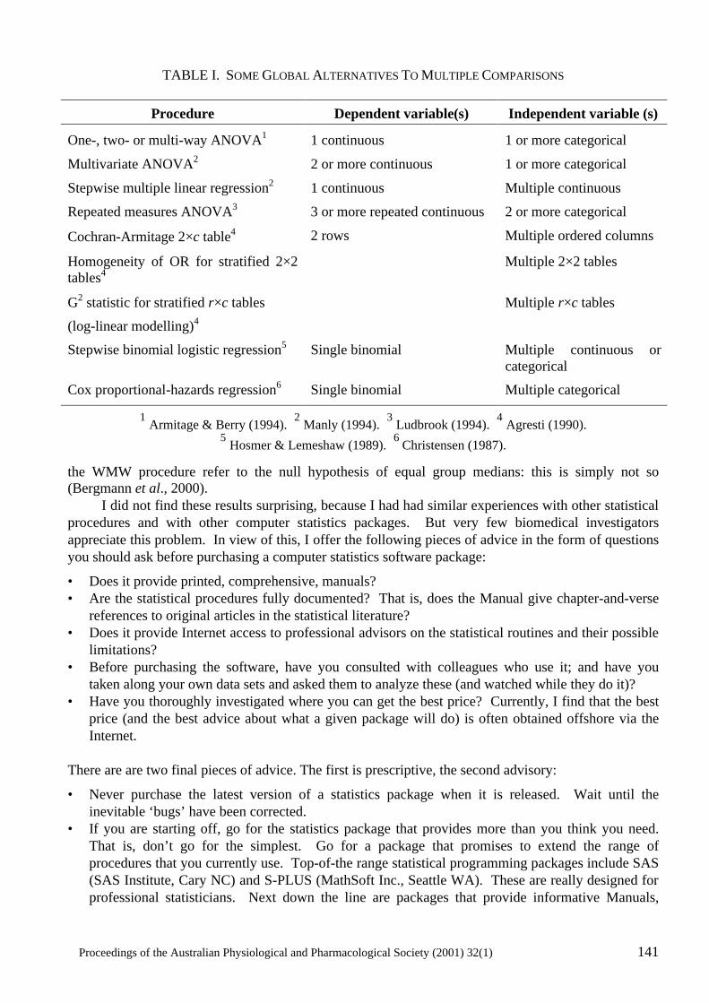
Proceedings of the Australian Physiological and Pharmacological Society (2001) 32(1) 141
TABLE I. SOME GLOBAL ALTERNATIVES TO MULTIPLE COMPARISONS
Procedure Dependent variable(s) Independent variable (s)
One-, two- or multi-way ANOVA1 1 continuous 1 or more categorical
Multivariate ANOVA2 2 or more continuous 1 or more categorical
Stepwise multiple linear regression2 1 continuous Multiple continuous
Repeated measures ANOVA3 3 or more repeated continuous 2 or more categorical
Cochran-Armitage 2×c table4 2 rows Multiple ordered columns
Homogeneity of OR for stratified 2×2 tables4
Multiple 2×2 tables
G2 statistic for stratified r×c tables
(log-linear modelling)4
Multiple r×c tables
Stepwise binomial logistic regression5 Single binomial Multiple continuous or categorical
Cox proportional-hazards regression6 Single binomial Multiple categorical
1 Armitage & Berry (1994). 2 Manly (1994). 3 Ludbrook (1994). 4 Agresti (1990). 5 Hosmer & Lemeshaw (1989). 6
Christensen (1987).
the WMW procedure refer to the null hypothesis of equal group medians: this is simply not so (Bergmann et al., 2000). I did not find these results surprising, because I had had similar experiences with other statistical procedures and with other computer statistics packages. But very few biomedical investigators appreciate this problem. In view of this, I offer the following pieces of advice in the form of questions you should ask before purchasing a computer statistics software package:
• Does it provide printed, comprehensive, manuals? • Are the statistical procedures fully documented? That is, does the Manual give chapter-and-verse
references to original articles in the statistical literature? • Does it provide Internet access to professional advisors on the statistical routines and their possible
limitations? • Before purchasing the software, have you consulted with colleagues who use it; and have you
taken along your own data sets and asked them to analyze these (and watched while they do it)? • Have you thoroughly investigated where you can get the best price? Currently, I find that the best
price (and the best advice about what a given package will do) is often obtained offshore via the Internet.
There are are two final pieces of advice. The first is prescriptive, the second advisory:
• Never purchase the latest version of a statistics package when it is released. Wait until the inevitable ‘bugs’ have been corrected.
• If you are starting off, go for the statistics package that provides more than you think you need. That is, don’t go for the simplest. Go for a package that promises to extend the range of procedures that you currently use. Top-of-the range statistical programming packages include SAS (SAS Institute, Cary NC) and S-PLUS (MathSoft Inc., Seattle WA). These are really designed for professional statisticians. Next down the line are packages that provide informative Manuals,
142 Proceedings of the Australian Physiological and Pharmacological Society (2001) 32(1)
online Internet help, and a wide variety of statistical procedures. These include SPSS (SPSS Inc., Chicago IL), SYSTAT (SPSS Inc., Chicago IL) and, perhaps, Statistica (StatSoft Inc, Tulsa OK). My advice is not to descend to the third level of elementary, ‘user-friendly’, and relatively cheap, programs (including spreadsheets).
Biostatisticians
The most obvious solution to the biostatistical difficulties that have been described above is to ensure that every group of biomedical investigators, large or small, has access to professional biostatistical advice. The emphasis is on ‘biostatistical’, not merely ‘statistical’, because investigators complain bitterly that if they approach a statistical consulting service they find that a great deal of their time (and money) is spent in explaining the biological problem to the statistician. A review of the 1998-2000 Annual Reports of the larger biomedical research institutes in Australia shows that the average cost of production of each publication is about $140 000. Diversion of even 1% of this should provide an excellent biostatistical consulting service. The same argument, though on a different scale, applies to smaller research groups. However, if biomedical investigators have the will to invest in biostatistical expertise, there is a very serious practical obstacle to finding a way. It is that there is a serious shortage of biostatisticians in Australia. Attention has been drawn to this in the context of epidemiology by Carlin (1997). A Biostatistics Collaboration of Australia has been formed. It has urged the Department of Health & Aged Care to set aside, within the Public Health Education and Research Program (PHERP), funds for the development of a three-tier biostatistical award structure to develop and deliver by distance mode a Graduate Certificate, Graduate Diploma, or Masters Degree in Biostatistics. The Biostatistics Collaboration has also developed a Curriculum Outline for such courses. Information on these matters can be obtained at the website www.ctc.usyd.edu.au/BCA. If the laudable initiatives undertaken by the Biostatistics Collaboration are successful, at least a start will have been made in rectifying the shortage of biostatisticians in Australia. But two problems will remain. One is to assure employment for the Diplomates and Graduates in Biostatistics within the broad field of biomedical research. The other touches the disciplines within which members of APPS work: usually laboratory experimental research, whether in humans, animals, tissues or cells (Ludbrook, 1998). It is not at all certain that the postgraduate qualifications proposed by the Biostatistics Collaboration will lead to an understanding of the experimental techniques and designs used in these research disciplines, or of the corresponding statistical techniques for analyzing the results of such experiments. Members of APPS and of other societies concerned with experimental biology should examine this question. References
Armitage, P. & Berry, G. (1994) Statistical Methods in Medical Research. 3rd edn. Oxford: Blackwell Scientific Publications.
Agresti, A. (1990) Categorical Data Analysis. New York: John Wiley & Sons.
Australian Code of Practice for the Care and Use of Animals for Scientific Purposes. 6th edn. (1997) Canberra: National Health and Medical Research Council.
Bergmann, R., Ludbrook, J. & Spooren, W.P.J.M. (2000) Different outcomes of the Wilcoxon-Mann-Whitney test from different statistics packages. The American Statistician, 54, 72-77.
Carlin, J. (1997) Biostatistical expertise in Australia: is there enough and are we doing the right training? (letter) Australasian Epidemiologist, 4, 12-13.
Christensen, E. (1989) Multivariate survival analysis using Cox’s regression model. Hepatology, 7, 1346-1358.
Proceedings of the Australian Physiological and Pharmacological Society (2001) 32(1) 143
Dunn, H.L. (1929) Application of statistical methods in physiology. Physiological Reviews, 9, 275-398.
Fisher, R.A. (1925) Applications of “Student’s” distribution. Metron, 5, 90-104.
Freiman, J.A., Chalmers, T.C., Smith, H. & Kuebler, R.R. (1978) The importance of beta, the type II error, and sample size in the design and interpretation of the randomized control trial. New England Journal of Medicine, 299, 691-694.
Friedman, L.M., Furberg, C.D. & DeMets, D.L. (1996) Fundamentals of Clinical Trials.. 3rd edn. St Louis: Mosby.
Gardner, M.J. & Altman, D.G. (1989) Statistics with confidence: confidence intervals and statistical guidelines. London: British Medical Journal.
Hosmer, D.W. & Lemeshaw, S. (1989) Applied Logistic Regression. New York: John Wiley & Sons.
Ludbrook, J. (1966) Collateral artery resistance in the human lower limb. Journal of Surgical Research, 6, 423-434.
Ludbrook, J. (1994) Repeated measurements and multiple comparisons in cardiovascular research. Cardiovascular Research, 28, 303-311.
Ludbrook, J. (1998) Multiple comparison procedures updated. Clinical and Experimental Pharmacology and Physiology, 25, 1032-1037.
Ludbrook, J. (1998) Biomedical laboratory scientists in Australia also need better access to biostatistical expertise (letter). Australasian Epidemiologist, 5, 14.
Ludbrook, J. (2000) Multiple inferences using confidence intervals. Clinical and Experimental Pharmacology and Physiology, 27, 212-215.
Ludbrook, J. & Dudley H. (1998) Why permutation tests are superior to t and F tests in biomedical research. The American Statistician, 52, 127-132.
McCance, I. (1989) The number of animals. News in Physiological Sciences, 4, 172-176.
Machin, D., Campbell, M.J., Fayers, P.M. & Pinol, A.P.Y. (1997) Sample Size Tables for Clinical Studies. 2nd edn. Oxford: Blackwell Science.
Manly, B.F.J. (1994) Multivariate Statistical Methods. 2nd edn. London: Chapman & Hall.
Miller, R.G. (1981) Simultaneous Statistical Inference. 2nd edn. New York: Springer-Verlag.
Mills, J.L. (1993) Data torturing. New England Journal of Medicine, 329, 1196-1199.
Wallenstein, S., Zucker, C.L. & Fleiss, J.L. (1980) Some statistical methods useful in circulation research. Circulation Research, 47, 1-9.
Walter, S.D. (1995) Methods of reporting statistical results from medical research studies. American Journal of Epidemiology, 141, 896-906.
144 Proceedings of the Australian Physiological and Pharmacological Society (2001) 32(1)
INTEGRATION OR DISINTEGRATION? Warwick P. Anderson, Department of Physiology, PO Box 13F, Monash University, Victoria 3800 In the coming decades, one of the greatest challenges in medical research will be to successfully integrate knowledge at many levels. Genomics gives us the power to understand the basic blocks from which life operates. But, as we move up the scale of complexity towards a fuller understanding of how whole individuals operate and what causes ill health, there is a rapidly increasing degree of complexity of interactions with environment - physical, social, cultural, psychological, time and so on. If physiologists grasp the opportunities that this brings, we can play a central role in helping to understand these complex relationships. To do this we will need to understand functional genomics, to incorporate bioinformatics in our approaches and to act as links between genetic reductionism and population health approaches. THIS PRE-ABSTRACT OF PROF ANDERSONS’S PAPER APPEARED IN THE PROCEEDINGS, ISSUE 32(1), AS 127P.
Proceedings of the Australian Physiological and Pharmacological Society (2001) 32(1) 145
INTEGRATIVE PHYSIOLOGY OF EXERCISE A Symposium Integrative Physiology of Exercise took place on Friday November 24th, 2000, during the Meeting of the Society at RMIT University in Melbourne. The Symposium contained five papers, three of which are reproduced here in pp. 146 – 168. These papers are to appear also in Clinical and Experimental Pharmacology and Physiooogy. The Symposium was chaired by Prof M. Hargreaves
146 Proceedings of the Australian Physiological and Pharmacological Society (2001) 32(1)
EXERCISE AND SKELETAL MUSCLE GENE EXPRESSION
David Cameron-Smith
School of Health Sciences, Deakin University, Burwood, Australia.
Abstract
1. Skeletal muscle is a complex and heterogenous tissue capable of remarkable adaptation in response to exercise training. The role of gene transcription, as an initial target to control protein synthesis, is poorly understood. 2. Mature myofibers contain several hundred nuclei, all of which maintain transcriptional competency, although the localised responsiveness of nuclei is not well known. Myofibers are capable of hypertrophy and recovery via the activation and myogenic differentiation of mononuclear satellite cells that fuse with the enlarging or repairing myofiber. 3. A single bout of exercise in human subjects is capable of activating the expression of many diverse groups of genes. 4. The impact of repeated exercise bouts, typical of exercise training, on gene expression has yet to receive systematic investigation. 5. The molecular program elicited by resistance exercise and endurance exercise differs markedly. Muscular hypertrophy following resistance exercise is dependent upon the activation of satellite cells and their subsequent myogenic maturation. Endurance exercise requires the simultaneous activation of mitochondrial and nuclear genes to enable mitochondrial biogenesis. 6. Future analysis of the regulation of genes by exercise may combine high throughput technologies, such as gene-chips, enabling the rapid detection and analysis of changes in the expression of many thousands of genes. Background
Mature skeletal muscle is a remarkably adaptive tissue, able to demonstrate significant regeneration, hypertrophy and metabolic adaptation (Goldspink, 1998). One of the most powerful stimuli for inducing skeletal muscle cellular re-organisation is exercise training. In adults, depending upon the type and duration of training, there can be appreciable changes in the size of the muscle mass and fiber-type composition, in addition to increased contractile activity and metabolic characteristics of the myofiber population (Booth et al., 1998). Considerable research has focused on the phenotypic nature of the adaptations, measured as alterations in protein abundance and activity, metabolic pathway flux and anatomical alterations. Yet surprisingly few studies have systematically addressed the role and importance of gene expression, despite the sometimes marked increase in protein levels. In this review the role of gene expression in skeletal muscle adaptability will be examined. Attention will be given to the differential gene programs elicited by varied exercise interventions, the hypothetical adaptations in gene expression following repeated exercise (training) and the emerging technologies that hold promise in analysing the responsiveness of many thousands of genes to exercise interventions. Regulation of Gene Transcription
The draft sequence of the human genome published February 2001 had as a major finding that the human genome contains approximately 30,000 genes (Venter et al., 2001), one-quarter of the number predicted only 12 months earlier (Liang et al., 2000). The smaller than anticipated gene
Proceedings of the Australian Physiological and Pharmacological Society (2001) 32(1) 147
number encodes approximately 85,000 different mRNAs, due to alternative mRNA splicing and variable polyadenylation (Beaudoing et al., 2000). mRNA synthesis or gene transcription is catalyzed by multisubunit RNA polymerase II, which is under the interacting and complex regulation of the chromatin, histone acetylation and many nuclear proteins, including ‘transcription factors’ (Macfarlane, 2000). Transcription factors comprise a diverse population of DNA-binding proteins, which in many instances are the terminal protein linking a complex signalling sequence with the activation of gene transcription. The activation of gene transcription results in the transient synthesis of mRNA, which may undergo modification to generate the mature transcript. The mature transcript exits the nucleus via the nuclear pores and when coupled to the translational machinery of the ribsome, is translated into the encoded peptide. Unique Regulation of Gene Transcription in Skeletal Muscle
Skeletal muscle is a complex tissue containing predominantly multinucleated mature myofibers. Each myofiber arises from the fusion of many hundred mononucleated progenator cells known as satellite cells, which are capable of undergoing myogenic programming and differentiation into mature myocytes (Seale & Rudnicki, 2000). Satellite cells remain present within skeletal muscle, even into old age, retaining their proliferative potential (Marsh et al., 1997). Satellite cell proliferation and subsequent differentiation into myocytes enables repair and hypertrophy of existing myofibers or the generation of new myofibers. Within mature myofibers the many nuclei remain transcriptionally competent and theoretically capable of upregulated gene synthesis (Newlands et al., 1998). However, significant compartmentalisation of gene expression is evident, with genes for the α-subunit of the acetylcholine receptor and α-dystrobrevin (involved in synapse maturation) predominantly expressed by nuclei in close proximity to the neuromuscular junction (Fontaine et al., 1988). This is further supported by the limited diffusion of mRNA within structurally complex cells (Rossi et al., 2000). Yet the expression of both endogenous muscle-specific genes and housekeeping genes is not uniform along the length of a myofiber, despite shared cytoplasm and evenly protein requirement along the myofiber (Newlands et al., 1998). The differences in individual nuclei action might be explained in terms of instability of the transcriptional complexes, such that small fluctuations in low abundance enhancers or transcription factors increase stability and regulate gene expression stochastically, in an off/on mode, rather than regulating the rate of transcription (McAdams & Arkin, 1997). To date it has yet to be determined whether exercise elicits the widespread activation of nuclei or alternatively localised nuclei have sustained periods of transcriptional activity. However, the large number of nuclei and the involvement of the entire length of the myofiber in contractile events suggest marked synthesis of new mRNA species is possible in myofibers. Exercise Regulates Gene Transcription
Numerous studies have now demonstrated that following a single exercise bout significant elevations in the concentration of mRNA species including; metabolic (Koval et al., 1998; Kraniou et al., 2000; Wadley et al., 2001), coordinatory (Puntschart et al., 1998; Gustafsson et al., 1999), and immunomodulatory (Febbraio & Koukoulas, 2000) genes are observed in muscle samples from healthy human subjects. In response to exercise gene transcription may be activated within seconds of contraction initiation, through to hours after the cessation of exercise or following the restoration of nutrient stores (Neufer et al., 1998). For example, the mRNA abundance of the fos proto-oncogene family (c-fos & fosB) has been reported to be more than 20-fold higher 4 minutes after the commencement of treadmill running in adult subjects (Puntschart et al., 1998). Yet, the majority of studies have demonstrated that gene expression is most significantly enhanced in the recovery period, following the completion of the exercise bout (Neufer et al., 1998). Using a novel run-on protocol to
148 Proceedings of the Australian Physiological and Pharmacological Society (2001) 32(1)
measure nuclear mRNA abundance, Neufer and colleagues (Pilegaard et al., 2000) have demonstrated widespread activation of genes in the hours following varying modes of exercise. Thus the predominant transcriptional response to exercise may be present in the recovery phase of exercise, rather than during the exercise bout. Preliminary data from our laboratory analysing the expression of 184 genes simultaneously using gene-array technology (Research Genetics, Human Genefilter) in human subjects in which biopsies were collected prior and immediately at the end of 40 minute cycling exercise (70% VO2 max) has supported these findings. Of the analysed genes more than 85% demonstrated a greater than 1.5 fold reduction in abundance (Cameron-Smith, unpublished data), demonstrating the predominant action may be the inhibition of transcription or degradation of mRNA during exercise, prior to selective transcriptional activity once exercise has ceased. Exercise Training and Gene Transcription
The impact on gene expression of exercise performed without prior familiarisation or training is likely to differ markedly from the response to repeated exercise bouts or the trained response. Few studies have systematically examined the changes in mRNA abundance following repeated bouts of the same exercise protocol, either maintained at the same absolute workload or matched relative to the improvements in exercise performance observed with training. Furthermore, the contribution of transcriptional (mRNA synthesis) versus translational (mRNA stability or translation efficiency) adaptations to a training-induced increase in protein levels are poorly understood. It is difficult to correlate mRNA with protein levels due to both the transient nature of mRNA following exercise and the protracted synthesis and longer-half lives of proteins. Caution must therefore be applied to the analysis of adaptive changes in both mRNA responses to exercise and the impact transcriptional compared with translational events contribute to protein synthesis. Although few studies have examined the transient changes in mRNA levels following an initial exercise bout and again after an adaptation or training period, significant adaptation is likely to occur. Of the available data it has been shown that eight weeks of leg knee-extensor training markedly attenuated (3-fold) the expression of vascular endothelial growth factor (VEGF) mRNA at a workload adjusted to increase as exercise capacity improved with training (Richardson et al., 2000). An alternative adaptive response to repeated exercise might be the selective activation of mRNA species which require repeated bouts of exercise prior to any measurable increase in mRNA abundance. In our laboratory, the expression of several genes involved in skeletal muscle fat transport (fatty acid transport protein) and oxidation (carnitine palmitoyltransferase 1) were not increased following a single exercise bout, but demonstrated increased basal and post-exercise expression following 9 days of 1 hour cycle training (Tunstall, unpublished data). Similarly, adaptations such as inhibitions in gene expression requiring repeated bouts of exercise have also been shown. After a single exercise bout no measurable impact has been observed on the expression of the myosin heavy chain (MHC) IIa isoform, yet 7 days of training led to reduced mRNA levels after the last exercise bout, demonstrating that suppression was evident only after repeated exercise bouts (O’Neill et al., 1999). Further studies are clearly required to elucidate the differential impact exercise training has on the expression of genes and the contribution this exerts in adaptive alterations in the cellular protein concentrations. Post-translational Events and Exercise Training
In experimental models of increased muscle loading, increased protein synthesis rate proceeds changes in mRNA abundance, implying that the efficiency of protein synthesis is enhanced (Welle et al., 1999). Several studies have now examined the activation of the rate-limiting step of protein synthesis, the initiation step in which the mRNA transcript is coupled to the ribosomal machinery. Several kinases involved in the initiation step of protein synthesis, including; the 70-kDA S6 protein
Proceedings of the Australian Physiological and Pharmacological Society (2001) 32(1) 149
kinase (p70S6K), protein kinase B (PKB/Akt) and terminal members of the MAPK kinase pathway (ERK and p38), have all recently been identified to be up-regulated by exercise (Nader & Esser, 2001). The extensive remodeling of muscle with exercise training also requires the activation of protein degradation pathways for removal of proteins no longer required. The ubiquitin-proteasome pathway is the major mechanism of selective protein degradation (Lecker et al., 1999). Chronic motor nerve stimulation for 28 days of the rabbit tibialis anterior muscle markedly increased total proteasome activity of muscle extracts, with upregulated protein abundance of the 20S proteasome subunit and two regulatory proteins, PA700 and PA28 (Ordway et al., 2000). Transcriptional Regulation in Exercise
• Resistance Exercise
A pronounced adaptive response to high-intensity or weight-bearing exercise interventions is muscle hypertrophy. The increased mass of active muscle groups is achieved by an increase in the volume of individual myofibers (Green et al., 1999). The enlarged myofiber can only expand with the insertion of new nuclei, as a constant ratio of nuclei to cytoplasmic volume is maintained throughout all hypertrophic responses (McCall et al., 1998). Thus hypertrophy is dependent upon the proliferative activation of satellite cells and their myogenic differentiation (Seale & Rudnicki, 2000), prior to fusion with the existing myofiber (Garry et al., 2000). Progression of satellite cells into myoblasts involves the regulation of muscle-specific proteins belonging to the basic-helix-loop-helix family of transcription factors. Members including: MyoD, myogenin, myf-5, MRF-4 and MEF-2 that collectively function as dominant activators of skeletal muscle differentiation (Perry & Rudnick, 2000). In the quiescent state these myogenic factors are expressed at very low levels in satellite cells, but once activated their abundance increases markedly (Cooper et al., 1999). Considerable progress has been made in elucidating the signaling pathways responsible for the activation of the myogenic differentiation pathway highlighting the pivotal role of endogenously derived factors regulated by shear- or stress-forces including, integrin-linked kinases (Huang et al., 2000), and increased intracellular calcium flux, activating the calcineurin intracellular signalling pathway (Olson & Williams, 2000). Exogenous hormone activation of IGF-1, which may participate in the activation of calcineurin (Musaro et al., 1999), in addition to angiotensin II (Gordon et al., 2001) and fibroblast growth factors (Scata et al., 1999) may also coordinate myogenic differentiation of satellite cells. • Endurance exercise
In contrast, endurance exercise typically results in a shift in myofibrillar components towards an increased abundance of slow isoform proteins, together with up-regulated mitochondrial and oxidative metabolism enzyme levels (Holloszy & Coyle, 1984). Interestingly, induction of slow myofibrillar isoform genes may also be dependent upon calcium and the calcineurin pathway (Delling et al., 2000). Thus an apparent anomalous situation arises in which calcium activation of calcineurin is pivotal in two vastly differing transcriptional programs, either hypertrophy or the activation of the slow myofiber cellular pattern and mitochondrial biogenesis. Partial explanation is evident in the pattern of intracellular calcium released by tonic low frequency motor nerve activity typical of endurance exercise, as compared to the high amplitude and short duration response of intense hypertophic physical activity (Chin et al., 1998). The resultant differing patterns of activation may result in the recruitment of alternative calcium-responsive kinases (Dolmetsch et al., 1997), or depend upon kinases being activated simultaneously to stimulate an integrative downstream regulator (Wu et al., 2000). Such a downstream integration of calcium signaling might be controlled by the myogenic transcription factor MEF-2, the activity of which is regulated by multiple signal cascades including calcineurin and calmodulin-dependent protein kinase pathways (Wu et al., 2000). However, much is to be discovered
150 Proceedings of the Australian Physiological and Pharmacological Society (2001) 32(1)
about how these pathways might intersect with those necessary for mitochondrial proliferation which requires the coordination of both nuclear genes and the genes contained within the mitochondria’s own DNA (Hood, 2001). Analysis Strategies
Current individual mRNA species are analysed using either hybridisation techniques (northern blotting or RNAse protection) or polymerase chain reaction (PCR) amplification of individual genes. Normalisation of gene abundance is based on comparison to genes that have a constant gene expression, the housekeeping genes. These techniques enable the semi-quantitative analysis of single or small numbers of genes and are labour intensive. Given that any phenotypic adaptation may require the activation and inhibition of many thousands of genes simultaneously, these techniques will provide limited new data on the scope or complexity of the adaptation. New technologies, particular high throughput gene scanning tools (including gene-chips) will provide dramatic insights into the complex patterns of gene expression necessary for the phenotype adaptation in muscle. The use of gene-chip technology to probe for patterns of gene expression have been applied to the analysis of fiber-type differences in gene expression (Campbell et al., 2001), skeletal muscle aging and species differences (Welle et al., 2001) and the molecular pathophysiology of muscular dystrophies (Chen et al., 2000). Gene chip technology, not withstanding the potential pitfalls (Knight, 2001), will enable the mass screening of genetic response to a wide variety of stimuli, including different exercise interventions. Importantly the inclusion of expressed sequence tags (ESTs), which represent partial gene sequences of genes of no described function, on gene-chips may aid in the identification of new and novel genes involved in the adaptive response of exercise to physical activity. Future Issues
There is much to be discovered in the pathways of gene regulation in response to exercise. Currently little is known of the complex integration of competing signalling cascades prior to the activation of exercise-sensitive genes. Further confounding these investigations is the large differences in individual responsiveness to exercise. The population differences in exercise capacity are unlikely to be due to polymorphisms of single genes, although the recent examination of the angiotensin-converting enzyme (ACE) gene has yielding interesting associations with performance in several (Williams et al., 2000; Folland et al., 2000), but not all studies (Rankinen et al., 2000). The recently published genomic scan of the HERITAGE family training study identified a number of potential genetic markers which correlate with changes in body composition (Chagnon et al., 2001). Such gene-scanning studies will increasingly define areas of the human genome that are linked to individual differences in exercise responsiveness. Beyond these issues will be the difficult task of converting genetic findings into outcomes that can benefit the health of diseased individuals, including the obese and the diabetic. Acknowledgements
The author wishes to thank the collaborations and support of Professor Mark Hargreaves, Professor Greg Collier and Dr Rodney Snow (School of Health Sciences, Deakin University). References
Beaudoing, E., Freier, S., Wyatt, J.R., Claverie, J.M. & Gautheret, D. (2000) Patterns of variant polyadenylation signal usage in human genes. Genome Research, 10, 1001-1010.
Proceedings of the Australian Physiological and Pharmacological Society (2001) 32(1) 151
Booth, F., Tseng, B., Fluck, M. & Carson, J. (1998) Molecular and cellular adaptation of muscle in response to physical training. Acta Physiologica Scandanavica, 162, 343-350.
Campbell, W.G., Gordon, S.E., Carlson, C.J., Pattison, J.S., Hamilton, M.T. & Booth, F.W. (2001) Differential global gene expression in red and white skeletal muscle. American Journal of Physiology: Cell Physiology, 280, C763-768.
Chagnon, Y.C., Rice, T., Perusse, L., Borecki, I.B., Ho-Kim, M.-A., Lacaille, M., et al. (2001) Genomic scan for genes affecting body composition before and after training in caucasians from HERITAGE. Journal of Applied Physiology, 90, 1777-1787.
Chen, Y.-W., Zhao, P., Borup, R. & Hoffman, E.P. (2000) Expression profiling in the muscular dystrophies: identification of novel aspects of molecular pathophysiology. Journal of Cell Biology, 161, 1321-1336.
Chin, E.R., Olson, E.N., Richardson, J.A., Yang, Q., Humphries, C., Shelton, J.M., et al. (1998) A calcineurin-dependent transcriptional pathway controls skeletal muscle fiber type. Genes and Development, 12, 2499-2509.
Cooper, R.N., Tajbakhsh, S., Mouly, V., Cossu, G., Buckingham, M. & Butler-Browne, G.S. (1999) In vivo satellite cell activation via Myf5 and MyoD in regenerating mouse skeletal muscle. Journal of Cell Science, 112, 2895-2901.
Delling, U., Tureckova, J., Lim, H., De Windt, L., Rotwein, P. & Molkentin, J. (2000) A calcineurin-NFATc3-dependent pathway regulates skeletal muscle differentiation and slow myosin heavy-chain expression. Molecular and Cellular Biology, 20, 6600-6611.
Dolmetsch, R.E., Lewis, R.S., Goodnow, C.C. & Healy, J.I. (1997) Differential activation of transcription factors induced by Ca2+ response amplitude and duration. Nature, 386, 855-858.
Febbraio, M. & Koukoulas, I. (2000) HSP72 gene expression progessively increases in human skeletal muscle during prolonged, exhaustive exercise Journal of Applied Physiology, 89, 1055-1060.
Folland, J., Leach, B., Little, T., Hawker, K., Myerson, S., Montgomery, H., et al. (2000) Angiotensin-converting enzyme genotype affects the response of human skeletal muscle to functional overload. Experimental Phyiology, 85, 575-579.
Fontaine, B., Sassoon, D., Buckingham, M. & Changeux, J.P. (1988) Detection of the nicotinic acetylcholine receptor alpha-subunit mRNA by in situ hybridization at neuromuscular junctions of 15-day-old chick striated muscles. EMBO Journal, 7, 603-609.
Garry, D., Meeson, A., Elterman, J., Zhao, Y., Yang, P. & Bassel-Duby, R. (2000) Myogenic stem cell function is impaired in mice lacking the forkhead/winged helix protein MNF. Proceedings of the National Academy of Sciences USA, 97, 5416-5421.
Goldspink, G. (1998) Selective gene expression during adaptation of muscle in response to different physiological demands. Comparative Biochemistry and Physiology B, 120, 5-15.
Gordon, S.E., Davis, B.S., Carlson, C.J. & Booth, F.W. (2001) ANG II is required for optimal overload-induced skeletal muscle hypertrophy. American Journal of Physiology. Endocrinology and Metabolism, 280, 150-159.
Green, H., Goreham, C., Ouyang, J., Ball-Burnett, M. & Ranney, D. (1999) Regulation of fiber size, oxidative potential, and capillarization in human muscle by resistance exercise. American Journal of Physiology: Regulatory, Integrative and Comparative Physiology, 276, R591-596.
Gustafsson, T., Puntschart, A., Kaijser, L., Jansson, E. & Sundberg, J. (1999) Exercise-induced expression of angiogenesis-related transcription and growth factors in human skeletal muscle. American Journal of Physiology: Heart and Circulatory Physiology, 276, H679-685.
152 Proceedings of the Australian Physiological and Pharmacological Society (2001) 32(1)
Holloszy, J.O. & Coyle, E.F. (1984) Adaptations of skeletal muscle to endurance exercise and their metabolic consequences. Journal of Applied Physiology, 56, 831-838.
Hood, D.A. (2001) Contractile activity-induced mitochondrial biogenesis in skeletal muscle. Journal of Applied Physiology, 90, 1137-1157.
Huang, Y., Li, J., Zhang, Y. & Wu, C. (2000) The roles of integrin-linked kinase in the regulation of myogenic differentiation. Journal of Cell Biology, 150, 861-872.
Knight, J. (2001) When the chips are down. Nature, 410, 860-861.
Koval, J.A., DeFronzo, R.A., O'Doherty, R.M., Printz, R., Ardehali, H., Granner, D.K., et al. (1998) Regulation of hexokinase II activity and expression in human muscle by moderate exercise. American Journal of Physiology: Endocrinology & Metabolism, 274, E304-308.
Kraniou, Y., Cameron-Smith, D., Misso, M., Collier, G. & Hargreaves, M. (2000) Effects of exercise on GLUT-4 and glycogenin gene expression in human skeletal muscle. Journal of Applied Physiology, 88, 794-796.
Lecker, S.H., Solomon, V., Mitch, W.E. & Goldberg, A.L. (1999) Muscle protein breakdown and the critical role of the ubiquitin-proteasome pathway in normal and disease states. Journal of Nutrition, 129, 227S-237S.
Liang, F., Holt, I., Pertea, G., Karamycheva, S., Salzberg, S.L. & Quackenbush, J. (2000) Gene index analysis of the human genome estimates approximately 120,000 genes. Nature Genetics, 25, 239-240.
Macfarlane, W.M. (2000) Demystified…..transcription. Molecular Pathology, 53, 1-7.
Marsh, D.R., Criswell, D.S., Carson, J.A. & Booth, F.W. (1997) Myogenic regulatory factors during regeneration of skeletal muscle in young, adult, and old rats. Journal of Applied Physiology, 83, 1270-1275.
McAdams, H.H. & Arkin, A. (1997) Stochastic mechanisms in gene expression. Proceedings of the National Academy of Sciences USA, 94, 814-819.
McCall, G., Allen, D., Linderman, J., Grindeland, R., Roy, R., Mukka, V., et al. (1998) Maintenance of myonuclear domain size in rat soleus after overload and growth hormone/IGF-I treatment. Journal of Applied Physiology, 84, 1407-1412.
Musaro, A., McCullagh, K., Naya, F., Olson, E. & Rosenthal, N. (1999) IGF-1 induces skeletal myocyte hypertrophy through calcineurin in association with GATA-2 and NF-ATc1. Nature, 400, 581-585.
Nader, G.A. & Esser, K.A. (2001) Intracellular signaling specificity in skeletal muscle in response to different modes of exercise. Journal of Applied Physiology, 90, 1936-1942.
Neufer, P., Ordway, G. & Williams, R. (1998) Transient regulation of c-fos, aB-crystallin, and hsp70 in muscle during recovery from contractile activity. American Journal of Physiology: Cell Physiology, 274, C341-346.
Newlands, S., Levitt, L., Robinson, C., Karpf, A., Hodgson, V., Wade, R., et al. (1998) Transcription occurs in pulses in muscle fibers. Genes & Development, 12, 2748-2758.
Olson, E. & Williams, R. (2000) Calcineurin signaling and muscle remodeling. Cell, 101, 689-692.
O'Neill, D., Zheng, D., Anderson, W., Dohm, G. & Houmard, J. (1999) Effect of endurance exercise on myosin heavy chain gene regulation in human skeletal muscle. American Journal of Physiology: Regulatory, Integrative and Comparative Physiology, 276, R414-419.
Ordway, G., Neufer, P., Chin, E. & DeMartino, G. (2000) Chronic contractile activity upregulates the proteasome system in rabbit skeletal muscle. Journal of Applied Physiology, 88, 1134-1141.
Proceedings of the Australian Physiological and Pharmacological Society (2001) 32(1) 153
Perry, R. & Rudnick, M. (2000) Molecular mechanisms regulating myogenic determination and differentiation. Frontiers in Bioscience, 5, 750-767.
Pilegaard, H., Ordway, G., Saltin, B. & Neufer, P. (2000) Transcriptional regulation of gene expression in human skeletal muscle during recovery from exercise. American Journal of Physiology. Endocrinology and Metabolism, 279, E806-814.
Puntschart, A., Wey, E., Jostarndt, K., Vogt, M., Wittwer, M., Widmer, H.R., et al. (1998) Expression of fos and jun genes in human skeletal muscle after exercise. American Journal of Physiology: Cell Physiology, 274, C129-137.
Rankinen, T., Perusse, L., Gagnon, J., Chagnon, Y.C., Leon, A.S., Skinner, J.S., et al. (2000) Angiotensin-converting enzyme ID polymorphism and fitness phenotype in the HERITAGE family study. Journal of Applied Physiology, 88, 1029-1035.
Richardson, R., Wagner, H., Mudaliar, S., Saucedo, E., Henry, R. & Wagner, P. (2000) Exercise adaptation attenuates VEGF gene expression in human skeletal muscle. American Journal of Physiology: Heart and Circulatory Physiology, 279, H772-778.
Rossi, S.G., Vazquez, A.E. & Rotundo, R.L. (2000) Local control of acetylcholinestrerase gene expression in multinucleated skeletal muscle fibers: Individual nuclei respond to signals from the overlying plasma membrane. Journal of Neuroscience, 20, 919-928.
Scata, K.A., Bernard, D.W., Fox, J. & Swain, J.L. (1999) FGF receptor availability regulates skeletal myogenesis. Experimental Cell Research, 250, 10-21.
Seale, P. & Rudnicki, M.A. (2000) A new look at the origin, function, and "stem-cell" status of muscle satellite cells. Developmental Biology, 218, 115-124.
Venter, J.C., Adams, M.D., Myers, E.W. et. al. (2001) The sequence of the human genome. Science, 291, 1304-1351.
Wadley, G.D., Tunstall, R.J., Sanigorski, A., Collier, G.R., Hargreaves, M. & Cameron-Smith D. (2001) Differential effects of exercise on insulin-signaling gene expression in human skeletal muscle. Journal of Applied Physiology, 90, 436-440.
Welle, S., Bhatt, K. & Thornton, C. (1999) Stimulation of myfibrillar synthesis by exercise is mediated by more efficient translation of mRNA Journal of Applied Physiology, 86, 1220-1225.
Welle, S., Brooks, A. & Thornton, C.A. (2001) Senescence-related changes in gene expression in muscle: similarities and differences between mice and men. Phyiological Genomics, 5, 67-73.
Williams, A., Rayson, M., Jubb, M., World, M., Woods, D., Hayward, M., et al. (2000) The ACE gene and performance. Nature, 403, 614.
Wu, J., Naya, F.J., McKinsey, T.A., Mercer, B., Shelton, J.M., Chin, E.R., Simard, A.R., Michel, R.N., Bassel-Duby, R., Olson, E.N. & Williams, R.S. (2000) MEF2 responds to multiple calcium-regulated signals in the control of skeletal muscle fibre type. EMBO Journal, 19, 1963-1973.
154 Proceedings of the Australian Physiological and Pharmacological Society (2001) 32(1)
REGULATION OF SKELETAL MUSCLE GLUCOSE UPTAKE DURING EXERCISE Erik A. Richter, Copenhagen Muscle Research Centre, University of Copenhagen, Denmark Skeletal muscle glucose uptake is increased by exercise. The increased uptake is a function of both an increase in muscle glucose extraction and in muscle blood flow. The latter is quantitatively the most important factor since muscle blood flow may increase up to 20 fold whereas glucose extraction only increases 2-3-(4) fold (Richter, 1996). The contraction-induced increase in glucose transport is in large measure due to translocation of GLUT4 to the surface membrane as indicated by the almost total absence of contraction-induced muscle glucose transport in GLUT4 knockout mice (Zisman et al., 2000). GLUT4 is expressed in a muscle fiber type specific manner. Yet, in human quadriceps muscle the difference in GLUT4 expression between type I and type II fibers is only in the order of 20% (Daugaard et al., 2000) which is much less than in rat skeletal muscle. Physical training increases muscle GLUT4 expression but decreases muscle glucose utilization during submaximal exercise. We have recently shown that this is due to a decreased translocation of GLUT4 to the surface membrane in trained muscle (Richter et al., 1998). However, if exercise is carried out at a high relative work load (80-100% of peak VO2max) glucose uptake is in fact increased in trained muscle. Furthermore, the increase seems to be dependent on the training-induced increase in muscle GLUT4 protein expression (Kristiansen et al., 2000).Contraction-induced muscle glucose transport and GLUT4 translocation is dependent on the pre-contraction muscle glycogen concentration, however only in fast-twitch fibers (Derave et al., 1999). The exact mechanism behind this effect of glycogen is unknown but may be related to glycogen dependent activation of 5`AMP-kinase. However, recently we have described that in contracting rat slow-twitch muscle activation of AMPK is dissociated from activation of glucose transport (Derave et al., 2000). AMPK is activated in an intensity and isoform specific manner in human skeletal muscle during exercise (Wojtaszewski et al., 2000). Its role in regulation of muscle glucose uptake is, however, still unclear. Daugaard, J.R., Nielsen, J.N., Kristiansen, S., Andersen, J.L., Hargreaves, M. & Richter, E.A. (2000)
Diabetes, 49, 1092-1095. Derave, W., Ai, H., Ihlemann, J., Witters, L.A., Kristiansen, S., Richter, E.A. & Ploug, T. (2000)
Diabetes, 49, 1281-1287. Derave, W., Lund, S., Holman, G.D., Wojtaszewski, J.F.P., Pedersen, O. & Richter, E.A. (1999)
American Journal of Physiology, 277, E1103-E1110. Kristiansen, S., Gade, J., Wojtaszewski, J.F., Kiens, B. & Richter, E.A. (2000) Journal of Applied
Physiology, 89, 1151-1158. Richter,E.A. (1996) In: Handbook of Physiology. Section 12: Exercise: Regulation and integration of
multiple systems. ed. Rowell, L.B. & Shepherd, J.T. pp. 912-951. New York: Oxford University Press.
Richter, E.A., Jensen, P., Kiens, B. & Kristiansen, S. (1998) American Journal of Physiology, 274, E89-E95.
Wojtaszewski, J.F.P., Nielsen, P., Hansen B.F., Richter, E.A. & Kiens, B. (2000) Journal of Physiology, 528, 221-226.
Zisman, A., Peroni, O.D., Abel, E.D., Michael, M.D., Mauvais-Jarvis, F., Lowell, B.B., Wojtaszewski, J.F., Hirshman, M.F., Virkamaki, A., Goodyear, L.J., Kahn, C.R. & Kahn, B.B. (2000) Nature Medicine, 6, 924-928.
THIS PRE-ABSTRACT OF DR RICHTER’S PAPER APPEARED IN THE PROCEEDINGS, ISSUE 32(1), AS 129P.
Proceedings of the Australian Physiological and Pharmacological Society (2001) 32(1) 155
GENDER APSECTS OF LIPID METABOLISM DURING EXERCISE Bente Kiens, Copenhagen Muscle Research Centre, University of Copenhagen, Denmark Conflicting results appear in the literature regarding gender differences in substrate utilisation during exercise. Thus, some studies, based on RER determinations, have revealed that females utilise lipids to a greater extent during exercise at the same relative workload than males, whereas other studies have revealed similar contribution of lipids and carbohydrates to the oxidative metabolism during exercise. One reason for this discrepancy could be ascribed to different experimental protocols. Furthermore, studies vary considerably regarding the degree of matching, the training status of the subjects, the menstrual status of the females, phase of menstrual cycle where testing of the females occurred, the nutritional status of the subjects and type, duration and intensity of exercise performed. Different lipid sources, albumin-bound fatty acids (FA), VLD lipoprotein-triacylglycerol and triacylglycerol within the skeletal muscle, could contribute to the overall lipid oxidation during exercise. Possible gender differences in the relative contribution from these three lipid sources is not well known. Recently we studied well-controlled, well-matched untrained and trained females and males exercised on a bicycle ergometer at the same relative submaximal work load (60 % peak VO2) for 90 min. The femoral artery and vein were catheterized and a muscle biopsy was obtained before and after exercise. Females were studied in the mid-follicular phase. We found similar contribution from carbohydrates and lipids to oxidative metabolism, evaluated by means of RER. Despite similar arterial FA concentrations during exercise we observed that plasma FA net uptake across the exercising leg was higher in the trained males than in the untrained. In both the untrained and the trained females plasma net uptake of FA was similar and not different from that in the untrained males. The content of intramuscular located triacylglycerol (IMTG) at rest was significantly larger in the females than in the males, irrespective of training status, even though both females and males followed a similar carbohydrate-rich diet during the 8 days preceding the experimental trial. During exercise utilisation of IMTG was not observed in the male subjects. This supports our earlier findings and is in accordance with other studies but in contrast to some. On the contrary, irrespective of training status, the females utilised significant amounts of intramuscularly located triacylglycerols during submaximal exercise at the same relative workload as the males. The mechanism behind this gender difference is unknown. A possible underlying mechanism could be a higher concentration of TG-lipase responsible for the lipolysis of IMTG and/or a higher catecholamine sensitivity of this TG-lipase in females compared to males. THIS PRE-ABSTRACT OF DR KIENS’ PAPER APPEARED IN THE PROCEEDINGS, ISSUE 32(1), AS 130P.
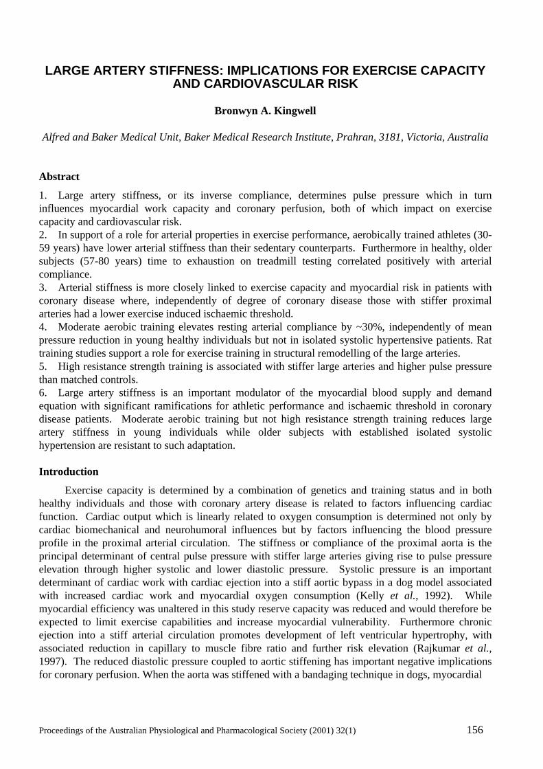
Proceedings of the Australian Physiological and Pharmacological Society (2001) 32(1) 156
LARGE ARTERY STIFFNESS: IMPLICATIONS FOR EXERCISE CAPACITY
AND CARDIOVASCULAR RISK
Bronwyn A. Kingwell
Alfred and Baker Medical Unit, Baker Medical Research Institute, Prahran, 3181, Victoria, Australia Abstract
1. Large artery stiffness, or its inverse compliance, determines pulse pressure which in turn influences myocardial work capacity and coronary perfusion, both of which impact on exercise capacity and cardiovascular risk. 2. In support of a role for arterial properties in exercise performance, aerobically trained athletes (30-59 years) have lower arterial stiffness than their sedentary counterparts. Furthermore in healthy, older subjects (57-80 years) time to exhaustion on treadmill testing correlated positively with arterial compliance. 3. Arterial stiffness is more closely linked to exercise capacity and myocardial risk in patients with coronary disease where, independently of degree of coronary disease those with stiffer proximal arteries had a lower exercise induced ischaemic threshold. 4. Moderate aerobic training elevates resting arterial compliance by ~30%, independently of mean pressure reduction in young healthy individuals but not in isolated systolic hypertensive patients. Rat training studies support a role for exercise training in structural remodelling of the large arteries. 5. High resistance strength training is associated with stiffer large arteries and higher pulse pressure than matched controls. 6. Large artery stiffness is an important modulator of the myocardial blood supply and demand equation with significant ramifications for athletic performance and ischaemic threshold in coronary disease patients. Moderate aerobic training but not high resistance strength training reduces large artery stiffness in young individuals while older subjects with established isolated systolic hypertension are resistant to such adaptation. Introduction
Exercise capacity is determined by a combination of genetics and training status and in both healthy individuals and those with coronary artery disease is related to factors influencing cardiac function. Cardiac output which is linearly related to oxygen consumption is determined not only by cardiac biomechanical and neurohumoral influences but by factors influencing the blood pressure profile in the proximal arterial circulation. The stiffness or compliance of the proximal aorta is the principal determinant of central pulse pressure with stiffer large arteries giving rise to pulse pressure elevation through higher systolic and lower diastolic pressure. Systolic pressure is an important determinant of cardiac work with cardiac ejection into a stiff aortic bypass in a dog model associated with increased cardiac work and myocardial oxygen consumption (Kelly et al., 1992). While myocardial efficiency was unaltered in this study reserve capacity was reduced and would therefore be expected to limit exercise capabilities and increase myocardial vulnerability. Furthermore chronic ejection into a stiff arterial circulation promotes development of left ventricular hypertrophy, with associated reduction in capillary to muscle fibre ratio and further risk elevation (Rajkumar et al., 1997). The reduced diastolic pressure coupled to aortic stiffening has important negative implications for coronary perfusion. When the aorta was stiffened with a bandaging technique in dogs, myocardial
Proceedings of the Australian Physiological and Pharmacological Society (2001) 32(1) 157
Figure 1. Schematic diagram illustrating the circular nature of the relationship between large artery stiffness, physical work capacity and cardiac risk. Stiff large arteries lead to pulse pressure elevation which in turn increases cardiac afterload and reduces coronary perfusion. The resultant reduction in myocardial performance both diminishes exercise capacity and increases cardiac risk. This condition is not conducive to activity and may therefore lead to diminished fitness, a condition associated with further stiffening of the large arteries. While it is not clear whether a predisposition to stiff arteries or low physical fitness is the incipient event in this cycle it is clear that once initiated a vicious cycle promoting disease progression and diminished physical capacity ensues.
perfusion particularly in the subendocardial region was significantly reduced (Watanabe et al., 1993). Aortic stiffness is thus a key factor modulating the relationship between myocardial blood supply and demand with important consequences for myocardial work capacity. Such effects would be expected to impact on both exercise capacity and cardiovascular risk (Figure 1). Arterial stiffness and exercise capacity in healthy individuals
Maximum cardiac output is the main determinant of exercise capacity in healthy individuals and may be influenced by large artery stiffness. In support of a role for arterial properties in exercise performance, lower central arterial stiffness has been associated with higher aerobic fitness levels using pulse contour analysis (Feske et al., 1988), pulse wave velocity (Eugene et al., 1986), magnetic resonance imaging (Tarnawski et al., 1994) and by our group in aerobically trained athletes aged 30-59 years using multiple methodologies (Kingwell et al., 1995). Furthermore systemic arterial compliance, a functional measure of arterial properties throughout the arterial system, correlated positively (r=0.37; p<0.01) and β-index an ultrasound derived measure of aortic stiffness in the arch region, correlated inversely with maximal oxygen consumption (VO2max) (r=-0.44; p<0.001). Furthermore in older
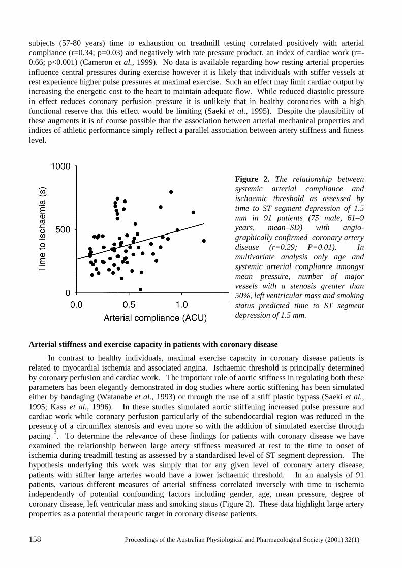
158 Proceedings of the Australian Physiological and Pharmacological Society (2001) 32(1)
subjects (57-80 years) time to exhaustion on treadmill testing correlated positively with arterial compliance (r=0.34; p=0.03) and negatively with rate pressure product, an index of cardiac work (r=-0.66; p<0.001) (Cameron et al., 1999). No data is available regarding how resting arterial properties influence central pressures during exercise however it is likely that individuals with stiffer vessels at rest experience higher pulse pressures at maximal exercise. Such an effect may limit cardiac output by increasing the energetic cost to the heart to maintain adequate flow. While reduced diastolic pressure in effect reduces coronary perfusion pressure it is unlikely that in healthy coronaries with a high functional reserve that this effect would be limiting (Saeki et al., 1995). Despite the plausibility of these augments it is of course possible that the association between arterial mechanical properties and indices of athletic performance simply reflect a parallel association between artery stiffness and fitness level.
Figure 2. The relationship between systemic arterial compliance and ischaemic threshold as assessed by time to ST segment depression of 1.5 mm in 91 patients (75 male, 61±9 years, mean±SD) with angio-graphically confirmed coronary artery disease (r=0.29; P=0.01). In multivariate analysis only age and systemic arterial compliance amongst mean pressure, number of major vessels with a stenosis greater than 50%, left ventricular mass and smoking status predicted time to ST segment depression of 1.5 mm.
Arterial stiffness and exercise capacity in patients with coronary disease
In contrast to healthy individuals, maximal exercise capacity in coronary disease patients is related to myocardial ischemia and associated angina. Ischaemic threshold is principally determined by coronary perfusion and cardiac work. The important role of aortic stiffness in regulating both these parameters has been elegantly demonstrated in dog studies where aortic stiffening has been simulated either by bandaging (Watanabe et al., 1993) or through the use of a stiff plastic bypass (Saeki et al., 1995; Kass et al., 1996). In these studies simulated aortic stiffening increased pulse pressure and cardiac work while coronary perfusion particularly of the subendocardial region was reduced in the presence of a circumflex stenosis and even more so with the addition of simulated exercise through pacing 3. To determine the relevance of these findings for patients with coronary disease we have examined the relationship between large artery stiffness measured at rest to the time to onset of ischemia during treadmill testing as assessed by a standardised level of ST segment depression. The hypothesis underlying this work was simply that for any given level of coronary artery disease, patients with stiffer large arteries would have a lower ischaemic threshold. In an analysis of 91 patients, various different measures of arterial stiffness correlated inversely with time to ischemia independently of potential confounding factors including gender, age, mean pressure, degree of coronary disease, left ventricular mass and smoking status (Figure 2). These data highlight large artery properties as a potential therapeutic target in coronary disease patients.
Proceedings of the Australian Physiological and Pharmacological Society (2001) 32(1) 159
Effects of aerobic training on arterial stiffness
Since a more compliant proximal circulation may increase exercise performance and perhaps represent an important therapeutic target, it is important to understand the effects of acute and chronic aerobic exercise on large artery stiffness (Figure 1). Specifically it is necessary to determine whether exercise training can increase large artery compliance in previously sedentary individuals. More difficult to address is whether a causative relationship exists between training induced changes in compliance and effects on haemodynamics and VO2max. Healthy individuals
A single 30 minute cycling bout at 65% VO2max increased arterial compliance by 40% 30 minutes post exercise, but values returned to resting levels by 60 minutes post exercise (Kingwell et al., 1997). After training 3 times per week for 4 weeks using the same protocol resting arterial compliance (24 hours after the last exercise bout) was elevated by ~30% (Cameron & Dart, 1994). Since compliance is inversely related to mean distending pressure it is important to determine whether this training effect was simply a result of reduced mean arterial pressure. To this end Wistar-Kyoto rats were allowed to train spontaneously in exercise wheels from 4 to 20 weeks of age and aortic cross-sectional compliance examined in organ baths (Kingwell et al., 1997, 1998). The slope of the diameter-tension curve was higher in trained rats indicating that training induced structural aortic adaptations. While arterial elasticity and fitness increased in parallel in both human and rat studies it is not possible to determine whether a causative relationship exists. Isolated Systolic Hypertension
Isolated systolic hypertension represents a clinical manifestation of large artery stiffening. Although the beneficial effects of lowering systolic blood pressure on cardiovascular events in ISH have been proven, the effectiveness of antihypertensive drug therapy may be limited by the detrimental effects of further reductions in diastolic pressure (Tonkin & Wing, (1996). Exercise training is a promising alternate therapy which targets the underlying cause of isolated systolic hypertension in addition to multiple beneficial effects on other risk factors (Jennings et al., 1986; Nelson et al., 1986; Hagberg et al., 1989; Kingwell & Jennings, 1993). To investigate this further a randomised cross-over study of 8 weeks of cycling (3 times per week, 30 minutes) and 8 weeks of sedentary activity was employed. Despite achieving a significant training effect as evidenced by increases in both VO2max and maximum work load, this regimen had no impact on either blood pressure or large artery mechanical properties. Since similar exercise programs have been shown to improve both blood pressure and arterial stiffness in younger subjects, the data suggest that the large artery stiffening associated with ISH is resistant to modification through short term aerobic training. Such irreversibility is perhaps not surprising when the mechanisms responsible for arterial stiffening are considered. These include fatigue induced degradation of elastic fibres, collagen accumulation, calcification and atherosclerostic lesions. Longer term intervention may have been more effective however, training from a younger age appears to the most effective way of preventing the age related decline in large artery elastic function (Tanaka et al., 1998). Effects of strength training on arterial stiffness
While the evidence linking aerobic training with improved arterial elastic function is strong, there is less data pertaining to the effects of strength training. The abrupt and large pressure elevations associated with muscular strength training result in concentric left ventricular hypertrophy and would also be expected to impact on the structure and function of the aorta. In a cross-sectional
160 Proceedings of the Australian Physiological and Pharmacological Society (2001) 32(1)
analysis, exclusively high resistance, strength trained athletes had stiffer large arteries with an associated elevation in both brachial and carotid pulse pressure (Bertovic et al., 1999). Such stiffening may protect the aorta from excessive expansion during acute lifting and would not be expected to limit lifting capacity. Such adaptations may however limit aerobic capacity while the clinical implications with regard to cardiovascular risk are currently unknown. Conclusion
In conclusion large artery stiffness is an important modulator of the myocardial blood supply and demand equation which has significant ramifications for athletic performance and for ischaemic threshold in coronary disease patients. Moderate aerobic training but not high resistance strength training reduces large artery stiffness in young individuals while older subjects with established isolated systolic hypertension are resistant to such adaptation. Acknowledgements
Many thanks to AProf Anthony Dart for thought-provoking discussions surrounding large artery stiffness and exercise capacity. References
Bertovic, D., Waddell, T., Gatzka, C., Cameron, J., Dart, A. & Kingwell, B. (1999) Muscular strength training is associated with low arterial compliance and high pulse pressure. Hypertension, 33, 1385-1391.
Cameron, J. & Dart, A. (1994) Exercise training increases total systemic arterial compliance. American Journal of Physiology, 266, H693-H701.
Cameron, J.D., Rajkumar, C., Kingwell, B.A., Jennings, G.L & Dart, A.M. (1999) Higher systemic arterial compliance is associated with greater exercise time and lower blood pressure in a young older population. Journal of the American Geriatrics Society, 47, 653-656.
Eugene, M., Vandewalle, H., Bertholon, J.F. & Teillac, A. (1986) Arterial elasticity and physical working capacity in young men. Journal of Applied Physiology, 61, 1720-1723.
Feske, W., Finkelstein, S.M., Francis, G. & Cohn, J.N. (1988) Arterial vascular compliance response to exercise in hypertension. Biomedical Sciences Instrumentation, 24, 161-165.
Hagberg, J., Montain, S., Martin, W. & Ehsani, A. (1989) Effect of exercise training in 60- to 69-year old persons with essential hypertension. American Journal of Cardiology, 64, 348-353.
Jennings, G., Nelson, L., Nestel, P., Esler. M., Korner, P., Burton, D. & Bazelmans, J. (1986) The effects of changes in physical activity on major cardiovascular risk factors, hemodynamics, sympathetic function, and glucose utilization in man: a controlled study of four levels of activity. Circulation, 73, 30-40.
Kass, D.A, Saeki, A., Tunin, R.S. & Recchia, F.A. (1996) Adverse influence of systemic vascular stiffening on cardiac dysfunction and adaptation to acute coronary occlusion. Circulation, 93, 1533-1541.
Kelly, R.P., Tunin, R. & Kass, D.A. (1992) Effect of reduced aortic compliance on cardiac efficiency and contractile function of in situ canine left ventricle. Circulation Reseqarch, 71, 490-502.
Kingwell, B. & Jennings, G. (1993) A comparison of the effects of walking and other exercise programs upon blood pressure in normal subjects. Medical Journal of Australia, 158, 234-238.
Kingwell, B., Arnold, P., Jennings, G. & Dart, A. (1997) Spontaneous running increases aortic compliance in Wistar-Kyoto rats. Cardiovascular Research, 35, 132-137.
Proceedings of the Australian Physiological and Pharmacological Society (2001) 32(1) 161
Kingwell, B., Berry, K., Cameron, J., Jennings, G. & Dart, A. (1997) Arterial compliance increases after moderate-intensity cycling. American Journal of Physiology, 273, H2186-H2191.
Kingwell, B., Cameron, J., Gillies, K., Jennings, G. & Dart, A. (1995) Arterial compliance may influence baroreflex function in athletes and hypertensives. American Journal of Physiology, 268, H411-H418.
Kingwell, B.A., Arnold, P.J, Jennings, G.L. & Dart, A.M. (1998) The effects of voluntary running on cardiac mass and aortic compliance in Wistar-Kyoto and spontaneously hypertensive rats. Journal of Hypertension, 16, 181-185.
Nelson, L., Jennings, G., Esler, M. & Korner, P. (1986) Effect of changing levels of physical activity on blood pressure and hemodynamics in essential hypertension. Lancet, ii, 473-476.
Rajkumar, C., Cameron, J.D., Christophidis, N., Jennings, G.L. & Dart, A.M. (1997) Reduced systemic arterial compliance is associated with left ventricular hypertrophy and diastolic dysfunction in older people [see comments]. Journal of the American Geriatrics Society, 45, 803-808.
Saeki, A., Recchia, F. & Kass, D.A. (1995) Systolic flow augmentation in hearts ejecting into a model of stiff aging vasculature. Influence on myocardial perfusion-demand balance. Circulation Research, 76, 132-141.
Tanaka, H., DeSouza, C.A. & Seals, D.R. (1998) Absence of age-related increase in central arterial stiffness in physically active women. Arteriosclerosis, Thrombosis and Vascular Biology, 18, 127-132.
Tarnawski, M., Cybulski, G., Doorly, D., Dumoulin, C., Darrow, R. & Caro, C. (1994) Noninvasive determination of local wavespeed and distensibility of the femoral artery by comb-excited Fourier velocity-encoded magnetic resonance imaging: measurements on athletic and nonathletic human subjects. Heart & Vessels, 9, 194-201.
Tonkin, A. & Wing, L. (1996) Management of isolated systolic hypertension. Drugs, 51, 738-749.
Watanabe, H., Ohtsuka, S., Kakihana, M. & Gugishita, Y. (1993) Coronary circulation in dogs with an experimental decrease in aortic compliance. Journal of the American College of Cardiology, 21, 1497-1506.
162 Proceedings of the Australian Physiological and Pharmacological Society (2001) 32(1)
ADAPTATIONS OF SKELETAL MUSCLE TO PROLONGED, INTENSE ENDURANCE TRAINING
John A. Hawley
Exercise Metabolism Group, School of Medical Sciences, Faculty of Life Sciences, RMIT University,
Bundoora, Victoria 3083, Australia. Summary
1. The ability to sustain the highest average power output or speed for a given distance/duration depends on the rate and efficiency at which chemical energy can be converted into mechanical energy for skeletal muscle contraction. 2. Training regimens for enhancing performance power output should, therefore, aim to induce multiple adaptations in muscle that i) increase the rate of energy production from both aerobic and oxygen-independent pathways, ii) maintain tighter metabolic control (i.e. match ATP production with ATP hydrolysis), iii) minimise cellular disturbances, iv) increase economy of movement and v) improve the working muscles resistance to fatigue during exercise. 3. Skeletal muscle adaptation is the sum of a number of interdependent training-induced responses that include i) morphological transformations, ii) changes in the pattern of substrate utilisation from carbohydrate-based fuels to fat based fuels, iii) less perturbation in acid-base status and iv) up regulation of a variety of mitogenic signalling pathways. 4. A premise is that the high-volume, high-intensity training regimens undertaken by endurance athletes induce a multitude of coordinated metabolic, molecular and celullar adaptations that ultimately exert a major influence on performance capacity. Introduction
An individuals ability to sustain the highest average power output (W) or speed (m min-1) for a given duration or distance depends on the rate and efficiency at which chemical energy can be converted into mechanical energy for skeletal muscle contraction. Training for performance enhancement should, therefore, aim to induce multiple physiological and metabolic adaptations that enable an individual to i) increase the rate of energy production from both aerobic and oxygen-independent pathways, ii) maintain tighter metabolic control (i.e. match ATP production with ATP hydrolysis), iii) minimise cellular disturbances, iv) increase economy of movement and v) improve the working muscles resistance to fatigue during exercise. This review summarises some of the major training-induced adaptations in skeletal muscle that are likely to exert a major influence on performance capacity. The training stimulus
The key components of a training programme are volume, intensity and frequency (Figure 1). The sum of these inputs can be termed the training stimulus, or training impulse that either enhance (fitness) or decrease (fatigue) performance capacity (Bannister et al., 1986). Training responses are directly related to the volume of work undertaken and are specific to the activity performed and the corresponding muscle recruitment patterns (Gollnick et al., 1971). For example, increases in mitochondrial density and oxidative enzyme activity are greatest in the skeletal muscles that are engaged directly in training (Gollnick et al., 1971) implying that the signal for adaptive changes are local rather than systemic (Saltin et al., 1976).
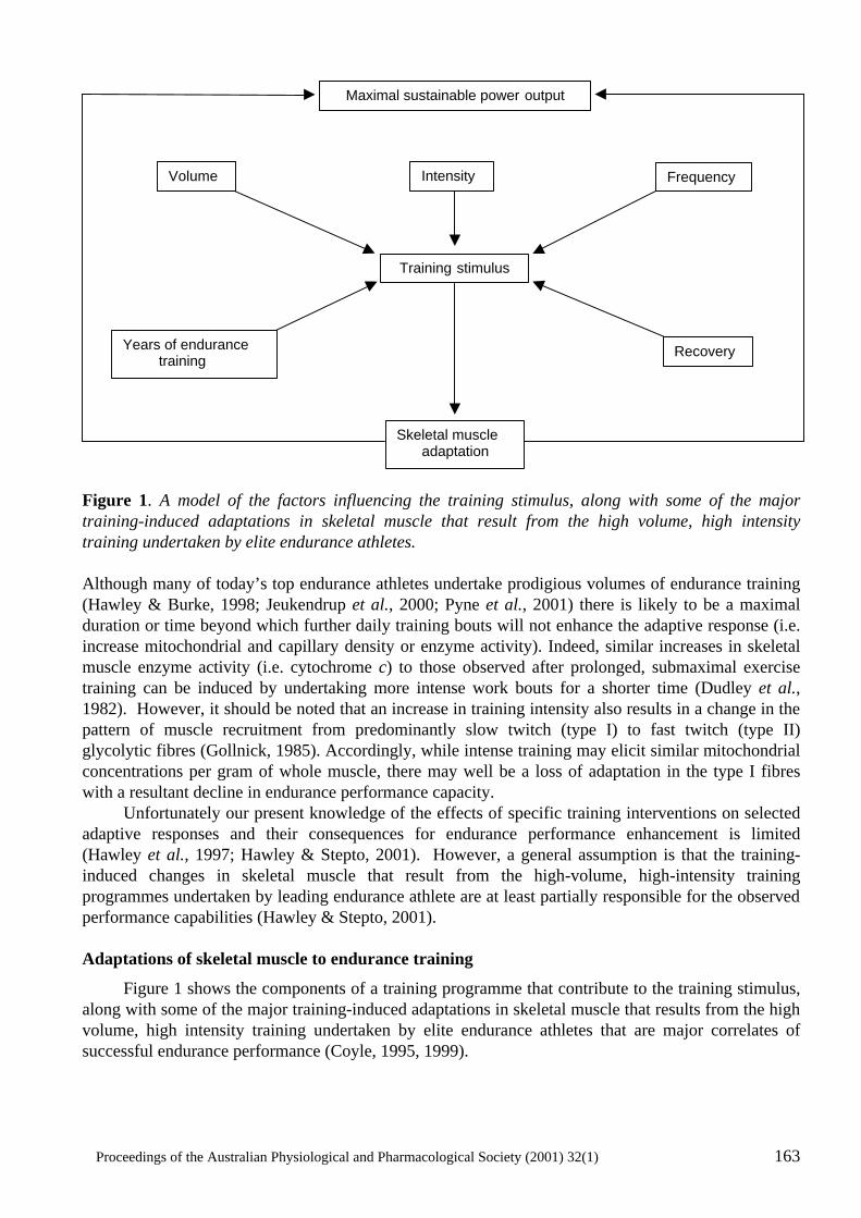
Proceedings of the Australian Physiological and Pharmacological Society (2001) 32(1) 163
Figure 1. A model of the factors influencing the training stimulus, along with some of the major training-induced adaptations in skeletal muscle that result from the high volume, high intensity training undertaken by elite endurance athletes. Although many of today’s top endurance athletes undertake prodigious volumes of endurance training (Hawley & Burke, 1998; Jeukendrup et al., 2000; Pyne et al., 2001) there is likely to be a maximal duration or time beyond which further daily training bouts will not enhance the adaptive response (i.e. increase mitochondrial and capillary density or enzyme activity). Indeed, similar increases in skeletal muscle enzyme activity (i.e. cytochrome c) to those observed after prolonged, submaximal exercise training can be induced by undertaking more intense work bouts for a shorter time (Dudley et al., 1982). However, it should be noted that an increase in training intensity also results in a change in the pattern of muscle recruitment from predominantly slow twitch (type I) to fast twitch (type II) glycolytic fibres (Gollnick, 1985). Accordingly, while intense training may elicit similar mitochondrial concentrations per gram of whole muscle, there may well be a loss of adaptation in the type I fibres with a resultant decline in endurance performance capacity. Unfortunately our present knowledge of the effects of specific training interventions on selected adaptive responses and their consequences for endurance performance enhancement is limited (Hawley et al., 1997; Hawley & Stepto, 2001). However, a general assumption is that the training-induced changes in skeletal muscle that result from the high-volume, high-intensity training programmes undertaken by leading endurance athlete are at least partially responsible for the observed performance capabilities (Hawley & Stepto, 2001).
Adaptations of skeletal muscle to endurance training
Figure 1 shows the components of a training programme that contribute to the training stimulus, along with some of the major training-induced adaptations in skeletal muscle that results from the high volume, high intensity training undertaken by elite endurance athletes that are major correlates of successful endurance performance (Coyle, 1995, 1999).
Maximal sustainable power output
Volumeme
Training stimulus
Years of endurance training
Recovery
Skeletal muscle adaptation
Frequencyccy
Intensity
164 Proceedings of the Australian Physiological and Pharmacological Society (2001) 32(1)
A comprehensive multi-factorial model has been described in detail previously (Hawley & Stepto, 2001). The reader is also referred to the excellent reviews of other workers who have described the physiological (Coyle, 1995, 1999), metabolic (Coyle & Holloszy, 1984; Coyle, 1995, biochemical (Holloszy & Booth, 1976; Holloszy et al., 1977; Coyle & Holloszy, 1984); molecular and cellular (Booth & Thomason, 1991) and mitogenic (Hiyashi et al., 1999) adaptation of muscle in response to exercise. Here an attempt is made to integrate the basic macro-components of the model and assess their overall impact for enhancing performance capacity. Morphological
During recent years it has been popular to determine the muscle fibre composition of elite athletes in different types of events. The most interesting findings are that sprint-trained athletes have a marked predominance of type II fibres in their leg muscles and endurance trained athletes a high proportion of type I fibres (Costill et al., 1976; Saltin et al., 1977). As type I fibres possess a higher capillary density and oxidative potential than type II fibres (Costill et al., 1976; Saltin et al., 1977) it not surprising to find that a high proportion of type I fibres in the vastus lateralis muscle is associated with a lower submaximal oxygen cost (i.e. a greater gross efficiency) during exercise Coyle et al., 1992) possibly because of a lower ATP turnover during contraction-shortening (Wendt & Gibbs, 1974). Cross-sectional data reveal that the number of type I fibres in the trained musculature is related to the numbers of years of prior endurance training (Coyle et al., 1991). This would imply that either those individuals who have a predominance of type I fibres advance into elite endurance sport via a ‘natural selection’ process, or that there is a training-induced interconversion between the fibre types. While there is evidence to suggest that there is a change in the ratio of type IIa to type IIb fibres with endurance training (Saltin et al., 1977), there are no longitudinal data to support type II to type I interconversion in already well-trained endurance athletes. Such studies are warranted because small improvements in gross mechanical efficiency have the potential to result in large enhancements in performance power output (Jeukendrup et al., 2000). Fuel supply
Perhaps one of the most impressive adaptations to regularly performed endurance training is that well-trained endurance athletes deplete their muscle glycogen stores less rapidly during submaximal standardized exercise compared to untrained individuals (Hermansen et al., 1967). The decreased carbohydrate utilisation during submaximal exercise in the trained state is compensated for by a proportional increase in fat oxidation, reflected by a lower respiratory exchange ratio at both the same absolute and relative exercise intensity (Coggan & Williams, 1995). The training-induced shift in substrate selection at the same absolute power output or speed has been attributed to the improved respiratory control sensitivity that results from the increased mitochondrial density (Holloszy & Booth, 1976; Holloszy et al., 1977; Coyle & Holloszy, 1984). However, this phenomenon can only partially explain the glycogen ‘sparing’ effect of endurance training. For example, Coyle et al. (1988) reported that in well-trained cyclists with similar maximal oxygen uptake (VO2max) values (66-68 ml kg min-1), mitochondrial enzyme activity and capillary density, utilisation of muscle glycogen during 30 min of cycling at 79% of VO2max was two-fold higher (65 vs. 28 mmol kg-1) in those individuals with a high power output at lactate threshold. Similarly, Westgarth Taylor et al. (1997) have reported that short-term (3 wk) high-intensity interval training in already well-trained cyclists significantly decreased the rates of carbohydrate oxidation over a range of exercise intensities (60-80% of VO2max) despite no changes in muscle oxidative capacity. Thus, although the early training-induced shifts in substrate selection (from carbohydrate to fat) are likely caused by improved muscle respiratory capacity, other factors must be important for the subsequent shifts in the patterns of fuel metabolism seen after a period of intensified training in already well-trained individuals. Such factors could include a greater
Proceedings of the Australian Physiological and Pharmacological Society (2001) 32(1) 165
supply of fat due to an increase in intramuscular triglyceride concentration (Hurley et al., 1986) and/or morphological adaptations such as a greater recruitment of active muscle mass (Coyle, 1995). Whatever the precise mechanism, it is clear that the training-induced shift in substrate selection by working muscles plays a major role in the increases in endurance capacity that occur after a period of training. Acid-base status.
An individuals maximal sustainable power output or speed is highly related to their lactate threshold (Coyle et al., 1988). Accordingly, the rate of lactate disposal or disappearance (Rd) must be greater than or equal to its rate of appearance (Ra) or production for steady-state blood/plasma lactate concentrations to prevail. In this regard, the capacity to transport lactate across the sarcolemma is significantly higher in endurance-trained athletes compared to untrained individuals (Pilegaard et al., 1994). Furthermore, the highest lactate transporter values are observed in those endurance athletes who incorporate high-intensity anaerobic workouts into their training regimens (Pilegaard et al., 1999). Such workouts undertaken twice per week for as little as 3 wk have been reported to increase muscle buffering capacity by 16% in already well-trained athletes (Weston et al., 1997). These results strongly suggest that a large volume of endurance training alone may be insufficient stimulus to improve the ability to transport lactate and might explain why a short-term supramaximal training programme (6 sessions of 12 x 30 s workbouts at 650 W) was just as effective at improving 40 km cycle time (lasting ~1 h) as longer aerobic interval sets (Stepto et al., 1999).
Finally, individuals with a high proportion of type I fibres in their active musculature have higher monocarboxyltae transporters (MCT), particularly the isoform MCT1, than untrained individuals (Pilegaard et al., 1999). As a muscle with predominantly type II fibres has ~50% of the lactate transport capacity compared to a muscle composed mainly of type I fibres (Pilegaard et al., 1999) and as endurance athletes have a predominance of type I fibres (Costill et al., 1976; Saltin et al., 1977), the functional significance of these membrane-bound transporters for prolonged, intense submaximal endurance performance is obvious. Mitogenic
Chronic endurance training has major effects on a variety of cellular growth and metabolic processes in skeletal muscle. As discussed previously, training-induced adaptations are specific to both the volume and intensity of exercise, implying independent control mechanisms. A major focus of signaling research during the past decade has been the study of three parallel mitogen-activated protein kinase (MAP) kinase cascades (Hiyashi et al., 1999) that are activated by a variety of environmental stress and growth factors. Indeed, the MAP kinase pathway has been implicated as a major signaling system by which cells transduce extracellular cues into intracellular responses (Booth & Watson, 1985; Seger & Krebs, 1995). Aronson et al. (1997) were the first to report that the MAP kinase signalling pathway was activated in human skeletal muscle in response to a single bout of moderate-intensity exercise. Expanding on these findings, Widegren et al. (1998) demonstrated that exercise had divergent effects on parallel MAP kinase pathways in skeletal muscle. Using one-legged cycle ergometry, these workers showed that exercise markedly increased ERK1/2 phosphorylation and to a lesser extent, phosphorylation of p38 MAP kinase (Widegren et al., 1998). Yu et al. (2000) provided evidence that habitual exercise training is associated with differential protein expression of the MAP kinase pathways. These workers obtained resting muscle samples from untrained and moderately trained humans and found that expression of total extracellular regulated kinase (ERK) 1/2 was 190% greater in muscle from trained subjects (Yu et al., 2000). Recently Widegren et al. (2000) have reported that the MAP kinase pathway increases in an exercise-intensity dependent manner, while Osman et al.
166 Proceedings of the Australian Physiological and Pharmacological Society (2001) 32(1)
(2001) have shown that changes in the activity of this signalling pathway are significantly correlated with the increase in citrate synthase activity observed after training. Taken collectively these data suggest that at least some of the training responses of skeletal muscle may be mediated by MAP kinase activation. As an acute bout of exercise induces transient increases in skeletal muscle gene transcription (Neufer & Dohm, 1993) even in individuals with a history of endurance training (Stepto et al., 2001), the activation of various signaling pathways in response to exercise would appear to be central to the upregulation of a variety of metabolic and mitogenic responses that are likely to induce the adaptations seen in skeletal muscle after repeated bouts of exercise. References
Aronson, D., Violan, M.A., Dufrense, S.D. et al. (1997) Exercise stimulates the mitogen-activated protein kinase pathway in human skeletal muscle. Journal of Clinical Investigation, 99, 1251-1257.
Bannister, E.W., Good, P., Holman, G. et al. (1986) Modeling the training response. In: The 1984 Olympic Scientific Congress Proceedings. Sport and Elite Performers. ed. Landers, D.M. pp. 7-23. Illinois: Champaign.
Booth, F.W. & Thomason, D.B. (1991) Molecular and cellular adaptation of muscle in response to exercise: Perspectives of various models. Physiological Reviews, 7,: 541-585.
Booth, F.W. & Watson, P.S. (1985) Control of adaptations in protein levels in response to exercise. Federation Proceedings, 44, 2293-2300.
Coggan, A.R. & Williams, B.D. (1995) Metabolic adaptations to endurance training: substrate metabolism during exercise. In: Exercise Metabolism. Human Kinetics. ed. Hargreaves, M. pp. 177-210. Illinois: Champaign.
Costill, D.L., Daniels, J., Evans, W. et al. (1976) Skeletal muscle enzymes and fiber composition in male and female track athletes. Journal of Applied Physiology, 40, 149-154.
Coyle, E.F. & Holloszy, J.O. (1984) Adaptations of skeletal muscle to endurance exercise and their metabolic consequences. Journal of Applied Physiology, 56, 831-838.
Coyle, E.F. (1995) Integration of the physiological factors determining endurance performance ability. In: Exercise and Sport Sciences Reviews. ed. J.O. Holloszy, J.O. 23, 25-63.
Coyle, E.F. (1999) Physiological determinants of endurance exercise performance. Journal of Science and Medicine in Sports; 2, 181-189.
Coyle, E.F., Coggan, A.R., Hopper, M.K. et al. (1988) Determinants of endurance in well-trained cyclists. Journal of Applied Physiology, 64, 2622-2630.
Coyle, E.F., Feltner, M.E., Kautz, S.A. et al. (1991) Physiological and biomechanical factors associated with elite endurance cycling performance. Medicine and Science in Sports and Exercise.; 23, 93-107.
Coyle, E.F., Sidossis, L.S., Horowitz, J.F. & Beltz, J.D. (1992) Cycling efficiency is related to the percentage of type I muscle fibers. Medicine and Science in Sports and Exercise, 24, 782-788.
Dudley, G.A., Abraham, W.M. & Terjung, R.L. (1982) Influence of exercise intensity and duration on biochemical adaptations in skeletal muscle. Journal of Applied Physiology, 53, 844-850.
Gollnick, P.D. (1985) Metabolism of substrates: energy substrate metabolism during exercise and as modified by training. Federation Proceedings, 44, 353-357.
Gollnick, P.D., Armstrong, R.B., Saubert, C.W. et al. (1971) Enzyme activity and fiber composition in skeletal muscle of untrained and trained men. Journal of Applied Physiology, 33, 312-319.
Proceedings of the Australian Physiological and Pharmacological Society (2001) 32(1) 167
Hawley, J.A. & Burke, L.M. (1998) Training and Nutritional Strategies for Sport. pp. 107-140. Sydney: Allen and Unwin.
Hawley, J.A. & Stepto. N.K. (2001) Adaptations to training in endurance cyclists. Implications for performance. Sports Medicine, 31, (In press).
Hawley, J.A., Myburgh, K.H., Noakes, T.D. et al. (1997) Training techniques to improve fatigue resistance and endurance performance. Journal of Sports Science, 15, 325-333.
Hermansen, L., Hultman, E. & Saltin, B. (1967) Muscle glycogen during prolonged severe exercise. Acta Physiologica Scandanavica, 71, 129-139,.
Hiyashi, T., Dufrense, S.D., Aronson, D., et al. (1999) Intracellular signaling pathways in contracting skeletal muscle. In: Biochemistry of Exercise X. Human Kinetics. ed. Hargreaves, M. & Thompson, D.B. pp. 19-34. Illinois: Champaign.
Holloszy, J.O. & Booth, F.W. (1976) Biochemical adaptations to endurance exercise in muscle. Annual Review of Physiology, 38, 273-291.
Holloszy, J.O., Rennie, M.J., Hickson, R.C. et al. (1977) Physiological consequences of the biochemical adaptations to endurance exercise. Annals of the New York Academy of Sciences, 301, 440-450.
Hurley, B.F., Nemeth, P.M., Martin, W.H. et al. (1986) Muscle triglyceride utilization during exercise: effect of endurance training. Journal of Applied Physiology, 60, 562-567.
Jeukendrup, A.E., Craig, N.P. & Hawley, J.A. (2000) The bioenergetics of world class cycling. Journal of Science and Medicine in Sports, 3, 414-433.
Neufer, P.D. & Dohm, G.L. (1993) Exercise induces a transient increase in transcription of the GLUT-4 gene in skeletal muscle. American Journal of Physiology. Cell Physiology, 34, C1597-1603.
Osman, A.A., Hancock, J., Hunt, D.G. et al. (2000) Exercise training increases ERK2 activity in skeletal muscle of obese Zucker rats. Journal of Applied Physiology, 90, 454-460.
Pilegaard, H., Bangsbo, J., Richter, E.A. et al. (1994) Lactate transport studied in sarcolemmal giant vesicles from human muscle biopsies: relation to training status. Journal of Applied Physiology, 77, 1858-1862.
Pilegaard, H., Domino, K., Noland, T. et al. (1999) Effect of high-intensity exercise training on lactate/H+
transport capacity in human skeletal muscle. American Journal of Physiology, 276, E255-261.
Pilegaard, H., Terzis, G., Halestrap, A. et al. (1999) Distribution of the lactate/ H+ transporter isoforms MCT1 and MCT4 in human skeletal muscle. American Journal of Physiology, 276, E843-848.
Pyne, D.B., Lee, H. & Swanick, K.M. (2001) Monitoring the lactate threshold in world-ranked swimmers. Medicine and Science in Sports and Exercise, 33, 291-297.
Saltin, B., Henriksson, J., Nygaard, E., et al. (1977) Fiber types and metabolic potentials of skeletal muscles in sedentary man and endurance runners. Annals of the New York Academy of Sciences, 301, 3-29.
Saltin, B., Nazar, K., Costill, D.L., Stein, E., et al. (1976) The nature of the training response: peripheral and central adaptations to one-legged exercise. Acta Physiologica Scandanavica, 96, 289-305.
Seger, R. & Krebs, E.G. (1995) The MAP kinase signaling cascade. FASEB Journal, 9, 726-735.
Stepto, N.K., Hawley, J.A., Dennis, S.C. et al. (1999) Effects of different interval-training programs on cycling time-trial performance. Medicine and Science in Sports and Exercise, 31, 736-741.
Stepto, N.K., Yu, M., Fryer, L.D.G., Chibalin, A.V., Carling, D., Krook, A., Hawley, J.A. & Zierath, J.R. (2001) Metabolic and mitogenic signal transduction in response to intense exercise in endurance-trained humans. Medicine and Science in Sports and Exercise, 33, S230.
168 Proceedings of the Australian Physiological and Pharmacological Society (2001) 32(1)
Wendt, I.R. & Gibbs, C.L. (1974) Energy production of mammalian fast-and slow-twitch muscles during development. American Journal of Physiology, 226, 644-647.
Westgarth-Taylor, C., Hawley, J.A., Rickard, S.. et al. (1997) Metabolic and performance adaptations to interval training in endurance-trained cyclists. European Journal of Applied Physiology, 75, 298-304.
Weston, A.R., Myburgh, K.H., Lindsay, F.H. et al. (1997) Skeletal muscle buffering capacity and endurance performance after high-intensity interval training by well-trained cyclists. European Journal of Applied Physiology,; 75: 7-13.
Widegren, U., Jiang, X.J., Krook, A. et al. (1998) Divergent effect of exercise on metabolic and mitogenic signalling pathways in human skeletal muscle. FASEB Journal, 12, 1379-1389.
Widegren, U., Wretman, C., Lionikas, A. et al. (2000) Influence of exercise intensity on ERK/MAP kinase signalling in human skeletal muscle. Pflügers Archiv, 441, 317-322.
Yu, M., Blomstrand, E., Chibalin, A.V. et al. (2000) Exercise-associated differences in an array of proteins involved in signal transduction and glucose transport. Journal of Applied Physiology, 90, 29-34.
Proceedings of the Australian Physiological and Pharmacological Society (2001) 32(1) 169
CELLULAR AND MECHANICAL COUPLING IN THE ARTERIAL WALL A Symposium Cellular and Mechanical Coupling in the Arterial Wall took place on Friday November 24th, 2000, during the Meeting of the Society at RMIT University in Melbourne. The Symposium contained five papers, four of which are reproduced here in pp. 170 – 210. These papers are to appear also in Clinical and Experimental Pharmacology and Physiology. The Symposium was chaired by Prof M.A. Hill and Prof C. Triggle.
170 Proceedings of the Australian Physiological and Pharmacological Society (2001) 32(1)
CELLULAR SIGNALLING IN ARTERIOLAR MYOGENIC CONSTRICTION:
INVOLVEMENT OF TYROSINE-PHOSPHORYLATION PATHWAYS
Timothy V. Murphy, Brian E. Spurrell and Michael A. Hill
Microvascular Biology Group, School of Medical Sciences, RMIT University, Bundoora, Victoria 3083, Australia.
Summary An increase in transmural pressure in arterioles results in a shortening of vascular smooth muscle cells with subsequent constriction of the vessel. The mechanisms underlying this myogenic contraction are not fully understood, however the obligatory role of increases in intracellular [Ca2+] and myosin light-chain phosphorylation have been demonstrated. The response shows a relationship with smooth muscle cell membrane potential and influx of extracellular Ca2+ through voltage-operated Ca2+-channels (VOCCs). Mechanically-sensitive channels and possibly release of Ca2+ from intracellular stores may play a role. However there are other components of myogenic contraction that cannot be explained by a Ca2+-MLCK mechanism, for example the initial sensing of alterations in transmural pressure, whether sustained myogenic constriction involves myofilament Ca2+-sensitization or remodelling of the vessel wall in response to a maintained increase in transmural pressure. In an attempt to investigate these areas, recent studies have examined a role for tyrosine-phosphorylation pathways in pressure-induced contraction of arterioles. In pressurised rat cremaster arterioles, tyrosine kinase inhibitors dilated vessels showing spontaneous myogenic tone and tyrosine phosphatase inhibitors caused vasoconstriction. However pressure-induced myogenic constriction of vessels persisted in the presence of these agents. Biochemical studies revealed that phosphotyrosine formed at a relatively slow rate (significant after 5 minutes, with maximal increase after approximately 15 minutes) in response to increased vessel transmural pressure, in contrast to myosin light-chain phosphorylation or the time course of myogenic constriction itself (maximum within 1 min). Taken together these observations support the idea of a role for tyrosine-phosphorylation pathways in longer-term responses to increased transmural pressure rather than acute myogenic constriction. Phosphotyrosine formation was also more closely correlated to vessel wall tension (pressure x diameter) than the diameter alone of the arterioles. The identity of the tyrosine-phosphorylated proteins requires further investigation, however there is some evidence supporting roles for cSrc-type tyrosine kinases and p44MAP-kinase. The longer-term responses of blood vessels to increased transmural pressure which may involve tyrosine-phosphorylation pathways include maintenance of myogenic constriction and vessel wall remodelling. Myogenic responses
Myogenic responsiveness of arterioles refers to the adjustments to vasomotor tone following alterations in transmural pressure. In vivo, such alterations are caused by changes in intra-luminal blood pressure with vessels constricting in response to an increase in pressure, or dilating in response to a reduction in pressure. In practice the normal resting blood pressure results in a maintained or spontaneous level of myogenic constriction, termed myogenic tone. Studies have shown myogenic activity is an inherent property of vascular smooth muscle, with little evidence for a direct role for factors derived from the endothelium, vessel nerve supply or paracrine or endocrine substances,
Proceedings of the Australian Physiological and Pharmacological Society (2001) 32(1) 171
although such signals interact with myogenic tone (for reviews see Osol, 1995; Davis & Hill, 1999; Schubert & Mulvany, 1999; Davis et al., 2001). The intracellular signalling mechanisms involved in myogenic responses are not fully understood. This review will focus on the evidence supporting the important role of Ca2+-calmodulin-myosin light chain kinase pathway in myogenic contraction of vascular smooth muscle and our recent studies on the possible role of tyrosine-phosphorylation pathways. Intracellular [Ca2+] and myosin light-chain phosphorylation
Myogenic constriction is heavily dependent on smooth muscle intracellular [Ca2+] (Uchida & Bohr, 1969; Meininger et al., 1991; Zou et al., 1995, 2000; Hill et al., 2001). Increases in vessel transmural pressure are accompanied by an increase in cytosolic [Ca2+] and removal of extracellular Ca2+ completely abolishes myogenic activity, suggesting Ca2+ entry into cells from the extracellular space is a major factor in myogenic constriction. A number of studies (Harder 1984, Brayden & Nelson 1992; Knot & Nelson, 1995; Kotecha et al., 1999; Potocnik et al., 2000) have noted a strong correlation between vessel transmural pressure and smooth muscle cell membrane potential, raising the possibility that Ca2+ enters the cells through voltage-dependent mechanisms. Consistent with a role for pressure-induced depolarization, blockers of L-type voltage-operated Ca2+-channels (VOCCs) including nifedipine, verapamil and nicardipine have been shown to greatly reduce myogenic responses (Knot & Nelson, 1992; Wesselman et al., 1996; Potocnik et al., 2000). The role of other VOCCs has not been thoroughly investigated. In the rat cremaster arteriole the T-channel blocker mibefradil produced a vasodilatory and [Ca2+]-lowering effect at concentrations consistent with blockade of L-channels (Potocnik et al., 2000), however a study in small arterioles demonstrated a dilating effect of mibefradil when L-channel antagonists were ineffective, suggested a role for T-channels in this model (Gustafsson et al., 2001). Although inhibition of the L-type VOCCs abolished myogenic responses in rat cremaster arteriole the pressure-induced depolarisation was not blocked by nifedipine, suggesting no role for VOCCs in initiating the depolarisation (Kotecha et al., 2000). The contribution of other Ca2+-sources to myogenic contraction is yet to be established firmly (Hill et al, 2001). In addition to VOCCs, arteriolar smooth muscle cells possess ion channels sensitive to cell membrane stretch (Davis et al., 1992a; Kirber et al., 1988; Langton, 1993; Wu & Davis 2001) which may be activated by vessel distension arising from an increase in intra-luminal pressure. However these channels have relative permeability for K+ and Na+ compared to Ca2+ and are thought to contribute to the membrane depolarisation following cell stretch rather than Ca2+-influx (Davis et al, 1992b). The importance of cell stretch alone as a stimulus is uncertain. In the rat cremaster arteriole, either a ‘slow’ or ‘instantaneous’ increase in transmural pressure from 50 to 120 mmHg resulted in similar increases intracellular [Ca2+] and reduction in vessel diameter, despite the lack of overt cell stretch in the former model (Hill et al., 2000). Furthermore an acute longtitudinal (as opposed to radial) stretch of arterioles caused a rapid, transient increase in intracellular smooth muscle [Ca2+], similar to that observed in the initial stages of pressure-induced cell stretch, however the longtitudinal stretch did not cause vessel constriction (Hill et al., 2000). These observations suggest that simple ‘stretching’ of smooth muscle cells alone is not an obligatory factor in myogenic activity. Vascular smooth muscle cells also possess intracellular stores of Ca2+, however the role of intracellular Ca2+ release in myogenic constriction of blood vessels is largely unknown. Intracellular Ca2+ stores may be released by activation of phospholipase C and generation of the second messenger inositol 1,4,5-trisphosphate (IP3). A study in rat renal arteries (Narayanan et al., 1994) and conduit arteries (Matsumoto et al., 1995) showed increased phospholipase-C activation and generation of IP3 in response to increased wall tension, however there is no compelling evidence supporting a role for pressure-induced intracellular Ca2+ release in myogenic contraction. Such studies are complicated by the fact that removal of extracellular Ca2+ abolishes myogenic tone. Indirect evidence supporting a role for intracellular Ca2+-release comes from the observation that depletion of intracellular Ca2+
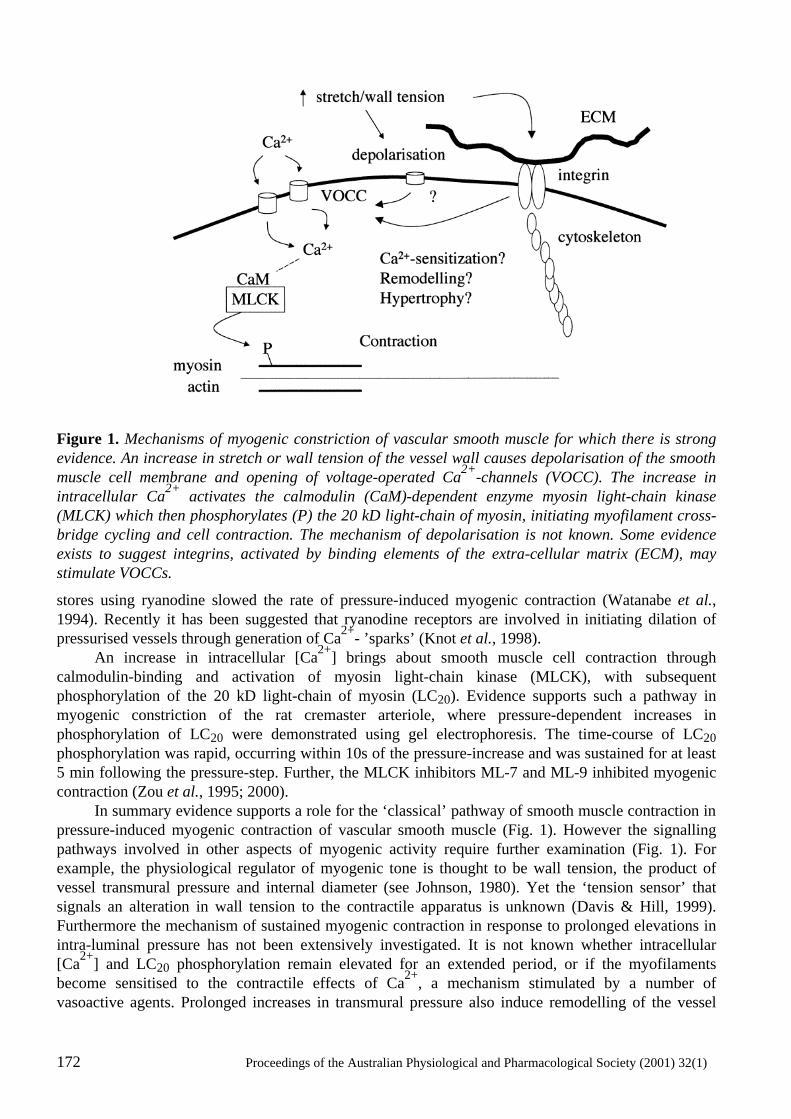
172 Proceedings of the Australian Physiological and Pharmacological Society (2001) 32(1)
Figure 1. Mechanisms of myogenic constriction of vascular smooth muscle for which there is strong evidence. An increase in stretch or wall tension of the vessel wall causes depolarisation of the smooth muscle cell membrane and opening of voltage-operated Ca2+-channels (VOCC). The increase in intracellular Ca2+ activates the calmodulin (CaM)-dependent enzyme myosin light-chain kinase (MLCK) which then phosphorylates (P) the 20 kD light-chain of myosin, initiating myofilament cross-bridge cycling and cell contraction. The mechanism of depolarisation is not known. Some evidence exists to suggest integrins, activated by binding elements of the extra-cellular matrix (ECM), may stimulate VOCCs.
stores using ryanodine slowed the rate of pressure-induced myogenic contraction (Watanabe et al., 1994). Recently it has been suggested that ryanodine receptors are involved in initiating dilation of pressurised vessels through generation of Ca2+- ’sparks’ (Knot et al., 1998). An increase in intracellular [Ca2+] brings about smooth muscle cell contraction through calmodulin-binding and activation of myosin light-chain kinase (MLCK), with subsequent phosphorylation of the 20 kD light-chain of myosin (LC20). Evidence supports such a pathway in myogenic constriction of the rat cremaster arteriole, where pressure-dependent increases in phosphorylation of LC20 were demonstrated using gel electrophoresis. The time-course of LC20 phosphorylation was rapid, occurring within 10s of the pressure-increase and was sustained for at least 5 min following the pressure-step. Further, the MLCK inhibitors ML-7 and ML-9 inhibited myogenic contraction (Zou et al., 1995; 2000). In summary evidence supports a role for the ‘classical’ pathway of smooth muscle contraction in pressure-induced myogenic contraction of vascular smooth muscle (Fig. 1). However the signalling pathways involved in other aspects of myogenic activity require further examination (Fig. 1). For example, the physiological regulator of myogenic tone is thought to be wall tension, the product of vessel transmural pressure and internal diameter (see Johnson, 1980). Yet the ‘tension sensor’ that signals an alteration in wall tension to the contractile apparatus is unknown (Davis & Hill, 1999). Furthermore the mechanism of sustained myogenic contraction in response to prolonged elevations in intra-luminal pressure has not been extensively investigated. It is not known whether intracellular [Ca2+] and LC20 phosphorylation remain elevated for an extended period, or if the myofilaments become sensitised to the contractile effects of Ca2+, a mechanism stimulated by a number of vasoactive agents. Prolonged increases in transmural pressure also induce remodelling of the vessel
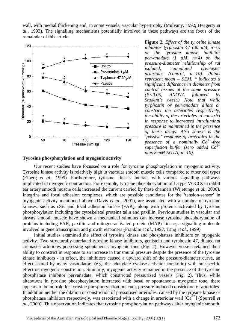
Proceedings of the Australian Physiological and Pharmacological Society (2001) 32(1) 173
wall, with medial thickening and, in some vessels, vascular hypertrophy (Mulvany, 1992; Heagerty et al., 1993). The signalling mechanisms potentially involved in these pathways are the focus of the remainder of this article.
Figure 2. Effect of the tyrosine kinase inhibitor tyrphostin 47 (30 µM, n=6) or the tyrosine kinase inhibitor pervanadate (1 µM, n=4) on the pressure-diameter relationship of rat isolated, cannulated cremaster arterioles (control, n=10). Points represent mean ± SEM. * indicates a significant difference in diameter from control tissues at the same pressure (P<0.05, ANOVA followed by Student’s t-test.) Note that while tyrphostin or pervanadate dilate or constrict the arterioles respectively, the ability of the arterioles to constrict in response to increased intraluminal pressure is maintained in the presence of these drugs. Also shown is the ‘passive’ response of arterioles in the presence of a nominally Ca2+-free superfusion buffer (zero added Ca2+ plus 2 mM EGTA; n=10).
Tyrosine phosphorylation and myogenic activity
Our recent studies have focussed on a role for tyrosine phosphorylation in myogenic activity. Tyrosine kinase activity is relatively high in vascular smooth muscle cells compared to other cell types (Elberg et al., 1995). Furthermore, tyrosine kinases interact with various signalling pathways implicated in myogenic contraction. For example, tyrosine phosphorylation of L-type VOCCs in rabbit ear artery smooth muscle cells increased the current carried by these channels (Wijetunge et al., 2000). Integrins and focal adhesion complexes, which are possible candidates for the ‘tension-sensor’ in myogenic activity mentioned above (Davis et al., 2001), are associated with a number of tyrosine kinases, such as cSrc and focal adhesion kinase (FAK), along with proteins activated by tyrosine phosphorylation including the cytoskeletal proteins talin and paxillin. Previous studies in vascular and airway smooth muscle have shown a mechanical stimulus can increase tyrosine phosphorylation of proteins including FAK, paxillin and mitogen-activated protein (MAP) kinase, a signalling molecule involved in gene transcription and growth responses (Franklin et al., 1997; Tang et al., 1999). Initial studies examined the effect of tyrosine kinase and phosphatase inhibitors on myogenic activity. Two structurally-unrelated tyrosine kinase inhibitors, genistein and tyrphostin 47, dilated rat cremaster arterioles possessing spontaneous myogenic tone (Fig. 2). However vessels retained their ability to constrict in response to an increase in transmural pressure despite the presence of the tyrosine kinase inhibitors - in effect, the inhibitors caused a upward shift of the pressure-diameter curve, an effect shared by many vasodilators (e.g. the adenylate cyclase-activator forskolin) with no specific effect on myogenic constriction. Similarly, myogenic activity remained in the presence of the tyrosine phosphatase inhibitor pervanadate, which constricted pressurized vessels (Fig. 2). Thus, while alterations in tyrosine phosphorylation interacted with basal or spontaneous myogenic tone, there appears to be no role for tyrosine phosphorylation in acute, pressure-induced constriction of arterioles. In addition neither the dilation or constriction of pressurised arterioles, caused by the tyrosine kinase or phosphatase inhibitors respectively, was associated with a change in arteriolar wall [Ca2+] (Spurrell et al., 2000). This observation indicates that tyrosine phosphorylation pathways alter myogenic smooth
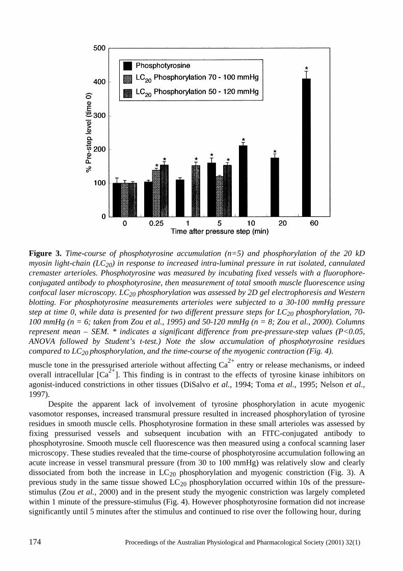
174 Proceedings of the Australian Physiological and Pharmacological Society (2001) 32(1)
Figure 3. Time-course of phosphotyrosine accumulation (n=5) and phosphorylation of the 20 kD myosin light-chain (LC20) in response to increased intra-luminal pressure in rat isolated, cannulated cremaster arterioles. Phosphotyrosine was measured by incubating fixed vessels with a fluorophore-conjugated antibody to phosphotyrosine, then measurement of total smooth muscle fluorescence using confocal laser microscopy. LC20 phosphorylation was assessed by 2D gel electrophoresis and Western blotting. For phosphotyrosine measurements arterioles were subjected to a 30-100 mmHg pressure step at time 0, while data is presented for two different pressure steps for LC20 phosphorylation, 70-100 mmHg (n = 6; taken from Zou et al., 1995) and 50-120 mmHg (n = 8; Zou et al., 2000). Columns represent mean ± SEM. * indicates a significant difference from pre-pressure-step values (P<0.05, ANOVA followed by Student’s t-test.) Note the slow accumulation of phosphotyrosine residues compared to LC20 phosphorylation, and the time-course of the myogenic contraction (Fig. 4).
muscle tone in the pressurised arteriole without affecting Ca2+ entry or release mechanisms, or indeed overall intracellular [Ca2+]. This finding is in contrast to the effects of tyrosine kinase inhibitors on agonist-induced constrictions in other tissues (DiSalvo et al., 1994; Toma et al., 1995; Nelson et al., 1997). Despite the apparent lack of involvement of tyrosine phosphorylation in acute myogenic vasomotor responses, increased transmural pressure resulted in increased phosphorylation of tyrosine residues in smooth muscle cells. Phosphotyrosine formation in these small arterioles was assessed by fixing pressurised vessels and subsequent incubation with an FITC-conjugated antibody to phosphotyrosine. Smooth muscle cell fluorescence was then measured using a confocal scanning laser microscopy. These studies revealed that the time-course of phosphotyrosine accumulation following an acute increase in vessel transmural pressure (from 30 to 100 mmHg) was relatively slow and clearly dissociated from both the increase in LC20 phosphorylation and myogenic constriction (Fig. 3). A previous study in the same tissue showed LC20 phosphorylation occurred within 10s of the pressure-stimulus (Zou et al., 2000) and in the present study the myogenic constriction was largely completed within 1 minute of the pressure-stimulus (Fig. 4). However phosphotyrosine formation did not increase significantly until 5 minutes after the stimulus and continued to rise over the following hour, during
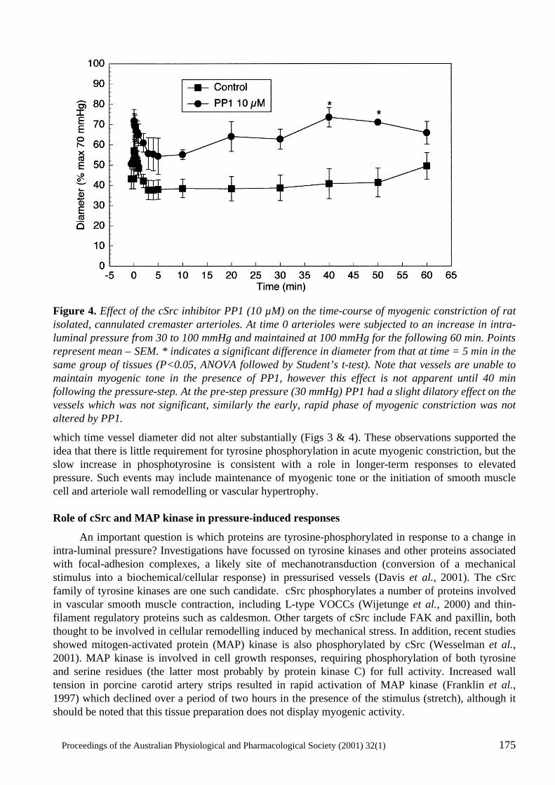
Proceedings of the Australian Physiological and Pharmacological Society (2001) 32(1) 175
Figure 4. Effect of the cSrc inhibitor PP1 (10 µM) on the time-course of myogenic constriction of rat isolated, cannulated cremaster arterioles. At time 0 arterioles were subjected to an increase in intra-luminal pressure from 30 to 100 mmHg and maintained at 100 mmHg for the following 60 min. Points represent mean ± SEM. * indicates a significant difference in diameter from that at time = 5 min in the same group of tissues (P<0.05, ANOVA followed by Student’s t-test). Note that vessels are unable to maintain myogenic tone in the presence of PP1, however this effect is not apparent until 40 min following the pressure-step. At the pre-step pressure (30 mmHg) PP1 had a slight dilatory effect on the vessels which was not significant, similarly the early, rapid phase of myogenic constriction was not altered by PP1.
which time vessel diameter did not alter substantially (Figs 3 & 4). These observations supported the idea that there is little requirement for tyrosine phosphorylation in acute myogenic constriction, but the slow increase in phosphotyrosine is consistent with a role in longer-term responses to elevated pressure. Such events may include maintenance of myogenic tone or the initiation of smooth muscle cell and arteriole wall remodelling or vascular hypertrophy. Role of cSrc and MAP kinase in pressure-induced responses
An important question is which proteins are tyrosine-phosphorylated in response to a change in intra-luminal pressure? Investigations have focussed on tyrosine kinases and other proteins associated with focal-adhesion complexes, a likely site of mechanotransduction (conversion of a mechanical stimulus into a biochemical/cellular response) in pressurised vessels (Davis et al., 2001). The cSrc family of tyrosine kinases are one such candidate. cSrc phosphorylates a number of proteins involved in vascular smooth muscle contraction, including L-type VOCCs (Wijetunge et al., 2000) and thin-filament regulatory proteins such as caldesmon. Other targets of cSrc include FAK and paxillin, both thought to be involved in cellular remodelling induced by mechanical stress. In addition, recent studies showed mitogen-activated protein (MAP) kinase is also phosphorylated by cSrc (Wesselman et al., 2001). MAP kinase is involved in cell growth responses, requiring phosphorylation of both tyrosine and serine residues (the latter most probably by protein kinase C) for full activity. Increased wall tension in porcine carotid artery strips resulted in rapid activation of MAP kinase (Franklin et al., 1997) which declined over a period of two hours in the presence of the stimulus (stretch), although it should be noted that this tissue preparation does not display myogenic activity.
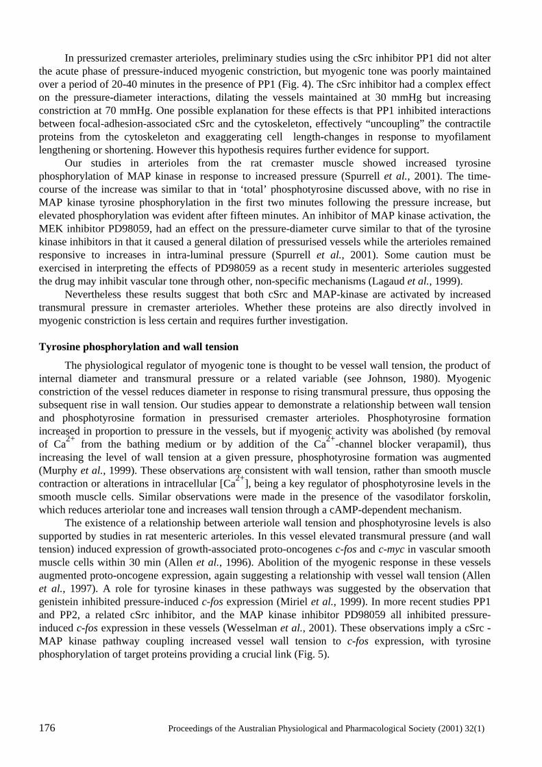
176 Proceedings of the Australian Physiological and Pharmacological Society (2001) 32(1)
In pressurized cremaster arterioles, preliminary studies using the cSrc inhibitor PP1 did not alter the acute phase of pressure-induced myogenic constriction, but myogenic tone was poorly maintained over a period of 20-40 minutes in the presence of PP1 (Fig. 4). The cSrc inhibitor had a complex effect on the pressure-diameter interactions, dilating the vessels maintained at 30 mmHg but increasing constriction at 70 mmHg. One possible explanation for these effects is that PP1 inhibited interactions between focal-adhesion-associated cSrc and the cytoskeleton, effectively “uncoupling” the contractile proteins from the cytoskeleton and exaggerating cell length-changes in response to myofilament lengthening or shortening. However this hypothesis requires further evidence for support. Our studies in arterioles from the rat cremaster muscle showed increased tyrosine phosphorylation of MAP kinase in response to increased pressure (Spurrell et al., 2001). The time-course of the increase was similar to that in ‘total’ phosphotyrosine discussed above, with no rise in MAP kinase tyrosine phosphorylation in the first two minutes following the pressure increase, but elevated phosphorylation was evident after fifteen minutes. An inhibitor of MAP kinase activation, the MEK inhibitor PD98059, had an effect on the pressure-diameter curve similar to that of the tyrosine kinase inhibitors in that it caused a general dilation of pressurised vessels while the arterioles remained responsive to increases in intra-luminal pressure (Spurrell et al., 2001). Some caution must be exercised in interpreting the effects of PD98059 as a recent study in mesenteric arterioles suggested the drug may inhibit vascular tone through other, non-specific mechanisms (Lagaud et al., 1999). Nevertheless these results suggest that both cSrc and MAP-kinase are activated by increased transmural pressure in cremaster arterioles. Whether these proteins are also directly involved in myogenic constriction is less certain and requires further investigation. Tyrosine phosphorylation and wall tension
The physiological regulator of myogenic tone is thought to be vessel wall tension, the product of internal diameter and transmural pressure or a related variable (see Johnson, 1980). Myogenic constriction of the vessel reduces diameter in response to rising transmural pressure, thus opposing the subsequent rise in wall tension. Our studies appear to demonstrate a relationship between wall tension and phosphotyrosine formation in pressurised cremaster arterioles. Phosphotyrosine formation increased in proportion to pressure in the vessels, but if myogenic activity was abolished (by removal of Ca2+ from the bathing medium or by addition of the Ca2+-channel blocker verapamil), thus increasing the level of wall tension at a given pressure, phosphotyrosine formation was augmented (Murphy et al., 1999). These observations are consistent with wall tension, rather than smooth muscle contraction or alterations in intracellular [Ca2+], being a key regulator of phosphotyrosine levels in the smooth muscle cells. Similar observations were made in the presence of the vasodilator forskolin, which reduces arteriolar tone and increases wall tension through a cAMP-dependent mechanism. The existence of a relationship between arteriole wall tension and phosphotyrosine levels is also supported by studies in rat mesenteric arterioles. In this vessel elevated transmural pressure (and wall tension) induced expression of growth-associated proto-oncogenes c-fos and c-myc in vascular smooth muscle cells within 30 min (Allen et al., 1996). Abolition of the myogenic response in these vessels augmented proto-oncogene expression, again suggesting a relationship with vessel wall tension (Allen et al., 1997). A role for tyrosine kinases in these pathways was suggested by the observation that genistein inhibited pressure-induced c-fos expression (Miriel et al., 1999). In more recent studies PP1 and PP2, a related cSrc inhibitor, and the MAP kinase inhibitor PD98059 all inhibited pressure-induced c-fos expression in these vessels (Wesselman et al., 2001). These observations imply a cSrc - MAP kinase pathway coupling increased vessel wall tension to c-fos expression, with tyrosine phosphorylation of target proteins providing a crucial link (Fig. 5).
Proceedings of the Australian Physiological and Pharmacological Society (2001) 32(1) 177
Figure 5. Possible divergent signalling mechanisms in the myogenic response. The early, fast phase of the myogenic response involves depolarisation-induced influx of Ca2+ through VOCCs and activation of CaM-dependent MLCK (see legend to Fig. 1). The delayed accumulation of phosphotyrosine suggests tyrosine-phosphorylation pathways may be involved in more slowly activated-pathways in response to increased transmural pressure, such as cytoskeletal remodelling or hypertrophic (growth) pathways. Evidence suggests roles for extra-cellular matrix (ECM)-coupled integrins, cSrc, focal adhesion kinase (FAK) and mitogen-activated protein kinase (MAPK) in these responses. Conclusions and future directions
The above discussion may have implications for the ways in which blood vessels respond to increased intra-luminal pressure and the modulation of a variable related to wall tension, wall stress. Wall stress is vessel wall tension (pressure x diameter) as a ratio of wall thickness. Thus a similar increment in intra-luminal pressure causes a proportionately greater increase in wall stress in a thin-walled vessel, for example an arteriole or venule, than a thick-walled vessel such as the aorta. In response to a sustained increase in intra-luminal pressure, a blood vessel may reduce the resulting increase in wall stress by either reducing its internal diameter or increasing wall thickness. In practice, these two responses are manifested as increased myogenic tone and/or vessel wall remodelling and hypertrophy (see Parker et al., 2000). The consequences of these two responses to increased vessel wall stress can be seen in various animal models of hypertension. Hypertrophic remodelling of the vessel wall, including medial thickening, is observed in those vessels which demonstrate minimal myogenic reactivity, for example the aorta and femoral, renal and superior mesenteric arteries. Skeletal muscle arterioles, which possess robust pressure-induced myogenic constriction, display ‘eutrophic’ remodelling, a decrease in arteriolar diameter without an overt increase in wall thickness or any evidence of hypertrophy (Mulvany, 1999; Parker et al., 2000). This may be a consequence of sustained myogenic constriction. Myogenic responses are increased in isolated arterioles from hypertensive rats (Dunn et al., 1998; Falcone & Meininger, 1999). Thus, vessels may respond to a sustained increase in intra-luminal pressure by constricting or initiating a growth response.
178 Proceedings of the Australian Physiological and Pharmacological Society (2001) 32(1)
Observed in this context, cell signalling in pressure-induced myogenic constriction may be resolved into two temporally- and biochemically-distinct pathways. The initial ‘fast’ myogenic constriction is due to rapid contraction of vascular smooth muscle cells mediated by Ca2+-entry through VOCCs, activation of MLCK and increased LC20 phosphorylation. The later responses to increased pressure, including sustained myogenic constriction and vessel wall remodelling, may be mediated by tyrosine-phosphorylation pathways involving cSrc, MAP kinase and c-fos (figure 5). Such a model leaves other questions unanswered: is the same tension-sensor (integrin/focal adhesion complex) responsible for both phases of the pressure-induced response? What are the roles of other signalling molecules involved in vascular smooth muscle contraction, such as protein kinase C and rho-activated kinase? How is the stimulus maintained once myogenic constriction has reduced wall tension? These questions require further investigation which will require sophisticated imaging, cellular and molecular approaches. References
Allen, S.P., Liang, H.M., Hill M.A. & Prewitt, R.L. (1996) Elevated pressure stimulates proto-oncogene expression in isolated rat mesenteric arterioles. American Journal of Physiology, 271, H1517-H1523.
Allen, S.P., Wade, S.S. & Prewitt, R.L. (1997) Myogenic tone attenuates pressure-induced gene expression in isolated small arteries. Hypertension, 30, 203-208.
Brayden, J.E. & Nelson, M.T. (1992) Regulation of arterial tone by activation of calcium-dependent potassium channels. Science, 256, 532-535.
Davis, M.J., Donovitz, J.A. & Hood, J.D. (1992a) Stretch-activated single-channel and whole cell currents in vascular smooth muscle cells. American Journal of Physiology, 262, C1083-C1088.
Davis, M.J. & Hill, M.A. (1999) Signaling mechanisms underlying the vascular myogenic response. Physiological Reviews, 79, 387-423.
Davis, M.J., Meininger, G.A. & Zaweija, D.C. (1992b) Stretch-induced increases in intracellular calcium of isolated vascular smooth muscle cells. American Journal of Physiology, 263, H1292-H1299.
Davis, M.J., Wu, X., Nurkiewicz, T., Kawasaki, J., Davis, G.E., Hill, M.A. & Meininger, G.A. (2001) Integrins and mechanotransduction of the vascular myogenic response. American Journal of Physiology, 280, H1427-H1433.
DiSalvo, J., Fitzer, G. & Semenchuk, L.A. (1994) Protein tyrosine phosphorylation, cellular Ca2+ and Ca2+-sensitivity for contraction of smooth muscle. Canadian Journal of Physiology and Pharmacology, 72, 1434-1439.
Dunn, W.R., Wallis, S.J. & Gardiner, S.M. (1998) Remodelling and enhanced myogenic tone in cerebral resistance arteries isolated from genetically hypertensive Brattleboro rats. Journal of Vascular Research, 35, 18-26.
Elberg, G., Li, J., Leibovitch, A. & Shechter, Y. (1995) Non-receptor cytosolic tyrosine kinases from various rat tissues. Biochimica et Biophysica Acta, 1269, 299-306.
Falcone, J.C. & Meininger, G.A. (1999) Endothelin mediates a component of the enhanced myogenic responsiveness of arterioles from hypertensive rats. Microcirculation, 6, 305-313.
Franklin, M.T., Wang, C.L.A. & Adam, L.P. (1997) Stretch-dependent activation and desensitization of mitogen-activated protein kinase in carotid arteries. American Journal of Physiology, 273, C1819-C1827.
Gustafsson, F., Andreasen, D., Salomonsson, M., Jensen, B.L. & Holstein-Rathlou, N. (2001) Conducted vasoconstriction in rat mesenteric arterioles: role for dihydropyridine-insensitive Ca(2+) channels. American Journal of Physiology, 280, H582-H590.
Proceedings of the Australian Physiological and Pharmacological Society (2001) 32(1) 179
Harder, D.R. (1984) Pressure-dependent membrane depolarization in cat middle cerebral artery. Circulation Research, 55, 197-202.
Hill, M.A., Zou, H., Davis, M.J., Potocnik, S.J. & Price, S. (2000) Transient increases in diameter and [Ca2+]i following acute pressure increases are not obligatory for myogenic constriction. American Journal of Physiology, 278, H345-H352.
Hill, M.A., Zou, H., Potocnik, S.J., Meininger, G.A. & Davis, M.J. (2001) Arteriolar smooth muscle mechanotransduction: Ca2+ signaling pathways underlying myogenic reactivity. American Journal of Physiology,
Johnson, P.C. (1980) The myogenic response. In: Handbook of Physiology. The Cardiovascular System. Vascular Smooth Muscle. Section 2, vol. II, pp. 409-442. Bethesda, MD: American Physiological Society.
Kirber, M.T., Walsh, J.V. & Singer, J.J. (1988) Stretch-activated ion channels in smooth muscle: a mechanism for the initiation of stretch-induced contraction. Pfluger’s Archives, 412, 339-345.
Knot, H.J. & Nelson, M.T. (1995) Regulation of membrane potential and diameter by voltage-dependent K+-channels in rabbit myogenic cerebral arteries. American Journal of Physiology, 269, H348-H355.
Knot, H.J., Standen, N.B. & Nelson, M.T. (1998) Ryanodine receptors regulate arterial diameter and wall [Ca2+] in cerebral arteries of rat via Ca2+-dependent K+ channels. Journal of Physiology, 508, 211-221.
Kotecha, N., Potocnik, S.J. & Hill, M.A. (2000) Relationship between membrane potential and calcium entry in myogenic contraction of skeletal muscle arterioles. Proceedings of the Australian Physiological and Pharmacological Society, 31, 88P.
Lagaud, G.J.L., Lam, E., Lui, A., van Breeman, C. & Laher, I. (1999) Nonspecific inhibition of myogenic tone by PD98059, a MEK1 inhibitor, in rat middle cerebral arteries. Biochemical and Biophysical Research Communications, 257, 523-527.
Laher, I. & Bevan, J.A. (1989) Stretch of vascular smooth muscle activates tone and 45Ca2+ influx. Journal of Hypertension, 7 (Suppl. 4), S17-S20.
Langton, P.D. (1993) Calcium channel currents from isolated myocytes of rat basilar artery are stretch-sensitive. Journal of Physiology, 471, 1-11.
Matsumoto, H., Barron, C.B. & Coburn, R.F. (1995) Smooth muscle stretch-activated phospholipase C activity. American Journal of Physiology, 268, C458-C465.
Meininger, G.A., Zaweija, D.C., Falcone, J.C., Hill, M.A. & Davey, J.P. (1991) Calcium measurement in isolated arterioles during myogenic and agonist stimulation. American Journal of Physiology, 261, H950-H959.
Miriel, V.A., Allen, S.P., Schriver, S.D. & Prewitt, R.L. (1999) Genistein inhibits pressure-induced expression of c-fos in isolated mesenteric arteries. Hypertension, 34, 132-137.
Mulvany, M.J. (1992) Vascular growth in hypertension. Journal of Cardiovascular Pharmacology, 20 (Suppl. 1), S1-S11.
Mulvany, M.J. (1999) Vascular remodelling of resistance vessels: can we define this? Cardiovascular Research, 41, 9-13.
Murphy, T.V., Spurrell, B.E. & Hill, M.A. (1999) Dissociation between myogenic response and tyrosine phosphorylation following an increase in intraluminal pressure in rat cremaster arterioles. Proceedings of the Australian Physiological and Pharmacological Society, 30, 82P.
Narayanan, J., Imig, N., Roman, R.J. & Harder, D.R. (1994) Pressurization of isolated renal arteries increases inositol trisphosphate and diacylglycerol. American Journal of Physiology, 266, H1840-H1845.
180 Proceedings of the Australian Physiological and Pharmacological Society (2001) 32(1)
Nelson. S.R., Chien, T. & DiSalvo, J. (1997) Genistein sensitivity of calcium transport pathways in serotonin-activated vascular smooth muscle cells. Archives of Biochemistry and Biophysics, 345, 65-72.
Osol, G. (1995) Mechanotransduction by vascular smooth muscle. Journal of Vascular Research, 32, 275-292.
Parker, S.B., Dobrian, A.D., Wade, S.S. & Prewitt, R.L. (2000) AT(1) receptor inhibition does not reduce arterial wall hypertrophy or PDGF-A expression in renal hypertension. American Journal of Physiology, 278, H613-H622.
Potocnik, S.J., Murphy, T.V., Kotecha, N. & Hill, M.A. (2000) Effects of mibefradil and nifedipine on arteriolar myogenic responsiveness and intracellular Ca2+. British Journal of Pharmacology, 131, 1065-1072.
Schubert, R. & Mulvany, M.J. (1999) The myogenic response: established facts and attractive hypotheses. Clinical Science (Colchester), 96, 313-326.
Smeda, J.S., Lombard, J.H., Madden, J.A. & Harder, D.R. (1987) The effect of alkaline pH and transmural pressure on arterial constriction and membrane potential of hypertensive cerebral arteries. Pfluger’s Archives, 408, 239-242.
Spurrell, B.E., Murphy, T.V. & Hill, M.A. (2000) Tyrosine phosphorylation modulates arteriolar tone but is not fundamental to the myogenic response. American Journal of Physiology, 278, H373-H382.
Spurrell, B.E., Murphy, T.V. & Hill, M.A. (2001) Tyrosine phosphorylation of MAP kinase following increased intra-luminal pressure or epidermal growth factor in cannulated arterioles. FASEB Journal, 15 (4), A43.
Tang, D., Mehta, D. & Gunst, S.J. (1999) Mechanosensitive tyrosine phosphorylation of paxillin and focal adhesion kinase in tracheal smooth muscle. American Journal of Physiology, 276, C250-C258.
Toma, C., Jensen, P.E., Prieto, D., Hughes, A., Mulvany, M.J. & Aalkjaer, C. (1995) Effects of tyrosine kinase inhibitors on the contractility of rat mesenteric arteries. British Journal of Pharmacology, 114, 1266-1272.
Uchida, E. & Bohr, D.F. (1969) Myogenic tone in isolated perfused resistance vessels from rats. American Journal of Physiology, 216, 1343-1350.
Watanabe, J., Horiguchi, S., Karibe, A., Keitoku, M., Takeuchi, M., Saito, S., Takishima, T. & Shirato, K. (1994) Effects of ryanodine on development of myogenic response in rat small skeletal arteries. Cardiovascular Research, 28, 480-484.
Wijetunge, S., Lymn, J.S. & Hughes, A.D. (2000) Effects of protein tyrosine kinase inhibitors on voltage-operated calcium channel currents in vascular smooth muscle cells and pp60c-src kinase activity. British Journal of Pharmacology, 129, 1347-1354.
Wesselman, J.P., Dobrian, A.D., Schriver, S.D. & Prewitt, R.L. (2001) Src tyrosine kinases and extracellular signal-regulated kinase 1/2 mitogen-activated protein kinases mediate pressure-induced c-fos expression in cannulated rat mesenteric small arteries. Hypertension, 37, 955-960.
Wesselman, J.P.M., VanBavel, E., Pfaffendorf, M. & Spaan, J.A.E. (1996) Voltage-operated calcium channels are essential for the myogenic responsiveness of cannulated rat small arteries. Journal of Vascular Research, 33, 32-41.
Wu, X. & Davis, M.J. (2001) Characterization of stretch-activated cation current in coronary smooth muscle cells. American Journal of Physiology, 280, H1751-H1761, 2001.
Zou, H., Ratz, P.H. & Hill, M.A. (1995) Role of myosin phosphorylation and [Ca2+]i in myogenic reactivity and arteriolar tone. American Journal of Physiology, 269, H1590-H1596.
Zou, H., Ratz, P.H. & Hill, M.A. (2000) Temporal aspects of Ca 2+ and myosin phosphorylation during myogenic and agonist-induced arteriolar constriction. Journal of Vascular Research, 37, 556-567.
Proceedings of the Australian Physiological and Pharmacological Society (2001) 32(1) 181
HETEROGENEITY IN THE DISTRIBUTION OF VASCULAR GAP
JUNCTIONS AND CONNEXINS: IMPLICATIONS FOR FUNCTION
C.E. Hill, N. Rummery, H. Hickey and S.L. Sandow
Division of Neuroscience, John Curtin School of Medical Research, Australian National University, Canberra, ACT, 0200, Australia.
Summary
1. Gap junctions, which are comprised of members of a family of membrane proteins called connexins (Cxs), permit the transfer of electrical and chemical information between adjacent cells in a wide variety of tissues. The aim of the present study was to compare the expression of Cxs 37, 40 and 43 in the smooth muscle and endothelium of a large elastic artery and two smaller muscular arteries of the rat. Serial section electron microscopy was also used to determine the presence of pentalaminar gap junctions in the smooth muscle and the incidence of myoendothelial gap junctions between the smooth muscle and endothelial cells in muscular arteries of different size. 2. Using immunohistochemistry, Cxs 37, 40 and 43 were found in the endothelium of the aorta, caudal and basilar arteries, with Cx 43 being the least abundant. Cx 43 was readily observed throughout the muscle layers of the aorta, but was not detected in the media of the caudal or basilar arteries. Cx 40 was not detected in the media of either artery, while very fine punctate staining was observed with Cx 37 antibodies in the media of the caudal and basilar arteries but not in the aorta.. 3. Real time polymerase chain reaction showed that the expression of mRNA for Cx 43 was three-fold greater in the aorta than in the caudal artery of the rat. 4. At the ultrastructural level, small pentalaminar gap junctions (<100 nm) were found between the fine processes of adjacent smooth muscle cells and also between the smooth muscle and endothelial cells. The incidence of myoendothelial gap junctions in the mesenteric vascular bed and in the caudal artery increased as vessel size decreased. 5. In summary, heterogeneity exists within the vascular system with regard to the distribution of gap junctions and their constituent Cxs. Such variation will have important consequences for the coordination and propagation of vascular responses. In muscular arteries in comparison to elastic arteries, Cx 37 may be more important than Cx 43 for cell coupling within the smooth muscle layers. The correlation between the incidence of myoendothelial gap junctions and the role of endothelium-derived hyperpolarising factor, relative to nitric oxide, in vasodilatory responses suggests that they play an important physiological role in the regulation of vascular tone. Introduction
Gap junctions, which are comprised of members of the connexin family of membrane proteins (Cxs), permit the transfer of electrical and chemical information between adjacent cells in a wide variety of tissues (Goodenough et al., 1996; White & Paul, 1999). Three Cxs (37, 40 and 43) are expressed in the walls of blood vessels (see Christ et al., 1996; Severs, 1999). The vascular endothelium expresses all three of these Cxs (Yeh et al., 1998;Gabriels & Paul, 1998), although variations in expression do occur in different vessels and species and under different physiological conditions (Severs, 1999; Yeh et al., 1998; Cowan et al., 1998). In the media, Cx 43 has generally been considered to be the predominant Cx (Severs et al., 2001). The majority of studies reporting the expression of Cx 43 in vascular smooth muscle have, however, been concerned with large elastic arteries, such as the aorta (Lash et al., 1990; Yeh et al., 1997b; Hong & Hill, 1998; van Kempen & Jongsma, 1999; Severs et al., 2001). Indeed, Hong and Hill (1998) were unable to detect Cx 43 in the
182 Proceedings of the Australian Physiological and Pharmacological Society (2001) 32(1)
media of a number of muscular arteries in the rat and several other studies have also failed to detect Cx 43 in the media of coronary arteries of rats and other species (Yeh et al., 1997b; Hong & Hill, 1998; Bastide et al., 1993; Bruzzone et al., 1993; Gros et al., 1994; Verheule et al., 1997). On the other hand, expression of Cxs 40 and 43 has been reported in the media of rat pial arterioles (Little et al., 1995), coronary arteries of some species (van Kempen & Jongsma, 1999) and human umbilical vessels (Van Rijen et al., 1997). Cx 40 has also been reported in the aorta of some species, but is absent in the rat (Yeh et al., 1997a; van Kempen & Jongsma, 1999). Finally, two reports have recently described expression of Cx 37 in vascular smooth muscle (Nakamura et al., 1999; van Kempen & Jongsma, 1999). Physiological studies have provided evidence of electrical coupling between smooth muscle cells, between endothelial cells and between the two cell layers in various different arteries (see Christ et al., 1996). The absence of light microscopic data describing the expression of known Cxs in the media of a number of muscular arteries may therefore suggest that the gap junctions in these vessels are considerably smaller than those in larger arteries, like the aorta (Hong & Hill, 1998). At the ultrastructural level, gap junctions are characterised by a pentalaminar appearance due to the close proximity of the outer membranes of the adjacent cells (Goodenough et al., 1996). While such gap junctional structures are readily observed between vascular endothelial cells (Yeh et al., 1998; Sandow & Hill, 2000), they are rarely described in vascular smooth muscle (Beny & Connat, 1992; Sandow & Hill, 2000). Studies demonstrating true gap junctions connecting the endothelial cells with the inner layer of smooth muscle cells are also rare (Spagnoli et al., 1982; Aydin et al. 1991). These latter structures are candidates for the role of the endothelium-derived hyperpolarizing factor (EDHF) whose activity varies in different sized vessels and in the same vessel in different species (Triggle et al., 1999; Hill et al., 2001). The present study has compared the expression of Cxs 37, 40 and 43 in the aorta, caudal and basilar arteries of the rat using immunohistochemistry and real time polymerase chain reaction (PCR), in an attempt to determine whether Cx 43 is indeed expressed in the media of muscular arteries. Serial section electron microscopy of the caudal and mesenteric arteries has also been used to investigate the incidence and size of gap junctions between smooth muscle cells and of myoendothelial gap junctions (MEGJs) in different sized vessels in which the role of EDHF has been reported to vary (Shimokawa et al., 1996). Methods
All experiments were conducted in line with the Australian Code of Practice for the Care and Use of Animals for Scientific Purposes endorsed by the National Health and Medical Research Council, the Commonwealth Scientific and Industrial Research Organisation, the Australian Research Council and the Australian Vice Chancellors’ Committee (6th edition, 1997), under a protocol approved by the Animal Experimentation Ethics Committee of the Australian National University. Immunohistochemistry
Wistar rats were killed with an overdose of ether anaesthetic and the thoracic aorta, caudal and basilar arteries removed and impregnated with 30% sucrose in phosphate buffered saline (PBS) before freezing in Cryo-M-Bed (Bright Instrument Company Ltd., England). Cryosections (10 µm) were incubated at room temperature for 1 hour with rabbit or sheep antibodies against Cxs 37, 40 and 43 (1:250), diluted in 2% bovine serum albumin, 0.2% Triton-X in PBS and then for one hour with Cy3-conjugated anti-rabbit or anti-goat immunoglobulins (1:100; Jackson Immunoresearch Laboratories Inc, PA) diluted in 0.01% Triton-X in PBS. All sections were mounted in buffered glycerol and viewed under a Leica confocal microscope fitted with appropriate filters.
Proceedings of the Australian Physiological and Pharmacological Society (2001) 32(1) 183
The antibodies against Cx 37 were raised in rabbits against a peptide corresponding to amino acids 318 to 333 of the C-terminus of mouse Cx 37 (Alpha Diagnostic International Tx, USA), similar to those used in previous studies of Cx 37 expression (315-331)(Delorme et al., 1997) or in sheep against amino acids 266 to 281 (Severs, 1999). The antibodies against Cx 40 were raised in sheep against a peptide corresponding to amino acids 254 to 270 of Cx 40, used in previous studies (Severs, 1999). Antibodies against Cx 43 were raised in rabbits (Zymed, CA), but the identity of the antigenic peptide is unknown. To test the specificity of the staining observed, sections were incubated without primary antibody or the primary antibody was preincubated for 1 hour at room temperature with a 10-fold excess by weight of the peptide against which the antibody was raised, before application to the sections. Isolation of mRNA and reverse transcription-polymerase chain reaction (RT-PCR)
Messenger RNA was prepared from the aorta and caudal arteries. All tissues were dissected from 4 week old rats, killed humanely with an overdose of ether anaesthetic, and placed into cold RNAzol B (Tel-test Inc.) for RNA extraction according to the manufacturer’s instructions. RNA was reverse-transcribed (42°C for 1 h, 50°C for 1 h, followed by 10 min at 90°C) using oligo dT primers (100 ng/µl, Stratagene) and reverse transcriptase (200 U/µl, GIBCO BRL) so that the final concentration of RNA was the same in all samples. Reactions controlling for genomic DNA contamination contained no reverse transcriptase enzyme. Real Time PCR
Real time PCR of cDNA from the thoracic aorta and caudal artery was undertaken in an Applied Biosystems ABI Prism 7700 sequence detection system, using subtype specific primers for Cx 43 (forward 5’-GAG ATG CAC CTG AAG CAG ATT GAA-3’ and reverse 5’-GAT GTT CAA AGC GAG AGA CAC CAA-3’) at a final concentration of 800 nM to yield a product of 308 bp. Reactions were performed with 100 ng and 33 ng of starting cDNA for each of the arteries and an annealing temperature of 65 °C. Increase in Cx 43 product in each sample was measured as an increase in fluorescence due to the presence of Sybr green in the reaction mix. Ct values were defined as the cycle number when the fluorescence was first detectable above background. Comparable reactions without DNA or without enzyme were included as controls. Electron microscopy
Adult Wistar Kyoto rats were anaesthetized with an intraperitoneal injection of ketamine and rompun (44 and 8 mg/kg, respectively). A central segment of the first-order branch of the superior mesenteric artery was fixed in glutaraldehyde and paraformaldehyde in 0.1 mmol/L sodium cacodylate, pH 7.4 and processed for electron microscopy using standard procedures. Samples were also taken from the second and third order mesenteric branches and caudal artery of the rat. Serial sections were collected on Formvar (0.3 to 0.5% in ethylene dichloride with grey to silver interference colors). Vessel circumference of each arterial section was estimated as the length of the internal elastic lamina. The number of smooth muscle cell layers in the media was determined by averaging the number of smooth muscle cell profiles ≥5 µm in length, from the outer edge of the internal elastic lamina to the inner edge of the external elastic lamina along each of four linear plots 90o apart, from each of 3 vessels each from a different animal. Methods for serial section analysis of MEGJs were as previously described (Sandow & Hill, 2000).
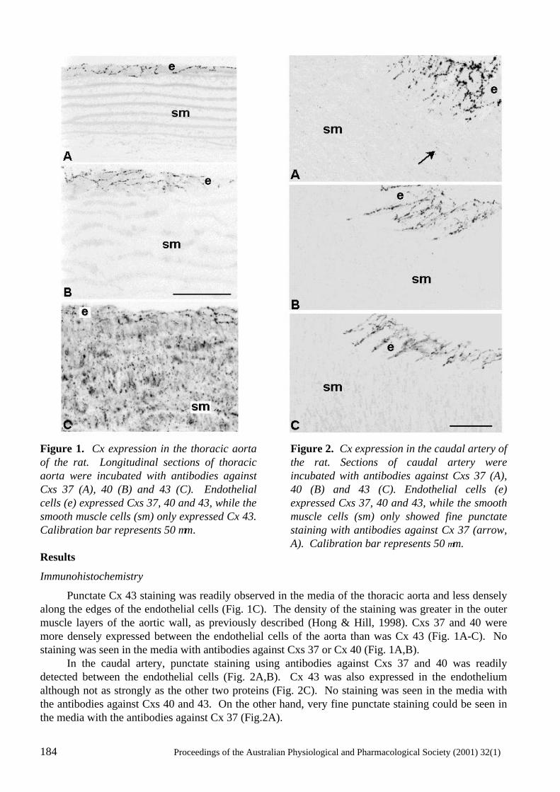
184 Proceedings of the Australian Physiological and Pharmacological Society (2001) 32(1)
Figure 1. Cx expression in the thoracic aorta of the rat. Longitudinal sections of thoracic aorta were incubated with antibodies against Cxs 37 (A), 40 (B) and 43 (C). Endothelial cells (e) expressed Cxs 37, 40 and 43, while the smooth muscle cells (sm) only expressed Cx 43. Calibration bar represents 50 µm.
Figure 2. Cx expression in the caudal artery of the rat. Sections of caudal artery were incubated with antibodies against Cxs 37 (A), 40 (B) and 43 (C). Endothelial cells (e) expressed Cxs 37, 40 and 43, while the smooth muscle cells (sm) only showed fine punctate staining with antibodies against Cx 37 (arrow, A). Calibration bar represents 50 µm.
Results
Immunohistochemistry
Punctate Cx 43 staining was readily observed in the media of the thoracic aorta and less densely along the edges of the endothelial cells (Fig. 1C). The density of the staining was greater in the outer muscle layers of the aortic wall, as previously described (Hong & Hill, 1998). Cxs 37 and 40 were more densely expressed between the endothelial cells of the aorta than was Cx 43 (Fig. 1A-C). No staining was seen in the media with antibodies against Cxs 37 or Cx 40 (Fig. 1A,B). In the caudal artery, punctate staining using antibodies against Cxs 37 and 40 was readily detected between the endothelial cells (Fig. 2A,B). Cx 43 was also expressed in the endothelium although not as strongly as the other two proteins (Fig. 2C). No staining was seen in the media with the antibodies against Cxs 40 and 43. On the other hand, very fine punctate staining could be seen in the media with the antibodies against Cx 37 (Fig.2A).
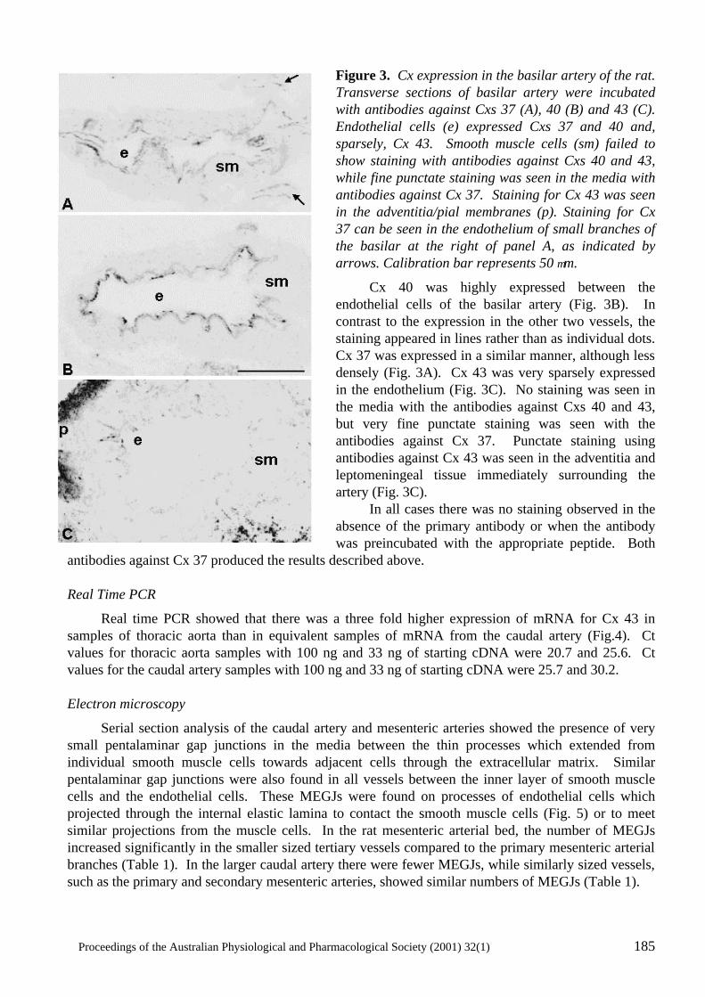
Proceedings of the Australian Physiological and Pharmacological Society (2001) 32(1) 185
Figure 3. Cx expression in the basilar artery of the rat. Transverse sections of basilar artery were incubated with antibodies against Cxs 37 (A), 40 (B) and 43 (C). Endothelial cells (e) expressed Cxs 37 and 40 and, sparsely, Cx 43. Smooth muscle cells (sm) failed to show staining with antibodies against Cxs 40 and 43, while fine punctate staining was seen in the media with antibodies against Cx 37. Staining for Cx 43 was seen in the adventitia/pial membranes (p). Staining for Cx 37 can be seen in the endothelium of small branches of the basilar at the right of panel A, as indicated by arrows. Calibration bar represents 50 µm.
Cx 40 was highly expressed between the endothelial cells of the basilar artery (Fig. 3B). In contrast to the expression in the other two vessels, the staining appeared in lines rather than as individual dots. Cx 37 was expressed in a similar manner, although less densely (Fig. 3A). Cx 43 was very sparsely expressed in the endothelium (Fig. 3C). No staining was seen in the media with the antibodies against Cxs 40 and 43, but very fine punctate staining was seen with the antibodies against Cx 37. Punctate staining using antibodies against Cx 43 was seen in the adventitia and leptomeningeal tissue immediately surrounding the artery (Fig. 3C). In all cases there was no staining observed in the absence of the primary antibody or when the antibody was preincubated with the appropriate peptide. Both
antibodies against Cx 37 produced the results described above. Real Time PCR
Real time PCR showed that there was a three fold higher expression of mRNA for Cx 43 in samples of thoracic aorta than in equivalent samples of mRNA from the caudal artery (Fig.4). Ct values for thoracic aorta samples with 100 ng and 33 ng of starting cDNA were 20.7 and 25.6. Ct values for the caudal artery samples with 100 ng and 33 ng of starting cDNA were 25.7 and 30.2. Electron microscopy
Serial section analysis of the caudal artery and mesenteric arteries showed the presence of very small pentalaminar gap junctions in the media between the thin processes which extended from individual smooth muscle cells towards adjacent cells through the extracellular matrix. Similar pentalaminar gap junctions were also found in all vessels between the inner layer of smooth muscle cells and the endothelial cells. These MEGJs were found on processes of endothelial cells which projected through the internal elastic lamina to contact the smooth muscle cells (Fig. 5) or to meet similar projections from the muscle cells. In the rat mesenteric arterial bed, the number of MEGJs increased significantly in the smaller sized tertiary vessels compared to the primary mesenteric arterial branches (Table 1). In the larger caudal artery there were fewer MEGJs, while similarly sized vessels, such as the primary and secondary mesenteric arteries, showed similar numbers of MEGJs (Table 1).
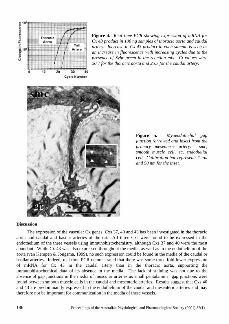
186 Proceedings of the Australian Physiological and Pharmacological Society (2001) 32(1)
Figure 4. Real time PCR showing expression of mRNA for Cx 43 product in 100 ng samples of thoracic aorta and caudal artery. Increase in Cx 43 product in each sample is seen as an increase in fluorescence with increasing cycles due to the presence of Sybr green in the reaction mix. Ct values were 20.7 for the thoracic aorta and 25.7 for the caudal artery.
Figure 5. Myoendothelial gap junction (arrowed and inset) from the primary mesenteric artery. smc, smooth muscle cell, ec, endothelial cell. Calibration bar represents 1 µm and 50 nm for the inset.
Discussion
The expression of the vascular Cx genes, Cxs 37, 40 and 43 has been investigated in the thoracic aorta and caudal and basilar arteries of the rat. All three Cxs were found to be expressed in the endothelium of the three vessels using immunohistochemistry, although Cxs 37 and 40 were the most abundant. While Cx 43 was also expressed throughout the media, as well as in the endothelium of the aorta (van Kempen & Jongsma, 1999), no such expression could be found in the media of the caudal or basilar arteries. Indeed, real time PCR demonstrated that there was some three fold lower expression of mRNA for Cx 43 in the caudal artery than in the thoracic aorta, supporting the immunohistochemical data of its absence in the media. The lack of staining was not due to the absence of gap junctions in the media of muscular arteries as small pentalaminar gap junctions were found between smooth muscle cells in the caudal and mesenteric arteries. Results suggest that Cxs 40 and 43 are predominantly expressed in the endothelium of the caudal and mesenteric arteries and may therefore not be important for communication in the media of these vessels.
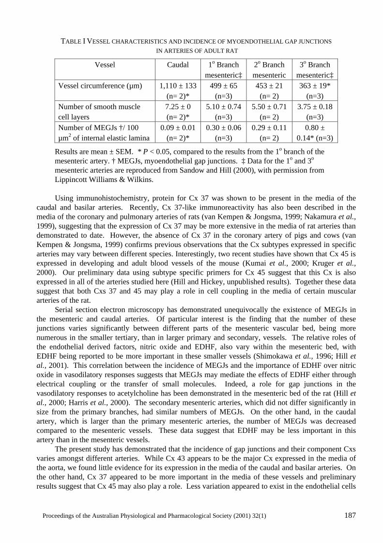
Proceedings of the Australian Physiological and Pharmacological Society (2001) 32(1) 187
TABLE I VESSEL CHARACTERISTICS AND INCIDENCE OF MYOENDOTHELIAL GAP JUNCTIONS IN ARTERIES OF ADULT RAT
Vessel Caudal 1o Branch mesenteric‡
2o Branch mesenteric
3o Branch mesenteric‡
Vessel circumference (µm) 1,110 ± 133 (n= 2)*
499 ± 65 (n=3)
453 ± 21 (n= 2)
363 ± 19* (n=3)
Number of smooth muscle cell layers
7.25 ± 0 (n= 2)*
5.10 ± 0.74 (n=3)
5.50 ± 0.71 (n= 2)
3.75 ± 0.18 (n=3)
Number of MEGJs †/ 100 µm2 of internal elastic lamina
0.09 ± 0.01 (n= 2)*
0.30 ± 0.06 (n=3)
0.29 ± 0.11 (n= 2)
0.80 ± 0.14* (n=3)
Results are mean ± SEM. * P < 0.05, compared to the results from the 1o branch of the mesenteric artery. † MEGJs, myoendothelial gap junctions. ‡ Data for the 1o and 3o mesenteric arteries are reproduced from Sandow and Hill (2000), with permission from Lippincott Williams & Wilkins.
Using immunohistochemistry, protein for Cx 37 was shown to be present in the media of the caudal and basilar arteries. Recently, Cx 37-like immunoreactivity has also been described in the media of the coronary and pulmonary arteries of rats (van Kempen & Jongsma, 1999; Nakamura et al., 1999), suggesting that the expression of Cx 37 may be more extensive in the media of rat arteries than demonstrated to date. However, the absence of Cx 37 in the coronary artery of pigs and cows (van Kempen & Jongsma, 1999) confirms previous observations that the Cx subtypes expressed in specific arteries may vary between different species. Interestingly, two recent studies have shown that Cx 45 is expressed in developing and adult blood vessels of the mouse (Kumai et al., 2000; Kruger et al., 2000). Our preliminary data using subtype specific primers for Cx 45 suggest that this Cx is also expressed in all of the arteries studied here (Hill and Hickey, unpublished results). Together these data suggest that both Cxs 37 and 45 may play a role in cell coupling in the media of certain muscular arteries of the rat. Serial section electron microscopy has demonstrated unequivocally the existence of MEGJs in the mesenteric and caudal arteries. Of particular interest is the finding that the number of these junctions varies significantly between different parts of the mesenteric vascular bed, being more numerous in the smaller tertiary, than in larger primary and secondary, vessels. The relative roles of the endothelial derived factors, nitric oxide and EDHF, also vary within the mesenteric bed, with EDHF being reported to be more important in these smaller vessels (Shimokawa et al., 1996; Hill et al., 2001). This correlation between the incidence of MEGJs and the importance of EDHF over nitric oxide in vasodilatory responses suggests that MEGJs may mediate the effects of EDHF either through electrical coupling or the transfer of small molecules. Indeed, a role for gap junctions in the vasodilatory responses to acetylcholine has been demonstrated in the mesenteric bed of the rat (Hill et al., 2000; Harris et al., 2000). The secondary mesenteric arteries, which did not differ significantly in size from the primary branches, had similar numbers of MEGJs. On the other hand, in the caudal artery, which is larger than the primary mesenteric arteries, the number of MEGJs was decreased compared to the mesenteric vessels. These data suggest that EDHF may be less important in this artery than in the mesenteric vessels. The present study has demonstrated that the incidence of gap junctions and their component Cxs varies amongst different arteries. While Cx 43 appears to be the major Cx expressed in the media of the aorta, we found little evidence for its expression in the media of the caudal and basilar arteries. On the other hand, Cx 37 appeared to be more important in the media of these vessels and preliminary results suggest that Cx 45 may also play a role. Less variation appeared to exist in the endothelial cells
188 Proceedings of the Australian Physiological and Pharmacological Society (2001) 32(1)
where all three Cxs were expressed, although the absolute amounts appeared to differ between vessels. Connections via MEGJs between the endothelial and smooth muscle cell layers were found to vary in their incidence in vessels of different size , becoming more numerous as vessel size decreased. These changes can be correlated with changes in the relative importance of EDHF versus nitric oxide in vasodilatory responses, which in turn would have important consequences for the coordination and propagation of vascular responses. Acknowledgements
The authors wish to thank the National Heart Foundation of Australia for support through a Grant-in-Aid. References
Aydin, F., Rosenblum, W.I. & Povlishock, J.T. (1991) Myoendothelial junctions in human brain arterioles. Stroke, 22, 1592-1597.
Bastide, B., Neyses, L., Ganten, D., Paul, M., Willecke, K. & Traub, O. (1993) Gap junction protein connexin40 is preferentially expressed in vascular endothelium and conductive bundles of rat myocardium and is increased under hypertensive conditions. Circulation Research, 73, 1138-1149.
Beny, J.-L. & Connat, J. L. (1992) An electron-microscopic study of smooth muscle cell dye coupling in the pig coronary arteries. Role of gap junctions. Circulation Research, 70 , 49-55.
Bruzzone, R., Haefliger, J.-A., Gimlich, R.L. & Paul, D.L. (1993) Connexin40, a component of gap junctions in vascular endothelium, is restricted in its ability to interact with other connexins. Molecular Biology of the Cell, 4, 7-20.
Christ, G.J., Spray, D.C., El-Sabban, M., Moore, L.K. & Brink, P.R. (1996) Gap junctions in vascular tissues. Evaluating the role of intercellular communication in the modulation of vasomotor tone. Circulation Research, 79, 631-646.
Cowan, D.B., Lye, S.J. & Langille, B.L. (1998) Regulation of vascular connexin43 gene expression by mechanical loads. Circulation Research, 82, 786-793.
Delorme, B., Dahl, E., Jarry-Guichard, T., Briand, J.P., Willecke, K., Gros, D. & Theveniau-Ruissy, M. (1997) Expression pattern of connexin gene products at the early developmental stages of the mouse cardiovascular system. Circulation Research, 81, 423-437.
Gabriels, J.E. & Paul, D.L. (1998) Connexin43 is highly localized to sites of disturbed flow in rat aortic endothelium but connexin37 and connexin40 are more uniformly distributed. Circulation Research, 83, 636-643.
Goodenough, D.A., Goliger, J.A. & Paul, D.L. (1996) Connexins, connexons, and intercellular communication. Annual Review of Biochemistry, 65, 475-502.
Gros, D., Jarry-Guichard, T., Ten Velde, I., De Maziere, A., Van Kempen, M.J.A., Davoust, J., Briand, J.P., Moorman, A.F.M. & Jongsma, H.J. (1994) Restricted distribution of connexin40, a gap junctional protein, in mammalian heart. Circulation Research, 74, 839-851.
Harris, D., Martin, P.E., Evans, W.H., Kendall, D.A., Griffith, T.M. & Randall, M.D. (2000) Role of gap junctions in endothelium-derived hyperpolarizing factor responses and mechanisms of K(+)-relaxation. European Journal of Pharmacology, 402, 119-128.
Hill, C.E., Hickey, H. & Sandow, S.L. (2000) Role of gap junctions in acetylcholine-induced vasodilation of proximal and distal arteries of the rat mesentery. Journal of the Autonomic Nervous System, 81, 122-127.
Proceedings of the Australian Physiological and Pharmacological Society (2001) 32(1) 189
Hill, C.E., Phillips, J.K. & Sandow, S.L. (2001) Heterogeneous control of blood flow amongst different vascular beds. Medicinal Research Reviews, 21, 1-60.
Hong, T. & Hill, C.E. (1998) Restricted expression of the gap junctional protein connexin43 in the arterial system of the rat. Journal of Anatomy, 193, 583-593.
Kruger, O., Plum, A., Kim, J., Winterhager, E., Maxeiner, S., Hallas, G., Kirchhoff, S., Traub, O., Lamers, W.H. & Willecke, K. (2000) Defective vascular development in connexin 45-deficient mice. Development, 127, 4179-4193.
Kumai, M., Nishii, K., Nakamura, K., Takeda, N., Suzuki, M. & Shibata, Y. (2000) Loss of connexin45 causes a cushion defect in early cardiogenesis. Development, 127, 3501-3512.
Lash, J.A., Critser, E.S. & Pressler, M.L. (1990) Cloning of a gap junctional protein from vascular smooth muscle and expression in two-cell mouse embryos. Journal of Biological Chemistry, 265, 13113-13117.
Little, T.L., Beyer, E.C. & Duling, B.R. (1995) Connexin 43 and connexin 40 gap junctional proteins are present in arteriolar smooth muscle and endothelium in vivo. American Journal of Physiology, 268, H729-H739.
Nakamura, K., Inai, T. & Shibata, Y. (1999) Distribution of gap junction protein connexin 37 in smooth muscle cells of the rat trachea and pulmonary artery. Archives of Histology and Cytology, 62, 27-37.
Sandow, S.L. & Hill, C.E. (2000) The incidence of myoendothelial gap junctions in the proximal and distal mesenteric arteries of the rat is suggestive of a role in EDHF-mediated responses. Circulation Research, 86, 341-346.
Severs, N.J. (1999) Cardiovascular disease. Novartis Foundation Symposium, 219, 188-211.
Severs, N.J., Rothery, S., Dupont, E., Coppen, S.R., Yeh, H.I., Ko, Y.S., Matsushita, T., Kaba, R. & Halliday, D. (2001) Immunocytochemical analysis of connexin expression in the healthy and diseased cardiovascular system. Microscopy Research and Technique, 52, 301-322.
Shimokawa, H., Yasutake, H., Fujii, K., Owada, K., Nakaike, R., Fukumoto, Y., Takayanagi, T., Nagao, T., Egashira, K., Fujishima, M. & Takeshita, A. (1996) The importance of the hyperpolarizing mechanism increases as the vessel size decreases in endothelium-dependent relaxations in rat mesenteric circulation. Journal of Cardiovascular Pharmacology, 28, 703-711.
Spagnoli, L.G., Villaschi, S., Neri, L. & Palmeri, G. (1982) Gap junctions in myoendothelial bridges of rabbit carotid arteries. Experientia, 38, 124-125.
Triggle, C.R., Dong, H., Waldron, G.J. & Cole, W.C. (1999) Endothelium-derived hyperpolarizing factor(s): Species and tissue heterogeneity. Clinical and Experimental Pharmacology and Physiology, 26, 176-179.
Van Kempen, M.J. & Jongsma, H.J. (1999) Distribution of connexin37, connexin40 and connexin43 in the aorta and coronary artery of several mammals. Histochemistry and Cell Biology, 112, 479-486.
Van Rijen, H.V., Van Kempen, M.J., Analbers, L.J., Rook, M.B., Van Ginneken, A.C., Gros, D. & Jongsma, H.J. (1997) Gap junctions in human umbilical cord endothelial cells contain multiple connexins. American Journal of Physiology, 272, C117-C130.
Verheule, S., Van Kempen, M.J.A., Te Welscher, P.H.J.A., Kwak, B.R. & Jongsma, H.J. (1997) Characterisation of gap junction channels in adult rabbit atrial and ventricular myocardium. Circulation Research, 80, 673-681.
White, T.W. & Paul, D.L. (1999) Genetic diseases and gene knockouts reveal diverse connexin functions. Annual Review of Physiology, 61, 283-310.
190 Proceedings of the Australian Physiological and Pharmacological Society (2001) 32(1)
Yeh, H.I., Dupont, E., Coppen, S., Rothery, S. & Severs, N.J. (1997a) Gap junction localization and connexin expression in cytochemically identified endothelial cells of arterial tissue. Journal of Histochemistry and Cytochemistry 45, 539-550.
Yeh, H.I., Lupu, F., Dupont, E. & Severs, N.J. (1997b) Upregulation of connexin43 gap junctions between smooth muscle cells after balloon catheter injury in the rat carotid artery. Arteriosclerosis, Thrombosis, and Vascular Biology, 17, 3174-3184.
Yeh, H.I., Rothery, S., Dupont, E., Coppen, S. & Severs, N.J. (1998) Individual gap junction plaques contain multiple connexins in arterial endothelium. Circulation Research, 83, 1248-1263.
Proceedings of the Australian Physiological and Pharmacological Society (2001) 32(1) 191
191
CELLULAR COUPLING AND CONDUCTED VASOMOTOR RESPONSES
T.O. Neild and G.J. Crane
Department of Human Physiology, Flinders University, Adelaide, Australia.
Summary
1. This brief review examines the concept of spreading vasodilator responses in arteriolar trees, its physiological relevance and possible mechanisms.
2. The most likely mechanisms involve spread of hyperpolarization though tissues in the vessel wall, made possible by electrical coupling between the cells. It is generally agreed that there is coupling between cells within the muscle and endothelial layers, but coupling between the two layers is not always present.
3. The passive electrical properties of arterioles can be modelled, using different techniques depending on the complexity of branching of the tissue. Comparison of experimental results with the model indicates that hyperpolarization can spread further than expected from passive properties alone, implying that spreading vasodilatation may be an active process.
Introduction
Efficient regulation of blood flow requires co-ordinated changes in the diameter of both arterioles and the slightly larger arteries (feed arteries) that supply them. Textbooks often give the impression that the arterioles are much more important than the feed arteries because of their smaller diameter and the dependence of flow on the radius to the fourth power, but a simple calculation (Fig. 1) shows that when vessels are coupled in series they all contribute to the resistance to flow. Changes in diameter of the arterioles alone may have only a small effect on flow if the feed arteries do not change, especially if the feed arteries are long. Physiologically, one of the most important areas where blood flow must be coordinated throughout vascular trees is skeletal muscle. In working skeletal muscle vasodilator metabolites act on arterioles among the muscle fibres, but dilation of these arterioles alone is insufficient to produce the increase in blood flow needed to sustain the high metabolism. It is only when the dilation spreads to the feed arteries outside the muscle that blood flow increases sufficiently to meet the demand (see Lash, 1966 for a comprehensive review). The dilation of the feed arteries occurs so rapidly after the onset of exercise (10 - 20 seconds; VanTeeffelen & Segal, 2000) that it seems unlikely to be the result of diffusion of metabolites out of the muscle, and other experiments have ruled out a blood-borne substance. In this review we shall not consider the nature of the vasodilator substance, but only the mechanism by which the vasodilation spreads from the smallest vessels and trees to the larger ones. Mechanism of spreading vasodilator responses
Some of the first observations in this field were made by Krogh and his co-workers (Krogh, Harrop & Rehberg, 1922). They worked on arterioles in the frog foot web, observing the dilation caused by applying a crystal of silver nitrate to the skin. The dilation would spread well beyond the web on which the crystal was placed and in many ways resembled the dilation produced by mechanical or thermal stimulation in the human skin. In keeping with contemporary thinking it was assumed that such spatially extended responses had to be nerve mediated and so Krogh et al were surprised when they found that the responses could still be obtained in a denervated limb. They postulated some novel type of nerve fibre which remained after the sciatic nerve had been cut, but later work confirmed their result that this type of response was in general not dependent on nerves.
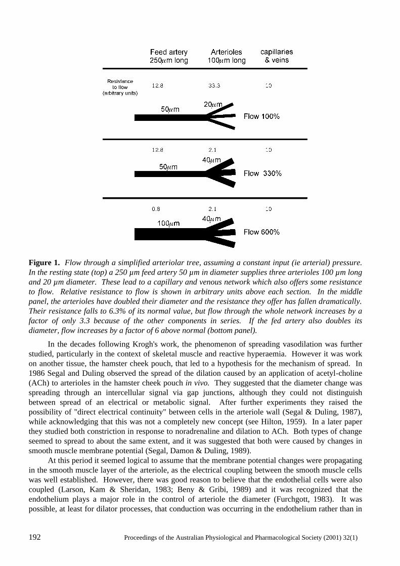
192 Proceedings of the Australian Physiological and Pharmacological Society (2001) 32(1)
Figure 1. Flow through a simplified arteriolar tree, assuming a constant input (ie arterial) pressure. In the resting state (top) a 250 µm feed artery 50 µm in diameter supplies three arterioles 100 µm long and 20 µm diameter. These lead to a capillary and venous network which also offers some resistance to flow. Relative resistance to flow is shown in arbitrary units above each section. In the middle panel, the arterioles have doubled their diameter and the resistance they offer has fallen dramatically. Their resistance falls to 6.3% of its normal value, but flow through the whole network increases by a factor of only 3.3 because of the other components in series. If the fed artery also doubles its diameter, flow increases by a factor of 6 above normal (bottom panel).
In the decades following Krogh's work, the phenomenon of spreading vasodilation was further studied, particularly in the context of skeletal muscle and reactive hyperaemia. However it was work on another tissue, the hamster cheek pouch, that led to a hypothesis for the mechanism of spread. In 1986 Segal and Duling observed the spread of the dilation caused by an application of acetyl-choline (ACh) to arterioles in the hamster cheek pouch in vivo. They suggested that the diameter change was spreading through an intercellular signal via gap junctions, although they could not distinguish between spread of an electrical or metabolic signal. After further experiments they raised the possibility of "direct electrical continuity" between cells in the arteriole wall (Segal & Duling, 1987), while acknowledging that this was not a completely new concept (see Hilton, 1959). In a later paper they studied both constriction in response to noradrenaline and dilation to ACh. Both types of change seemed to spread to about the same extent, and it was suggested that both were caused by changes in smooth muscle membrane potential (Segal, Damon & Duling, 1989). At this period it seemed logical to assume that the membrane potential changes were propagating in the smooth muscle layer of the arteriole, as the electrical coupling between the smooth muscle cells was well established. However, there was good reason to believe that the endothelial cells were also coupled (Larson, Kam & Sheridan, 1983; Beny & Gribi, 1989) and it was recognized that the endothelium plays a major role in the control of arteriole the diameter (Furchgott, 1983). It was possible, at least for dilator processes, that conduction was occurring in the endothelium rather than in
Proceedings of the Australian Physiological and Pharmacological Society (2001) 32(1) 193
193
the smooth muscle. Conduction through the endothelium was favoured by some authors who suggested that the longitudinal orientation of the endothelial cells would favour the spread of membrane potential changes, compared to the smooth muscle where the cells were wrapped circumferentially (Hass & Duling, 1997). Coupling between vascular endothelial cells has now been demonstrated by several groups, in vivo, in isolated vessels in vitro, and between endothelial cells in culture. The coupling between the muscle and the endothelium could present a further complication. Such coupling had been suggested by several anatomists who are had observed close appositions between endothelial and smooth muscle cells (Rhodin, 1967; Beacham, Konishi & Hunt, 1976). The possibility of muscle to endothelial electrical coupling was initially discounted by electrophysiologists, but in the last ten years it has been detected in several situations. It does not seem to be as universal or static as coupling between cells of the same type (muscle or endothelium). Muscle to endothelium electrical coupling has been demonstrated in isolated vessels from various species (Beny & Pacicca, 1989; Xia, Little & Duling, 1995, Neild, Crane & Kotecha, 1999) but it has also been found to be absent in some vessels (Hirst et al., 1997; Welsh & Segal, 1998; Emerson & Segal, 2000). Perhaps the most interesting data come from the hamster cheek pouch, in which the muscle and endothelium are coupled in isolated vessels in vitro but not coupled in the same vessels in vivo. The molecular structures responsible for the electrical coupling have yet to be determined. The situation is closest to resolution for the endothelium, where conventional gap junctions composed of connexin (Cx) proteins are almost certainly responsible. The dye Lucifer Yellow passes easily between endothelial cells - a good indicator of the presence of gap junctions. Some experiments have indicated that endothelial cells are immunoreactive for Cx43, but there appear to be some regions of the circulation where Cx43 is not present (Sandow & Hill, 2000). The situation regarding coupling between muscle cells or muscle and endothelial cells is quite different. Lucifer Yellow passes through neither of these pathways under any experimental conditions. There is some suggestion that other dyes may do so, but the experiments are sometimes difficult to interpret as the more mobile dyes also tend to leak out of the cells. A variety of Cx proteins (or mRNA for them) can be detected muscle and endothelial tissue (see Hill et al., 2001, in this symposium), but the physiological situation remains unclear. Electrical coupling between cells in the arteriole wall
Whatever the structures present which might enable electrical coupling, the ultimate question is whether the coupling is strong enough to be physiologically relevant. Experiments to detect and measure coupling involve causing a change in membrane potential in one region of a blood vessel and measuring the membrane potential change at some distant point. If there is a change at the distant point then there must be coupling between some of the intervening cells. Working out which cells are coupled and how strong the coupling is presents an additional challenge. Blood vessels, particularly arterioles, are always part of branching tree-like structures. Predicting the spread of membrane potential changes in such structures requires complex calculations using techniques that are not accessible to many physiologists. Fortunately the basic principles were worked out in the context of the branching dendritic fields of neurones, not only the theory but also the practical application (e.g. Thurbon et al., 1998). The same principles can be applied to branching arteriolar trees as long as the vessel wall can be treated as one homogeneous layer (Segal & Neild, 1996). However, if there is electrical coupling between two layers within the wall the situation is more complex. Equations that describe the spread of membrane potential changes in unbranched pieces of vessel are relatively simple and have been derived (Crane et al., 2001), but for branching structures numerical methods are necessary (Crane, Hines & Neild, 2001). These equations can be used to predict the passive spread of membrane potential changes through vascular networks. A surprising result emerges from these calculations. Although the
194 Proceedings of the Australian Physiological and Pharmacological Society (2001) 32(1)
endothelial cells are long and oriented in the longitudinal direction, they do not form a better pathway for signal conduction than the muscle (Crane, Kotecha et al., 2001). This is due to the fact that they are very thin and thus their cytoplasm presents a significant resistance to current flow. In a "typical" arteriole with conservative values for endothelial and muscle cell membrane resistances and cytoplasmic resistivities, membrane potential changes spread to about the same extent in the intact arteriole with both cell layers coupled as they do in the muscle layer alone after disruption of the endothelium. Experiments which measure the spread and attempt to quantitate it (perhaps in terms of a length constant) are thus unlikely to reveal the extent to which muscle and endothelium are coupled. A better strategy to answer this question is to compare the input resistance of the muscle layer of vessels before and after endothelial removal. Although the potential for error is large when passing currents through high resistance electrodes, the effects on input resistance of the muscle layer expected when removing a coupled endothelium are also large. In practice the experiment is feasible, and has provided evidence for coupling between muscle and endothelium in guinea-pig intestinal arterioles (Neild, Crane & Kotecha, 1999; Yamamoto, Imaeda & Suzuki, 1999; Crane, Kotecha et al., 2001; Coleman Tare & Parking ton, 2001). Even without a quantitative analysis, there is evidence that in some vessels the endothelium and muscle are electrically coupled, whereas in others they are not. It is not clear yet whether coupling or lack of coupling is a fundamental property of a particular vessel or something that can be changed by local conditions. This point is illustrated by work on the hamster cheek pouch arterioles where in vitro cannulated vessels appeared to have coupling between the two layers, whereas this was not present in vivo in anaesthetized hamsters. We have no reason to doubt the validity of either set of results (we have verified the in vivo result ourselves, in experiments in Prof Segal's laboratory at the Pierce Laboratory). The next stage should be to measure rather than simply detect the coupling, and determine whether it can be modulated. Spread of hyperpolarization
The physiological significance of coupling in the control of blood flow was alluded to above. We have calculated the of spread of membrane potential changes in branching networks where a membrane potential change is generated in the smallest arterioles and spreads upstream to feed arteries, and in general we doubt that passive spread of hyperpolarization is great enough to explain the vasodilatation observed in tissues such as working skeletal muscle (Crane, Kotecha et al., 2001). On the other hand, experiments by on of us (Glenis Crane, at the Pierce Institute) clearly show that in the hamster cheek pouch arterioles hyperpolarizations in either the muscle or the endothelial layer in response to ACh spread further than could be accounted for by passive properties. Furthermore, in the majority of experiments the hyperpolarizations failed to decline in size or even increased within the first millimetre from the initiation point. This behaviour could only be explained if there was some additional current source appearing as the hyperpolarization spread. Opening of a voltage dependent K+ channel would be an obvious mechanism, although no channel with exactly the right properties has been described. In summary, spreading diameter changes in arteriolar trees are probably controlled largely by changes in membrane potential in the smooth muscle and endothelial layers. These changes may be the same or different in the two layers, depending on whether the layers are electrically coupled. Evidence to date suggests that coupling may vary between species or tissues, and with different experimental conditions. If coupling can be measured rather than just detected in a qualitative manner, it may be found that it is a physiologically modulated variable. However, any analysis of membrane potential changes in the vessels should take into account the possibility of active propagation of membrane potential changes rather than just passive spread.
Proceedings of the Australian Physiological and Pharmacological Society (2001) 32(1) 195
195
References
Beacham, W.S., Konishi, A. & Hunt, C.C. (1976) Observations on the microcirculatory bed in the rat mesocecum using differential interference contrast microscopy in vivo and electron microscopy. American Journal of Anatomy, 146, 385-425.
Beny, J.L. & Gribi, F. (1989) Dye and electrical coupling of endothelial cells in situ. Tissue and Cell 21, 797-802.
Beny, J.L. & Pacicca, C. (1994) Bidirectional electrical communication between smooth muscle endothelial cells in the pig coronary artery. American Journal of Physiology, 266, H1465-H1472.
Coleman, H.A., Tare, M. & Parkington, H.C. (2001) K+ currents underlying the action of endothelium-derived hyperpolarizing factor in guinea-pig, rat and human blood vessels. Journal of Physiology, 531, 359-373.
Crane, G.J, Hines, M.L. & Neild, T.O. (2001) Simulating the spread of membrane potential changes in arteriolar networks. Microcirculation, 8, 33-43.
Crane, G.J.,. Kotecha, N., Luff, S.E. & Neild, T.O. (2001) Electrical coupling between smooth muscle and endothelium in arterioles. Submitted to Physics in Medicine and Biology.
Emerson, G.G. & Segal, S.S. (2000) Endothelial cell pathway for conduction of hyperpolarization and vasodilation along hamster feed artery Circulation Research, 86, 94-100.
Furchgott, R.F. (1983) Role of endothelium in responses of vascular smooth muscle. Circulation Research, 53, 557-573.
Hass, T.L. & Duling, B.R. (1997) Morphology favors an endothelial cell pathway for longitudinal conduction within arterioles. Microvascular Research, 53, 113-120.
Hill, C.E., Rummery, N., Hickey, H. & Sandow, S.L. (2001) Heterogeneity in the distribution of vascular gap junctions and connexins: implications for function. Proceedings of the Australian Physiological and Pharmacological Society, 32(1), 181-190.
Hilton, S.M. (1959) A peripheral arterial conducting mechanism underlying dilatation of the femoral artery and concerned with functional vasodilatation in skeletal muscle. Journal of Physiology, 149, 93-111.
Hirst, G.D.S, Edwards, F.R., Gould, D.J., Sandow, S.L. & Hill, C.E. (1997) Electrical properties of iridial arterioles of the rat. American Journal of Physiology, 273, H2465 -H2472.
Krogh, A., Harrop, G.A. & Rehberg, P.B. (1922) Studies on the physiology of capillaries. III. The innervation of the blood vessels in the hind legs of the frog. Journal of Physiology, 56, 179-189.
Larson, D.M., Kam, E.Y. & Sheridan, J.D. (1983) Junctional transfer in cultured vascular endothelium: I. Electrical coupling. Journal of Membrane Biology, 74, 103-113.
Lash, J.M. (1996) Regulation of skeletal muscle blood flow during contractions. Proceedings of the Society for Experimental Biology and Medicine, 211, 218-235.
Neild, T.O., Crane G.J. & Kotecha, N. (1999) Electrical connection between the endothelium and smooth muscle in guinea-pig intestinal submucosal arterioles. In "Progress in Microcirculation Research" ed. Neild, T.O. & Carati, C.J. Sydney: Australian and New Zealand Microcirculation Society.
Rhodin, J.A.G. (1967) The ultrastructure of mammalian arterioles and precapillary sphincters. Journal of Ultrastructural Research, 18, 181-223.
Sandow, S.L. & Hill, C.E. (2000) Incidence of myoendothelial gap junctions in the proximal and distal mesenteric arteries of the rat is suggestive of a role in endothelium-derived hyperpolarizing factor-mediated responses. Circulation Research, 86, 341-346
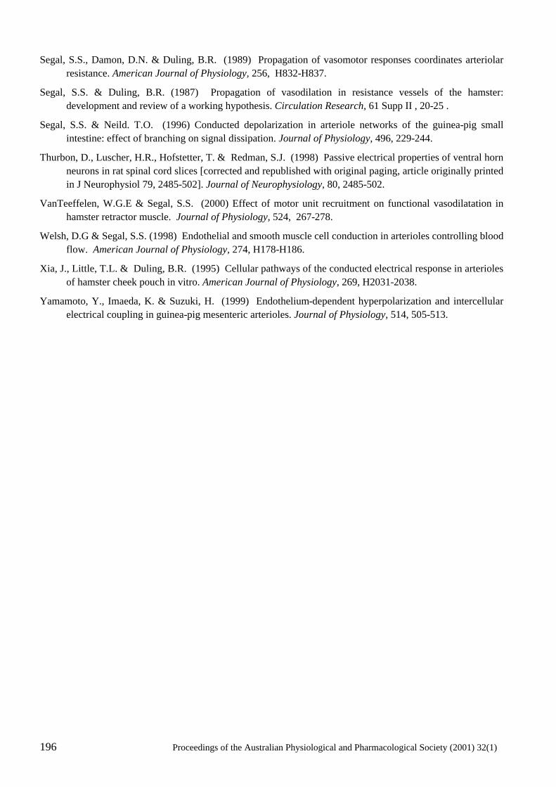
196 Proceedings of the Australian Physiological and Pharmacological Society (2001) 32(1)
Segal, S.S., Damon, D.N. & Duling, B.R. (1989) Propagation of vasomotor responses coordinates arteriolar resistance. American Journal of Physiology, 256, H832-H837.
Segal, S.S. & Duling, B.R. (1987) Propagation of vasodilation in resistance vessels of the hamster: development and review of a working hypothesis. Circulation Research, 61 Supp II , 20-25 .
Segal, S.S. & Neild. T.O. (1996) Conducted depolarization in arteriole networks of the guinea-pig small intestine: effect of branching on signal dissipation. Journal of Physiology, 496, 229-244.
Thurbon, D., Luscher, H.R., Hofstetter, T. & Redman, S.J. (1998) Passive electrical properties of ventral horn neurons in rat spinal cord slices [corrected and republished with original paging, article originally printed in J Neurophysiol 79, 2485-502]. Journal of Neurophysiology, 80, 2485-502.
VanTeeffelen, W.G.E & Segal, S.S. (2000) Effect of motor unit recruitment on functional vasodilatation in hamster retractor muscle. Journal of Physiology, 524, 267-278.
Welsh, D.G & Segal, S.S. (1998) Endothelial and smooth muscle cell conduction in arterioles controlling blood flow. American Journal of Physiology, 274, H178-H186.
Xia, J., Little, T.L. & Duling, B.R. (1995) Cellular pathways of the conducted electrical response in arterioles of hamster cheek pouch in vitro. American Journal of Physiology, 269, H2031-2038.
Yamamoto, Y., Imaeda, K. & Suzuki, H. (1999) Endothelium-dependent hyperpolarization and intercellular electrical coupling in guinea-pig mesenteric arterioles. Journal of Physiology, 514, 505-513.
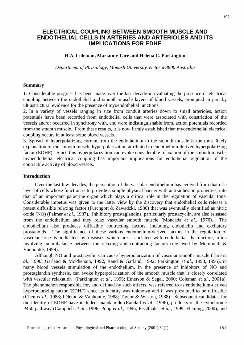
Proceedings of he Australian Physiological and Pharmacological Society (2001) 32(1) 197
197
ELECTRICAL COUPLING BETWEEN SMOOTH MUSCLE AND
ENDOTHELIAL CELLS IN ARTERIES AND ARTERIOLES AND ITS IMPLICATIONS FOR EDHF
H.A. Coleman, Marianne Tare and Helena C. Parkington
Department of Physiology, Monash University Victoria 3800 Australia
Summary
1. Considerable progress has been made over the last decade in evaluating the presence of electrical coupling between the endothelial and smooth muscle layers of blood vessels, prompted in part by ultrastructural evidence for the presence of myoendothelial junctions. 2. In a variety of vessels ranging in size from conduit arteries down to small arterioles, action potentials have been recorded from endothelial cells that were associated with constriction of the vessels and/or occurred in synchrony with, and were indistinguishable from, action potentials recorded from the smooth muscle. From these results, it is now firmly established that myoendothelial electrical coupling occurs in at least some blood vessels. 3. Spread of hyperpolarizing current from the endothelium to the smooth muscle is the most likely explanation of the smooth muscle hyperpolarization attributed to endothelium-derived hyperpolarizing factor (EDHF). Since this hyperpolarization can evoke considerable relaxation of the smooth muscle, myoendothelial electrical coupling has important implications for endothelial regulation of the contractile activity of blood vessels. Introduction
Over the last few decades, the perception of the vascular endothelium has evolved from that of a layer of cells whose function is to provide a simple physical barrier with anti-adhesion properties, into that of an important paracrine organ which plays a critical role in the regulation of vascular tone. Considerable impetus was given to the latter view by the discovery that endothelial cells release a potent diffusible relaxing factor (Furchgott & Zawadzki, 1980) that was eventually identified as nitric oxide (NO) (Palmer et al., 1987). Inhibitory prostaglandins, particularly prostacyclin, are also released from the endothelium and they relax vascular smooth muscle (Moncada et al., 1976). The endothelium also produces diffusible contracting factors, including endothelin and excitatory prostanoids. The significance of these various endothelium-derived factors in the regulation of vascular tone is indicated by diseases which are associated with endothelial dysfunction, often involving an imbalance between the relaxing and contracting factors (reviewed by Mombouli & Vanhoutte, 1999). Although NO and prostacyclin can cause hyperpolarization of vascular smooth muscle (Tare et al., 1990; Garland & McPherson, 1992; Rand & Garland, 1992; Parkington et al., 1993, 1995), in many blood vessels stimulation of the endothelium, in the presence of inhibitors of NO and prostaglandin synthesis, can evoke hyperpolarization of the smooth muscle that is closely correlated with vascular relaxation (Parkington et al., 1995; Emerson & Segal, 2000; Coleman et al., 2001a). The phenomenon responsible for, and defined by such effects, was referred to as endothelium-derived hyperpolarizing factor (EDHF) since its identity was unknown and it was presumed to be diffusible (Chen et al., 1988; Félétou & Vanhoutte, 1988; Taylor & Weston, 1988). Subsequent candidates for the identity of EDHF have included anandamide (Randall et al., 1996), products of the cytochrome P450 pathway (Campbell et al., 1996; Popp et al., 1996; Fisslthaler et al., 1999; Fleming, 2000), and
198 Proceedings of the Australian Physiological and Pharmacological Society (2001) 32(1)
K+ (Edwards et al., 1998), with no consensus (Fukao et al., 1997b; Plane et al., 1997; Zygmunt et al., 1997; Chataigneau et al., 1998; Petersson et al., 1998; Buus et al., 2000; Drummond et al., 2000; Lacy et al., 2000; Quilley & McGiff, 2000; Van de Voorde & Vanheel, 2000; Coleman et al., 2001a, b). An alternative explanation for the phenomenon of EDHF arose from ultrastructural evidence which showed that endothelial cells form protrusions through the elastic lamina to form close contact with smooth muscle cells (myoendothelial junctions) (Moore & Ruska, 1957; Rhodin, 1967). It was suggested that these junctions could provide a pathway for the movement of metabolites and/or flow of current between the two cell layers (Rhodin, 1967; Davies, 1986). The latter was proposed as an explanation for the conduction of vasodilatory responses along arteriolar walls (Segal & Duling, 1987; Xia et al., 1995) and shear stress-induced vasorelaxation resulting from endothelial hyperpolarization (Olesen et al., 1988). Along these lines, it has been considered that EDHF may represent electrotonic spread of hyperpolarizing current from the endothelial cells to the smooth muscle cells (Bény, 1990; Kühberger et al., 1994). An essential requirement of this proposition is that electrical coupling be demonstrated between the endothelial and smooth muscle layers. Myoendothelial electrical coupling in arteries
In an electrophysiological study of the pig coronary artery, Bény (1990) recorded simultaneously from Lucifer-Yellow identified endothelial cells and from smooth muscle cells and found that stimulation of the endothelium with bradykinin induced hyperpolarizing responses that had a “striking similarity” in both cell types, suggestive of electrical coupling. Stronger evidence for electrical coupling in these vessels was provided by an elegant study by Von der Weid and Bény (1993) when they recorded action potentials simultaneously from endothelial and smooth muscle cells, together with vessel contractile activity. This conclusion was based on the observation that native endothelial cells are unlikely to be capable of generating action potentials (reviewed by Adams, 1994; Nilius et al., 1997). Subsequently, the partition chamber was used to pass current steps into the smooth muscle of strips of arteries that had been de-endothelialized in the region of the stimulating electrodes. That electrotonic potentials could be recorded from dye-identified endothelial cells in these strips of artery (Bény, 1997) is further evidence of myoendothelial electrical coupling. In the rat aorta, Marchenko and Sage (1994) showed that the vasoconstrictors phenylephrine and endothelin-1, evoked action potential-like activity in endothelial cells in vessels with an intact layer of smooth muscle, but these agents had no effect on the membrane potential of the endothelium in tissues in which the smooth muscle layer had been removed by gentle peeling. Although the involvement of an excitatory diffusible factor could not be excluded, the results are consistent with electrotonic propagation of action potentials from the smooth muscle to the endothelium. Myoendothelial coupling in microvessels
Interest in the possibility of myoendothelial electrical coupling has been strong for smaller vessels, since current flowing from the endothelium would be expected to have a much larger effect on the single layer of smooth muscle of arterioles than in vessels with multiple layers of smooth muscle where the electrical load is much greater. Suggestive evidence for electrical coupling in hamster cheek pouch arterioles has been presented by Segal and Bény (1992) who recorded from dye-identified endothelial cells slow waves with active responses which were associated with vasomotion of the arterioles. In general, they found that there was no difference in the membrane potential recorded from dye-identified endothelial or smooth muscle cells. This was despite the absence of myoendothelial dye-coupling when the cells were injected with the fluorescent dye Lucifer Yellow. Subsequently, Little et al. (1995) demonstrated that myoendothelial dye coupling could occur in these arterioles. The dyes biocytin, ethidium bromide and carboxyfluorescein passed from endothelial cells to smooth muscle cells, though these dyes did not spread nearly as rapidly from the smooth muscle to the endo-
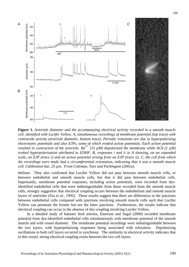
Proceedings of he Australian Physiological and Pharmacological Society (2001) 32(1) 199
199
Figure 1. Arteriole diameter and the accompanying electrical activity recorded in a smooth muscle cell, identified with Lucifer Yellow. A, simultaneous recordings of membrane potential (top trace) with contractile activity (arteriole diameter, bottom trace). Periodic transients are due to hyperpolarizing electrotonic potentials and also EJPs, some of which evoked action potentials. Each action potential resulted in contraction of the arteriole. Ba2+ (15 µM) depolarized the membrane while ACh (1 µM) evoked hyperpolarization attributed to EDHF. B, responses i and ii in A showing, on an expanded scale, an EJP (trace i) and an action potential arising from an EJP (trace ii). C, the cell from which the recordings were made had a circumferential orientation, indicating that it was a smooth muscle cell. Calibration bar, 25 µm. From Coleman, Tare and Parkington (2001a).
thelium. They also confirmed that Lucifer Yellow did not pass between smooth muscle cells, or between endothelial and smooth muscle cells, but that it did pass between endothelial cells. Importantly, membrane potential responses, including action potentials, were recorded from dye-identified endothelial cells that were indistinguishable from those recorded from the smooth muscle cells, strongly suggestive that electrical coupling occurs between the endothelium and smooth muscle layers of arterioles (Xia et al., 1995). These results suggest that there are differences in the junctions between endothelial cells compared with junctions involving smooth muscle cells such that Lucifer Yellow can permeate the former but not the latter junctions. Furthermore, the results indicate that electrical coupling can occur in the absence of dye coupling involving Lucifer Yellow. In a detailed study of hamster feed arteries, Emerson and Segal (2000) recorded membrane potential from dye-identified endothelial cells simultaneously with membrane potential of the smooth muscle and with vessel diameter. The membrane potential recordings were indistinguishable between the two layers, with hyperpolarizing responses being associated with relaxation. Depolarizing oscillations in both cell layers occurred in synchrony. The similarity in electrical activity indicates that in this vessel, strong electrical coupling exists between the two cell layers.
55
45
35
Dia
met
er (µ
m)
-80
-60
-40
-20
i ii
i
iiA C
B
2 min
Ba2+
AChMem
bran
e po
tent
ial (
mV
)
-60
-40
-20
1 s
Mem
bran
e po
tent
ial (
mV
)

200 Proceedings of the Australian Physiological and Pharmacological Society (2001) 32(1)
Figure 2. Arteriole diameter and the accompanying electrical activity recorded in an endothelial cell, identified with Lucifer Yellow. A, membrane potential (top trace) of an identified endothelial cell recorded simultaneously with arteriole contractile activity (diameter, bottom trace). Periodic transients are due to hyperpolarizing electrotonic potentials and also EJPs, some of which reached threshold to initiate action potentials. Ba2+ (20 µM) evoked depolarization while ACh (1 µM) evoked EDHFattributable hyperpolarization. Each action potential recorded from the endothelium was associated with contraction of the arteriole. B, the EJP (response i in A) and an action potential (response ii in A), on an expanded scale. C, the cell from which the recordings were made was a long thin cell running along the axis of the arteriole, indicating that it was an endothelial cell. Calibration bar, 25 µm. From Coleman, Tare and Parkington (2001a).
The possibility of myoendothelial electrical coupling in submucosal arterioles of the small intestine of guinea-pigs was assessed by recording the membrane potential responses to sympathetic nerve stimulation from Lucifer-Yellow filled endothelial or smooth muscle cells, simultaneously with contractile activity (Coleman et al., 2001a, b). Excitatory junction potentials (EJPs) were recorded from endothelial cells that were indistinguishable from those recorded from the smooth muscle (Fig.1 cf. Fig. 2). Occasionally the EJPs reached threshold for the initiation of action potentials, and those recorded from endothelial cells could not be distinguished from those action potentials recorded from the smooth muscle (Coleman et al., 2001a) (Fig. 1 cf. Fig. 2). Furthermore, ACh-induced hyperpolarizations, attributed to EDHF, were recorded from endothelial cells that were indistinguishable from those recorded from the smooth muscle in the same segment of arteriole (Coleman et al., 2001b). Thus, the endothelial and smooth muscle layers of the submucosal arterioles
65
55
45
1 s
2 min
Dia
met
er (µ
m)
-80
-60
-40
-20A C
B
ii
i
Ba2+
ACh
Mem
bran
e po
tent
ial (
mV
)
-60
-40
-20i
Mem
bran
e po
tent
ial (
mV
)
ii
Proceedings of he Australian Physiological and Pharmacological Society (2001) 32(1) 201
201
are coupled so tightly electrically that they function as a single electrical syncytium. The electrical coupling in these arterioles has recently been described mathematically (Crane et al., 1999). Vessels lacking myoendothelial electrical coupling
Although the evidence is now unequivocal for the presence of myoendothelial electrical coupling in a number of arterioles and arteries, this does not mean that it occurs in all vessels. In a detailed study of lymphatic vessels of the guinea-pig mesentery, Von der Weid and Van Helden (1997) recorded the membrane potential simultaneously from dye-identified cells in the endothelium and the smooth muscle. Responses to agonists had different time courses in the two layers, providing convincing evidence that myoendothelial electrical coupling does not occur in these vessels. When a similar approach was applied to a study of rat irideal arterioles, it was concluded that coupling between the muscle and endothelial layers might not occur in this vessel (Hirst et al., 1997). Although in vitro studies of hamster cheek pouch arterioles indicate the presence of myoendothelial electrical coupling in this vessel (see above), results from an in vivo study during blood flow showed that responses to agonists had different time courses in the two layers, indicating that electrical coupling did not occur in these vessels under these conditions (Welsh & Segal, 1998). The reason for the discrepancy with earlier studies of these arterioles is not clear. However, it does emphasize the possibility that myoendothelial electrical coupling may involve a dynamic dimension. This should not be unexpected since connexins, the building blocks of gap junctions, have been shown to have relatively short half lives (1-3 hrs), and are the targets of various kinases, with phosphorylation of connexins being implicated in a wide range of connexin processes influencing patency of gap junctions (reviewed by Lampe & Lau, 2000). Gap junction inhibitors
The strong evidence indicating the presence of myoendothelial electrical coupling, in at least some vessels, and its implications for EDHF, has raised considerable interest in methods to inhibit such coupling. Early studies focussed on the effects of the gap junction inhibitors heptanol, octanol and halothane. However, as demonstrated and discussed by Xia et al. (1995) and Chaytor et al. (1997), heptanol can alter the activity of a range of ion transport processes, thus altering membrane resistance and membrane potential, with interpretation of any results being equivocal at best. Halothane and octanol similarly have a range of non-specific effects, making them of limited use in the study of electrical coupling (discussed by Boitano & Evans, 2000). Derivatives of glycyrrhetinic (GA) acid have also been proposed to inhibit gap junctions, based on their ability to inhibit the transfer of citrulline metabolites between cells (Davidson et al., 1986; Davidson & Baumgarten, 1988), though there have been no detailed studies on their effects on electrical coupling. However, these triterpenoid saponins also have a range of non-specific effects, including inhibition of Na+/K+ ATPase (Terasawa et al., 1992). In guinea-pig submucosal arterioles, 18α-GA had little effect on input resistance, suggesting that there was little if any effect on electrical coupling. The effects of 18β-GA were associated with a small increase in input resistance, consistent with a weak inhibition of electrical coupling. Action potentials were partially inhibited by 18β-GA in the smooth muscle of submucosal arterioles (Fig. 3) and in the larger rat tail artery (Tare et al., 2000a). 18β-GA and its hemisuccinate form, carbenoxolone, reduced the ACh-induced hyperpolarization of endothelial cells in the rat mesenteric and guinea-pig coronary arteries and the submucosal arterioles (Tare et al., 2000a; Coleman et al., 2001a). Both 18β-GA and carbenoxolone evoked substantial depolarization of the endothelial and smooth muscle cells of submucosal arterioles (Imaeda et al., 2000; Coleman et al., 2001a), and carbenoxolone depolarized the guinea-pig coronary artery whereas 18β-GA hyperpolarized this artery (Tare et al., 2000a). Thus, the GA compounds have a variety of
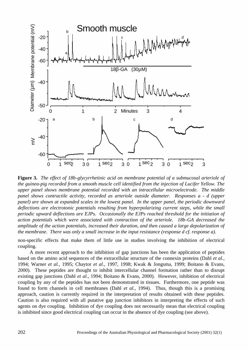
202 Proceedings of the Australian Physiological and Pharmacological Society (2001) 32(1)
Figure 3. The effect of 18β-glycyrrhetinic acid on membrane potential of a submucosal arteriole of the guinea-pig recorded from a smooth muscle cell identified from the injection of Lucifer Yellow. The upper panel shows membrane potential recorded with an intracellular microelectrode. The middle panel shows contractile activity, recorded as arteriole outside diameter. Responses a - d (upper panel) are shown at expanded scales in the lowest panel. In the upper panel, the periodic downward deflections are electrotonic potentials resulting from hyperpolarizing current steps, while the small periodic upward deflections are EJPs. Occasionally the EJPs reached threshold for the initiation of action potentials which were associated with contraction of the arteriole. 18β-GA decreased the amplitude of the action potentials, increased their duration, and then caused a large depolarization of the membrane. There was only a small increase in the input resistance (response d cf. response a).
non-specific effects that make them of little use in studies involving the inhibition of electrical coupling. A more recent approach to the inhibition of gap junctions has been the application of peptides based on the amino acid sequences of the extracellular structure of the connexin proteins (Dahl et al., 1994; Warner et al., 1995; Chaytor et al., 1997, 1998; Kwak & Jongsma, 1999; Boitano & Evans, 2000). These peptides are thought to inhibit intercellular channel formation rather than to disrupt existing gap junctions (Dahl et al., 1994; Boitano & Evans, 2000). However, inhibition of electrical coupling by any of the peptides has not been demonstrated in tissues. Furthermore, one peptide was found to form channels in cell membranes (Dahl et al., 1994). Thus, though this is a promising approach, caution is currently required in the interpretation of results obtained with these peptides. Caution is also required with all putative gap junction inhibitors in interpreting the effects of such agents on dye coupling. Inhibition of dye coupling does not necessarily mean that electrical coupling is inhibited since good electrical coupling can occur in the absence of dye coupling (see above).
0 1 2 3 4-50
-40
Dia
met
er (µ
m)
Minutes
-60
-40
-20 dcb
a
18β-GA (30µM)
Mem
bran
e po
tent
ial (
mV
)
0 1 2 3
-60
-40
-20 a
mV
0 1 2 3
b
Smooth muscle
sec 0 1 2 3
dc
sec
sec 0 1 2 3
sec
Proceedings of he Australian Physiological and Pharmacological Society (2001) 32(1) 203
203
Functional implications of myoendothelial electrical coupling
The main functional implications of myoendothelial electrical coupling currently centre on the actions of EDHF. Though there have been various suggestions as to the chemical identity of EDHF, such suggestions remain conjectural. Some of the strongest evidence as to the nature of EDHF was provided by a study of guinea-pig submucosal arterioles in which electrically short segments of arterioles were voltage-clamped with single intracellular microelectrodes and a switching amplifier (Coleman et al., 2001a). Since the endothelial and smooth muscle cells remained in their normal functional relationship, this approach enabled the recording of the membrane current that underlies the EDHF-attributed hyperpolarization. This current had biophysical and pharmacological characteristics indicative of K+ permeating Ca2+-activated K+ channels of intermediate conductance (IKCa) and small conductance (SKCa). Under the physiological gradient for K+ used in the study, the current-voltage relationships of the IKCa and SKCa current components were outwardly rectifying. In strong contrast, the application of raised K+ (additional 5 or 10 mM) substantially activated inward rectifier K+ channels (KIR) in these arterioles. Ouabain inhibited an outward current which had a relatively flat current-voltage relationship that was characteristic of the Na+/K+ ATPase and very dissimilar to that of the EDHF current. This observation therefore excludes any significant involvement of the Na+/K+ ATPase, whether activated by K+ or by other means. The lack of involvement of KIR channels and the Na+/K+ ATPase in the EDHF currents, and the differences between the responses attributed to EDHF and to K+ exclude K+ from being EDHF, at least in these arterioles (Coleman et al., 2001a, b) Epoxyeicosatrienoic acids (EETs), which are products of the cytochrome P450 pathway, have been reported to activate large conductance Ca2+-activated K+ channels (BKCa) (Zou et al., 1996; Baron et al., 1997; Eckman et al., 1998; Edwards et al., 2000). In submucosal arterioles, the hyperpolarization was insensitive to iberiotoxin, excluding any significant involvement of BKCa K+ channels. This suggests that EETs are unlikely to be a diffusible extracellular factor involved in the actions of EDHF in these arterioles. The application of endothelium-dependent vasodilators such as ACh, bradykinin and substance P to endothelial cells that are not in contact with smooth muscle results in endothelial hyperpolarization due to the activation of Ca2+-activated K+ channels, with IKCa and SKCa channels being the main channels implicated (Cannell & Sage, 1989; Mehrke & Daut, 1990; Vaca et al., 1992, 1996; Marchenko & Sage, 1993, 1996; Manabe et al., 1995; Frieden et al., 1999; Ohashi et al., 1999). The K+ currents evoked by ACh and substance P in voltage-clamped submucosal arterioles result from the activation of IKCa and SKCa channels (Coleman et al., 2001a, b) and are therefore likely to correspond to the Ca2+-activated K+ currents generated in the endothelial cells. If EDHF was a diffusible factor that activated ion channels in the smooth muscle, then the membrane current recorded in response to agonists such as ACh would be expected to consist of a smooth muscle current that was additional to the endothelial K+ current. Significantly, no additional components of K+ currents were recorded. Furthermore, IKCa channels have not been reported to occur in native smooth muscle cells. In view of agonist-induced hyperpolarization of isolated endothelial cells, the electrical coupling between the endothelial and smooth muscle cells, and the lack of evidence from the voltage clamp studies for additional components of K+ current that could have arisen in the smooth muscle, the most economical explanation for the phenomenon of EDHF is that it represents spread of hyperpolarizing current from the endothelium to the smooth muscle. That EDHF is able to evoke appreciable relaxation of blood vessels means that hyperpolarizing currents in the endothelial cells are likely to effect substantial relaxation of blood vessels via myoendothelial electrical coupling. However, there are significant regional differences in the contribution of EDHF to endothelium-dependent relaxation (reviewed by Hill et al., 2001) and this could be due to differences in the equivalent electrical circuitry of the various blood vessels. Parameters of importance include the resistance of the myoendothelial gap junctions, and the electrical load of the smooth muscle. If there are very few myoendothelial gap junctions, resulting in poor
204 Proceedings of the Australian Physiological and Pharmacological Society (2001) 32(1)
electrical coupling, then a large endothelial hyperpolarization may result in only a small hyperpolarization (EDHF) of the smooth muscle. With increasing electrical coupling, more endothelial current would be able to flow to the smooth muscle, and therefore have the potential to produce a greater EDHF response in the smooth muscle. Regional variations in myoendothelial gap junction density have been reported with a greater density of junctions found in the smaller arteries and arterioles (Rhodin, 1967; Sandow & Hill, 2000), consistent with the greater effectiveness of EDHF in more distal components of the vascular tree (Shimokawa et al., 1996). In this situation, one might expect little change in amplitude of the endothelial hyperpolarization along the vascular tree. Another important parameter which could cause an increased effectiveness in the more distal components of vascular trees is the electrical load of the smooth muscle. In larger blood vessels, endothelial current would be readily dissipated through many layers of smooth muscle, particularly if the myoendothelial electrical coupling was very strong, resulting in a small EDHF in the smooth muscle. In this situation, the endothelial hyperpolarization would be relatively small, being of similar amplitude to that in the smooth muscle. In contrast, in arterioles with only a single layer of smooth muscle, the much smaller electrical load of the smooth muscle means that the endothelial current is likely to produce a relatively large hyperpolarization, and therefore EDHF is likely to be much more significant in evoking vasorelaxation, as observed (Coleman et al., 2001a). Thus, a better understanding of regional differences in EDHF requires a comparison of smooth muscle hyperpolarization with that of endothelial hyperpolarization (Sandow et al., 2001). EDHF in disease
Endothelial regulation of vascular tone involves a balance between contracting and relaxing influences and perturbations in this balance are a feature of many vascular diseases. Thus, during diseases involving vasorelaxant dysfunction, the constricting influence is likely to be more significant (Mombouli & Vanhoutte, 1999). This situation occurs in the mesenteric artery of spontaneously hypertensive rats in which the EDHF-attributed hyperpolarization of smooth muscle is reduced in amplitude compared with that in normotensive control rats (Fujii et al., 1992). Diabetes is another disease in which EDHF-attributed hyperpolarization and relaxation are impaired (Fukao et al., 1997a; De Vriese et al., 2000; Wigg et al., 2001). The mechanisms underlying this impairment of EDHF, including the role of myoendothelial gap junctions, remain to be elucidated. Conclusions
Electrical coupling between the endothelium and smooth muscle of at least some arterial blood vessels is now firmly established. This electrical coupling is the most likely explanation for the phenomenon referred to as EDHF, and therefore endothelium-derived hyperpolarization (EDH) would now seem to be a more appropriate term. A good indication of the functional significance of this electrical coupling is indicated by the appreciable relaxation, particularly of the smaller, resistance arteries and arterioles, attributed to EDHF. However, the full functional implications of this electrical coupling are currently hindered by the lack of effective, selective inhibitors of the electrical coupling. Acknowledgements
The authors’ original work presented or cited in this review was supported by the National Health and Medical Research Council of Australia. This work was carried out during the tenure of a grant from the National Heart Foundation. References
Adams, D. J. (1994) Ionic channels in vascular endothelial cells. Trends in Cardiovascular Medicine, 4, 18-26.
Proceedings of he Australian Physiological and Pharmacological Society (2001) 32(1) 205
205
Baron, A., Frieden, M. & Bény, L. J. (1997) Epoxyeicosatrienoic acids activate a high-conductance, Ca2+-dependent K+ channel on pig coronary artery endothelial cells. Journal of Physiology, 504, 537-543.
Bény, J.-L. (1990) Endothelial and smooth muscle cells hyperpolarised by bradykinin are not dye coupled. American Journal of Physiology, 258, H836-H841.
Bény, J.-L. (1997) Electrical coupling between smooth muscle cells and endothelial cells in pig coronary arteries. Pflügers Archiv - European Journal of Physiology, 433, 364-367.
Boitano, S. & Evans, W. H. (2000) Connexin mimetic peptides reversibly inhibit Ca2+ signaling through gap junctions in airway cells. American Journal of Physiology, 279, L623-L630.
Buus, N. H., Simonsen, U., Pilegaard, H. K. & Mulvany, M. J. (2000) Nitric oxide, prostanoid and non-NO, non-prostanoid involvement in acetylcholine relaxation of isolated human small arteries. British Journal of Pharmacology, 129, 184-192.
Campbell, W. B., Gebremedhin, D., Pratt, P. F. & Harder, D. R. (1996) Identification of epoxyeicosatrienoic acids as endothelium-derived hyperpolarizing factors. Circulation Research, 78, 415-423.
Cannell, M. B. & Sage, S. O. (1989) Bradykinin-evoked changes in cytoplasmic calcium and membrane currents in cultured bovine pulmonary artery endothelial cells. Journal of Physiology, 419, 555-568.
Chataigneau, T., Félétou, M., Duhault, J. & Vanhoutte, P. M. (1998) Epoxyeicosatrienoic acids, potassium channel blockers and endothelium-dependent hyperpolarization in the guinea-pig carotid artery. British Journal of Pharmacology, 123, 574-580.
Chaytor, A. T., Evans, W. H. & Griffith, T. M. (1997) Peptides homologous to extracellular loop motifs of connexin 43 reversibly abolish rhythmic contractile activity in rabbit arteries. Journal of Physiology, 503, 99-110.
Chaytor, A. T., Evans, W. H. & Griffith, T. M. (1998) Central role of heterocellular gap junctional communication in endothelium-dependent relaxations of rabbit arteries. Journal of Physiology, 508, 561-573.
Chen, G., Suzuki, H. & Weston, A. H. (1988) Acetylcholine releases endothelium-derived hyperpolarizing factor and EDRF from rat blood vessels. British Journal of Pharmacology, 95, 1165-1174.
Coleman, H. A., Tare, M. & Parkington, H. C. (2001a) K+ currents underlying the action of endothelium-derived hyperpolarizing factor in guinea-pig, rat and human blood vessels. Journal of Physiology, 531.2, 359-373.
Coleman, H. A., Tare, M. & Parkington, H. C. (2001b) EDHF is not K+ but may be due to spread of current from the endothelium in guinea pig arterioles. American Journal of Physiology, 280, H2478-H2483.
Crane, G. J., Kotecha, N. & Neild, T. O. (1999) Electrical connection between the endothelium and the smooth muscle in guinea-pig intestinal submucosal arterioles. Progress in Microcirculation Research, 10, 54-56.
Dahl, G., Nonner, W. & Werner, R. (1994) Attempts to define functional domains of gap junction proteins with synthetic peptides. Biophysical Journal, 67, 1816-1822.
Davidson, J. S. & Baumgarten, I. M. (1988) Glycyrrhetinic acid derivatives: A novel class of inhibitors of gap-junctional intercellular communication. Structure-activity relationships. Journal of Pharmacology and Experimental Therapeutics, 246, 1104-1107.
Davidson, J. S., Baumgarten, I. M. & Harley, E. H. (1986) Reversible inhibition of intercellular junctional communication by glycyrrhetinic acid. Biochemical and Biophysical Research Communications, 134, 29-36.
206 Proceedings of the Australian Physiological and Pharmacological Society (2001) 32(1)
Davies, P.F. (1986) Biology of Disease: Vascular cell interactions with special reference to the pathogensis of atherosclerosis. Laboratory. Investigation, 55, 5-24.
De Vriese, A. S., Verbeuren, tj, Van de Voorde, J., Lameire, N. H. & Vanhoutte, P. M. (2000) Endothelial dysfunction in diabetes. British Journal of Pharmacology, 130, 963-974.
Drummond, G. R., Selemidis, S. & Cocks, T. M. (2000) Apamin-sensitive, non-nitric oxide (NO) endothelium-dependent relaxations to bradykinin in the bovine isolated coronary artery: no role for cytochrome P450 and K+. British Journal of Pharmacology, 129, 811-819.
Eckman, D. M., Hopkins, N., McBride, C. & Keef, K. D. (1998) Endothelium-dependent relaxation and hyperpolarization in guinea-pig coronary artery: role of epoxyeicosatrienoic acid. British Journal of Pharmacology, 124, 181-189.
Edwards, G., Dora, K. A., Gardener, M. J., Garland, C. J. & Weston, A. H. (1998) K+ is an endothelium-derived hyperpolarizing factor in rat arteries. Nature, 396, 269-272.
Edwards, G., Thollon, C., Gardener, M. J., Félétou, M., Vilaine, J. P., Vanhoutte, P. M. & Weston, A. H. (2000) Role of gap junctions and EETs in endothelium-dependent hyperpolarization of porcine coronary artery. British Journal of Pharmacology, 129, 1145-1154.
Emerson, G. G. & Segal, S. S. (2000) Electrical coupling between endothelial cells and smooth muscle cells in hamster feed arteries - Role in vasomotor control. Circulation Research, 87, 474-479.
Félétou, M. & Vanhoutte, P. M. (1988) Endothelium-dependent hyperpolarization of canine coronary smooth muscle. British Journal of Pharmacology, 93, 515-524.
Fisslthaler, B., Popp, R., Kiss, L., Potente, M., Harder, D. R., Fleming, I. & Busse, R. (1999) Cytochrome P450 2C is an EDHF synthase in coronary arteries. Nature, 401, 493-497.
Fleming, I. (2000) Cytochrome P450 2C is an EDHF synthase in coronary arteries. Trends in Cardiovascular Medicine, 10, 166-170.
Frieden, M., Sollini, M. & Bény, J.-L. (1999) Substance P and bradykinin activate different types of KCa currents to hyperpolarize cultured porcine coronary artery endothelial cells. Journal of Physiology, 519, 361-371.
Fujii, K., Tominaga, M., Ohmori, S., Kobayashi, K., Koga, T., Takata, Y. & Fujishima, M. (1992) Decreased endothelium-dependent hyperpolarization to acetylcholine in smooth muscle of the mesenteric artery of spontaneously hypertensive rats. Circulation Research, 70, 660-669.
Fukao, M., Hattori, Y., Kanno, M., Sakuma, I. & Kitabatake, A. (1997a) Alterations in endothelium-dependent hyperpolarization and relaxation in mesenteric arteries from streptozotocin-induced diabetic rats. British Journal of Pharmacology, 121, 1383-1391.
Fukao, M., Hattori, Y., Kanno, M., Sakuma, I. & Kitabatake, A. (1997b) Evidence against a role of cytochrome P450-derived arachidonic acid metabolites in endothelium-dependent hyperpolarization by acetylcholine in rat isolated mesenteric artery. British Journal of Pharmacology, 120, 439-446.
Furchgott, R. F. & Zawadzki, J. V. (1980) The obligatory role of endothelial cells in the relaxation of arterial smooth muscle by acetylcholine. Nature, 288, 373-376.
Garland, C. J. & McPherson, G. A. (1992) Evidence that nitric oxide does not mediate the hyperpolarization and relaxation to acetylcholine in the rat small mesenteric artery. British Journal of Pharmacology, 105, 429-435.
Hill, C. E., Phillips, J. K. & Sandow, S. L. (2001) Heterogeneous control of blood flow amongst different vascular beds. Medicinal Research Reviews, 21, 1-60.
Proceedings of he Australian Physiological and Pharmacological Society (2001) 32(1) 207
207
Hirst, G. D. S., Edwards, F. R., Gould, D. J., Sandow, S. L. & Hill, C. E. (1997) Electrical properties of iridial arterioles of the rat. American Journal of Physiology, 273, H2465-H2472.
Imaeda, K., Yamamoto, Y., Fukuta, H., Koshita, M. & Suzuki, H. (2000) Hyperpolarization-induced dilatation of submucosal arterioles in the guinea-pig ileum. British Journal of Pharmacology, 131, 1121-1128.
Kühberger, E., Groschner, K., Kukovetz, W. R. & Brunner, F. (1994) The role of myoendothelial cell contact in non-nitric oxide-,non-prostanoid-mediated endothelium-dependent relaxation of porcine coronary artery. British Journal of Pharmacology, 113, 1289-1294.
Kwak, B. R. & Jongsma, H. J. (1999) Selective inhibition of gap junction channel activity by synthetic peptides. Journal of Physiology, 516, 679-685.
Lacy, P. S., Pilkington, G., Hanvesakul, R., Fish, H. J., Boyle, J. P. & Thurston, H. (2000) Evidence against potassium as an endothelium-derived hyperpolarizing factor in rat mesenteric small arteries. British Journal of Pharmacology, 129, 605-611.
Lampe, P. D. & Lau, A. F. (2000) Regulation of gap junctions by phosphorylation of connexins. Archives of Biochemistry and Biophysics, 384, 205-215.
Little, T. L., Xia, J. & Duling, B. R. (1995) Dye tracers define differential endothelial and smooth muscle coupling patterns within the arteriolar wall. Circulation Research, 76, 498-504.
Manabe, K., Ito, H., Matsuda, H. & Noma, A. (1995) Hyperpolarization induced by vasoactive substances in intact guinea-pig endocardial endothelial cells. Journal of Physiology, 484, 25-40.
Marchenko, S. M. & Sage, S. O. (1993) Electrical properties of resting and acetylcholine-stimulated endothelium in intact rat aorta. Journal of Physiology, 462, 735-751.
Marchenko, S. M. & Sage, S. O. (1994) Smooth muscle cells affect endothelial membrane potential in rat aorta. American Journal of Physiology, 267, H804-H811.
Marchenko, S. M. & Sage, S. O. (1996) Calcium-activated potassium channels in the endothelium of intact rat aorta. Journal of Physiology, 492, 53-60.
Mehrke, G. & Daut, J. (1990) The electrical response of cultured guinea-pig coronary endothelial cells to endothelium-dependent vasodilators. Journal of Physiology, 430, 251-272.
Mombouli, J. V. & Vanhoutte, P. M. (1999) Endothelial dysfunction: From physiology to therapy. Journal of Molecular and Cellular Cardiology, 31, 61-74.
Moncada, S., Gryglewski, R. J., Bunting, S. & Vane, J. R. (1976) An enzyme isolated from arteries transforms prostaglandin endoperoxides to an unstable substance that inhibits platelet aggregation. Nature, 263, 663-665.
Moore, D. H. & Ruska, H. (1957) The fine structure of capillaries and small arteries. Journal of Biophysical and Biochemical Cytology, 3, 457-475.
Nilius, B., Viana, F. & Droogmans, G. (1997) Ion channels in vascular endothelium. Annual Review of Physiology, 59, 145-170.
Ohashi, M., Satoh, K. & Itoh, T. (1999) Acetylcholine-induced membrane potential changes in endothelial cells of rabbit aortic valve. British Journal of Pharmacology, 126, 19-26.
Olesen, S. P., Clapham, D. E. & Davies, P. F. (1988) Haemodynamic shear stress activates a K+ current in vascular endothelial cells. Nature, 331, 168-170.
Palmer, R. M. J., Ferrige, A. G. & Moncada, S. (1987) Nitric oxide release accounts for the biological activity of endothelium-derived relaxing factor. Nature, 327, 524-526.
208 Proceedings of the Australian Physiological and Pharmacological Society (2001) 32(1)
Parkington, H. C., Tare, M., Tonta, M. A. & Coleman, H. A. (1993) Stretch revealed three components in the hyperpolarization of guinea-pig coronary artery in response to acetylcholine. Journal of Physiology, 465, 459-476.
Parkington, H. C., Tonta, M. A., Coleman, H. A. & Tare, M. (1995) Role of membrane potential in endothelium-dependent relaxation of guinea-pig coronary arterial smooth muscle. Journal of Physiology, 484, 469-480.
Petersson, J., Zygmunt, P. M., Jönsson, P. & Högestätt, E. D. (1998) Characterization of endothelium-dependent relaxation in guinea pig basilar artery - Effect of hypoxia and role of cytochrome P450 mono-oxygenase. Journal of Vascular Research, 35, 285-294.
Plane, F., Holland, M., Waldron, G. J., Garland, C. J. & Boyle, J. P. (1997) Evidence that anandamide and EDHF act via different mechanisms in rat isolated mesenteric arteries. British Journal of Pharmacology, 121, 1509-1511.
Popp, R., Bauersachs, J., Hecker, M., Fleming, I. & Busse, R. (1996) A transferable, $-naphthoflavone-inducible, hyperpolarizing factor is synthesized by native and cultured porcine coronary endothelial cells. Journal of Physiology, 497, 699-709.
Quilley, J. & McGiff, J. C. (2000) Is EDHF an epoxyeicosatrienoic acid? Trends in Pharmacological Sciences, 21, 121-124.
Rand, V. E. & Garland, C. J. (1992) Endothelium-dependent relaxation to acetylcholine in the rabbit basilar artery: importance of membrane hyperpolarization. British Journal of Pharmacology, 106, 143-150.
Randall, M. D., Alexander, S. P. H., Bennett, T., Boyd, E. A., Fry, J. R., Gardiner, S. M., Kemp, P. A., McCulloch, A. I. & Kendall, D. A. (1996) An endogenous cannabinoid as an endothelium-derived vasorelaxant. Biochemical and Biophysical Research Communications, 229, 114-120.
Rhodin, J. A. G. (1967) The ultrastructure of mammalian arterioles and precapillary sphincters. Journal of Ultrastructure Research, 18, 181-223.
Sandow, S. L. & Hill, C. E. (2000) Incidence of myoendothelial gap junctions in the proximal and distal mesenteric arteries of the rat is suggestive of a role in endothelium-derived hyperpolarizing factor-mediated responses. Circulation Research, 86, 341-346.
Sandow, S. L., Tare, M., Coleman, H. A., Hill, C. E. & Parkington, H. C. (2001) EDHF in femoral arteries. Progress in Microcirculation Research, 11, (in press).
Segal, S. S. & Bény, J.-L. (1992) Intracellular recording and dye transfer in arterioles during blood flow control. American Journal of Physiology, 263, H1-H7.
Segal, S. S. & Duling, B. R. (1987) Propagation of vasodilation in resistance vessels of the hamster: Development and review of a working hypothesis. Circulation Research, 61 (suppl II), II20-II25.
Shimokawa, H., Yasutake, H., Fujii, K., Owada, K., Nakaike, R., Fukumoto, Y., Takayanagi, T., Nagao, T., Egashira, K., Fujishima, M. & Takeshita, A. (1996) The importance of the hyperpolarizing mechanism increases as the vessel size decreases in endothelium-dependent relaxations in rat mesenteric circulation. Journal of Cardiovascular Pharmacology, 28, 703-711.
Tare, M., Parkington, H. C., Coleman, H. A., Neild, T. O. & Dusting, G. J. (1990) Hyperpolarization and relaxation of arterial smooth muscle caused by nitric oxide derived from the endothelium. Nature, 346, 69-71.
Tare, M., Coleman, H. A. & Parkington, H. C. (2000a) The actions of glycyrrhetinic acid derivatives on hyperpolarization of identified endothelial cells in arteries. Proceedings of the Australian Physiological and Pharmacological Society, 31, 68P.
Proceedings of he Australian Physiological and Pharmacological Society (2001) 32(1) 209
209
Tare, M., Parkington, H. C. & Coleman, H. A. (2000b) EDHF, NO and a prostanoid: hyperpolarization-dependent and -independent relaxation in guinea-pig arteries. British Journal of Pharmacology, 130, 605-618.
Taylor, S. G. & Weston, A. H. (1988) Endothelium-derived hyperpolarizing factor: a new endogenous inhibitor from the vascular endothelium. Trends in Pharmacological Sciences, 9, 272-274.
Terasawa, T., Okada, T., Hara, T. & Itoh, K. (1992) Glycyrrhetinic acid derivatives as potent inhibitors of Na+, K+-ATPase. Synthesis and structure-activity relationships. European Journal of Medicinal Chemistry, 27, 345-351.
Vaca, L., Schilling, W. P. & Kunze, D. L. (1992) G-protein-mediated regulation of a Ca2+-dependent K+ channel in cultured vascular endothelial cells. Pflügers Archiv - European Journal of Physiology, 422, 66-74.
Vaca, L., Licea, A. & Possani, L. D. (1996) Modulation of cell membrane potential in cultured vascular endothelium. American Journal of Physiology, 270, C819-C824.
Van de Voorde, J. & Vanheel, B. (2000) EDHF-mediated relaxation in rat gastric small arteries: Influence of ouabain/Ba2+ and relation to potassium ions. Journal of Cardiovascular Pharmacology, 35, 543-548.
Von der Weid, P.-Y. & Bény, J.-L. (1993) Simultaneous oscillations in the membrane potential of pig coronary artery endothelial and smooth muscle cells. Journal of Physiology, 471, 13-24.
Von der Weid, P.-Y. & Van Helden, D. F. (1997) Functional electrical properties of the endothelium in lymphatic vessels of the guinea-pig mesentery. Journal of Physiology, 504, 439-451.
Warner, A., Clements, D. K., Parikh, S., Evans, W. H. & DeHaan, R. L. (1995) Specific motifs in the external loops of connexin proteins can determine gap junction formation between chick heart myocytes. Journal of Physiology, 488, 721-728.
Welsh, D. G. & Segal, S. S. (1998) Endothelial and smooth muscle cell conduction in arterioles controlling blood flow. American Journal of Physiology, 274, H178-H186.
Wigg, S. J., Tare, M., Tonta, M. A., O'Brien, R. C., Meredith, I. T. & Parkington, H. C. (2001) Comparison of the effects of diabetes mellitus on an EDHF-dependent artery. American Journal of Physiology, (in press).
Xia, J., Little, T. L. & Duling, B. R. (1995) Cellular pathways of the conducted electrical response in arterioles of hamster cheek pouch in vitro. American Journal of Physiology, 269, H2031-H2038.
Zou, A., Fleming, J. T., Falck, J. R., Jacobs, E. R., Gebremedhin, D., Harder, D. R. & Roman, R. J. (1996) Stereospecific effects of epoxyeicosatrienoic acids on renal vascular tone and K+-channel activity. American Journal of Physiology, 270, F822-F832.
Zygmunt, P. M., Hogestatt, E. D., Waldeck, K., Edwards, G., Kirkup, A. J. & Weston, A. H. (1997) Studies on the effects of anandamide in rat hepatic artery. British Journal of Pharmacology, 122, 1679-1686.

210 Proceedings of the Australian Physiological and Pharmacological Society (2001) 32(1)
Proceedings of the Australian Physiological and Pharmacological Society (2001) 32(1) 1P
PREPARATION OF ABSTRACTS OF COMMUNICATIONS AND DEMONSTRATIONS TO THE AUSTRALIAN PHYSIOLOGICAL AND PHARMACOLOGICAL SOCIETY 1. McCance*, formerly of the Department of Physiology, Monash University, Clayton, Victoria 3168, Australia. Material should be submitted on a floppy disk that can be read by an IBM-compatible computer, OR by E-mail to [email protected]. The number of words should not exceed 600. The format should conform with the style of abstracts in recent Issues of the Proceedings. This abstract is an example, except for its use of headings. Title. The title should be in grammatical English without a full stop. Name(s) and Institute address of author(s). The name of the author who will present the communication should be listed first or underlined. The address should identify where the work was done. Employ an asterisk in conjunction with a footnote to give an author's address if it differs from where the work was done. Where no author is a Society member, the introducing member must be identified in parentheses immediately following the address i.e. (Introduced by ...........) Text. The text should contain enough detail to be self-explanatory. If abbreviations are used they must be defined at first appearance (but not in the title). S.I. units should be used as recommended by Baron (1988). For statistical notation see McCance (2001). Citations. Use the Harvard system as employed by the Journal of Physiology, except that the ampersand (&) should appear only in references and bracketed citations, not in the text. Work by three or more authors may be cited using the form Aitkin et al. (1993) (e.g.) unless it will lead to confusion. References. The cited works must be listed after the text, alphabetically by the authors' surnames, then chronologically if necessary. The reference must include all authors' names, year of publication, full title of journal (e.g., Journal of Physiology, not J. Physiol.), volume number and first and last pages of the article. For a book, include the title, editor, edition if applicable, specific page references if applicable, city of publication and publisher (see, e.g., Baron, 1988). Footnotes. An example of a footnote is shown below. A line will appear between the references and the footnote(s). Figure. Only one figure (line drawing or half tone) is permitted. It should bear no title or legend and be unnumbered. Its location should be within the text (not before or after), be appropriately described in the text and referred to as "the Figure". Lettering should be approximately 12 pt. Table. Only one table is permitted. It should be typed in the appropriate position in the text and ruled with 0.5 mm black lines. It should have no number title or legend and be referred to as "the Table". Animal Experiments. Note that Domestic rule 11(5) (see Members Handbook) states that "All abstracts that deal with animal experimentation in vivo should include the names, doses (where applicable) and modes of administration of all anaesthetic, tranquilizing and muscle relaxant drugs employed". Users of in vitro materials should make it clear that tissue was removed from anaesthetized or dead animals. The inclusion of such information is for the protection of authors and the Society. Baron, D.N. (1988) In: Units, Symbols and Abbreviations, ed. Baron, D.N. pp. 1-64. London: The Royal Society of Medicine. McCance, I. (2001) Proceedings of the Australian Physiological and Pharmacological Society, 32(1), 2P. *Address for correspondence with the Editor and submission of material for the "Proceedings": P.O. Box 2371, Mt. Waverley, Vic. 3149.
2P Proceedings of the Australian Physiological and Pharmacological Society (2001) 32(1)
STANDARDIZATION OF STATISTICAL NOTATION IN THE PROCEEDINGS OF THE AUSTRALIAN PHYSIOLOGICAL AND PHARMACOLOGICAL SOCIETY 1. McCance,* formerly of the Department of Physiology, Monash University, Clayton, Victoria 3168, Australia. The book by Ellis (1972), previously recommended as a guide for symbols and abbreviations, is superseded by Baron (1988), q.v. The entries under "standard error", however, still suggest that some clarification is required before this work can be used to standardize statistical notation for the "Proceedings". The guide-lines that follow are intended to provide this clarification. In all respects other than in reference to the standard error the abbreviations recommended here are those of Baron (1988). Abbreviation for number of observations. Use lower case (italic) n. Abbreviationfor probability. Upper case (italic) P should be used, rather than p (Pressure) or p (pico-). Standard deviation and standard error. Authors should cite standard deviations when they are concerned to describe the variability of individual values about their mean. In such cases the data are likely to be descriptive rather than strictly experimental. Baron (1988) suggests SD or s: we prefer to use SD (N.B. A change - this differs from previously recommended s.d. (Ellis, 1972)). Authors should cite standard errors when they are concerned to indicate the precision, as an estimate, of a particular statistic such as a mean. The preferred abbreviation is SE (change, no longer s.e.) in all cases where it is clear to which statistic the SE is referred, but (in accordance with the notation used in Clinical and Experimental Pharmacology and Physiology) authors may use SEM for the SE of a mean. When quoting a SE it is preferable to include n, the number of observations on which the SE is based (see below). The use of ± . It is normal practice, in the "Proceedings" and elsewhere, to use ± to introduce a SE. In the "Proceedings" we will disregard the ruling against this usage in Baron (1988). This matter is discussed by Bliss (1967), who agrees that "in the recent literature" a term following a ± sign is unambiguously a SE. Inappropriate(?) SE. Authors should consider seriously their objective for including SE when these include variability that is not involved in the statistical test of significance used in the study, for example, the SE of means when the analysis uses a paired "t" test. In such cases it may be preferable to quote the SE of the mean difference as well as or instead of the individual SE. Improper SE. Authors should remember that the precision of a statistic or a comparison between statistics will usually involve variability "between animals (experimental units)" . A value of n that is inflated by the inclusion of multiple observations on the same animal (experimental unit) must not be used as the basis for calculating the SE, or for carrying out tests of significance, where the generalization is between animals. Baron, D.N. (1988) In: Units, Symbols and Abbreviations, ed. Baron, D.N., pp. 1-64. London: The Royal Society of Medicine. Bliss, C.I. (1967) Statistics in Biology, p. 137. New York: McGraw-Hill. Ellis, G. (1972) In: Units, Symbols and Abbreviations, ed. Ellis, G. Revised edition, pp. 1-36. London: The Royal Society of Medicine. *Address for correspondence with the Editor and submission of material for the "Proceedings": P.O. Box 2371, Mt. Waverley, Vic. 3149.
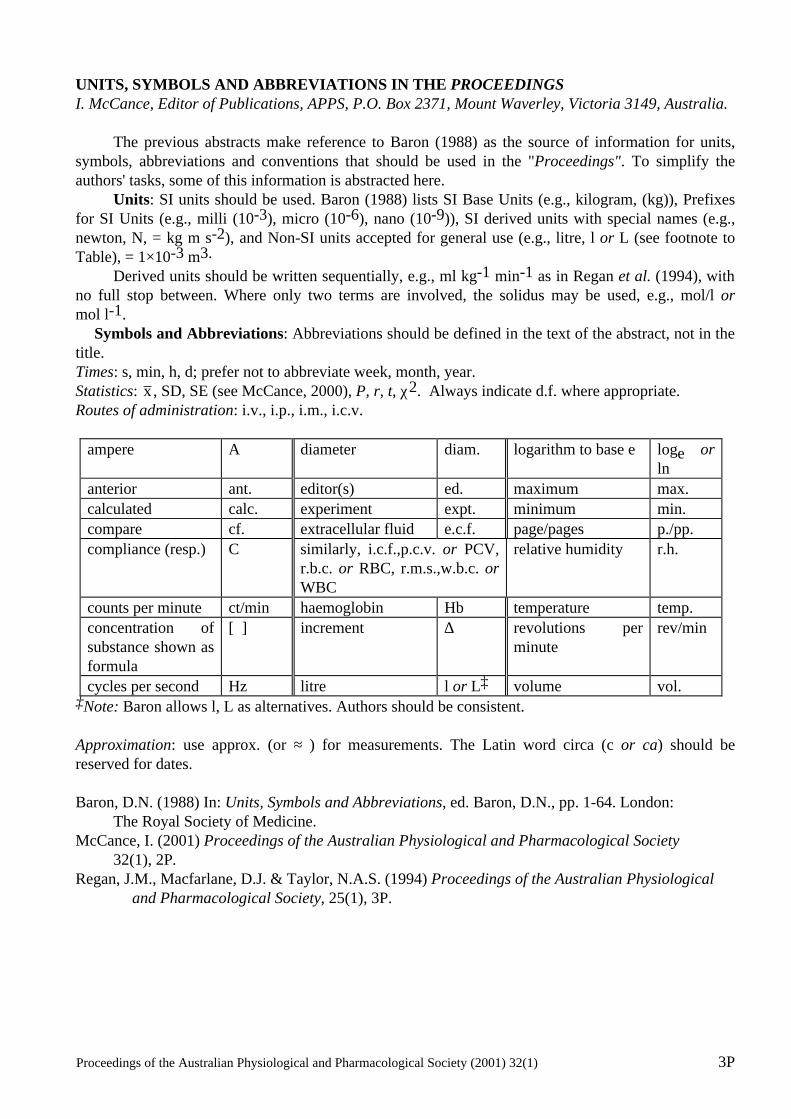
Proceedings of the Australian Physiological and Pharmacological Society (2001) 32(1) 3P
UNITS, SYMBOLS AND ABBREVIATIONS IN THE PROCEEDINGS I. McCance, Editor of Publications, APPS, P.O. Box 2371, Mount Waverley, Victoria 3149, Australia. The previous abstracts make reference to Baron (1988) as the source of information for units, symbols, abbreviations and conventions that should be used in the "Proceedings". To simplify the authors' tasks, some of this information is abstracted here. Units: SI units should be used. Baron (1988) lists SI Base Units (e.g., kilogram, (kg)), Prefixes for SI Units (e.g., milli (10-3), micro (10-6), nano (10-9)), SI derived units with special names (e.g., newton, N, = kg m s-2), and Non-SI units accepted for general use (e.g., litre, l or L (see footnote to Table), = 1×10⁻³ m3. Derived units should be written sequentially, e.g., ml kg-1 min-1 as in Regan et al. (1994), with no full stop between. Where only two terms are involved, the solidus may be used, e.g., mol/l or mol l-1. Symbols and Abbreviations: Abbreviations should be defined in the text of the abstract, not in the title. Times: s, min, h, d; prefer not to abbreviate week, month, year. Statistics: x , SD, SE (see McCance, 2000), P, r, t, χ2. Always indicate d.f. where appropriate. Routes of administration: i.v., i.p., i.m., i.c.v.
ampere A diameter diam. logarithm to base e loge or ln
anterior ant. editor(s) ed. maximum max. calculated calc. experiment expt. minimum min. compare cf. extracellular fluid e.c.f. page/pages p./pp. compliance (resp.) C similarly, i.c.f.,p.c.v. or PCV,
r.b.c. or RBC, r.m.s.,w.b.c. or WBC
relative humidity r.h.
counts per minute ct/min haemoglobin Hb temperature temp. concentration of substance shown as formula
[ ] increment ∆ revolutions per minute
rev/min
cycles per second Hz litre l or L‡ volume vol. ‡Note: Baron allows l, L as alternatives. Authors should be consistent. Approximation: use approx. (or ≈ ) for measurements. The Latin word circa (c or ca) should be reserved for dates. Baron, D.N. (1988) In: Units, Symbols and Abbreviations, ed. Baron, D.N., pp. 1-64. London: The Royal Society of Medicine. McCance, I. (2001) Proceedings of the Australian Physiological and Pharmacological Society 32(1), 2P. Regan, J.M., Macfarlane, D.J. & Taylor, N.A.S. (1994) Proceedings of the Australian Physiological and Pharmacological Society, 25(1), 3P.
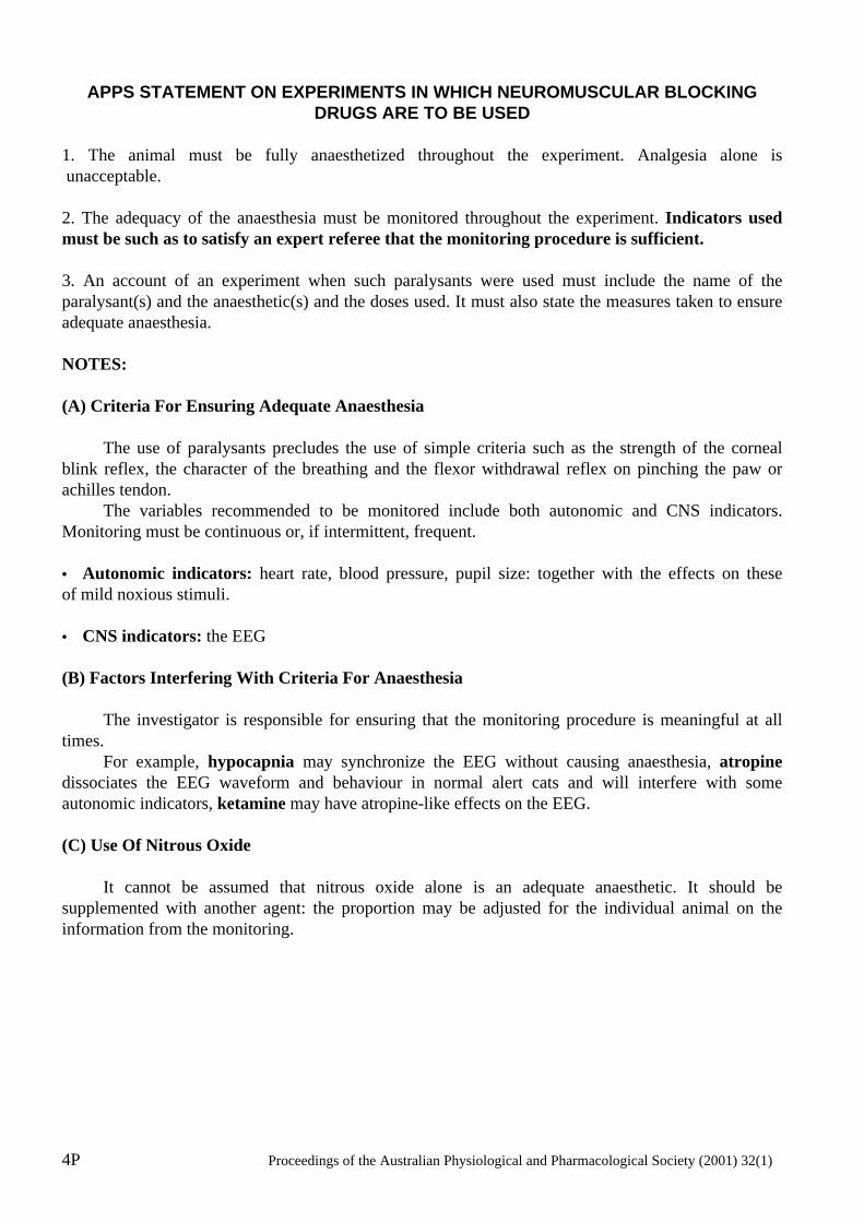
4P Proceedings of the Australian Physiological and Pharmacological Society (2001) 32(1)
APPS STATEMENT ON EXPERIMENTS IN WHICH NEUROMUSCULAR BLOCKING DRUGS ARE TO BE USED
1. The animal must be fully anaesthetized throughout the experiment. Analgesia alone is unacceptable. 2. The adequacy of the anaesthesia must be monitored throughout the experiment. Indicators used must be such as to satisfy an expert referee that the monitoring procedure is sufficient. 3. An account of an experiment when such paralysants were used must include the name of the paralysant(s) and the anaesthetic(s) and the doses used. It must also state the measures taken to ensure adequate anaesthesia. NOTES: (A) Criteria For Ensuring Adequate Anaesthesia The use of paralysants precludes the use of simple criteria such as the strength of the corneal blink reflex, the character of the breathing and the flexor withdrawal reflex on pinching the paw or achilles tendon. The variables recommended to be monitored include both autonomic and CNS indicators. Monitoring must be continuous or, if intermittent, frequent. • Autonomic indicators: heart rate, blood pressure, pupil size: together with the effects on these of mild noxious stimuli. • CNS indicators: the EEG (B) Factors Interfering With Criteria For Anaesthesia The investigator is responsible for ensuring that the monitoring procedure is meaningful at all times. For example, hypocapnia may synchronize the EEG without causing anaesthesia, atropine dissociates the EEG waveform and behaviour in normal alert cats and will interfere with some autonomic indicators, ketamine may have atropine-like effects on the EEG. (C) Use Of Nitrous Oxide It cannot be assumed that nitrous oxide alone is an adequate anaesthetic. It should be supplemented with another agent: the proportion may be adjusted for the individual animal on the information from the monitoring.