Probing the structure of multi-stranded guanine-rich DNA complexes by Raman spectroscopy and...
13
Ž . Biophysical Chemistry 79 1999 11]23 Probing the structure of multi-stranded guanine-rich DNA complexes by Raman spectroscopy and enzymatic degradation Karen Poon, Robert B. Macgregor, Jr. U Department of Pharmaceutical Sciences, Faculty of Pharmacy, Uni ¤ ersity of Toronto, 19 Russell St, Toronto, Ontario, Canada M5S 2S2 Received 13 July 1998; received in revised form 9 February 1999; accepted 9 February 1999 Abstract Ž . Ž . The multi-stranded DNA complexes formed by the oligonucleotides d T GTG , Tel, and d T G , TG, were 15 4 2 4 15 15 examined by nuclease digestion and Raman spectroscopy. Both Tel and TG can aggregate to form structures consisting of multiple, parallel-oriented DNA strands with two independent structural domains. Overall, the structures of the TG and Tel aggregates appear similar. According to the Raman data, the majority of bases are in C29-endoranti conformation. The interaction of guanines at the 39-ends in both complexes stabilizes the complexes and protects them from degradation by exonuclease III. The 59-extensions remain single-stranded and the thymines are accessible to single-strand-specific nuclease digestion. The extent of enzymatic cleavage at the junction at the 59 end of the 15 thymines implies a conformational change between this part of the molecule and the guanine-rich region. The differential enzymatic sensitivity of the complexes suggests there are variations in backbone conforma- tions between TG and Tel aggregates. TG aggregates were more resistant to digestion by DNase I, Mung Bean nuclease, and S1 nuclease than Tel complexes. It is proposed that the lower DNase I sensitivity may be partly due to the more stable backbone exhibited by TG than Tel complexes. Structural uniformity along the guanine core of TG is suggested, as there is no indication of structural discontinuities or protected sites in the guanine-rich regions of TG aggregates. The lower extent of digestion by Mung Bean nuclease at the 39 end implies that these bases are inaccessible to the enzyme. This suggests that there is minimal fraying at the ends, which is consistent with the extreme thermal stability of the TG aggregates. Q 1999 Elsevier Science B.V. All rights reserved. Keywords: Raman spectroscopy; DNA structures; Nuclease digestion; Telomere; Electrophoresis Ž . Ž . Abbre ¤ iations: TG, d T G ; Tel, d T GTG ; ps, parallel stranded; aps, antiparallel stranded; TBE, 89 mM Tris, 89 mM boric 15 15 15 4 2 4 Ž . acid, 2 mM EDTA pH 8.0 U Corresponding author. Tel.: q1-416-978-7332; fax: q1-416-978-8511; e-mail: [email protected] 0301-4622r99r$ - see front matter Q 1999 Elsevier Science B.V. All rights reserved. Ž . PII: S 0 3 0 1 - 4 6 2 2 99 00034-4
-
Upload
karen-poon -
Category
Documents
-
view
217 -
download
3
Transcript of Probing the structure of multi-stranded guanine-rich DNA complexes by Raman spectroscopy and...
Ž .Biophysical Chemistry 79 1999 11]23
Probing the structure of multi-stranded guanine-richDNA complexes by Raman spectroscopy and enzymatic
degradation
Karen Poon, Robert B. Macgregor, Jr.U
Department of Pharmaceutical Sciences, Faculty of Pharmacy, Uni ersity of Toronto, 19 Russell St, Toronto, Ontario,Canada M5S 2S2
Received 13 July 1998; received in revised form 9 February 1999; accepted 9 February 1999
Abstract
Ž . Ž .The multi-stranded DNA complexes formed by the oligonucleotides d T G T G , Tel, and d T G , TG, were15 4 2 4 15 15examined by nuclease digestion and Raman spectroscopy. Both Tel and TG can aggregate to form structuresconsisting of multiple, parallel-oriented DNA strands with two independent structural domains. Overall, thestructures of the TG and Tel aggregates appear similar. According to the Raman data, the majority of bases are inC29-endoranti conformation. The interaction of guanines at the 39-ends in both complexes stabilizes the complexesand protects them from degradation by exonuclease III. The 59-extensions remain single-stranded and the thyminesare accessible to single-strand-specific nuclease digestion. The extent of enzymatic cleavage at the junction at the 59end of the 15 thymines implies a conformational change between this part of the molecule and the guanine-richregion. The differential enzymatic sensitivity of the complexes suggests there are variations in backbone conforma-tions between TG and Tel aggregates. TG aggregates were more resistant to digestion by DNase I, Mung Beannuclease, and S1 nuclease than Tel complexes. It is proposed that the lower DNase I sensitivity may be partly due tothe more stable backbone exhibited by TG than Tel complexes. Structural uniformity along the guanine core of TG issuggested, as there is no indication of structural discontinuities or protected sites in the guanine-rich regions of TGaggregates. The lower extent of digestion by Mung Bean nuclease at the 39 end implies that these bases areinaccessible to the enzyme. This suggests that there is minimal fraying at the ends, which is consistent with theextreme thermal stability of the TG aggregates. Q 1999 Elsevier Science B.V. All rights reserved.
Keywords: Raman spectroscopy; DNA structures; Nuclease digestion; Telomere; Electrophoresis
Ž . Ž .Abbre¨iations: TG, d T G ; Tel, d T G T G ; ps, parallel stranded; aps, antiparallel stranded; TBE, 89 mM Tris, 89 mM boric15 15 15 4 2 4Ž .acid, 2 mM EDTA pH 8.0
U Corresponding author. Tel.: q1-416-978-7332; fax: q1-416-978-8511; e-mail: [email protected]
0301-4622r99r$ - see front matter Q 1999 Elsevier Science B.V. All rights reserved.Ž .PII: S 0 3 0 1 - 4 6 2 2 9 9 0 0 0 3 4 - 4
( )K. Poon, Robert B. Macgregor r Biophysical Chemistry 79 1999 11]2312
1. Introduction
Guanosine can self-associate via hydrogenw xbonds to form guanine]guanine base pairs 1,2
w xand guanine quartets 3 , giving rise to variousDNA structures formed by different guanine-richsequences, for example, telomeric DNA com-
w x w x w xplexes 4]7 , G-wires 8 , and frayed wires 9 .Recent interest in the structure and thermody-namics of these complexes arises from their bio-logical importance, their potential as targets fortherapeutic agents, and their ability to form self-assembling nanostructures.
The most important biological example of gua-nine-rich sequences are telomeres, which consistof short, simple guanine-rich sequences repeatedhundreds or thousands of times at the end ofeukaryotic chromosomes. Telomeres are impli-cated in mechanisms regulating aging and cancerand they are important in the prevention of chro-
w xmosomal fusion and degradation 10,11 . Muta-tions in telomeric sequences can delay or blockcell division in anaphase by inhibiting chromo-
w xsome separation 12,13 . Investigations into thefactors important in determining the structureand stability of guanine-rich sequences is impor-tant to the understanding of the molecular mech-anisms underlying these cellular events.
In a previous study, we reported that substitu-tion of guanine residues for the thymine spacerswithin the telomereic-like sequence G T G , al-4 2 4ters the physical and chemical properties of the
w xaggregates formed by this oligonucleotide 14 .The interactions stabilizing the structures formedby oligonucleotides with uninterrupted runs of
Ž . Ž .guanine residues such as, d T G , TG , are15 15distinct from the interactions stabilizing com-plexes formed by oligonucleotides with a
Ž . Ž .telomere-like sequence, d T G T G , Tel . For15 4 2 4example, the guanine N7 sites are required for
w xthe formation of tetrads by Tel 3 , no aggregationis observed after methylation of this site bydimethylsulfate. However, methylation of guanineN7 sites does not impair the aggregation of TGw x14 . Although the guanine]guanine interactionsin the TG complexes apparently do not involvehydrogen bonding with the N7 sites, the thermal
stability of these complexes is extremely highwhen compared to that of the telomeric DNA
w xcomplexes 14 . We have proposed that the un-usual stability of the aggregates formed byoligonucleotides such as TG may be biologicallyrelevant. Mutation of a non-guanine residue toguanine would result in the formation of a longrun of consecutive guanines, which could thenform extremely stable complexes that result in theinterruption of cell division, especially during
w xanaphase 12,13 .The complexes formed by Tel and TG also
exhibit differences in the stoichiometry of theaggregates and preference of cation for stabiliza-
w xtion 14 . We have shown that the successivelyhigher states of aggregation of TG resolved byelectrophoresis differ from each other by theaddition of a single additional strand of the par-
w xent oligonucleotide 14 . This differs from thebehavior of the Tel complexes, which are com-posed of one, two, four, or multiples of fourcopies of the parent oligonucleotide. These andother data imply that the guanine]guanine inter-actions in the structures formed by TG and Teldiffer.
In this report, we have examined the structureof the TG and Tel aggregates by analyzing theirsensitivity to enzymatic degradation and by Ra-man spectroscopy. The positions and intensitiesof the Raman lines are sensitive to the main-chainconformation; therefore, we use this technique todiagnose the global structures of these DNA com-plexes. Because of the sensitivity of enzyme activ-ity to the structure of the substrate, it is possibleto deduce information concerning the structureof the substrate by analyzing the relative rates ofenzymatic degradation. This is best done in com-parison with another, known structure and byemploying a range of different enzymes. We haveinvestigated the susceptibility of TG and Tel ag-gregates to degradation by four nucleases, usingsingle and double stranded DNA as structuralstandards. Our results reveal that the aggregatesof both TG and Tel have parallel strand orienta-tion. And that despite the similarities of the cova-lent structure of the parent oligonucleotides, TGcomplexes are more resistant to enzymatic cleav-age than Tel complexes implying there are struc-

( )K. Poon, Robert B. Macgregor r Biophysical Chemistry 79 1999 11]23 13
tural differences between these two aggregates.Both of these multistranded structures are moreresistant as well as than the normal double-stranded and single-stranded DNA.
2. Material and methods
2.1. DNA preparation
The oligonucleotides were purchased from theHospital for Sick Children Biotechnology ServiceCentre, Toronto. The lyophilized, deprotected,cartridge-purified oligonucleotide were dissolvedin 89 mM Tris, 89 mM Boric acid, 2 mM EDTA,
Ž .pH 8.0 TBE .
2.2. Raman spectroscopy
Raman spectra were excited with the 514.5-nmline of an argon-ion laser; the radiant power onthe sample was 50 mW. The spectra were col-lected on a SPEX model 1877, 0.6-m Triple Spec-trometer with a liquid nitrogen cooled CCD de-
Ž .tector Princeton Instrument Co. . For a typicalexperiment, 50 l-min exposures were accumulatedand averaged.
The aggregate-forming oligonucleotides, TGand Tel were dissolved in TBE and incubatedwith either 1 M MgCl at 908C for 30 min or 1 M2KCl at 908C for 5 min prior to acquisition ofRaman spectra.
2.3. Nuclease digestion
The DNA oligomers were labeled with 32 Pw 32 xusing g- P ATP and polynucleotide kinase and
subsequently desalted using a P-6 Bio-Spin Chro-Ž .matography column Bio-Rad, Inc. . The aggre-
gates of TG and Tel were prepared with 200 mMMgCl at 908C for 30 min or 200 mM KCl at 908C2for 5 min. The heated samples were then slowlycooled to room temperature before use. Single
Ž .stranded d T N and double stranded15 15Ž . Ž .T N .d A N were used as controls.15 15 15 15
We followed two protocols in the nuclease di-gestion experiments. To mimic the condition inextracellular fluid, all digestions were carried out
Žin Hank’s solution at 378C 5 mM KCl, 0.3 mM
KH PO , 138 mM NaCl, 4.0 mM NaHCO , 0.32 4 3.mM Na HPO , 5.6 mM D-glucose . For one set of2 4
experiments we used extremely high concentra-tions of the enzyme to degrade the aggregates.One-minute incubations at 378C with 249 U of
Ž .Exonuclease III Exo III , 332 U of S1 nuclease,or 156 U of Mung Bean nuclease were used. Inanother series of experiments more moderateamounts of these enzymes were used and theincubation period was varied. Nucleases used in
Žthis part of the study were: Exonuclease III 4.98. ŽUrml in 20 mM MgCl , DNase I 3 Urml in 202
. ŽmM MgCl , S1 6.64 Urml in 20 mM MgCl and2 2. Ž0.1 mM ZnSO , and Mung Bean nuclease 6.244
.Urml in 20 mM MgCl and 0.1 mM ZnSO . For2 4both of the nuclease digestion experiments, 1 mlof sample was incubated for the desired length oftime with 5 ml of a solution containing the en-zyme at the appropriate concentration. The reac-tion was stopped by adding 2 ml of 0.5 M EDTA.The digested products were then examined byelectrophoresis on native and denaturing poly-acrylamide gels. The native gels were run at 48C.
3. Results
3.1. Raman spectroscopy
In TBE, in the absence of added cations, TG,and Tel, containing only guanosine and thymidine
Ž .residues, exhibit similar Raman spectra Fig. 1 .TG complexes formed in the presence of Mg2q
and Tel aggregates formed in the presence of Kq
y1 Ž .had a prominent peak at 837 cm Fig. 1 . Thispeak is characteristic of B-form DNA with the
w xfuranose rings in the C29-endo conformation 15 .The peaks at 837 cmy1 and 1091 cmy1 are indica-tors of the local backbone geometry, and theirposition implies that the backbone is similar to
w xthat of B-DNA 15,16 . The marker band for thephosphate group at 1091 cmy1 was employed for
w xnormalization of intensities 17 . The spectra ofboth aggregates exhibited an intense band at 746cmy1 indicating that a majority of thymineresidues adopt the C29-endoranti conformationŽ .Fig. 1 .
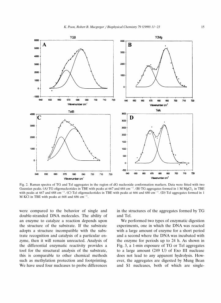
Fig. 2 displays the Raman spectral region from
( )K. Poon, Robert B. Macgregor r Biophysical Chemistry 79 1999 11]2314
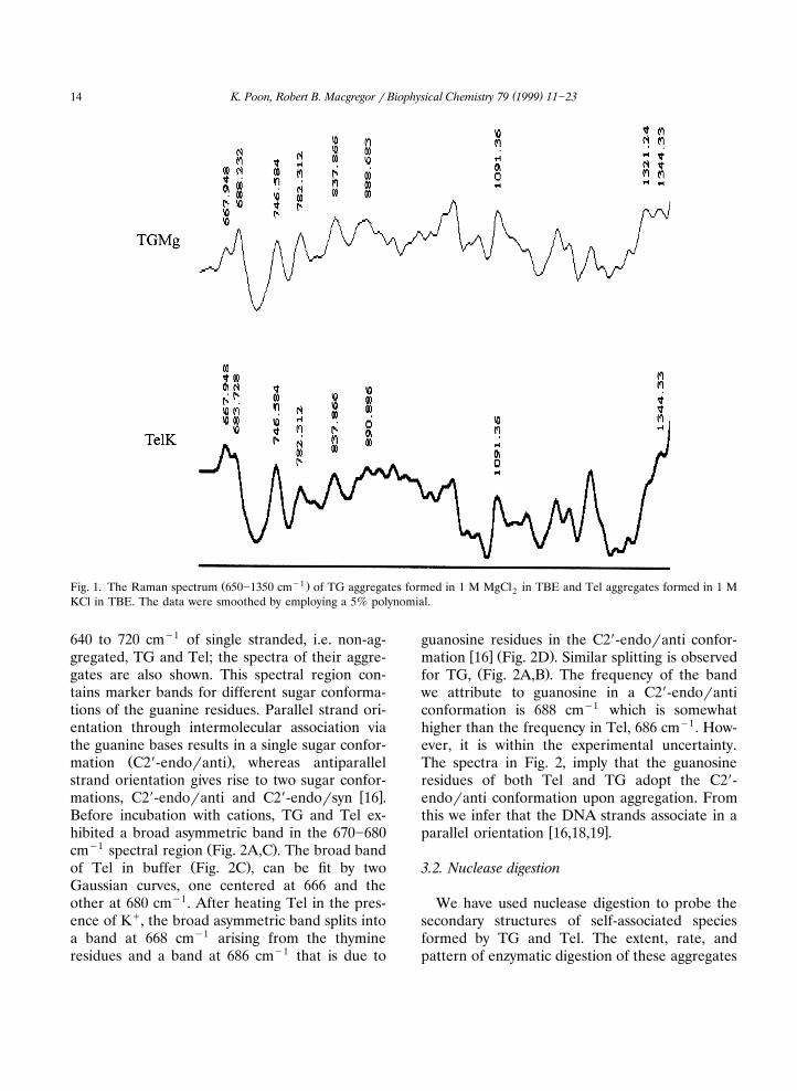
Ž y1 .Fig. 1. The Raman spectrum 650]1350 cm of TG aggregates formed in 1 M MgCl in TBE and Tel aggregates formed in 1 M2KCl in TBE. The data were smoothed by employing a 5% polynomial.
640 to 720 cmy1 of single stranded, i.e. non-ag-gregated, TG and Tel; the spectra of their aggre-gates are also shown. This spectral region con-tains marker bands for different sugar conforma-tions of the guanine residues. Parallel strand ori-entation through intermolecular association viathe guanine bases results in a single sugar confor-
Ž .mation C29-endoranti , whereas antiparallelstrand orientation gives rise to two sugar confor-
w xmations, C29-endoranti and C29-endorsyn 16 .Before incubation with cations, TG and Tel ex-hibited a broad asymmetric band in the 670]680
y1 Ž .cm spectral region Fig. 2A,C . The broad bandŽ .of Tel in buffer Fig. 2C , can be fit by two
Gaussian curves, one centered at 666 and theother at 680 cmy1. After heating Tel in the pres-ence of Kq, the broad asymmetric band splits intoa band at 668 cmy1 arising from the thymineresidues and a band at 686 cmy1 that is due to
guanosine residues in the C29-endoranti confor-w x Ž .mation 16 Fig. 2D . Similar splitting is observedŽ .for TG, Fig. 2A,B . The frequency of the band
we attribute to guanosine in a C29-endoranticonformation is 688 cmy1 which is somewhathigher than the frequency in Tel, 686 cmy1. How-ever, it is within the experimental uncertainty.The spectra in Fig. 2, imply that the guanosineresidues of both Tel and TG adopt the C29-endoranti conformation upon aggregation. Fromthis we infer that the DNA strands associate in a
w xparallel orientation 16,18,19 .
3.2. Nuclease digestion
We have used nuclease digestion to probe thesecondary structures of self-associated speciesformed by TG and Tel. The extent, rate, andpattern of enzymatic digestion of these aggregates
( )K. Poon, Robert B. Macgregor r Biophysical Chemistry 79 1999 11]23 15
Fig. 2. Raman spectra of TG and Tel aggregates in the region of dG nucleoside conformation markers. Data were fitted with twoŽ . y1 Ž .Gaussian peaks. A TG oligonucleotides in TBE with peaks at 667 and 684 cm . B TG aggregates formed in 1 M MgCl in TBE2
y1 Ž . y1 Ž .with peaks at 667 and 688 cm . C Tel oligonucleotides in TBE with peaks at 666 and 680 cm . D Tel aggregates formed in 1M KCl in TBE with peaks at 668 and 686 cmy1.
were compared to the behavior of single anddouble-stranded DNA molecules. The ability ofan enzyme to catalyze a reaction depends uponthe structure of the substrate. If the substrateadopts a structure incompatible with the subs-trate recognition and catalysis of a particular en-zyme, then it will remain unreacted. Analysis ofthe differential enzymatic reactivity provides atool for the structural analysis of the substrate,this is comparable to other chemical methodssuch as methylation protection and footprinting.We have used four nucleases to probe differences
in the structures of the aggregates formed by TGand Tel.
We performed two types of enzymatic digestionexperiments, one in which the DNA was reactedwith a large amount of enzyme for a short periodand a second where the DNA was incubated withthe enzyme for periods up to 24 h. As shown inFig. 3, a 1-min exposure of TG or Tel aggregates
Ž .to a large amount 249 U of Exo III nucleasedoes not lead to any apparent hydrolysis. How-ever, the aggregates are digested by Mung Beanand S1 nucleases, both of which are single-

( )K. Poon, Robert B. Macgregor r Biophysical Chemistry 79 1999 11]2316
Ž .Fig. 3. Digestion with exonuclease III 249 U , Mung BeanŽ . Ž .156 U and S1 endonucleases 332 U for a 1-min incubation
Ž . 32 Ž . 32at 378C. A P-Tel. B P-TG.
Ž .strand-specific Fig. 3 . Comparison of the relativeintensities of the bands demonstrated that theTG aggregates are more resistant than Tel com-plexes to enzymatic degradation.
The aggregates formed by TG or Tel oligonu-cleotides are inefficiently cleaved upon extended
Ž .incubation with Exo III Fig. 4 . During the first 5min, almost 40% of the double stranded refer-ence DNA was digested while only 1]2% of the
ŽTG or Tel aggregates were degraded data not.shown . After 24 h, only 3% of the TG or Tel
complexes were degraded compared with 70]80%digestion of double-stranded or single-stranded
Ž .DNA data not shown . The incomplete cleavageof double-stranded and single-stranded DNA isdue to the phosphate content of Hank’s solutionwhich would interfere with the action of the en-zymes. Despite this, it is clear that extent ofdigestion of the TG and Tel complexes by Exo IIIis dramatically lower than that of the double-stranded or single-stranded DNA. Although TGand Tel complexes have comparable rates of di-gestion, the two thymine spacers within the Telsequence are preferably cut by Exo III nucleaseŽ .Fig. 5 .
Ž .Fig. 4. Native electrophoretic gel 15% showing the digestion products of TG and Tel complexes. The complexes were digestedŽ . Ž . Žwith DNase I 15 Ursample for 1 and 24 h , Exonuclease III ExoIII, 24.9 Ursample for 1 and 24 h , Mung Bean nuclease MB,
. Ž .31.2 Ursample for 24 h and S1 33.2 Ursample for 24 h . Note that neither sample exhibited any digestion after 1 hr incubationwith Exo, so the gel patterns reflect the original samples of TG and Tel complexes.

( )K. Poon, Robert B. Macgregor r Biophysical Chemistry 79 1999 11]23 17
Ž . Ž .Fig. 5. Twenty percent denaturing gel showing the digestion products of A TG and B Tel complexes. The complexes wereŽ . Ž . Ž .digested with DNase 1 15 Ursample for 1 and 24 h , Exonuclease III Exo III, 24.9 Ursample for 1 and 24 h , Mung Bean MB
Ž . Ž .nuclease 31.2 Ursample for 24 h and S1 33.2 Ursample for 24 h .
TG and Tel aggregates were digested by DNaseI, with only 20]30% aggregates remaining at the
Ž .end of a 24-h incubation Fig. 4 . However, bothof the aggregated species were more resistantthan double or single stranded DNA toward this
Ž .enzyme data not shown . The single-strandedthymines are more extensively degraded than the
Ž .guanines for both TG and Tel aggregates Fig. 5 .Examination of the rate of digestion by DNase Ishows that the TG aggregates are more resistantto cleavage than the Tel complexes, especially
Ž .during the first hour of digestion Fig. 4 . Al-though more cutting was observed in theguanine-rich region of the Tel complexes, in ei-ther aggregate, this region was inefficiently di-
Ž .gested Fig. 5 .Figs. 4 and 6, show that the single-stranded
thymine residues at the 59 end of both the TGand Tel aggregates are accessible to digestion bythe single-strand-specific enzymes Mung Beannuclease and S1 nuclease. At the end of a 24-hincubation, 40]50% degradation was observed inTG aggregates and 80]90% in Tel complexes
Ž .data not shown . The difference in degradationrate was more dramatic during the first hour of
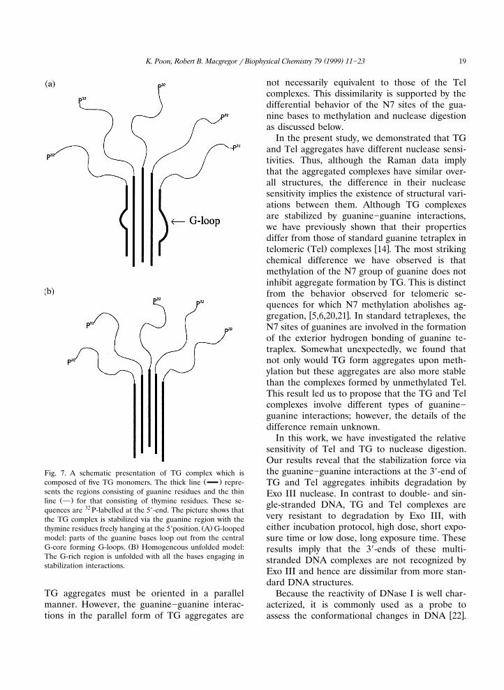
Ž .incubation Fig. 6 . The data show that MungBean nuclease apparently digests Tel complexesfrom the 39-end, where they are stabilized byguanine]guanine interactions, and proceeds to-wards the 59-end. S1 and Mung Bean nucleasesrarely cut within the G-run region of the TGcomplexes. This argues against the existence ofsignificant number of guanine bases looped out
Žfrom the central G-core of the aggregates Fig..7a . For both types of aggregates, hypersensitive
cleavage sites at the junctions between the gua-nine and thymine residues were observed for the
Ž .single-strand-specific nucleases Fig. 6 .
4. Discussion
We have previously shown that the formationof stable multistranded aggregates by Tel,Ž .d T G T G , is promoted by potassium ions but15 4 2 4
w xnot by magnesium ions 14 . Modification of thesequence within the guanine-rich region to

( )K. Poon, Robert B. Macgregor r Biophysical Chemistry 79 1999 11]2318
Fig. 6. Twenty percent denaturing gel showing the digestion products of TG and Tel complexes. The complexes were exposed toŽ . Ž . Ž .Mung Bean MB nuclease 31.2 Ursample for 5, 10, 30, and 60 min and S1 nuclease 33.2 Ursample for 5, 10, 30, and 60 min .
Ž . Ž .d T G TG , one observes the formation of15 15stable multistranded aggregates in the presence
w xof either potassium or magnesium ions 14 . Weshowed that the complexes formed by TG in thepresence of Mg2q have chemical and physicalproperties distinct from those of Tel aggregates.We anticipated that the origin of these distinctproperties would lie in structural differencesbetween TG and Tel aggregates. However, theRaman data presented here show that the sugarconformation of the TG and Tel aggregates aresimilar. Both types of aggregates consist of multi-ple, parallel DNA strands, in which the sugarshave B conformation and the majority of basesare in the C29-endoranti conformation.
Although telomeric DNA sequences can formeither parallel or anti-parallel multi-strandedcomplexes, their ability to adopt different confor-mations is determined by cation conditions andthe number of thymine spacers within the
w xtelomeric sequences 4 . Studies using the
telomeric sequence of Oxytricha No¨a showedthat it could form a parallel-stranded complex athigh concentrations of either sodium or potas-sium ions while anti-parallel species are prevalentat low cation concentrations. Since the salt con-centrations used in the present studies werehigher than those reported necessary to form
w xparallel stranded species 16 , it is expected thatparallel strand arrangement would be favored.One way to obtain an anti-parallel structure in-volves the interaction of two hairpin structures.For a tetraplex, this requires that the thyminespacers within the telomeric sequence loop out ofthe helix allowing the oligonucleotide chain tofold-back on itself and the guanine bases to en-
w xgage in tetraplex interactions 4 . A minimum oftwo thymine spacers is needed to form a stable
w xfold-back structure 4 . The absence of a thyminespacer in the TG oligonucleotide would appear toeliminate the possibility of forming antiparallelfold-back structures, implying that the strands in
( )K. Poon, Robert B. Macgregor r Biophysical Chemistry 79 1999 11]23 19
Fig. 7. A schematic presentation of TG complex which isŽ .composed of five TG monomers. The thick line repre-
sents the regions consisting of guanine residues and the thinŽ .line } for that consisting of thymine residues. These se-
quences are 32 P-labelled at the 59-end. The picture shows thatthe TG complex is stabilized via the guanine region with the
Ž .thymine residues freely hanging at the 59position. A G-loopedmodel: parts of the guanine bases loop out from the central
Ž .G-core forming G-loops. B Homogeneous unfolded model:The G-rich region is unfolded with all the bases engaging instabilization interactions.
TG aggregates must be oriented in a parallelmanner. However, the guanine]guanine interac-tions in the parallel form of TG aggregates are
not necessarily equivalent to those of the Telcomplexes. This dissimilarity is supported by thedifferential behavior of the N7 sites of the gua-nine bases to methylation and nuclease digestionas discussed below.
In the present study, we demonstrated that TGand Tel aggregates have different nuclease sensi-tivities. Thus, although the Raman data implythat the aggregated complexes have similar over-all structures, the difference in their nucleasesensitivity implies the existence of structural vari-ations between them. Although TG complexesare stabilized by guanine]guanine interactions,we have previously shown that their propertiesdiffer from those of standard guanine tetraplex in
Ž . w xtelomeric Tel complexes 14 . The most strikingchemical difference we have observed is thatmethylation of the N7 group of guanine does notinhibit aggregate formation by TG. This is distinctfrom the behavior observed for telomeric se-quences for which N7 methylation abolishes ag-
w xgregation, 5,6,20,21 . In standard tetraplexes, theN7 sites of guanines are involved in the formationof the exterior hydrogen bonding of guanine te-traplex. Somewhat unexpectedly, we found thatnot only would TG form aggregates upon meth-ylation but these aggregates are also more stablethan the complexes formed by unmethylated Tel.This result led us to propose that the TG and Telcomplexes involve different types of guanine]guanine interactions; however, the details of thedifference remain unknown.
In this work, we have investigated the relativesensitivity of Tel and TG to nuclease digestion.Our results reveal that the stabilization force viathe guanine]guanine interactions at the 39-end ofTG and Tel aggregates inhibits degradation byExo III nuclease. In contrast to double- and sin-gle-stranded DNA, TG and Tel complexes arevery resistant to degradation by Exo III, witheither incubation protocol, high dose, short expo-sure time or low dose, long exposure time. Theseresults imply that the 39-ends of these multi-stranded DNA complexes are not recognized byExo III and hence are dissimilar from more stan-dard DNA structures.
Because the reactivity of DNase I is well char-acterized, it is commonly used as a probe to
w xassess the conformational changes in DNA 22 .

( )K. Poon, Robert B. Macgregor r Biophysical Chemistry 79 1999 11]2320
This enzyme binds to the minor groove of the BDNA double helix forming contacts with thephosphates that are opposite each other on thetwo strands. DNase I catalyzes the hydrolysis ofthe 39O-P phosphodiester bond in single and dou-ble stranded DNA by nucleophilic attack. Its re-activity is strongly influenced by the size of thehelix groove and the spacing between the phos-
w xphates across the minor groove 22 , but notbase-pair arrangement. The cleavage rate is sensi-tive to local, sequence-dependent conformational
w xvariations 22 , for example, local helical twistw x23 . Cleavage is inhibited when the disposition ofthe phosphates no longer resembles that of B-
w xDNA 22 .As shown in Fig. 5, most of the DNase I cutting
for both TG and Tel aggregates occurred in the59 thymine residues rather than within the gua-nine residues at the 39-end. The guanine]guanineinteractions in both TG and Tel complexes seemto significantly reduce the cutting rate of DNaseI. This reflects that the minor grooves of both TGand Tel complexes are different from that ofB-DNA. This is consistent with our finding thatTG and Tel complexes are parallel multi-strandedcomplexes; therefore, the groove width, radialsymmetry and accessibility of the phosphates willdiffer from those of double stranded molecules.This is reflected in their relatively lower rate ofcleavage by DNase I. Neither hypersensitiveDNase I cleavage sites, nor any protected siteswere found within the guanine cores.
The 59 non-guanine regions of telomeric se-quences and molecules like TG have been shownto protrude from the aggregated structures as
w xsingle stranded arms 7,9,14 . Consistent with thisstructure we found that the 59-thymine extensionsof both complexes were accessible to digestion bysingle-stranded-specific nucleases.
S1 and Mung Bean nucleases both preferen-tially cut at the junctions between the guaninecores and the single stranded thymines of the TGand Tel aggregates. Other enzymatic studies havereported preferred cleavage at conformationaljunctions, for example between right-handed B-
w xDNA and left-handed Z-DNA 30 , and betweenw xdouble stranded and triple stranded DNA 31 .
The hypersensitive sites at the junctions of Teland TG aggregates suggest, not surprisingly, aconformational difference between the 39 gua-nines and the 59 thymine extensions.
The variation in the ability of nucleases torecognize the aggregates as substrates is a mani-festation of their structural differences. The find-ing that TG aggregates are digested by DNase Iat a lower rate than the Tel complexes impliesthat the backbone structures of the aggregates
w xare not the same. Thomas et al. 24 have shownthat uninterrupted runs of guanines are very rigid,perhaps the lack of conformational flexibility isresponsible for the limited extent of DNase cleav-
w xage 25 . The thymine loop linking the two sets offour guanine bases in the telomeric sequence was
Ž .preferentially cleaved by S1 nuclease Fig. 6 . S1nuclease recognizes and cleaves exposed phos-phodiester bonds in loops more readily than those
w xin a double helix 22 . This enzyme readily cutsbonds close to the 59-end of a hairpin and the
w xcleavage proceeds rapidly toward the 59-end 22 .Similar to DNase I, S1 is relatively insensitive tosequence; however, the rate of catalysis dependson the structural details of sugar]phosphate
w xbackbone 22 . The higher cutting rate of S1 withinthe thymine loop of Tel aggregates implies thatthese phosphodiester groups are more exposedthan those of the four adjacent guanines. Thedata shown in Fig. 6 also revealed that once thethymine loop was nicked, S1 nuclease can proceedalong the guanines which are stabilized by gua-
Ž .nine tetrads Fig. 8b . It seems unlikely that thisresult is a secondary cleavage effect of cutting
w xfrom the 39-end. Drew 22 has clearly shown thata phosphate group at the 39-side of the cleavagesite of the substrate is necessary for S1 nucleaseto cleave. The absence of a phosphate group atthe 39-end of the Tel aggregates would hindercutting from this end.
Although S1 and Mung Bean nucleases aresingle-strand-specific, they can cleave doublestranded molecules because of base pair fraying
w xat the ends of double stranded DNA 26]28 . Inthe present study, we found the 39-ends of Telwere more susceptible to single-stranded-specific
Žnuclease digestion than duplex DNA data not

( )K. Poon, Robert B. Macgregor r Biophysical Chemistry 79 1999 11]23 21
.shown . This was not observed for the TG aggre-gates. Fraying of DNA involves a localized, rapidassociation and dissociation of the base pairs atthe end of the DNA strands. For an enzymaticreaction to proceed, the lifetime of the dissoci-ated state has to be long enough to allow the
enzyme to bind. Thus, the greater the extent offraying, the greater is the chance of enzymaticcleavage. The higher digestion rate of Tel at the39-end by single-stranded-specific nucleases may
Ž .reflect extensive fraying at this end Fig. 8a .Under similar experimental conditions, Mung
Ž . Ž .Fig. 8. A schematic presentation of TG complex which is digested by Mung Bean MB and S1 nucleases. The thick lineŽ .represents the regions consisting of guanine residues and the thin line } for that consisting of thymine residues. The drawing
shows that the TG complex is formed by four Tel monomers and is stabilized via the guanine region. These sequences are32 Ž . Ž .P-labelled at the 59-end. A MB is shown to cut at the fraying region of the 39-end. B S1 is shown to preferentially cut at thethymine loop and proceed with cleavage in the 59 direction.
( )K. Poon, Robert B. Macgregor r Biophysical Chemistry 79 1999 11]2322
w xBean nuclease cleaves more slowly than S1 29 ,however, we found that it hydrolyzes the 39-termi-nal guanine bases of the Tel aggregates more
Ž .efficiently than S1 Fig. 6 . This may be due to thefact that S1 preferentially cleaves at positions
w xwith a 39 phosphate 22 , there is none at the39-end of the Tel aggregates. Mung Bean nucle-ase apparently does not require a 39 phosphate,
Žthus cutting can proceed from the 39-end Fig..8a .We do not detect much single-stranded cutting
along the G-run of the TG aggregates. This indi-cates that there are few single-stranded loopsformed along the G-run region. This arguesagainst the G-looped model of the TG aggregatesŽ .Fig. 7a in which some of the guanine bases arelooping out from the G-core region and do notform a stabilizing interaction with those along thecentral G-core. Instead, the data is consistent
Ž .with a homogeneous G-core structure Fig. 7b inwhich all the guanine bases interact with eachother.
The backbone heterogeneity in the guanine-richregion of Tel due to the interspersed thymineresidues enhances the reaction of the single-stranded-specific enzymes. Although a certain de-gree of backbone heterogeneity within the gua-nines may exist in TG aggregates, no hypersensi-tive sites within the run of guanines wereobserved, and overall, these structures are muchmore stable than the Tel aggregates with respectto cleavage by S1 or Mung Bean nuclease. Thisfinding is consistent with TG and Tel aggregateshaving different variations of backbone confor-mations. The absence of fraying implied by thelack of enzymatic cleavage is also consistent withthe greater thermal stability of the TG complexes.The resistance of TG aggregates toward cleavageby these enzymes may have the same structuralorigin as the thermal stability of TG aggregates.
In summary, the multistranded aggregatesformed by TG and Tel appear to have structuresthat are globally similar. Both types of aggregatesconsist of multiple parallel DNA strands, but theyexhibit differences in their stoichiometry andthermal stability. Generally, TG aggregates arequite resistant to nuclease cleavage. The nuclease
digestion results imply that the guanines in Teland TG aggregates do not adopt the same localconformation. There is no evidence of loopingout of bases within the guanines of the TG aggre-gates; all of the guanines are uniformly accessi-ble, or resistant, to cleavage. These data are con-sistent with the existence of a uniform structure
Žalong the guanine core of the TG aggregates Fig..8b . The accessibility of the guanines to cleavage
by single strand-specific nucleases imply that thereis fraying at the 39 end of the Tel aggregates.However, fraying does not appear to be extensivein the TG aggregates.
Acknowledgements
The authors would like to thank Dr X. Gu ofthe Photonics Resource Facility, University ofToronto for help with the Raman measurements.This work was supported in part by a grant fromGlaxo Wellcome Canada; K. Poon was supportedby an Ontario Graduate Scholarship.
References
w x1 M. Gellert, M.N. Lipsett, D.R. Davies, Helix formationŽ .by guanylic acid. Proc. Natl. Acad. Sci. U.S.A. 48 1962
2013]2018.w x2 I.G. Panyutin, O.I. Kovalsky, E.I. Budowsky, G-DNA: A
twice-folded DNA structure adopted by single-strandedŽ .oligo dG and its implications for telomeres. Nucleic
Ž .Acids Res. 17 1989 8257]8271.w x3 M. Lu, Q. Guo, N.R. Kallenbach, Thermodynamics of
G-tetraplex formation by telomeric DNAs. BiochemistryŽ .32 1993 598]601.
w x4 Q. Guo, M. Lu, N.R. Kallenbach, Effect of thyminetract length on the structure and stability of model
Ž .telomeric sequences. Biochemistry 32 1993 3596]3603.w x5 D. Sen, W. Gilbert, A sodium]potassium switch in the
Ž .formation of four-stranded G4-DNA. Nature 344 1990410]414.
w x6 D. Sen, W. Gilbert, Novel DNA superstructures formedŽ .by telomere-like oligomers. Biochemistry 31 1992
65]70.w x7 W.I. Sundquist, A. Klug, Telomeric DNA dimerizes by
formation of guanine tetrads between hairpin loops.Ž .Nature 342 1989 825]829.
w x8 T.C. Marsh, J. Vesenka, E. Henderson, A new DNAnanostructure, the G-wire, imaged by scanning probe
Ž .microscopy. Nucleic Acids Res. 23 1995 696]700.w x9 E. Protozanova, R.B. Macgregor, Jr., Frayed wires: A
thermally stable form of DNA with two distinct struc-Ž .tural domains. Biochemistry 35 1996 16638]16645.
( )K. Poon, Robert B. Macgregor r Biophysical Chemistry 79 1999 11]23 23
w x10 M.S. Rhyu, Telomeres, telomerase, and immortality. J.Ž .Natl. Cancer Inst. 87 1995 884]894.
w x11 L.L. Sandell, V.A. Zakian, Loss of a yeast telomere:Ž .arrest, recovery, and chromosome loss. Cell 75 1993
729]739.w x12 K.E. Kirk, B.P. Harmon, I.K. Reichardt, J.W. Sedat,
E.H. Blackburn, Block in anaphase chromosome separa-tion caused by a telomerase template mutation. Science
Ž .275 1997 1478]1481.w x13 R.S. Hawley, Unresolvable endings: defective telomeres
Ž .and failed separation. Science 275 1997 1478]1481.w x14 K. Poon, R.B. Macgregor, Jr., Unusual behavior exhib-
ited by multistranded guanine-rich DNA complexes.Ž .Biopolymers 45 1998 427]434.
w x15 C. Otto, G.A. Thomas, K. Rippe, T.M. Jovin, W.L.Peticolas, The hydrogen-bonding structure in parallel-stranded duplex DNA is reverse Watson]Crick.
Ž .Biochemistry 30 1991 3062]3069.w x16 T. Miura, G.J. Thomas, Jr., Structural polymorphism of
telomere DNA: interquadruplex and duplex]quadruplexconversions probed by Raman spectroscopy. Biochem-
Ž .istry 33 1994 7848]7856.w x17 J.M. Benevides, A.H.J. Wang, G.A. Van der Marel, G.J.
Thomas, Jr., Crystal and solution structures of the B-Ž .DNA dodecamer d CGCAAATTTGCG probed by Ra-
man spectroscopy: heterogeneity in the crystal structuredoes not persist in the solution structure. Biochemistry
Ž .27 1988 931]938.w x Ž .18 Y. Wang, D.J. Patel, Guanine residues in d T2AG3 and
Ž .d T2G4 form parallel-stranded potassium cationstabilized G-quadruplexes with anti glycosidic torsion
Ž .angles in solution. Biochemistry 31 1992 8112]8119.w x19 G. Gupta, A.E. Garcia, Q. Guo, M. Lu, N.R. Kallen-
bach, Structure of a parallel-stranded tetramer of theOxytricha telomeric DNA sequence dT G . Biochemistry4 4
Ž .32 1993 7098]7103.w x20 D. Sen, W. Gilbert, Formation of parallel four-stranded
complexes by guanine-rich motifs in DNA and its impli-Ž .cations for meiosis. Nature 334 1988 364]366.
w x21 E.A. Venczel, D. Sen, Parallel and antiparallel G-DNAstructures from a complex telomeric sequence.
Ž .Biochemistry 32 1993 6220]6228.w x22 H.R. Drew, Structural specificities of five commonly
Ž .used DNA nucleases. J. Mol. Biol. 176 1984 535]557.w x23 G.P. Lomonossoff, P.J.G. Butler, A. Klug, Sequence-de-
pendent variation in the conformation of DNA. J. Mol.Ž .Biol. 149 1981 745]760.
w x24 T. Miura, G.J. Thomas, Jr., Structure and dynamics ofinterstrand guanine association in quadruplex telomeric
Ž .DNA. Biochemistry 34 1995 9645]9654.˚w x25 D. Suck, A. Lahm, C. Oefner, Structure refined to 2 A
of a nicked DNA octanucleotide complex with DNase I.Ž .Nature 332 1988 464]468.
w x26 S.C. Sung, M. Laskowski, Sr., A nuclease from MungŽ .bean sprouts. J. Biol. Chem. 237 1962 506]511.
w x27 W.D. Kroeker, D. Kowalski, M. Laskowski, Sr., Mungbean nuclease I. Terminally directed hydrolysis of native
Ž .DNA. Biochemistry 15 1976 4463]4467.w x28 R.C. Wiegand, G.N. Godson, C.M. Radding, Specificity
of the S1 nuclease from Aspergillus oryzae. J. Biol.Ž .Chem. 250 1975 8848]8855.
w x29 G.A. Ghangas, R. Wu, Specific hydrolysis of the cohe-sive ends of bacteriophage lDNA by three single
Ž .strand-specific nucleases. J. Biol. Chem. 250 19754601]4606.
w x30 C.K. Singleton, J. Klysik, R.D. Wells, Conformationalflexibility of junctions between contiguous B- and Z-DNAs in supercoiled plasmids. Proc. Natl. Acad. Sci.
Ž .U.S.A. 80 1983 2447]2451.w x31 A. Bacolla, F.Y.H. Wu, Mung bean nuclease cleavage
pattern at a polypurine.polypyrimidine sequence up-stream from the mouse metallothionein-1 gene. Nucleic
Ž .Acids Res. 19 1991 1639]1647.