PRIVILEGED COMMUNICATION Prospective Lung … Goldstraw, MB, FRCS . Royal Brompton Hospital,...
32
IASLC Prospective LC Staging: Protocol 27MAR2009 – Page 1 of 32 Activation Date: 27MAR2009 PRIVILEGED COMMUNICATION The International Association for the Study of Lung Cancer (IASLC) Prospective Lung Cancer Staging Project PROCOTOL FOR PURPOSE OF GRANT APPLICATION AND ETHICS REVIEW IASLC Staging Committee, Full Committee Chair Peter Goldstraw, MB, FRCS Royal Brompton Hospital, Imperial College, London Full Committee Vice Chair T Subcommittee Chair Ramón Rami-Porta, MD Hospital Mutua de Terrassa, Terrassa, Spain N Subcommittee Chair Valerie Rusch, MD Memorial Sloan-Kettering Cancer Center, New York, New York, United States of America M Subcommittee Chair Pieter E. Postmus, MD, PhD Vrije Universiteit University Medical Center, Amsterdam, The Netherlands Small Cell Lung Cancer Subcommittee Chair Frances A. Shepherd, MD Princess Margaret Hospital and the University of Toronto, Toronto, Ontario, Canada Prognostic Factors Subcommittee Chair Jean-Paul Sculier, MD Institute Jules Bordet, Brussels, Belgium Validation and Methodology Subcommittee Chair Patti A. Groome, PhD Queen’s Cancer Research Institute, Kingston, Ontario, Canada Statistics and Data Management John Crowley, PhD Cancer Research and Biostatistics (CRAB), Seattle, Washington, United States of America 27MAR2009 ACTIVATION DOCUMENT
Transcript of PRIVILEGED COMMUNICATION Prospective Lung … Goldstraw, MB, FRCS . Royal Brompton Hospital,...

IASLC Prospective LC Staging: Protocol 27MAR2009 – Page 1 of 32
Activation Date: 27MAR2009
PRIVILEGED COMMUNICATION
The International Association for the Study of Lung Cancer (IASLC)
Prospective Lung Cancer Staging Project
PROCOTOL FOR PURPOSE OF GRANT APPLICATION AND ETHICS REVIEW
IASLC Staging Committee, Full Committee Chair Peter Goldstraw, MB, FRCS Royal Brompton Hospital, Imperial College, London Full Committee Vice Chair T Subcommittee Chair Ramón Rami-Porta, MD Hospital Mutua de Terrassa, Terrassa, Spain N Subcommittee Chair Valerie Rusch, MD Memorial Sloan-Kettering Cancer Center, New York, New York, United States of America M Subcommittee Chair Pieter E. Postmus, MD, PhD Vrije Universiteit University Medical Center, Amsterdam, The Netherlands Small Cell Lung Cancer Subcommittee Chair Frances A. Shepherd, MD Princess Margaret Hospital and the University of Toronto, Toronto, Ontario, Canada Prognostic Factors Subcommittee Chair Jean-Paul Sculier, MD Institute Jules Bordet, Brussels, Belgium Validation and Methodology Subcommittee Chair Patti A. Groome, PhD Queen’s Cancer Research Institute, Kingston, Ontario, Canada Statistics and Data Management John Crowley, PhD Cancer Research and Biostatistics (CRAB), Seattle, Washington, United States of America
27MAR2009 ACTIVATION DOCUMENT

IASLC Prospective LC Staging: Protocol 27MAR2009 – Page 2 of 32
TABLE OF CONTENTS PROCOTOL ........................................................................................................................... 1 TABLE OF CONTENTS ....................................................................................................... 2 1.0 OBJECTIVES ................................................................................................................... 3 2.0 BACKGROUND .............................................................................................................. 4 3.0 STUDY DESIGN ............................................................................................................. 5 4.0 SAMPLE SELECTION .................................................................................................... 6 5.0 INTERVENTION ............................................................................................................. 6 6.0 ELIGIBILITY CRITERIA ............................................................................................... 6 7.0 DESCRIPTIVE FACTORS .............................................................................................. 7 8.0 STATISTICAL CONSIDERATIONS ............................................................................. 7 9.0 DE-IDENTIFICATION OF DATA ................................................................................. 7 10.0 PROPERTY OF THE DATA BASE AND PUBLICATION POLICY ......................... 8 11.0 DATA COLLECTION PROCESSES ............................................................................ 8 12.0 OVERSIGHT BY ISC VALIDATION AND METHODOLOGY COMMITTEE ....... 9 13.0 SECONDARY USE OF THE IASLC LUNG CANCER DATA BASE ....................... 9 14.0 MEMBERSHIP OF THE IASLC STAGING COMMITTEE ..................................... 10 15.0 REFERENCES ............................................................................................................. 11 16.0 SITE APPLICATION MATERIALS ........................................................................... 13 17.0 SUMMARY OF STAGING CRITERIA ..................................................................... 19 18.0 APPENDIX - DATA ELEMENTS .............................................................................. 32
27MAR2009 ACTIVATION DOCUMENT

IASLC Prospective LC Staging: Protocol 27MAR2009 – Page 3 of 32
1.0 OBJECTIVES
1.1. The primary objectives of this study are: 1.1.1. To assess the prognostic value and validity of each component of the
seventh edition of the tumor, node, metastasis (TNM) classification for lung cancer with respect to the overall survival of patients with newly diagnosed lung cancer.
1.1.2. To identify and validate additional descriptors for possible inclusion in future revisions to the TNM classification.
1.2. T-Component objectives:
1.2.1. To assess the prognostic impact of tumor size 1.2.2. To assess the classification capacity of each descriptor defining T-status. 1.2.3. To study new conditions not included in the present T (e.g., differences
between parietal pleura invasion and rib invasion).
1.3. N-Component objectives: 1.3.1. To assess the prognostic impact of N-status. 1.3.2. To explore the prognostic impact of involved lymph node “zones” within N1
and N2 categories. 1.3.3. To assess the prognostic impact of:
a. Nodal extent (single vs multiple station involvement in N1 and N2 locations),
b. Nodal size, i.e. the largest involved node within the relevant N category, and c. Individual nodes being involved in each nodal category.
1.3.4 To assess the prognostic impact of extracapsular extension. 1.3.5 To assess the prognostic impact of the N3 nodal location, i.e. contralateral
mediastinum, ipsilateral or contralateral supraclavicular fossa.
1.4 M-Component objectives: 1.4.4 To assess the prognostic impact of M-status, especially those descriptors
now included within the new category of M1a included in the 7th edition. 1.4.5 To assess the prognostic impact of:
1.4.5.1 Single metastasis in a single organ 1.4.5.2 Multiple metastases in a single organ, and 1.4.5.3 Multiple metastases in several organs.
1.5 Objectives regarding other prognostic factors:
1.5.1 To assess the prognostic impact of histologic type and grade. 1.5.2 To assess the reliability of staging methods utilized in clinical staging (for
those tumors with pre-treatment and post-surgical classification). 1.5.3 To assess the prognostic impact of complete, incomplete, and uncertain
resections, according to the proposed definitions of the IASLC. 1.5.4 To assess the prognostic impact of clinical factors, including co-morbidity
and pulmonary function tests. 1.5.5 To assess the prognostic impact of maximum standard uptake value (SUV
max), at the primary site and in any positive nodal sites, for those patients with positron emission tomography (PET) scans in the pre-treatment staging.
27MAR2009 ACTIVATION DOCUMENT

IASLC Prospective LC Staging: Protocol 27MAR2009 – Page 4 of 32
2.0 BACKGROUND
The objectives of the Lung Cancer Staging Project of the International Association for the Study of Lung Cancer (IASLC) (1) have been achieved with the submission of recommendations for the seventh edition of the tumor, node, metastasis (TNM) classification for lung cancer to the International Union Against Cancer (UICC) and to the American Joint Committee on Cancer (AJCC). These core recommendations and the methodology used in the analysis of the retrospective database have been published (2, 3, 4, 5, 6), as have additional publications on small cell lung cancer, carcinoid tumors, and prognostic factors (7, 8, and 9).
The limitations of the analysis of the retrospective database derive from the fact that
most databases that contributed cases to the international database were not designed to study the TNM classification of lung cancer. The most important consequence was that, while the clinical or pathologic T status was recorded in most of the databases, few included the finer details, such as the specific anatomic sites of tumor extension. For this reason, most of the descriptors that define T3 and T4 tumors could not be validated in this retrospective study (2). The same is true for the potential subdivision of the N1 and N2 nodal spread based on the number of involved nodes/nodal stations or nodal zones (3), and for the differences in the various forms of M1 disease (4).
In addition, subtle differences between nodal maps used in different parts of the world – e.g. the Mountain and Dressler 1997 modification to the American Thoracic Society (ATS) (10) map and the Naruke-Japan Lung Cancer Society map (11, 12) – complicated previous attempts to analyze international data on nodal involvement. A new IASLC nodal map has been proposed and requires validation (13).
To overcome the limitations of the Retrospective Project, the International Staging
Committee (ISC) of the IASLC is proposing a Prospective Project (14) with the general objective to refine future editions of the TNM classification for lung cancer. This would imply the validation of all T, N, and M descriptors, with special attention to those that could not be validated with the analysis of the retrospective database, and the investigation of the prognostic value of other descriptors that are not included in the present TNM classification.
These objectives would be achieved by collecting a large prospective international database. This would, as far as possible, correct the geographical omissions and disproportionate spread of treatment modalities, which were inevitable in the retrospective data base, and would include both non-small and small cell bronchogenic carcinomas at the time of the initial diagnosis. In addition, at a recent IASLC workshop in London, there was international support for the inclusion of specific neuroendocrine subsets into the prospective data set (15). Pleural mesothelioma is to be covered in a separate data set at a later point.
27MAR2009 ACTIVATION DOCUMENT

IASLC Prospective LC Staging: Protocol 27MAR2009 – Page 5 of 32
The prospective dataset has been designed to achieve the proposed objectives. The
project’s success will depend on the extent of the international participation and the quality of the data. The Retrospective Project showed that quality of the data is even more important than its size. It will, therefore, be pivotal to collect complete data, especially when registering the elements directly related to the pre-treatment and post-surgical classification of lung cancer, the analysis of which is the main objective of this project.
Useful information not related to the anatomic extent of the disease can also be derived from the prospectively registered data. The inclusion of the methods used in the clinical staging will allow us to explore their reliability in those patients undergoing lung resection, in whom the pre-treatment and post-surgical classifications can be compared. The IASLC has published consensus guidelines on clinical staging based on the best evidence available in clinical practice (16, 17, 18). With the information of the prospective database, the IASLC would be able to review those guidelines based on contemporary data specifically collected for the purpose of staging analysis.
One of the objectives of the TNM classification is to assign a prognosis based on the anatomical extent of the disease. However, there are other factors that influence prognosis of lung cancer that are not related to its anatomic extension. Sex, age and comorbidity (19, 20, 21), biological parameters (22), and molecular and genetic factors are known to influence prognosis, but have never been integrated, along with the TNM classification, into a valid, clinically useful prognostic system. Information on comorbidity and basic blood analyses is easily available from most patients. Because the study of molecular and genetic factors is not standardized or universally available, information on the accessibility of these data would be collected, rather than the actual endpoints. This could facilitate international collaborative research among contributing centers. The maximum standard uptake value, which has shown prognostic relevance (23), will also be registered in those patients undergoing PET scan in the pre-treatment staging of their tumors.
The prospective registration of all these anatomic and non-anatomic parameters will allow us to address most of the issues that are expected from the International Staging Committee of the IASLC (24). This document is provided to collaborating institutions so that we may standardize the processes and procedures for conducting this prospective study across multiple institutions.
3.0 STUDY DESIGN
This is an international, multi-institutional cohort study that will collect detailed information on the extent of disease, personal and demographic characteristics, comorbid illness status, treatment and survival of newly-diagnosed lung cancer patients. Ideally, an inception cohort will be enrolled prospectively at each site, and data will be collected using a standardized abstraction tool. However, because it is unlikely that accrual goals will be met using this option alone, sites may alternatively petition the ISC to transfer data from an existing database.
27MAR2009 ACTIVATION DOCUMENT

IASLC Prospective LC Staging: Protocol 27MAR2009 – Page 6 of 32
Data completeness and logic checks will be conducted on an ongoing basis. Analyses will be conducted at CRAB. Each participating institution will have access to their own patients’ data and will be eligible to conduct secondary investigations of the larger database subject to approval by the IASLC (25). 4.0 SAMPLE SELECTION Based on the experience from the original retrospective study, three types of study samples will be targeted, depending on the nature of the collaborating institution: population-based, institution-based and clinical series. In each case, the intention is to describe the experience of an unselected group of patients. Sample selection for each is described below. 4.1 Population-based sample selection will likely involve enhancement of a population-based cancer registry with the data elements required for this study. All patients diagnosed within the study period may be included or a random sample of same. Documentation of the population coverage of the registry will be required for a sample to fall into this category. 4.2 Institution-based sample selection will likely involve the capture of information on all newly-diagnosed lung cancer patients seen at that institution during the period of the study. Usually, this involves the use of an institution’s tumor registry that will be enhanced with the data elements required for this study. Description of the institution’s referral pattern will be required. 4.3 Clinical series sample selection will capture information on an inception cohort of all newly-diagnosed patients presenting to a defined clinical service during the period of the study. All such patients will be tracked with documentation regarding data completeness and losses to follow up. In considering applications for participation in the project, the ISC will grant preference to sites which implement one of the above methods of sample selection. 5.0 INTERVENTION
Subjects will not be assigned to any specific intervention as a result of inclusion in this observational data base.
6.0 ELIGIBILITY CRITERIA 6.1 Subjects must have newly diagnosed non-small or small cell bronchogenic carcinomas, including the newly classified neuroendocrine tumors (ref WHO) and carcinoid tumors (included for the first time in the 7th edition of TNM) with documentation of TNM classification prior to treatment.
27MAR2009 ACTIVATION DOCUMENT

IASLC Prospective LC Staging: Protocol 27MAR2009 – Page 7 of 32
6.2 Lung cancer must be confirmed by histology or cytology.
6.3 Patients will be staged according to the seventh edition of the tumor, node, metastasis (TNM) classification for lung cancer.
7.0 DESCRIPTIVE FACTORS
Patients will be described by pretreatment T, N, and M status, treatment (surgically managed vs not), by country of origin and study sample type. Enrollment will be monitored with respect to these descriptive factors, with two objectives in mind: 1) to track recruitment of specific subgroups defined by geography, stage, or treatment modality with a view to targeting additional institutions and/or clinical settings if under-representation exists and 2) to demonstrate that the study sample is unbiased with regard to subject selection. 8.0 STATISTICAL CONSIDERATIONS If 3,000 subjects were accrued to each of the seven stage groups (IA, IB, IIA, IIB, IIIA, IIIB, and IV), for a total of 21,000 subjects, the power to detect a hazard ratio of 1.2 in comparisons of survival within any stage group would be 90%, using a two-sided logrank test at level .05. This calculation assumes exponential survival and three years of follow-up after a two-year accrual period. It is recognized, however, that stage is not evenly distributed in the lung cancer population. According to validation results from the retrospective IASLC staging project, roughly 40% of the subjects or more may be stage IV. If 10% (2,100) of the subjects were accrued from each of the six lower stage groups (IA, IB, IIA, IIB, IIIA and IIIB) and 40% (8,400) to stage IV, a total of 21,000 subjects would provide at least 80% power to detect a hazard ratio of 1.2 within any stage group. 9.0 DE-IDENTIFICATION OF DATA All sites must agree to gather identifiable private information of research subjects in compliance with applicable law and with respect and regards for human subjects. Each participating institution will secure approval of the project from their local Research Ethics Board. Participating sites must agree unequivocally to prohibit release of individually identifiable private data to CRAB for research purposes. CRAB will receive only ‘coded’ data for analysis. The ‘coded’ data sent to CRAB must not be able to be linked to individual research subjects, either directly or indirectly through the coding system, by any member of CRAB’s research team . Where personal identifiers might inadvertently be included with data received, CRAB will delete/destroy this identified data, and immediately notify the site to replace with de-identified data.
27MAR2009 ACTIVATION DOCUMENT

IASLC Prospective LC Staging: Protocol 27MAR2009 – Page 8 of 32
While visiting the site, CRAB staff may access or utilize individually private information but these activities become subject to the oversight of the site’s Institutional Review Board. At no time will CRAB employees record any private information. Because only de-identified data are submitted to CRAB, as an institution, CRAB is not considered to be “engaged” in human subjects’ research for this project. 10.0 PROPERTY OF THE DATA BASE AND PUBLICATION POLICY
Each institution will retain full access and publishing rights to its own data;
however, the collective database will be the property of the IASLC, and CRAB will be responsible for its management, storage, and analysis.
Publications related to the objectives of the Prospective Project of the IASLC Staging Committee (i.e., publications providing recommendations for changes in the TNM classification) will be planned, researched, analysed, and written by the members of the respective Subcommittees, and will follow the same authorship pattern used for the publications on the retrospective data: chairperson of the subcommittee, members of the subcommittee in alphabetical order, Chairman of the Staging Committee, on behalf of the IASLC Staging Committee, and participating institutions. 11.0 DATA COLLECTION PROCESSES Identification and training of data collectors will be left to the discretion of the participating institution. Information about these data collectors including how many are being employed and the nature of their professional training will be reported to the ISC. With the exception of the outcome data, most of the data collected for this study will occur around the time of diagnosis and treatment. The last date of follow up and vital status of each study subject will be updated at each follow-up visit with a frequency of no less than once per year. Institutions approved by the ISC for participation in this Prospective Project will enter the data online using a secure, web-based data entry system or transfer data from an existing database. Access to the online data entry system will be available via the IASLC web site at http://www.iaslc.org. Designed and administered by Cancer Research and Biostatistics (CRAB), the system will incorporate extensive, between-field logic checks and provide a query system enabling communication between CRAB and the institutions regarding the data. At the discretion of the Principal Investigator for each institution, the system will provide select users the ability to download all data entered by that institution. Transfer of existing, external data will be initially be limited to selected partners from the retrospective project and centers that facilitate correction of geographical gaps identified in the retrospective data. Additional sites may be recruited to meet the accrual goals, provided standards regarding data quality and completeness are met.
27MAR2009 ACTIVATION DOCUMENT

IASLC Prospective LC Staging: Protocol 27MAR2009 – Page 9 of 32
It is the intent of the project to follow each subject until death, provided there is sufficient funding to maintain this follow-up. As of the date of issuance of this protocol, the IASLC has agreed to sponsor collection of baseline data via the web-based data entry system through the year 2010 and collection of follow-up data through 2012. 12.0 OVERSIGHT BY ISC VALIDATION AND METHODOLOGY COMMITTEE Data collection is subject to measurement error between and within raters, particularly when subjective judgment is involved. Whenever possible, a single, experienced rater should be used to reduce observer variability. The ISC Validation and Methodology Committee will monitor population coverage, losses to follow up, and missing data rates at each site and report their findings to the full committee. 13.0 SECONDARY USE OF THE IASLC LUNG CANCER DATA BASE The IASLC Staging Project has a duty to ensure that the data within its database are used to maximum benefit for the good of patients and the lung cancer community, within ethical constraints and the agreements entered into with individual databases. All requests for the secondary use of the database will be subjected to the following review mechanism: An initial, outline proposal should be submitted to the chair of the committee. This will be reviewed by e-mail by a sub-committee consisting of the chair person, a CRAB member of the committee, and the chair of the relevant sub-committee. If the request is considered to be a reasonable proposal, the applicant will be asked to submit a full application containing the following, additional documents:
a) A full proposal setting out the details of the study, methods, population under study, data required from the database and proposed time lines.
b) A full list of the participants to the study and proposals for involvement by members of the committee and CRAB. The study should include as primary authors at least one medical member of the committee and one CRAB member of the committee.
c) A supportive letter from CRAB confirming that the necessary data is obtainable from the data base and that the quality and volume of that data is adequate to answer the question posed.
d) Confirmation that the applicant and all other parties who may be considered to hold intellectual property rights will adhere to the highest scientific and ethical standards, including but not exclusively:
a. Will respect the IASLC ownership of the data and will not seek to use the information provided for any other use without the agreement of the IASLC.
b. Will respect the anonymity of the clinical data. c. Will submit any publication or presentation for scrutiny by the committee,
and in addition, by those database proprietors with whom there exists prior agreements, before submission. The committee reserves the right to deny publication in extreme situations.
27MAR2009 ACTIVATION DOCUMENT

IASLC Prospective LC Staging: Protocol 27MAR2009 – Page 10 of 32
d. Will publish any submission in a format agreed with the committee,
including the format of the title, and acknowledging the participation of the IASLC, the committee members, CRAB and the database proprietors. The acknowledgment of our sponsors will be recognized in a format agreed with them from time to time.
e. Will submit publications, in the first place, to the Journal of Thoracic Oncology, the official journal of the IASLC.
The full proposal will be circulated to the full committee by e-mail and the committee’s view collected by the chairman. If consensus is not reached the proposal will be discussed at the next meeting of the committee. Revisions or additional material may be requested before a final decision is reached. The committee’s decision is final and there will be no appeal structure.
14.0 MEMBERSHIP OF THE IASLC STAGING COMMITTEE P. Goldstraw (Chairperson), Royal Brompton Hospital, Imperial College, London, UK; H. Asamura, National Cancer Centre Hospital, Tokyo, Japan; D. Ball, Peter MacCallum Cancer Centre, East Melbourne, Australia; V. Bolejack, Cancer Research and Biostatistics, Seattle, Washington, USA; E. Brambilla, Laboratoire de Pathologie Cellulaire, Grenoble Cedex, France; P.A. Bunn, University of Colorado Health Sciences, Denver, Colorado; D. Carney, Mater Misericordiae Hospital, Dublin, Ireland; K. Chansky, Cancer Research and Biostatistics, Seattle, Washington, USA; J. Crowley, Cancer Research and Biostatistics, Seattle, Washington, USA; D Giroux, Cancer Research And Biostatistics, Seattle, Washington, USA; P. Groome, Queen’s Cancer Research Institute, Kingston, Ontario, Canada; P. Van Houtte, Institute Jules Bordet, Bruxelles, Belgium; J. R. Jett, Mayo Clinic, Rochester, Minnesota, USA; C. Kennedy, University of Sydney, Sydney, Australia; H Kondo, Shizuoka Cancer Centre, Shizuoka, Japan; M. Krasnik, Gentofte Hospital, Copenhagen, Denmark; J. van Meerbeeck, University Hospital, Ghent, Belgium; H Pass, New York Medical Centre, New York, USA; E.F. Patz, Duke University Medical Center, Durham, North Carolina, USA; P.E Postmus, Vrije Universiteit Medical Center, Amsterdam, the Netherlands; R. Rami-Porta, Hospital Universitario Mutua de Terrassa, Terrassa, Spain; V. Rusch, Memorial Sloan-Kettering Cancer Center, New York, USA; N Saijo, National Cancer Center Hospital East, Chiba, Japan; JP. Sculier, Institute Jules Bordet, Bruxelles, Belgium; F.A. Shepherd, University of Toronto, Toronto, Ontario, Canada; L. Sobin, Armed Forces Institute of Pathology, Washington, DC; W. Travis, Memorial Sloan-Kettering Cancer Center, New York, USA; M. Tsuboi, Tokyo Medical University, Tokyo, Japan; E Vallieres, Swedish Cancer Institute, Seattle, Washington, USA; J. Vansteenkiste, Leuven Lung Cancer Group, Belgium; H Watanabe, National Cancer Centre Hospital, Tokyo, Japan.
27MAR2009 ACTIVATION DOCUMENT

IASLC Prospective LC Staging: Protocol 27MAR2009 – Page 11 of 32
15.0 REFERENCES
1. Goldstraw P, Crowley JJ. The International Association for the Study of Lung Cancer International staging project on lung cancer. J Thorac Oncol 2006; 1: 281-286.
2. Rami-Porta R, Ball D, Crowley J et al. The IASLC lung cancer staging project: proposals for the revision of the T descriptors in the forthcoming (seventh) edition of the TNM classification for lung cancer. J Thorac Oncol 2007; 2: 593-602.
3. Rusch VW, Crowley J, Giroux DJ et al. The IASLC lung cancer project: proposals for the revision of the N descriptors in the forthcoming seventh edition of the TNM classification for lung cancer. J Thorac Oncol 2007; 2: 603-612.
4. Postmus PE, Brambilla E, Chansky K et al. The IASLC lung cancer project: proposals for revision of the M descriptors in the forthcoming (seventh) edition of the TNM classification of lung cancer. J Thorac Oncol 2007; 2: 686-693.
5. Goldstraw P, Crowley J, Chansky K et al. The IASLC lung cancer project: proposals for the revision of the TNM stage groupings in the forthcoming (seventh) edition of the TNM classification of malignant tumours. J Thorac Oncol 2007; 2: 706-714.
6. Groome PA, Bolejack V, Crowley JJ et al. The IASLC lung cancer project: validation of the proposals for revision of the T, N, and M descriptors and consequent stage groupings in the forthcoming (seventh) edition of the TNM classification of malignant tumours. J Thorac Oncol 2007; 2: 694-705.
7. Shepherd FA, Crowley J, Van Houtte P et al. The IASLC lung cancer staging project: proposals regarding the clinical staging of small cell lung cancer in the forthcoming (seventh) edition of the tumor, node, metastasis classification for lung cancer. J Thorac Oncol 2007; 2: 1067-1077.
8. Travis W, Giroux D, Chansky K et al. The IASLC lung cancer staging project: proposals for the inclusion of broncho-pulmonary carcinoid tumors in the forthcoming (seventh) edition of the TNM classification for lung cancer. J Thorac Oncol 2008; 3: 1384-1390.
9. Sculier JP, Chansky K, Crowley J et al. The impact of additional prognostic factors on survival and their relationship with the anatomical extent of disease expressed by the 6th edition of the TNM classification of malignant tumors and the proposals for the 7th edition. J Thorac Oncol 2008; 3: 457-466.
10. Mountain CF, Dresler CM. Regional lymph node classification for lung cancer staging. Chest 1997; 111: 1718-1723.
11. Naruke T, Suemasu K, Ishikawa S. Lymph node mapping and curability at various levels of metastasis in resected lung cancer. J Thorac Cardiovasc Surg 1978; 76: 832-839.
12. Japan Lung Cancer Society. Classification of lung cancer. First English edition. Tokyo: Kanehara Publishing; 2000.
13. Rusch V, Asamura H, Watanabe H et al. 2009. The IASLC Lung Cancer Staging Project: A Proposal for a New International Lymph Node Map in the Forthcoming Seventh Edition of the TNM Classification for Lung Cancer. J Thorac Oncol in press.
14. Giroux D, Rami-Porta R, Chansky K et al. The IASLC lung cancer staging project: proposals for the inclusion of broncho-pulmonary carcinoid tumors in the
27MAR2009 ACTIVATION DOCUMENT

IASLC Prospective LC Staging: Protocol 27MAR2009 – Page 12 of 32
forthcoming (seventh) edition of the TNM classification for lung cancer. J Thorac Oncol 2009; In press.
15. Lim E, Goldstraw P, Nicholson, A. Proceedings of the IASLC International Workshop on Advances in Pulmonary Neuroendocrine Tumours 2007. J Thorac Oncol 2007; 3:1194-1201.
16. Goldstraw P, Bureau G, Cullen M et al. Pretreatment minimal staging for non-small cell lung cancers: a consensus report. Lung Cancer 1991; 7: 7-9.
17. Goldstraw P, Rocmans P, Ball D et al. Pretreatment minimal staging for non-small cell lung cancer: an updated consensus report. Lung Cancer 1994; 11 (Suppl. 3): S1-S4.
18. Postmus PE, Rocmans P, Asamura H et al. Consensus report IASLC workshop Bruges, September 2002: pre-treatment minimal staging for non-small cell lung cancer. Lung Cancer 2003; 42 (Suppl. 1): S3-S6.
19. López-Encuentra A, Bronchogenic Carcinoma Co-operative Group. Comorbidity in operable lung cancer; a multicenter descriptive study on 2992 patients. Lung Cancer 2002; 35: 263-269.
20. López-Encuentra A, Astudillo J, Cerezal J et al. Prognostic value of chronic obstructive pulmonary disease in 2994 cases of lung cancer. Eur J Cardiothorac Surg 2005; 27: 8-13.
21. López-Encuentra A, Gómez de la Cámara A, Rami-Porta R et al. Previous tumour as a prognostic factor in stage I non-small cell lung cancer. Thorax 2007; 62: 386-390.
22. Gómez de la Cámara A, López-Encuentra A, Ferrando P et al. Heterogeneity of prognostic profiles in non-small cell lung cancer: too many variables but a few relevant. Eur J Epidemiol 2005; 20: 907-914.
23. Berghmans T, Dusart M, Paesmans M et al. Primary tumor standardized uptake value (SUVmax) measured on fluorodeoxyglucose positron emission tomography (FDG-PET) is of prognostic value for survival in non-small cell lung cacner (NSCLC): a systematic review and meta-analysis (MA) by the European Lung cancer Working Party for the IASLC Lung Cancer Staging Project. J Thorac Oncol 2008; 3: 6-12.
24. Mountain C. The Odyssey of lung cancer staging: The new frontier. Lung Cancer 2005; 49 (Suppl. 2): S26.
25. Goldstraw P, Rami-Porta R, Crowley JJ. Editorial, Secondary use of IASLC data base. J Thorac Oncol 2009; In press.
27MAR2009 ACTIVATION DOCUMENT

IASLC Prospective LC Staging: Protocol 27MAR2009 – Page 13 of 32
16.0 SITE APPLICATION MATERIALS This section includes the necessary application materials for any site interested in contributing data in the prospective project. Account request forms are also provided in this section for sites approved to enter data online. 16.1 Site Cohort Description 16.2 Data Use Agreement Note: We kindly request that the information supplied on this form be typed rather than handwritten, signatures excepted. CRAB will be pleased to provide a customized Data Use Agreement to each site with the contact information preprinted on the form upon request. 16.3 Database Account Request Form
27MAR2009 ACTIVATION DOCUMENT

IASLC Prospective LC Staging: Protocol 27MAR2009 – Page 14 of 32
27MAR2009 ACTIVATION DOCUMENT

IASLC Prospective LC Staging: Protocol 27MAR2009 – Page 15 of 32
DATA USE AGREEMENT
INVESTIGATORS: PLEASE FAX TO: 001-206-342-1688, Attn: Diana Lowry, CRAB
TO (Participating Institution): Principal Investigator:
This Data Agreement is effective as of this day of , by and between Cancer Research And Biostatistics (“CRAB”) and . [participating institution] agrees that identifiable private information of research subjects was gathered in compliance with applicable law and with respect and regards for human subjects. [participating institution] agrees unequivocally to prohibit release of individually identifiable private data to CRAB for research purposes. CRAB will receive only ‘coded’ data for analysis. [participating institution] agrees that the ‘coded’ data sent to CRAB cannot be linked to individual research subjects, either directly or indirectly through the coding system, by any member of CRAB’s research team. Where personal identifiers might inadvertently be included with data received, CRAB will delete/destroy this identified data, and immediately notify [participating institution] to replace with de-identified data. While visiting [participating institution], CRAB employees may access or utilize individually private information but these activities become subject to the oversight of ’s [participating institution] Institutional Review Board. At no time will CRAB employees record any private information. CRAB, as an institution, is not considered to be “engaged” in human subjects research for this project. Participating Institution: ______________________________________ _________________________ Signature Date Name: Title: [Institutional Official] Institution Address: Fax Number: International Association for the Study of Lung Cancer: Prospective IASLC Staging Project _______________________________________ ___________________________ Signature Date Name: Peter Goldstraw, MB, FRCS Title: Chairman, IASLC International Staging Committee Cancer Research And Biostatistics _____________________________________ ________________________ Signature Date Name: John Crowley, PhD Title: President and CEO
27MAR2009 ACTIVATION DOCUMENT

IASLC Prospective LC Staging: Protocol 27MAR2009 – Page 16 of 32
IASLC Prospective Lung Cancer Staging Project
Database Account Request Form
Instructions
• For an institution and its personnel to use the IASLC International Lung cancer Staging Project Prospective Database study website, the institution must send a request to CRAB to create accounts for the institution and its authorized users.
• To request that institution and/or user accounts be created or changed, the institution must complete and submit an Account Request Form.
• Accounts for up to three users can be requested on each page of the form. • Each user must have a unique email address. Users cannot share the same email
address. • Please fill in all required information. CRAB cannot fill in missing or incomplete
items. • Account for the PI at the institution must be created before subjects can be
registered at the institution. (Even if the PI does not intend to access the study website, an account for the PI must be created.)
• If a person moves to a different institution, de-activate their account and set up a new account at the new institution.
• If the fax number or email address changes, check the This is a change to the current account box, fill in the user name, and list the new contact information.
• Please email the completed form as an attachment to [email protected]. • CRAB will notify you when the account setup is complete. • At the first attempt to login, new users will enter their user ID and email address
to verify their identity. A temporary password will then be emailed to the user.
Definitions
• Principal Investigator (PI): Can view and submit data.
• Study Coordinator (SC)/Clinical Research Coordinator (CRC): Can view and submit data.
27MAR2009 ACTIVATION DOCUMENT

IASLC Prospective LC Staging: Protocol 27MAR2009 – Page 17 of 32
IASLC Prospective Lung Cancer Staging Project
Database Account Request Form
Use this form to request or change an Institution or User account or to deactivate an existing account. Definitions of account types (PI, Study Coordinator) are listed on the Instruction page. Email the completed form to [email protected].
Institution Name Address City, Region, Zip Code
User Information
This is a change to the current account
Name (First, Last, Title)
Fax Number
Email Address PI Study Coordinator De-activate account (user name only required)
User Information
This is a change to the current account
Name (First, Last, Title)
Fax Number
Email Address PI Study Coordinator De-activate account (user name only required)
User Information
This is a change to the current account
Name (First, Last, Title)
27MAR2009 ACTIVATION DOCUMENT

IASLC Prospective LC Staging: Protocol 27MAR2009 – Page 18 of 32
Fax Number
Email Address PI Study Coordinator De-activate account (user name only required)
Form submitted to CRAB on by
27MAR2009 ACTIVATION DOCUMENT

IASLC Prospective LC Staging: Protocol 27MAR2009 – Page 19 of 32
17.0 SUMMARY OF STAGING CRITERIA 17.1 Definitions for T, N, and M Descriptors
T (Primary Tumor)
TX Primary tumor cannot be assessed, or tumor proven by the presence of malignant cells in sputum or bronchial washings but not visualized by imaging or bronchoscopy
T0 No evidence of primary tumor
Tis Carcinoma in situ
T1 Tumor less than or equal to 3 cm in greatest dimension, surrounded by lung or visceral pleura, without bronchoscopic evidence of invasion more proximal than the lobar bronchus (i.e., not in the main bronchus)a
T1a Tumor less than or equal to 2 cm in greatest dimension
T1b Tumor > 2 cm but less than or equal to 3 cm in greatest dimension
T2
Tumor > 3 cm but less than or equal to 7 cm or tumor with any of the following features (T2 tumors with these features are classified T2a if less than or equal to 5 cm) Involves main bronchus, greater than or equal to 2 cm distal to the carina Invades visceral pleura Associated with atelectasis or obstructive pneumonitis that extends to the hilar region but does not involve the entire lung
T2a Tumor > 3 cm but less than or equal to 5 cm in greatest dimension
T2b Tumor > 5 cm but less than or equal to 7 cm in greatest dimension
T3
Tumor > 7 cm or one that directly invades any of the following: chest wall (including superior sulcus tumors), diaphragm, phrenic nerve, mediastinal pleura, parietal pericardium; or tumor in the main bronchus (< 2 cm distal to the carinaa but without involvement of the carina; or associated atelectasis or obstructive pneumonitis of the entire lung or separate tumor nodule(s) in the same lobe
T4 Tumor of any size that invades any of the following: mediastinum, heart, great vessels, trachea, recurrent laryngeal nerve, esophagus, vertebral body, carina, separate tumor nodule(s) in a different ipsilateral lobe
27MAR2009 ACTIVATION DOCUMENT

IASLC Prospective LC Staging: Protocol 27MAR2009 – Page 20 of 32
N (Regional Lymph Nodes)
NX Regional lymph nodes cannot be assessed
N0 No regional lymph node metastases
N1 Metastasis in ipsilateral peribronchial and/or ipsilateral hilar lymph nodes and intrapulmonary nodes, including involvement by direct extension
N2 Metastasis in ipsilateral mediastinal and/or subcarinal lymph node(s)
N3 Metastasis in contralateral mediastinal, contralateral hilar, ipsilateral or contralateral scalene, or supraclavicular lymph node(s)
M (Distant Metastasis)
MX Distant metastasis cannot be assessed
M0 No distant metastasis
M1 Distant metastasis
M1a Separate tumor nodule(s) in a contralateral lobe; tumor with pleural nodules or malignant pleural (or pericardial) effusionb
M1b Distant metastasis a The uncommon superficial spreading tumor of any size with its invasive component limited to the bronchial wall, which may extend proximally to the main bronchus, is also classified as T1a. b Most pleural (and pericardial) effusions with lung cancer are due to tumor. In a few patients, however, multiple cytopathologic examinations of pleural (pericardial) fluid are negative for tumor, and the fluid is nonbloody and is not an exudate. Where these elements and clinical judgement dictate that the effusion is not related to the tumor, the effusion should be excluded as a staging element and the patient should be classified as M0.
From Goldstraw et al., J Thorac Oncol 2007;8:706-714, used with permission. For the T2 category, visceral pleural invasion is defined as invasion to the surface of the visceral pleura or invasion beyond the elastic layer. Based on a review of published literature, the IASLC Staging Committee recommends that elastic stains be used in cases where it is difficult to identify invasion of the elastic layer by hematoxylin and eosin (H&E) stains. A tumor that falls short of completely traversing the elastic layer is defined as PL 0. A tumor that extends through the elastic layer is defined as PL1 and one that extends to the surface of the visceral pleural as PL2. Either PL1 or PL2 status allows classification of the primary tumor as T2. Extension of the tumor to the parietal pleura is defined as PL3 and categorizes the primary tumor as T3. Direct tumor invasion into an adjacent ipsilateral lobe (i.e. invasion across a fissure) is classified as T2a.
27MAR2009 ACTIVATION DOCUMENT

IASLC Prospective LC Staging: Protocol 27MAR2009 – Page 21 of 32
17.2 IASLC 2009 Nodal Map (From Rusch et al., J Thorac Oncol 2009; In press, used with permission.)
27MAR2009 ACTIVATION DOCUMENT

IASLC Prospective LC Staging: Protocol 27MAR2009 – Page 22 of 32
Comparison of the Naruke, MD-ATS and IASLC Lymph Node Maps with respect to the anatomical definitions for each lymph node
station.
Japan Lung Cancer Society Map MD-ATS Map IASLC Map
#1 Low cervical, supraclavicular and sternal notch nodes
Located in the area of the upper 1/3 of the intrathoracic trachea. Boundary level from the upper margin of the subclavian artery or the apex to the crossing point of the upper margin of the left brachiocephalic vein and the midline of the trachea
Nodes lying above a horizontal line at the upper rim of the brachiocephalic (left innominate) vein where it ascends to the left, crossing in front of the trachea at its midline
Upper border: lower margin of cricoid cartilage Lower border: clavicles bilaterally and, in the midline, the upper border of the manubrium, 1R designates right-sided nodes, 1L, left-sided nodes in this region For lymph node station 1, the midline of the trachea serves as the border between 1R and 1L
27MAR2009 ACTIVATION DOCUMENT

IASLC Prospective LC Staging: Protocol 27MAR2009 – Page 23 of 32
Japan Lung Cancer Society Map MD-ATS Map IASLC Map
#2 Paratracheal lymph nodes #2 Upper paratracheal nodes
Located in the area between the superior mediastinal lymph nodes (#1) and the tracheobronchial lymph nodes (#4). Paratracheal lymph nodes with primary tumor can be defined as ipsilateral lymph nodes; paratracheal lymph nodes without primary tumor can be defined as contralateral lymph nodes
Nodes lying above a horizontal line drawn tangential to the upper margin of the aortic arch and below the inferior boundary of No. 1 nodes
2R: Upper border: apex of the right lung and pleural space, and in the midline, the upper border of the manubrium Lower border: intersection of caudal margin of innominate vein with the trachea As for lymph node station 4R, 2R includes nodes extending to the left lateral border of the trachea 2L: Upper border: apex of the left lung and pleural space, and in the midline, the upper border of the manubrium Lower border: superior border of the aortic arch
27MAR2009 ACTIVATION DOCUMENT

IASLC Prospective LC Staging: Protocol 27MAR2009 – Page 24 of 32
Japan Lung Cancer Society Map MD-ATS Map IASLC Map
#3 Pretracheal lymph nodes #3 Pre-vascular and retrotracheal nodes
Located in the area anterior to the trachea and inferior to the superior mediastinal lymph nodes (#1). On the right side, the boundary is limited to the posterior wall of the superior vena cava. On the left side, the boundary is limited to the posterior wall of the brachiocephalic vein #3a Anterior mediastinal lymph nodes On the right side, located in the area anterior to the superior vena cava. On the left side, the boundary is limited to the line connecting the left bracheocephalic vein and the ascending aorta #3p Retrotracheal mediastinal lymph nodes/Posterior mediastinal lymph nodes Located in the retrotracheal or posterior area of the trachea
Prevascular and retrotracheal nodes may be designated 3A and 3P; midline nodes are considered to be ipsilateral
3a: Prevascular On the right upper border: apex of chest lower border: level of carina anterior border: posterior aspect of sternum posterior border: anterior border of superior vena cava On the left: upper border: apex of chest lower border: level of carina anterior border: posterior aspect of sternum posterior border: left carotid artery 3p: Retrotracheal upper border: apex of chest lower border: carina
27MAR2009 ACTIVATION DOCUMENT

IASLC Prospective LC Staging: Protocol 27MAR2009 – Page 25 of 32
Japan Lung Cancer Society Map MD-ATS Map IASLC Map
#4 Tracheobronchial lymph nodes #4 Lower paratracheal nodes
Located in the area superior to the carina. On the right side, located medial to the azygos vein. On the left side, located in the area surrounded by the medial wall of the aortic arch
The lower paratracheal nodes on the right lie to the right of the midline of the trachea between a horizontal line drawn tangential to the upper margin of the aortic arch and a line extending across the right main bronchus at the upper margin of the upper lobe bronchus, and contained within the mediastinal pleural envelope; the lower paratracheal nodes on the left lie to the left of the midline of the trachea between a horizontal line drawn tangential to the upper margin of the aortic arch and a line extending across the left main bronchus at the level of the upper margin of the left upper lobe bronchus, medial to the ligamentum arteriosum and contained within the mediastinal pleural envelope. Researchers may wish to designated the lower paratracheal nodes as No. 4s (superior) and No. 4i (inferior) subsets for study purposes; the No. 4s nodes may be defined by a horizontal line extending across the trachea and drawn tangential to the cephalic border of the azygos vein; the No. 4i nodes may be defined by the lower boundary of No. 4s and the lower boundary of no. 4, as described above
4R: includes right paratracheal nodes, and pretracheal nodes extending to the left lateral border of trachea upper border: intersection of caudal margin of innominate vein with the trachea lower border: lower border of azygos vein 4L: includes nodes to the left of the left lateral border of the trachea, medial to the ligamentum arteriosum upper border: upper margin of the aortic arch lower border: upper rim of the left main pulmonary artery
27MAR2009 ACTIVATION DOCUMENT
IASLC Prospective LC Staging: Protocol 27MAR2009 – Page 26 of 32
Japan Lung Cancer Society Map MD-ATS Map IASLC Map
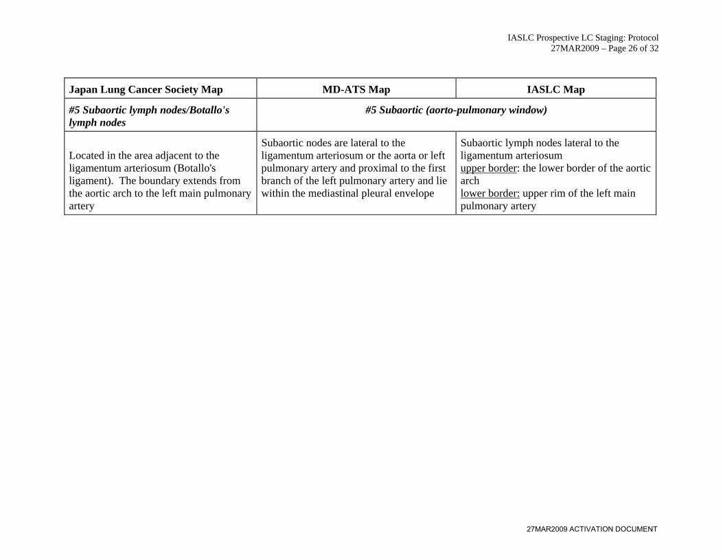
#5 Subaortic lymph nodes/Botallo's lymph nodes
#5 Subaortic (aorto-pulmonary window)
Located in the area adjacent to the ligamentum arteriosum (Botallo's ligament). The boundary extends from the aortic arch to the left main pulmonary artery
Subaortic nodes are lateral to the ligamentum arteriosum or the aorta or left pulmonary artery and proximal to the first branch of the left pulmonary artery and lie within the mediastinal pleural envelope
Subaortic lymph nodes lateral to the ligamentum arteriosum upper border: the lower border of the aortic arch lower border: upper rim of the left main pulmonary artery
27MAR2009 ACTIVATION DOCUMENT
IASLC Prospective LC Staging: Protocol 27MAR2009 – Page 27 of 32
Japan Lung Cancer Society Map MD-ATS Map IASLC Map
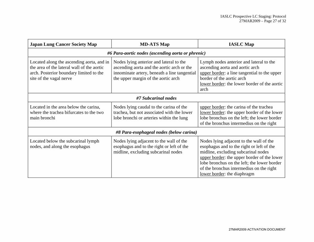
#6 Para-aortic nodes (ascending aorta or phrenic)
Located along the ascending aorta, and in the area of the lateral wall of the aortic arch. Posterior boundary limited to the site of the vagal nerve
Nodes lying anterior and lateral to the ascending aorta and the aortic arch or the innominate artery, beneath a line tangential the upper margin of the aortic arch
Lymph nodes anterior and lateral to the ascending aorta and aortic arch upper border: a line tangential to the upper border of the aortic arch lower border: the lower border of the aortic arch
#7 Subcarinal nodes
Located in the area below the carina, where the trachea bifurcates to the two main bronchi
Nodes lying caudal to the carina of the trachea, but not associated with the lower lobe bronchi or arteries within the lung
upper border: the carina of the trachea lower border: the upper border of the lower lobe bronchus on the left; the lower border of the bronchus intermedius on the right
#8 Para-esophageal nodes (below carina)
Located below the subcarinal lymph nodes, and along the esophagus
Nodes lying adjacent to the wall of the esophagus and to the right or left of the midline, excluding subcarinal nodes
Nodes lying adjacent to the wall of the esophagus and to the right or left of the midline, excluding subcarinal nodes upper border: the upper border of the lower lobe bronchus on the left; the lower border of the bronchus intermedius on the right lower border: the diaphragm
27MAR2009 ACTIVATION DOCUMENT

IASLC Prospective LC Staging: Protocol 27MAR2009 – Page 28 of 32
Japan Lung Cancer Society Map MD-ATS Map IASLC Map
#9 Pulmonary ligament nodes
Located in the area of the posterior and the lower edge of the inferior pulmonary vein
Nodes lying within the pulmonary ligament, including those in the posterior wall, and lower part of the inferior pulmonary vein
Nodes lying within the pulmonary ligament upper border: the inferior pulmonary vein lower border: the diaphragm
27MAR2009 ACTIVATION DOCUMENT
IASLC Prospective LC Staging: Protocol 27MAR2009 – Page 29 of 32
Japan Lung Cancer Society Map MD-ATS Map IASLC Map
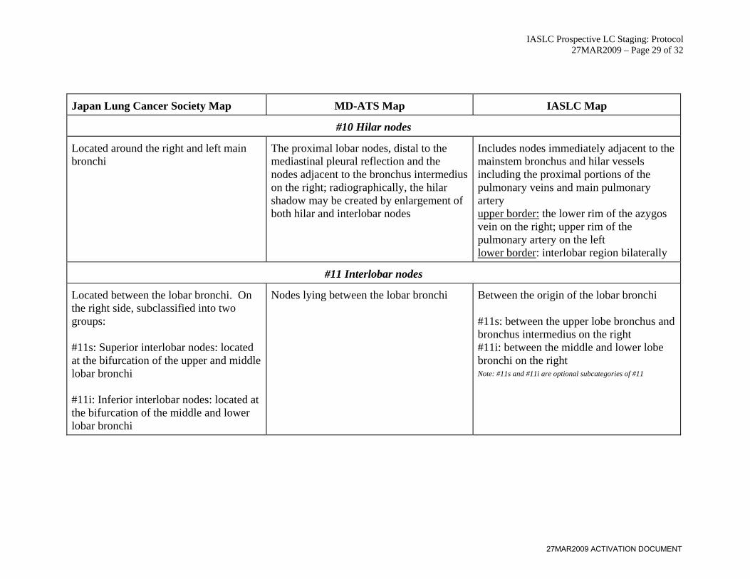
#10 Hilar nodes
Located around the right and left main bronchi
The proximal lobar nodes, distal to the mediastinal pleural reflection and the nodes adjacent to the bronchus intermedius on the right; radiographically, the hilar shadow may be created by enlargement of both hilar and interlobar nodes
Includes nodes immediately adjacent to the mainstem bronchus and hilar vessels including the proximal portions of the pulmonary veins and main pulmonary artery upper border: the lower rim of the azygos vein on the right; upper rim of the pulmonary artery on the left lower border: interlobar region bilaterally
#11 Interlobar nodes
Located between the lobar bronchi. On the right side, subclassified into two groups: #11s: Superior interlobar nodes: located at the bifurcation of the upper and middle lobar bronchi #11i: Inferior interlobar nodes: located at the bifurcation of the middle and lower lobar bronchi
Nodes lying between the lobar bronchi Between the origin of the lobar bronchi #11s: between the upper lobe bronchus and bronchus intermedius on the right #11i: between the middle and lower lobe bronchi on the right Note: #11s and #11i are optional subcategories of #11
27MAR2009 ACTIVATION DOCUMENT

IASLC Prospective LC Staging: Protocol 27MAR2009 – Page 30 of 32
Japan Lung Cancer Society Map MD-ATS Map IASLC Map
#12 Lobar nodes
Located in the area around the lobar branches, which are subclassified into three groups: #12u: Upper lobar lymph nodes #12m: Middle lobar lymph nodes #12l: Lower lobar lymph nodes
Nodes adjacent to the distal lobar bronchi Adjacent to the lobar bronchi
#13 Segmental nodes
Located along the segmental branches Nodes adjacent to the segmental bronchi Adjacent to the segmental bronchi
#14 Sub-segmental nodes
Located along the subsegmental branches Nodes around the subsegmental bronchi Adjacent to the subsegmental bronchi From Rusch et al., J Thorac Oncol 2009; In press, used with permission.
27MAR2009 ACTIVATION DOCUMENT

IASLC Prospective LC Staging: Protocol 27MAR2009 – Page 31 of 32
17.3 Stage Groupings
Descriptors, Proposed T and M Categories, and Stage Groupings
Sixth Edition
T/M Descriptor
7th Edition
T/M N0 N1 N2 N3
T1 (less than or equal to 2 cm) T1a IA IIA IIIA IIIB
T1 (>2-3 cm) T1b IA IIA IIIA IIIB
T2 (less than or equal to 5 cm) T2a IB IIA IIIA IIIB
T2 (>5-7 cm) T2b IIA IIB IIIA IIIB
T2 (>7 cm)
T3
IIB IIIA IIIA IIIB
T3 invasion IIB IIIA IIIA IIIB
T4 (same lobe nodules) IIB IIIA IIIA IIIB
T4 (extension) T4
IIIA IIIA IIIB IIIB
M1 (ipsilateral lung) IIIA IIIA IIIB IIIB
T4 (pleural effusion) M1a
IV IV IV IV
M1 (contralateral lung) IV IV IV IV
M1 (distant) M1b IV IV IV IV
Cells in bold indicate a change from the sixth edition for a particular TNM category
From Goldstraw et al., J Thorac Oncol 2007;8:706-714, used with permission.
27MAR2009 ACTIVATION DOCUMENT

IASLC Prospective LC Staging: Protocol 27MAR2009 – Page 32 of 32
18.0 APPENDIX - DATA ELEMENTS In order to illustrate the full complement of data elements to be collected in the prospective IASLC Prospective Lung Cancer Staging Project, the data entry screens corresponding to each “form” in the online system are provided in the Appendix.
27MAR2009 ACTIVATION DOCUMENT