Prior knowledge of HPV status improves detection of CIN2 ...
7
ONCOLOGY Prior knowledge of HPV status improves detection of CIN2 by cytology screening Ina H. Benoy, MSc, PhD; Davy Vanden Broeck, MSc, PhD; Maya J. Ruymbeke, MD; Shaira Sahebali, MD, PhD; Marc Arbyn, MD, PhD; Johannes J. Bogers, MD, PhD; Marleen Temmerman, MD, PhD; Christophe E. Depuydt, MSc, PhD OBJECTIVE: The objective of the study was to investigate whether knowl- edge of human papillomavirus (HPV) deoxyribonucleic acid test results in- creases sensitivity of guided cytology screening for the detection of cervical intraepithelial neoplasia (CIN)-2 or higher-grade cervical lesions. STUDY DESIGN: This was a prospective colposcopy-controlled study of 2905 BD SurePath samples to identify cases with CIN2 within a 24 month follow-up period. Sensitivity and specificity to detect CIN2 was evaluated, comparing guided cytology screening with and without prior knowledge of HPV status. RESULTS: Prior knowledge of HPV status resulted in significantly higher detection rate of CIN2 compared with screening blinded to HPV status (P .005) with limited loss of specificity (P .026). Gain in sensitivity is higher in older women (43.8%, P .008) vs in younger women (10.2%, P .317), whereas loss of specificity is more pronounced in younger women (P .001) vs older women (P .729). CONCLUSION: Guided cytological screening performed with prior knowledge of HPV status results in an improved detection of CIN2 or higher-grade lesions. Key words: cervical cancer, cervical cytology, human papillomavirus, human papillomavirus genotypes, real-time polymerase chain reaction Cite this article as: Benoy IH, Vanden Broeck D, Ruymbeke MJ, et al. Prior knowledge of HPV status improves detection of CIN2 by cytology screening. Am J Obstet Gynecol 2011;205:569.e1-7. T he recognition of the strong causal relationship between persistent in- fection with high-risk (HR) human pap- illomavirus (HPV) (HR-HPV) types, and cervical cancer and its precursors, has resulted in the development assays that detect viral nucleic acids as an alter- native for or as an adjunct to cervical cy- tology. 1 One can distinguish assays that detect all HR-HPV types as a group and genotyping tests that distinguish indi- vidual HPV types. 2 Liquid-based cytol- ogy is now gradually replacing conven- tional cytological testing, because of practical advantages (quicker interpretation, easy ancillary molecular testing, and possibility of computerized guided screening), in spite of the lack of evidence that it increases the detection of cervical intraepithelial neoplasia (CIN)-2 or higher-grade cervical abnormalities. 3-5 Cytological screening combined HR- HPV testing and HPV-based screening followed by cytology triage have been evaluated as more sensitive than cytol- ogy screening alone. 6 Compared with cy- tology alone, this screening strategy im- proves detection of precancerous growths but with a certain increase in the number of false-positive tests. 6-8 Recent random- ized trials have confirmed that HPV-based screening, in women older than 30-35 years followed by cytology triage results in detection of more CIN2 or worse lesions compared with cytology screening. More- over, longitudinal results of these trials have demonstrated that women with a negative HPV test have a lower risk of CIN3 and even invasive cancer. 9,10 This study aimed to evaluate the influ- ence of knowing the different HR-HPV genotypes present in cervical specimens before performing guided cytological screening. MATERIALS AND METHODS Study population In this prospective, colposcopy-controlled study, we enrolled 3126 voluntary partici- pants from August 2005 until February 2007 (Figure 1). Samples were collected during opportunistic routine health checks by 11 selected gynecologists in Flanders From the Department of Molecular Pathology (RIATOL) Sonic Healthcare, Antwerp, Belgium (Drs Benoy, Sahebali, Bogers, and Depuydt) and Department of Applied Molecular Biology Research (AMBIOR), University of Antwerp (Dr Bogers), Antwerp; the International Centre for Reproductive Health (ICRH), Ghent University (Drs Vanden Broeck and Temmerman), and Jan Palfijn Hospital and Department of Obstetrics and Gynaecology, University Hospital (Dr Ruymbeke), Ghent; and the Unit of Cancer Epidemiology/Belgian Cancer Centre, Scientific Institute of Public Health, Brussels (Dr Arbyn), Belgium. Received Dec. 13, 2010; revised March 2, 2011; accepted June 28, 2011. The first 2 authors contributed equally to the study and manuscript. M.A. is supported by the Belgian Foundation Against Cancer, Brussels, Belgium, and the European Commission (DG Sanco, Luxembourg) through the ECCG Project (European Cooperation on Development and Implementation of Cancer Screening and Prevention Guidelines, IARC, Lyon, France). D.V.B. is supported by the Flemish Fund for Scientific Research. The authors report no conflict of interest. Reprints: Ina Benoy, PhD, Emiel Vloorsstraat 9, 2020 Antwerp, Belgium. [email protected]. 0002-9378/$36.00 • © 2011 Mosby, Inc. All rights reserved. • doi: 10.1016/j.ajog.2011.06.101 Research www. AJOG.org DECEMBER 2011 American Journal of Obstetrics & Gynecology 569.e1
Transcript of Prior knowledge of HPV status improves detection of CIN2 ...

Research www.AJOG.org
ONCOLOGY
Prior knowledge of HPV status improves detectionof CIN2� by cytology screeningIna H. Benoy, MSc, PhD; Davy Vanden Broeck, MSc, PhD; Maya J. Ruymbeke, MD;Shaira Sahebali, MD, PhD; Marc Arbyn, MD, PhD; Johannes J. Bogers, MD, PhD;Marleen Temmerman, MD, PhD; Christophe E. Depuydt, MSc, PhD
OBJECTIVE: The objective of the study was to investigate whether knowl-edge of human papillomavirus (HPV) deoxyribonucleic acid test results in-creases sensitivity of guided cytology screening for the detection of cervicalintraepithelial neoplasia (CIN)-2 or higher-grade cervical lesions.
STUDY DESIGN: This was a prospective colposcopy-controlled study of2905 BD SurePath samples to identify cases with CIN2� within a 24month follow-up period. Sensitivity and specificity to detect CIN2� wasevaluated, comparing guided cytology screening with and without priorknowledge of HPV status.
RESULTS: Prior knowledge of HPV status resulted in significantly higher
detection rate of CIN2� compared with screening blinded to HPV statusObstet Gynecol 2011;205:569.e1-7.
otaaostna
0002-9378/$36.00 • © 2011 Mosby, Inc. All rights reserved. • doi: 10.1016
(P � .005) with limited loss of specificity (P � .026). Gain in sensitivityis higher in older women (43.8%, P � .008) vs in younger women(10.2%, P � .317), whereas loss of specificity is more pronounced inyounger women (P � .001) vs older women (P � .729).
CONCLUSION: Guided cytological screening performed with priorknowledge of HPV status results in an improved detection of CIN2 orhigher-grade lesions.
Key words: cervical cancer, cervical cytology, human papillomavirus,
human papillomavirus genotypes, real-time polymerase chain reactionCite this article as: Benoy IH, Vanden Broeck D, Ruymbeke MJ, et al. Prior knowledge of HPV status improves detection of CIN2� by cytology screening. Am J
tpbo
The recognition of the strong causalrelationship between persistent in-
fection with high-risk (HR) human pap-illomavirus (HPV) (HR-HPV) types,and cervical cancer and its precursors,has resulted in the development assaysthat detect viral nucleic acids as an alter-native for or as an adjunct to cervical cy-tology.1 One can distinguish assays thatdetect all HR-HPV types as a group andgenotyping tests that distinguish indi-vidual HPV types.2 Liquid-based cytol-
From the Department of Molecular PathologyBelgium (Drs Benoy, Sahebali, Bogers, and DBiology Research (AMBIOR), University of ACentre for Reproductive Health (ICRH), GheTemmerman), and Jan Palfijn Hospital and DUniversity Hospital (Dr Ruymbeke), Ghent; aCancer Centre, Scientific Institute of Public H
Received Dec. 13, 2010; revised March 2, 2011
The first 2 authors contributed equally to the stu
M.A. is supported by the Belgian Foundation AgEuropean Commission (DG Sanco, LuxembourgCooperation on Development and ImplementatiGuidelines, IARC, Lyon, France). D.V.B. is supp
The authors report no conflict of interest.
Reprints: Ina Benoy, PhD, Emiel Vloorsstraat 9,
gy is now gradually replacing conven-ional cytological testing, because of practicaldvantages (quicker interpretation, easyncillary molecular testing, and possibilityf computerized guided screening), inpite of the lack of evidence that it increaseshe detection of cervical intraepithelialeoplasia (CIN)-2 or higher-grade cervicalbnormalities.3-5
Cytological screening combined HR-HPV testing and HPV-based screeningfollowed by cytology triage have been
IATOL) Sonic Healthcare, Antwerp,ydt) and Department of Applied Molecularerp (Dr Bogers), Antwerp; the Internationalniversity (Drs Vanden Broeck and
rtment of Obstetrics and Gynaecology,the Unit of Cancer Epidemiology/Belgianth, Brussels (Dr Arbyn), Belgium.
cepted June 28, 2011.
nd manuscript.
t Cancer, Brussels, Belgium, and therough the ECCG Project (Europeanf Cancer Screening and Prevention
d by the Flemish Fund for Scientific Research.
0 Antwerp, Belgium. [email protected].
/j.ajog.2011.06.101
DECEMBER 2011 Americ
evaluated as more sensitive than cytol-ogy screening alone.6 Compared with cy-ology alone, this screening strategy im-roves detection of precancerous growthsut with a certain increase in the numberf false-positive tests.6-8 Recent random-
ized trials have confirmed that HPV-basedscreening, in women older than 30-35years followed by cytology triage results indetection of more CIN2 or worse lesionscompared with cytology screening. More-over, longitudinal results of these trialshave demonstrated that women with anegative HPV test have a lower risk ofCIN3 and even invasive cancer.9,10
This study aimed to evaluate the influ-ence of knowing the different HR-HPVgenotypes present in cervical specimensbefore performing guided cytologicalscreening.
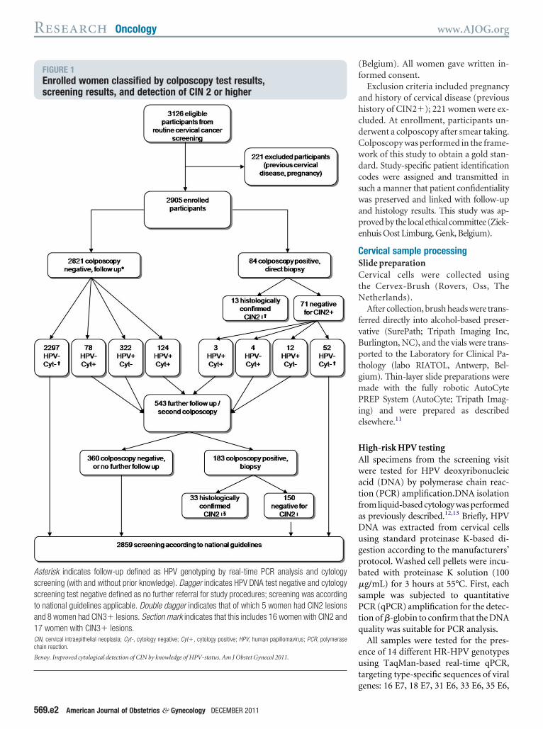
MATERIALS AND METHODSStudy populationIn this prospective, colposcopy-controlledstudy, we enrolled 3126 voluntary partici-pants from August 2005 until February2007 (Figure 1). Samples were collectedduring opportunistic routine health checks
(Repuntwnt Uepandeal
; ac
dy a
ains) th
on oorte
202
by 11 selected gynecologists in Flanders
an Journal of Obstetrics & Gynecology 569.e1
Dugpb
Research Oncology www.AJOG.org
569.e2 American Journal of Obstetrics & Gynecology DECEMBER 2011
(Belgium). All women gave written in-formed consent.
Exclusion criteria included pregnancyand history of cervical disease (previoushistory of CIN2�); 221 women were ex-cluded. At enrollment, participants un-derwent a colposcopy after smear taking.Colposcopy was performed in the frame-work of this study to obtain a gold stan-dard. Study-specific patient identificationcodes were assigned and transmitted insuch a manner that patient confidentialitywas preserved and linked with follow-upand histology results. This study was ap-proved by the local ethical committee (Ziek-enhuis Oost Limburg, Genk, Belgium).
Cervical sample processingSlide preparationCervical cells were collected usingthe Cervex-Brush (Rovers, Oss, TheNetherlands).
After collection, brush heads were trans-ferred directly into alcohol-based preser-vative (SurePath; Tripath Imaging Inc,Burlington, NC), and the vials were trans-ported to the Laboratory for Clinical Pa-thology (labo RIATOL, Antwerp, Bel-gium). Thin-layer slide preparations weremade with the fully robotic AutoCytePREP System (AutoCyte; Tripath Imag-ing) and were prepared as describedelsewhere.11
High-risk HPV testingAll specimens from the screening visitwere tested for HPV deoxyribonucleicacid (DNA) by polymerase chain reac-tion (PCR) amplification.DNA isolationfrom liquid-based cytology was performedas previously described.12,13 Briefly, HPV
NA was extracted from cervical cellssing standard proteinase K-based di-estion according to the manufacturers’rotocol. Washed cell pellets were incu-ated with proteinase K solution (100
�g/mL) for 3 hours at 55°C. First, eachsample was subjected to quantitativePCR (qPCR) amplification for the detec-tion of �-globin to confirm that the DNAquality was suitable for PCR analysis.
All samples were tested for the pres-ence of 14 different HR-HPV genotypesusing TaqMan-based real-time qPCR,targeting type-specific sequences of viral
FIGURE 1Enrolled women classified by colposcopy test results,screening results, and detection of CIN 2 or higher
Asterisk indicates follow-up defined as HPV genotyping by real-time PCR analysis and cytologyscreening (with and without prior knowledge). Dagger indicates HPV DNA test negative and cytologyscreening test negative defined as no further referral for study procedures; screening was accordingto national guidelines applicable. Double dagger indicates that of which 5 women had CIN2 lesionsand 8 women had CIN3� lesions. Section mark indicates that this includes 16 women with CIN2 and17 women with CIN3� lesions.CIN, cervical intraepithelial neoplasia; Cyt-, cytology negative; Cyt�, cytology positive; HPV, human papillomavirus; PCR, polymerasechain reaction.
Benoy. Improved cytological detection of CIN by knowledge of HPV-status. Am J Obstet Gynecol 2011.
genes: 16 E7, 18 E7, 31 E6, 33 E6, 35 E6,

Benoy. Improved cytological detection of CIN by knowledge of HPV-status. Am J Obstet Gynecol 2011.
www.AJOG.org Oncology Research
39 E7, 45 E7, 51 E6, 52 E7, 56 E7, 58 E6,59 E7, 66 E6, and 68 E7. Presence of low-risk HPV types, for instance, HPV6,HPV53, and HPV67, were considerednegative for HPV infection.
Slide classificationBD FocalPoint readingAll liquid-based cytology samples werefirst scanned with the BD-FocalPointsystem followed by guided assistedscreening with BD-SlideWizard, accord-ing to the manufacturer’s instructions(BD Diagnostics-TriPath, Burlington,NC).14 The FocalPoint system classifies25% of all slides as no further review(NFR) in which the probability of intra-epithelial lesions is extremely low. Theother 75% are categorized in quintile1-5, in which quintile 1 has the highestprobability of abnormality, based onslide scores. In this study, also, the NFRslides were screened cytologically.
BD SurePath liquid-based cytologyusing guided screening with a BD-Slide-Wizard was used to compare screeningwith and without prior knowledge ofHR-HPV status for each selected sample.
Bethesda classificationThe cytological results were classified ac-cording to the Bethesda system 2001,15
using the classes negative for intraepithe-lial lesions or malignancy, atypical squa-mous cells of undetermined significance(ASC-US), atypical squamous cells ofundetermined significance that cannotexclude high-grade squamous intraepi-thelial lesions (ASC-H), low-grade squa-mous intraepithelial lesions (LSIL), andhigh-grade squamous intraepithelial le-sions (HSIL). Slides were read by 2 inde-pendent cytologists in arbitrary order sothat for any sample, it was equally likelythat the cytology with or without priorknowledge would take the first reading.High-grade cervical disease was consid-ered to comprise histological grade CIN2or higher (CIN2, CIN3, adenocarcinoma insitu, invasive squamous cell carcinoma).
Cytology with/withoutprior knowledgeCytology was performed twice on everysample, with and without knowledge of
HPV DNA test results. Reading was doneTABLE 1Characteristics of the population
CharacteristicTotal population(n � 2905)
General.....................................................................................................................................................................................................................................
Age, y Median � 42.7 (first quartile � 32.9; third quartile � 51.9).....................................................................................................................................................................................................................................
�30 y 562 (19.3%).....................................................................................................................................................................................................................................
�30 y 2343 (80.7%)..............................................................................................................................................................................................................................................
Viral characteristics.....................................................................................................................................................................................................................................
HR-HPV positive 473 (16.3%).....................................................................................................................................................................................................................................
HR-HPV (�30 y) 143 (25.4%).....................................................................................................................................................................................................................................
HR-HPV (�30 y) 329 (14.0%)............................................................................................................................................................................................................................
Type 16 110 (23.3%)............................................................................................................................................................................................................................
Type 51 98 (20.7%)............................................................................................................................................................................................................................
Type 31 75 (15.9%)............................................................................................................................................................................................................................
Type 59 50 (10.6%)............................................................................................................................................................................................................................
Type 39 47 (9.9%)............................................................................................................................................................................................................................
Type 52 44 (9.3%)............................................................................................................................................................................................................................
Type 56 44 (9.3%)............................................................................................................................................................................................................................
Type 18 41 (8.7%)............................................................................................................................................................................................................................
Type 66 28 (5.9%)............................................................................................................................................................................................................................
Type 33 321 (4.4%)............................................................................................................................................................................................................................
Type 35 20 (4.2%)............................................................................................................................................................................................................................
Type 58 20 (4.2%)............................................................................................................................................................................................................................
Type 53 17 (3.6%)............................................................................................................................................................................................................................
Type 45 9 (1.9%)..............................................................................................................................................................................................................................................
Single HR-HPV infection 372 (78.6%)..............................................................................................................................................................................................................................................
Multiple HR-HPV infection 3101 (21.4%)..............................................................................................................................................................................................................................................
Median number of infections 1..............................................................................................................................................................................................................................................
Histology 256 (8.8%).....................................................................................................................................................................................................................................
Normal/CIN 1 210 (7.2%).....................................................................................................................................................................................................................................
CIN 2 21 (0.7%).....................................................................................................................................................................................................................................
CIN 3 25 (0.9%)..............................................................................................................................................................................................................................................
Cytology n � 2905
Without priorknowledge, n (%)
With priorknowledge, n (%) P value
Normal 2718 (93.6) 2685 (92.4) .10.....................................................................................................................................................................................................................................
ASC-US 108 (3.7) 110 (3.8) .94.....................................................................................................................................................................................................................................
LSIL 46 (1.6) 67 (2.3) .05.....................................................................................................................................................................................................................................
ASC-H 8 (0.3) 15 (0.5) .21.....................................................................................................................................................................................................................................
HSIL 25 (0.9) 27 (0.9) .89..............................................................................................................................................................................................................................................
ASC-US, atypical squamous cells of undetermined significance; CIN, cervical intraepithelial neoplasia; HPV, human papillo-mavirus; HR-HPV, high-risk human papillomavirus; HSIL, high-grade squamous intraepithelial lesions; LSIL, low-grade squa-mous intraepithelial lesions.
DECEMBER 2011 American Journal of Obstetrics & Gynecology 569.e3

ioHHHsv(toaCw
ppa
icvcet
ttidsapC
Research Oncology www.AJOG.org
in arbitrary order, and it was ensuredthat every slide was read by different cy-tologists, arbitrarily selected from a poolof cytologists. In both cytological read-ings, demographic and clinical informa-tion was provided. Cytological interpre-tation with knowledge of HPV statusincluded information on the type andthe type-specific viral load. All slideswere reviewed by cytopathologists,blinded from HPV DNA tests results butaware of the screening results of thecytologists.
Follow-up and assessmentof study endpointsAll women underwent colposcopy at en-rollment and participants with cervicalabnormalities were referred for biopsy(Figure 1). Women with a normal col-poscopy result (or a negative histologyresult if a biopsy was taken) were consid-ered as free of CIN. Women with a pre-cancerous lesion or cervical cancer werereferred for further management. Thenumber of detected CIN 2 or highergrade cases in a 24 month follow-up pe-riod was used as study endpoint.
Data managementAll results from cytology, histology, treat-
FIGURE 2Cytology according to prior knowle
Asterisk indicates that the significant difference wwithout prior knowledge of HPV (P � .05).HPV, human papillomavirus; LSIL, low-grade squamous intraepith
Benoy. Improved cytological detection of CIN by knowledge o
ment/follow-up, and HPV status were
569.e4 American Journal of Obstetrics & Gynecolo
entered in a database. Each patient wasallocated a unique patient identification(ID) number. This patient ID numberwas used to link the different cytological,histological, and virological data.
Statistical analysisThe statistical package R version 2.10.1 wasused for data analysis.16 HR-HPV positiv-ty was defined as the presence of 1 or moref the following 14 HPV types: HPV16,PV18, HPV31, HPV33, HPV35, HPV39,PV45, HPV51, HPV52, HPV56, HPV58,PV59, HPV66, and HPV68. Estimates of
ensitivity, specificity, negative predictivealue (NPV), and positive predictive valuePPV) for detection of CIN 2� were ob-ained for both screening strategies: cytol-gy with prior knowledge of HPV typesnd cytology without prior knowledge.onfidence intervals (CIs) for proportionsere calculated by Wilson’s method.17
Differences in proportions were tested us-ing McNemar statistics for paired compar-ison.18 The 95% CIs were computed for
roportions, differences, and ratios of pro-ortions. All statistical tests were 2 sided,nd P � .05 was considered statistically
significant.Women were divided by age into 2 cat-
e
found for LSIL when comparing reading with and
lesions.
V-status. Am J Obstet Gynecol 2011.
egories; younger (�30 years old) and older
gy DECEMBER 2011
(�30 years old). The difference in sensitiv-ty to detect CIN2� in the 2 age groups andorresponding specificities and predictivealues were assessed. A receiver operatingharacteristics (ROC) plot was used tovaluate/visualize the true positive rate vshe false-negative rate.
Histologically verified CIN 2� caseshat were detected within 24 months af-er enrollment at routine screening werencluded. Women who had a cytologicaliagnosis of ASC-US or worse were clas-ified as having abnormal cytology. Sep-rate analyses were performed to com-are the sensitivity in the detection ofIN 2 or worse.
RESULTSCharacteristics of the populationIn total, 3126 women were eligible forinclusion of the current study. After ex-clusion of 221 subjects with antecedentsof cervical neoplasia and/or pregnancy,2905 participants were included in thefinal study group. The median age was42.7 years (first quartile, 32.9; third quar-tile, 51.9). Eighty-four percent of thestudy population belonged to the targetage group of 25-64 years, for whomscreening is recommended in Belgium.16
The proportion of younger (�30 years)women in the study population was19.3%, with HPV prevalence of 25.4%,whereas 80.7% of women were 30 yearsold or older with HPV prevalence of14.0% (P � .001) (Table 1).
Of the 2905 women in the study, 473(16.3%) tested positive for at least 1 HR-HPV type. Single HPV infection was foundin 78.6% of the HR-HPV-infected womenand 21.4% were infected with 2 or moreHR-HPV types. The maximum number ofmultiple HR-HPV infection per womanwas 5. The most common type was HPV16(23.2%), followed by HPV51 (20.7%),HPV31 (15.9%), and HPV59 (10.6%), re-spectively. A high prevalence of HR-HPVinfections was observed in women with cy-tological abnormalities (Table 1).
Forty-six histologically confirmed CIN2 or worse cases were found. Five CIN 2and 8 CIN 3 subjects were detected duringthe first colposcopy visit and 16 CIN 2 and17 CIN3 subjects during subsequent visits
dg
as
elial
f HP
(Figure 1). Two (9.5%) of 21 women with

btes
kdbkft.
9fr
w
oyio
(
www.AJOG.org Oncology Research
CIN 2 were HR-HPV negative. All CIN 3�were HR-HPV positive.
Figure 2 shows a decrease in the num-er of slides ranked as normal when cy-ology was performed with prior knowl-dge; nevertheless, this decrease was nottatistically significant (P � .10). Con-
currently, when prior knowledge wasavailable, more slides were ranked as ab-normal in comparison with blinded cy-tology, but this difference was also notstatistically significant (P � .06). Of allthe slides ranked abnormal, a significantincrease in the number of LSIL ratedslides was noted (P � .05) when prior
nowledge was available. No significantifferences in cytological results betweenlinded cytology and reading with priornowledge of HPV DNA could be foundor the remaining cytological categories,hat is, ASC-US, ASC-H, and HSIL (P �47; P � .11; and P � .44 respectively).
Figure 3 shows more detailed shifts in cy-tological categories.
When prior knowledge of HPV DNAwas available, the sensitivity of guidedcytology to detect CIN 2� was signifi-cantly higher as compared with detec-tion without prior knowledge (P �.005). Guided cytology, with and with-out prior knowledge of HPV status,showed a sensitivity of 76.1% (95% CI,62.1– 86.1%) and 58.7% (95% CI, 44.3–71.7%), respectively (Table 2). For CIN3�, the sensitivities were 64.0% (95%CI, 44.5–79.7%) with 52.0% (95% CI,33.5– 69.9%) without prior knowledge.
The positive predictive value of cytol-ogy for CIN2� and CIN3� were not sta-tistically significantly higher with re-vealed HPV status than those for blindedscreening. Relative PPV equals 1.11 (95%CI, 0.70–1.75) and 1.05 (95% CI, 0.53–2.10, for CIN2�and CIN 3�, respectively.There was a significant decrease in specific-ity when prior knowledge was involved(P � .026), 93.6% (95% CI, 92.7%–4.5%) vs 94.4% (95% CI 93.6%–95.2%)or screening with and without knowledge,espectively (Table 2).
The prevalence of CIN 2�varied by age:2.5% and 1.3%, respectively, younger than30 years or 30 years or older. Overall, cytol-ogy screening showed higher sensitivity inyounger than in older women. The gain in
sensitivity induced by knowing the HPVstatus was observed in both age groups butwas highest in women aged 30 years old orolder:�43.8%,whereas�10.2%inyounger
omen) (Figure 4).No loss in specificity was observed for
lder women (P � .729), whereas inounger women an important drop in spec-ficitycouldbefound(P� .001).Knowledgef HPV status yielded higher PPVs in older
FIGURE 3Prior knowledge of HPV DNA induc
SC-H, high-grade squamous intraepithelial lesions; ASC-US, aapillomavirus; HSIL, high-grade squamous intraepithelial lesions
ntraepithelial lesion and malignancy.
enoy. Improved cytological detection of CIN by knowledge o
TABLE 2Test protocol comparison with deteconfirmed CIN2� lesions
VariableCytology withoutprior knowledge (%
Total population..........................................................................................................
Sensitivity 27/46 (58.7)..........................................................................................................
Specificity 2699/2859 (94.4)..........................................................................................................
NPV 2699/2718 (99.3)..........................................................................................................
PPV 27/187 (14.4)...................................................................................................................
Age (�30 y)..........................................................................................................
Sensitivity 11/14 (78.5)..........................................................................................................
Specificity 506/548 (92.3)..........................................................................................................
NPV 506/509 (99.4)..........................................................................................................
PPV 11/53 (20.75)...................................................................................................................
Age (�30 y)..........................................................................................................
Sensitivity 16/32 (50.0)..........................................................................................................
Specificity 2192/2310 (94.9)..........................................................................................................
NPV 2192/2208 (99.3)..........................................................................................................
PPV 16/134 (11.9)...................................................................................................................
All comparisons evaluate the difference between cytological sccomparison of 2 matched proportions was used.CIN, cervical intraepithelial neoplasia; NPV, negative predictive
Benoy. Improved cytological detection of CIN by knowledge of
DECEMBER 2011 Americ
P � .347) but not younger women (P �.511). Overall, there were no remarkable dif-ferences in the NPVs by screening procedureor by age strata (Table 2).
COMMENTThis study demonstrates that priorknowledge of presence of HR-HPV types
shifts in cytology outcome
al squamous cells of undetermined significance; HPV, human, low-grade squamous intraepithelial lesions; NILM, negative for
V-status. Am J Obstet Gynecol 2011.
histologically
Cytology withprior knowledge (%) P value
..................................................................................................................
35/46 (76.1) .005..................................................................................................................
2675/2859 (93.6) .026..................................................................................................................
2675/2686 (99.6) .211..................................................................................................................
35/219 (16.0) .771..................................................................................................................
..................................................................................................................
12/14 (85.7) .317..................................................................................................................
479/548 (87.4) � .001..................................................................................................................
479/481 (99.6) .943..................................................................................................................
12/81 (14.8) .511..................................................................................................................
..................................................................................................................
23/32 (71.9) .008..................................................................................................................
2195/2310 (95.0) .729..................................................................................................................
2195/2204 (99.6) .242..................................................................................................................
23/138 (16.7) .347..................................................................................................................
ng without vs with prior knowledge. McNemar statistics for
e; PPV, positive predictive value.
ed
A typicp ; LSILi
B f HP
ct
)
.........
.........
.........
.........
.........
.........
.........
.........
.........
.........
.........
.........
.........
.........
.........
reeni
valu
HPV-status. Am J Obstet Gynecol 2011.
an Journal of Obstetrics & Gynecology 569.e5

rHssowuscmp
Research Oncology www.AJOG.org
increased cytological detection of CIN 2grade lesions or worse. Compared withcytology alone, knowledge of the HPVstatus resulted in a gain in sensitivity of30%, a slight loss in specificity, however,without loss of positive predictive value.
The prevalence of HR-HPV genotypesin our study population was similar tothe national distribution, indicating thata representative sample was taken.11 Itemains to be elucidated how to utilizePV DNA testing: as a complementary
creening method to cytology or as atand-alone screening followed by cytol-gy-based triage of HR-HPV-positiveomen. A major concern regarding these of HPV DNA tests in cervical cancercreening has been their lower specificityompared with cytology, which results inore women needing to go through re-
eat testing and diagnostic procedures.7
Overall, cytological screening withprior knowledge resulted in an increasednumber of abnormal smears, in particu-lar LSIL.
Our observations are in concordance
FIGURE 4ROC plot
In both young women (diamonds), a limited gain iolder women (squares), a substantial gain in senClosed symbols represent screening without prioedge was applied.Benoy. Improved cytological detection of CIN by knowledge o
with results from certain randomized
569.e6 American Journal of Obstetrics & Gynecolo
trials indicating that HPV screening fol-lowed by cytology triage identifies moreCIN2� than cytology screening alone.19
These findings can be explained only by amore cautious evaluation of HPV-posi-tive slides by the cytotechnologist alertedby knowledge of HPV status.
A striking finding was that knowingthe HPV status prior to cytologicalscreening was more sensitive in both agegroup, but the gain was higher in women30 years old or older (young women:78.5% vs 85.7%; older women: 50.0% vs71.9%). Prior knowledge of HPV statusresulted in a loss in specificity in younger(92.3% vs 87.4%) but not in older women:(94.9% vs 95.0%).
HR-HPV infection is very common,especially in younger women.19 Amongwomen, whose smears are processed inour laboratory, HR-HPV prevalencereaches a peak of at least 20% amongwomen between the ages of 18 and 30years of age, with a subsequent decline toapproximately 10% among women in
nsitivity is combined with a loss of specificity. Inity is paralleled by virtually no loss of specificity.nowledge and open symbols when prior knowl-
V-status. Am J Obstet Gynecol 2011.
their 30s and less thereafter.11
gy DECEMBER 2011
The lower specificity and PPV (albeitnot significant in our study) in womenyounger than 30 years old is not unex-pected because infections usually aretransient and are not accompanied byhigh-grade neoplasia at this age group.Moreover, HPV-induced increase insensitivity for CIN2� has to be inter-preted with caution because of the highprobability of overdiagnosis in this agegroup.10
As illustrated in Figure 4, gain in sen-sitivity is most substantial in olderwomen, this combined with virtually noloss of specificity. In contrast, a gain insensitivity is accompanied by an in-creased false positivity rate. Hence, ourfindings suggest that prior knowledgeof HPV is particularly useful in olderwomen and support the guidelines of theAmerican Cancer Society recommend-ing women above 30 cytological screen-ing (conventional or liquid-based) every3 years in combination with an HPVtest.
Diagnostic accuracy studies in whichverification of the outcome is incom-plete and mainly restricted to screen-positive subjects may be vulnerable toverification bias, resulting in overesti-matation of sensitivity and underestima-tion of specificity.20
In our study, colposcopy was incorpo-rated in the enrollment phase and ineventual follow-up visits to avoid verifi-cation bias.
From this study, no relevant conclu-sions could be drawn toward test perfor-mance with CIN3� as endpoint becauseof a lack of statistical power to determinesensitivity and specificity of the 2 screen-ing strategies. Further research in largercohorts needs to be conducted to assessthe significant impact of prior knowledgeas a complementary screening strategy. Awoman’s HPV status together with infor-mation on the exact type and perhaps viralload involved in the infection may have farmore importantclinical significance. Itwillallow more close monitoring of the naturalhistory of the disease and identify the all-important persistent infections.
More research is required to investigatethe influence of the type of knowledge thatshould be provided prior to cytological
n sesitivr k
f HP
reading. The criteria for defining abnor-

www.AJOG.org Oncology Research
malities, because of the subjectivity of bothcytology and histology in cervical screen-ing programs, could be more quantitativeto ensure more objective and accuratemeasurements.
An important limitation of this studywas the sample size, although more than3000 women were screened, conclusionscould be formulated only with CIN2 asan endpoint. No conclusive answerswere formulated toward the question ofwhether screening with prior knowledgeof HPV improves sensitivity to detectCIN3 or higher grade lesions. Extra sam-ple size could also allow for analysis formore information on the nature of theprior knowledge (HPV type, viral load,etc).
From a methodological point of view,the use of colposcopy/biopsy has inher-ent limitations, and imperfections to-ward disease certification could havebeen overcome by using random biopsy.Women were recruited from the normalBelgian screening population, based onopportunistic screening scheme. Here apotential bias could be recognized.
Summarizing, the real home messageis that sensitivity for CIN2� markedlyincreased with minimal effect on speci-ficity in both young and old but mostimportantly those greater than or equalto 30. The loss in specificity was limitedto the younger age group. No loss of PPVwas noted between the 2 strategies. Therewas no decline in NPV with both groupsor by age, which is the most importantfactor for a screening test.
The decrease in specificity in youngpatients is to be expected and supportsthe use of American Society for Colpos-copy and Cervical Pathology guidelines,which advocate avoiding interventionwith colposcopy in the young to allowregression of HPV infection and low-
grade lesions. fACKNOWLEDGMENTSWe wish to thank the cytotechnologists, KristinVan Belle, Sabrina Van Belle, Linda De Kreyger,and Sandy Van Den Eynde, and laboratorytechnicians, Rachel Baveco, Ludo Boels, An-neleen Cremers, Carmen de Maerschalck, Na-dine Deckers, Kathlijne De Preter, Sabine Dier-ickx, Brenda Gabriels, Inge Goegebeur, KarenIleghems, Kim Van Beek, Miranda Vervoort, andMarleen Wauters, for excellent technical sup-port. Chantal Vanden Bosch offered secretarialsupport. We are sincerely grateful to CarolyneBosire and Joris Meys for statistical advice andmanuscript preparation. M.A. is supported bythe Belgian Foundation Against Cancer (Brus-sels, Belgium) and the European Commission(DG Sanco, Luxembourg) through the ECCGproject (European Cooperation on Develop-ment and Implementation of Cancer screeningand prevention guidelines, International Agencyfor Research on Cancer, Lyon, France). D.V.B.is supported by the Flemish Fund for Scientificresearch.
REFERENCES1. Breitenecker G. Cervical cancer screening:past—present—future. Der Pathologe 2009;2:128-35.2. Vince A, Lepej SZ. Diagnostic methods andtechniques in cervical cancer prevention Part II:molecular diagnostics of HPV infection. MedGlas Ljek Komore Zenicko-Doboj Kantona2010;7;18-25.3. Arbyn M, Bergeron C, Klinkhamer P, Martin-Hirsch P, Siebers AG, Bulten J. Liquid com-pared with conventional cervical cytology: asystematic review and meta-analysis. ObstetGynecol 2008;111:167-77.4. Schiffman M, Solomon D. Screening andprevention methods for cervical cancer. JAMA2009;302:1809-10.5. Sawaya GF. Evidence-based medicine ver-sus liquid-based cytology. Obstet Gynecol2008;111:2-3.6. Naucler P, Ryd W, Törnberg S, Strand A, etal. Efficacy of HPV DNA testing with cytologytriage and/or repeat HPV DNA testing in primarycervical cancer screening. Natl Cancer Inst2009;101;88-99.7. Arbyn M, Sasieni P, Meijer CJ, Clavel C, Ko-liopoulos G, Dillner J. Chapter 9: Clinical appli-cations of HPV testing: a summary of meta-analyses. Vaccine 2006;24(Suppl 3):S78-89.8. Cuzick J, Arbyn M, Sankaranarayanan R, etal. Overview of human papillomavirus-based
and other novel options for cervical cancerDECEMBER 2011 Americ
screening in developed and developing coun-tries. Vaccine 2008;26(Suppl 10):K29-41.9. Arbyn M, Ronco G, Meijer CJLM, Naucler P.Trials comparing cytology with HPV screening.Lancet Oncol 2009;10:935-6.10. Ronco G, Giorgi-Rossi P, Carozzi F, et al.Efficacy of human papillomavirus testing for thedetection of invasive cervical cancers and cer-vical intraepithelial neoplasia: a randomisedcontrolled trial. Lancet Oncol 2010;11:249-57.11. Arbyn M, Benoy I, Simoens C, Bogers J,Beutels P, Depuydt C. Prevaccination distribu-tion of human papillomavirus types in womenattending at cervical cancer screening in Bel-gium. Cancer Epidemiol Biomarkers Prev 2009;18;321-30.12. Depuydt C, Benoy IH, Bailleul EJ, Vande-pitte J, Vereecken AJ, Bogers JJ. Improved en-docervical sampling and HPV viral load detec-tion by Cervex-Brush Combi. Cytopathology2006;17;374-81.13. Depuydt CE, Boulet GA, Horvath CA,Benoy IH, Vereecken AJ, Bogers JJ. Compari-son of MY09/11 consensus PCR and type-spe-cific PCRs in the detection of oncogenic HPVtypes. J Cell Mol Med 2007;11;881-91.14. Wilbur DC, Prey MU, Miller WM, PawlickGF, Colgan TJ, Dax Taylor D. Detection of highgrade squamous intraepithelial lesions and tu-mors using the AutoPap System: results of aprimary screening clinical trial. Cancer 1999;87:354-8.15. Solomon D, Davey D, Kurman R, MoriartyA, O’Connor D, Prey M, Raab S, Sherman M,Wilbur D, Wright T Jr, Young N. The 2001Bethesda System: terminology for reporting re-sults of cervical cytology. JAMA 2002;287:2114-9.16. R Development Core Team. R: a languageand environment for statistical computing.2009. Available at: http://www.r-project.org/.Accessed Aug. 26, 2011.17. Agresti A. An introduction to categoricaldata analysis, 2nd ed. Hoboken, NJ: Wiley-In-terscience; 2007.18. Field A. Discovering statistics using SPSS,3rd ed. London, UK: Sage Publications; 2009.19. Leinonen M, Nieminen P, Kotaniemi-Tal-onen L, et al. Age-specific evaluation of primaryhuman papillomavirus screening vs conven-tional cytology in a randomized setting. J NatlCancer Inst 2009;101:1612-23.20. Gaffikin L, McGrath J, Arbyn M, BlumenthalP. Avoiding verification bias in screening testevaluation in resource poor settings; a casestudy from Zimbabwe. Clin Trials 2008;5:
496-503.an Journal of Obstetrics & Gynecology 569.e7