![Composite cell sheet for periodontal regeneration ...cells (BMMSCs) have also been widely applied in the tissue-engineering repair of periodontal tissue defects [16, 17]. The research](https://static.fdocuments.us/doc/165x107/60fa10b5777c6d50b8631a62/composite-cell-sheet-for-periodontal-regeneration-cells-bmmscs-have-also-been.jpg)
Printed in Singapore. All rights reserved … · Does periodontal tissue regeneration really work?...
12
Does periodontal tissue regeneration really work? D IETER D. B OSSHARDT &A NTON S CULEAN Periodontitis is an infectious disease that causes destruction of the tooth-attachment apparatus. Untreated periodontitis results in progressive attach- ment loss that may eventually lead to early tooth loss. Fortunately, research has provided evidence that in most situations chronic periodontal diseases can be treated [reviewed in Ref. (29)]. There is also evidence that periodontally involved teeth have a good chance of survival, provided that therapy, patient compliance and maintenance care are appropriate [reviewed in Ref. (29)]. There are a broad range of treatment op- tions available, but only a few may be regarded as truly regenerative procedures. According to a position paper from the American Academy of Periodontology (29), periodontal regenerative procedures include soft tissue grafts, bone replacement grafts, root biomod- ifications, guided tissue regeneration, and combina- tions thereof, for osseous, furcation and recession defects. Regeneration is defined as the reproduction or reconstitution of a lost or injured part of the body in such a way that the architecture and function of the lost or injured tissues are completely restored. The aim of regenerative periodontal therapy is to restore the structure and function of the periodontium. This means that the structure and function of the gingiva, alveolar bone, root cementum and periodontal liga- ment must be restored (Figs 1 and 2). By contrast, periodontal repair implies healing without restoration of the tooth-attachment apparatus and is often asso- ciated with the formation of a long junctional epi- thelium (Figs 3–5). Detachment of the junctional epithelium from the tooth surface (i.e. the formation of a periodontal pocket), disconnection of periodontal ligament fiber attachment to the root surface via cementum, and bone loss, are hallmarks of peri- odontitis. New attachment of junctional epithelium to the tooth surface and of connective tissue fibers to the root surface are very critical components of true periodontal regeneration. New connective tissue attachment requires the formation of new cementum to a previously diseased root surface that was modi- fied following periodontal therapy. Needless to say, in order to increase the attachment function of a tooth, the periodontal connective tissue fibers also have to insert into newly formed bone (Fig. 6). While less concern exists about the new epithelial attachment, new connective tissue attachment is much more critical. Concerns include predictability and the amount of new connective tissue attachment, as well as the strength of the regenerated interface between the treated root surface and the new cementum. As formation of cementum is essential for the attach- ment of periodontal ligament fibers to the root surface, much research has been devoted to under- standing cementogenesis (for reviews, see Refs 3, 7, 9, 26, 30, 61, 62, 81). Not all studies that claim to have achieved periodontal regeneration have utilized histological techniques. Methods of assessing periodontal regen- eration have been reviewed previously (56). Clinically, the outcome of a regenerative periodontal treatment is assessed by clinical parameters (periodontal probing, radiographs and re-entry evaluations). These methods are, however, inappropriate for demonstrating true attachment gain. Histology continues to be the only reliable method of evaluating the efficacy of a therapy aimed at achieving periodontal regeneration. Accord- ing to the World Workshop in Periodontics of the American Academy of Periodontology (1996), the requirements for a periodontal treatment to be con- sidered a regenerative procedure are as follows: (i) human histology demonstrating new cementum, periodontal ligament and bone coronal to the former defect base; (ii) controlled human clinical trials dem- onstrating improved clinical probing attachment and bone levels; and (iii) controlled animal histological studies revealing new cementum, periodontal liga- ment and bone. 208 Periodontology 2000, Vol. 51, 2009, 208–219 Printed in Singapore. All rights reserved Ó 2009 John Wiley & Sons A/S PERIODONTOLOGY 2000
Transcript of Printed in Singapore. All rights reserved … · Does periodontal tissue regeneration really work?...
Does periodontal tissueregeneration really work?
DI E T E R D. BO S S H A R D T & AN T O N SC U L E A N
Periodontitis is an infectious disease that causes
destruction of the tooth-attachment apparatus.
Untreated periodontitis results in progressive attach-
ment loss that may eventually lead to early tooth loss.
Fortunately, research has provided evidence that in
most situations chronic periodontal diseases can be
treated [reviewed in Ref. (29)]. There is also evidence
that periodontally involved teeth have a good chance
of survival, provided that therapy, patient compliance
and maintenance care are appropriate [reviewed in
Ref. (29)]. There are a broad range of treatment op-
tions available, but only a few may be regarded as
truly regenerative procedures. According to a position
paper from the American Academy of Periodontology
(29), periodontal regenerative procedures include soft
tissue grafts, bone replacement grafts, root biomod-
ifications, guided tissue regeneration, and combina-
tions thereof, for osseous, furcation and recession
defects. Regeneration is defined as the reproduction
or reconstitution of a lost or injured part of the body in
such a way that the architecture and function of the
lost or injured tissues are completely restored. The
aim of regenerative periodontal therapy is to restore
the structure and function of the periodontium. This
means that the structure and function of the gingiva,
alveolar bone, root cementum and periodontal liga-
ment must be restored (Figs 1 and 2). By contrast,
periodontal repair implies healing without restoration
of the tooth-attachment apparatus and is often asso-
ciated with the formation of a long junctional epi-
thelium (Figs 3–5). Detachment of the junctional
epithelium from the tooth surface (i.e. the formation
of a periodontal pocket), disconnection of periodontal
ligament fiber attachment to the root surface via
cementum, and bone loss, are hallmarks of peri-
odontitis. New attachment of junctional epithelium to
the tooth surface and of connective tissue fibers to the
root surface are very critical components of true
periodontal regeneration. New connective tissue
attachment requires the formation of new cementum
to a previously diseased root surface that was modi-
fied following periodontal therapy. Needless to say, in
order to increase the attachment function of a tooth,
the periodontal connective tissue fibers also have to
insert into newly formed bone (Fig. 6). While less
concern exists about the new epithelial attachment,
new connective tissue attachment is much more
critical. Concerns include predictability and the
amount of new connective tissue attachment, as well
as the strength of the regenerated interface between
the treated root surface and the new cementum. As
formation of cementum is essential for the attach-
ment of periodontal ligament fibers to the root
surface, much research has been devoted to under-
standing cementogenesis (for reviews, see Refs 3, 7, 9,
26, 30, 61, 62, 81).
Not all studies that claim to have achieved
periodontal regeneration have utilized histological
techniques. Methods of assessing periodontal regen-
eration have been reviewed previously (56). Clinically,
the outcome of a regenerative periodontal treatment is
assessed by clinical parameters (periodontal probing,
radiographs and re-entry evaluations). These methods
are, however, inappropriate for demonstrating true
attachment gain. Histology continues to be the only
reliable method of evaluating the efficacy of a therapy
aimed at achieving periodontal regeneration. Accord-
ing to the World Workshop in Periodontics of the
American Academy of Periodontology (1996), the
requirements for a periodontal treatment to be con-
sidered a regenerative procedure are as follows: (i)
human histology demonstrating new cementum,
periodontal ligament and bone coronal to the former
defect base; (ii) controlled human clinical trials dem-
onstrating improved clinical probing attachment and
bone levels; and (iii) controlled animal histological
studies revealing new cementum, periodontal liga-
ment and bone.
208
Periodontology 2000, Vol. 51, 2009, 208–219
Printed in Singapore. All rights reserved
� 2009 John Wiley & Sons A/S
PERIODONTOLOGY 2000
Well-controlled human histological studies with
appropriate controls are very rare. Furthermore,
reproduction of results from well-designed,
well-controlled and well-conducted animal studies
within humans may be difficult. In practical terms
we assume that once a regenerative technique has
revealed regenerative potential, as evidenced by
histology, any positive clinical findings are often
automatically equated with periodontal regeneration.
There have been several recent detailed reviews of
guided tissue regeneration and therefore this manu-
script provides an overview of the current state of the
field, stepping back from the details of individual
studies in an attempt to answer the question, �does
periodontal tissue regeneration really work?� It also
aims to set the scene for two further manuscripts
within this volume of Periodontology 2000 that
address novel approaches to cell-based methods of
regeneration and tissue engineering.
Scaling and root planing usinghand instruments
Scaling and root planing are basic, traditional and
effective mechanical methods for treating periodon-
tal diseases. The aim of scaling and root planing is to
remove the bacterial biofilm, calculus and contami-
nated cementum. Numerous studies have proven the
effectiveness of reducing the bacterial load, and thus
controlling the subgingival microflora, by scaling and
root planing [reviewed in Refs (20, 57)]. Research in
animals and in humans (10, 16, 17, 44, 69, 73, 74)
indicates that the formation of new connective tissue
attachment following scaling and root planing or flap
surgery is not predictable. Although some new con-
nective tissue attachment may form, a long junc-
tional epithelium is what predictably establishes itself
on the root surface (Fig. 7). Therefore, scaling and
root planing cannot be regarded as a regenerative
procedure, although its efficacy in treating chronic
periodontitis is beyond doubt.
Sonic ⁄ ultrasonic scalers andlasers
Lasers or sonic ⁄ ultrasonic instruments may be used
as an alternative treatment or as an adjunctive
treatment to mechanical scaling and root planing.
However, findings from a human histological study
have failed to show predictable periodontal regener-
ation following scaling and root planing using
ultrasonic instruments. The healing occurred pre-
dominantly through a long junctional epithelium,
while formation of a new connective tissue attach-
ment occurred only occasionally and was confined to
the apical portion of the pockets (69). In a recent
systematic review, it was concluded that there is
insufficient clinical evidence to support the use of
CO2, neodymium-doped yttrium–aluminum–garnet
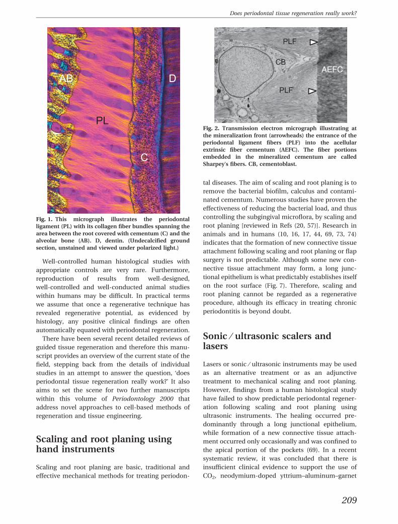
Fig. 2. Transmission electron micrograph illustrating at
the mineralization front (arrowheads) the entrance of the
periodontal ligament fibers (PLF) into the acellular
extrinsic fiber cementum (AEFC). The fiber portions
embedded in the mineralized cementum are called
Sharpey�s fibers. CB, cementoblast.
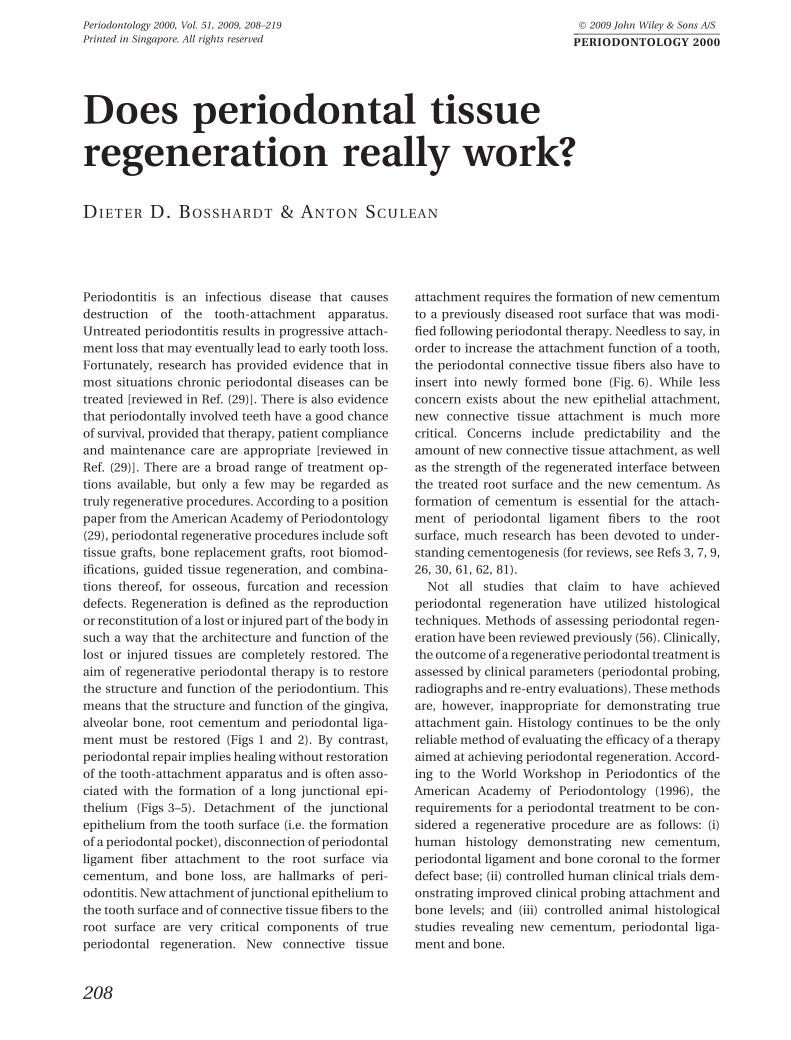
Fig. 1. This micrograph illustrates the periodontal
ligament (PL) with its collagen fiber bundles spanning the
area between the root covered with cementum (C) and the
alveolar bone (AB). D, dentin. (Undecalcified ground
section, unstained and viewed under polarized light.)
209
Does periodontal tissue regeneration really work?
(Nd:YAG), or neodymium-doped yttrium–aluminum–
perovskite (Nd:YAP) lasers, or different diode laser
wavelengths (63). Only the erbium-doped yttrium–
aluminum–garnet (Er:YAG) laser appears to be
suitable for the nonsurgical treatment of chronic
periodontitis (35, 63). However, there is insufficient
evidence to suggest that any specific wavelength of
laser is superior to conventional root surface
treatment (i.e. root scaling and planing) (21, 63).
Regarding histological evidence for periodontal
regeneration, only one animal study to date has
concluded that both the Er:YAG laser and an ultra-
sonic device might support the formation of a new
connective tissue attachment (64).
The use of Er:YAG laser radiation during perio-
dontal surgery was also evaluated in an animal study
by Mizutani et al. (51). Class III furcation defects
were experimentally induced in six beagle dogs and
randomly treated, using a split-mouth design, with
either an Er:YAG laser or hand instruments. The
histological analysis 3 months following surgery re-
vealed similar amounts of new connective tissue
attachment formation in both groups, but signifi-
cantly higher bone formation in the laser group.
Comparable results were also found in a case report
study evaluating, clinically and histologically, the
healing following flap surgery and defect debride-
ment with an Er:YAG laser in six patients with one
advanced intrabony defect (67). The histologic
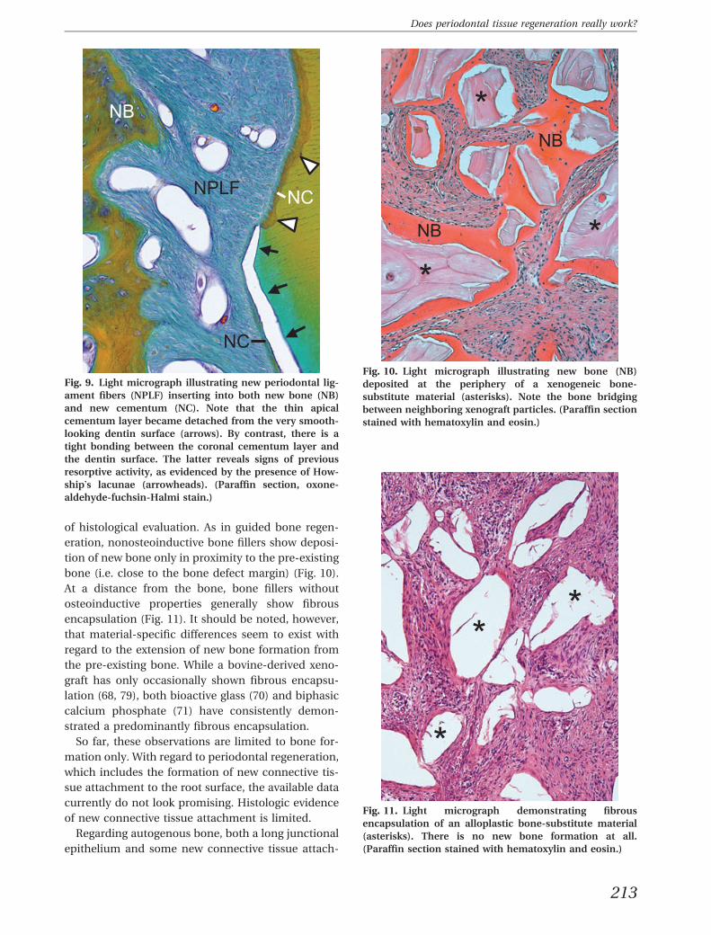
Fig. 4. Light micrograph illustrating formation of a long
junctional epithelium (LJE) ending at the coronal-most
end of regenerated cementum (C). D, dentin. (Paraffin
section stained with hematoxylin and eosin.)
A B C
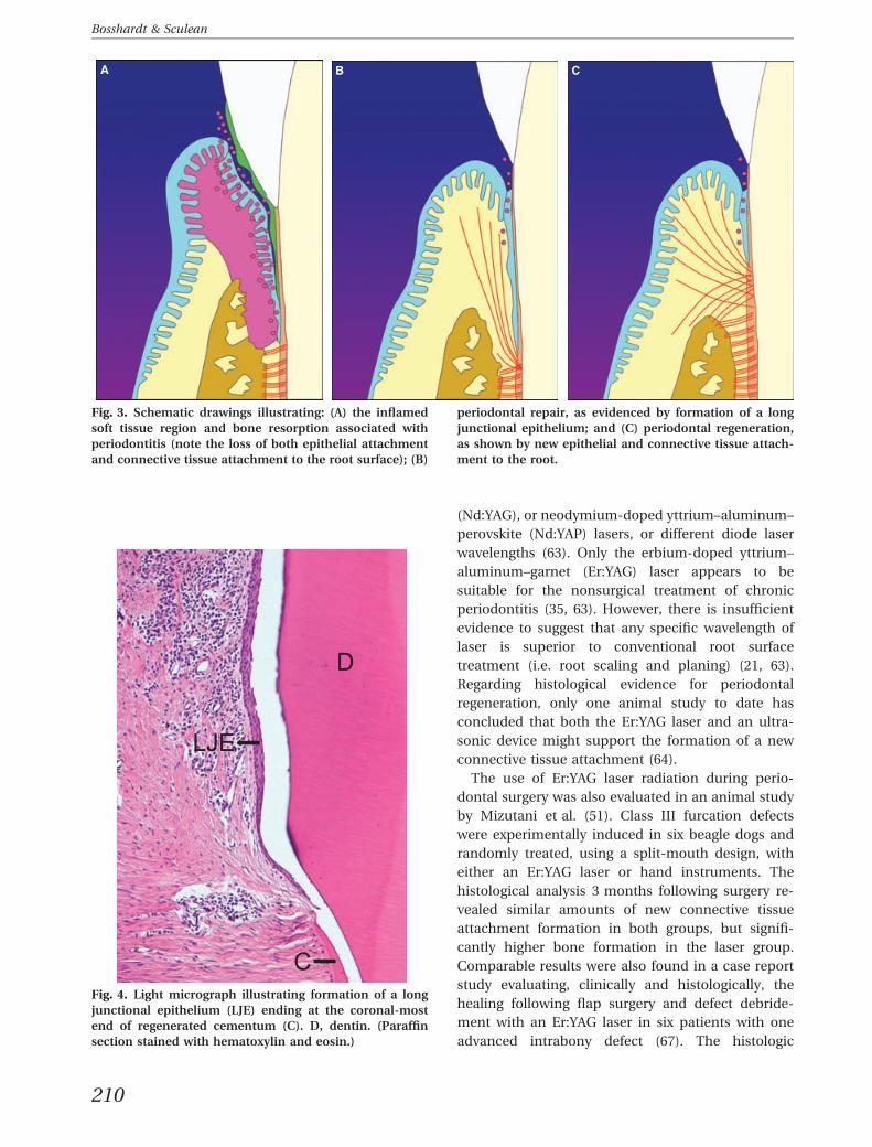
Fig. 3. Schematic drawings illustrating: (A) the inflamed
soft tissue region and bone resorption associated with
periodontitis (note the loss of both epithelial attachment
and connective tissue attachment to the root surface); (B)
periodontal repair, as evidenced by formation of a long
junctional epithelium; and (C) periodontal regeneration,
as shown by new epithelial and connective tissue attach-
ment to the root.
210
Bosshardt & Sculean

analysis revealed that in four out of the six specimens
the healing was mainly characterized by formation of
a long junctional epithelium along the instrumented
root surface, while cementum formation was only
occasionally found and was limited to the most apical
part of the defects. Formation of new connective
tissue attachment was only found in two out of the six
specimens. In one of these two specimens, the new
attachment was also accompanied by new bone
(67). However, periodontal regeneration at diseased
root surfaces was observed following an Nd:YAG
laser-assisted new attachment procedure in humans
(80).
In conclusion, there are currently insufficient data
to support the use of sonic ⁄ ultrasonic devices or
lasers in promoting periodontal regeneration.
Root surface conditioning
While the aim of root surface debridement is to
reduce the amount of bacteria and endotoxins on the
root surface, treatment of the root surface with
demineralizing agents such as acids or EDTA
primarily aims to expose collagen fibrils. To achieve
this, the smear layer must be removed and the min-
eralized component of the superficial layer of
cementum or dentin needs to be decalcified. The
clinical and histological effects of this type of root
surface treatment have been discussed previously in
excellent reviews (46, 48, 78). The biological concept
behind root surface demineralization is to improve
blood clot adhesion to exposed collagen fibrils. Sta-
bilization of the coagulum may have a positive effect
on wound healing and is regarded as an important
contributing factor in achieving periodontal regen-
eration [reviewed in Ref. (55)]. Mesenchymal cells
may preferentially adhere to the blood clot-stabilized
root surface and the apical migration of epithelial
cells may be reduced. Originally, citric acid was used
because of its ability to detoxify the root surface. As
reports have shown that treatment with citric acid
and phosphoric acid can result in root resorption and
ankylosis (1, 47), the chelator EDTA, which possesses
a significantly higher pH than acids and is therefore a
more gentle agent, appears to be a better choice.
Irrespective of the type of demineralizing agent used,
it cannot be claimed that demineralization of the root
surface per se is a regenerative procedure. It may,
however, have a positive effect on wound healing and
be used as a component of, or a step within, regen-
erative procedures (e.g. in combination with enamel
matrix proteins).
An underestimated issue may be the mechanical
strength between the treated root surface and
new cementum. Tissue separation between new
Fig. 5. Transmisson electron micrograph showing junc-
tional epithelium (JE) attachment to the cementum (C)
layer.
Fig. 6. Light micrograph illustrating true periodontal
regeneration as demonstrated by new periodontal liga-
ment fibers (NPLF) inserting into new bone (NB) and new
cementum (NC). (Paraffin section, oxone-aldehyde-
fuchsin-Halmi stain.)
211
Does periodontal tissue regeneration really work?

cementum and the treated root surface is a very
common finding in experimental periodontal regen-
eration studies [reviewed in Ref. (62)]. As tissue pro-
cessing for paraffin histology is prone to shrinkage
alterations, these tissue gaps are widely believed to
represent artifacts. The presence of plaque bacteria in
such gaps (Fig. 8), however, suggests that not all gaps
are artifactual in nature (8). Moreover, cemento–
dentinal tears have been confirmed, by radiographic
observations, to be present in periodontally involved
teeth and after regenerative periodontal therapy (15,
18, 33, 34, 36, 45, 49, 52), and are associated with
rapid periodontal breakdown. Thus, appropriate root
surface conditioning may not only provide a bio-
compatible surface for cell attachment, cell spreading
and matrix deposition, but might also improve
mechanical interfacial bonding and therefore could
be an issue of clinical relevance. It is striking that
histology often discloses that root surfaces naturally
conditioned by odontoclasts appear to provide a
better substrate for new cementum attachment than
otherwise modified root surfaces (Fig. 9), as also
discussed by Schroeder (62).
Bone grafts and bone substitutematerials
Autogenous bone, allogeneic bone, xenogeneic bone
substitutes and alloplastic materials, hereafter col-
lectively referred to as bone fillers, have all been used
with the aim of achieving periodontal regeneration
(22, 58, 78). A systematic review has shown that
clinical parameters are improved when intrabony
and Class II furcation defects are treated with bone
fillers (58). The rationale behind the use of bone
fillers is to take advantage of one or more of the
following properties of such materials, namely
osteoconduction, osteoinduction and osteogenesis,
induced by transferred cells that are capable of dif-
ferentiating into osteoblasts (5, 6, 37). Not all three
properties apply to every type of bone filler. While the
contribution of transferred cells to new tissue for-
mation may be overestimated, osteoconduction is
the most powerful property of bone fillers to support
new bone. While re-entry surgery or radiographs
demonstrate impressive volume gains, the actual
ratio of filler material to new bone cannot be deter-
mined using these methods. The exact nature of
changes occurring around bone fillers in osseous
periodontal defects can only be determined by means
Fig. 8. Transmission electron micrograph illustrating an
interfacial gap that formed between the instrumented root
surface and new cementum (NC) following regenerative
periodontal therapy. Note that the gap between old
cementum (C) and new cementum is filled with bacteria.
Fig. 7. Formation of a long junctional epithelium (LJE)
and partial periodontal regeneration, as evidenced by the
formation of new cementum (NC) and new bone (NB). The
arrowhead points to the apical end of the junctional epi-
thelium, whereas the arrow demarcates the apical border
of the defect. This is a section from a monkey tooth
5 months after treatment with Emdogain�. (Paraffin sec-
tion stained with hematoxylin and eosin.)
212
Bosshardt & Sculean
of histological evaluation. As in guided bone regen-
eration, nonosteoinductive bone fillers show deposi-
tion of new bone only in proximity to the pre-existing
bone (i.e. close to the bone defect margin) (Fig. 10).
At a distance from the bone, bone fillers without
osteoinductive properties generally show fibrous
encapsulation (Fig. 11). It should be noted, however,
that material-specific differences seem to exist with
regard to the extension of new bone formation from
the pre-existing bone. While a bovine-derived xeno-
graft has only occasionally shown fibrous encapsu-
lation (68, 79), both bioactive glass (70) and biphasic
calcium phosphate (71) have consistently demon-
strated a predominantly fibrous encapsulation.
So far, these observations are limited to bone for-
mation only. With regard to periodontal regeneration,
which includes the formation of new connective tis-
sue attachment to the root surface, the available data
currently do not look promising. Histologic evidence
of new connective tissue attachment is limited.
Regarding autogenous bone, both a long junctional
epithelium and some new connective tissue attach-
Fig. 9. Light micrograph illustrating new periodontal lig-
ament fibers (NPLF) inserting into both new bone (NB)
and new cementum (NC). Note that the thin apical
cementum layer became detached from the very smooth-
looking dentin surface (arrows). By contrast, there is a
tight bonding between the coronal cementum layer and
the dentin surface. The latter reveals signs of previous
resorptive activity, as evidenced by the presence of How-
ship�s lacunae (arrowheads). (Paraffin section, oxone-
aldehyde-fuchsin-Halmi stain.)
Fig. 10. Light micrograph illustrating new bone (NB)
deposited at the periphery of a xenogeneic bone-
substitute material (asterisks). Note the bone bridging
between neighboring xenograft particles. (Paraffin section
stained with hematoxylin and eosin.)
Fig. 11. Light micrograph demonstrating fibrous
encapsulation of an alloplastic bone-substitute material
(asterisks). There is no new bone formation at all.
(Paraffin section stained with hematoxylin and eosin.)
213
Does periodontal tissue regeneration really work?

ment are observed [reviewed in Refs (40, 78)]. It may
therefore be concluded that histologic evaluation of
human and animal studies suggest that the treatment
of periodontal osseous defects with autogenous bone
grafts lacks predictability and that only limited
amounts of new connective tissue attachment form.
Bone allograft materials are generally used as either
freeze-dried bone allografts or as demineralized
freeze-dried bone allografts. The effects of allogeneic
bone grafts on the regeneration of intra-osseous
periodontal defects have also been extensively re-
viewed (22, 40, 78). The conclusions are controversial
and range from optimistic to no osteoinductive
effects for demineralized freeze-dried bone allografts.
Wide variation has been observed in the ability of
commercially available demineralized freeze-dried
bone allografts preparations to induce new bone
formation, which may be related to donor age and to
the content of bone-inducing factors in the donor
bone (22). Concerning periodontal regeneration, one
human histologic study showed that implantation of
freeze-dried bone allografts into intrabony perio-
dontal defects resulted in a long junctional epithe-
lium, but no new connective tissue attachment (24).
By contrast, the use of demineralized freeze-dried
bone allografts resulted in histologic evidence of
periodontal regeneration (11, 12). While allografts are
widely used in the USA, they are less popular in
Europe, partly because of restrictive local regulations
within the European Union. The frequently observed
resorption may be another reason for reluctant
(limited) use of allografts.
Concerning xenogeneic and allogeneic bone sub-
stitutes, there is only vague histologic evidence that
the formation of new connective tissue attachment is
enhanced. As xenografts and allografts were often
tested together with a barrier membrane, the often-
observed histological evidence of new connective
tissue attachment is probably related to the barrier
function and not to the supporting materials.
Encapsulation in soft connective tissue is a common
observation of these bone-substitute materials
[reviewed in Ref. (40)].
The basic problem pertaining to all bone filler
materials is that a biologic rationale for the regener-
ation of the periodontium is missing. Bone grafts or
bone substitute materials do not possess the ability to
regenerate lost connective tissue attachment. The
formation of a connective tissue attachment to the
root surface may be absent or may not progress
beyond what can be achieved with conventional
therapy (i.e. open flap surgery alone). The osteocon-
ductive, osteoinductive and ⁄ or osteogenic properties
of such materials can at best support new bone for-
mation. Their efficacy is proven in conjunction with
barrier membranes in guided bone regeneration
procedures (i.e. for augmentation of bone deficien-
cies or defects to install dental implants). The
mechanical membrane support may also be benefi-
cial for guided tissue regeneration approaches around
teeth, as the bone-filler particles support the barrier
membrane and prevent its collapse into the defect.
Guided tissue regeneration
Guided tissue regeneration is a technique that is
based on a solid biologic principle. The rationale
behind guided tissue regeneration is to use a physical
barrier (barrier membrane or simple membrane) to
selectively guide cell proliferation and tissue expan-
sion within tissue compartments [reviewed in Refs
(40, 41)] (Fig. 12). The barrier membrane prevents
gingival epithelium and connective tissue expansion
and favors migration of cells from the periodontal
ligament and alveolar bone into the periodontal
defect. The foundation for the development of the
guided tissue regeneration principle was informed by
the realization that the periodontal ligament is of
central importance to the regenerative processes of
the tooth-attachment apparatus (13, 28, 39, 42, 50,
54). Numerous experimental animal studies have
Fig. 12. Schematic drawing illustrating the four com-
partments from which cells can grow into the periodontal
defect and repopulate the root surface after periodontal
therapy: (1) oral gingival epithelium; (2) gingival connec-
tive tissue; (3) bone from the alveolar process; and (4)
periodontal ligament.
214
Bosshardt & Sculean

proven that this principle leads to periodontal
regeneration, and human histology has documented
that periodontal regeneration can be achieved [re-
viewed in Refs (40, 41)]. In the early days, the devel-
opment of the guided tissue regeneration technique
raised hope that lost periodontal tissues can be
regenerated in a predictable manner for many defect
types. There are, however, some drawbacks. For
everyday clinical practice, patient and defect selec-
tion may not be as rigorous as in well-designed
clinical studies, and the recall schedule is usually less
rigorous. Another problem relates to the exposure of
membranes to the oral environment and their inevi-
table contamination with bacteria [reviewed in Ref.
(2)]. Nonresorbable membranes are particularly
prone to exposure to the oral environment. As a
consequence, bacterial contamination and infection
may result in delayed wound healing and poor
regenerative outcomes. Biodegradable collagen
membranes possess a lower risk of exposure and do
not need a second surgical procedure for their re-
moval. As collagen membranes possess fewer favor-
able mechanical properties than nonresorbable
membranes, a bone filler is needed to prevent their
collapse into the defect area. A recent systematic re-
view came to the conclusion that most preclinical
studies have histologically demonstrated periodontal
regeneration when grafting materials are combined
with barrier membranes (65).
The guided tissue regeneration technique is sensi-
tive and technically demanding. Outcome improve-
ments through the development of new types of
barrier membrane (e.g. resorbable collagen mem-
branes, degradable synthetic membranes) may solve
some of the reported problems. However, harmful
degradation products of synthetic membranes, and
difficulties encountered in attempts to seal off the
gingival compartment against the space occupied by
periodontal ligament and bone without interfering
with the very important re-establishment of the
junctional epithelium, may hamper such attempts.
Growth ⁄ differentiation factors
For many years, research has attempted to use bio-
logically active molecules to achieve periodontal
regeneration. Among these molecules are: extracellular
matrix proteins and cell-attachment factors; media-
tors of cell metabolism and activity; and growth ⁄differentiation factors. Growth factors regulate cell
proliferation, cell activity, chemotaxis and ⁄ or cell
differentiation. Numerous growth factors, alone or in
combination, have been tested for periodontal
regeneration in animal experiments. Among these are
insulin-like growth factors, fibroblast growth factors,
epidermal growth factor, platelet-derived growth
factors, vascular endothelial growth factor, parathy-
roid hormone, transforming growth factor-b and
bone morphogenetic proteins. In addition, the clini-
cal effectiveness of recombinant human platelet-de-
rived growth factor-BB, platelet-rich plasma and
peptide P-15 has been tested for the treatment of
intra-osseous and furcation defects (76). A tremen-
dous amount of work has resulted in an enormous
number of original articles that have reported upon
the efficacy of added growth factors or related bio-
active agents in animal and human periodontal de-
fect models. The outcomes of these experiments have
been exhaustively presented and discussed in a
considerable number of reviews (14, 19, 23, 27, 43, 53,
59, 60, 72). The most promising growth factors ap-
pear to be the bone morphogenetic proteins, partic-
ularly bone morphogenetic protein-2 and bone
morphogenetic protein-7, the same growth factors
that are approved and applied in the orthopedic field
for hard-to-heal cases (i.e. nonunion, open tibial
fractures and spinal fusions), but only when all other
treatment options have failed. What can we learn
from all these studies and discussions? The transla-
tion of knowledge about the functions of bone mor-
phogenetic proteins and other growth factors in
embryonic development, tissue formation and
homeostasis, and bone healing, into a clinically
applicable solution with the aim to regenerate lost
periodontal tissues, appears to be very difficult, if not
impossible, at the present point in time. Critical issue
include: (i) the complexity of the periodontium,
which consists of four different tissues; (ii) the use of
very high doses of bone morphogenetic proteins; (iii)
the ideal carrier has still not been found; and (iv) the
enormous costs that are associated with recombinant
human bone morphogenetic proteins in relation to
relatively small and non-life-threatening periodontal
defects for which other treatment options exist.
What can be learned from all of these studies?
Despite the fact that very heterogeneous preclinical
studies have been performed (i.e. different species,
different defect designs, different growth factor do-
ses, single or combined use with other growth factors,
different vehicles), most authors concluded that
the growth factors evaluated achieved successful
periodontal regeneration and it is just a matter of
time until their therapeutic application. However,
despite a long history of preclinical evaluation with
promising results, the routine use of growth factors as
215
Does periodontal tissue regeneration really work?
therapeutic agents for periodontal regeneration is not
yet a reality. Were preclinical data interpreted too
optimistically? Or is it just too simplistic to think that
one therapeutically applied growth factor can really
restore the complexity of the periodontium?
Enamel matrix proteins
Compared with growth factors such as bone mor-
phogenetic proteins, enamel matrix proteins emerged
relatively late as a therapeutic option for periodontal
regeneration. Even more surprising is that their entry
into the dental practice occurred long before an ade-
quate number of studies was available that allowed a
scientifically sound explanation to be provided for the
positive effects of enamel matrix proteins on peri-
odontal wound healing and regeneration. Many clini-
cal studies have shown positive effects of an enamel
matrix derivative (Emdogain�, Institute Straumann
AG, Basel, Switzerland) for the treatment of peri-
odontitis [e.g. (75)], and many reviews of clinical and
histological studies document such beneficial effects
(25, 38, 66, 77). Concerning periodontal regeneration,
numerous histological studies have shown the for-
mation of new cementum and new bone with inserting
connective tissue fibers (Fig. 13). Of major interest is
the biological concept behind the therapeutic use of
enamel matrix proteins for periodontal regeneration.
Based on circumstantial evidence, the original idea
emerged that there was a causal relationship between
enamel matrix proteins and cementogenesis (31, 32).
However, such a cause–effect relationship has never
been proven experimentally. Over a period of more
than a decade, more than 100 nonclinical and non-
histological studies formed a basis that allowed the
development of a comprehensive picture of what ap-
pears to be responsible for supporting periodontal
regeneration [reviewed in Ref. (4)]. Overall, these data
provide evidence for enamel matrix proteins to
support wound healing and new periodontal tissue
formation. However, as with any other regenerative
technique, patient and defect selection and appropri-
ate recall programs are mandatory for successful out-
comes. Furthermore, the clinician�s experience and
skills, and a biological understanding of periodontal
wound healing and regeneration, are certainly of
additional advantage.
Conclusions
The answer to the question, �Does periodontal tissue
regeneration really work�, may simply be, �Yes, it
does�. As a proof of principle, many histological
studies, mainly performed in animals, have pro-
vided evidence that various treatment modalities
have regenerative potential. However, human stud-
ies comparing regenerative procedures with the
standard of care alone as a control are lacking. In
human studies, usually hopeless (i.e. irrational to
treat) teeth are used, because of ethical consider-
ations. It should, however, always be borne in mind
that these teeth may possess a considerably lower
regenerative potential than less affected or peri-
odontally healthy teeth. Furthermore, the number of
treated human teeth scheduled for histological
assessment is always at the lower end. Many studies
give ample scope for interpretation and sometimes
they convey the feeling that a wide margin is left for
the imagination. It is not that difficult to find a
photo micrograph showing new connective tissue
attachment to the root surface. How meaningful are
such data, when taking into consideration that other
teeth treated in the same way show formation of a
long junctional epithelium? Moreover, even in the
same treated defect, both periodontal regeneration
and repair (i.e. formation of a long junctional epi-
thelium) can occur (Fig. 7). Do histomorphometric
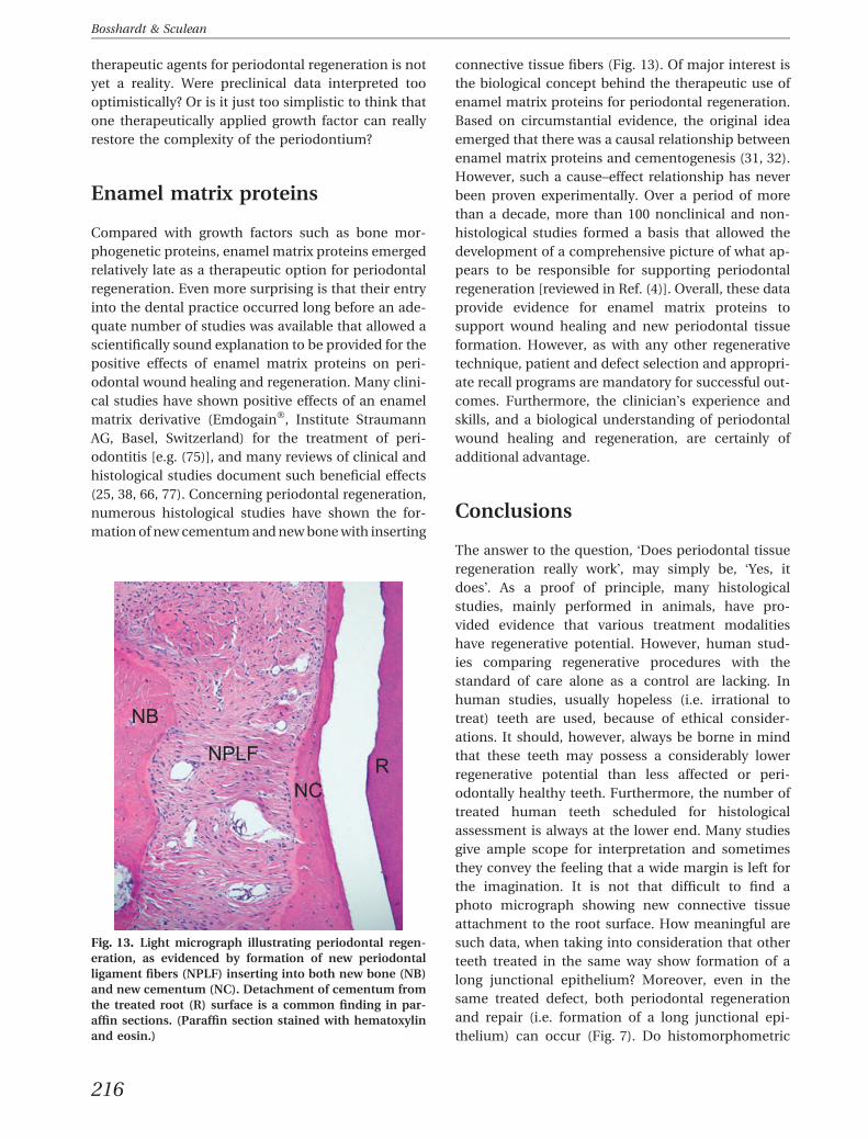
Fig. 13. Light micrograph illustrating periodontal regen-
eration, as evidenced by formation of new periodontal
ligament fibers (NPLF) inserting into both new bone (NB)
and new cementum (NC). Detachment of cementum from
the treated root (R) surface is a common finding in par-
affin sections. (Paraffin section stained with hematoxylin
and eosin.)
216
Bosshardt & Sculean

measurements overcome these shortcomings? We
very much rely on statistical significance rather than
clinical significance. How shall we deal with the fact
that a regenerative procedure may produce 0.0 mm
of new attachment around one tooth, whereas other
teeth show, for the same technique, a broad range
from a fraction of a millimeter to a few milli-
meters? And even if there are statistically significant
differences, one question remains: �Does statistical
significance equate to clinical significance?�. Further-
more, in many clinical situations where regenerative
techniques are used, despite significant probing
depth reduction and gains of clinical attachment,
residual defects still remain. Currently, there is
limited knowledge on the issue of to what extent
such remaining deep sites (i.e. residual pockets) are
prone to bacterial recolonization and subsequent
deterioration. All these issues need to be discussed
and re-evaluated. Better outcome criteria, such as a
threshold for what is �sufficient� new attachment,
need to be established for regenerative treatment
modalities in order to obtain a seal of approval that
is accepted worldwide and subsequently applied.
Our perspective on the current evidence is that
regenerative periodontal therapies to date can only
restore a fraction of the original tissue volume in
extent. Thus, complete periodontal restoration may
still be regarded as an illusion. When it comes to
predictability and a substantial extent of new
attachment formation, there are only a few regener-
ative techniques available. Guided tissue regenera-
tion and enamel matrix proteins certainly have a
regenerative potential. However, these regenerative
techniques do not relieve the dentist from his
responsibilities. As with so many other sensitive
techniques, important aspects to be considered as
outcome determining variables include: (i) appro-
priate patient and defect selection; (ii) correct appli-
cation of a regenerative device or a technique; and
(iii) the dentist�s experience and skills. Finally, it is
striking to realize how little biology is considered.
Minimally invasive surgical techniques for improved
wound stabilization and sufficient time for healing
should be developed. Finally, it should still be borne
in mind that the structural and interactive complexity
of periodontal tissues is probably one of the reasons
why it is so difficult to regenerate the periodontium.
References
1. Bogle G, Adams D, Crigger M, Klinge B, Egelberg J. New
attachment after surgical treatment and acid conditioning
of roots in naturally occurring periodontal disease in dogs.
J Periodontal Res 1981: 16: 130–133.
2. Bornstein MM, von Arx T, Bosshardt DD, Buser D. Prop-
erties of barrier membranes. In: Buser D, editor. 20 Years of
Guided Bone Regeneration in Implant Dentistry. 2nd edn.
Chicago, IL: Quintessenz, 2009: 47–69.
3. Bosshardt DD. Are cementoblasts a subpopulation of os-
teoblastsorauniquephenotype? JDentRes2005:84: 390–406.
4. Bosshardt DD. Biological mediators and periodontal
regeneration: a review of enamel matrix proteins at the
cellular and molecular levels. J Clin Periodontol 2008: 35
(Suppl. 8): 87–105.
5. Bosshardt DD, Hjørting-Hansen E, Buser D. The fate of the
autogenous bone graft. Forum Implantologicum 2009: 5:
4–11.
6. Bosshardt DD, Schenk RK. GBR. Bone regeneration: bio-
logic basis. In: Buser D, editor. 20 Years of Guided Bone
Regeneration in Implant Dentistry. 2nd edn. Chicago, IL:
Quintessenz, 2009: 15–45.
7. Bosshardt DD, Schroeder HE. Cementogenesis reviewed: a
comparison between human premolars and rodent molars.
Anat Rec 1996: 245: 267–292.
8. Bosshardt DD, Sculean A, Windisch P, Pjetursson BE, Lang
NP. Effects of enamel matrix proteins on tissue formation
along the roots of human teeth. J Periodontal Res 2005: 40:
158–167.
9. Bosshardt DD, Selvig KA. Dental cementum: the dynamic
tissue covering of the root. Periodontol 2000 1997: 13: 41–75.
10. Bowers GM, Chadroff B, Carnevale R, Mellonig J, Corio R,
Emerson J, Stevens M, Romberg E. Histologic evaluation of
new attachment apparatus formation in humans. Part I.
J Periodontol 1989: 60: 664–674.
11. Bowers GM, Chadroff B, Carnevale R, Mellonig J, Corio R,
Emerson J, Stevens M, Romberg E. Histologic evaluation of
new attachment apparatus formation in humans. Part II.
J Periodontol 1989: 60: 675–682.
12. Bowers GM, Chadroff B, Carnevale R, Mellonig J, Corio R,
Emerson J, Stevens M, Romberg E. Histologic evaluation of
new attachment apparatus formation in humans. Part III.
J Periodontol 1989: 60: 683–693.
13. Boyko GA, Melcher AH, Brunette DM. Formation of new
periodontal ligament by periodontal ligament cells im-
planted in vivo after culture in vitro. A preliminary study of
transplanted roots in the dog. J Periodontal Res 1981: 16:
73–88.
14. Caffesse RG, Quinones CR. Polypeptide growth factors and
attachment proteins in periodontal wound healing and
regeneration. Periodontol 2000 1993: 1: 69–79.
15. Camargo PM, Pirih FQ, Wolinsky LE, Lekovic V, Kamrath H,
White SN. Clinical repair of an osseous defect associated
with a cemental tear: a case report. Int J Periodontics
Restorative Dent 2003: 23: 79–85.
16. Caton J, Nyman S. Histometric evaluation of periodontal
surgery. I. The modified Widman flap procedure. J Clin
Periodontol 1980: 7: 212–223.
17. Caton J, Nyman S, Zander H. Histometric evaluation of
periodontal surgery. II. Connective tissue attachment levels
after four regenerative procedures. J Clin Periodontol 1980:
7: 224–231.
18. Chou J, Rawal YB, O�Neil JR, Tatakis DN. Cementodentinal
tear: a case report with 7-year follow-up. J Periodontol
2004: 75: 1708–1713.
217
Does periodontal tissue regeneration really work?
19. Cochran DL, Wozney JM. Biological mediators for
periodontal regeneration. Periodontol 2000 1999: 19: 40–
58.
20. Cobb CM. Clinical significance of non-surgical periodontal
therapy: an evidence-based perspective of scaling and root
planing. J Clin Periodontol 2002: 29 (Suppl. 2): 6–16.
21. Cobb CM. Lasers in periodontics: a review of the literature.
J Periodontol 2006: 77: 545–564.
22. Committee on Research, Science and Therapy of the
American Academy of Periodontology. Tissue banking of
bone allografts used in periodontal regeneration. J Perio-
dontol 2001: 76: 834–838.
23. Dereka XE, Markopopoulou CE, Vrotsos IA. Role of growth
factors on periodontal repair. Growth Factors 2006: 24:
260–267.
24. Dragoo MR, Kaldahl WB. Clinical and histological evalua-
tion of alloplasts and allografts in regenerative periodontal
surgery in humans. Int J Periodontics Restorative Dent 1983:
3: 8–29.
25. Esposito M, Grusovin MG, Coulthard P, Worthington HV.
Enamel matrix derivative (Emdogain) for periodontal tissue
regeneration in intrabony defects. Cochrane Database Syst
Rev 2005: 19: 1–34.
26. Foster BL, Popowics TE, Fong HK, Somerman MJ. Ad-
vances in defining regulators of cementum development
and periodontal regeneration. Curr Top Dev Biol 2007: 78:
47–126.
27. Giannobile WV, Somerman MJ. Growth and amelogenin-
like factors in periodontal wound healing. A systematic
review. Ann Periodontol 2003: 8: 193–204.
28. Gould TR, Melcher AH, Brunette DM. Migration and divi-
sion of progenitor cell populations in periodontal ligament
after wounding. J Periodontal Res 1980: 15: 20–42.
29. Greenwell H, Committee on Research, Science and Ther-
apy, American Academy of Periodontology. Position paper:
guidelines for periodontal therapy. J Periodontol 2001: 72:
1624–1628.
30. Grzesik WJ, Narayanan AS. Cementum and periodontal
wound healing and regeneration. Crit Rev Oral Biol Med
2002: 13: 474–484.
31. Hammarstrom L. Enamel matrix, cementum development
and regeneration. J Clin Periodontol 1997: 24: 669–677.
32. Hammarstrom L, Heijl L, Gestrelius S. Periodontal regen-
eration in a buccal dehiscence model in monkeys after
application of enamel matrix proteins. J Clin Periodontol
1997: 24: 669–677.
33. Haney JM, Leknes KN, Lie T, Selvig KA, Wikesjo UM. Ce-
mental tear related to rapid periodontal breakdown: a case
report. J Periodontol 1992: 63: 220–224.
34. Harrel SK, Wright JM. Treatment of periodontal destruction
associated with a cemental tear using minimally invasive
surgery. J Periodontol 2000: 71: 1761–1766.
35. Ishikawa I, Aoki A, Takasaki AA, Mizutani K, Sasaki KM,
Yuichi I. Application of lasers in periodontics: true inno-
vation or myth? Periodontol 2000 2009: 50: 90–126.
36. Ishikawa I, Oda S, Hayashi J, Arakawa S. Cervical cemental
tears in older patients with adult periodontitis. J Period-
ontol 1996: 67: 15–20.
37. Jensen SS, Bosshardt DD, Buser D. Bone grafts and bone
substitute materials for GBR procedures. In: Buser D, edi-
tor. 20 Years of Guided Bone Regeneration in Implant
Dentistry. 2nd edn. Chicago, IL: Quintessenz, 2009: 71–96.
38. Kalpidis CDR, Ruben MP. Treatment of intrabony peri-
odontal defects with enamel matrix derivative: a literature
review. J Periodontol 2002: 73: 1360–1376.
39. Karring T, Isidor F, Nyman S, Lindhe J. New attachment
formation on teeth with a reduced but healthy periodontal
ligament. J Clin Periodontol 1985: 12: 51–60.
40. Karring T, Lindhe J, Cortellini P. Regenerative periodontal
therapy. In: Lindhe J, Karring T, Lang NP, editors. Clinical
Periodontology and Implant Dentistry. 4th edn. Oxford, UK:
Blackwell Munksgaard, 2003: 650–704.
41. Karring T, Nyman S, Gottlow J, Laurell L. Development of
the biological concept of guided tissue regeneration –
animal and human studies. Periodontol 2000 1993: 1: 26–35.
42. Karring T, Nyman S, Lindhe J. Healing following implan-
tation of periodontitis-affected roots into bone tissue.
J Clin Periodontol 1980: 7: 96–105.
43. King G, Cochran DL. Factors that modulate the effects of
bone morphogenetic protein-induced periodontal regen-
eration: a critical review. J Periodontol 2002: 73: 925–936.
44. Listgarten MA, Rosenberg MM. Histological study of repair
following new attachment procedures in human peri-
odontal lesions. J Periodontol 1979: 50: 333–344.
45. Leknes KN, Lie T, Selvig KA. Cemental tear: a risk factor in
periodontal attachment loss. J Periodontol 1996: 67: 583–
588.
46. Lowenguth RA, Blieden TM. Periodontal regeneration: root
surface demineralization. Periodontol 2000 1993: 1: 54–68.
47. Magnusson I, Claffey N, Bogle S, Garrett S, Egelberg J. Root
resorption following periodontal flap procedures in mon-
key. J Periodontal Res 1985: 20: 79–85.
48. Mariotti A. Efficacy of clinical root surface modifiers in the
treatment of periodontal disease. A systematic review. Ann
Periodontol 2003: 8: 205–226.
49. Marquam BJ. Atypical localized deep pocket due to a
cemental tear: case report. J Contemp Dent Pract 2003: 4:
52–64.
50. Melcher AH. On the repair potential of periodontal tissues.
J Periodontol 1976: 47: 256–260.
51. Mizutani K, Aoki A, Takasaki AA, Kinoshita A, Hayashi C,
Oda S, Ishikawa I. Periodontal tissue healing following flap
surgery using Er:YAG laser in dogs. Lasers Surg Med 2006:
38: 314–324.
52. Muller HP. Cemental tear treated with guided tissue
regeneration: a case report 3 years after initial treatment.
Quintessence Int 1999: 30: 111–115.
53. Nakashima M, Reddi AH. The application of bone mor-
phogenetic proteins to dental tissue engineering. Nat
Biotechnol 2003: 21: 1025–1032.
54. Nyman S, Karring T, Lindhe J, Planten S. Healing following
implantation of periodontitis-affected roots into gingival
connective tissue. J Clin Periodontol 1980: 7: 394–401.
55. Polimeni G, Xiropaidis AV, Wikesjo UME. Biology and
principles of periodontal wound healing ⁄ regeneration.
Periodontol 2000 2006: 41: 30–47.
56. Reddy MS, Jeffcoat MK. Methods of assessing periodontal
regeneration. Periodontol 2000 1999: 19: 87–103.
57. Research, Science and Therapy Committee of the American
Academy of Periodontology. Treatment of plaque-induced
gingivitis, chronic periodontitis, and other clinical condi-
tions. J Periodontol 2001: 72: 1790–1800.
58. Reynolds MA, Aichelmann-Reidy MA, Branch-Mays GL,
Gunsolley JC. The efficacy of bone replacement grafts in
218
Bosshardt & Sculean

the treatment of periodontal osseous defects. A systematic
review. Ann Periodontol 2003: 8: 227–265.
59. Ripamonti U, Reddi AH. Tissue engineering, morphogen-
esis, and regeneration of the periodontal tissues by bone
morphogenetic proteins. Crit Rev Oral Biol Med 1997: 8:
154–163.
60. Ripamonti U, Renton L. Bone morphogenetic proteins and
the induction of periodontal tissue regeneration. Period-
ontol 2000 2006: 41: 73–87.
61. Saygin NE, Giannobile WV, Somerman MJ. Molecular and
cell biology of cementum. Periodontol 2000 2000: 24:
73–98.
62. Schroeder HE. Biological problem of regenerative ce-
mentogenesis: synthesis and attachment of collagenous
matrices on growing and established root surfaces. Int Rev
Cytol 1992: 142: 1–59.
63. Schwarz F, Aoki A, Becker J, Sculean A. Laser application in
non-surgical periodontal therapy: a systematic review.
J Clin Periodontol 2008: 35 (Suppl. 8): 29–44.
64. Schwarz F, Jepsen S, Herten M, Aoki A, Sculean A. Immu-
nohistochemical characterization of periodontal wound
healing following nonsurgical treatment with fluorescence
controlled Er:YAG laser radiation in dogs. Lasers Surg Med
2007: 39: 428–440.
65. Sculean A, Nikolidakis D, Schwarz F. Regeneration of
periodontal tissues: combinations of barrier membranes
and grafting materials – biological foundation and pre-
clinical evidence: a systematic review. J Clin Periodontol
2008: 35 (Suppl. 8): 106–116.
66. Sculean A, Rathe F, Junker R, Becker J, Schwarz F, Arweiler
N. The use of Emdogain in periodontal and osseous
regeneration. Schweiz Monatsschr Zahnmed 2007: 117:
598–606.
67. Sculean A, Schwarz F, Windisch P, Keglevich T, Gerharz D,
Becker J. Clinical and histologic evaluation of human
intrabony defects treated with an Er:YAG laser. Perio 2004:
1: 345–352.
68. Sculean A, Stavropoulos A, Windisch P, Keglevich T,
Karring T, Gera I. Healing of human intrabony defects
following regenerative periodontal therapy with a bovine-
derived xenograft and guided tissue regeneration. Clin Oral
Investig 2004: 8: 70–74.
69. Sculean A, Windisch P, Keglevich T, Gera I. Histologic
evaluation of human intrabony defects following non-sur-
gical periodontal therapy with and without application of
an enamel matrix protein derivative. Int Periodontol 2003:
74: 153–160.
70. Sculean A, Windisch P, Keglevich T, Gera I. Clinical and
histologic evaluation of an enamel matrix protein
derivative combined with a bioactive glass for the treat-
ment of intrabony periodontal defects in humans. Int J
Periodontics Restorative Dent 2005: 25: 139–147.
71. Sculean A, Windisch P, Scendroi-Kiss D, Horvath A, Rosta
P, Becker J, Gera I, Schwarz F. Clinical and histologic
evaluation of an enamel matrix protein derivative com-
bined with a biphasic calcium phosphate for the treatment
of human intrabony periodontal defects. J Periodontol
2008: 79: 1991–1999.
72. Shimono M, Ishikawa T, Ishikawa H, Matsuzaki H,
Hashimoto S, Muramatsu T, Shima K, Matsuzaka K-I,
Inoue T. Regulatory mechanisms of periodontal regenera-
tion. Microsc Res Tech 2003: 60: 491–502.
73. Stahl S, Froum S, Kushner L. Healing responses of human
teeth following the use of debridement grafting and citric
acid root conditioning. II. Clinical and histologic observa-
tions: one year post-surgery. J Periodontol 1983: 54: 325–338.
74. Steiner SS, Crigger M, Egelberg J. Connective tissue
regeneration to periodontal diseased teeth, II. Histologic
observation of cases following replaced flap surgery.
J Periodontal Res 1981: 16: 109–116.
75. Tonetti MS, Lang NP, Cortellini P, Suvan JE, Adriaens P,
Dubravec D, Fonzar A, Fourmousis I, Mayfield L, Rossi R,
Silvestri M, Tiedemann C, Topoll H, Vangsted T, Wallkamm
B. Enamel matrix proteins in the regenerative therapy of
deep intrabony defects. A multicenter randomized con-
trolled clinical trial. J Clin Periodontol 2002: 29: 317–325.
76. Trombelli L, Farina R. Clinical outcomes with bioactive
agents alone or in combination with grafting or guided
tissue regeneration. J Clin Periodontol 2008: 35 (Suppl. 8):
117–135.
77. Venezia E, Goldstein M, Boyan BD, Schwartz Z. The use of
enamel matrix derivative in the treatment of periodontal
defects: a literature review and meta-analysis. Crit Rev Oral
Biol Med 2004: 15: 382–402.
78. Wang HL, Greenwell H, Fiorellini J, Giannobile W, Of-
fenbacher S, Salkin L, Townsend C, Sherida P, Genco RJ,
Research, Science and Therapy Committee. Periodontal
regeneration. J Periodontol 2005: 76: 1601–1622.
79. Windisch P, Szendroi-Kiss D, Horvath A, Suba Z, Gera I,
Sculean A. Reconstructive periodontal therapy with
simultaneous ridge augmentation. A clinical and histolog-
ical case series report. Clin Oral Investig 2008: 12: 257–264.
80. Yukna RA, Carr RL, Evans GH. Histologic evaluation of an
Nd:YAG laser-assisted new attachment procedure in hu-
mans. Int J Periodontics Restorative Dent 2007: 27: 577–587.
81. Zeichner-David M. Regeneration of periodontal tissues:
cementogenesis revisited. Periodontol 2000 2006: 41: 196–
217.
219
Does periodontal tissue regeneration really work?


![Enamel matrix derivative [Emdogain(R)] for …...[Intervention Review] Enamel matrix derivative (Emdogain®) for periodontal tissue regeneration in intrabony defects Marco Esposito1,](https://static.fdocuments.us/doc/165x107/5e7b0259219c7e285a26632d/enamel-matrix-derivative-emdogainr-for-intervention-review-enamel-matrix.jpg)


