Principles of biochemical tests commonly used in the...
203
In the name of God By: Dr. S. S. Khoramrooz, Ph.D. Department of Microbiology, Faculty of Medicine, Yasuj University of Medical Sciences, Yasuj, Iran Principles of biochemical tests commonly used in the identification of gram-negative bacteria Yasouj University of Medical Science Department Of Microbiology 1 Dr. Khoramrooz
Transcript of Principles of biochemical tests commonly used in the...

In the name of God
By: Dr. S. S. Khoramrooz, Ph.D.
Department of Microbiology, Faculty of Medicine,
Yasuj University of Medical Sciences, Yasuj, Iran
Principles of biochemical tests
commonly used in the identification
of gram-negative bacteria
Yasouj University of Medical Science Department
Of
Microbiology
1 Dr. Khoramrooz

Introduction
Historically, the identification of bacteria based on:
Colony morphology,
Gram staining,
Biochemical testing.
Based on the phenotype of microorganisms
This method required a lot of time and lots of space in incubators.
Over time, biochemical tests were miniaturized and multitest systems were developed
Resulting in time and space savings.
More recently, automated systems have been developed
2 Dr. Khoramrooz
Clinical microbiology entered the next generation of test systems for the identification of bacteria:
Molecular biology assays.
Based on nucleic acid sequences, are highly sensitive, specific, and rapid, thereby providing accurate results in a few hours or less.
More accurate than examining the phenotype.
In this lecture discuss the principles of biochemical tests commonly used in the identification of gram-negative bacteria.
3 Dr. Khoramrooz

Characters of Enterobacteriaceae
All Enterobacteriaceae
Gram-negative rods
Ferment glucose with acid production
Reduce nitrates into nitrites
Oxidase negative
Facultative anaerobic
Motile except Shigella and Klebsiella
Non-capsulated except Klebsiella
Non-fastidious
Grow on bile containing media (MacConkey agar)
4 Dr. Khoramrooz

Classification of Enterobacteriaceae
Enterobacteriaceae
Lactose fermenters
E. coli, Citrobacter, Klebsiella, Enterobacter
Non-lactose fermenter
Salmonell, Shigella Proteus, Yersinia
There are several selective and differential media used to
isolate distinguishes between LF & LNF
The most important media are:
MacConkey agar
Eosin Methylene Blue (EMB) agar
Salmonella Shigella (SS) agar
In addition to Kiligler Iron agar (KIA) 5 Dr. Khoramrooz

Tests To Know Case Study Tests
Indole
Methyl Red/Voges Proskauer
Citrate
H2S production in SIM
Urea hydrolysis
Motility
Lactose fermentation
Glucose fermentation & gas production
Decarboxylation of amino acis
Fermentation of sugars
Reaction on selective media 6 Dr. Khoramrooz

7 Dr. Khoramrooz

Selection of Primary Isolation Media
Used to recover the significant species of bacteria from specimens that may harbor a mixture of microorganisms.
Microbiologists must know the composition of each formula and the purpose and relative concentration of each chemical or compound that is included.
Not sufficient to know that bile salts are included in selective media
In SS agar contains about 5 times the concentration of bile salts compared with MacConkey agar and is more inhibitory to E.coli and more selective for the recovery of Salmonella species from stool cultures.
8 Dr. Khoramrooz

For the recovery of the Enterobacteriaceae from clinical, three general types of media are available:
1) nonselective media for primary isolation (e.g., blood agar);
2) Selective or differential agars (e.g., MacConkey and Hektoen enteric agars);
3) enrichment broths.
9 Dr. Khoramrooz

Selective Isolation Media
MacConkey and EMB agars are only moderately inhibitory and are designed primarily to prevent growth of gram positive bacteria from mixed cultures.
10 Dr. Khoramrooz

Growth of Enterobacteriaceae on MacConkey agar
Uninoculated plate Lactose non feremters Salmonella, Shigella,
Proteus
Lactose feremters E. coli, Citrobacter
Klebsiella, Enterobacter
Colorless colonies Pink colonies
11 Dr. Khoramrooz

MacConkey Agars
Intended Use
MacConkey agars are slightly selective and differential plating media mainly used for the detection and isolation of gram-negative organisms from clinical, dairy, food, water, pharmaceutical, cosmetic, and other industrial sources.
MacConkey Agar is used for isolating and differentiating lactose-fermenting from lactose-nonfermenting gram-negative enteric bacilli.
MacConkey Agar Base is used with added carbohydrate in differentiating coliforms based on fermentation reactions.
12 Dr. S. S. Khoramrooz

MacConkey Agar without Crystal Violet is used for isolating and differentiating enteric microorganisms while permitting growth of staphylococci and enterococci.
The medium can be used also to separate Mycobacterium fortuitum and M. chelonae from other rapidly growing mycobacteria.
MacConkey Agar without Crystal Violet or Salt and MacConkey Agar without Salt are used for isolating and differentiating gram-negative bacilli while suppressing the swarming of most Proteus species.
13 Dr. S. S. Khoramrooz

Principles of the Procedure
Lactose is a fermentable carbohydrate.
When lactose is fermented, a local pH drop around the colony cause a color change in the pH indicator (neutral red) and bile precipitation.
14 Dr. S. S. Khoramrooz

Bile salts, bile salts no. 3, oxgall and crystal violet are selective agents that inhibit growth of gram-positive organisms.
Magnesium sulfate is a source of divalent cations. Agar is the solidifying agent.
15 Dr. S. S. Khoramrooz

16 Dr. S. S. Khoramrooz

Expected Results
Lactose-fermenting organisms grow as pink to brick-red colonies with or without a zone of precipitated bile.
Lactose-nonfermenting organisms grow as colorless or clear colonies.
Swarming by Proteus spp. is reduced on MacConkey agars without salt.
17 Dr. S. S. Khoramrooz

Dr. S. S. Khoramrooz 18

Limitations of the Procedure
1. Although MacConkey media are selective primarily for gram-negative enteric bacilli, biochemical and, if indicated, serological testing using pure cultures are recommended for complete identification.
2. Incubation of MacConkey Agar plates under increased CO2 has been reported to reduce the growth and recovery of a number of strains of gram-negative bacilli.
19 Dr. S. S. Khoramrooz

Intended Use
Eosin Methylene Blue Agar, Levine is a slightly selective and differential plating medium for the isolation of gram-negative enteric bacteria.
Principles of the Procedure
The eosin Y and methylene blue dyes in Levine EMB Agar render the medium slightly selective in that they inhibit gram- positive bacteria to a limited degree.
20 Dr. S. S. Khoramrooz

These dyes also play a role in differentiating between lactose fermenters and lactose nonfermenters due to the presence or absence of dye uptake in the bacterial colonies.
Coliforms, as lactose-fermenting organisms, are visualized as blue-black colonies, whereas colonies of Salmonella and Shigella, as lactose nonfermenters, appear colorless, transparent or amber.
Some gram-positive bacteria, such as fecal streptococci, staphylococci and yeasts, will grow on this medium and usually form pinpoint colonies.
21 Dr. S. S. Khoramrooz

22 Dr. S. S. Khoramrooz

Expected Results
Typical colonial morphology on Eosin Methylene
Blue Agar, Levine is as follows:
23 Dr. S. S. Khoramrooz

Dr. S. S. Khoramrooz 24

Highly Selective Isolation Media Used Primarily for Gastrointestinal Specimens
Media are made highly selective by the addition of a variety of inhibitors to their formulas, generally in higher concentrations than in MacConkey and EMB agars.
These media are used primarily to inhibit the growth of E. coli and other "coli forms," but they allow Salmonella and Shigella species to grow out from stool specimens
The most commonly used are Salmonella-Shigella (SS) agar, xylose lysine deoxycholate (XLD) agar, and Hektoen enteric (HE) agar.
25 Dr. Khoramrooz

Intended Use
XL (Xylose Lysine) Agar Base is used for the
isolation and differentiation of enteric pathogens
and, when supplemented with appropriate
additives, as a base for selective enteric media.
XLD Agar is the complete Xylose Lysine
Desoxycholate Agar, a moderately selective
medium recommended for isolation and
differentiation of enteric pathogens, especially
Shigella species.
26 Dr. S. S. Khoramrooz

Principles of the Procedure
Xylose is incorporated into the medium because it is fermented by practically all enterics except for the shigellae.
This property enables the differentiation of Shigella species.
Lysine is included to enable the Salmonella group to be differentiated from the nonpathogens.
Without lysine, salmonellae rapidly would ferment the xylose and be indistinguishable from nonpathogenic species.
27 Dr. S. S. Khoramrooz

After the salmonellae exhaust the supply of xylose, the lysine is attacked via the enzyme lysine decarboxylase, with reversion to an alkaline pH, which mimics the Shigella reaction.
To prevent similar reversion by lysine-positive coliforms, lactose and sucrose (saccharose) are added to produce acid in excess.
Degradation of xylose, lactose and sucrose generates acid products, which in the presence of the pH indicator phenol red, causes a color change in the medium from red to yellow.
28 Dr. S. S. Khoramrooz

To add to the differentiating ability of the
formulation, an H2S indicator system, consisting
of sodium thiosulfate and ferric ammonium citrate,
is included for the visualization of the hydrogen
sulfide produced, resulting in the formation of
colonies with black centers.
The nonpathogenic H2S producers do not
decarboxylate lysine; therefore, the acid reaction
produced by them prevents the blackening of the
colonies.
29 Dr. S. S. Khoramrooz

XLD Agar is both a selective and differential
medium.
It utilizes sodium desoxycholate as the selective
agent and, therefore, it is inhibitory to gram-
positive microorganisms.
30 Dr. S. S. Khoramrooz

31 Dr. S. S. Khoramrooz

32 Dr. S. S. Khoramrooz

Expected Results
Degradation of xylose, lactose and sucrose generates acid products, causing a color change in the medium from red to yellow.
Hydrogen sulfide production under alkaline conditions causes colonies to develop black centers.
This reaction is inhibited by the acid conditions that accompany carbohydrate fermentation.
Lysine decarboxylation in the absence of lactose and sucrose fermentation causes reversion to an alkaline condition and the color of the medium changes back to red.
Typical colonial morphology and reactions on XLD Agar are as follows:
33 Dr. S. S. Khoramrooz

34 Dr. S. S. Khoramrooz

Dr. S. S. Khoramrooz 35

Dr. S. S. Khoramrooz 36

Limitations of the Procedure
1. Red, false-positive colonies may occur with some Proteus and Pseudomonas species.
2. Incubation in excess of 48 hours may lead to false-positive results.
3. S. Paratyphi A, S. Choleraesuis, S. pullorum and S. gallinarum may form red colonies without black centers, thus resembling Shigella species.
4. Some Proteus strains will give black-centered colonies on XLD Agar.
37 Dr. S. S. Khoramrooz

41 Dr. Khoramrooz

42 Dr. Khoramrooz

43 Dr. Khoramrooz

44 Dr. Khoramrooz

Intended Use
Hektoen Enteric (HE) Agar is a moderately selective
medium used in qualitative procedures for the
isolation and cultivation of gram-negative enteric
microorganisms, especially Shigella, from a variety
of clinical and nonclinical specimens.
45 Dr. S. S. Khoramrooz

Principles of the Procedure
The selective nature of Hektoen Enteric Agar is
due to the incorporation of bile salts in the
formulation.
These substances inhibit gram-positive organisms
but also can be toxic for some gram-negative
strains.
46 Dr. S. S. Khoramrooz

This medium contains three carbohydrates, lactose, sucrose (saccharose) and salicin, for optimal differentiation of enteric pathogens
The lactose concentration is higher than in many other media used for enterics in order to aid in the visualization of enteric pathogens and minimize the problem of delayed lactose fermentation.
Ferric ammonium citrate and sodium thiosulfate in the medium enable the detection of hydrogen sulfide production.
The indicator system, consisting of acid fuchsin and bromthymol blue, has a lower toxicity than that of many other enteric media, resulting in improved recovery of enteric pathogens.
47 Dr. S. S. Khoramrooz

Procedure
A nonselective medium should also be streaked to
increase the chance of recovery when the
population of gram-negative organisms is low and
to provide an indication of other organisms present
in the specimen.
Incubate plates, protected from light, at 35 ± 2°C
for 18-24 hours.
48 Dr. S. S. Khoramrooz

Dr. S. S. Khoramrooz 49
DO NOT AUTOCLAVE.

Dr. S. S. Khoramrooz 50

Dr. S. S. Khoramrooz 51

Dr. S. S. Khoramrooz 52
mixed with normal fecal flora. Salmonella

Dr. S. S. Khoramrooz 53

Hektoen Enteric (HE) Agar:
Composition • Peptone 1.2%
• Yeast extract 0.3%
• Bile salts 0.9%
• Lactose 1.2%
• Sucrose 1.2%
• Salicin 0.2%
• Sodium chloride 0.5%
• Ferric ammonium citrate
• Acid fuchsin
• Thymol blue
• Agar 1.4%
• pH = 7.6
54 Dr. Khoramrooz

HE Agar: Growth of Enteric
Pathogens and Commensals
• High bile salt concentration inhibits growth of gram-positive and gram-negative intestinal commensals, and thereby selects for pathogenic Salmonella (bile-resistant growth) present in fecal specimens.
• Salmonella species as non-lactose and non-sucrose fermenters that produce H2S form colorless colonies with black centers.
• Shigella species (non-lactose and non-sucrose fermenters, no H2S production) form colorless colonies.
• Lactose and sucrose fermenters (E. coli, K. pneumoniae) form orange to yellow colonies due to acid production.
55 Dr. Khoramrooz

Intended Use
Bismuth Sulfite Agar is a highly selective medium
used for isolating Salmonella spp., particularly
Salmonella Typhi, from food and clinical
specimens.
56 Dr. S. S. Khoramrooz

Principles of the Procedure
Dextrose is an energy source.
Bismuth sulfite indicator and brilliant green are complementary in inhibiting gram-positive bacteria and members of the coliform group, while allowing Salmonella to grow luxuriantly.
Ferrous sulfate is included for detection of H2S production.
When H2S is present, the iron in the formula is precipitated, giving positive cultures the characteristic brown to black color with metallic sheen.
57 Dr. S. S. Khoramrooz

58 Dr. S. S. Khoramrooz

For isolation of Salmonella spp. from clinical
specimens, inoculate fecal specimens and rectal
swabs onto a small area of one quadrant of the
Bismuth Sulfite Agar plate and streak for isolation.
This will permit the development of discrete
colonies.
Incubate plates at 35°C.
Examine at 24 hours and again at 48 hours for
colonies resembling Salmonella spp.
59 Dr. S. S. Khoramrooz

Expected results
The typical discrete S. Typhi surface colony is black and surrounded by a black or brownish-black zone which may be several times the size of the colony.
By reflected light, preferably daylight, this zone exhibits a distinctly characteristic metallic sheen.
Plates heavily seeded with S. Typhi may not show this reaction except near the margin of the mass inoculation.
60 Dr. S. S. Khoramrooz

In these heavy growth areas, this organism
frequently appears as small light green colonies.
This fact emphasizes the importance of inoculating
plates so that some areas are sparsely populated
with discrete S. Typhi colonies.
Other strains of Salmonella produce black to
green colonies with little or no darkening of the
surrounding medium.
61 Dr. S. S. Khoramrooz

Dr. S. S. Khoramrooz 62

Heat with frequent agitation and boil for 1 minute
to completely dissolve the powder.
DO NOT AUTOCLAVE.
Use the medium the same day it is prepared.
63 Dr. S. S. Khoramrooz

64 Dr. Khoramrooz

Intended Use
Brilliant Green Agar is a highly selective medium for
the isolation of Salmonella other than S. Typhi from
feces and other materials.
Principles of the Procedure
Brilliant green dye inhibits gram-positive bacteria and a
majority of gram-negative bacilli.
Phenol red serves as a pH indicator and yields a yellow
color as a result of acid production in the fermentation of
the lactose and/or sucrose in the medium.
65 Dr. S. S. Khoramrooz

66 Dr. S. S. Khoramrooz

Procedure
A less selective medium and a nonselective medium should also be streaked to increase the chance of recovery when the population of gram-negative organisms is low and to provide an indication of other organisms present in the specimen.
Incubate plates, protected from light, at 35 ± 2°C for 18-24 hours.
If negative after 24 hours, reincubate an additional 24 hours.
67 Dr. S. S. Khoramrooz

Dr. S. S. Khoramrooz 68
Salmonella on BPLS Agar. The colonies
are red because the bacterium does not
ferment lactose or sucrose.
Escherichia coli on BPLS Agar.
The colonies are yellow due to the
low pH which is caused by the
production of acid during
fermentation of lactose and/or
sucrose.

69 Dr. S. S. Khoramrooz

Differential Identification Characteristics
70 Dr. Khoramrooz

Carbohydrate utilization
Lactose degradation; Differentiate those (lactose fermenter LF]) and those that are nonlactose fermenters (NLFs).
Examples of sugars used to differentiate bacteria include lactose, maltose, rhamnose, sucrose, raffinose, and arabinose
Bacteria can utilize carbohydrates: oxidation (aerobically), fermentatively (anaerobically),or both.
Bacteriat hat can grow either aerobically or anaerobically are called facultative anaerobes.
Some bacteria are asacchrolytic.
They do not use any carbohydrate; instead they use other organic molecules for energy and carbon sources.
71 Dr. Khoramrooz

Oxidation/Fermentation (O/F) Test
Principle :
To determine the ability of bacteria to breakdown glucose oxidative or fermentative
O/F medium ( Hugh and Leifson Medium) is formulated to detect weak acids produced from saccharolytic M.O.
O/F medium contains
Sugar (glucose 1%)
Low percentage of Agar and Peptone
pH indicator (Bromothymol blue)
• Alkaline Blue
• Neutral Green
• Acidic Yellow
72 Dr. Khoramrooz

Intended Use
OF (Oxidation Fermentation) media are used for the determination of oxidative and fermentative metabolism of carbohydrates by gram-negative rods on the basis of acid reaction in either the open or closed system.
Summary and Explanation
OF Medium was developed by Hugh and Leifson who described the taxonomic significance of fermentative versus oxidative metabolism of carbohydrates by gram-negative bacteria.
73 Dr. S. S. Khoramrooz

They showed that when an organism is inoculated into two tubes of OF Basal Medium containing a carbohydrate and the medium in one of the tubes is covered with melted petrolatum prior to incubation, the patterns of metabolism are of differential significance.
Oxidative organisms only produce an acid reaction in the open tube with little or no growth and no acid formation in the covered tube.
Fermentative organisms will produce an acid reaction in both types of tubes.
Changes in the covered agar are considered to be due to true fermentation, while changes in the open tubes are due to oxidative utilization of the carbohydrate present.
If the carbohydrate is not utilized by either method, there is no acid production in either tube.
Dr. S. S. Khoramrooz 74

Principles of the Procedure
Dextrose is the most important carbohydrate for
use in OF Basal Medium; however, certain
organisms may metabolize other carbohydrates
even if they are unable to utilize dextrose.
Prepared tubed media containing arabinose,
dextrose, dulcitol, fructose, galactose, lactose,
maltose, mannose, raffinose, rhamnose, salicin,
sorbitol, sucrose and xylose are provided.
75 Dr. S. S. Khoramrooz

76 Dr. S. S. Khoramrooz

Procedure
Inoculate a pair of OF tubes of each carbohydrate used with each organism being tested.
The tubes should be stabbed to approximately 1/4 inch from the bottom using an inoculating needle and a light inoculum.
Overlay one tube of each pair with sterile mineral oil.
Incubate tubes at 35 ± 2°C in an aerobic atmosphere for 48 hours.
Do not discard as negative until after 4 days of incubation.
77 Dr. S. S. Khoramrooz

Expected Results
Record results as acid (A) or alkaline/no change (–).
Also record whether or not the organism is motile
as evidenced by the appearance of growth away
from the line of inoculation.
Typical reaction patterns are as follows.
78 Dr. S. S. Khoramrooz

Limitations of the Procedure
1. The acid reaction produced by oxidative organisms is apparent at the surface and gradually spreads throughout the medium.
If the oxidation is weak or slow, however, an initial alkaline reaction at the surface of the open tube may persist for several days and eventually convert to an acid reaction.
2. If an organism is unable to grow on OF Basal Medium, Cowan recommends adding either 2% serum or 0.1% yeast extract to each carbohydrate tube.
79 Dr. S. S. Khoramrooz
Dr. S. S. Khoramrooz 80

Dr. S. S. Khoramrooz 82

Triple Sugar lron Agar Kligler Iron Agar
Lactose Fermentation
Glucose fermentation
Gas Production (H2 & CO2 )
H2S Production
83 Dr. Khoramrooz

glucose
lactose
Ferrous sulfate
pH indicator: phenol red
TSI and KIA
Proteins
84 Dr. Khoramrooz

85 Dr. Khoramrooz

Intended Use
Triple Sugar Iron Agar (TSI Agar) is used for the differentiation of gram-negative enteric bacilli based on carbohydrate fermentation and the production of hydrogen sulfide.
Principles of the Procedure
TSI Agar contains three sugars (dextrose, lactose and sucrose),
Phenol red for detecting carbohydrate fermentation
86 Dr. S. S. Khoramrooz

Ferrous ammonium sulfate for detection of hydrogen sulfide production (indicated by blackening in the butt of the tube).
Carbohydrate fermentation is indicated by the production of gas and a change in the color of the pH indicator from red to yellow.
To facilitate the detection of organisms that only ferment dextrose, the dextrose concentration is one-tenth the concentration of lactose or sucrose.
The small amount of acid produced in the slant of the tube during dextrose fermentation oxidizes rapidly, causing the medium to remain red or revert to an alkaline pH.
In contrast, the acid reaction (yellow) is maintained in the butt of the tube because it is under lower oxygen tension.
87 Dr. S. S. Khoramrooz

After depletion of the limited dextrose, organisms
able to do so will begin to utilize the lactose or
sucrose.
To enhance the alkaline condition of the slant, free
exchange of air must be permitted by closing the
tube cap loosely.
If the tube is tightly closed, an acid reaction
(caused solely by dextrose fermentation) will also
involve the slant.
88 Dr. S. S. Khoramrooz

89 Dr. S. S. Khoramrooz
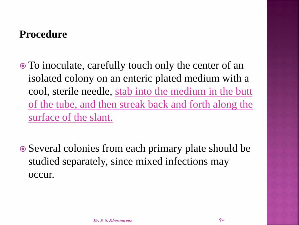
Procedure
To inoculate, carefully touch only the center of an
isolated colony on an enteric plated medium with a
cool, sterile needle, stab into the medium in the butt
of the tube, and then streak back and forth along the
surface of the slant.
Several colonies from each primary plate should be
studied separately, since mixed infections may
occur.
90 Dr. S. S. Khoramrooz

Incubate with caps loosened at 35°C and examine
after 18-24 hours for carbohydrate fermentation,
gas production and hydrogen sulfide production.
Any combination of these reactions may be
observed.
Do not incubate longer than 24 hours because
the acid reaction in the slant of lactose and sucrose
fermenters may revert to an alkaline reaction.
91 Dr. S. S. Khoramrooz

Dr. S. S. Khoramrooz 92

Expected Results
Carbohydrate fermentation is indicated by a yellow coloration of the medium.
If the medium in the butt of the tube becomes yellow (acidic), but the medium in the slant becomes red (alkaline), the organism being tested only ferments dextrose (glucose).
A yellow (acidic) color in the slant and butt indicates that the organism being tested ferments dextrose, lactose and/or sucrose.
A red (alkaline) color in the slant and butt indicates that the organism being tested is a nonfermenter.
93 Dr. S. S. Khoramrooz

Hydrogen sulfide production results in a black
precipitate in the butt of the tube.
Gas production is indicated by splitting and
cracking of the medium.
94 Dr. S. S. Khoramrooz

1. Hydrogen sulfide production may be evident on Kligler Iron Agar but negative on Triple Sugar Iron Agar.
Studies by Bulmash and Fulton showed that the utilization of sucrose could suppress the enzymatic mechanisms responsible for H2S production.
Padron and Dockstader8 found that not all H2S-positive Salmonella are positive on TSI.
2. Sucrose is added to TSI to eliminate some sucrose-fermenting lactose-nonfermenters such as Proteus and Citrobacter spp.
3. Further biochemical tests and serological typing must be performed for definite identification and confirmation of organisms.
95 Dr. S. S. Khoramrooz

4. Do not use an inoculating loop to inoculate a tube of Triple Sugar Iron Agar.
While stabbing the butt, mechanical splitting of the medium occurs, causing a false positive result for gas production.
5. A pure culture is essential when inoculating Triple Sugar Iron Agar.
If inoculated with a mixed culture, irregular observations may occur.
6. Tubes should be incubated with caps loosened.
This allows a free exchange of air, which is necessary to enhance the alkaline condition on the slant.
96 Dr. S. S. Khoramrooz

Red/Red Alkaline /Alkaline K/K Lactose -/Glucose –
Yellow/Yellow Acid/Acid A/A Lactose +/Glucose +
Red/Yellow Alkaline/Acid K/A Lactose -/Glucose +
Gas - H2S -
Red/Yellow Alkaline/Acid K/A Lactose -/Glucose +
Gas + H2S -
Red/Yellow Alkaline/Acid K/A Lactose -/Glucose +
Gas - H2S +
Red/Black Alkaline/Acid K/A Lactose -/Glucose +
Gas + H2S +
97 Dr. Khoramrooz

Example
Result
Reaction on KIA
H2S Slant
color
Butt
color
Non fermenter
e.g.
Pseudomonas
Alk/Alk/-
(No action on sugars)
Negative Red Red
LNF
e.g. Shigella A/Alk/-
(Glucose fermented
without H2S)
Negative
Red Yellow
LNF
e.g. Salmonella &
Proteus
A/Alk/+
(Glucose fermented
with H2S)
Positive
black in
butt Red Yellow
LF
e.g. E. coli,
Klebsiella,
Enterobacter
A/A/-
(three sugars are
fermented)
Negative Yellow Yellow
Result
98 Dr. Khoramrooz

Ortho- Nitrophenyl - β –D- galactopyranosideTest
Organisms that are delayed lactose-fermenters appear as nonfermenting colonies.
The o-nitrophenyl β–D- galactopyranoside (ONPG) and the p-nitrophenyl-B-D-galactopyranoside (PNPG) tests determine whether the organism is a delayed lactose fermenter (one that lacks the enzyme β-galactoside permease but possesses β-galactosidase) or a true nonlactose fermenter (NLF).
The test can be performed by making a heavy suspension of bacteria in sterile saline and adding commercially prepared ONPG disks or tablets.
The suspension is incubated at 37' C, and positive results can generally be seen within 6 hours.
99 Dr. Khoramrooz

Dr. Khoramrooz 100

IMViC Test
Indole, Methyl Red, Voges-Prosakaur, Citrate (IMViC) Tests:
The following four tests comprise a series of important determinations that are collectively called the IMViC series of reactions
The IMViC series of reactions allows for the differentiation of the various members of Enterobacteriaceae.
101 Dr. Khoramrooz

IMViC: Indole test Principle
Certain microorganisms can metabolize tryptophan by tryptophanase
The enzymatic degradation leads to the formation of pyruvic acid, indole and ammonia
The presence of indole is detected by addition of Kovac's reagent.
Tryptophane amino acids
Tryptophanase Indole + Pyurvic acid + NH3
Kovac’s Reagent
Red color in upper organic layer` 102 Dr. Khoramrooz

IMViC: Indole test
Result:
A bright pink color in the top
layer indicates the presence of
indole
The absence of color means that
indole was not produced i.e.
indole is negative
Special Features:
Used in the differentiation of genera
and species. e.g. E. coli (+) from
Klebsiella (-).
Positive test e.g. E. coli
Negative test e.g. Klebsiella
103 Dr. Khoramrooz

Intended Use
SIM Medium is used to differentiate enteric bacilli
on the basis of sulfide production,indole
formation and motility.
104 Dr. S. S. Khoramrooz

Summary and Explanation
Hydrogen sulfide production, indole formation
and motility are distinguishing characteristics
which aid in the identification of the
Enterobacteriaceae, especially Salmonella and
Shigella.
SIM Medium, therefore ,is useful in the process of
identification of enteric pathogens.
105 Dr. S. S. Khoramrooz

Principles of the Procedure
Sodium thiosulfate and ferrous ammonium sulfate are indicators of hydrogen sulfide production.
The ferrous ammonium sulfate reacts with H2S gas to produce ferrous sulfide,a black precipitate.
The casein peptone is rich in tryptophan,which is attacked by certain microorganisms resulting in the production of indole.
The indole is detected by the addition of chemical reagents following the incubation period.
106 Dr. S. S. Khoramrooz

Motility detection is possible due to the semisolid
nature of the medium.
Growth radiating out from the central stab line
indicates that the test organism is motile.
107 Dr. S. S. Khoramrooz

108 Dr. S. S. Khoramrooz

Procedure
Loosen caps, boil and cool before use.
Using growth from a pure culture, stab an
inoculating needle two-thirds of the distance to
the bottom in the center of the tube.
Incubate tubes with loosened caps for18-24 hours
at 35±2°C in anaerobic atmosphere.
109 Dr. S. S. Khoramrooz

Dr. S. S. Khoramrooz 110

IMViC test Methyl Red-Voges Proskauer (MR-VP) Tests
Glucose
Acidic pathway
Mixed acids pH less than 4.4
Methyl Red
indicator
Red color
Principle
MR positive E. coli
Or Neutral pathway
Acety methyl carbinol (ACETOIN)
solution A
solution B
Pink color VP positive Klebsiella
111 Dr. Khoramrooz

Butylene Glycol Pathway of Glucose Fermentation
In the butylene glycol pathway
pyruvic acid to acetoin and butylene glycol.
Acetoin and butylene glycol are detected by oxidation to diacteyl at an alkaline pH.
Addition of -naphthol which forms a red-colored complex with diacetyl.
Important biochemical property used for the identification of Klebsiella, Enterobacter, and Serratia.
112 Dr. Khoramrooz

IMViC test: MRVP test
Inoculate the tested organism into MRVP broth
Incubate the tubes at 37°C for 24 hours
• For methyl red: Add 6-8 drops of methyl
red reagent.
• For Voges-Proskauer: Add 12 drops of
solution A (-naphthol), mix, 4 drops of
Solution B (40% KOH), mix
Method
113 Dr. Khoramrooz

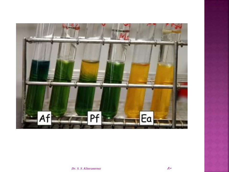
IMViC test: MR/VP test
Results
Methyl Red test Voges-Proskauer test
Red: Positive MR (E. coli)
Yellow or orange: Negative MR (Klebsiella)
Pink: Positive VP (Klebsiella)
No pink: Negative VP (E. coli) 114 Dr. Khoramrooz

Intended Use
MR-VP Medium and MR-VP Broth (Methyl Red-
Voges Proskauer Medium/Broth, also known as
Buffered Peptone- Glucose Broth) are used for the
differentiation of bacteria by means of the methyl
red and Voges-Proskauer reactions.
115 Dr. S. S. Khoramrooz
Principles of the Procedure
Methyl red-positive organisms produce high levels of acid during fermentation of dextrose, overcome the phosphate buffer system and produce a red color upon the addition of the methyl red pH indicator.
In the Voges-Proskauer test, the red color produced by the addition of potassium hydroxide to cultures of certain microbial species is due to the ability of the organisms to produce a neutral end product, acetoin (acetylmethylcarbinol), from the fermentation of dextrose.
The acetoin is oxidized in the presence of oxygen and alkali to produce a red color.
This is a positive Voges-Proskauer reaction.
116 Dr. S. S. Khoramrooz

117 Dr. S. S. Khoramrooz

Procedure
Using a light inoculum, inoculate tubes of MR-VP media with 18- to 24-hour pure cultures.
Incubate tubes aerobically at 35 ― 2�‹C for a minimum of 48 hours but preferably for 5 days.
Prepare the methyl red indicator: 0.1 g of methyl red in 300 mL of 95% ethyl alcohol.
Add sufficient purified water to make 500 mL.
After the appropriate incubation period, aseptically remove aliquots (1 mL for the VP test) of the medium and conduct the following tests:
118 Dr. S. S. Khoramrooz

1. Methyl Red Test:
Add 5 drops of methyl red indicator to an aliquot of the broth.
Interpret the color result immediately.
2. Voges-Proskauer Test:
Empty the contents (15 drops) from the reagent A dropper
5 drops from the reagent B dropper into 1 mL of broth culture.
Shake well after the addition of each reagent to aerate the sample.
119 Dr. S. S. Khoramrooz

Expected Results
1. Methyl Red Test
a. Positive – red color at surface of the medium.
b. Negative – yellow color at surface of the medium.
2. Voges-Proskauer Test
A positive reaction is indicated by the development of a distinct red color which occurs within 5 minutes.
Certain species within Enterobacteriaceae genera may react differently or give variable results.
Consult appropriate texts for reactions of specific species.
120 Dr. S. S. Khoramrooz

Limitations of the Procedure
1. Results of the MR and VP tests need to be used in conjunction with other biochemical tests to differentiate genus and species within the Enterobacteriaceae.
2. A precipitate may form in the potassium hydroxide reagent solution.
This precipitate has not been shown to reduce the effectiveness of the reagent.
121 Dr. S. S. Khoramrooz

3. Most members of the family Enterobacteriaceae give either a positive MR test or a positive VP test.
However, certain organisms such as Hafnia alvei and Proteus mirabilis may give a positive result for both tests.
4. Incubation time for the Methyl Red test cannot be shortened by increasing the dextrose concentration in the medium or by heavily inoculating the broth.
5. Incubate MR-negative tests for more than 48 hours and test again.
Dr. S. S. Khoramrooz 122

6. Read the VP test at 48 hours. Increased incubation may produce acid conditions in the broth that will interfere with reading the results.
7. VP reagents must be added in the order and the amounts specified or a weak-positive or false-negative reaction may occur.
A weak-positive reaction may be masked by a copper-like color
which may form due to the reaction of KOH and α-naphthol.
8. Read the VP test within 1 hour of adding the reagents. The KOH and α-naphthol may react to form a copper-like color, causing a potential false-positive interpretation.
9. Due to the possible presence of acetoin, diacetyl or related substances in certain raw materials, the use of media low in these substances (such as MR-VP media) is recommended for this test.
123 Dr. S. S. Khoramrooz

124 Dr. S. S. Khoramrooz

Citrate Utilization Test Principle:
Citrate Na2CO3
Alkaline,↑pH
Blue colour Bromothymol blue
Simmone’s Citrate media
Positive test: Klebsiella, Enterobacter, Citrobacter
CO2 + Na + H2O Pyruvate
Positive test
Negative test: E. coli
Contains Citrate as a sole of C source
125 Dr. Khoramrooz

Citrate Utilization Test
Incubate at 37°C for 24 hours.
Method
Streak a Simmon's Citrate agar slant with
the organism
126 Dr. Khoramrooz

Citrate Utilization Test
Examine for growth (+)
Growth on the medium is accompanied by a rise in pH to change the medium from its initial green color to deep blue
Result
Positive Klebsiella, Enterobacter Negative
E. coli 127 Dr. Khoramrooz

Intended Use
Simmons Citrate Agar is used for the differentiation of
gram negative bacteria on the basis of citrate utilization.
Principles of the Procedure
Organisms able to utilize ammonium dihydrogen
phosphate and sodium citrate as the sole sources of
nitrogen and carbon, respectively, will grow on this
medium and produce an alkaline reaction as evidenced
by a change in the color of the bromthymol blue
indicator from green (neutral) to blue (alkaline).
128 Dr. S. S. Khoramrooz


Procedure
Inoculate slants with growth from a pure culture using a light inoculum.
Incubate all tubes for 4 days at 35 ± 2°C in an aerobic atmosphere.
Expected Results
A positive reaction is indicated by growth with an intense blue color in the slant.
A negative reaction is evidenced by no growth to trace growth with no change in color (medium remains dark green).
130 Dr. S. S. Khoramrooz

Dr. S. S. Khoramrooz 131

Dr. S. S. Khoramrooz 132

Dr. S. S. Khoramrooz 133

Amino Acid Decarboxylation
Enterobacteriaceae contain decarboxylases with substrate specificity for amino acids, and are detected using Moeller decarboxylase broth overlayed with mineral oil for anaerobiosis.
Moeller broth contains:
glucose for fermentation,
peptone and beef extract,
amino acid, pyridoxal,
pH indicator bromcresol purple.
134 Dr. Khoramrooz

Amino Acid Decarboxylation
If an Enterobacteriaceae contains amino acid decarboxylase, amines produced by decarboxylase action cause an alkaline pH, and bromcresol purple turns purple.
Lysine, ornithine, and arginine are utilized.
A base broth without amino acid is included in which glucose fermentation acidifies the broth, turning the bromcresol purple yellow.
135 Dr. Khoramrooz

Amino Acid Decarboxylation1
Lysine → Cadaverine
Ornithine → Putrescine
Arginine → Citrulline → Ornithine → Putrescine
1Conversion of arginine to citrulline is a dihydrolase reaction
136 Dr. Khoramrooz

Amino Acid Decarboxylation
Decarboxylation patterns are essential for the genus identification of Klebsiella, Enterobacter, Escherichia, and Salmonella.
Decarboxylation patterns are also essential for the species identification of Enterobacter aerogenes, Enterobacter cloacae, Proteus mirabilis, and Shigella sonnei.
137 Dr. Khoramrooz

Amino Acid Decarboxylation
Lys Orn Arg
Klebsiella + – –
Enterobacter +/– + +/–
Escherichia + +/– –/+
Salmonella + + +
138 Dr. Khoramrooz

Amino Acid Decarboxylation
Lys Orn Arg
E. aerogenes + + –
E. cloacae – + +
P. mirabilis – + –
P. vulgaris – – –
Shigella D – + –
Shigella A-C – – –
139 Dr. Khoramrooz

Phenylalanine Deaminase Reaction
Enterobacteriaceae utilize amino acids in a variety of ways including deamination.
Phenylalanine is an amino acid that forms the keto acid phenylpyruvic acid when deaminated.
Phenylpyruvic acid is detected by addition of ferric chloride that forms an intensely dark olive-green colored complex when binding to phenylpyruvic acid.
The deamination of phenylalanine is an important biochemical property of Proteus, Morganella, and Providencia.
140 Dr. Khoramrooz

Intended Use
Decarboxylase media are used in the biochemical differentiation of gram-negative enteric bacilli based on the production:
Arginine dihydrolase
Lysine decarboxylase
Ornithine decarboxylase
Decarboxylase Medium Base, with added arginine, lysine or ornithine is used for the same purpose.
Lysine Decarboxylase Broth is used for differentiating microorganisms based on lysine decarboxylation.
141 Dr. S. S. Khoramrooz

Summary and Explanation
Moeller introduced the decarboxylase media for detecting the production of lysine and ornithine decarboxylase and arginine dihydrolase.
These media are a useful adjunct to other biochemical tests for the speciation and identification of the Enterobacteriaceae and other gram-negative bacilli.
The production of OD is particularly useful for differentiating Klebsiella and Enterobacter species.
Klebsiella species are non-motile and, except for K. ornithinolytica, do not produce ornithine decarboxylase, while most Enterobacter species are motile and, except for E. agglomerans, usually produce this enzyme.
142 Dr. S. S. Khoramrooz

Principles of the Procedure
Pyridoxal is an enzyme co-factor for the amino acid decarboxylase.
Dextrose is a fermentable carbohydrate.
Bromcresol purple and cresol red are pH indicators.
The amino acids lysine, ornithine or arginine are added to the basal medium at a concentration of 10.0 g/L to detect the production of the enzyme specific for these substrates.
143 Dr. S. S. Khoramrooz

When the medium is inoculated with a bacterium
that is able to ferment dextrose, acids are produced
that lower the pH of the medium and change the
color of the indicator from purple to yellow.
The acidic condition also stimulates decarboxylase
activity.
If the organism produces the appropriate enzyme,
the amino acid in the medium is degraded, yielding
a corresponding amine.
144 Dr. S. S. Khoramrooz

Dr. S. S. Khoramrooz 145

Decarboxylation of lysine yields cadaverine.
while decarboxylation of ornithine yields putrescine.
Arginine is first hydrolyzed to form ornithine, which is then decarboxylated to form putrescine.
The production of these amines elevates the pH of the medium, changing the color of the indicator from yellow to purple or violet.
If the organism does not produce the appropriate enzyme, the medium remains acidic (yellow).
146 Dr. S. S. Khoramrooz

Each isolate to be tested must also be inoculated into a tube of the basal medium that does not contain the amino acid.
If this tube becomes alkaline, the test is invalid.
To obtain the appropriate reactions, the inoculated tubes must be protected from air with a layer of sterile mineral oil.
Exposure to air may cause alkalinization at the surface of the medium, which could cause a decarboxylase-negative organism to appear positive.
147 Dr. S. S. Khoramrooz

148 Dr. S. S. Khoramrooz

Expected Results
Compare the color of tubes of media containing the specific amino acids with the color of control tubes of basal media (without amino acid) that have been inoculated with the same isolate.
If inoculated control tubes show an alkaline reaction, the test is invalid; i.e.,
Improperly performed or the test organisms
Degrade the peptone sufficiently to produce an alkaline reaction in the absence of a specific amino acid.
The medium becomes purple to violet if the reaction is positive (alkaline).
A yellow color indicates a negative test; i.e., the organism does not produce the appropriate enzyme.
149 Dr. S. S. Khoramrooz

150 Dr. S. S. Khoramrooz

The lysine iron agar (LIA) test is a tubed agar slant.
It contains the amino acid lysine, glucose, ferric ammonium
citrate, and sodium thiosulfate.
The pH indicator is bromcresol purple.
LIA is used primarily to determine whether the bacteria
decarboxylate or deaminate lysine.
H2S production is also detected in this medium.
LIA is inoculated in the same manner as a TSI agar slant.
LIA is most useful in conjunction with TSI in screening stool
specimens for the presence of enteric pathogens, differentiating
Salmonella spp. (lysine-positive) from Citrobacter spp. (lysine-
negative).
Dr. S. S. Khoramrooz 151

Decarboxylation occurs anaerobically only; the
presence of a dark purple butt is positive for lysine
decarboxylation.
The production of H2S can mask the purple color
in the butt of the tube.
Because H2S production in LIA occurs only in an
alkaline environment, a black precipitate indicating
H2S is also a positive result for decarboxylation.
Dr. S. S. Khoramrooz 152

LIA is also useful in differentiating Proteus,
Morganella, and Providencia spp. from most other
members of Enterobacteriaceae
This group of enterics deaminates (attacks the NH2
group instead of the carboxyl group) amino acids.
In the LIA slant, deamination of lysine turns the
original light purple color slant to a plum or reddish
purple color; the butt turns yellow because of glucose
fermentation.
Dr. S. S. Khoramrooz 153

Dr. S. S. Khoramrooz 154

IPViC Reactions for Initial Grouping of the Enterobacteriaceae
Indole
Phenylalanine deaminase
Voges-Proskauer
Citrate
155 Dr. Khoramrooz

Urease Test
Urea agar contains urea and phenol red
Urease is an enzyme that catalyzes the conversion of urea to CO2 and NH3
Ammonia combines with water to produce ammonium hydroxide, a strong base which ↑ pH of the medium.
↑ in the pH causes phenol red r to turn a deep pink. This is indicative of a positive reaction for urease
Urea Urease
CO2 + NH3 H2O
NH4 OH ↑ in pH
Phenol Red
Pink Positive test
Streak a urea agar tube with the organism
incubate at 37°C for 24 h
Method
Principle
156 Dr. Khoramrooz

Urease Test
If color of medium turns from yellow to pink indicates positive test.
Proteus give positive reaction after 4 h while Kelebsiella and Enterobacter gave positive results after 24 h
Result
Positive test Negative test
157 Dr. Khoramrooz

Intended Use
Urea Agar and Urease Test Broth are used for the
differentiation of organisms, especially the
Enterobacteriaceae, on the basis of urease
production.
158 Dr. S. S. Khoramrooz

Principles of the Procedure
The urea medium of Rustigian and Stuart is particularly suited for the differentiation of Proteus species from other gram negative enteric bacilli capable of utilizing urea.
Unable to do so in Urease Test Broth because of limited nutrients and the high buffering capacity of the medium.
To provide a medium with greater utility, Urea Agar was devised by Christensen with peptone and dextrose included and reduced buffer content to promote more rapid growth of many of the Enterobacteriaceae and permit a reduction in incubation time.
159 Dr. S. S. Khoramrooz

The complete Urea Agar contains 15.0 g/L of agar
in addition to the ingredients in the base
medium.
When organisms utilize urea, ammonia is formed
during incubation which makes the reaction of
these media alkaline, producing a red-pink color.
Consequently, urease production may be detected
by the change in the phenol red indicator.
160 Dr. S. S. Khoramrooz

Urease medium

162 Dr. S. S. Khoramrooz

Dehydrated Product
BBL™ Urea Agar Base
1. Dissolve 29 g of the powder in 100 mL of purified water. Mix thoroughly. Sterilize by filtration.
2. Suspend 15 g of agar in 900 mL of purified water.
3. Autoclave at 121°C for 15 minutes.
4. Cool to 50°C and add 100 mL of the sterile Urea Aga Base.
5. Mix thoroughly and dispense aseptically in sterile tubes.
6. Cool tubed medium in a slanted position so that deep butts are formed.
7. Do not remelt the complete medium.
8. Test samples of the finished product for performance using stable, typical control cultures.
163 Dr. S. S. Khoramrooz

Procedure
Using a heavy inoculum (2 loopfuls) of growth from
an 18- to 24-hour pure culture (TSI Agar or other
suitable medium), inoculate the broth or agar
(streaking back and forth over the entire slant
surface).
164 Dr. S. S. Khoramrooz

Do not stab the butt since it serves as a color control.
For broth, shake tubes gently to suspend the bacteria.
Incubate tubes with loosened caps at 35 ― 2�‹C in an
incubator or water bath.
Observe reactions after 2, 4, 6, 18, 24 and 48 hours.
For agar, continue to check every day for a total of 6
days; even longer incubation periods may be necessary.
165 Dr. S. S. Khoramrooz

The production of urease is indicated by an intense
pink-red (red-violet) color on the slant or
throughout the broth.
The color may penetrate into the agar (butt); the
extent of the color indicates the rate of urea
hydrolysis.
166 Dr. S. S. Khoramrooz

A negative reaction is no color change.
The agar medium remains pale yellow to buff; the
broth remains yellowish orange.
167 Dr. S. S. Khoramrooz

Urea Agar Base
1. The alkaline reaction produced in this medium after prolonged incubation may not be caused by urease activity.
False positive reactions may occur due to the utilization of peptones (especially in slant agar by Pseudomonas aeruginosa, for example) or other proteins which raise the pH due to protein hydrolysis and the release of excessive amino acid residues.
To eliminate possible protein hydrolysis, perform a control test with the same test medium without urea.
2. Do not heat or reheat the medium because urea decomposes very easily.
168 Dr. S. S. Khoramrooz

3. Urea Agar detects rapid urease activity of only the urease
positive Proteus species.
For results to be valid for the detection of Proteus, the results
must be read within the first 2-6 hours after incubation.
Urease-positive Enterobacter, Citrobacter or Klebsiella, in
contrast, hydrolyze urea much more slowly, showing
only slight penetration of the alkaline reaction into the butt
of the medium in 6 hours and requiring 3-5 days to
change the reaction of the entire butt.
169 Dr. S. S. Khoramrooz

Urea Broth
1. To rule out false positives due to protein
hydrolysis (as opposed to urea hydrolysis) that
may occur in the medium after prolonged
incubation, perform a control test with the same
test medium without urea.
2. Do not heat or reheat the medium because urea
decomposes very easily.
170 Dr. S. S. Khoramrooz

3. The high buffering system in this medium masks urease activity in organisms that are delayed positive.
This medium is therefore recommended for the detection of urease activity in all Proteus spp., Providencia rettgeri and urease-positive Providencia stuartii.
M. morganii slowly hydrolyzes urea and may require approximately a 36 hour incubation for a strong urease-positive reaction to occur.
If in doubt as to a result, compare with an uninoculated tube or incubate for an additional 24 hours.
4. Variations in the size of the inoculum can affect the time required to reach positive (alkaline, pH 8.1) results.
171 Dr. S. S. Khoramrooz

Motility From left to right: + – +
172 Dr. Khoramrooz

SIM Sulfide, Indole, Motility
Dr. Khoramrooz 173

Initial Grouping of the Enterobacteriaceae
(VP=Voges Proskauer, PDA=Phenylalanine
Deaminase)
GENERA VP PDA
Klebsiella POSITIVE NEGATIVE
Enterobacter POSITIVE NEGATIVE
Serratia POSITIVE NEGATIVE
Hafnia POSITIVE NEGATIVE
Pantoea POSITIVE NEGATIVE
175 Dr. Khoramrooz

Initial Grouping of the
Enterobacteriaceae
GENERA VP PDA
Proteus1 NEGATIVE POSITIVE
Morganella NEGATIVE POSITIVE
Providencia NEGATIVE POSITIVE
1Proteus mirabilis: 50% of strains VP positive176 Dr. Khoramrooz

Initial Grouping of the
Enterobacteriaceae
GENERA VP PDA
Escherichia NEGATIVE NEGATIVE
Shigella NEGATIVE NEGATIVE
Edwardsiella NEGATIVE NEGATIVE
Salmonella NEGATIVE NEGATIVE
Citrobacter NEGATIVE NEGATIVE
Yersinia NEGATIVE NEGATIVE
177 Dr. Khoramrooz

Initial Grouping of the
Enterobacteriaceae1
GENERA INDOLE CITRATE
Escherichia POSITIVE NEGATIVE
Shigella
Yersinia
POSITIVE2
POSITIVE3
NEGATIVE
NEGATIVE
Edwardsiella POSTIVE NEGATIVE
1VP negative, PDA negative 2Shigella groups A, B, and C variably positive
for indole production (25-50%), group D
Shigella negative. 3Yersinia enterocolitica 50% positive
178 Dr. Khoramrooz
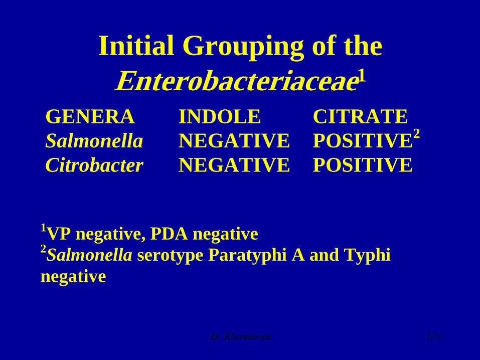
Initial Grouping of the
Enterobacteriaceae1
GENERA INDOLE CITRATE
Salmonella NEGATIVE POSITIVE2
Citrobacter NEGATIVE POSITIVE
1VP negative, PDA negative 2Salmonella serotype Paratyphi A and Typhi
negative
179 Dr. Khoramrooz

Key Characteristics of the
Enterobacteriaceae
TSI ON GAS H2S VP IND CIT PDA UR MO LYS OR AR
E
coli
A/A + + + + + +/
/
+ Shi
A-
C
Ak/
A /
+
Shi
D
Ak/
A + + Ed Ak/
A + + + + + + Sal Ak/
A + + + + + + +/
Cit A/A
Ak/
A
+ + + + +/
+ /
+
+/
Yer A/A
+ +/
+/
RT
(1) +
(1) RT=room temperature 180 Dr. Khoramrooz

Key Characteristics of the
Enterobacteriaceae
TSI ON GAS H2S VP IND CIT PDA UR MO LYS OR AR
Kle
pne
A/A + + + + + +
Kle
oxy
A/A + + + + + + +
En
aer
A/A + + + + + + +
En
cloa
A/A + + + + +/ + + +
Serr
(1)
A/A + + + + + + +
Haf Ak/
A + + + + + + Pan A/A
Alk/
A
+ /+ +/ /+ +/ /+ /+
(1) Produces DNase, lipase, and gelatinase 181 Dr. Khoramrooz

Key Characteristics of the
Enterobacteriaceae
TSI ON GAS H2S VP IND CIT PDA UR MO LYS OR AR
Prot
mir
a
Ak/
A + + +/ +/ + + +s +
Prot
vulg
A/A +/ + + /+ + + +s
Mor Ak/
A + + + + + + Pro
v
Ak/
A + + + + +
s = swarming motility
182 Dr. Khoramrooz

Biochemical Characteristics of
Escherichia coli and Shiglla
E. coli E. coli O157:H7 Shigella
TSI A/Ag A/Ag Alk/A
Lactose + + –
ONPG + + –/+1
Sorbitol + – +/–
Indole + + +/–
Methyl red + + +
VP – – –
Citrate – – –
Lysine + + –
Motility + + –
1Shigella sonnei (group D) ONPG +
183 Dr. Khoramrooz

Biochemical Characteristics of
Salmonella
Most Serotypes Typhi Paratyphi A
TSI Alk/A Alk/A Alk/A
H2S (TSI) + + (weak) –
Citrate + – –
Lysine + + –
Ornithine + – +
Dulcitol + – +
Rhamnose + – +
Indole – – –
Methyl red + + +
VP – – –
184 Dr. Khoramrooz

185 Dr. Khoramrooz

The End
186 Dr. Khoramrooz

t
Dr. Khoramrooz 187

Dr. Khoramrooz 188

Intended Use
Malonate Broth is used for differentiating Enterobacter from Escherichia based on malonate utilization.
Principles of the Procedure
Malonate Broth contains ammonium sulfate, which is the sole source of nitrogen in the medium;
Sodium malonate is the sole source of carbon.
Increased alkalinity resulting from malonate utilization causes the indicator, bromthymol blue, to change color from green to blue.
189 Dr. S. S. Khoramrooz

190 Dr. S. S. Khoramrooz

Procedure
1. Inoculate tubes with a loopful of test organism.
2. Incubate at 35 ― 2�‹C for 18-48 hours.
3. Examine tubes for a change in the color of the medium from green to blue.
Expected Results
Malonate utilization is indicated by a change in the color of the medium from green to blue:
Positive: Blue
Negative: Green
191 Dr. S. S. Khoramrooz

192 Dr. S. S. Khoramrooz

Intended Use
SS Agar and Salmonella Shigella Agar are
moderately selective and differential media for the
isolation of pathogenic enteric bacilli, especially
those belonging to the genus Salmonella.
This formulation is not recommended for the
primary isolation of Shigella.
193 Dr. S. S. Khoramrooz

Principles of the Procedure
SS Agar and Salmonella Shigella Agar are designated as moderately selective media based upon the degree of inhibition of gram-positive microorganisms that they inhibit due to their content of bile salts, brilliant green and citrates.
Differentiation of enteric organisms is achieved by the incorporation of lactose in the medium.
Organisms that ferment lactose produce acid which, in the presence of the neutral red indicator, results in the formation of red colonies.
Lactose nonfermenters form colorless colonies.
194 Dr. S. S. Khoramrooz

The latter group contains the majority of the
intestinal pathogens, including Salmonella and
Shigella.
The sodium thiosulfate and ferric citrate enable
the detection of hydrogen sulfide production as
evidenced by colonies with black centers.
195 Dr. S. S. Khoramrooz

Procedure
A nonselective medium should also be streaked to increase the chance of recovery when the population of gram-negative organisms is low and to provide an indication of other organisms present in the specimen.
Incubate plates, protected from light, at 35 ± 2°C for 18-24 hours.
If negative after 24 hours, reincubate an additional 24 hours.
196 Dr. S. S. Khoramrooz

Dr. S. S. Khoramrooz 197

Expected Results
Typical colonial morphology on Salmonella
Shigella Agar is as follows:
198 Dr. S. S. Khoramrooz

199 Dr. S. S. Khoramrooz

Limitation of the Procedure
Due to the relatively high level of selectivity, some
Shigella strains may not grow on SS Agar and
Salmonella Shigella Agar and, therefore, these
media are not recommended for the primary
isolation of Shigella.
Media recommended for the isolation of Shigella are Hektoen Enteric and XLD agars.
200 Dr. S. S. Khoramrooz

Intended Use
Selenite Broth (Selenite-F Broth) is used as an enrichment medium for the isolation of Salmonella from feces, urine, water, foods and other materials of sanitary importance.
Principles of the Procedure
The peptone provides essential nitrogenous and carbon compounds.
The lactose in the medium serves to maintain a uniform pH.
201 Dr. S. S. Khoramrooz

When selenite is reduced by the growth of bacteria, alkali is produced, and such increase in pH would lessen the toxicity of the selenite and result in overgrowth of extraneous bacteria.
The acid produced by lactose fermentation serves to maintain a neutral or slightly decreased pH.
The function of the phosphate is two-fold; it serves to maintain a stable pH and lessens the toxicity of the selenite, thus increasing the capacity of the medium.
Sodium selenite inhibits many species of grampositive and gram-negative bacteria including enterococci and coliforms.
202 Dr. S. S. Khoramrooz

203 Dr. S. S. Khoramrooz

Dr. S. S. Khoramrooz 204

Procedure
For feces and other solid materials, suspend 1-2 g of the specimen in the broth (approximately 10-15% by volume) and emulsify with an inoculating needle, if necessary.
Incubate tubes with loosened caps at 35 ± 2°C for up to 24 hours.
Subcultures should be made after 12-18 hours of incubation, if possible.
Coliforms will tend to overgrow the pathogens if incubated longer than 24 hours.
205 Dr. S. S. Khoramrooz

Expected Results
After incubation, there should be an increase in the
number of pathogens that the medium is designed
to select for and enrich.
Subculture onto appropriate selective and
differential media (e.g., MacConkey Agar,
Hektoen Enteric Agar, XLD Agar, XLT4 Agar,
CHROMagar™ Salmonella) to isolate
pathogens for identification.
206 Dr. S. S. Khoramrooz

Limitation of the Procedure
Enrichment broths should not be used as the sole
isolation medium.
They are to be used in conjunction with selective
and nonselective plating media to increase the
probability of isolating pathogens, especially when
they may be present in small numbers.
207 Dr. S. S. Khoramrooz

The End
208 Dr. Khoramrooz