PRIMARY TUMOURS OF CHILDREN* - Archives of...
7
PRIMARY TUMOURS OF THE LIVER IN INFANTS AND CHILDREN* BY H. WILLIAM CLATWORTHY, JR., E. THOMAS BOLES, JR. and WILLIAM A. NEWTON From the Departments of Surgery and Pathology, the Ohio State University Medical School and the Columbus Children's Hospital Primary liver tumours occur in infants and children with sufficient frequency to justify careful evaluation of current techniques in diagnosis and management and to review the pathological classification of this group of neoplasms. In the past nine years (1950-1958) 15 such cases have been diagnosed at the Columbus Children's Hospital, all presenting as clinical problems; an analysis of these cases forms the substance of this report. During the same period of time 46 cases of neuroblastoma and 17 cases of embryoma of the kidney (Wilms' tumour) have been treated at this institution. Although the treatment of liver tumours, as judged from a resume of individual case reports in recent years, has been encouraging, the overall results achieved in significant series have been disappointing and have perhaps fostered a generally hopeless outlook for young patients with such lesions (Donovan and Santulli, 1946; Wangensteen, 1951; Lortat-Jacob and Robert, 1952; Quattlebaum, 1953; Brunschwig, 1953; Pack and Baker, 1953; Lorimer, 1955; Clatworthy and Boles, 1956). In the series from Babies' Hospital in New York City analysed by Andersen (1951) there was only one long-term survivor in 12 cases occurring over a 15-year period. This patient remained well over a six-year follow-up period after excision of the left lobe of the liver had been performed for a mesen- chymoma. Gross, in a review at the Boston Children's Hospital, reports two apparent cures out of 18 such cases. One of these received radiotherapy for multiple haemangiomas and has been in excellent health for seven years thereafter; the second has been well over a 10-year period after having a wedge resection of a small haemartoma. Safe techniques for the resection of the left lobe of the liver have been available for many years, and * A paper read at a meeting of the British Association of Paediatric Surgeons held in Liverpool in June, 1959. there are numerous reported successful cases of this procedure applied to removal of neoplasms confined to this lobe. Most of these were performed on adult patients, but a number were in children. More recently the technique for total resection of the right lobe of the liver was evolved, whereby the dangers of haemorrhage, air embolism, bile peritonitis and subsequent liver failure are minimized (Wangen- steen, 1951; Lortat-Jacob and Robert, 1952; Brunschwig, 1953; Pack and Baker, 1953; Quattle- baum, 1953; Lorimer, 1955). The application of one or the other of these procedures should permit the surgical excision of most liver tumours which are not centrally placed or diffuse or multicentric lesions. Diagnosis The vast majority of primary neoplasms of the liver produce symptoms in the first two years of life. Only two of the 15 cases in this series had the onset of symptoms after 2 years of age, and in three instances the clinical manifestations were present in the neonatal period. Abdominal enlargement due to a palpable abdominal mass is by far the most common present- ing complaint. The distribution of these tumours in the liver, and hence the location of the mass, seems to follow the distribution of the liver mass. Of these 15 tumours, nine were in the right lobe, three in the left and two were situated centrally. One was diffuse. In most instances the mass was in the right upper quadrant, and less often in the mid-epigastrium. The mass moves with respirations. Its characteristics on palpation vary considerably, but most tumours are hard and rounded and inseparable from the liver edge. Transillumination is a useful tool in diagnosis since a liver tumour which transilluminates is almost certainly a large lymphangioma. Other clinical manifestations observed in this 22 copyright. on 14 July 2018 by guest. Protected by http://adc.bmj.com/ Arch Dis Child: first published as 10.1136/adc.35.179.22 on 1 February 1960. Downloaded from
Transcript of PRIMARY TUMOURS OF CHILDREN* - Archives of...

PRIMARY TUMOURS OF THE LIVER IN INFANTS ANDCHILDREN*
BY
H. WILLIAM CLATWORTHY, JR., E. THOMAS BOLES, JR. and WILLIAM A. NEWTONFrom the Departments of Surgery and Pathology, the Ohio State University Medical School and the Columbus
Children's Hospital
Primary liver tumours occur in infants andchildren with sufficient frequency to justify carefulevaluation of current techniques in diagnosis andmanagement and to review the pathologicalclassification of this group of neoplasms. In thepast nine years (1950-1958) 15 such cases have beendiagnosed at the Columbus Children's Hospital, allpresenting as clinical problems; an analysis of thesecases forms the substance of this report. Duringthe same period of time 46 cases of neuroblastomaand 17 cases of embryoma of the kidney (Wilms'tumour) have been treated at this institution.Although the treatment of liver tumours, as
judged from a resume of individual case reports inrecent years, has been encouraging, the overallresults achieved in significant series have beendisappointing and have perhaps fostered a generallyhopeless outlook for young patients with suchlesions (Donovan and Santulli, 1946; Wangensteen,1951; Lortat-Jacob and Robert, 1952; Quattlebaum,1953; Brunschwig, 1953; Pack and Baker, 1953;Lorimer, 1955; Clatworthy and Boles, 1956). Inthe series from Babies' Hospital in New York Cityanalysed by Andersen (1951) there was only onelong-term survivor in 12 cases occurring over a15-year period. This patient remained well over asix-year follow-up period after excision of the leftlobe of the liver had been performed for a mesen-chymoma. Gross, in a review at the BostonChildren's Hospital, reports two apparent cures outof 18 such cases. One of these received radiotherapyfor multiple haemangiomas and has been in excellenthealth for seven years thereafter; the second hasbeen well over a 10-year period after having a wedgeresection of a small haemartoma.
Safe techniques for the resection of the left lobeof the liver have been available for many years, and
* A paper read at a meeting of the British Association of PaediatricSurgeons held in Liverpool in June, 1959.
there are numerous reported successful cases of thisprocedure applied to removal of neoplasms confinedto this lobe. Most of these were performed on adultpatients, but a number were in children. Morerecently the technique for total resection of the rightlobe of the liver was evolved, whereby the dangersof haemorrhage, air embolism, bile peritonitis andsubsequent liver failure are minimized (Wangen-steen, 1951; Lortat-Jacob and Robert, 1952;Brunschwig, 1953; Pack and Baker, 1953; Quattle-baum, 1953; Lorimer, 1955). The application ofone or the other of these procedures should permitthe surgical excision of most liver tumours whichare not centrally placed or diffuse or multicentriclesions.
DiagnosisThe vast majority of primary neoplasms of the
liver produce symptoms in the first two years of life.Only two of the 15 cases in this series had the onsetof symptoms after 2 years of age, and in threeinstances the clinical manifestations were present inthe neonatal period.Abdominal enlargement due to a palpable
abdominal mass is by far the most common present-ing complaint. The distribution of these tumoursin the liver, and hence the location of the mass,seems to follow the distribution of the liver mass.Of these 15 tumours, nine were in the right lobe,three in the left and two were situated centrally.One was diffuse. In most instances the mass wasin the right upper quadrant, and less often in themid-epigastrium. The mass moves with respirations.Its characteristics on palpation vary considerably,but most tumours are hard and rounded andinseparable from the liver edge. Transilluminationis a useful tool in diagnosis since a liver tumourwhich transilluminates is almost certainly a largelymphangioma.
Other clinical manifestations observed in this22
copyright. on 14 July 2018 by guest. P
rotected byhttp://adc.bm
j.com/
Arch D
is Child: first published as 10.1136/adc.35.179.22 on 1 F
ebruary 1960. Dow
nloaded from

PRIMARY TUMOURS OF THE LIVER IN INFANTS AND CHILDREN
series include anorexia, fever, weight loss and rarelyjaundice. This latter sign was observed in only asingle case in this series, a 14-year-old girl who diedfrom a liver cell carcinoma associated with post-necrotic cirrhosis. In a single instance, heart failurein a newborn infant progressed to death at 8 weeks,with the pathological findings of diffuse haemangi-omas in the liver, skin and pancreas, as has beenpreviously described by Winters, Robinson andBates (1954) and by Levick and Rubie (1953).
Roentgenological studies are useful in distinguish-ing hepatic tumours from embryomas of the kidneyand neuroblastomas. An intravenous pyelogram isparticularly helpful. The kidneys function normallyand are usually in normal position in right-sidedtumours, although the pelvis and calices may becompressed. Distortion of the pelvis and calices,as seen with a Wilms' tumour, is not present, nor islateral displacement of the kidney, as with a neuro-blastoma, common. The mottled calcification socharacteristic of neuroblastoma was not observed,although calcification has been described in someliver tumours. Barium studies of the gastro-intestinal tract show displacement of these visceraaway from the tumour as one would expect. Down-ward displacement of the duodenum and hepaticflexure of the colon is seen with neoplasms of theright lobe, in contrast to the forward or medialdisplacement usually seen with a renal embryoma orneuroblastoma.Laboratory tests have been of little diagnostic
value in these cases. Liver function studies were allessentially normal except in the case of the childwith diffuse post-necrotic cirrhosis. Abnormalitiesin the liver function tests should lead one to questionthe diagnosis of primary liver tumour. For example,a diagnostic error was made in a newborn infant,admitted at 2 days of age with jaundice and a largeepigastric mass. The jaundice persisted as well asother laboratory evidence of disturbed liver function.At operation at 3 weeks of age the mass proved tobe a hard, nodular, grey-white "tumour' occupyingthe left lobe. Resection of the left lobe was per-formed and the child recovered without difficulty.However, on microscopical examination this provedto be a large cirrhotic nodule possibly associatedwith post-intrauterine hepatitis, with marked fibroustissue proliferation, replacing the normal architectureof the hepatic tissue. The hepatic cells were com-pletely or partially destroyed and showed irregulararrangement. The bile ducts appeared to beincreased. It is interesting that the jaundice dis-appeared within a month after operation and theinfant has been perfectly well over a subsequenteight-month period.
ClassificationThe wide variety of gross and microscopical
characteristics of primary liver tumours and therelatively small number of such cases collected at anyone institution have made the adoption of a uniformclassification difficult (Warvi, 1944; Edmondson,1956). To add to the problem is the fact that theclinical behaviour is not always correlated with themicroscopical appearance, so that a classificationbased on the pathological data alone appears to beunsatisfactory. For practical reasons it thereforeseems appropriate to group these tumours in threebroad categories:
1. Tumours of Hepatic Cell Origin. This groupis made up of all tumours derived from entodermalcells and includes hepatomas, cholangiomas and thecholangiohepatomas. There were no adenomas inthis series and the hepatomas have invariablybehaved as malignant lesions, although the histo-logical appearance may suggest benignity (Fig. laand b).
2. Tumours of Supporting Structures. Thisgroup comprises those tumours of mesodermal originincluding fibromas, haemangiomas, lymphangiomas,fibroangiomas and mesenchymomas. Such neo-plasms may be benign, as are most of those in thisseries, or malignant.
3. Mixed Tumours and Teratomas. Such tumourscontain cells derived from more than one germ layer.Osteoid tissue, for example, may be seen in liver celltumours. Teratomas are distinctly rare (Fig. 2).
In this series hepatoma was the most commontumour encountered, occurring in 10 cases. Fivewere in the right lobe, two in the left and two werecentrally positioned. In eight cases symptomsdeveloped in the first two years of life, and the ninthpatient had his initial symptoms at 2 years 11 monthsof age. None of these nine patients had associatedcirrhosis so commonly found in adult cases. How-ever, as already mentioned, the tenth case occurredin a 14-year-old girl with post-necrotic cirrhosis ofthe liver. There were no instances of adenomas orexclusively bile duct tumours among these cases andonly a single case of histologically benign cholangio-hepatoma occurring in the right lobe of a 10-week-old infant.Among the tumours of supporting tissue origin
were one diffuse haemangioma, two lymphangiomasand one fibroangioma. There were no mesenchy-momas, mixed tumours or teratomas. Both
23
copyright. on 14 July 2018 by guest. P
rotected byhttp://adc.bm
j.com/
Arch D
is Child: first published as 10.1136/adc.35.179.22 on 1 F
ebruary 1960. Dow
nloaded from

24 ARCHIVES OF DISEASE IN CHILDHOOD
FIG. I (a).-Gross and microscopical appearances of a well-differ-entiated hepatoma of left lobe of liver successfully excised from a15-month-old infant who subsequently died of undifferentiatedhepatic and pulmonary metastases seven months after original
resection.
I. .. .-- --.. .. .... ...
FIG. I (b).-Gross and microscopical appearances of a very rapidlygrowing anaplastic hepatoma of left lobe of liver easily resected withwide margin of normal liver substance in a 2-year-old infant whosuccunmbed to multiple recurrences in right lobe of liver within five
months.
copyright. on 14 July 2018 by guest. P
rotected byhttp://adc.bm
j.com/
Arch D
is Child: first published as 10.1136/adc.35.179.22 on 1 F
ebruary 1960. Dow
nloaded from
PRIMARY TUMOURS OF THE LIVER IN INFANTS AND CHILDREN 25
FIG. 2.-Gross and microscopical appearances of mixed tumour ofliver in a young child; it contained highly undifferentiated hepatoma
as well as areas of osteoid tissue.
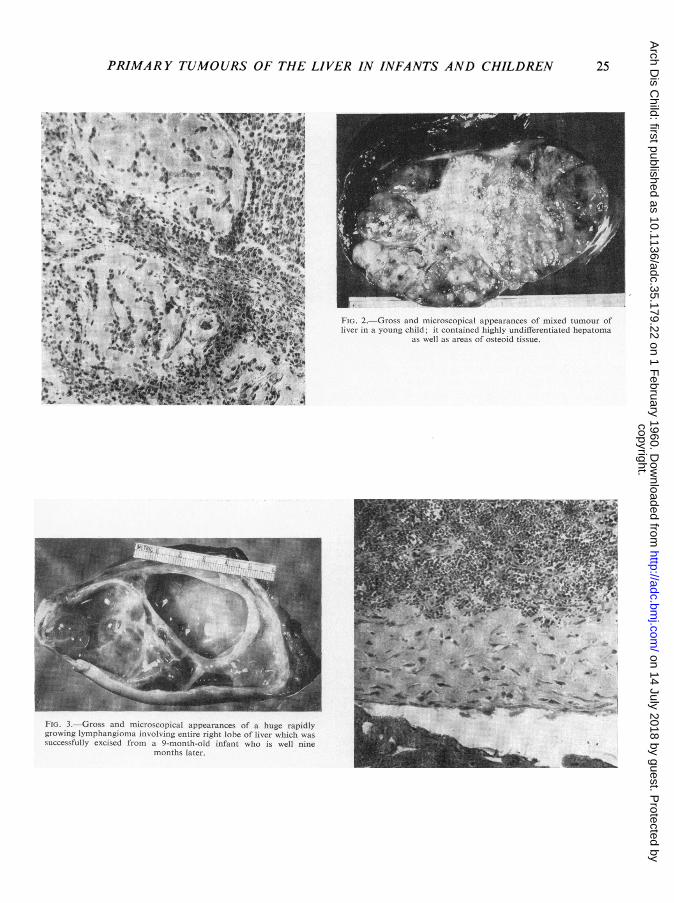
FrG. 3.-Gross and microscopical appearances of a huge rapidlygrowing lymphangioma involving entire right lobe of liver which wassuccessfully excised from a 9-month-old infant who is well nine
months later.
copyright. on 14 July 2018 by guest. P
rotected byhttp://adc.bm
j.com/
Arch D
is Child: first published as 10.1136/adc.35.179.22 on 1 F
ebruary 1960. Dow
nloaded from
ARCHIVES OF DISEASE IN CHILDHOOD
lymphangiomas were found in the right lobe ofinfants, one 41 months and the other 9 months ofage (Fig. 3). The patient with diffuse haemangio-matosis, already noted, had symptoms at birth. Theliver of this patient had numerous small capillaryhaemangiomas scattered diffusely throughout allportions of the liver parenchyma. The single caseof fibroangioma was a newborn infant with theneoplasm originating in the left lobe.There is a strikingly similar gross appearance of
many of these tumours, so that differentiation by thismeans alone is often quite unreliable. Similarly, thedifferentiation microscopically of benign andmalignant liver cell tumours is occasionally virtuallyimpossible. In one of the hepatomas in this seriesthe cytology and architecture were most suggestiveofa benign neoplasm, but the presence of metastasesestablished the malignant nature of the tumour.
TreatmentEffective treatment of primary hepatic neoplasms
is at present limited almost entirely to surgicalresection. Radiotherapy may be of value inhaemangiomas, as suggested by Gross, and in oneof our cases of inoperable hepatoma radiotherapyresulted in definite regression in size of the tumourmass. Systemic chemotherapy has not been of valueto date.
In tumours limited to either the right or left lobe,surgical removal by total right or left lobectomy isa practical procedure. Such procedures takeadvantage of the anatomical division of the organinto right and left lobes, each with a largely separateblood supply from the hepatic artery and portalvein. Control of the blood supply and the bile ductsystem at the hilum of the liver plus similar controlof the hepatic veins draining into the inferior venacava make possible such resections without excessiverisk from haemorrhage, air embolism, or bileperitonitis.
In such a procedure for a tumour involving theright lobe the diagnosis is confirmed and operabilitydetermined through an abdominal incision. Ifresection appears possible the abdominal incisionis joined or extended by an incision into the rightchest through the seventh or eighth intercostal space,and the right diaphragm is divided down to the venacava. Umbilical tapes are then placed around theinferior vena cava just below the right atrium andabove the renal veins. The hilar structures are freedand the appropriate branches of the hepatic artery,portal vein and hepatic duct are carefully identified,ligated and divided. The short hepatic veins fromthe right lobe to the vena cava are next ligated anddivided. This is the most dangerous part of the
procedure since extension of the tumour into thisarea may not be appreciated until this part of thedissection is under way, and if such extension isencountered there is a considerable risk of haemor-rhage or air embolism. The previously placed tapeson the vena cava and control of the hilar structurespermit control of such a situation. Following thisthe liver is transected to the right of the falciformligament down to the vena cava. During the actualtransection the hilar structures are cross-clampedwith a Pott's clamp. Interruption of the bloodsupply by this means can safely be tolerated by theliver for about 15 minutes, and probably longerwith the controlled moderate hypothermia (90°-92° F.) customarily employed in these cases. Allvisible blood vessels and bile ducts are ligated as thedissection proceeds. If the gallbladder is not in-volved, it may be peeled out of its bed before thetransection and used after removal of the lobe forretrograde injection of a methylene blue solution.By this means additional bile ducts can be visualizedand ligated. The falciform ligament and posteriorperitoneum are used to reperitonealize the rawsurface. If this is not sufficient, a layer of com-pressed Ivalon may be sutured in place to cover theremaining raw area. Penrose drains are placed todrain the right subdiaphragmatic space so that alocalized collection of bile or a bile peritonitis willnot occur.The left lobe can be mobilized more easily than
the right and this can ordinarily be done through anupper abdominal incision by dividing the peritonealattachment to the diaphragm and the falciformligament. It is quite easy to place a row of inter-locking mattress sutures just to the left of thefalciform ligament and resect the left lobe. It hasnot been found necessary to perform a hilar dis-section or to occlude the hilar structures during thisprocedure. Again, however, reperitonealization ofthe raw surface and drainage are important steps.
ResultsThere have been no long-term survivors in the
10 patients with hepatoma. In one, the diagnosiswas not made until post-mortem examination andno treatment was given. In two, only explorationand biopsy were performed, and both died withinthree months. In two others, exploration andbiopsy were followed by radiotherapy. The tumourmass strikingly decreased in size in one of these,but none the less the patient lived only six monthsafter completion of the treatment. The other livedonly four months after therapy.
Right lobectomy was performed in four patientsand there were three early deaths. One of these
26
copyright. on 14 July 2018 by guest. P
rotected byhttp://adc.bm
j.com/
Arch D
is Child: first published as 10.1136/adc.35.179.22 on 1 F
ebruary 1960. Dow
nloaded from

PRIMARY TUMOURS OF THE LIVER IN INFANTS AND CHILDREN
patients died from operative haemorrhage, and asecond from haemorrhage and air embolism. Athird unfortunate death resulted from staphylococcalperitonitis and septicaemia two weeks after opera-tion and at autopsy no tumour was found. Allthree deaths must be considered preventable. Thefirst two cases were in retrospect inoperable becauseof tumour invasion across the midline posteriorlyin the area of the hepatic veins. The third deathpresumably followed contamination in the operatingroom or via the drain site. The fourth patient whohad a resection of the right lobe died at hometwo and a half months after operation following abrief acute illness suggestive of intestinal obstruc-tion. The patient was not returned to the hospitalduring this illness and no autopsy was performed.A left lobectomy was performed in the final patient,and he died with massive metastases in the rightlobe seven months after resection. This last patienthad been known by his parents to have an abdominallump since 3 months of age, but no treatment wassought until 15 months later at which time a hugetumour in the left lobe was found.The single patientwith a cholangio-hepatoma had a
resection of the right lobe of the liver at 10 weeksof age (Fig. 4). He is living and well 12 monthslater with no evidence of recurrence.Both patients with giant lymphangiomas had
tumours occupying almost all of the right lobe.Resections of the right lobe were done at 5 and9 months, and both patients are in good health
FIG. 4.-A 10-week-old infant one week after total right hepatectomyfor cholangio-hepatoma.
FIG. 5.-Microscopical appearance of liver removed at autopsy froman 8-week-old infant with multiple diffuse haemangiomas who had
been under treatment since birth for 'heart failure'.
six and a half years and nine months later. Theinfant with a fibroangioma of the left lobe had aleft hepatectomy at 3 weeks of age, and is well14 months later.None of the long-term survivors after resection
of the right or left lobe shows any evidence ofimpairment of liver function. Although in all casesthere was a plateau in weight gain for a few weeksafter operation, subsequent growth and develop-ment patterns have been entirely normal.The patient with diffuse haemangiomas of the
liver, as well as of the pancreas and skin, was treatedfor heart failure from birth and died at 8 weeks ofage. The diagnosis was made from the autopsyfindings (Fig. 5).
DiscussionPrimary liver tumours are not rare paediatric
problems. Without treatment the prognosis is poorin both the malignant and benign varieties, asindicated by the larger reported series (Andersen,1951; Gross, 1953; Lee, Newstedt and Siddall,1956). With adequate surgical resection the prog-nosis in cases of benign tumours is good, and aftersuch treatment normal growth and freedom fromliver impairment may be expected. The outlookfor patients with malignant tumours, even afteroperation, has thus far been poor, but with the
27
copyright. on 14 July 2018 by guest. P
rotected byhttp://adc.bm
j.com/
Arch D
is Child: first published as 10.1136/adc.35.179.22 on 1 F
ebruary 1960. Dow
nloaded from

28 ARCHIVES OF DISEASE IN CHILDHOOD
development of more adequate techniques inachieving wide resections there is reason to believethat success may be achieved. One remarkableexception is the case reported by Lee et al. (1956)of an infant with a huge hepatoma confirmed bybiopsy. No treatment was given but spontaneousregression occurred and the child was reported ingood health 10 years later.
Establishing a clinical diagnosis of a primary livertumour is ordinarily not difficult; however, thedifferentiation of a benign from a malignant tumourclinically or at operation may be quite impossible.Two such dilemmas occurred in this series. Sincesurgical excision currently affords the only chanceof eradicating such lesions, it is our opinion that inthe absence of distant metastases isolated lesionsshould be removed.The techniques of resection of either the right or
the left lobe are sufficiently well developed to permittheir application with a reasonable assurance ofsuccess. The major problems are haemorrhage, airembolism and bile peritonitis. An anatomicaltechnique with control of the afferent and efferentblood supply, controlled hypothermia, reperitoneal-ization of the cut surface and adequate drainageare methods which contribute to the success of theseprocedures.
SummaryThe majority of primary liver tumours which
present as clinical problems in children are malignantand occur in the first two years of life. Such tumoursdevelop in a normal liver, and are not super-imposed on a cirrhotic process as in adults.From the practical standpoint these neoplasms
can be classified as (1) tumours derived from livercells, (2) tumours derived from supporting cells,
and (3) mixed tumours. Those derived from livercells are malignant in the vast majority of cases,although the microscopical picture of some is quitebenign.The solid tumours of the liver should be treated
in a uniform manner, adequate surgical excisionthrough normal liver tissues being the best availableprocedure.A surgical technique based on sound anatomical
principles and planned to minimize the risks ofhaemorrhage, air embolism and bile peritonitismakes removal of these tumours by a resection ofeither the right or left hepatic lobes a logical andpractical procedure.
REFERENCESAndersen, Dorothy H. (1951). Tumours of Infancy and Childhood.
Cancer, 4, 890.Brunschwig, A. (1953). Surgery of Hepatic Neoplasms. Cancer, 6,
725.Clatworthy, H. W., Jr. and Boles, E. T. Jr. (1956). Right Lobectomy
of the Liver in Children. Surgery, 39, 850.Donovan, E. J. and Santulli, T. V. (1946). Resection of the Left Lobe
of the Liver for Mesenchymoma. Ann. Surg., 124, 90.Edmondson, H. A. (1956). Differential Diagnosis of Tumors and
Tumor-like Lesions ofLiver in Infancy and Childhood. A.M.A.J. Dis. Child., 91, 168.
Gross, Robert E. (1953). The Surgery of Infancy and Childhood.p. 532. Saunders & Co., Philadelphia.
Lee, C. M. Jr., Newstedt, J. R. and Siddall, H. S. (1956). LargeAbdominal Tumors of Childhood (Other than Wilms' Tumoror Neuroblastoma). Ann. Surg., 143, 803.
Levick, C. B. and Rubie, J. (1953). Haemangioendothelioma of theLiver Simulating Congenital Heart Disease in an Infant. Arch.Dis. Childh., 28, 49.
Lorimer, W. S. Jr. (1955). Right Hepatolobectomy for PrimaryMesenchymoma of the Liver. Ann. Surg., 141, 246.
Lortat-Jacob, J. L. and Robert, H. G. (1952). H6patectomie droitereglee. Presse med., 60, 549.
Pack, G. T. and Baker, H. W. (1953). Total Right Hepatic Lobec-tomy. Ann. Surg., 138, 253.
Quattlebaum, J. K. (1953). Massive Resection of the Liver, Ann.Surg., 137, 787.
Wangensteen, 0. H. (1951). Cancer of the Esophagus and theStomach. American Cancer Society, Inc., p. 92.
Warvi, W. N. (1944). Primary Neoplasms of the Liver. Arch. Path.,37, 367.
Winters, R. W., Robinson, S. J. and Bates, G. (1954). Hemangiomaof the Liver with Heart Failure: A Case Report. Pediatrics,14, 117.
copyright. on 14 July 2018 by guest. P
rotected byhttp://adc.bm
j.com/
Arch D
is Child: first published as 10.1136/adc.35.179.22 on 1 F
ebruary 1960. Dow
nloaded from