Primary Shoot -Stem Structure - Amazon...
7
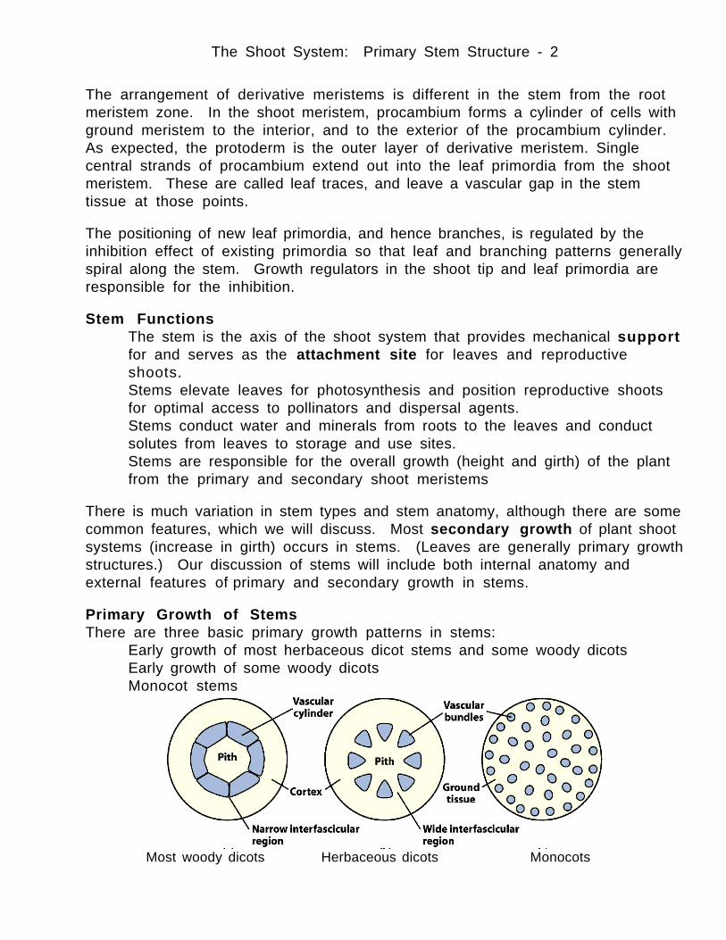
The Shoot System: Primary Stem Structure - 1 Shoot System The shoot system comprises the leaves and stems of plants. Leaves are located at nodes on the stem; the distance along the stem between nodes is known as an internode. Shoots develop from shoot meristem, which contains the apical shoot tip meristem from the epicotyl of the embryo, leaf primordia, and bud primordia, which are embryonic lateral shoot systems. Flowers, the reproductive organs of angiosperms, are modified shoots. Shoot meristems are located at the growing tips of stems, and in buds. Shoot Meristems The apical meristem is divided into two regions: tunica and corpus. The tunica meristem cells divided in a plane the produces additional surface meristem. The corpus meristem cells under the tunica divide in a plane that adds bulk to the shoot meristem. Generally there are two tunica layers and one corpus layer. As a shoot grows, buds are laid down by shoot meristem in the axils of leaf primordia. The repeating units of leaf and bud primordia are called phytomeres . These buds are dormant meristems that are activated at some later time in growth. Stem tissues are produced from the same three derivative meristems as root tissues are: Protoderm is responsible for Epidermis Ground Meristem differentiates into ground tissues Procambium produces the vascular tissues Coleus stem tip, l.s. Shoot tip, xs SEM of shoot tip
Transcript of Primary Shoot -Stem Structure - Amazon...
The Shoot System: Primary Stem Structure - 1
Shoot SystemThe shoot system comprises the leaves and stems of plants. Leaves are locatedat nodes on the stem; the distance along the stem between nodes is known as aninternode. Shoots develop from shoot meristem, which contains the apical shoottip meristem from the epicotyl of the embryo, leaf primordia, and bud primordia,which are embryonic lateral shoot systems. Flowers, the reproductive organs ofangiosperms, are modified shoots. Shoot meristems are located at the growingtips of stems, and in buds.
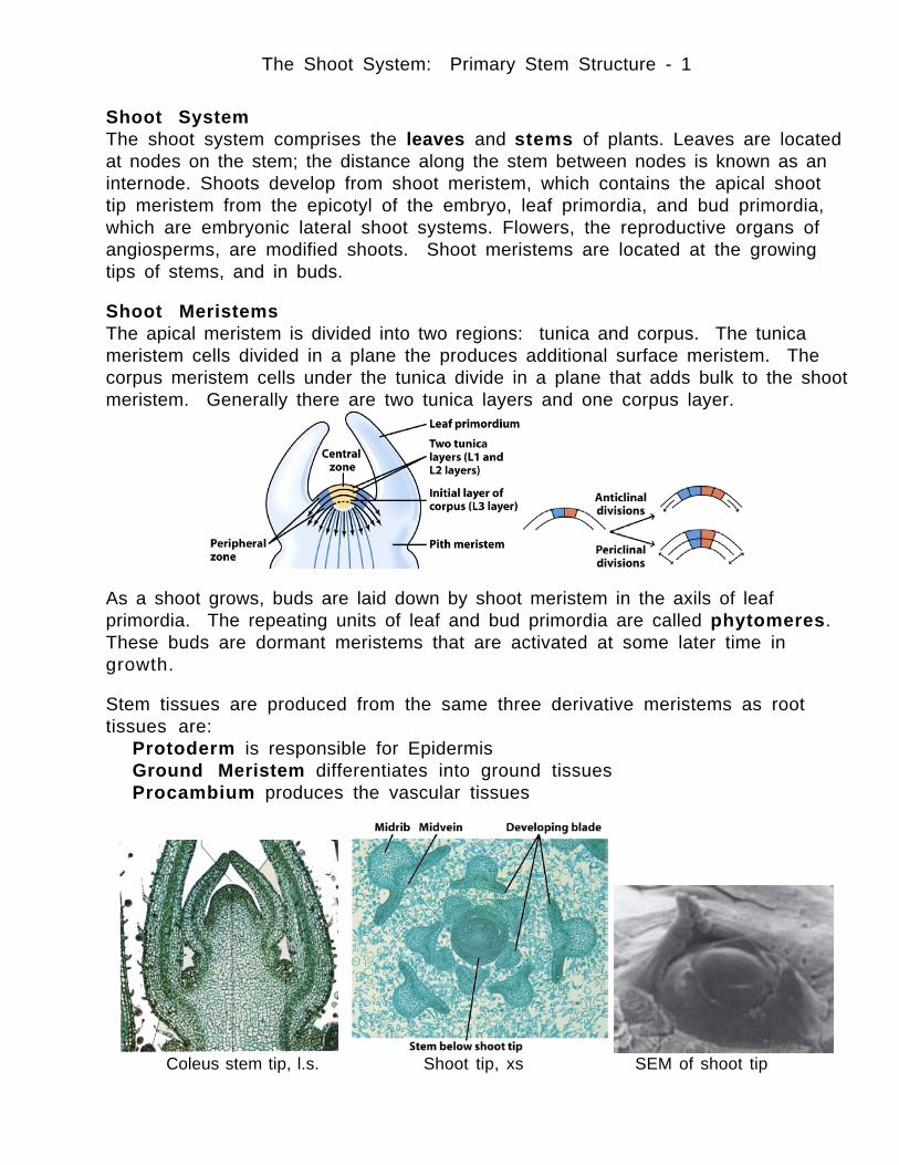
Shoot MeristemsThe apical meristem is divided into two regions: tunica and corpus. The tunicameristem cells divided in a plane the produces additional surface meristem. Thecorpus meristem cells under the tunica divide in a plane that adds bulk to the shootmeristem. Generally there are two tunica layers and one corpus layer.
As a shoot grows, buds are laid down by shoot meristem in the axils of leafprimordia. The repeating units of leaf and bud primordia are called phytomeres.These buds are dormant meristems that are activated at some later time ingrowth.
Stem tissues are produced from the same three derivative meristems as roottissues are:
Protoderm is responsible for EpidermisGround Meristem differentiates into ground tissuesProcambium produces the vascular tissues
Coleus stem tip, l.s. Shoot tip, xs SEM of shoot tip
The Shoot System: Primary Stem Structure - 2
The arrangement of derivative meristems is different in the stem from the rootmeristem zone. In the shoot meristem, procambium forms a cylinder of cells withground meristem to the interior, and to the exterior of the procambium cylinder.As expected, the protoderm is the outer layer of derivative meristem. Singlecentral strands of procambium extend out into the leaf primordia from the shootmeristem. These are called leaf traces, and leave a vascular gap in the stemtissue at those points.
The positioning of new leaf primordia, and hence branches, is regulated by theinhibition effect of existing primordia so that leaf and branching patterns generallyspiral along the stem. Growth regulators in the shoot tip and leaf primordia areresponsible for the inhibition.
Stem Functions• The stem is the axis of the shoot system that provides mechanical support
for and serves as the attachment site for leaves and reproductiveshoots.
• Stems elevate leaves for photosynthesis and position reproductive shootsfor optimal access to pollinators and dispersal agents.
• Stems conduct water and minerals from roots to the leaves and conductsolutes from leaves to storage and use sites.
• Stems are responsible for the overall growth (height and girth) of the plantfrom the primary and secondary shoot meristems
There is much variation in stem types and stem anatomy, although there are somecommon features, which we will discuss. Most secondary growth of plant shootsystems (increase in girth) occurs in stems. (Leaves are generally primary growthstructures.) Our discussion of stems will include both internal anatomy andexternal features of primary and secondary growth in stems.
Primary Growth of StemsThere are three basic primary growth patterns in stems:
• Early growth of most herbaceous dicot stems and some woody dicots• Early growth of some woody dicots• Monocot stems
Most woody dicots Herbaceous dicots Monocots
The Shoot System: Primary Stem Structure - 3
While the arrangement of tissues in stems differs from roots, the tissues are thesame, and by now, should be becoming familiar.
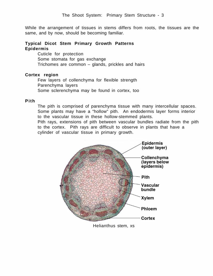
Typical Dicot Stem Primary Growth PatternsEpidermis
• Cuticle for protection• Some stomata for gas exchange• Trichomes are common – glands, prickles and hairs
Cortex region• Few layers of collenchyma for flexible strength• Parenchyma layers• Some sclerenchyma may be found in cortex, too
Pith• The pith is comprised of parenchyma tissue with many intercellular spaces.• Some plants may have a “hollow” pith. An endodermis layer forms interior
to the vascular tissue in these hollow-stemmed plants.• Pith rays, extensions of pith between vascular bundles radiate from the pith
to the cortex. Pith rays are difficult to observe in plants that have acylinder of vascular tissue in primary growth.
Helianthus stem, xs
The Shoot System: Primary Stem Structure - 4
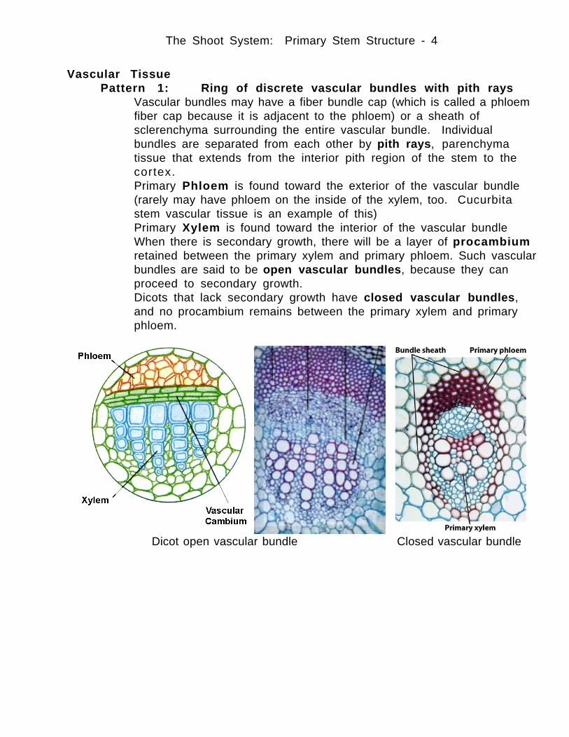
Vascular TissuePattern 1: Ring of discrete vascular bundles with pith rays
• Vascular bundles may have a fiber bundle cap (which is called a phloemfiber cap because it is adjacent to the phloem) or a sheath ofsclerenchyma surrounding the entire vascular bundle. Individualbundles are separated from each other by pith rays, parenchymatissue that extends from the interior pith region of the stem to thecortex.
• Primary Phloem is found toward the exterior of the vascular bundle(rarely may have phloem on the inside of the xylem, too. Cucurbitastem vascular tissue is an example of this)
• Primary Xylem is found toward the interior of the vascular bundle• When there is secondary growth, there will be a layer of procambium
retained between the primary xylem and primary phloem. Such vascularbundles are said to be open vascular bundles, because they canproceed to secondary growth.
• Dicots that lack secondary growth have closed vascular bundles,and no procambium remains between the primary xylem and primaryphloem.
Dicot open vascular bundle Closed vascular bundle
The Shoot System: Primary Stem Structure - 5
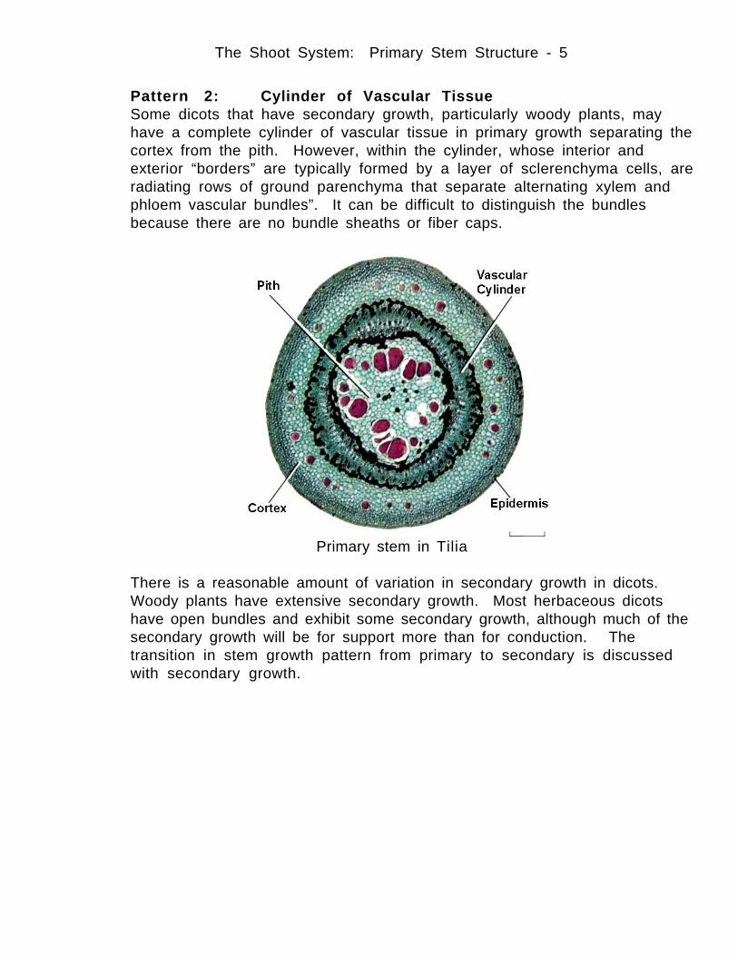
Pattern 2: Cylinder of Vascular TissueSome dicots that have secondary growth, particularly woody plants, mayhave a complete cylinder of vascular tissue in primary growth separating thecortex from the pith. However, within the cylinder, whose interior andexterior “borders” are typically formed by a layer of sclerenchyma cells, areradiating rows of ground parenchyma that separate alternating xylem andphloem vascular bundles”. It can be difficult to distinguish the bundlesbecause there are no bundle sheaths or fiber caps.
Primary stem in Tilia
There is a reasonable amount of variation in secondary growth in dicots.Woody plants have extensive secondary growth. Most herbaceous dicotshave open bundles and exhibit some secondary growth, although much of thesecondary growth will be for support more than for conduction. Thetransition in stem growth pattern from primary to secondary is discussedwith secondary growth.
The Shoot System: Primary Stem Structure - 6
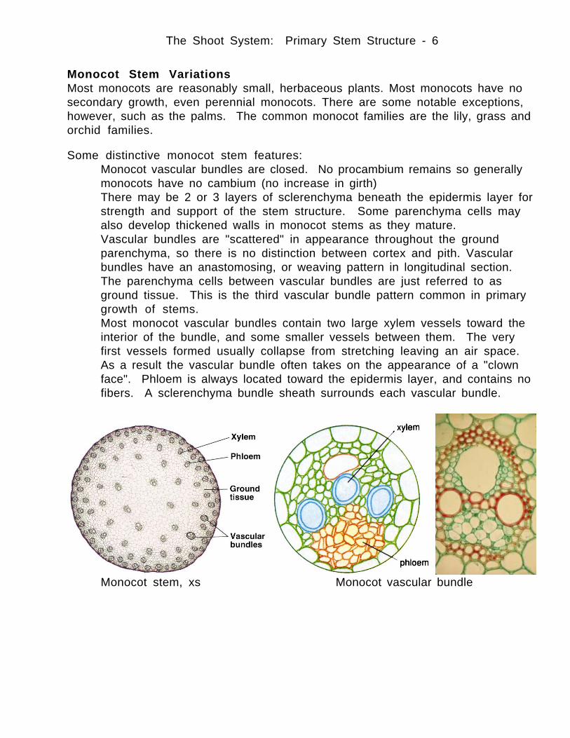
Monocot Stem VariationsMost monocots are reasonably small, herbaceous plants. Most monocots have nosecondary growth, even perennial monocots. There are some notable exceptions,however, such as the palms. The common monocot families are the lily, grass andorchid families.
Some distinctive monocot stem features:• Monocot vascular bundles are closed. No procambium remains so generally
monocots have no cambium (no increase in girth)• There may be 2 or 3 layers of sclerenchyma beneath the epidermis layer for
strength and support of the stem structure. Some parenchyma cells mayalso develop thickened walls in monocot stems as they mature.
• Vascular bundles are "scattered" in appearance throughout the groundparenchyma, so there is no distinction between cortex and pith. Vascularbundles have an anastomosing, or weaving pattern in longitudinal section.The parenchyma cells between vascular bundles are just referred to asground tissue. This is the third vascular bundle pattern common in primarygrowth of stems.
• Most monocot vascular bundles contain two large xylem vessels toward theinterior of the bundle, and some smaller vessels between them. The veryfirst vessels formed usually collapse from stretching leaving an air space.As a result the vascular bundle often takes on the appearance of a "clownface". Phloem is always located toward the epidermis layer, and contains nofibers. A sclerenchyma bundle sheath surrounds each vascular bundle.
Monocot stem, xs Monocot vascular bundle
The Shoot System: Primary Stem Structure - 7
Special Variations in some MonocotsHollow StemsA central pith cavity develops from cells of the ground tissue that are destroyedduring early growth. This gives an appearance of a ring of vascular bundles, similarto dicot patterns.
Intercalary MeristemsMany grasses have meristem layers at the bases of nodes, which provide for non-apical growth and enlargement of cells throughout the plant stems. (Which is whywe have to mow lawns).
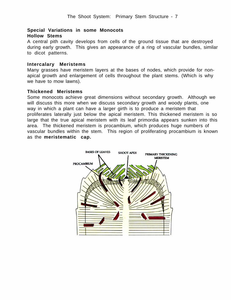
Thickened MeristemsSome monocots achieve great dimensions without secondary growth. Although wewill discuss this more when we discuss secondary growth and woody plants, oneway in which a plant can have a larger girth is to produce a meristem thatproliferates laterally just below the apical meristem. This thickened meristem is solarge that the true apical meristem with its leaf primordia appears sunken into thisarea. The thickened meristem is procambium, which produces huge numbers ofvascular bundles within the stem. This region of proliferating procambium is knownas the meristematic cap.


![[PPT]Shoot House Slideshow Presentation - Pennsylvaniaftig.png.pa.gov/Training/Documents/Shoot House/Shoot... · Web viewCAPABILITIES two story enclosed shoot house constructed of](https://static.fdocuments.us/doc/165x107/5ae5190a7f8b9a495c8f743e/pptshoot-house-slideshow-presentation-houseshootweb-viewcapabilities-two.jpg)