PRIMARY LYMPHOSARCOMA OF THE - thorax.bmj.comthorax.bmj.com/content/thoraxjnl/17/3/190.full.pdf ·...
11
Thorax (1962), 17, 190. PRIMARY LYMPHOSARCOMA OF THE LUNG BY C. W. H. HAVARD, J. B. NICHOLS, AND A. G. STANSFELD From the Departments of Medicine and Pathology, St. Bartholomew's Hospital, London (RECEIVED FOR PUBLICATION NOVEMBER 28, 1961) Pulmonary involvement in disseminated lympho- sarcoma is comparatively common. Vieta and Craver (1941) found an incidence of nearly 25% by radiographic assessment in a series of 239 patients with lymphosarcoma* and a similar proportion of a smaller number coming to necropsy. This figure agrees closely with the post-mortem incidence reported by Robbins (1953) in an even larger series. More recently, Rosenberg, Diamond, Jaslowitz, and Craver (1961) found approximately 40% with lung involvement in a necropsy series of 277 cases of lymphosarcoma. At St. Bartholomew's Hospital, 86 patients with histologically proven lymphosarcomat have been admitted over the last ten years. In 12 of these patients there was radiographic evidence of pulmonary involvement. Primary lymphosarcoma of the lung is, on the other hand, a rare disease. Sugarbaker and Craver (1940) found only one instance of a primary lung tumour in a series of 196 cases of histologically proven lymphosarcoma. Its importance, however, far outweighs the incidence, for it is a slowly growing tumour which tends to remain localized for a long time, spreading by direct extension into neighbouring structures, rather than metastasizing by the blood or lymph streams. The tumour is therefore often amenable to complete surgical excision. The diagnosis of this disease is difficult, for symptoms are usually mild or absent and physical examination will at the most reveal signs of consolidation. The radiographic picture is non- specific and in most instances has given rise to a suspicion of bronchial carcinoma. Baron and Whitehouse (1961) have, however, emphasized the patency of the bronchi in pulmonary lympho- sarcoma and have drawn attention to radiographic appearances which they describe as an 'air bronchogram.' Bronchoscopy has seldom proved helpful in diagnosis in spite of the frequent * The term is used in the general sense by these authors to include all types of " malignant lymphoma." t The term is here used in the restricted sense to mean a tumour of lymphoid cells. infiltration of bronchial walls by the tumour. Cytological examination of the sputum has on at least one occasion led to a pre-operative diagnosis (Jackson, Bertoli, and Ackerman, 1951), but usually thoracotomy and lung biopsy are necessary to establish the diagnosis. Reports of about 50 cases of primary lympho- sarcoma of the lung have been published, although in some of these the widespread dissemination of the disease makes the pulmonary origin uncertain. Recent reviews have appeared in the American literature (Cooley, McDonald and Clagett, 1956; Rose, 1957; Sternberg, Sidransky, and Ochsner, 1959; Baron and Whitehouse, 1961) and sub- sequent cases have been reported by Prichard and Bradshaw (1961). Although Willis (1960) briefly mentions a case of pulmonary lymphosarcoma, no detailed report of these tumours has appeared in the British literature. For this reason our account of the clinical and pathological features of three further cases of primary lymphosarcoma of the lung is presented. CASE REPORTS CASE 1.-J. H. is an engineer aged 48 years. In April, 1954, he developed pleurisy and a radiograph two months later revealed an area of consolidation in the right lower lobe (Fig. 1). He was admitted to hospital in November, 1954. At this time he did not complain of any symptoms although he admitted losing 11 lb. (5 kg.) in weight over the previous year. There were no physical signs of disease apart from a small mobile mass in the right axilla which was attached to the skin but not to deeper structures. It had been present for six years, according to the patient, and was considered to be a sebaceous cyst. The haemoglobin was 15.7 g. per 100 ml. Leucocytes numbered 8,400 per c.mm., of which 73% were neutrophils, 24% lymphocytes, and 3% monocytes. The E.S.R. was 6 mm./hr. (Westergren). Examina- tion of the sputum failed to show acid-fast bacilli or evidence of fungal infection. Bronchoscopy was normal. In view of the fact that the patient had been taking oily nasal drops for many years on account of nasal catarrh, a diagnosis of lipoid pneumonia was on 7 July 2018 by guest. Protected by copyright. http://thorax.bmj.com/ Thorax: first published as 10.1136/thx.17.3.190 on 1 September 1962. Downloaded from
Transcript of PRIMARY LYMPHOSARCOMA OF THE - thorax.bmj.comthorax.bmj.com/content/thoraxjnl/17/3/190.full.pdf ·...

Thorax (1962), 17, 190.
PRIMARY LYMPHOSARCOMA OF THE LUNGBY
C. W. H. HAVARD, J. B. NICHOLS, AND A. G. STANSFELDFrom the Departments of Medicine and Pathology, St. Bartholomew's Hospital, London
(RECEIVED FOR PUBLICATION NOVEMBER 28, 1961)
Pulmonary involvement in disseminated lympho-sarcoma is comparatively common. Vieta andCraver (1941) found an incidence of nearly 25%by radiographic assessment in a series of 239patients with lymphosarcoma* and a similarproportion of a smaller number coming tonecropsy. This figure agrees closely with thepost-mortem incidence reported by Robbins (1953)in an even larger series. More recently, Rosenberg,Diamond, Jaslowitz, and Craver (1961) foundapproximately 40% with lung involvement in anecropsy series of 277 cases of lymphosarcoma.At St. Bartholomew's Hospital, 86 patients withhistologically proven lymphosarcomat have beenadmitted over the last ten years. In 12 of thesepatients there was radiographic evidence ofpulmonary involvement.Primary lymphosarcoma of the lung is, on the
other hand, a rare disease. Sugarbaker and Craver(1940) found only one instance of a primary lungtumour in a series of 196 cases of histologicallyproven lymphosarcoma. Its importance, however,far outweighs the incidence, for it is a slowlygrowing tumour which tends to remain localizedfor a long time, spreading by direct extension intoneighbouring structures, rather than metastasizingby the blood or lymph streams. The tumour istherefore often amenable to complete surgicalexcision.The diagnosis of this disease is difficult, for
symptoms are usually mild or absent and physicalexamination will at the most reveal signs ofconsolidation. The radiographic picture is non-specific and in most instances has given rise toa suspicion of bronchial carcinoma. Baron andWhitehouse (1961) have, however, emphasized thepatency of the bronchi in pulmonary lympho-sarcoma and have drawn attention to radiographicappearances which they describe as an 'airbronchogram.' Bronchoscopy has seldom provedhelpful in diagnosis in spite of the frequent
* The term is used in the general sense by these authors toinclude all types of " malignant lymphoma."
t The term is here used in the restricted sense to mean a tumourof lymphoid cells.
infiltration of bronchial walls by the tumour.Cytological examination of the sputum has onat least one occasion led to a pre-operativediagnosis (Jackson, Bertoli, and Ackerman, 1951),but usually thoracotomy and lung biopsy arenecessary to establish the diagnosis.
Reports of about 50 cases of primary lympho-sarcoma of the lung have been published, althoughin some of these the widespread dissemination ofthe disease makes the pulmonary origin uncertain.Recent reviews have appeared in the Americanliterature (Cooley, McDonald and Clagett, 1956;Rose, 1957; Sternberg, Sidransky, and Ochsner,1959; Baron and Whitehouse, 1961) and sub-sequent cases have been reported by Prichard andBradshaw (1961). Although Willis (1960) brieflymentions a case of pulmonary lymphosarcoma, nodetailed report of these tumours has appeared inthe British literature. For this reason our accountof the clinical and pathological features of threefurther cases of primary lymphosarcoma of thelung is presented.
CASE REPORTSCASE 1.-J. H. is an engineer aged 48 years. In
April, 1954, he developed pleurisy and a radiographtwo months later revealed an area of consolidationin the right lower lobe (Fig. 1). He was admitted tohospital in November, 1954. At this time he did notcomplain of any symptoms although he admittedlosing 11 lb. (5 kg.) in weight over the previous year.There were no physical signs of disease apart from asmall mobile mass in the right axilla which wasattached to the skin but not to deeper structures. Ithad been present for six years, according to thepatient, and was considered to be a sebaceous cyst.The haemoglobin was 15.7 g. per 100 ml. Leucocytesnumbered 8,400 per c.mm., of which 73% wereneutrophils, 24% lymphocytes, and 3% monocytes.The E.S.R. was 6 mm./hr. (Westergren). Examina-tion of the sputum failed to show acid-fast bacilli orevidence of fungal infection. Bronchoscopy wasnormal.
In view of the fact that the patient had been takingoily nasal drops for many years on account of nasalcatarrh, a diagnosis of lipoid pneumonia was
on 7 July 2018 by guest. Protected by copyright.
http://thorax.bmj.com
/T
horax: first published as 10.1136/thx.17.3.190 on 1 Septem
ber 1962. Dow
nloaded from

PRIMARY LYMPHOSARCOMA OF THE LUNG
25-9-54FIG. 1. Case 1. Chest radiograph in September, 1954.
17 12-55FIG. 2. Case 1. Chest radiograph in December, 1955.
191
on 7 July 2018 by guest. Protected by copyright.
http://thorax.bmj.com
/T
horax: first published as 10.1136/thx.17.3.190 on 1 Septem
ber 1962. Dow
nloaded from

oIG. 3. Case 1. Chest radiograph in June, 1958.
*..v....,.. . . . . . .~~~~~~~~~~~~~~~~~~~~~~~~~~~~~~~~~~~~~~~~~~~~~~~~~~~~~~~......
FIG. 4. Case 1. Chest radiograph in July, 1961.
on 7 July 2018 by guest. Protected by copyright.
http://thorax.bmj.com
/T
horax: first published as 10.1136/thx.17.3.190 on 1 Septem
ber 1962. Dow
nloaded from

FIG. 5. Case 2. Chest radiograph before operation.
FIG. 6. Case 2. Chest radiograph one year after operation.
on 7 July 2018 by guest. Protected by copyright.
http://thorax.bmj.com
/T
horax: first published as 10.1136/thx.17.3.190 on 1 Septem
ber 1962. Dow
nloaded from
C. W. H. HAVARD, J. B. NICHOLS, and A. G. STANSFELD
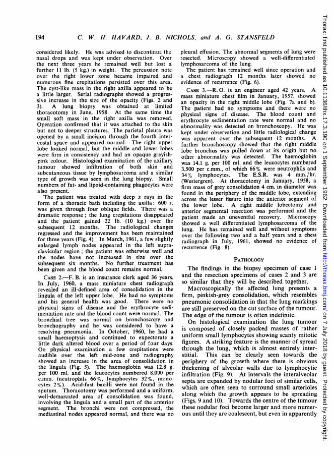
considered likely. He was advised to discontinue thenasal drops and was kept under observation. Overthe next three years he remained well but lost afurther 11 lb. (5 kg.) in weight. The percussion noteover the right lower zone became impaired andnumerous fine crepitations persisted over this area.The cyst-like mass in the right axilla appeared to bea little larger. Serial radiographs showed a progres-sive increase in the size of the opacity (Figs. 2 and3). A lung biopsy was obtained at limitedthoracotomy in June, 1958. At the same time thesmall soft mass in the right axilla was removed.Operation confirmed that it was attached to the skinbut not to deeper structures. The parietal pleura wasopened by a small incision through the fourth inter-costal space and appeared normal. The right upperlobe looked normal, but the middle and lower lobeswere firm in consistency and had an opaque greyish-pink colour. Histological examination of the axillarytumour showed infiltration of both skin andsubcutaneous tissue by lymphosarcoma and a similartype of growth was seen in the lung biopsy. Smallnumbers of fat- and lipoid-containing phagocytes werealso present.The patient was treated with deep x rays in the
form of a thoracic bath including the axilla: 600 r.was given through four oblique fields. There was adramatic response; the lung crepitations disappearedand the patient gained 22 lb. (10 kg.) over thesubsequent 12 months. The radiological changesregressed and the improvement has been maintainedfor three years (Fig. 4). In March, 1961, a few slightlyenlarged lymph nodes appeared in the left supra-clavicular region; the patient was otherwise well andthe nodes have not increased in size over thesubsequent six months. No further treatment hasbeen given and the blood count remains normal.CASE 2.-F. B. is an insurance clerk aged 36 years.
In July, 1960, a mass miniature chest radiographrevealed an ill-defined area of consolidation in thelingula of the left upper lobe. He had no symptomsand his general health was good. There were nophysical signs of disease and the erythrocyte sedi-mentation rate and the blood count were normal. Thebronchial tree was normal on bronchoscopy andbronchography and he was considered to have aresolving pneumonia. In October, 1960, he had asmall haemoptysis and continued to expectorate alittle dark altered blood over a period of four days.On physical examination a few crepitations wereaudible over the left mid-zone and radiographyshowed an increase in the area of consolidation inthe lingula (Fig. 5). The haemoglobin was 12.8 g.per 100 ml. and the leucocytes numbered 8,000 perc.mm. (neutrophils 66%, lymphocytes 32%, mono-cytes 2%). Acid-fast bacilli were not found in thesputum. Thoracotomy was performed and a uniform,well-demarcated area of consolidation was found,involving the lingula and a small part of the anteriorsegment. The bronchi were not compressed, themediastinal nodes appeared normal, and there was no
pleural effusion. The abnormal segments of lung wereresected. Microscopy showed a well-differentiatedlymphosarcoma of the lung.The patient has remained well since operation and
a chest radiograph 12 months later showed noevidence of recurrence (Fig. 6).CASE 3.-R. 0. is an engineer aged 42 years. A
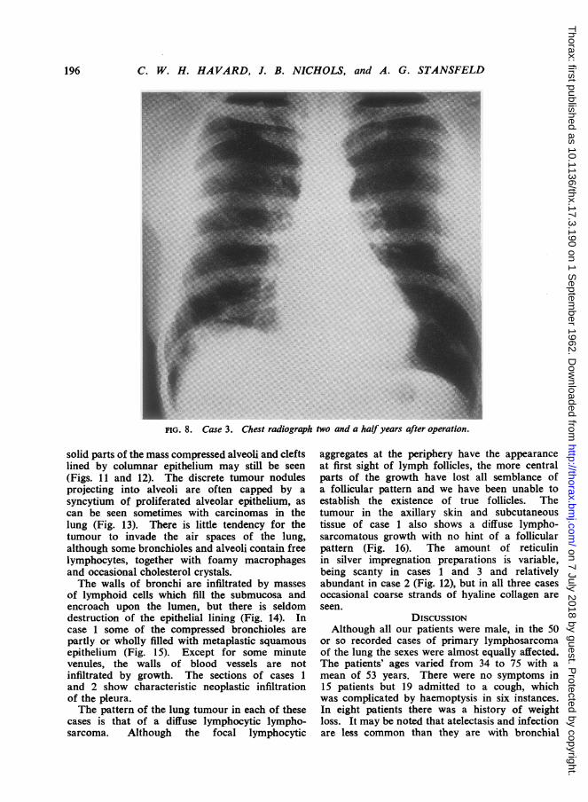
mass miniature chest film in January, 1957, showedan opacity in the right middle lobe (Fig. 7a and b).The patient had no symptoms and there were nophysical signs of disease. The blood count anderythrocyte sedimentation rate were normal and noabnormality was detected on bronchoscopy. He waskept under observation and little radiological changewas apparent over the subsequent 12 months. Afurther bronchoscopy showed that the right middlelobe bronchus was pulled down at its origin but noother abnormality was detected. The haemoglobinwas 14.1 g. per 100 ml. and the leucocytes numbered3,500 per c.mm., of which 66% were neutrophils and34% lymphocytes. The E.S.R. was 4 mm./hr.(Westergren). At thoracotomy in January, 1958, afirm mass of grey consolidation 4 cm. in diameter wasfound in the periphery of the middle lobe, extendingacross the lesser fissure into the anterior segment ofthe lower lobe. A right middle lobectomy andanterior segmental resection was performed and thepatient made an uneventful recovery. Microscopyshowed a well differentiated lymphosarcoma of thelung. He has remained well and without symptomsover the following two and a half years and a chestradiograph in July, 1961, showed no evidence ofrecurrence (Fig. 8).
PATHOLOGYThe findings in the biopsy specimen of case 1
and the resection specimens of cases 2 and 3 areso similar that they will be described together.
Macroscopically the affected lung presents afirm, pinkish-grey consolidation, which resemblespneumonic consolidation in that the lung markingsare still preserved on the cut surface of the tumour.The edge of the tumour is often indefinite.On histological examination the lung tumour
is composed of closely packed masses of ratheruniform small lymphocytes showing scanty mitoticfigures. A striking feature is the manner of spreadthrough the lung, which is almost entirely inter-stitial. This can be clearly seen towards theperiphery of the growth where there is obviousthickening of alveolar walls due to lymphocyticinfiltration (Fig. 9). At intervals the interalveolarsepta are expanded by nodular foci of similar cells,which are often seen to surround small arteriolesalong which the growth appears to be spreading(Figs. 9 and 10). Towards the centre of the tumourthese nodular foci become larger and more numer-ous until they are coalescent, but even in apparently
194
on 7 July 2018 by guest. Protected by copyright.
http://thorax.bmj.com
/T
horax: first published as 10.1136/thx.17.3.190 on 1 Septem
ber 1962. Dow
nloaded from

FIG. 7. Case 3: (above) Postero-anterior and (below) lateral chest radiographsbefore operation.
on 7 July 2018 by guest. Protected by copyright.
http://thorax.bmj.com
/T
horax: first published as 10.1136/thx.17.3.190 on 1 Septem
ber 1962. Dow
nloaded from
C. W. H. HAVARD, J. B. NICHOLS, and A. G. STANSFELD
FIG. 8. Case 3. Chest radiograph two and a halfyears after operation.
solid parts of the mass compressed alveoli and cleftslined by columnar epithelium may still be seen(Figs. 11 and 12). The discrete tumour nodulesprojecting into alveoli are often capped by asyncytium of proliferated alveolar epithelium, ascan be seen sometimes with carcinomas in thelung (Fig. 13). There is little tendency for thetumour to invade the air spaces of the lung,although some bronchioles and alveoli contain freelymphocytes, together with foamy macrophagesand occasional cholesterol crystals.The walls of bronchi are infiltrated by masses
of lymphoid cells which fill the submucosa andencroach upon the lumen, but there is seldomdestruction of the epithelial lining (Fig. 14). Incase 1 some of the compressed bronchioles arepartly or wholly filled with metaplastic squamousepithelium (Fig. 15). Except for some minutevenules, the walls of blood vessels are notinfiltrated by growth. The sections of cases 1and 2 show characteristic neoplastic infiltrationof the pleura.The pattern of the lung tumour in each of these
cases is that of a diffuse lymphocytic lympho-sarcoma. Although the focal lymphocytic
aggregates at the periphery have the appearanceat first sight of lymph follicles, the more centralparts of the growth have lost all semblance ofa follicular pattern and we have been unable toestablish the existence of true follicles. Thetumour in the axillary skin and subcutaneoustissue of case 1 also shows a diffuse lympho-sarcomatous growth with no hint of a follicularpattern (Fig. 16). The amount of reticulinin silver impregnation preparations is variable,being scanty in cases 1 and 3 and relativelyabundant in case 2 (Fig. 12), but in all three casesoccasional coarse strands of hyaline collagen areseen.
DISCUSSIONAlthough all our patients were male, in the 50
or so recorded cases of primary lymphosarcomaof the lung the sexes were almost equally affected.The patients' ages varied from 34 to 75 with amean of 53 years. There were no symptoms in15 patients but 19 admitted to a cough, whichwas complicated by haemoptysis in six instances.In eight patients there was a history of weightloss. It may be noted that atelectasis and infectionare less common than they are with bronchial
196
on 7 July 2018 by guest. Protected by copyright.
http://thorax.bmj.com
/T
horax: first published as 10.1136/thx.17.3.190 on 1 Septem
ber 1962. Dow
nloaded from

FIG. 9. Case 1. Low-power view showing interstitial FIG. 10. Case 3. Nodularfoci in the interstitial tissue ofspread and spurious follicular pattern (haematoxylin the lung at the periphery of the tumour (haematoxylinandeosin x 18). andeosin x 60).
_8 J . / - ; . \ . X . W t35 ( _ ; v . J T~~~~~~~~~~~~~~~~~~~~~~~
FIG. 11. Case 2. Central part of tumour showing preser-vation of epithelial-lined spaces (haematoxylin andeosin x 65).
1-J(
\~~~~~~~~~~~~
%X'~~j% iA4W4W~~~7,,
r .-t- , ~
& Y 44PV
)T, / t
FIG. 12. Case 2. Similar field to that in Fig. 1 1, showingabundant reticulin in the tumour. The residual airspaces are sharply outlined (silver impregnation x 65).
I ..loil 1.5BE''.
on 7 July 2018 by guest. Protected by copyright.
http://thorax.bmj.com
/T
horax: first published as 10.1136/thx.17.3.190 on 1 Septem
ber 1962. Dow
nloaded from

M.*~~~.W.~~~~~~
"' t
~~~~~~~.~ ~ ~ ~ V
W* A
!L,Sm,~~ ~ ~ ~ ~ ~~V~~I
FI. 13. Cae3 ycta a feihtu oeiga I.14 ae2 yiloa naiflrtn h alo
4 41
**~~~~~~~~~~~~~~~~~~*~~~~~~~~~~~~44~~~~~~~~~~~~~~~~~ #4 ~ ~ ~ ~ ~ ~~~~.4
..4W~~~~4
FIG. 15 FIG. 16
FIG. 15. Case 1. Squamou.s nzetaplasia in a bronchiole surrounded by tumour (hlaelatoxylin and cosin x 400).FIG. 16. Case 1. Margin ofaxillary tumour showing lyinphosarcoma infiltrating the skin and subcutis (haenmatoxylin
and eosin x 85).
on 7 July 2018 by guest. Protected by copyright.
http://thorax.bmj.com
/T
horax: first published as 10.1136/thx.17.3.190 on 1 Septem
ber 1962. Dow
nloaded from

PRIMARY LYMPHOSARCOMA OF THE LUNG
carcinoma. The blood count and erythrocytesedimentation rate were normal in our patients.The initial treatment was by surgery alone in
28 patients, and surgery followed by immediateradiotherapy in 16 patients. Four patientsreceived radiotherapy alone.Of the 28 patients who received surgical treat-
ment alone, nine were known to be alive and wellfive years later, although in two recurrence oflymphosarcoma had required treatment withradiotherapy. Of the 19 patients who had beenfollowed up for less than five years, 11 werereported alive and well, having survived forperiods varying between three months and threeyears after operation. Eight patients are knownto have died, but at least three of these died fromunrelated causes.Of the 16 patients who received surgery and
post-operative radiotherapy, four were alive andwithout evidence of recurrence five years later andone other was alive and well seven years afterlobectomy, having had a hilar recurrence treatedby radiotherapy. The 11 patients who had beenfollowed up for less than five years had survivedfor periods of one to four years after operation.Two patients had died, six and 21 months aftersurgery.Four patients received radiotherapy alone as
their disease was inoperable. Two died within14 months of treatment, one (our case 1) is aliveand well seven and a half years after the onset,and another ten years after the onset of the disease.
It is difficult to compare the prognosis ofprimary pulmonary lymphosarcoma with that oflymphosarcoma elsewhere for two reasons. Firstthe follow-up data on most of the reported casesof pulmonary lymphosarcoma are inadequate, andsecondly the widespread dissemination of thedisease makes the pulmonary origin of some casesuncertain. Nevertheless ten patients with primarypulmonary lymphosarcoma have been observedfor periods of seven or more years. In four ofthese there was no evidence of recurrence, oneof them surviving 14 years after operation anddying from an unrelated cause. In five patientsthe disease recurred, but certain survival periodsof 72, 81, 10, 12, and 131 years were recorded.
It seems clear from these figures that primarylymphosarcoma of the lung carries a betterprognosis than lymphosarcoma in general. Notonly has long survival after treatment beenrecorded, but in several patients there was radio-graphic evidence that the tumour had been presentfor a year or more before any treatment wasinstituted. In our first case this period was four
years and in another reported case it was as longas five years.There are possibly two factors that determine
the relatively benign behaviour of primarylymphosarcoma of the lung, namely a slow rateof growth and a tendency to spread locally ratherthan metastasize. The seemingly uncharacteristicbehaviour may raise doubts about the neoplasticnature of this condition, but histological examina-tion of the lung tumours will dispel such doubts,for the appearances are unquestionably those of aninfiltrating neoplasm, which is in most respectsindistinguishable from lymphosarcoma occurringelsewhere. There is no histological resemblance toany form of pulmonary granuloma and the growthof the tumour is progressive though slow. Further-more, it is well known that primary lympho-sarcomas of some other viscera often display asimilar type of behaviour (Shimkin, Oppermann,Low-Beer and Mettier, 1954; Harrison, 1960).The slow rate of growth of primary pulmonary
lymphosarcoma may in part be related to thedegree of differentiation of these tumours, for themajority are of lymphocytic type with a strikinglyuniform cytology. There is, however, someevidence that even reticulum cell sarcomas whichare primary in the lung show a better prognosisthan corresponding types of growth which arisein the mediastinal or other lymph nodes (Sternberget al., 1959). It is doubtful whether the sameapplies to pulmonary Hodgkin's disease, butprobably few, if any, of these tumours are reallyprimary in the lung.A remarkable feature of the reported cases of
primary lymphosarcoma of the lung is the lack ofmediastinal lymph node involvement. This hasbeen noted so often, even in patients who havebeen under observation for long periods beforeoperation, that the finding of extensive mediastinallymph node involvement at thoracotomy wouldappear to throw doubt upon the diagnosis ofprimary pulmonary lymphosarcoma. The media-stinal lymph nodes may be found enlarged, buthistological examination generally shows non-specific reactive changes only. A long delaybefore the regional lymph nodes are affected hasalso been noted many times with primary lympho-sarcoma of the gastrointestinal tract (Gall, 1943;Azzopardi and Menzies, 1960). The reasons forthe failure to spread in pulmonary as in othervisceral lymphosarcomas are obscure. It cannoteasily be attributed to a lack of infiltrative capacity,since local spread in the lung is often extensivewith pleural invasion and transgression of inter-lobar fissures.
199
on 7 July 2018 by guest. Protected by copyright.
http://thorax.bmj.com
/T
horax: first published as 10.1136/thx.17.3.190 on 1 Septem
ber 1962. Dow
nloaded from

C. W. H. HAVARD, J. B. NICHOLS, and A. G. STANSFELD
Sternberg et al. (1959) found a follicular patternin some parts of each of the four lymphocyticlymphomas of the lung which they reported. Wewere unable to find true follicles in our threepatients. The rounded, nodular masses oflymphocytes produced an appearance suggestiveof lymph follicles, but in our view this patternis a consequence of the interstitial manner ofspread in the lung. This type of spread is notpeculiar to primary lymphosarcoma of the lung,for we have found it also in secondary lympho-sarcomas and reticulum cell sarcomas of the lung.Secondary lymphomas, however, show a muchgreater tendency to destroy the lung parenchymaand invade the walls of blood vessels and thelumina of bronchi. The relatively abundantproduction of reticulin in case 2 and the findingof strands of collagen in all three tumours areprobably a reflection of the slow rate of growth,as seems to be the case with lymphosarcoma inother sites.Antecedent factors in primary lymphosarcoma
of the lung are unknown and the use of oily nasaldrops by our first patient is probably of nosignificance, but it is of interest that Hall andBlades' (1959) second patient gave a similar historyand was thought to have a patch of lipoidpneumonia in the lung opposite to that in whichthe lymphosarcoma arose. To our knowledgethere is no recorded evidence to suggest thatlymphosarcoma of the lung has ever developedsecondarily to lipoid pneumonia. One of thepatients of Sternberg et al. (1959) with a lympho-cytic lymphoma of the lung had a history ofrecurrent respiratory infections and 18 monthsafter lung resection the patient developedsymptoms characteristic of Sj0gren's syndrome.The concurrence of these two uncommonconditions is interesting in view of the heavylymphocytic infiltration characteristically found inthe salivary and other upper respiratory glandsin Sj0gren's syndrome, but these authors makeno comment about a possible association betweenthem.
SUMMARY AND CONCLUSIONSThe clinical and pathological details of three
cases of primary lymphosarcoma of the lung are
reported. Though an uncommon tumour, primarylymphosarcoma of the lung is a definite clinicalentity. The tumour tends to remain localized fora long time and is thus often amenable to completesurgical excision. The diagnosis has rarely beenmade before histological examination of thetumour. It is usually a well-differentiated,lymphocytic lymphosarcoma, which spreadsinterstitially with relatively little destruction oftissue. The prognosis of primary pulmonarylymphosarcoma, like that of some other viscerallymphosarcomas, is better than that of lympho-sarcoma arising in lymph nodes. The treatmenthas usually been surgical, as thoracotomy has beennecessary to establish the diagnosis. In view ofthe extreme radiosensitivity of these tumours,radiotherapy might theoretically be the treatmentof choice. Since this appears to be a potentiallycurable disease, surgical excision should befollowed by radiotherapy.
We should like to record our thanks to Dr. NevilleOswald, to Dr. H. J. Partington, and to Mr. K. S.Mullard for permitting us to publish records of thepatients under their care, and to Dr. T. E. W. Goodierand Dr. E. Nassau for the loan of histological sectionsrelating to cases 2 and 3, to Mr. N. K. Harrison forthe reproductions of radiographs, and to Mr. PeterCrocker for the photomicrographs.
REFERENCESAzzopardi, J. G., and Menzies, T. (1960). Brit. J. Surg., 47, 358.Baron, M. G., and Whitehouse, W. M. (1961). Amer. J. Roentgenol.,
85, 294.Cooley, J. C., McDonald, J. R., and Clagett, 0. T. (1956). Ann.
Surg., 143, 18.Gall, E. A. (1943). Ibid., 118, 1064.Hall, E. R., and Blades, B. (1959). Dis. Chest, 36, 571.Harrison, C. V. (1960). In Recent Advances in Pathology, 7th ed.
p. 49, ed. C. V. Harrison. Churchill, London.Jackson. E.,Bertoli, F.,and Ackerman,L. V.(1951). J. thorac. Surg.
21, 7.Prichard, R. W., and Bradshaw, H. H. (1961). Arch. Path., 71, 420.Robbins, L. L. (1953). Cancer, 6, 80.Rose, A. H. (1957). J. thorac. Surg., 33, 254.Rosenberg, S. A., Diamond, H. D., Jaslowitz, B., and Craver, L. F.
(1961). Medicine (Baltimore), 40, 31.Shimkin, M. B.. Oppermann, K. C., Low-Beer, B. V. A., and Mettier,
S. R. (1954). Ann. intern. Med., 40, 1095.Sternberg, W. H., Sidransky, H., and Ochsner, S. (1959). Cancer,
12, 806.Sugarbaker, E. D., and Craver, L. F. (1940). J. Amer. med. Ass.,
115, 17.Vieta, J. O., and Craver, L. F. (1941). Radiology, 37, 138.Willis, R. A. (1960). Pathology of Tumours, 3rd ed., p. 772. Butter-
worths, London.
200
on 7 July 2018 by guest. Protected by copyright.
http://thorax.bmj.com
/T
horax: first published as 10.1136/thx.17.3.190 on 1 Septem
ber 1962. Dow
nloaded from