
Primary Congenital Hydrocephalusadc.bmj.com/content/archdischild/43/231/516.full.pdf · Primary...
12
Arch. Dis. Childh., 1968, 43, 516. Primary Congenital Hydrocephalus Long-term Results of Controlled Therapeutic Trial JOHN LORBER and R. B. ZACHARY From the Department of Child Health, University of Sheffield, and Children's Hospital, Sheffield Holter's invention of the non-retum valve and its use in ventriculo-caval drainage operations has been a revolutionary step in the treatment of hydro- cephalus. The valves effectively control intra- cranial pressure and prevent the back flow of blood from the vascular system. This technique has been in use in this country since January 1958. In our unit 48 such operations were carried out during 1958, using the Holter valve. Having acquired technical experience and confidence in the operation, we decided to assess its value and limitations, and to define the indications for and the timing of this procedure by fully controlled therapeutic trials. In this paper we describe one such trial, in which all the patients suffered from uncomplicated congenital hydrocephalus. The second trial cover- ing the treatment of congenital hydrocephalus in infants who were born with meningomyelocele will be described in a separate publication. Material and Methods During 1959 and 1960 30 infants (19 boys and 11 girls) under 1 year of age were admitted with primary hydro- cephalus. The hydrocephalus, as far as could be ascertained, was due neither to intrauterine infections (e.g. toxoplasmosis) nor to postnatal cerebral trauma or infection. There was no associated spina bifida cystica, or other complex multiple congenital deformities. All were under 1 year of age on admission and 11 were under 2 months, but 6 were over 6 months of age (Table I). After the diagnosis of hydrocephalus was established by pneumoencephalography or ventriculography, they were allocated by random selection to one of two groups. One group was treated by operation immediately after the diagnosis was made, and the other group acted as controls; they were not operated on at least until it became clear that conservative treatment had failed. There were 5 patients who were not allocated to either group, because they already had such advanced hydro- cephalus that immediate operative treatment appeared essential. Received May 1, 1968. The degree of hydrocephalus was determined by ventriculography (Lorber, 1961). Among 12 patients allocated to the treated group, 2 infants had Grade 2 hydrocephalus, with a cerebral mantle measuring between 15 and 35 mm., and 10 had Grade 3 hydro- cephalus, with a cerebral mantle of less than 15 mm. Among the 13 controls there were 8 infants with Grade 2, and 5 with Grade 3 hydrocephalus. Thus the treated group was accidentally weighted with more severe or more advanced cases. This adverse weighting is probably related to the age on admission. The 'controls' were admitted at an earlier age (9 under 3 months against 4 in the treated group). Had they come a little later, their hydrocephalus would have been more advanced, as indeed was demonstrated by the precipitous rise in their head circumference after inclusion in the trial. Finally, in the 5 not included in the controlled investi- gation the cerebral mantle was extremely thin. Admission of new cases to the trial ceased at the end of 1960, because intermediate analysis of the results already indicated a trend which led to a change in policy. All children were examined at regular intervals at 6, 12, 18, and 24 months of age, and then at each birthday. Developmental assessments were made at each visit by one of us (J.L.), and independent assessment was made by educational psychologists immediately before their fifth birthday, to assess them for scholastic purposes. Their assessment continued subsequently, and school TABLE I Primary Congenital Hydrocephalus, 1959/60 Number 30 Age on Admission (mth.) <1 .. .. .. .. .. .. 5 1... .. .. .. .. .. 6 2-3. 6 4-5 .7 6-11. 6 Grade of Hydrocephalus 1 0 2 .10 3 .20 Sex Boys .19 Girls .11 516 copyright. on 20 June 2018 by guest. Protected by http://adc.bmj.com/ Arch Dis Child: first published as 10.1136/adc.43.231.516 on 1 October 1968. Downloaded from
Transcript of Primary Congenital Hydrocephalusadc.bmj.com/content/archdischild/43/231/516.full.pdf · Primary...

Arch. Dis. Childh., 1968, 43, 516.
Primary Congenital HydrocephalusLong-term Results of Controlled Therapeutic Trial
JOHN LORBER and R. B. ZACHARYFrom the Department of Child Health, University of Sheffield, and Children's Hospital, Sheffield
Holter's invention of the non-retum valve and itsuse in ventriculo-caval drainage operations has beena revolutionary step in the treatment of hydro-cephalus. The valves effectively control intra-cranial pressure and prevent the back flow of bloodfrom the vascular system. This technique has beenin use in this country since January 1958. In ourunit 48 such operations were carried out during1958, using the Holter valve. Having acquiredtechnical experience and confidence in the operation,we decided to assess its value and limitations, and todefine the indications for and the timing of thisprocedure by fully controlled therapeutic trials.In this paper we describe one such trial, in whichall the patients suffered from uncomplicatedcongenital hydrocephalus. The second trial cover-ing the treatment of congenital hydrocephalus ininfants who were born with meningomyelocele willbe described in a separate publication.
Material and Methods
During 1959 and 1960 30 infants (19 boys and 11 girls)under 1 year of age were admitted with primary hydro-cephalus. The hydrocephalus, as far as could beascertained, was due neither to intrauterine infections(e.g. toxoplasmosis) nor to postnatal cerebral trauma orinfection. There was no associated spina bifida cystica,or other complex multiple congenital deformities. Allwere under 1 year of age on admission and 11 were under2 months, but 6 were over 6 months of age (Table I).
After the diagnosis of hydrocephalus was establishedby pneumoencephalography or ventriculography, theywere allocated by random selection to one of two groups.One group was treated by operation immediately afterthe diagnosis was made, and the other group acted ascontrols; they were not operated on at least until itbecame clear that conservative treatment had failed.There were 5 patients who were not allocated to eithergroup, because they already had such advanced hydro-cephalus that immediate operative treatment appearedessential.
Received May 1, 1968.
The degree of hydrocephalus was determined byventriculography (Lorber, 1961). Among 12 patientsallocated to the treated group, 2 infants had Grade 2hydrocephalus, with a cerebral mantle measuringbetween 15 and 35 mm., and 10 had Grade 3 hydro-cephalus, with a cerebral mantle of less than 15 mm.Among the 13 controls there were 8 infants with Grade 2,and 5 with Grade 3 hydrocephalus. Thus the treatedgroup was accidentally weighted with more severe ormore advanced cases. This adverse weighting isprobably related to the age on admission. The 'controls'were admitted at an earlier age (9 under 3 months against4 in the treated group). Had they come a little later,their hydrocephalus would have been more advanced,as indeed was demonstrated by the precipitous rise intheir head circumference after inclusion in the trial.
Finally, in the 5 not included in the controlled investi-gation the cerebral mantle was extremely thin.Admission of new cases to the trial ceased at the end
of 1960, because intermediate analysis of the resultsalready indicated a trend which led to a change in policy.
All children were examined at regular intervals at 6,12, 18, and 24 months of age, and then at each birthday.Developmental assessments were made at each visit byone of us (J.L.), and independent assessment was madeby educational psychologists immediately before theirfifth birthday, to assess them for scholastic purposes.Their assessment continued subsequently, and school
TABLE IPrimary Congenital Hydrocephalus, 1959/60
Number 30
Age on Admission (mth.)<1 .. .. .. .. .. .. 5
1... .. .. .. .. .. 62-3. 64-5 .76-11. 6
Grade of Hydrocephalus1 0
2 .103 .20
SexBoys .19Girls .11
516
copyright. on 20 June 2018 by guest. P
rotected byhttp://adc.bm
j.com/
Arch D
is Child: first published as 10.1136/adc.43.231.516 on 1 O
ctober 1968. Dow
nloaded from

Primary Congenital Hydrocephalusreports were obtained about their progress. During1966 and 1967 they had psychometric assessment byMr. J. Parsons, research psychologist at the Unit.
Physical examination included a full neurologicalassessment, with special reference to the appearance ofthe optic fundi. Tests of visual acuity and of visualfield were undertaken when this was indicated.The Holter shunt was examined for its patency by
palpation of the 'pump' behind the ear, and by radio-graphy for the position of the lower end of the tube in theright auricle or in the main veins entering it. If the tubecould be compressed easily with the finger and filled uprapidly when the finger was removed, it was consideredto be working satisfactorily.
Results
The results presented are those that applied inOctober 1967. As a controlled therapeutic investi-gation, this study failed in one respect. As timewent on, all but 2 of the 13 infants allocated to thecontrol group had to be operated on because ofrapidly increasing head circumference or other signsof neurological deterioration. In fact, it appearsretrospectively that some of those who were operatedon at a later stage should probably have beenoperated on sooner.
In the control group the interval between theallocation into the trial and the operation was
between 4 weeks and 4 months in 7 infants; it was12 months, 16 months, 18 months, and 3 years,respectively, in one each (Table II).Two operated children had a shunt in position
for so short a time that in some respects these couldbe considered as untreated cases.The first was operated on at 20 months of age but
developed septicaemia with colonization of thevalve. The shunt was removed within 3 weeks andwas not reinserted until he was 7 years of age.The second was first seen and operated on at
11 months of age, when he was already severelyretarded. He reacted so badly with high fever tothe procedure that the shunt had to be removed 6weeks later. A new shunt was inserted 6 yearslater, without complication, but he remains pro-
foundly retarded.There are only 2 control children who have not had
a shunt to date. Observation and treatment of largegroups of older hydrocephalic children suggest,moreover, that it is by no means certain that thehydrocephalus even in these 2 is truly arrested. Itmay well be that their intracranial pressure is stillhigher than normal. Though at present they arewell, with average intelligence and with no seriousneurological handicap, deterioration could set in atany time, requiring reinvestigation and possiblyoperative treatment. Such sequence of events hasbeen commonly observed in hydrocephalic childrenof an older age-group (Lorber, to be published).As there is no true control group left, the results
obtained in the entire group of 30 children will beanalysed together.
Mortality (Table III). Up to the end ofOctober 1967, 5 children (17%) had died. Deathwas directly due to the hydrocephalus or its treat-ment in 3, probably due to it in 1, and almostcertainly was unconnected with it in the fifth.The youngest (Case 1) died at 8 months of age.
The skin over the Holter valve broke down, the valvebecame exposed, and the baby developed meningitisdue to Staphylococcus aureus. (The whole shuntingsystem had to be removed and the baby died fromthe combined effects of meningitis and untreatedhydrocephalus.)The next (Case 2), according to age at death, was
a boy who died at home at 2 years 10 months of age.He was not operated on until he was 12 months of
TABLE IIReason for and Timing of Delayed Operative Treatment in Control Cases
Age (mth.) Age (mth.)Case No. Grade on at Reason for Operation
Allocation Operation
6 2 2 3 Precipitous increase of head circumference7 3 1 3* Precipitous increase of head circumference8 2 51 9t Precipitous increase of head circumference9 3 4 5k Precipitous increase of head circumference
10 2 1 I 3k Precipitous increase of head circumference11 3 3 5 Precipitous increase of head circumference12 3 3k 7 Precipitous increase of head circumference
2 3 I 2 12 Precipitous increase of head circumference and mental and physical deterioration13 2 18 Precipitous increase of head circumference and mental and physical deterioration14 2 4 20 Progressive increase in head circumference, removed after 3 wk.;
reinserted 5 yr. later15 2 1 3k yr. Very big head; hemiplegia; papilloedema; vomiting
517
copyright. on 20 June 2018 by guest. P
rotected byhttp://adc.bm
j.com/
Arch D
is Child: first published as 10.1136/adc.43.231.516 on 1 O
ctober 1968. Dow
nloaded from

TABLE IIIDeaths
Case No. Sex Grade Age at Death Cause(yr.)
1 F 3 8/12 Staphylococcal meningitis resulting from exposed valvewhich had to be removed; necropsy
2 M 3 2 10/12 Not sure; at home; extreme hydrocephalus; severeretardation, blindness, spasticity
3 M 3 5 Blocked shunt; removal; meningitis; acuteintracranial hypertension; necropsy
4 F 2 6 Bronchopneumonia; necropsy5 M 3 7 Detachment and blockage of ventricular catheter;
intraventricular haemorrhage; necropsy
age. By then his head circumference was 58 cm.and he was profoundly retarded and blind. Theoperation arrested the growth of his head, but didnot help him otherwise. (It is not known whetherhis shunt became blocked before he died.)The third child (Case 3), who was mentally
normal with a moderate spastic paraplegia, died asa result of a blocked shunt at 5 years of age. Theblockage could not be overcome by 3 operations inquick succession. He developed streptococcalmeningitis. External drainage of CSF did notcontrol his intracranial tension, and though hismeningitis cleared up he died as a result of acutecerebellar coning.The fourth child (Case 4) died at home at the age
of 6 years. When last seen she was retarded but well,and her head was of normal size. She died ofpneumonia. The necropsy report specified that theshunting system was in complete order.The fifth and last child (Case 5) who died had
extreme hydrocephalus which was first operated onat 8 weeks of age. His development had beenretarded throughout the 7 years of his life. At 16months of age blockage of the upper catheter wasrelieved by the insertion of a new one. At 7 yearshis ventricular catheter became detached and hadslipped into the lateral cerebral ventricle. A newcatheter was inserted, but much bleeding took place,both into the wound and the lateral ventricle,requiring several transfusions. He developed bila-teral hemiplegia, and it was apparent that the new
shunting system also became blocked. 5 furtheroperations were performed within 12 days but to noavail. Necropsy showed that a large blood clotoccupied his distended cerebral ventricles.
Survivors (Table IV). There are 25 survivors,who are now 7 to 9 years of age. All but one attendour clinic regularly; one child moved to the Londonarea, but postal contact is maintained with him.These 25 children are classified into 4 groups:
(a) 9 normal children, (b) 4 normal children butwith large heads, (c) 8 children with normalintellect but with neurological lesions, and (d) 4children with intellectual as well as neurologicalsequelae.
(a) Normal children. There are 9 children whoare physically and mentally normal and whose headcircumference is at or below the 90th centile.Their IQ is between 80 and 128. All have beenoperated on, but in 2, who were controls, theoperation was delayed up to 30 and 37 weeks of age(Case 8 and Case 12, Table II). There are only 2among these 9 who did not require further opera-tions for their hydrocephalus.The other 7 had to have further operations to deal
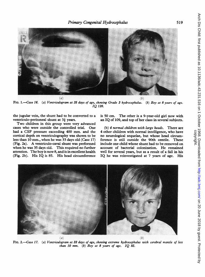
with problems related to the shunt. In spite of hishydrocephalus, one boy (Case 16) (Fig. la and b)now 8 years of age, has an IQ of 128. His headcircumference rose to 42 cm. by the time he wasoperated on at 5 weeks. Subsequently the rate ofgrowth of his head slowed down and by 18 monthsof age his head circumference fell below the 90thcentile. After two blockages due to thrombosis of
TABLE IVPrimary Congenital Hydrocephalus, 1959/60 Series:
Assessment October 1967. Age of Survivors:7-9 Years
Educa-Neurological Condition Mentally tionally Ament
Normal Sub-normal
Normal head circumference55 cm. or less .. 9 _ -
Normal head circumference56-62 cm. 4 _
Varying handicaps (blindness,paraplegia, etc.) 8 3 1
Total .. 21 3 1
518 Lorber and Zachary
copyright. on 20 June 2018 by guest. P
rotected byhttp://adc.bm
j.com/
Arch D
is Child: first published as 10.1136/adc.43.231.516 on 1 O
ctober 1968. Dow
nloaded from
Primary Congenital Hydrocephalus
(a) (b)FIG. 1.-Case 16. (a) Ventriculogram at 35 days of age, showing Grade 3 hydrocephalus. (b) Boy at 8 years of age.
IQ 128.
the jugular vein, the shunt had to be converted to aventriculo-peritoneal shunt at 3j years.Two children in this group were very advanced
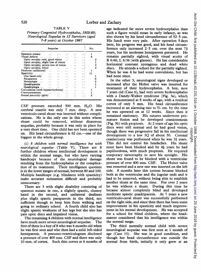
cases who were outside the controlled trial. Onehad a CSF pressure exceeding 400 mm. and thecortical depth on ventriculography was shown to beless than 10 mm., when he was 33 days old (Case 17)(Fig. 2a). A ventriculo-caval shunt was performedwhen he was 35 days old. This required no furtherattention. The boy is now 8, and is in excellent health(Fig. 2b). His IQ is 85. His head circumference
is 50 cm. The other is a 9-year-old girl now withan IQ of 109, and top of her class in several subjects.
(b) 4 normal children with large heads. There are4 other children with normal intelligence, who haveno neurological sequelae, but whose head circum-ference is still outside the 90th centile. Theseinclude one child whose shunt had to be removed onaccount of bacterial colonization. He remainedwell for several years, but as a result of a fall in hisIQ he was reinvestigated at 7 years of age. His
(a) (bIFIG. 2.-Case 17. (a) Ventriculogram at 33 days of age, showing extreme hydrocephalus with cerebral mantle of less
than 10 mm. (b) Boy at 8 years of age. IQ 85.
519
copyright. on 20 June 2018 by guest. P
rotected byhttp://adc.bm
j.com/
Arch D
is Child: first published as 10.1136/adc.43.231.516 on 1 O
ctober 1968. Dow
nloaded from
Lorber and ZacharyTABLE V
Primary Congenital Hydrocephalus, 1959/60;Neurological Sequelae in 12 Survivors (aged
7-9 years) at October 1967
Sequelae No.
Spasmus nutans 1Visual defects. 8
Optic atrophy only, good vision . . . 2Optic atrophy, slight loss of vision 1Optic atrophy, severe loss of vision 3Optic atrophy, blind . 2
Deafness (unilateral).1Spasticity.11One hand only. 1Paraparesis . .6Hemiplegia . . 2Quadriparesis. 1Quadriplegia . .1
Convulsions (with hypsarrhythmia). 1Craniosynostosis 1Sexual precocity (girls) 2
CSF pressure exceeded 500 mm. H20. Hiscerebral mantle was only 7 mm. deep. A newventriculo-caval shunt was inserted without compli-cations. He is the only one in this series whoseshunt could be removed, without disastroussequelae, probably because it was only in place fora very short time. One child has not been operatedon. His head circumference is 62 cm.-one of thebiggest in the whole group.
(c) 8 children with normal intelligence but withneurological sequelae (Table V). There are 8further children whose intellectual development iswithin the normal range, but who have varyinghandicaps because of the neurological damageresulting from the hydrocephalus or the complica-tion of its treatment. Their intelligence quotientis in the lower ranges ofnormal, between 80 and 100.Multiple handicaps (e.g. blindness with spasticity)make accurate estimation difficult and probablyunnecessary.There are 3 with slight disability consisting of
spasmus nutans in one, a slightly spastic, clumsyhand in the second, and unilateral deafnessplus slight spastic paraparesis in the third, notsufficient though to keep him from walking andgoing to ordinary school. A fourth child also hasslight spastic weakness of her legs but she also haspale optic discs and impaired vision.The remaining 4 children with normal intelligence
have much more severe neurological sequelae. Thefirst ofthese is a boy of 8 who was 6 months old whenhe was first seen and who then had a mild left-sidedhemiparesis. A pneumo-ventriculogram discloseda pressure of over 400 mm. CSF and there was only10 mm. of cortex. Such thin cortex at 6 months of
age indicated far more severe hydrocephalus thansuch a figure would mean in early infancy, as was
also shown by his head circumference of 52*5 cm.
His fundi were very pale. After operation 5 dayslater, his progress was good, and his head circum-ference only increased 2-5 cm. over the next 7!years, but his moderate hemiparesis persisted. Heremains partially sighted, with visual acuity ofR 6/60, L 6/36 (with glasses). He has considerablehorizontal constant nystagmus and dead whitediscs. He attends a school for the partially sighted.When he was 4 he had some convulsions, but hashad none since.
In the other 3, neurological signs developed or
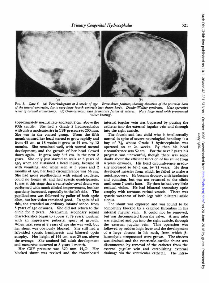
increased after the Holter valve was inserted fortreatment of their hydrocephalus. A boy, now7 years old (Case 6), had very severe hydrocephaluswith a Dandy-Walker syndrome (Fig. 3a), whichwas demonstrated at 8 weeks of age, with a residualcortex of only 5 mm. His head circumferenceincreased at an alarming rate to 51 cm. by the timehe was operated on at 12 weeks. Since then itremained stationary. His sutures underwent pre-
mature fusion and he developed craniostenosis(Fig. 3b) with proptosis. At 2 years of age his opticdiscs were still normal and he had good vision,though there was progressive fall in his intellectualdevelopment to a low IQ of about 50. Coronalcraniectomy was performed when he was 33 years.This did not control his headaches. His shuntmust have been blocked and by 41 years he hadpapilloedema, with much proptosis. He requiredtemporary tarsorraphy to save his eyesight. Hisshunt was found to be blocked with a ventricularpressure of over 400 mm. CSF. The Holter valvewas removed and a new one was inserted on the leftside. 8 months later this system became blockedboth at the ventricular and the jugular ends and ithad to be removed, without being able to establishanother shunt at the same time. For over 2 yearshe was without a shunt. During this time hebecame almost completely blind and developedmoderate spastic quadriparesis. At 7 years a newventriculo-caval shunt was successfully performedon the right side, and since then there has been someimprovement in his spasticity and much improve-ment in his mental development. He was acceptedfor a school for blind children, where the head-master considered that his intelligence was withinthe normal range.The third mentally normal child with severe
neurological sequelae was first seen at 1 month ofage (Case 15). She was in good condition, andthough her head circumference was outside thenormal from birth, initially it only grew at an
520
copyright. on 20 June 2018 by guest. P
rotected byhttp://adc.bm
j.com/
Arch D
is Child: first published as 10.1136/adc.43.231.516 on 1 O
ctober 1968. Dow
nloaded from
Primary Congenital Hydrocephalus
We.,! /. wS as4 z- w 9
(a) (b)FIG. 3.-Case 6. (a) Ventriculogram at 8 weeks of age. Brow-down position, showing elevation of the posterior hornof the lateral ventricles, due to very large fourth ventricle (not shown here). Dandy-Walker syndrome. Note operativeresult of coronal craniectomy. (b) Craniostenosis with premature fusion of sutures. Note large head with pronounced
'silver beating'.
approximately normal rate and kept 2 cm. above the90th centile. She had a Grade 2 hydrocephaluswith only a moderate rise in CSF pressure to 200 mm.She was in the control group. From the fifthmonth onward her head started to grow rapidly andfrom 45 cm. at 18 weeks it grew to 55 cm. by 12months. She remained well, with normal mentaldevelopment, and the growth of her head sloweddown again. It grew only 3 5 cm. in the next 2years. She only just started to walk at 3 years ofage, when she sustained a head injury, became illwith vomiting, and when seen at 3 years and 2months of age, her head circumference was 64 cm.She had gross papilloedema with retinal exudates,could no longer sit, and had spastic quadriparesis.It was at this stage that a ventriculo-caval shunt wasperformed with much clinical improvement, but herspasticity increased, especially in the left side. Thepapilloedema was followed by pallor of both opticdiscs, but her vision remained good. In spite of allthis, she attended an ordinary infants' school from5 years of age onwards. She did not return to theclinic for 2 years. Meanwhile, secondary sexualcharacteristics began to appear at 71 years, togetherwith an impressive pubertal spurt of growth.When next seen at 8 years of age she was well, buther shunt was obviously blocked. She still had aleft-sided spastic hemiparesis and bilateral opticatrophy. Her height of 145 cm. was 23 cm. abovethe average. She attained full adult developmentand menarche occurred at 8 years 1 month.Her CSF pressure was 370 mm. H20. Her
blocked shunt was revised and the thrombosed
internal jugular vein was bypassed by putting thecatheter into the external jugular vein and throughinto the right auricle.The fourth and last child who is intellectually
normal in spite of severe neurological handicap is aboy of 74, whose Grade 3 hydrocephalus wasoperated on at 24 weeks. By then his headcircumference was 52 cm. For the next 7 years hisprogress was uneventful, though there was somedoubt about the efficient function of his shunt from4 years onwards. His head circumference gradu-ally increased to 62 5 cm. by 74 years. He thendeveloped measles from which he failed to make aquick recovery. He became drowsy, with headachesand vomiting, but was not returned to the clinicuntil some 7 weeks later. By then he had very littleresidual vision. He had bilateral secondary opticatrophy with tortuous retinal vessels. There wasspastic weakness of both legs with bilateral ankleclonus.The shunt was explored and was found to be
completely blocked by a calcified thrombus in hisinternal jugular vein. It could not be removed,but was disconnected from the valve. A new tubewas attached and put into the right auricle, throughthe external jugular vein. This operation wasfollowed by sudden high fever and the developmentof a large abscess in his neck, from which P-haemolytic streptococci were grown. The abscesswas drained and the ventriculo-cardiac shunt wasdisconnected by removal of the catheter from theexternal jugular vein and establishing externaldrainage via the ventricular catheter. The intra-
521
copyright. on 20 June 2018 by guest. P
rotected byhttp://adc.bm
j.com/
Arch D
is Child: first published as 10.1136/adc.43.231.516 on 1 O
ctober 1968. Dow
nloaded from

Lorber and Zacharyventricular pressure was 380 mm. H20, underanaesthesia. A burr-hole was made on the left sideof the skull, and through this soluble penicillin wasinjected daily into the cerebral ventricle to preventventriculitis which might have resulted from theabscess surrounding the ventricular shunt on theright side. With this and with antibiotic treatmentventriculitis was avoided, the infection cleared up,and 8 days after the first operation a left-sidedventriculo-peritoneal shunt was established. Thewhole shunting system on the right was removed.Meanwhile the spastic weakness of his legs hadincreased. After the first operation he had verylittle eyesight, but after the establishment ofsatisfactory drainage there was progressive improve-ment. Five months later his vision was R 6/36 andL 6/18. In spite of his poor vision and his spasticparaparesis he went back to his ordinary school.Recovery of vision after prolonged blindness hasbeen described in a larger series of patients else-where (Lorber, 1967).
It is possible that these complications would havebeen avoided by attending to his shunt before he hadany symptoms.
(d) 4 children with intellectual and neurologicalsequelae (Table V). These 4 survivors arementally and physically handicapped: 2 areeducationally subnormal and 2 are even moreseverely retarded; 2 have a normal-sized head, thehead circumference of the others is from 57 cm. to60 cm. One of the moderately retarded childrenhas a spastic paraparesis and bilateral optic atrophy,but his vision is good. The other, now 8, had amost severe degree of hydrocephalus, so that by 14weeks her head circumference reached 57 cm. Herintraventricular pressure was over 400 mm. H2Oand her cerebral mantle was less than 5 mm. wide.A ventriculo-caval shunt at this time arrested thegrowth of her head, but her development was slowand her vision was very poor. She had searchingnystagmus with bilateral optic atrophy. Twoblockages of the shunt were corrected, but shedeveloped moderate spastic weakness apart from heralmost total blindness. Sexual precocity wasanother feature. Menarche occurred at 7 yearsand 5 months of age, and menstruation wasregularly established by 8 years. At this age herhead was still of the same size as when she was 14weeks old.The third child, a girl of 8, was normal until she
was 7 years 10 months, when she developedintracranial hypertension as a result of jugular veinthrombosis. She was sent to a local hospitalunfamiliar with her case. She had 5 operations
in quick succession, and during this time hershunt system has been twice removed. Thisled to extreme illness and her life was in balance.Nevertheless, at the fifth procedure a new ventriculo-facial vein shunt was established, and this led to areturn of her CSF pressure to normal and to goodhealth, with a residual but rapidly improving spasticparaparesis. Her IQ dropped from above 100 to62 as a result of this illness. This, and many othercases illustrate the grave dangers from rapidlymounting intracranial tension, if a previouslyfunctioning shunt is removed for any reason,beyond the age of infancy, and when the child'ssutures are unable to separate further.
Finally, there is only one child who is profoundlyretarded, who is unable to walk, and who is subjectto fits. In addition, he has a spastic quadriplegiaand has poor vision with optic atrophy. His EEGshows the pattern of hypsarrhythmia. This boywas 11 months old (the oldest in the series) when hefirst came to be investigated and treated. Thoughhis hydrocephalus was not severe (Grade 2, pressure220 mm. H20, head circumference 49 cm.) it wasprobably the association with hypsarrhythmia whichcaused his profound retardation. He had thetypical history of developmental arrest at about5 months of age, concurrent with the developmentof 'salaam' convulsions. He responded uniquelyto the insertion of the ventriculo-caval shunt for hebecame extremely ill with high fever, which persis-ted over several weeks, without any ascertainableinfective cause. CSF was normal, blood cultureswere sterile, and there was no leucocytosis. Therewas no response to antibiotic treatment. Immedi-ately on removal of the shunt 6 weeks after itsinsertion the fever ceased. No organisms werecultured from the components of the shunt. It wasthought that possibly this was a hypersensitivityreaction to some component of the system. Thisboy remained without a shunt for just over 6 years.His head circumference rose to 60 cm. He wasbedridden and kept in a long-term hospital. Evenso, it was felt that a new shunt might save his sightand possibly benefit him in other ways, and aventriculo-caval shunt was inserted without anyfebrile reaction, in February 1967. He improvedconsiderably but it is too early to judge just howmuch benefit will accrue.
Head circumference related to results(Table IV). As might be expected, children whoat the time of their last assessment still had anabnormally large head fared worse. Only 4 out of12 children whose head circumference is 56 cm. ormore are intellectually and neurologically normal as
522
copyright. on 20 June 2018 by guest. P
rotected byhttp://adc.bm
j.com/
Arch D
is Child: first published as 10.1136/adc.43.231.516 on 1 O
ctober 1968. Dow
nloaded from

Primary Congenital Hydrocephaluscompared with 9 of 13 whose head is smaller thanthat. Conversely, only 1 of 13 with a normal-sizedhead is retarded and has physical handicaps as well,as compared with 3 out of 12 whose head is undulylarge. Nevertheless, there are no surviving childrenwith extreme enlargement of the head. The childwith the largest head circumference of 64 cm. is atall, attractive girl who goes to an ordinary schoolin spite of her handicaps (Case 15). There are 6children whose head circumference is already 60 cm.or more, and hence it will always be above thenormal. None ofthe children who had a ventriculo-caval shunt procedure performed at the first occa-sion, and who were able to retain it, belong to thisgroup of 6.
Revisions (Tables VI and VII)(a) Number of children who required revision. One
of the principal risks and disadvantages of allshunting procedures, including the ventriculo-caval shunt using the Holter valve, is the need forrepeated operative procedures, colloquially knownas 'revisions', to deal with the many complicationsthat arise.Of the 28 children who had a primary ventriculo-
caval shunt, 23 have already required one or moreoperative procedures to deal with such complica-tions.
Altogether, 52 further operations were performedin these 23, or an average of over 2 per patient in anaverage of nearly 6 years after insertion of the shunt:4 children have already had 4-7 further operations,often in quick succession.
(b) Reasons for revision. The reasons for therevisions are given in Table VIII, together with thesteps taken. Trouble with the catheter in thejugular vein alone was the reason for furtheroperations in 13 instances, usually due to blockageby thrombosis, with or without calcification. Thesymptoms and signs of such blockages are irrita-bility, headache, vomiting, drowsiness, diminishingeyesight, development ofor increase in spasticity, andconvulsions. Usually, however, these symptomstake several weeks or even months to develop.
Blockage of the jugular catheter may be diagnosedeven before there are obvious symptoms, andeffective action can be taken to prevent illness,neurological sequelae, and intellectual deterioration.The signs differ in infants whose fontanelle is stillopen from those seen in older children. In infantsan increased fontanelle tension in conjunction withcontinued or newly recurring abnormally rapid rateof growth of the skull, together with a feeling offullness of the compressible silastic part of the valve,
TABLE VIPrimary Congenital Hydrocephalus,
'Revisions'1959/60 Series;
Number at risk .28
No revision required to date. 5
Number with 1 revision .12 1Number with 2 revisions. 3 23Number with 3 revisions 4Number with 4 to 7 revisions. 4
Total number of procedures .52Average number per patient .2 *2
TABLE VIIPrimary Congenital Hydrocephalus, 1959/60 Series;
'Revisions'
Reason for Operation Action Taken No.
Jugular catheter thrombosis Ventriculo-peritoneal shunt 5Jugular catheter thrombosis New catheter in external
(and calcification) jugular vein 6Short jugular catheter Lengthened 1Jugular catheter detached Reconnected 1
Peritoneal shunt blocked Freed 5
Ventricular catheter blocked Replaced by new catheter 7Ventricular catheter Replaced by new catheter 1
disconnectedExploration for doubtful Replaced by new shunt 3
blockage
Blockage both ends of shunt Removal of whole shunt, notimmediately replaced(major error) 4
Meningitis septicaemia, Removal of whole shunt 3persistent fever
Infection, ventricular External drainage 4haemorrhage
Craniosynostosis Craniectomy 1Temporary absence of shunt, New V-C or V-P shunt 7
intracranial tensionMiscellaneous 4
TABLE VIIICongenital Hydrocephalus, 1959/60,Intelligence Quotient of 25 Survivors
IQ No. of Cases
120-129 290-119 12
70-89. 750-69. 2<50. 2
Total .25
523
copyright. on 20 June 2018 by guest. P
rotected byhttp://adc.bm
j.com/
Arch D
is Child: first published as 10.1136/adc.43.231.516 on 1 O
ctober 1968. Dow
nloaded from
Lorber and Zacharyare the most important signs. In properly function-ing valves this tube is normally full of CSF, but notunder tension, and on pressure it readily emptiesand quickly refills from above. If the jugularcatheter is blocked, the tension in the tube is high,it feels stiff, and cannot be readily compressed. Inolder children the head cannot grow rapidly anymore and the mounting intracranial tension usuallyshows itself by tortuosity of the retinal vessels, theirdistension, and eventually papilloedema. All thesesigns may be present before the child has anysubjective complaint. The ophthalmoscope is there-fore the most essential diagnostic tool in the routineexamination of these children. Blockage or impendingblockage of the jugular catheter may also be diag-nosed or suspected, if on radiography of the chestthe tip of the radio-opaque catheter is seen to haverisen out of the cardiac atrium and is at the level ofthe 2nd rib or even higher in the neck. Electiverevision may be carried out where the tube has risenout of the atrium into the superior vena cava even ifthere are no symptoms, but this was the reason foroperation in only one child in this series. Theprobable need for revision on this evidence has beenrecognized only more recently. Many more suchrevisions might have been done otherwise, anticipa-ting and preventing the trouble which followedlater.
Blockage of the jugular catheter is confirmed onthe operation table. On detaching the distalcatheter from the lower valve, no blood wells upfrom the jugular vein and none may be aspiratedwith a syringe. If the intraventricular pressure ismeasured, it is shown to be raised. The patency ofthe catheter in the cerebral ventricle is shown byfree flow of CSF from the upper catheter (ifdetached from the valve), or by dripping CSF fromthe lower end of the valve. This observation alsoproves the patency of the valves themselves.
Blockage of the jugular catheter may be overcomeby removing it and replacing it by a new longercatheter, inserted into the internal jugular vein at aslightly lower level in some cases; into the externaljugular vein or some minor vein in the neck in othercases. With experience, an alternative vein canoften be found, but if not, the shunt may be con-verted into a ventriculo-peritoneal shunt, drainingthe CSF into the peritoneal cavity. This wascarried out on 5 occasions in this group of patients.The peritoneal end of the shunt may also get blockedby fibrin, omentum, or the formation of an enclosedcyst round the peritoneal 'button'. This occurred5 times in this series. The symptoms and signs arevery similar, except that the compressible tubebetween the valves may not be so stiff, and compres-
sion is easier because of the extra length of the tubeand the larger dead space in the system.
Blockage or other problems connected with theventricular catheter occurred in 12 instances, inaddition to 4 others, in which both ends of theshunt became blocked. Such a blockage is moredangerous than a jugular catheter block, becausethe onset of symptoms is more sudden, it leads tomore rapid increase in intracranial tension, and maylead to coma, convulsions, and death in a matter ofhours in some cases. The signs are similar to thosefound in jugular blocks, except that the symptomsand the papilloedema develop much more quickly.The feel of the tube is different. Here no fluid canenter it from above, so it feels flat and empty, orremains empty after compression. A soft, emptytube, however, is not necessarily due to blockage ofthe ventricular catheter: it may merely mean thatthe pressure gradient, at the time, is such that nofluid need escape from the cerebral ventricles. Insuch cases the child is well, has no signs of intra-cranial hypertension, and no action need be taken.If there is clinical evidence of obstruction, andexploration shows that both upper and lower tubesare patent, the fault must lie in the valve itself.This is rare and did not occur in this series thoughwe have seen it in more recent cases (Corkery andZachary, 1967).Removal of the shunt in the belief that it is no
longer necessary is to be avoided as it may lead to thesame sudden rise in intracranial tension as blockageof the system. It may even lead to death, for it isvery difficult to reinsert a shunt after the cerebralventricles become of near normal size as aresult of satisfactory treatment up to that time.Diminution of the size of the ventricles is indeedone of the main causes of blockage of the ventricularcatheter, because it becomes embedded in cerebralsubstances. Another cause is obstruction by thechoroid plexus which is less likely if the ventricularcatheter is passed well forwards towards the anteriorhorn of the lateral ventricle.
Various infections connected with the shuntnecessitated its removal or exteriorization in 6instances. Meningitis, colonization of the valve, orexposure of the valve by skin breakdown wereresponsible for the need for revision in this group.This was often followed, at a later date, by reinser-tion of a new shunt after the infection was overcome.These are usually grave events, which may lead todeath or serious neurological deterioration in thepatient.An unusual, but interesting and well-recognized
complication of the operation is the development ofcraniosynostosis, leading to proptosis, visual defect,
524
copyright. on 20 June 2018 by guest. P
rotected byhttp://adc.bm
j.com/
Arch D
is Child: first published as 10.1136/adc.43.231.516 on 1 O
ctober 1968. Dow
nloaded from

Primary Congenital Hydrocephalusand increased intracranial tension, which occurredin one patient in this series and who requiredcraniectomy for this reason (Case 6).At least 3 and probably 4 of the 5 children who
died lost their life as a result of complications withthe shunt, and in at least 8 others these complicationsled to physical or intellectual deterioration.
Schooling(A) Ordinary infants' and primary schools (18
children). Twelve of the 13 children with normalintelligence and who have no neurological sequelaeattend the ordinary infants' and primary schools.The thirteenth is unsettled and moves from placeto place for social reasons. Five handicappedchildren also attend ordinary schools in spite of veryconsiderable difficulties in at least 2 children. It isthe policy in our Unit to encourage children to goto an ordinary school, whenever possible.
(B) Special schools and training centres (7 children).Only 2 children attend schools for the physicallyhandicapped. Another goes to a school for thepartially sighted, and the fourth goes to a BlindSchool. One child has home tuition, because thereis no school for the physically handicapped near hishome and his mother will not let him go far fromhome. The sixth child in this group is in a trainingcentre for the severely retarded, and the last isresident in a home for defective children.
Intelligence (Table VIII). The IQ was asses-sed using the Terman-Merrill scale (Form L or M),or other special tests, where applicable, to blind andother handicapped children. As is seen in TableVIII, there are 2 children with an IQ of 120 or over,12 have IQs ranging from 90-119, and 7 between70-89. It is also apparent, however, that there aremore children among the lower IQ ranges thanwould be expected in the general population. Thefour children with the higher IQ (109, 111, 120, and128) were all treated 'early'. None of the 'controls'achieved an IQ of more than 101.
DiscussionMany series, some quite large, have now been
published detailing the results of treatment ofhydrocephalus using ventriculo-caval shunt opera-tions, but in most of these, cases of hydrocephalusof various aetiology were combined together, andthere has been no published series in which allpatients had primary uncomplicated hydrocephalusof congenital origin. Neither has there been anyseries published in which concurrent controls wereincluded in the protocol of the investigations.
Finally, in most series the period of follow-up wasshort and there was a great scatter of the period ofobservation from the time the operation wasperformed.
This therapeutic trial shows that in an unselectedgroup of infants with 'primary' congenital hydro-cephalus the progression of the untreated diseasewas consistently and often rapidly unfavourable.Operative interference became necessary in thelarge majority of those who were initially allottedto the 'control' group. In many the operation hadto be performed because of a precipitous rise in headcircumference, but in others even more seriousneurological symptoms and signs developed, suchas spastic weakness or paralysis of hemiplegic orparaplegic distribution, or the development of eyedefects, such as papilloedema or optic atrophyassociated with visual defect. Severe intellectualdeterioration was another feature in untreatedchildren, or in those whose shunt became ineffective,or in whom it had to be removed. The so-called'spontaneous arrest', at least as far as it concernscongenital hydrocephalus unassociated with spinabifida, must be rare indeed. It is obvious that thereis a limit of head growth, but progressive neurologi-cal damage tends to occur, and most, though not allsuch patients, end up with serious intellectual, visual,and neurological handicap. A study of olderchildren, who were bom before the current techni-ques of treatment became available or widely used,also showed that most of those who were consideredto be 'arrested' cases inexorably progressed andbecame severely handicapped. Similar observa-tions were made by Schick and Matson (1961).
It is essential to point out that these remarks donot apply to many of those infants whose relativelymild hydrocephalus is associated with spina bifidacystica, and in whom the hydrocephalus is oftendetected as a result of systematic ventriculography(Lorber, 1961). About one-third of infants bornwith spina bifida and hydrocephalus do betterwithout surgical treatment of their hydrocephalus,because they do not run the risks of the late compli-cations of the shunting procedures. This problemwill be the subject of a separate report.
It is clear, however, that the infants in this serieswhose hydrocephalus was not of an extreme degreewhen surgical treatment was first undertaken didbetter than those who were referred very late or inwhom operation was delayed. Nevertheless, infantswith even the most extreme degree ofhydrocephalus,as long as it had not been untreated for manymonths, could be treated fully successfully. 2among 5 who were extremely severe cases arementally and physically normal at 8 and 9 years,
525
copyright. on 20 June 2018 by guest. P
rotected byhttp://adc.bm
j.com/
Arch D
is Child: first published as 10.1136/adc.43.231.516 on 1 O
ctober 1968. Dow
nloaded from

Lorber and Zacharyrespectively. There are several other childrentreated in our Unit, who made similar satisfactoryprogress in spite of such extreme hydrocephalusthat their head freely transilluminated beforeoperation. There is a much younger child in ourseries, whose head had to be punctured duringlabour to allow delivery, and yet he is intellectuallynormal. Even the most severe degree of hydro-cephalus is no contraindication to active treatment,though, of course, not all of such subjects will benormal individuals, and many will be severelyretarded in spite of early treatment (Lorber, 1968).The results obtained in this series were not
optimal and it is easy to see, looking back on the casehistories, that different action in several instancesmight well have led to better results. This, indeed,is one of the objects of a prospective study, whichwas carried out. Even so, the results here reportedcompare favourably with the very few other reportsin which untreated cases of similar compositionwere studied: Yashon, Jane, and Sugar (1965)reported on 47 untreated cases of hydrocephalus.Half of these died at an average age of 3 years. Ofthe survivors, only 9 were 'well' and only 4 (8%)had IQs ranging from 82-101. Only 63 (35%) ofHagberg and Sjorgen's (1966) 180 untreated patientssurvived to 6 to 24 years of age. Of these, only 23(13%) had no neurological sequelae, and 23 (13%)survived with normal intelligence. Though it isnot possible to discern clearly the results in this typeof hydrocephalus in Laurence and Coates' review(1962) in which all types of unoperated cases ofhydrocephalus were analysed, it is again apparentthat only about one-fifth survived with normalintelligence. These figures compare with an 83%prolonged survival rate in the current series, amongwhom 21 children or 70% of all cases, and 84% ofthe survivors have normal intelligence. In thisseries the range of intelligence quotient in childrentreated early was higher than in those whosesurgical treatment was delayed. The 4 childrenwith the highest IQ are in this group. There areno other comparable series of treated or untreatedcases reported, in which cases of uncomplicatedhydrocephalus can be separated from those associ-ated with spina bifida or other complications.
Nevertheless, the high incidence of complications,the need for further operative procedures, and thehigh late mortality resulting from these areindications for careful study of the optimal timingof primary operation and of the value of'elective' revision procedures at a time whenthe child has no symptoms of increased intracranialpressure or suggestion of a blocked system. It isnot known whether the existing shunts will be
satisfactory or necessary for these patients' continuedgood progress. It is not known how many willreach adult life in good health and able to competewith other individuals in normal circumstances.It is, however, evident, that though the techniqueof the current surgical treatment is the best that canbe offered today, search for better methods, and ifpossible without the necessity of indwelling foreignsubstances, should be continued.
SummaryThirty consecutive cases of primary congenital
infantile hydrocephalus unassociated with spinabifida cystica are reported. All were admitted andstudied during their first year of life (24 under 6months of age) and all were followed up to 7-9years of age. A controlled therapeutic trial wascarried out, in which infants were allocated byrandom sampling to immediate operative treatment,using the technique of ventriculo-caval shuntingand the Holter valve. All but 2 of the control caseshad to be operated on later because of the rapidprogression of their condition and the developmentof intellectual and neurological deterioration.There were 5 deaths, at least 3 and probably 4
being due to complications of the operative proce-dures. In at least 8 others, complications due tothe 'shunt' led to serious intellectual or neurologicaldeterioration in those who did well before blockageor infection of the shunt.
Nevertheless, the high survival figure of 83% andthe fact that 84% of the survivors are of normalintellect indicate the advance made in this field oftreatment. Half of the survivors have normal-sizedheads, and there are none with really gross cranialenlargement. Large heads in hydrocephalic child-ren are now avoidable with early treatment.
There are 13 children with varying neurologicalhandicaps of which visual defects of varying degreesand upper motor neurone lesions of varyingdistribution and severity are the most important.An interesting feature is sexual precocity in 2 girls.Convulsions are extremely rare.
Further advances in treatment are to be sought byearlier referral to specialized teams dealing withthese conditions; by the more accurate timing ofoperative interference; by the elective revision ofexisting shunts before they cease functioningeffectively and the early recognition of such defects.Among more distant objectives are techniques ofsurgical treatment in which the permanent insertionof foreign substances can be avoided.
Meanwhile, if adequately treated, most childrenwith uncomplicated hydrocephalus should survive,at least for many years, with normal-sized head,
526
copyright. on 20 June 2018 by guest. P
rotected byhttp://adc.bm
j.com/
Arch D
is Child: first published as 10.1136/adc.43.231.516 on 1 O
ctober 1968. Dow
nloaded from

Primary Congenital Hydrocephalus 527normal intellect, good vision, and little or noneurological handicap.
REFERENCESCorkery, J. J., and Zachary, R. B. (1967). Increased resistance
developing in Holter valves. Lancet, 2, 1331.Hagberg, B., and Sjorgen, I. (1966). The chronic brain syndrome
of infantile hydrocephalus. Amer. J. Dis. Child., 112, 189.Laurence, K. M., and Coates, S. (1962). The natural history of
hydrocephalus. Detailed analysis of 182 unoperated cases.Arch. Dis. Childh., 37, 345.
Lorber, J. (1961). Systematic ventriculographic studies in infantsbornwithmeningomyeloceleandencephalocele. Theincidenceand development of hydrocephalus. ibid., 36, 381.(1967). Recovery of vision following prolonged blindness inchildren with hydrocephalus or following pyogenic meningitis.Clin. Pediat. (Philad.), 6,699.
-(1968). The results of early treatment of extreme hydro-cephalus. Develop. Med. Child. Neurol., Suppl. In the press.
Schick, R. W., and Matson, D. D. (1961). What is arrested hydro-cephalus ?7J. Pediat., 58,791.
Yashon, D., Jane, J. A., and Sugar, 0. (1965). The course of severeuntreated infantile hydrocephalus. Prognostic significance ofthe cerebral mantle. J. Neurosurg., 23, 509.
copyright. on 20 June 2018 by guest. P
rotected byhttp://adc.bm
j.com/
Arch D
is Child: first published as 10.1136/adc.43.231.516 on 1 O
ctober 1968. Dow
nloaded from