Prevention and control of multi-drug-resistant Gram ... · VenatoRx and Wockhardt. Paid lectures...
44
Guidelines Prevention and control of multi-drug-resistant Gram-negative bacteria: recommendations from a Joint Working Party A.P.R. Wilson a, * , D.M. Livermore b , J.A. Otter c , R.E. Warren d , P. Jenks e , D.A. Enoch f , W. Newsholme g , B. Oppenheim h , A. Leanord i , C. McNulty j , G. Tanner k , S. Bennett l , M. Cann m , J. Bostock n , E. Collins o , S. Peckitt p , L. Ritchie q , C. Fry r , P. Hawkey s a Consultant Microbiologist, Department of Microbiology and Virology, University College London Hospitals, London, UK b Professor of Medical Microbiology, Norwich Medical School, University of East Anglia, Norwich, UK c Epidemiologist, Infection Prevention and Control, Imperial College Healthcare NHS Trust, London, UK d Retired Consultant Microbiologist, Shrewsbury and Telford Hospital NHS Trust, Shrewsbury, UK e Consultant Microbiologist, Plymouth Hospitals NHS Trust, Plymouth, UK f Consultant Microbiologist, Public Health England, Addenbrooke’s Hospital, Cambridge University Hospitals NHS Foundation Trust, Cambridge, UK g Consultant in Infectious Diseases, Infection Control and General Medicine, Department of Infection, St Thomas’ Hospital, London, UK h Consultant Microbiologist, University Hospitals Birmingham NHS Foundation Trust, Queen Elizabeth Hospital, Queen Elizabeth Medical Centre, Birmingham, UK i Consultant Microbiologist, Southern General Hospital, Glasgow, UK j Head of Primary Care Unit, Public Health England, and Honorary Visiting Professor Cardiff University, Microbiology Department, Gloucester Royal Hospital, Gloucester, UK k Patient Representative, Bristol, UK l Patient Representative, Member of Health Care Acquired Infections, Service Users Research Forum, Leicester, UK m Trustee, MRSA Action, Kirkham, UK n Patient Representative, Member of Health Care Acquired Infections, Service Users Research Forum, London, UK o Clinical Lead Infection Prevention, University Hospitals of Leicester, Leicester Royal Infirmary, Leicester, UK p Infection Prevention and Control Lead for North Yorkshire and Humber Commissioning Support Unit, Hull, UK q Nurse Consultant Infection Control, Infection Control Team/HAI Group, Health Protection Scotland, Glasgow, UK r Nursing Officer e Communicable Diseases Infectious Diseases and Blood Policy, Department of Health, London, UK s Professor of Clinical and Public Health Bacteriology, Consultant Medical Microbiologist, Public Health Laboratory, Birmingham Heartlands Hospital, Bordesley Green East, Birmingham, UK NICE has accredited the process used by the Healthcare Infection Society to produce its ‘Prevention and control of multi-drug-resistant Gram- negative bacteria: recommendations from a Joint Working Party’ guidelines. Accreditation is valid for 5 years from March 2015. More information on accreditation can be viewed at http://www.nice.org.uk/about/what-we-do/accreditation * Corresponding author. Address: Department of Microbiology and Virology, University College London Hospitals, 60 Whitfield Street, London W1T 4EU, UK. Tel.: þ44 (0) 2034479516. E-mail address: [email protected] (A.P.R. Wilson). Available online at www.sciencedirect.com Journal of Hospital Infection journal homepage: www.elsevierhealth.com/journals/jhin http://dx.doi.org/10.1016/j.jhin.2015.08.007 0195-6701/ª 2015 The Healthcare Infection Society. Published by Elsevier Ltd. All rights reserved. Journal of Hospital Infection 92 (2016) S1eS44
Transcript of Prevention and control of multi-drug-resistant Gram ... · VenatoRx and Wockhardt. Paid lectures...
ww.sciencedirect.com
Journal of Hospital Infection 92 (2016) S1eS44
Available online at w
Journal of Hospital Infection
journal homepage: www.elsevierhealth.com/journals / jhin
Guidelines
Prevention and control of multi-drug-resistantGram-negative bacteria: recommendations from aJoint Working Party
A.P.R. Wilson a,*, D.M. Livermore b, J.A. Otter c, R.E. Warren d, P. Jenks e,D.A. Enoch f, W. Newsholme g, B. Oppenheim h, A. Leanord i, C. McNulty j,G. Tanner k, S. Bennett l, M. Cannm, J. Bostock n, E. Collins o, S. Peckitt p,L. Ritchie q, C. Fry r, P. Hawkey s
aConsultant Microbiologist, Department of Microbiology and Virology, University College London Hospitals, London, UKb Professor of Medical Microbiology, Norwich Medical School, University of East Anglia, Norwich, UKc Epidemiologist, Infection Prevention and Control, Imperial College Healthcare NHS Trust, London, UKdRetired Consultant Microbiologist, Shrewsbury and Telford Hospital NHS Trust, Shrewsbury, UKeConsultant Microbiologist, Plymouth Hospitals NHS Trust, Plymouth, UKfConsultant Microbiologist, Public Health England, Addenbrooke’s Hospital, Cambridge University Hospitals NHS FoundationTrust, Cambridge, UKgConsultant in Infectious Diseases, Infection Control and General Medicine, Department of Infection, St Thomas’ Hospital,London, UKhConsultant Microbiologist, University Hospitals Birmingham NHS Foundation Trust, Queen Elizabeth Hospital, Queen ElizabethMedical Centre, Birmingham, UKiConsultant Microbiologist, Southern General Hospital, Glasgow, UKjHead of Primary Care Unit, Public Health England, and Honorary Visiting Professor Cardiff University, Microbiology Department,Gloucester Royal Hospital, Gloucester, UKk Patient Representative, Bristol, UKl Patient Representative, Member of Health Care Acquired Infections, Service Users Research Forum, Leicester, UKmTrustee, MRSA Action, Kirkham, UKn Patient Representative, Member of Health Care Acquired Infections, Service Users Research Forum, London, UKoClinical Lead Infection Prevention, University Hospitals of Leicester, Leicester Royal Infirmary, Leicester, UKp Infection Prevention and Control Lead for North Yorkshire and Humber Commissioning Support Unit, Hull, UKqNurse Consultant Infection Control, Infection Control Team/HAI Group, Health Protection Scotland, Glasgow, UKrNursing Officer e Communicable Diseases Infectious Diseases and Blood Policy, Department of Health, London, UKs Professor of Clinical and Public Health Bacteriology, Consultant Medical Microbiologist, Public Health Laboratory, BirminghamHeartlands Hospital, Bordesley Green East, Birmingham, UK
NICE has accredited the process used by the Healthcare Infection Society to produce its ‘Prevention and control of multi-drug-resistant Gram-negative bacteria: recommendations from a Joint Working Party’ guidelines. Accreditation is valid for 5 years from March 2015. More informationon accreditation can be viewed at http://www.nice.org.uk/about/what-we-do/accreditation* Corresponding author. Address: Department of Microbiology and Virology, University College London Hospitals, 60 Whitfield Street, London W1T
4EU, UK. Tel.: þ44 (0) 2034479516.E-mail address: [email protected] (A.P.R. Wilson).
http://dx.doi.org/10.1016/j.jhin.2015.08.0070195-6701/ª 2015 The Healthcare Infection Society. Published by Elsevier Ltd. All rights reserved.
A.P.R. Wilson et al. / Journal of Hospital Infection 92 (2016) S1eS44S2
Contents
1. Executive summary . . . . . . . . . . . . . . . . . . . . . . . . . . . . . . . . . . . . . . . . . . . . . . . . . . . . . . . . . . . . . . . . . . . . . . . . . . . . . . . . . . . . . . . . . . S2
2. Lay summary . . . . . . . . . . . . . . . . . . . . . . . . . . . . . . . . . . . . . . . . . . . . . . . . . . . . . . . . . . . . . . . . . . . . . . . . . . . . . . . . . . . . . . . . . . . . . . . S3
3. Introduction . . . . . . . . . . . . . . . . . . . . . . . . . . . . . . . . . . . . . . . . . . . . . . . . . . . . . . . . . . . . . . . . . . . . . . . . . . . . . . . . . . . . . . . . . . . . . . . . S3
4. Guideline Development Team . . . . . . . . . . . . . . . . . . . . . . . . . . . . . . . . . . . . . . . . . . . . . . . . . . . . . . . . . . . . . . . . . . . . . . . . . . . . . . . . . . . S3
4.1 Guideline Advisory Group . . . . . . . . . . . . . . . . . . . . . . . . . . . . . . . . . . . . . . . . . . . . . . . . . . . . . . . . . . . . . . . . . . . . . . . . . . . . . . . . . S3
4.2 Acknowledgements . . . . . . . . . . . . . . . . . . . . . . . . . . . . . . . . . . . . . . . . . . . . . . . . . . . . . . . . . . . . . . . . . . . . . . . . . . . . . . . . . . . . . S3
4.3 Source of funding . . . . . . . . . . . . . . . . . . . . . . . . . . . . . . . . . . . . . . . . . . . . . . . . . . . . . . . . . . . . . . . . . . . . . . . . . . . . . . . . . . . . . . S3
4.4 Disclosure of potential conflict of interest . . . . . . . . . . . . . . . . . . . . . . . . . . . . . . . . . . . . . . . . . . . . . . . . . . . . . . . . . . . . . . . . . . . . . S3
4.5 Relationship of authors with sponsors . . . . . . . . . . . . . . . . . . . . . . . . . . . . . . . . . . . . . . . . . . . . . . . . . . . . . . . . . . . . . . . . . . . . . . . . . S3
4.6 Responsibility for guidelines . . . . . . . . . . . . . . . . . . . . . . . . . . . . . . . . . . . . . . . . . . . . . . . . . . . . . . . . . . . . . . . . . . . . . . . . . . . . . . . S4
5. Working Party Report . . . . . . . . . . . . . . . . . . . . . . . . . . . . . . . . . . . . . . . . . . . . . . . . . . . . . . . . . . . . . . . . . . . . . . . . . . . . . . . . . . . . . . . . . S4
5.1 What is the Working Party Report? . . . . . . . . . . . . . . . . . . . . . . . . . . . . . . . . . . . . . . . . . . . . . . . . . . . . . . . . . . . . . . . . . . . . . . . . . . . S4
5.2 Why do we need a Working Party Report for these infections? . . . . . . . . . . . . . . . . . . . . . . . . . . . . . . . . . . . . . . . . . . . . . . . . . . . . . . . . S4
5.3 What is the purpose of the Working Party Report’s recommendations? . . . . . . . . . . . . . . . . . . . . . . . . . . . . . . . . . . . . . . . . . . . . . . . . . . S4
5.4 What is the scope of the guidelines? . . . . . . . . . . . . . . . . . . . . . . . . . . . . . . . . . . . . . . . . . . . . . . . . . . . . . . . . . . . . . . . . . . . . . . . . . S4
5.5 What is the evidence for these guidelines? . . . . . . . . . . . . . . . . . . . . . . . . . . . . . . . . . . . . . . . . . . . . . . . . . . . . . . . . . . . . . . . . . . . . . S4
5.6 Who developed these guidelines? . . . . . . . . . . . . . . . . . . . . . . . . . . . . . . . . . . . . . . . . . . . . . . . . . . . . . . . . . . . . . . . . . . . . . . . . . . . . S4
5.7 Who are these guidelines for? . . . . . . . . . . . . . . . . . . . . . . . . . . . . . . . . . . . . . . . . . . . . . . . . . . . . . . . . . . . . . . . . . . . . . . . . . . . . . . S4
5.8 How are the guidelines structured? . . . . . . . . . . . . . . . . . . . . . . . . . . . . . . . . . . . . . . . . . . . . . . . . . . . . . . . . . . . . . . . . . . . . . . . . . . S4
5.9 How frequently are the guidelines reviewed and updated? . . . . . . . . . . . . . . . . . . . . . . . . . . . . . . . . . . . . . . . . . . . . . . . . . . . . . . . . . . S4
5.10 Aim . . . . . . . . . . . . . . . . . . . . . . . . . . . . . . . . . . . . . . . . . . . . . . . . . . . . . . . . . . . . . . . . . . . . . . . . . . . . . . . . . . . . . . . . . . . . . . . S4
6. Summary of guidelines . . . . . . . . . . . . . . . . . . . . . . . . . . . . . . . . . . . . . . . . . . . . . . .. . . . . . . . . . . . . . . . . . . . . . . . . . . . . . . . . . . . . . . . . . S4
6.1 Surveillance . . . . . . . . . . . . . . . . . . . . . . . . . . . . . . . . . . . . . . . . . . . . . . . . . . . . . . . . . . . . . . . . . . . . . . . . . . . . . . . . . . . . . . . . . . S4
6.2 Screening . . . . . . . . . . . . . . . . . . . . . . . . . . . . . . . . . . . . . . . . . . . . . . . . . . . . . . . . . . . . . . . . . . . . . . . . . . . . . . . . . . . . . . . . . . . . S5
6.3 Prevention of transmission . . . . . . . . . . . . . . . . . . . . . . . . . . . . . . . . . . . . . . . . . . . . . . . . . . . . . . . . . . . . . . . . . . . . . . . . . . . . . . . . . S5
6.4 Cleaning and environment . . . . . . . . . . . . . . . . . . . . . . . . . . . . . . . . . . . . . . . . . . . . . . . . . . . . . . . . . . . . . . . . . . . . . . . . . . . . . . . . . S5
6.5 Miscellaneous . . . . . . . . . . . . . . . . . . . . . . . . . . . . . . . . . . . . . . . . . . . . . . . . . . . . . . . . . . . . . . . . . . . . . . . . . . . . . . . . . . . . . . . . . S5
7. Implementation of these guidelines . . . . . . . . . . . . . . . . . . . . . . . . . . . . . . . . . . . . . . . . . . . . . . . . . . . . . . . . . . . . . . . . . . . . . . . . . . . . . . S5
7.1 How can the guidelines be used to improve clinical effectiveness? . . . . . . . . . . . . . . . . . . . . . . . . . . . . . . . . . . . . . . . . . . . . . . . . . . . . . S5
7.2 How much will implementation of the guidelines cost? . . . . . . . . . . . . . . . . . . . . . . . . . . . . . . . . . . . . . . . . . . . . . . . . . . . . . . . . . . . . S5
7.3 Summary of audit measures . . . . . . . . . . . . . . . . . . . . . . . . . . . . . . . . . . . . . . . . . . . . . . . . . . . . . . . . . . . . . . . . . . . . . . . . . . . . . . . . S6
7.4 E-learning tools . . . . . . . . . . . . . . . . . . . . . . . . . . . . . . . . . . . . . . . . . . . . . . . . . . . . . . . . . . . . . . . . . . . . . . . . . . . . . . . . . . . . . . . . S6
8. Methodology . . . . . . . . . . . . . . . . . . . . . . . . . . . . . . . . . . . . . . . . . . . . . . . . . . . . . . . . . . . . . . . . . . . . . . . . . . . . . . . . . . . . . . . . . . . . . . . S6
8.1 Evidence appraisal . . . . . . . . . . . . . . . . . . . . . . . . . . . . . . . . . . . . . . . . . . . . . . . . . . . . . . . . . . . . . . . . . . . . . . . . . . . . . . . . . . . . . S6
8.2 Consultation process . . . . . . . . . . . . . . . . . . . . . . . . . . . . . . . . . . . . . . . . . . . . . . . . . . . . . . . . . . . . . . . . . . . . . . . . . . . . . . . . . . . . S6
9. Rationale for recommendations . . . . . . . . . . . . . . . . . . . . . . . . . . . . . . . . . . . . . . . . . . . . . . . . . . . . . . . . . . . . . . . . . . . . . . . . . . . . . . . . . . S7
9.1 Epidemiology . . . . . . . . . . . . . . . . . . . . . . . . . . . . . . . . . . . . . . . . . . . . . . . . . . . . . . . . . . . . . . . . . . . . . . . . . . . . . . . . . . . . . . . . . S7
9.2 Is there evidence of differences between organisms in respect of transmission, morbidity and mortality? . . . . . . . . . . . . . . . . . . . . . . . . . .S10
9.3 Surveillance . . . . . . . . . . . . . . . . . . . . . . . . . . . . . . . . . . . . . . . . . . . . . . . . . . . . . . . . . . . . . . . . . . . . . . . . . . . . . . . . . . . . . . . . . .S11
9.4 What is the evidence that infection prevention and control precautions prevent transmission? . . . . . . . . . . . . . . . . . . . . . . . . . . . . . . . . . .S15
9.5 What are the minimum standards to stop spread in public areas, primary care or care homes? . . . . . . . . . . . . . . . . . . . . . . . . . . . . . . . . . .S29
9.6 Are there organizational structures within a healthcare facility that play a role in the successful control of multi-drug-resistant Gram-negative
bacilli? . . . . . . . . . . . . . . . . . . . . . . . . . . . . . . . . . . . . . . . . . . . . . . . . . . . . . . . . . . . . . . . . . . . . . . . . . . . . . . . . . . . . . . . . . . . . .S29
10. Further research . . . . . . . . . . . . . . . . . . . . . . . . . . . . . . . . . . . . . . . . . . . . . . . . . . . . . . . . . . . . . . . . . . . . . . . . . . . . . . . . . . . . . . . . . . . S30
References . . . . . . . . . . . . . . . . . . . . . . . . . . . . . . . . . . . . . . . . . . . . . . . . . . . . . . . . . . . . . . . . . . . . . . . . . . . . . . . . . . . . . . . . . . . . . . . . S30
Appendix 1. Glossary . . . . . . . . . . . . . . . . . . . . . . . . . . . . . . . . . . . . . . . . . . . . . . . . . . . . . . . . . . . . . . . . . . . . . . . . . . . . . . . . . . . . . . . S40
Appendix 2. Guideline development . . . . . . . . . . . . . . . . . . . . . . . . . . . . . . . . . . . . . . . . . . . . . . . . . . . . . . . . . . . . . . . . . . . . . . . . . . . . . . S41
Appendix 3. Consultation stakeholders . . . . . . . . . . . . . . . . . . . . . . . . . . . . . . . . . . . . . . . . . . . . . . . . . . . . . . . . . . . . . . . . . . . . . . . . . . . . S42
Appendix 4. Continuing Professional Development material . . . . . . . . . . . . . . . . . . . . . . . . . . . . . . . . . . . . . . . . . . . . . . . . . . . . . . . . . . . . . . S43
Appendix AeG. Supplementary data . . . . . . . . . . . . . . . . . . . . . . . . . . . . . . . . . . . . . . . . . . . . . . . . . . . . . . . . . . . . . . . . . . . . . . . . . . . . . . S44
2 3
1. Executive summary
Multi-drug-resistant (MDR) Gram-negative bacterial in-fections have become prevalent in some European countries.Moreover, increased use of broad-spectrum antimicrobialagents selects organisms with resistance and, by increasingtheir numbers, increases their chance of spread. This reportdescribes measures that are clinically effective for preventingtransmission when used by healthcare workers in acute andprimary healthcare premises. Methods for systematic review1946e2014 were in accordance with SIGN 501 and the Cochrane
Collaboration; critical appraisal was applied using AGREEII.Accepted guidelines were used as part of the evidence baseand to support expert consensus. Questions for review werederived from the Working Party Group, which included patientrepresentatives in accordance with the Patient InterventionComparison Outcome (PICO) process. Recommendations aremade in the following areas: screening, diagnosis and infectioncontrol precautions including hand hygiene, single-room ac-commodation, and environmental screening and cleaning.Recommendations for specific organisms are given where thereare species differences. Antibiotic stewardship is covered in aseparate publication.
A.P.R. Wilson et al. / Journal of Hospital Infection 92 (2016) S1eS44 S3
2. Lay summary
MDR Gram-negative bacteria are bacteria (or germs) that areresistant to at least three different antibiotics. These bacteriaarecommonly found in thegut,where theydonoharm;however,they can cause infection at other body sites, mainly in patientswho are vulnerable due to other underlying diseases, injury orhospitalization. Infectionoftenhappenswhen thebacteriaenterthebody through anopenwoundor via amedical device suchas acatheter. Infections caused by MDR Gram-negative bacteria aredifficult to treat, and can cause additional pain to patients withslow wound healing and other complications such as pneumoniaor infection in the blood. This can prolong the length of stay inhospital and, in some cases, can cause death.
Some types of resistant Gram-negative bacteria can becarried on the skin rather than the gut, again with no obvioussigns or symptoms. ‘Colonization’ describes this carriage ofbacteria in the gut, on the skin or in the nose, throat or else-where on the body. Although the patients lack symptoms ofinfection, they may still need to be isolated/segregated and/orother contact precautions may be necessary in order to stoptheir resistant bacteria spreading to others.
3. Introduction
This guidance has been prepared by the Working Party toprovide advice on screening (testing), treatment and pre-cautions needed to prevent the spread of MDR Gram-negativebacteria. The guidance describes appropriate infection preven-tion and control precautions to includehandhygiene, equipmentand environmental cleaning and guidance on screening for MDRGram-negative bacteria. There is an accompanying guidelinedescribing best practice in antimicrobial prescribing and stew-ardship which should be used in conjunction with this report.
The Working Party comprises a group of medical microbiol-ogists and scientists, infectious disease physicians, infectioncontrol practitioners, epidemiologists and patient representa-tives. The patient representatives are lay members and havedirect experience of the treatment of healthcare-associatedinfections through personal experience and/or through mem-bership of the Healthcare-acquired Infection Service UsersResearch Forum, patient charities and/or through involvementin the development of National Institute for Health and CareExcellence (NICE) guidelines.
4. Guideline Development Team
4.1. Guideline Advisory Group
� Martin Kiernan, Nurse Consultant, Prevention and Control ofInfection, Southport and Ormskirk NHS Trust, Southport, UK
� Phil Wiffen, Cochrane Pain, Palliative and Supportive CareGroup Pain Research, Churchill Hospital Oxford,Nuffield Department of Clinical Neurosciences, Oxford, UK
� Karla Soares-Wieser, Enhance Reviews Ltd, Wantage, UK
4.2. Acknowledgements
The authors would like to acknowledge the support of theInfection Prevention Society (IPS) for their input into thedevelopment of these guidelines, as well as the associations,
societies, Royal Colleges and patient groups who helped withthe external review. APRW was supported, in part, by theNational Institute for Health Research University CollegeLondon Hospitals Biomedical Research Centre. The authorswish to thank Claire Brown, Senior Nurse Infection ControlHealth Protection Scotland for help in writing the staff andpatient information leaflets.
4.3. Source of funding
A grant from the British Infection Association (BIA), theHealthcare Infection Society (HIS) and the British Society forAntimicrobial Chemotherapy (BSAC) funded Karla Soares-Wieser and others at Enhance Reviews Ltd, Lyford, Wantageand Paul Wiffen to perform the systematic review.
4.4. Disclosure of potential conflict of interest
� APRW: Consultant on Drug Safety Monitoring Boards forRoche and Genentech. Advisory panel for 3M.
� DML: Advisory boards or consultancy for Achaogen, Ade-nium, Alere, Allecra, AstraZeneca, Basilea, Bayer, Bio-Versys, Cubist, Curetis, Cycle, Discuva, Forest, GSK,Longitude, Meiji, Pfizer, Roche, Shionogi, Tetraphase,VenatoRx and Wockhardt. Paid lectures for AOP Orphan,AstraZeneca, Bruker, Curetis, Merck, Pfizer and Leo. Rele-vant shareholdings in Dechra, GSK, Merck, Perkin Elmer andPfizer (collectively amounting to <10% of portfolio value).
� JAO: Part-time employment at Bioquell during the produc-tion of these guidelines. Paid lectures for 3M. Researchfunding from Pfizer and the Guy’s and St Thomas’ Charity.
� PJ: Advisory board for Baxter.� DAE: ECCMID conference attendance funded by Astellas andEumedica.
� BO: Advisory board for Astellas and Forest. Lecture for Alere.� CM: Travel expenses funded by Merieux Diagnostics.� MC: IPS conference attendance funded by corporate spon-sorship from Molnlycke Healthcare.
� PH: Consultancy for bioMerieux, Becton-Dickinson, Eumed-ica, Merck, Novartis, MagusCommunications, Pfizer andWyeth. Director of ModusMedica (medical education com-pany). Research funded by Merck, Novartis and Pfizer.
� All other authors declared no conflict of interest.
4.5. Relationship of authors with sponsors
BSAC, BIA and HIS commissioned the authors to undertakethe Working Party Report. IPS provided additional panelmembers and financial support for them to attend meetings.The authors are members of these societies.
4.6. Responsibility for guidelines
The views expressed in this publication are those of theauthors, and have been endorsed by the sponsoring societiesfollowing consultation.
5. Working Party Report
Date of publication: January 2016 (published online 16thNovember 2015)
A.P.R. Wilson et al. / Journal of Hospital Infection 92 (2016) S1eS44S4
5.1. What is the Working Party Report?
This report is a set of recommendations covering preventionof transmission of MDR Gram-negative bacteria (i.e. resistantto at least three different antibiotics).
Antimicrobial chemotherapy and stewardship are covered ina separate publication.
The Working Party recommendations have been developedsystematically through a multi-professional group based onpublished evidence. They should be used in the development oflocal protocols for all acute and long-term healthcare settings.
5.2. Why do we need a Working Party Report for theseinfections?
Colonization and infection by MDR Gram-negative bacteriahave become prevalent in some European countries. Heavy useof broad-spectrum agents selects for organisms with resis-tance, and increases their chance of spread. National antibi-otic consumption is increasing in the UK (6% increase in 2013compared with 2010).4 The spread of these infections risksincreasing the length of hospital stay, and has an adverse effecton the quality of care of patients. Public awareness of resis-tance and healthcare-associated infections is increasing, andthe paucity of new antimicrobial agents to treat these in-fections has resulted in the formulation of the five-year Anti-microbial Resistance Strategy by the Department of Health forEngland to address the problem. When outbreaks of infectioninvolving MDR strains occur, there is a considerable financial,physical and psychological cost. Unless controlled, outbreaksare likely to become more common and MDR strains willbecome endemic. Evidence-based infection prevention andassociated quality improvement methods are effective inreducing the number of infections with these organisms.
5.3. What is the purpose of the Working PartyReport’s recommendations?
This report describes measures that are clinically effectivefor preventing infections when used by healthcare workers inacute and long-term health care.
5.4. What is the scope of the guidelines?
Two sets of guidelines have been developed. This reportincludes appropriate infection prevention and control pre-cautions. The other report describes best-practice antimicro-bial prescribing and stewardship.5
5.5. What is the evidence for these guidelines?
In the preparation of these recommendations, systematicreviews of peer-reviewed research were undertaken. Expertopinion was also derived from published guidelines subjectedto validated appraisal.3 Evidence was assessed for methodo-logical quality and clinical applicability according to the pro-tocols of the Scottish Intercollegiate Guidelines Network(SIGN).
5.6. Who developed these guidelines?
A group of medical microbiologists, scientists, infectiousdisease physicians, infection control practitioners, epidemiol-ogists and patient representatives.
5.7. Who are these guidelines for?
Any healthcare practitioner can use these guidelines andadapt them for local use.Users are anticipated to include clinicalstaff (i.e. medical, nursing and paramedical staff) as well ashealthcare infection prevention and control teams. The guide-lines should be used to improve practice of infection prevention,and to help patients and their carers to understand the methodsavailable to prevent acquisition of antibiotic-resistant bacteria.
5.8. How are the guidelines structured?
Each section comprises an introduction, a summary of theevidence base with levels, and a recommendation graded ac-cording to the available evidence.
5.9. How frequently are the guidelines reviewed andupdated?
The guidelines will be reviewed at least every four years andupdated if change(s) in the evidence are sufficient to require achange in practice.
5.10. Aim
The primary aim of this report was to assess the currentevidence for prevention and control of MDR Gram-negativeinfections.
6. Summary of guidelines
The guidelines relate to MDR Gram-negative bacteria andhave been derived from current best peer-reviewed publica-tions and expert opinion. Table IV contains expert opinion.Each recommendation is associated with a class of supportingevidence, as follows.
6.1. Surveillance
1,2. The minimum susceptibility tests performed on all sig-nificant Gram-negative isolates should include mer-openem; in addition, cefpodoxime should be tested forEnterobacteriaceae, and ceftazidime should be testedfor Pseudomonas spp. Strong
3. Travel history (i.e. countries or known endemic areasvisited within previous year) should be collected for allpatients with carbapenemase-producing Gram-negativebacteria. Strong
4. Each healthcare organization should have access torobust microbiological arrangements for detecting andreporting all MDR Gram-negative organisms in routineclinical samples and for screening using highly-sensitivetests with a diagnostic turnaround time of<48 h. Conditional
A.P.R. Wilson et al. / Journal of Hospital Infection 92 (2016) S1eS44 S5
6.2. Screening
5. Active screening rather than passive surveillance is rec-ommended for high-risk specialties. Conditional
6. Patients at high risk of colonization or infection withcarbapenem-resistant organisms include those admittedto intensive care units (ICUs) and from long-term carefacilities (e.g. care homes). Conditional
7. Screening for rectal and wound carriage ofcarbapenemase-producing Enterobacteriaceae should beundertaken in patients at risk. Strong
8. All patients transferred from, or with a history ofadmission to, healthcare facilities with known endemiccarbapenemase-producing Enterobacteriaceae in thepreceding year should be screened. Strong
9. Screening for carbapenem-resistant Acinetobacter bau-mannii and MDR Pseudomonas aeruginosa is required inthe management of outbreaks. Strong
10. A rectal swab (with visible material) or stool sample (andurine sample if catheter present) should be used forscreening for MDR Enterobacteriaceae and P. aeruginosa.For Acinetobacter spp., skin sites should be sampled, or,if a catheter or endotracheal tube is present, urine orrespiratory secretions should be sampled. Conditional
11. In the event of secondary cases of carbapenem-resistantEnterobacteriaceae, standard infection control pre-cautions (SICPs) and contact precautions should bemonitored and re-inforced among clinical staff.Screening of patients not identified as carriers should berepeated weekly and on discharge from affected unitsuntil no new cases are identified for more than sevendays. Strong
12. Patients with previous samples with carbapenem-resistant or other MDR Gram-negative bacteria shouldbe screened at the time of admission. Conditional
6.3. Prevention of transmission
13. In addition to SICPs, apply contact precautions for thosepatients who present an infection risk. Strong
14. Where possible, single-room isolation should be providedfor patients with MDR Gram-negative bacterial infection/colonization, and contact precautions should becontinued for the duration of their stay. Conditional
15. Use disposable gloves and gowns or aprons to care forpatients with MDR Gram-negative bacteria:A. baumannii, carbapenem-resistant and extended-spectrum b-lactamase (ESBL)-producing Enter-obacteriaceae, P. aeruginosa. Strong
16. Identify and place infected and colonized patients insingle rooms where available in this order of priority:carbapenem-resistant Enterobacteriaceae, carbapenem-resistant A. baumannii, ESBL-producing Klebsiella spp.,carbapenemase-producing P. aeruginosa, ESBL-producingEscherichia coli and other Enterobacteriaceae, AmpCEnterobacteriaceae. Strong
17. If insufficient rooms are available, cohort patientsfollowing local risk assessment. Conditional
18. Hand hygiene is required before and after direct patientcontact; after contact with body fluids, mucous mem-branes and non-intact skin; after contact with the
immediate patient environment; and immediately afterthe removal ofgloves. Strong
6.4. Cleaning and environment
19. Environmental screening should be considered wherethere is unexplained transmission of MDR Gram-negativeorganisms or a possible common source for anoutbreak. Strong
20. Respiratory and other contaminated equipment shouldbe decontaminated (or respiratory secretions discarded)away from the immediate bed area in designatedcleaning sinks and not in handwash sinks. Strong
21. For P. aeruginosa, including MDR strains, at a minimum,in accordance with the organization’s water safety plan,a risk assessment should be made when levels of patientcolonization or infection rise in order to determine ifpoint-of-use filters should be installed or if taps need tobe changed. Strong
22. Terminal disinfection of vacated areas with hypochloriteshould be used in the control of outbreaks of infectiondue to MDR Gram-negative bacteria. Conditional
23. Hydrogen peroxide vapour should be considered as anadjunctive measure following cleaning of vacated isola-tion rooms/areas. Conditional
24. The routine use of selective decontamination of themouth or digestive tract is not recommended for controlof MDR Gram-negative bacteria. Conditional
6.5. Miscellaneous
25. Monitor hand hygiene of all staff when patient cohortingis being applied. Strong
7. Implementation of these guidelines
7.1. How can the guidelines be used to improveclinical effectiveness?
The guidelines can be used to inform local infection pre-vention and control guidance, and to direct clinical decision-making. They provide a framework for clinical audit toolsaiming to achieve quality improvement.
7.2. How much will implementation of the guidelinescost?
In most areas, there are no anticipated additional costsunless existing practice falls well below currently-acceptedbest practice. Failure to implement the recommendationswould result in greater costs in terms of economics and qualityof life. Screening and isolation will result in significant costpressures where they are not currently practised, but thesecosts are set against reduced transmission and fewer casesneeding antibiotic treatment. Prolonged isolation can haveadverse effects on a patient’s psychological health, so mayhave additional unexpected costs.

A.P.R. Wilson et al. / Journal of Hospital Infection 92 (2016) S1eS44S6
7.3. Summary of audit measures
The following are expressed as percentage compliance:
� All Gram-negative isolates requiring antibiotic treatmentare to be tested for susceptibility to meropenem (or allblood isolates should be tested).
� The microbiology laboratory reports all patients infected orcolonized with carbapenemase-producing Gram-negativebacteria to Public Health England (PHE) or an equivalentbody.
� All patients colonized or infected with carbapenem-resistant Enterobacteriaceae and A. baumannii are placedunder contact precautions within 6 h of identification.
� All patients colonized or infected with carbapenem-resistant Enterobacteriaceae and A. baumannii are placedunder contact precautions in a single room or cohort for theduration of their stay.
� Travel history is obtained at the time of admission for allacute hospital patients, and patients from endemic areasare screened.
7.4. E-learning tools
Continuing Professional Development questions and modelanswers are listed for self-assessment in Appendix 4.
8. Methodology
8.1. Evidence appraisal
Methods were in accordance with SIGN 501 and the CochraneCollaboration,2 and critical appraisal was applied usingAGREEII.3 Accepted guidelines were used as part of the evi-dence base and to support expert consensus. Questions forreview were derived from the Working Party Group, whichincluded patient representatives in accordance with the PICOprocess.1
K. Soares-Wiesner of Enhance Reviews Ltd and Dr P.Wiffen of Pain Research and Nuffield Department of ClinicalNeurosciences, Oxford University used a systematic reviewprocess. Guidelines and research studies were identified foreach search question. Systematic reviews, randomizedcontrolled trials and observational studies were includedand assessed by two reviewers. In context, observationalstudies included non-randomized controlled studies,controlled beforeeafter studies and interrupted timeseries.
All languages were searched. Search strategies for eacharea are given in the sections below. MeSH headings and free-text terms were used in the Cochrane Library (Issue 11 2012),Medline (1946e2012), Embase (1980e2012) and CumulatedIndex of Nursing and Allied Health Literature (CINAHL)(1984e2012). On 23rd May 2014, an update search was con-ducted on Medline alone using the same strategy for referencesafter 1st January 2013. Reference lists of included studies weresearched. Two review authors independently screened all ci-tations and abstracts identified, and screened full reports ofpotentially eligible studies (those that addressed reviewquestions in primary or systematic secondary research or a
clinical or in-use study). Disagreements were resolved by dis-cussion, and rationales for exclusion of studies were docu-mented. Pretested data extraction forms were used, and studycharacteristics and results were collected. Data were extrac-ted from observational studies for multiple-effect estimates:number of patients, adjusted and unadjusted effect estimateswith standard error or 95% confidence interval (CI), con-founding variables and methods used to adjust the analysis. Ifavailable, data were extracted from contingency tables. Riskof bias was assessed using SIGN critical appraisal checklists.Interrupted time series were assessed using the CochraneEffective Practice and Organisation of Care (EPOC) Group.1,6
Quality was judged by reported details of protection againstsecular changes (intervention independent of other changes)and detection bias (blinded assessment of primary outcomesand completeness of data). For outbreak patterns associatedwith particular pathogens, the Working Party made additionalsearches of descriptive studies to extract effectiveinterventions.
Clinical outcomes were mortality, effectiveness of treat-ment and length of hospital stay. Microbial outcome measureswere decreases in the prevalence of multi-drug resistanceamong Gram-negative bacteria, or decreases in colonizationor infection by specific Gram-negative pathogens. Risk ratios(RR) were used for dichotomous variables, and mean differ-ences with 95% CI were used for continuous outcomes.7 Ana-lyses were performed using Revman 5.2.8 SIGN summary tableswere used.
Evidence tables and judgement reports were presented anddiscussed by the Working Party, and guidelines were preparedaccording to the nature and applicability of the evidence, pa-tient preference and acceptability, and likely costs. Thestrength of evidence was defined by SIGN (Table I), and thestrength of recommendation was adopted from GRADE(Grading of Recommendations Assessment, Development andEvaluation) (Table II). The grading relates to the strength of thesupporting evidence and predictive power of the study designs,rather than the importance of the recommendation. Any dis-agreements between members were resolved by discussion.For some areas, only expert opinion is available; in such cases,a good practice recommendation has been made.
8.2. Consultation process
On completion, these guidelines were opened to consul-tation with the stakeholders listed in Appendix 1. The draftreport was placed on the HIS website for one month.Views were invited on format, content, local applicability,patient acceptability and recommendations. The WorkingParty considered and collated comments, and agreedrevisions.
9. Rationale for recommendations
9.1. Epidemiology
9.1.1. What is the definition of multi-drug-resistantGram-negative bacteria?
For the purposes of this guideline, MDR Gram-negativebacteria were defined as having three or more antimicrobial
Table I
Levels of evidence for intervention studies1,6
1þþ High-quality meta-analyses, systematic reviews of RCTs or RCTs with a very low risk of bias.1 þ Well-conducted meta-analyses, systematic reviews or RCTs with a low risk of bias.1 � Meta-analyses, systematic reviews or RCTs with a high risk of bias.a
2þþ High-quality systematic reviews of caseecontrol or cohort studies.High-quality caseecontrol or cohort studies with a very low risk of confounding or bias and a high probability that therelationship is causal.Interrupted time series with a control group: (i) there is a clearly defined point in time when the intervention occurred;and (ii) at least three data points before and three data points after the intervention.
2þ Well-conducted caseecontrol or cohort studies with a low risk of confounding or bias, and a moderate probability that therelationship is causal.Controlled beforeeafter studies with two or more intervention and control sites.
2� Caseecontrol or cohort studies with a high risk of confounding or bias and a significant risk that the relationship is not causal.Interrupted time series without a parallel control group: (i) there is a clearly defined point in time when the interventionoccurred; and (ii) at least three data points before and three data points after the intervention.Controlled beforeeafter studies with one intervention and one control site.
3 Non-analytic studies (e.g. uncontrolled beforeeafter studies, case reports, case series).4 Expert opinion. Legislation.
RCT, randomized controlled trial.a Studies with an evidence level of ‘1�‘ and ‘2�‘ should not be used as a basis for making a recommendation.
Table II
Recommendation grading1
Recommendation
Undesirable consequences clearly outweighdesirable consequences
Strong recommendation against
Undesirable consequences probably outweighdesirable consequences
Conditional recommendation against
Balance between desirable and undesirableconsequences is closely balanced or uncertain
Recommendation for research and possiblyconditional recommendation for use restricted to trials
Desirable consequences probably outweighundesirable consequences
Conditional recommendation for
Desirable consequences clearly outweighundesirable consequences
Strong recommendation for
A.P.R. Wilson et al. / Journal of Hospital Infection 92 (2016) S1eS44 S7
resistance mechanisms affecting different antibiotic classes.For a full discussion of the definitions in use, please refer to thecompanion paper.5
9.1.2. Which Gram-negative bacteria cause infectioncontrol problems?
Opportunistic Gram-negative bacteria that presentincreasing resistance issues include Enterobacteriaceae (E. coli,Klebsiella spp., Enterobacter spp., Serratia spp., Citrobacterspp., Proteeae) and the non-fermenters, P. aeruginosa andA. baumannii. Stenotrophomonas maltophilia is inherently MDRin most cases, but a less common cause of cross-infection.Gonococci are Gram-negative bacteria and are increasinglyresistant, but were excluded because relevant public healthcontrol actions are substantially different.
In this report, emphasis is placed on strains resistant to b-lactams, including carbapenems, cephalosporins and b-lacta-mase inhibitor combinations, and strains resistant to fluo-roquinolones insofar as these are the core components of mosttherapies for severe infections. Aminoglycosides are mostoften used as adjuncts to b-lactam therapy in severe infection,
whereas polymyxins are mainly used in cases where b-lactamscannot be used due to resistance. Resistance to these lattergroups of agents should nevertheless prompt concern, espe-cially where it is coupled with resistance to multiple b-lactams,as is often the case. Means of infection control remain the sameirrespective of the specific resistance.
9.1.3. What are the relative contributions of communityand hospital acquisition?
The mechanisms and time course of resistance accumu-lation by Gram-negative opportunists, both internationallyand in the UK, are reviewed in a companion paper.5 Thisintroduction, rather, is concerned with the distribution ofthese resistance types in hospitals, long-term care facilitiesand the community. The distinction between these sectors isincreasingly blurred, with many elderly patients moving backand forth between hospital and care homes,9 and with hos-pital stays becoming shorter, so that healthcare-associatedinfections often become apparent after hospital discharge10
or on re-admission. Consequently, MDR Gram-negative bac-teria e including those producing carbapenemases e are

A.P.R. Wilson et al. / Journal of Hospital Infection 92 (2016) S1eS44S8
increasingly seen in general practice specimens, principallyurine samples. Careful enquiry often reveals that the patientrecently received secondary care. The period of time thatmay elapse from acquisition in hospital, often in colonizationsites, to the development of an obvious infection in thecommunity is variable, and different papers use differentintervals when classifying infection diagnosed in the com-munity as ‘healthcare-associated’. Intervals of one to threemonths are commonly used to distinguish community acqui-sition from that acquired during hospital admission, but theliterature shows that carriage, and the potential for infec-tion, can persist for much longer periods (commonly oneyear); it is recommended that this longer period should beused.11
9.1.4. What is the evidence for reservoirs and spread ofmulti-drug-resistant Gram-negative bacteria in care homesand secondary care?
Enterobacteriaceae, P. aeruginosa and Acinetobacter spp.,whether resistant or not, can all be transferred amongvulnerable patients by staff vectors and contaminated equip-ment, leading to well-defined local clonal outbreaks.12 BothA. baumannii and Klebsiella pneumoniae (by virtue of theircapsules) can survive on dry surfaces, including hands.13,14 MDREnterobacteriaceae can colonize the gut, providing e withoutany symptoms e a reservoir for transfer to other body siteswhere infection may ensue, or transfer to other patients. Therisk of transmission is increased if the carrier experiencesdiarrhoea or incontinence.
In general, and excluding particular high-risk clones dis-cussed below, there is no evidence that MDR strains are morelikely to be associated with cross-infection than other strains.Enterobacteriaceae that owe carbapenem resistance to com-binations of ESBLs or AmpC b-lactamase activity together withporin loss are often thought to have impaired fitness, and to beless likely to spread among patients than those with carbape-nemases, but cross-infection by porin-deficient Enter-obacteriaceae has been reported from Italy, Korea andPortugal.15e17 In a nested caseecontrol study in the USA, me-chanical ventilation, pulmonary disease, days of antibiotictreatment and colonization pressure were associated withacquisition of carbapenem-resistant Enterobacteriaceae.18
Typing of K. pneumoniae in this study suggested clonal trans-mission within and between hospitals.
9.1.4.1. High-risk clones. Bacterial typing has revealed therole of ‘high-risk clones’ in the international spread of resis-tance.19 For example:
� The majority of fluoroquinolone-resistant ESBL-producingE. coli causing infection in hospitals and the communitybelong to sequence type (ST) 131-B2-O25b.20,21
� The growing prevalence of K. pneumoniae carbapenemase(KPC)-producing K. pneumoniae in hospitals (e.g. in Israel,Italy and the USA) substantially reflects the clonal expansionof ST258 variants with KPC-2 or -3 enzymes.22
� In Russia, Belarus and Kazakhstan, there is extensive noso-comial spread of ST235 P. aeruginosa, with VIM-2 carbape-nemase only susceptible to colistin.23
Except in the case of ST131 E. coli (where infection may bepreceded by a long period of innocuous gut carriage), UK
hospitals are minimally affected by these lineages, althoughboth ST258 K. pneumoniae and ST235 P. aeruginosa have beenrecorded.22,24 National clones that have achieved considerabletraction in the UK include A. baumannii OXA-23 clone 1,recorded at >60 hospitals.19,25 It remains uncertain whetherthis prevalence reflects site-to-site transfer via colonized pa-tients, or the selection, at multiple sites, of a pre-existing butpreviously rare subtype of this very clonal species. Focusinginfection control on specific types rather than resistances hasnot been explored for ESBL-producing E. coli.
E. coli ST131 has spread globally, and is transmitted withinhospitals, families, through pets and long-term care facilitieswhilst being very rare in food animals. It is often resistant tofluoroquinolones and multiple other antimicrobials, as well asproducing CTX-M ESBLs.26 The lineage can be distinguished byserotyping and polymerase chain reaction (PCR).27 Amongfaeces sent for culture from international travellers returningto the UK, many of which were from the Indian subcontinent,18% contained ESBL E. coli, mainly with CTX-M-15 enzymes,and 2.1% had ST131 strains with ESBL.28
9.1.4.2. Plasmid outbreaks. In this situation, a plasmid orfamily of related plasmids disseminate(s) among strains of oneor more species in a locale.29,30 This is the case, for example, inthe current spread of pKpQIL plasmids encoding KPC carbape-nemases in and around Manchester.31 Unlike in a clonaloutbreak, the isolates are diverse in terms of species, strainand in their antibiograms, which also reflect the host strain andany other plasmid(s) carried. Single-strain clusters occur withinthis overall diversity, but do not come to dominate the pictureas in a classical single-strain outbreak. It is inferred (althoughrarely proven) that frequent plasmid transfer among gut bac-teria leads to the diversity of strains involved.32 As there is nosingle ‘outbreak’ organism to target this scenario, it is morechallenging for infection control teams than a classicaloutbreak. Moreover, it presents a greater recognition challengeto the microbiology laboratory; consequently, reliable andconsistent application of SICPs is extremely important.
9.1.4.3. Outbreaks due to Pseudomonas aeruginosa contami-nation of water systems. Classical clonal outbreaks of hospitalinfection have a clear train of transmission if carriage is takeninto account, and, assuming consistent application of contactprecautions, can be controlled in a relatively short time if thestrain(s) are not re-introduced.12 However, a different epide-miology is seen occasionally, particularly with P. aeruginosa(MDR or not), when a single clone or small number of clonescauses infections in multiple patients in a unit or hospital, oftenwithout obvious links, over a prolonged period, sometimesextending over several years and with gaps of months betweencases.33,34 Such instances often reflect contamination of thehospital plumbing system by the pseudomonas clone(s), andcontrol may require modification/assessment, including, forexample, replacing sinks and toilets with easier-to-cleanmodelsless prone to splashback, educating staff to reduce blockagesand inappropriate storage, reviewing cleaning protocols, andreducing shower flow rates to minimize flooding.35,36
9.1.4.4. Long-term care facilities and the spread of multi-drug-resistant Enterobacteriaceae. Long-term care facilitiesare increasingly identified as reservoirs of antibiotic resis-tance, particularly for colonization. The data to support this

A.P.R. Wilson et al. / Journal of Hospital Infection 92 (2016) S1eS44 S9
view are considerable but are not based on systematic sur-veillance, except in France, where the incidence of ESBL-producing Enterobacteriaceae infection per 1000 days inlong-term care facilities increased from 0.07 in 1996 to 0.28 in2005. This largely reflected the proliferation of E. coli withCTX-M enzymes, which were later recognized as representa-tives of the international ST131 clone.37,38
Long-term care facilities range from establishments offeringassisted living to largely independent residents through tothose providing complex medical support.39,40 This spectrum ofcare varies between countries, reflecting healthcare organi-zation and cultural factors.41e43
The distribution of incontinent and catheterized residents islikely to influence the transmission of Gram-negative bacteria,including those with multi-drug resistance. Variation may bevery local; March et al. found that gut carriage of resistantbacteria varied across five subunits in one long-term care fa-cility in Bolzano, Italy.44 Overall, carriage was higher than inthe hospital’s geriatric unit, which perhaps had more knowl-edge and reliable application of infection prevention andcontrol precautions (75% of 111 in long-term care facility vs 22%of 45 in geriatric unit). In contrast, Gruber et al. in Germanyfound higher carriage rates of MDR bacteria in geriatric units(32.6%) than in nursing homes (18.5%) or ambulatory care(15.6%).45
While most resistance studies on long-term care facilitiesrelate to those caring for the elderly, spread of carbapenemaseproducers as gut colonizers has also been recorded in a carehome for children and young adults with neurodevelopmentalproblems.46
The literature supporting the view that long-term carefacilities constitute a reservoir of multi-drug resistance com-prises, firstly, numerous analyses showing that previous stay ina long-term care facility is a risk factor for later infectionswith MDR Gram-negative bacteria, including those with ESBLsand carbapenemases; and, secondly, multiple snapshot sur-veys showing frequent (although very variable) gut carriage ofESBL-producing E. coli and Klebsiella spp. among residents inlong-term care facilities, including in Australia, Belgium,France, Germany, Israel, Japan, Malaysia, the UK, Italy andthe USA where these enzymes have already proliferated inhospitals.44,47e51 Accumulation of MDR Gram-negative bacte-ria in long-term care facilities probably reflects a combinationof:
� the frequent transfer into long-term care facilities of pa-tients/residents who were initially colonized or infected inhospitals;
� oro-faecal transfer within long-term care facilities,reflecting breakdowns of personal hygiene in populationswith high rates of dementia and incontinence;
� frequent antibiotic use and its contingent selection pressureon the gut flora; and
� high rates of urinary tract catheterization.
Only one sizeable UK study of the carriage of MDR Gram-negative bacteria by nursing home residents has been pub-lished.9 This was conducted in Belfast from 2004 to 2006, earlyin the national dissemination of E. coli with CTX-M ESBLs. Thisstudy included 16 long-term care facilities, and found E. colithat were both ciprofloxacin-resistant and produced ESBLs infaeces from 119 of 294 residents (40.5%). This was a 40-fold
higher carriage rate than for diarrhoeal samples from com-munity patients. Virtually all (99%) of these MDR isolates wereST131 variants; half belonged to the CTX-M-15-positive ‘strainA’ variant that is common elsewhere in the UK.52,53 Two small(six- and 12-bed) long-term care facilities had no colonizedresidents, and others had up to 75% (18/24) colonized resi-dents, with considerable diversity among the ST131 variants atmany sites. Fluoroquinolone use and a history of urinary tractinfection were independently associated with carriage in amulti-variate model.9 Duration of nursing home residency didnot correlate with increased likelihood of carriage, although itseems likely that carriers commonly acquire their organismwithin their long-term care facilities.
9.1.5. Multi-drug resistance in the communityMulti-drug resistance remains uncommon among true
community-acquired infections in the UK, and few studies havecorrelated resistance in clinical infections and faecal carriagein these cases. Nevertheless, stool carriage of ESBL-producingfaecal E. coli was found in 11.3% of patients in Birmingham,rising to 22.8% in those with surnames suggesting a MiddleEastern or South Asian patrimony compared with 8.1% amongnames suggesting European patrimony. This differentialperhaps reflects frequent travel to parts of the world whereESBLs are common outside the hospital setting.54 A few refer-ences specifically indicated travel to South or East Asia as a riskfactor for acquisition of ESBL-producing E. coli in faeces. Car-riage is often persistent and, in Canada, prior travel to acountry with a high prevalence of ESBL-producing E. coli wasidentified as a risk factor for subsequent urinary infection withthese organisms, typically with the particular ESBL type prev-alent in the country visited.55
Most cases of infection or colonization by carbapenemase-producing Enterobacteriaceae and non-fermenters occur in hos-pital and healthcare settings, at least in Europe and NorthAmerica.22,56e58 However, in areas of high prevalence, particu-larly parts of the Indian subcontinent, it seems that a largereservoir of community carriers of carbapenemase-producingEnterobacteriaceae has been established, which likely eclipsesthehospital-based reservoir in termsofnumbers, butnot risk.59,60
MDR P. aeruginosa and other non-fermenters are an importantproblem in patients with cystic fibrosis, who also span the hospi-tal/community divide. There is a growing prevalence of high-riskclones, such as the Liverpool epidemic P. aeruginosa strain.61
Cross-infection occurs62 and can be interrupted by segregationof colonized and non-colonized patients with cystic fibrosis.63
Resistance is often extensive but evolves very variably in the in-dividual patient, and there is no specific resistance patternassociated with any of the successful cystic fibrosis lineages.64
9.1.6. What is the role of agricultural use of sewage andantibiotic treatment in veterinary practice in spreadingextended-spectrum b-lactamases?
Gut E. coli are ubiquitous in mammals, and MDR strains arereported repeatedly in both food and companion animals.65
Johnson et al. demonstrated that the same ESBL-producingE. coli strains can be shared among household members andtheir pet dog, although the direction of transmission is uncer-tain.66 Transmission of resistant E. coli down the food chain canoccur. At a population level, fluoroquinolone-resistant E. colifrom chickens and humans were reportedly more similar thanfluoroquinolone-resistant and -susceptible E. coli from

A.P.R. Wilson et al. / Journal of Hospital Infection 92 (2016) S1eS44S10
humans.67 However, sequence typing needs to be examined. Inthe Netherlands, the same E. coli strains, plasmids and ESBLgenes (blaCTX-M-1 and blaTEM-52) were found in humans, broilersand retail chicken meat.68 However, the ESBLs in retail chickenmeat in the UK are predominantly CTX-2 or -14-like,69,70 andtheir host strains are non-clonal, whereas clonal ST131 E. coliwith CTX-M-15 enzyme predominates among human ESBL iso-lates and is very rare in chicken meat.
Recently, a large UK, German and Dutch study found thatonly 1.2% of ESBL-producing E. coli from food animals resem-bled human ESBL-producing isolates. The authors concludedthat human-to-human faecal-oral or plasmid transmission wasconsiderably more important than food chain transmission, butnoted that food animals represent a reservoir (and evolutionsite) for resistant strains that may pose future challenges inhumans.71
9.1.7. What insights have national Escherichia colibacteraemia surveillance provided?
Bacteraemias caused by E. coli result from a variety of ae-tiologies including pre-existing urinary tract infection,indwelling urinary catheters and biliary-related infection. Insentinel surveillance undertaken by PHE, most cases arose inelderly patients in the community who had visited their generalpractitioner at least once in the preceding weeks with urinarytract infection, suggesting that co-morbidity or treatmentfailure may be a significant factor.72 One-third of patients withbacteraemia had received antibiotics for genitourinary infec-tion in the preceding four weeks, but the adequacy of treat-ment was not known. There is a notable rise in incidence in thesummer for all Gram-negative bacteraemias,73e76 and a num-ber of hypotheses are possible, including the role that hydra-tion status in the elderly has to play in predisposition toinfection. Reporting resistance data in E. coli bacteraemiahelps in making local risk assessments on patients transferredfrom other hospitals.77
9.1.8. Is there evidence for high-/low-risk areas within ahealthcare facility?
Sharing a room with a colonized patient and ICU admissionare risk factors for acquisition of carbapenem-resistantorganisms.78e80 A German point prevalence study of 56 hospi-tals in 2011 showed that, overall, prevalence of resistance washighest in ICUs (ESBL-producing E. coli 2.5% on ICU) and higheron medical wards compared with surgical wards,81 as also seenin a UK study.82 A European survey of 19,888 patients, mainly inBelgium and France, showed the highest prevalence ofhealthcare-acquired infection in ICUs (28.1%).83
Long-term care facilities report high prevalence of coloni-zation with MDR Gram-negative bacteria in residents comparedwith acute hospitals, associated with prolonged stay, antimi-crobial treatment and faecal incontinence.84,85 In one series ofcarbapenem-resistant Acinetobacter and Klebsiella isolates,over half were obtained from patients admitted from long-term acute care facilities.86
EvidenceICUs in acute hospitals and any long-term care facilities
have higher prevalence of MDR Gram-negative bacteria thangeneral wards. 2þ
Recommendation
Patients at high risk for colonization or infection withcarbapenem-resistant organisms include those admitted toICUs and from long-term care facilities (e.g. carehomes). Conditional
9.2. Is there evidence of differences betweenorganisms in respect of transmission, morbidity andmortality?
9.2.1. Resistant EnterobacteriaceaeEnterobacteriaceae are part of the gastrointestinal flora of
humans and animals, and some are readily transmitted,particularly in the healthcare setting (Table III). It remains un-clear why the E. coli ST131 lineage has been so successfulcompared with many other ESBL-producing strains.87 Trans-mission from patient to patient is believed to be mainly viahands of staff, although common environmental sources haveoccasionally been described and should be sought where noother plausible vectors can be found (e.g. ventilator equipmentor water supply).12,35,88,89 Infection prevention and control re-lies on the consistent application of SICPs (e.g. hand hygiene,appropriate use of personal protective equipment, and ensuringa clean and well-maintained care environment). Patientscreening, used as part of a bundle of infection prevention andcontrol measures, is effective for identifying carriage of ESBLsby E. coli, K. pneumoniae and Enterobacter spp.90e92
For colonization or infection with ESBL-producing bacteria,the presence of a gastrostomy, urinary catheter or nasogastrictube were risk factors.93e95 Antibiotic treatment has beenshown to select for ESBL-producing E. coli in a variety ofhealthcare settings.96 For some strains, piperacillin-tazobactam can select for quinolone-resistant bacteria thatproduce CTX-M,97 and carbapenem use is associated withacquisition of carbapenem-resistant E. coli.98
Screening for carriers with subsequent isolation of thoseidentified is effective in preventing transmission, and isimportant for early recognition.99 Awareness of carriage isimportant and, therefore, communications regarding thoseidentified to be infected or colonized with MDR strains isessential when transferring patients within and betweeninstitutions.
9.2.2. Acinetobacter baumanniiInfection control precautions against A. baumannii have
been adapted following experience with outbreaks, andgenerally address the organism’s major epidemic modes oftransmission and the excessive use of broad-spectrum antibi-otics (Table III). Control can sometimes be achieved when acommon source is identified and eliminated.12,100 A review of51 hospital outbreaks showed that 25 had common sources. Ofthese, 13 outbreaks were predominantly respiratory tract in-fections, and 12 were predominantly bloodstream or other in-fections. They were controlled by removal or disinfection andsterilization of contaminated ventilator (or related) equipmentor contaminated moist fomites.12
When neither common sources nor environmental reservoirsare identified, control has depended on surveillance and isola-tion of colonized and infected patients, along with promotingimprovements in the hand hygiene practices of healthcareworkers,101 and ensuring the aseptic care of vascular cathetersand endotracheal tubes.12 Increased cleaning of the generalcare environment has been the next most common outbreak

A.P.R. Wilson et al. / Journal of Hospital Infection 92 (2016) S1eS44 S11
intervention,12 reflecting the concern that Acinetobacter spp.can survive for months on wet or dry surfaces, thereby facili-tating nosocomial transmission.102 Disinfection regimens usedon surfaces include 0.1% hypochlorite103,104 and, increasingly,hydrogen peroxide vapour.105e109
Patient screening has been suggested in a number ofstudies.104,110,111 Several studies also advocate reduced pre-scribing of broad-spectrum antibiotics, such as fluoroquin-olones or carbapenems.12,112 Antibiotic exposure is often a riskfactor for an outbreak; however, the use of multipleinterventions and historical controls complicates interpreta-tion of these studies. Patient decolonization by skin cleansingwith chlorhexidine or the use of polymyxin on wounds, orally orby inhaled aerosol, has been an occasional adjunctive controlmeasure but may be a risk for development of resi-stance.113e115 Often, the use of a multi-factorial or ‘bundle’approach is the most effective way of controlling thisorganism.116
9.2.3. Pseudomonas aeruginosaSources and mechanisms of transmission vary, and surveil-
lance is complicated by the close association between patientand environmental isolates. Association with moist environ-mental sources is well documented, although significantpersistence on dry surfaces, including hospital linen and floors,with a range of 6 h to 16 months is reported.117 Water systemsact as a source of infection, or indicate environmentalcontamination from other sources (e.g. staff hands or re-usablecare equipment being cleaned in handwash sinks).34 Levels ofsink colonization are higher in critical care areas than generalwards.118
Transmission occurs via the hands of healthcare workers,contaminated either from patients or from the environment,and has been reviewed systematically by Loveday.34,119e124
Pseudomonal carriage on hands may be less persistent thanfor other Gram-negative bacteria, but other factors such asglove usage and artificial nails contribute.13,125e127 Patient-to-patient transmission can occur via the air among pa-tients with cystic fibrosis, with evidence of infectiousdroplet nuclei,128 or via patient hand and environmentalcontamination.129,130
Sporadic and epidemic strains tend to co-exist and may bedifficult to track without molecular typing.124,131 There is noevidence for the effect of routine surveillance on the control ofMDR Pseudomonas spp., but reports of outbreak interventionssupport the utility of screening.132,133
There is little evidence that isolating patients in singlerooms reduces endemic MDR Pseudomonas spp. levels. Inoutbreak settings, use of isolation measures as part of amulti-faceted infection control regime is usual, but directevidence for the impact of isolation alone is lacking.132,134e138
There is a risk of bias in outbreak reports, and balance be-tween desirable and undesirable effects of physical isolationshould be considered. There is a poor level of specific evi-dence as to the effect of hand hygiene, but expert opinionextrapolated from other situations supports the use of thismeasure as part of a wider infection prevention strat-egy.33,132,133,138,139 Care should be exercised with production,storage and turnover of cleaning products as the organism hasa degree of tolerance to disinfectants, and there is evidencefor pseudomonal contamination of detergent-type cleaningproducts.140,141
9.3. Surveillance
9.3.1. Selection of samples and antimicrobials to testIn order to support surveillance and infection control, na-
tional uniformity is needed in the testing of clinically signifi-cant isolates and in the detection of MDR strains. This mayinvolve widespread testing of organisms with antibiotics thatwould not ordinarily be used in the individual patient.
In particular, testing of parenteral agents against urinaryGram-negative isolates from community patients is necessary.This may impose costs on diagnostic laboratories withoutmatching benefits beyond earlier detection of spread of suchinfections. At present, the major requirement is detection ofcarbapenem-resistant organisms, although detection ofquinolone-resistant and ESBL-producing organisms is impor-tant. Plasmid transmission of carbapenemases to a wide vari-ety of Gram-negative species makes it difficult to beproscriptive. Validated, sensitive algorithms for testing need tobe developed if universal testing is not applied. Testingcephalosporin-resistant isolates solely for carbapenem resis-tancemaymiss strains with OXA-48 carbepenemases, but this isa useful minimum standard for detection of other carbapene-mases. Wider testing of temocillin may detect more OXA-48-producing strains.142
The basic phenotypic strategy to detect carbapenemaseproducers is to use a carbapenem as an indicator, and then toundertake supplementary tests to distinguish carbapenemaseproducers from those that have other carbapenem resistancemechanisms.143 Some carbapenemases may not be associatedwith clinical resistance to carbapenems, and tests that detecthydrolytic capacity [e.g. the modified Hodge/clover leaf test, orsynergy tests between carbapenems and boronates (to inhibitKPC enzymes) or EDTA (to inhibit metallo-carbapenemases)] aremore useful in identifying these strains. European Committee onAntimicrobial Susceptibility Testing (EUCAST) advice is thatEnterobacteriaceae with a minimum inhibitory concentration(MIC) for meropenem >0.12mg/L should be treated with suspi-cion, not just those with MICs above the clinical breakpoint of2mg/L; the screeningMICof>0.12mg/Lequates toa zonewithadiameter of <25mm on Mueller-Hinton agar.144 Ertapenem is amore sensitive indicator of carbapenemase production thanmeropenem or imipenem, but is less specific as it is affectedmore thanother carbapenemsbyporin-mediatedmechanisms. Itis also less used and tested. Meropenemor imipenemhavebetterspecificity and are to be recommended for screening for nationalsurveillance. EUCAST screening breakpoints should be used.143
Many laboratories do not test meropenem susceptibilityroutinely for all Gram-negative blood isolates. For nationalsurveillance of carbapenem resistance to be effective,phenotypic meropenem resistance must be tested for all bloodisolates, and resistance must be reported to central author-ities. However, provision for reporting meropenem-resistantGram-negative isolates from all body sites is important, andshould not burden laboratories excessively; electronic andpaper reporting systems should be made available. All sec-ondary and tertiary care hospitals, as well as private hospitals,should be included. Monitoring by identifying specific carba-penemases would require reference laboratory reports;therefore, local confirmatory tests are encouraged. AutomatedPCR methods are being developed or available for specificcarbapenemase gene detection, but are not yet widely used.Most meropenem resistance in P. aeruginosa is due to loss of

Table III
Dissecting the epidemiology of multi-drug-resistant (MDR) Gram-negative rods
Resistant Enterobacteriaceae MDR non-fermenters
AmpC, ESBL CPE Acinetobacter baumanniia Pseudomonas aeruginosa Stenotrophomonas
maltophilia
Microbiology Fermentative, oxidase-negative, motile or non-motile,facultatively anaerobic, rods
Non-fermentative, oxidase-negative, non-motile, obligateaerobic, coccobacilli330
Non-fermentative, oxidase-positive, motile, aerobic,rods331
Non-fermentative,b,332
motile, oxidase þ/�,obligate aerobic, rods333
Reservoirs Human and animal gastrointestinal tract, water Respiratory andgastrointestinal tract, drysurfaces330,334
Ubiquitous: plants, animals,moist environments331
Ubiquitous: plants, animals,humans, moistenvironments333,335
Sites ofcolonization
Gastrointestinal tract22 Skin, respiratory andgastrointestinal tract187,334,336
Gastrointestinal tract, moistbody sites (throat, nasalmucosa, axillary skin,perineum)337
Respiratory andgastrointestinaltract333,335,338
Duration ofcolonization
Months to more than one year339e341 Days to weeks334 e e
Clinicalmanifestation
Urinary tract (e.g. E. coli), pneumonia (e.g. K. pneumoniaeand Enterobacter spp.), intra-abdominal infection337,342
Ventilator-associatedpneumonia, catheter-relatedbloodstream and urinary tractinfections, woundinfections330,334
Pneumonia, urinary tract,surgical site, bloodstreaminfections, cystic fibrosis lung,burns331
Pneumonia, bloodstreaminfections; less commonly,urinary tract and woundinfections333,335
Environmentalsurvival
Hours to weeks on dry surfaces;117 contaminatedenvironment likely to play a minor role intransmission252,263
Weeks to months on drysurfaces;117,251 difficult toremove from surfaces bycleaning and disinfection103,106
Contaminates moist hospitalenvironments: tap aerators,respiratory therapyequipment337
Contaminates moist hospitalenvironments; can formbiofilms on surfaces; lowbiocide susceptibility333,335
Transmissionroutes
Hands (þþ), contaminated surfaces (þ/�)319 Contaminated surfaces (þþ),hands (þ), air (þ/�)31,252,334
Hands (þ), contaminated moistsurfaces (þ), air (þ/�), watersystems337,343
Hands (þ), contaminatedmoist surfaces (þ), air(þ/�)333,343
Antimicrobialresistance e
intrinsic
Ampicillin, first- and second-generation cephalosporins.344
Serratia and Proteeae spp. are intrinsically resistant topolymyxins
Ampicillin, amoxicillin-clavulanate, cefazolin,cefotaxime, ceftriaxone,ertapenem, trimethoprim,fosfomycin330
Some b-lactams andfluoroquinolones, macrolides,tetracyclines, cotrimoxazole345
Most agents exceptcotrimoxazole333,335
Antimicrobialresistance e
acquired
Penicillins (excepttemocillin), ESBLs,carbapenems (throughmechanisms other thanmore common acquiredcarbapenemases),aminoglycosides,sulphonamides,quinolones344,346
Most or all b-lactams,carbapenems, polymyxins(rarely) (exact profiledepends on particularcarbapenemase and any co-produced ESBL)31,347
Quinolones, aminoglycosides,b-lactams (includingcarbapenems), polymyxins,tigecycline330,348
Aminoglycosides, b-lactams(including carbapenems),monobactams,fluoroquinolones,polymyxins345
Trimethoprim/sulfamethoxazole
A.P.R
.Wilso
netal.
/Jo
urnalofHospita
lInfectio
n92
(2016)S1eS44
S12

Commonacquiredresistanceenzymes
AmpC (intrinsic inEnterobacter), ESBLs (TEM,SHV, CTX-M), variousaminoglycoside-modifyingenzymes
Carbapenemases (KPC, VIM,IMP, NDM)22
Various aminoglycoside-modifying enzymes orribosomal methyltransferase,class-D OXA typecarbapenemases330,349
Metallo-b-lactamases (VIM andIMP)345
sul genes (resistance tosulphonamide)
Mortality(bacteraemia)
Moderate/substantialincrease in attributablemortality342,350
Stark increase inattributablemortality22,262,351
Minimal increase in attributablemortality350
Moderate/substantial increasein attributable mortalitydepending on type ofinfection350,352
Minimal increase inattributable mortality353
Risk factors Hospital: prolonged hospitalstay, prior hospitalization,previous use of antibiotics,presence of indwellingcatheters, mechanicalventilationCommunity: older age,recurrent urinary tractinfections/prior invasiveprocedures (e.g.catheterization), knownfaecal carriage, contactwith healthcare facilities,antimicrobial treatment143
Prior antimicrobial use,length of stay, severity ofillness, mechanicalventilation, admission toICU, high procedure score,presence of wounds,positive culture from ablood isolate, transferbetween hospital unitswithin the same hospital,prior surgery, prior hospitalstay, proximity to othercolonized/infectedpatients, presence of abiliary catheter and recenttransplantation.168 For NDM,prior hospitalization onIndian subcontinent; forOXA-48, priorhospitalization in MiddleEast
(i) Major trauma, particularlyburns, surgery and battlefieldinjury; (ii) previousantimicrobial therapy; (iii)prolonged hospital and ICUstay; (iv) mechanicalventilation, drainage tubes andindwelling catheters; (v) highprevalence of MDRAcinetobacter spp. on the unit;(vi) proximity to othercolonized/infectedpatients330,349
(i) Prior use of antibiotics; (ii)mechanical ventilation; (iii)prolonged hospital and ICUstay; (iv) co-morbidities (e.g.cystic fibrosis, burnsunits)352,354
Severely compromisedhealth status, malignancy,indwelling devices (such asintravascular catheters andventilation tubes), exposureto broad-spectrumantimicrobials, long hospitalstay, ICU stay333,335
At-riskpopulation
Patients in acute, long-termand community settings;patients travelling to areasof high prevalence56
Patients in acute settings,particularly those withrecent travel to areas ofhigh prevalence22,355
Immunocompromised patientsin the ICU and burns units;330
rare cause of community-acquired infection334,356
Immunocompromised patientsin the ICU and burns units;patients with cystic fibrosis;345
rare cause of community-acquired infection337
Immunocompromisedpatients in the ICU; patientswith cancer and cysticfibrosis; rare cause ofcommunity-acquiredinfection333,357
Commoninternationalclones
E. coli ST131 with CTX-MESBLs19
K. pneumoniae ST258 withKPC enzymes19,22
International clones IeIII19,330 Clonal diversity.19 A fewinternational high-risk clones[e.g. ST111 (serotype O12)]acquire multi-drug resistance;spread of ST235 with VIMcarbapenemase in Russia,Belarus and Kazakhstan
Clonal diversity333,335
KPC, Klebsiella pneumoniae carbapenemase; E. coli, Escherichia coli; K. pneumoniae, Klebsiella pneumoniae; ESBL, extended-spectrum b-lactamase; CPE, carbapenem-producing Enter-obacteriaceae; ICU, intensive care unit.a From a taxonomic viewpoint, four species are virtually indistinguishable (A. baumannii, Acinetobacter calcoaceticus, genomic species 3 and genomic species 13TU) so are grouped together
as ‘A. calcoaceticuseA. baumannii complex’; however, A. baumannii is by far the most important human pathogen in this group. However, as methods commonly used to speciate Acinetobacterspp. in the clinical laboratory are unable to distinguish these species, the relative contribution of each to the burden of human disease is difficult to establish.b Recent data indicate that the ‘non-fermentative’ status of S. maltophilia should be re-assessed.
A.P.R
.Wilso
netal.
/Jo
urnalofHospita
lInfectio
n92
(2016)S1eS44
S13

A.P.R. Wilson et al. / Journal of Hospital Infection 92 (2016) S1eS44S14
OprD porin and not a carbapenemase. Sensitivity to ceftazi-dime, piperacillin-tazobactam and carbenicillin despite mer-openem resistance suggests this mechanism, and also suggeststhat neither high-level infection control action nor submissionto a reference laboratory is needed.
To detect ESBLs, E. coli, Klebsiella spp. and Proteus mir-abilis should be screened for clavulanate-reversed resistanceto ceftazidime and cefotaxime143 or cefpodoxime. AmpC pro-ducers are resistant to cefotaxime (reversed by cloxacillin),but susceptible (or intermediate) to cefepime. For AmpC-inducible genera, such as Enterobacter spp. and Citrobacterfreundii, comparison of cefepime and cefepime plus clav-ulanate discs can be used to detect additional presence ofESBLs. Confirmation of ESBL production is most easily accom-plished by comparing inhibition zones for discs with cephalo-sporin alone with discs containing clavulanic acid. A zoneexpansion of >5mm indicates ESBL production. Alternatively,an Etest strip is used to demonstrate at least eight-foldreduction in MIC.
Faster diagnostic methods may be considered, particularlyduring outbreaks, to allow more rapid isolation. Selectivemedia or combinations of non-selective media and a chromo-genic or genetic test achieve a result within 24 h. Detectionwithin a few hours is possible if molecular tests are applieddirectly to the clinical specimen, although this approach is stillnew.
Various selective commercial media are available to seekESBL, CTX-M ESBL or carbapenemase producers directly fromclinical specimens or early growth in blood culture bottles.Media-seeking ESBL producers often have good sensitivity butpoor specificity in distinguishing these organisms from strainsthat hyperproduce AmpC enzymes.145e147 The sensitivity ofmedia-seeking carbapenemases varies with the particularenzyme,148 with OXA-48 being the most difficult to detect dueto the low levels of resistance often conferred. The alternativeapproach is to seek ESBL or carbapenemase activity in coloniesgrowing on non-selective agars. Colorimetric and biochemicalapproaches include the following:
� The chromogenic oxyimino-cephalosporin HMRZ-86 turnsfrom yellow to red on hydrolysis.149 If used in combinationwith inhibitors, it can be used to distinguish strains withAmpC, ESBLs or metallo-carbapenemases, although KPCenzymes may be confused with AmpC and it is unclearwhether OXA-48 is detected.
� Acidimetric b-lactamase tests can be adapted to detectcarbapenemase producers, as in the ‘Carba-NP’ test where,again, some authors report problems in detecting OXA-48.150e152
� MALDI-ToF assays for carbapenemase activity, exploiting themolecular mass change that occurs when the b-lactammolecules are hydrolysed.153
Molecular tests can be used to seek b-lactamase genes inovernight cultures. One PCR/array system (Check-MDR CT03)can rapidly detect a wide range of relevant acquired AmpC,ESBL and carbapenemase genes, distinguishing between thoseencoding classical and extended-spectrum TEM and SHVtypes.154
PCR may be used directly on rectal swabs, without culture,and can give results within 1h of the specimen being taken.155
Sensitivity and specificity are good, although positive results
are often obtained for patients from whom the laboratory failsto grow a carbapenemase-producing pathogen.156 This is awider issue with molecular diagnostics when used directly onspecimens, and may either indicate a poor positive predictivevalue or that the culture is not the ‘gold standard’.157
RecommendationThe minimum susceptibility tests performed on all signifi-
cant Gram-negative isolates should include meropenem; inaddition, cefpodoxime should be tested for Enter-obacteriaceae, and ceftazidime should be tested for Pseudo-monas spp. Strong
9.3.1.1. When to seek reference laboratory typing of isolates
� To inform cross-infection and outbreak investigations.
� To seek a particular type associated with specific clinicalcharacteristic(s) (e.g. K1 capsular type of CC 23 ofK. pneumoniae associated with hypermucoviscosity andliver abscesses).
� To provide national/international context (e.g. in trackingthe spread of ‘high-risk clones’, such as ST258K. pneumonia, with KPC carbapenemases).
Typing results can never stand alone, and need to beinterpreted in the context of all available epidemiological,clinical and demographical data.158 Typing of isolates ishelpful to inform cross-infection and outbreak investigationsamong groups of patients with potential links. Comparison ofisolates without epidemiological linkage information mayresult in patients being linked in error, simply because theyshare the same international high-risk clone, or both haverepresentatives of a widespread cluster. Typing of environ-mental isolates may be helpful, especially where a piece ofequipment common to all the affected patients is impli-cated. However, it may also be confusing and needs to befocused. All environmental samples should have a clear linkto an affected patient; there is no point in typing environ-mental isolates on their own. Isolates from sink plug holes/drains may well match patient isolates, but this provideslittle information regarding the source as the isolate is likelyto have come from the patient rather than the patient havingacquired it from a drain. Large-scale environmental samplingis rarely helpful, and there should be a clear hypothesis as toa likely source and the link between that source and thepatient(s).
9.3.2. What national surveillance is performed and howshould it be developed?
National surveillance of antimicrobial resistance is essentialin detecting the emergence of new strains and resistancemechanisms, providing information for formularies andassessing the effect of control strategies. Outputs must betimely and tailored to the needs of medical and nursing staff,healthcare organizations and commissioners of health care.The World Health Organization (WHO) provides WHONETdatabase software, which is used for collecting data in someareas, while the European Antimicrobial Resistance Surveil-lance Network (EARS-NET) provides resistance information onblood and cerebrospinal fluid isolates across Europe.159 EARS-NET identified the early accumulation of carbapenemases inK. pneumoniae in Greece,160 although they are now also

A.P.R. Wilson et al. / Journal of Hospital Infection 92 (2016) S1eS44 S15
proliferating in other countries such as Italy. As such, travelhistory on admission can be a useful indicator of risk of carriageof MDR organisms.
In the UK (except Scotland), PHE collects susceptibility datain a voluntary scheme for bloodstream isolates of all species.The Second Generation Surveillance System is a web-enableddatabase application that collects both communicable diseasereports (which were previously collected by the UK CoSurvsystem and include bloodstream isolates) and antimicrobialresistance reports (which were previously collected by the UKAmSurv system).159 Antimicrobial resistance reporting isvoluntary and only applies to England, but now covers over 80%of laboratories. In May 2015, an enhanced surveillance systemfor the surveillance of carbapenemase-producing Gram-negative bacteria was launched. In addition, the BSAC Resis-tance Surveillance Project (http://www.bsacsurv.org) tracksthe prevalence of antibiotic resistance for a range of speciesand antibiotics in bacteraemia and lower respiratory tractinfection, based on collection and central testing of isolatesfrom a panel of 40 laboratories across the UK and Ireland.
EvidenceThere is a significant increasing trend of carbapenem-
resistant K. pneumoniae and other Gram-negative bacteria inmost European countries, with major proliferation in Greeceand Italy, suggesting a risk of occurrence in the UK. 2þ
RecommendationsLaboratories should test meropenem susceptibility in all
clinically significant Gram-negative isolates. StrongTravel history (i.e. countries or known endemic areas visited
within previous year) should be collected for all patients withcarbapenemase-producing Gram-negative bacteria. Strong
9.3.3. How should we undertake local screening, why is itimportant and how should it be interpreted?
Screening at hospital level is useful for infection control, andto track resistance types (e.g. carbapenemase producers) byrectal swabor stool onadmission,weekly duringhospital stayandat discharge (Table III). Rectal swabs have maximum sensitivityfor MDR pathogens (other than Acinetobacter spp.), but it iscritical to ensure the compliance of staff with guidance on howand when to take samples by means of audit and feedback, aswell as the specific actions arising from a positive result.161Whena carbapenem-resistant organism is identified (or an isolate withany other index resistance is sought), any epidemiologically-linked patients should be screened. Screening of other patientsdepends on an assessment of risk of shedding of the organism andduration of exposure, and is less likely to be required if the pa-tient has been isolated from admission.162
The primary purpose of local screening is the detection ofoutbreaks of resistant colonizing or infecting organisms withminimum delay. Few hospitals have sufficient single rooms toallow segregation of all patients at risk when they areadmitted. Therefore, local identification of carriers allowsprioritization of single rooms, potentially limiting spread.Hospital-level surveillance provides faster notification of anemergent problem than awaiting results from the referencelaboratory, particularly if a single clone and species isresponsible. Passive surveillance of clinical infections alonewill be too delayed to help to limit spread. In an outbreak,isolation and infection control precautions are only effective if
combined with active surveillance.161 A plasmid-basedoutbreak (e.g. carbapenemase producers) can be more diffi-cult to recognize because multiple bacterial species may beinvolved.
9.3.4. At what point should passive surveillance switchto active surveillance (screening)?
Examination of routine diagnostic tests or discharge sum-maries requires few resources compared with screening anentire ‘at-risk’ group. However, screening quickly identifiespatients colonized with MDR Gram-negative pathogens whorequire source isolation but who might otherwise be placed in ashared bay. Choosing to screen depends on available resources,outbreak progression and clinical characteristics. Current na-tional advice to screen patients at risk for carbapenemase-producing Enterobacteriaceae is made despite a low preva-lence of these organisms in most UK centres because the clin-ical risk of spread is thought to be high.162
Passive surveillance of routine cultures did not distinguish12 (86%) of 14 patients later found to have faecal carriage ofcarbapenemase-producing K. pneumoniae.163 MIC was highlydependent on the inoculum. On average, routine culturesidentify patient carriage of ESBL Enterobacteriaceae threedays later than active screening.164 The virulence of thestrain, host susceptibility and the sensitivity of the diag-nostic method will affect the efficiency of passive identifi-cation of patients.161 As such, no single recommendation canbe made.
EvidencePassive surveillance is less sensitive and slower in identi-
fying outbreaks of MDR Gram-negative infections than activescreening. 3
RecommendationActive screening rather than passive surveillance is recom-
mended for high-risk specialties. Conditional
9.4. What is the evidence that infection preventionand control precautions prevent transmission?
Trials of infection control strategies are difficult to mountwith sufficient power to determine efficacy, and most trials usea package of measures so the effect of single interventionscannot be extracted. A systematic review of infection controlprecautions in care settings for patients receiving stem cells ortreatment for cancer showed a combination of prophylacticantibiotics, control of air quality and isolation in a room wasassociated with a lower rate of mortality [odds ratio (OR) 0.60,95% CI 0.50e0.72] at 30 days.165 Gram-negative bacteraemiawas reduced by the package of measures. Gram-negative in-fections were significantly less common in patients who wereisolated, but there were insufficient data to assess the specificeffect on MDR strains. Environmental cleaning and screeningare discussed in other sections.
9.4.1. Are standard infection control precautionssufficient to stop transmission?
Existing national guidelines are unequivocal that SICPsshould be used by all staff, in all care settings, at all times, forall patients (adults, children and infants), whether infection isknown to be present or not, in order to ensure the safety of
A.P.R. Wilson et al. / Journal of Hospital Infection 92 (2016) S1eS44S16
those being cared for, staff and visitors in any environmentwhere care is given. SICPs are the basic infection preventionand control measures necessary to reduce the risk of trans-mission of infectious agents from both recognized and unrec-ognized sources of infection.
Sources of (potential) infection include blood and otherbody fluid secretions or excretions (excluding sweat), non-intact skin, mucous membranes, and any equipment or itemsin the care environment that could have becomecontaminated.
To be effective in protecting against infection risks, SICPsmust be used continuously by all staff. Patients who movefrequently between the hospital, the community and long-term care facilities may render location-based screeninginadequate as a means to identify outbreaks.
However, as underscored by recent systematic re-views,166,167 there is a paucity of evidence directly testinginfection prevention and control advice as related to Gram-negative organisms, particularly MDR strains. A similar lack ofevidence was noted in the ESCMID guidelines on preventingtransmission of MDR Gram-negative bacteria.161 Nevertheless,the European Centre for Disease Prevention and Control (ECDC)risk assessment on carbapenemase-producing Enterobacter-iaceae showed that there was agreement across Europe thatSICPs are an essential integral part of any strategy to controlMDR Gram-negative organisms.168 The supporting Europeansurvey of carbapenemase-producing Enterobacteriaceaeemphasized the importance of diagnosis, early containmentthrough patient screening and SICPs.169
A number of authoritative bodies have produced detailedguidance on carbapenemase-producing Enterobacteriaceae inparticular based on expert consensus; these emphasize theimportance of continuous implementation of SICPs, withparticular emphasis on hand hygiene.143,162,170,171 PHE, theCenters for Disease Control and Prevention, and ESCMID allrecommend contact precautions (patient isolation) in additionto SICPs for all colonized or infected patients with MDR Gram-negative bacteria, as well as those previously colonized and notknown to be free of these bacteria.161,162,170 All patients shouldbe assessed for transmission risk on or before arrival at the carearea, and reviewed for any changes in risk during their stay.
In an endemic setting (with constant challenge from ad-missions of colonized or infected patients), ESCMID does notrecommend isolation for ESBL E. coli. Other guidance empha-sizes basing isolation on a risk assessment while maintaininghigh levels of hand-hygiene compliance and environmentalcleaning.143 The ST131 clone of E. coli appears to be morereadily transmissible, and further study is needed.
Other guidelines use general principles based on a rangeof pathogens. Both National Evidence-Based Guidelines (EPIC3) and Health Protection Scotland’s National Infection Pre-vention and Control Manual specify good-practice standardsbased predominantly on expert consensus or Health andSafety legislation, rather than evidence from controlled tri-als.166,172
SICPs include166,173 the following elements:
� hand hygiene;� environmental cleanliness, including the decontaminationof patient care equipment, the safe management of linenand disposal of healthcare (clinical) waste;
� safe use and disposal of sharps;� aseptic practice;� respiratory hygiene; and� assessment of infection risk, use of personal protectiveequipment and patient placement.
Contact precautions entail donning personal protectiveequipment on room entry, and discarding before exitingthe patient room. A single room is preferred.173 Hand hygieneis performed before touching the patient, and prior towearing gloves for touching the patient and the patient’senvironment.
Strategies to minimize the transmission of pathogens,including MDR Gram-negative bacteria, will only be successfulif there is a reliable high level of compliance with SICPs andcontact precautions by all healthcare workers.174,175 Training,education, audit and feedback are therefore important. Lowlevels of compliance with hand hygiene and inappropriateglove usage are commonly described.174,176 Invasive medicaldevices breach the body’s natural defence mechanisms andincrease the likelihood of infection and colonization; there-fore, device avoidance and minimization are important.
EvidenceConsistent application of SICPs with contact precautions for
patients colonized or infected with MDR Gram-negative path-ogens reduces transmission. 3
RecommendationIn addition to SICPs, apply contact precautions for those
patients who present an infection risk. Strong
Good Practice Recommendation166
Apply and maintain SICPs in all care settings, at all times, forall patients.
9.4.2. Screening9.4.2.1. What is the role of screening in patients and staff?Early detection of patients colonized or infected with MDRGram-negative organisms is important for managing their sta-tus effectively, and for implementing timely interventions toprevent subsequent spread. Screening of potential coloniza-tion sites of patients (e.g. faeces) is essential in limiting thespread of carbapenemase producers in hospitals. Identificationof other MDR Enterobacteriaceae is useful to identify thosepatients who may need carbapenems if treated empirically.Although ESBL E. coli are often resistant to ciprofloxacin, theproportion varies widely by country.26,177,178
In a multi-centre German study of screening of patients withhaematological malignancies, colonization rates with ESBLEnterobacteriaceae varied between 5.3% and 21.8% of pa-tients.179 In a Korean study of ICU patients, 28% of 347 werefound to have ESBL Enterobacteriaceae faecal carriage onadmission, and another 12% acquired these organisms duringfollow-up in ICU. As assessed by pulsed-field gel electropho-resis, none of the acquisitions were nosocomial transmissions,but the methods used would not have readily identified plasmidoutbreaks.180 Routine screening of urine for ESBL E. coli andKlebsiella spp. followed by single-room isolation of carriers didnot result in any significant reduction in the number of ESBLproducers isolated from non-urinary sites in hospital.181 Againstthe background of a high prevalence of ESBL
A.P.R. Wilson et al. / Journal of Hospital Infection 92 (2016) S1eS44 S17
Enterobacteriaceae in Korea, routine screening of carriagesites was not cost-effective in ICUs.180 The risk of disseminationin the local community was high. In Europe, ST131 CTX-M-15strains are common, but specific screening for that strain hasnot been studied.
A nationwide intervention in Israel against a clonaloutbreak of ST258 K. pneumoniae (with a KPC carbapene-mase) was successful because it depended on mandatory pa-tient screening and isolation, and patient and staffcohorting.77 Short- and long-term care facilities wereinvolved, as the latter were a reservoir for re-introduction toacute units. Compliance with national guidelines was re-inforced by visits to facilities, reporting of carrier and isola-tion status, and contact tracing. In high-intensity units such asICUs, rectal swabs from all the patients on the ward wereassessed. Two rectal swabs negative by culture and one rectalswab negative by PCR were required before screening wasdiscontinued for an individual patient in any ward. The pro-gramme successfully reduced acquisition of carbapenem-resistant organisms from 55.5 to 4.8 instances per 100,000patient-days. Screening for non-fermenters such as Pseudo-monas spp. or Acinetobacter spp. is not supported by high-quality evidence, but may be performed in outbreaks(Tables III and IV).
There is usually no indication for screening faecal culturesfrom healthcare workers or family members, although goodpersonal hygiene should be emphasized. Outbreak in-vestigations that do not identify a single environmental sourcesuggest that transmission is occurring via the hands of hospitalstaff, but hand cultures are usually negative, presumablybecause contamination is transient.182 Gram-negative organ-isms isolated from nurses’ hands are, in most cases, differentfrom those causing significant infections in patients.13,183
However, outbreak reports are selective and open to bias.
EvidenceMandatory screening and full implementation of SICPs
combined with contact precautions throughout the area of careis effective in controlling clonal outbreaks of carbapenemase-producing pathogens. 2þþ
Routine screening of carriage sites for ESBL-producingEnterobacteriaceae for infection control purposes may not becost-effective if community transmission and carriage isfrequent. Screening and isolation of carriers of ESBL Klebsiellaspp. is more likely to be useful than that for ESBL Enterobacterspp., Serratia spp. or E. coli. However, specific screening forspecific clones has not been studied. 3
RecommendationScreening for rectal and wound carriage of carbapenemase-
producing Enterobacteriaceae should be undertaken in pa-tients at risk. Strong
Good Practice RecommendationRoutine screening of family contacts and staff is not
recommended.
9.4.2.2. What organisms should screening include? Enter-obacteriaceae and non-fermenters (i.e. A. baumannii andP. aeruginosa) constitute the majority of MDR Gram-negativepathogens causing healthcare-acquired infections.Carbapenem-resistant organisms should have priority, as
meropenem is currently the most widely used broad-spectrumantibiotic of last resort. Some MDR strains are readily trans-missible and require patient isolation. Most carbapenemresistance seen in Enterobacteriaceae, at least among refer-ence laboratory submissions, is now associated with productionof KPC, OXA-48, NDM and VIM carbapenemases; almost allcarbapenem resistance in A. baumannii is associated with OXA-23-, OXA-40-, OXA-51- and OXA-58-related carbapenemases.Carbapenem resistance in P. aeruginosa may involve carbape-nemase production, but is more commonly related to porinloss, which confers a narrow-spectrum carbapenem-specificresistance profile.
ESBL producers and plasmid or chromosomal AmpC Enter-obacteriaceae are resistant to a number of antibiotics, andinfection control precautions are used to prevent trans-mission. A major driver for the use of carbapenems is thesuspicion of the presence of ESBL. Screening for ESBL pro-ducers may therefore be useful in guiding and therebylimiting empirical use of carbapenem, although no confirma-tory reports are available. Where isolation facilities arelimited, cephalosporin-resistant Enterobacteriaceae casesand carriers should have a lower priority than patients car-rying carbapenem-resistant Enterobacteriaceae. Subject tolocal risk assessment, colonized patients with diarrhoea ordischarging wounds would usually take precedence for singlerooms over patients without those characteristics but thesame MDR organism.
RecommendationScreening for carbapenem-resistant A. baumannii and MDR
P. aeruginosa is required in management of outbreaks. Strong
9.4.2.3. Who, how and when to screen patients for multi-drug-resistant Gram-negative bacilli?
9.4.2.3.1. Who to screen. The potential risk factors identifiedfor colonization or infection with MDR Gram-negative organ-isms are similar and wide ranging, and include recent antimi-crobial treatment, presence of indwelling devices, severity ofillness, admission to an ICU, transfer between hospital units,residence in long-term care facilities, previous surgery, hospi-tal inpatient stay within the preceding year (particularlyoverseas in an endemic area), recent solid organ or stem celltransplantation, presence of wounds, presence of biliarycatheter and mechanical ventilation.38,161
Although only limited data are available from studies oninterhealthcare transmission of carbapenem-resistant Gram-negative bacteria within countries, a number of descriptivestudies indicate that cross-border transfer of patients isassociated with a risk of transmission of carbapenem-resistant organisms, particularly in respect of patients com-ing from the Middle East, India, Pakistan, Italy and Greece.168
This applies to patients transferred from endemic areas tohealthcare facilities in another country, and where patientshave received medical care abroad in areas with high rates ofcarbapenem-resistant organisms. Based on this, a recentECDC report recommended that all countries should developguidance for active screening of faeces of all patientstransferred from any healthcare facility in an endemicarea.161 Among the first 250 patients in the UK with an isolateproducing the NDM carbapenemase, 100 had a travel history,with half of these having travelled to the Indiansubcontinent.184

Table IV
Infection prevention and surveillance by organism: recommendations by organism/resistance
Recommendation Application in respect of
Resistant Enterobacteriaceae (AmpC, ESBL
and carbapenem-resistant organisms)
MDR non-fermenters
Acinetobacter baumannii Pseudomonas aeruginosa
1,2. Laboratory test for susceptibility tomeropenem for all significant Gram-negative isolates
Test susceptibility tomeropenem þ cefpodoxime
Test susceptibility to meropenem.Usually confined to ICU
Test susceptibility tomeropenem þ ceftazidime.Most carbapenem resistance is via loss ofOprD, and is less important for infectioncontrol than carbapenemases. Carbenicillinand ceftazidime sensitivity indicates OprDloss
3. Request international travel history forpatients with carbapenemase-producing Gram-negative bacteria
Find and record Not usually required e local acquisition Find and record
4. Diagnostics: detect and report all MDRGram-negative organisms (at leastthree resistance mechanisms) inclinical samples and screens in <48 h
Early recognition and infection controlintervention to reduce transmission
Early recognition and infection controlintervention to reduce transmission
Early recognition and infection controlintervention to reduce transmission
5. Active screening rather thanmonitoring of laboratory reports forhigh-risk specialities
Case finding/screening forcarbapenemase for ICU and other high-risk patients but not routinelyrecommended for ESBL/AmpC
In outbreaks, case finding/screening forICU and other high-risk patients.Otherwise, monitor laboratory reports
Poor evidence for screening e may beappropriate in high-risk units (e.g. cysticfibrosis units, burns units, haematologyunits) or in context of outbreak.Sporadic and epidemic strains co-exist, sotracking of outbreaks may be problematicwithout typing unless there is a clearphenotype (e.g. metallo-b-lactamase)
6. Risk assessment on admission to ICUand from long-term care facility
Risk assess on admission for carriage ofcarbapenem-resistantEnterobacteriaceae.ESBLs e not recommended but may beconsidered on admission from long-stayunits
Not recommended on admission exceptwhen outbreak in referring ward orhospital
Not recommended on admission exceptwhen outbreak in referring ward or hospital
7,8. Screen patients at risk forcarbapenem-resistantEnterobacteriaceae
Screen patients assessed as at risk/coming from endemic settings.Screening not recommended for familycontacts or staff
Screen in outbreaks.Screening not recommended for familycontacts or staff
Screen in outbreaks.Screening not recommended for familycontacts or staff
9. Type of sample for screening Rectal/stool Skin sites or, if a catheter orendotracheal tube is present, urine,rectal or respiratory secretion
Rectal/stool
A.P.R
.Wilso
netal.
/Jo
urnalofHospita
lInfectio
n92
(2016)S1eS44
S18
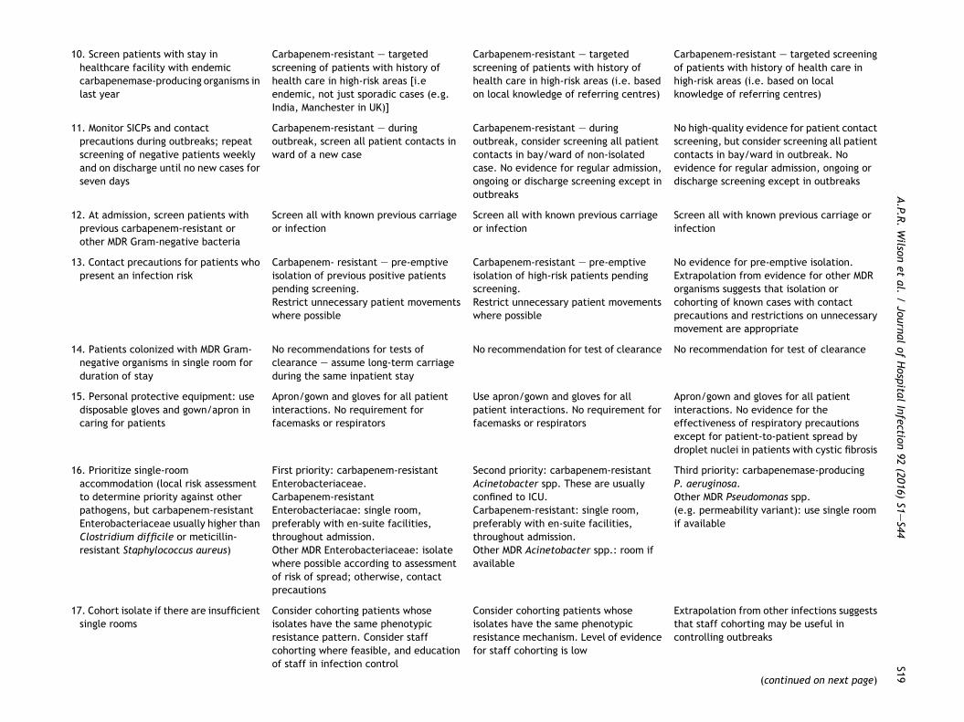
10. Screen patients with stay inhealthcare facility with endemiccarbapenemase-producing organisms inlast year
Carbapenem-resistant e targetedscreening of patients with history ofhealth care in high-risk areas [i.eendemic, not just sporadic cases (e.g.India, Manchester in UK)]
Carbapenem-resistant e targetedscreening of patients with history ofhealth care in high-risk areas (i.e. basedon local knowledge of referring centres)
Carbapenem-resistant e targeted screeningof patients with history of health care inhigh-risk areas (i.e. based on localknowledge of referring centres)
11. Monitor SICPs and contactprecautions during outbreaks; repeatscreening of negative patients weeklyand on discharge until no new cases forseven days
Carbapenem-resistant e duringoutbreak, screen all patient contacts inward of a new case
Carbapenem-resistant e duringoutbreak, consider screening all patientcontacts in bay/ward of non-isolatedcase. No evidence for regular admission,ongoing or discharge screening except inoutbreaks
No high-quality evidence for patient contactscreening, but consider screening all patientcontacts in bay/ward in outbreak. Noevidence for regular admission, ongoing ordischarge screening except in outbreaks
12. At admission, screen patients withprevious carbapenem-resistant orother MDR Gram-negative bacteria
Screen all with known previous carriageor infection
Screen all with known previous carriageor infection
Screen all with known previous carriage orinfection
13. Contact precautions for patients whopresent an infection risk
Carbapenem- resistant e pre-emptiveisolation of previous positive patientspending screening.Restrict unnecessary patient movementswhere possible
Carbapenem-resistant e pre-emptiveisolation of high-risk patients pendingscreening.Restrict unnecessary patient movementswhere possible
No evidence for pre-emptive isolation.Extrapolation from evidence for other MDRorganisms suggests that isolation orcohorting of known cases with contactprecautions and restrictions on unnecessarymovement are appropriate
14. Patients colonized with MDR Gram-negative organisms in single room forduration of stay
No recommendations for tests ofclearance e assume long-term carriageduring the same inpatient stay
No recommendation for test of clearance No recommendation for test of clearance
15. Personal protective equipment: usedisposable gloves and gown/apron incaring for patients
Apron/gown and gloves for all patientinteractions. No requirement forfacemasks or respirators
Use apron/gown and gloves for allpatient interactions. No requirement forfacemasks or respirators
Apron/gown and gloves for all patientinteractions. No evidence for theeffectiveness of respiratory precautionsexcept for patient-to-patient spread bydroplet nuclei in patients with cystic fibrosis
16. Prioritize single-roomaccommodation (local risk assessmentto determine priority against otherpathogens, but carbapenem-resistantEnterobacteriaceae usually higher thanClostridium difficile or meticillin-resistant Staphylococcus aureus)
First priority: carbapenem-resistantEnterobacteriaceae.Carbapenem-resistantEnterobacteriacae: single room,preferably with en-suite facilities,throughout admission.Other MDR Enterobacteriaceae: isolatewhere possible according to assessmentof risk of spread; otherwise, contactprecautions
Second priority: carbapenem-resistantAcinetobacter spp. These are usuallyconfined to ICU.Carbapenem-resistant: single room,preferably with en-suite facilities,throughout admission.Other MDR Acinetobacter spp.: room ifavailable
Third priority: carbapenemase-producingP. aeruginosa.Other MDR Pseudomonas spp.(e.g. permeability variant): use single roomif available
17. Cohort isolate if there are insufficientsingle rooms
Consider cohorting patients whoseisolates have the same phenotypicresistance pattern. Consider staffcohorting where feasible, and educationof staff in infection control
Consider cohorting patients whoseisolates have the same phenotypicresistance mechanism. Level of evidencefor staff cohorting is low
Extrapolation from other infections suggeststhat staff cohorting may be useful incontrolling outbreaks
(continued on next page)
A.P.R
.Wilso
netal.
/Jo
urnalofHospita
lInfectio
n92
(2016)S1eS44
S19
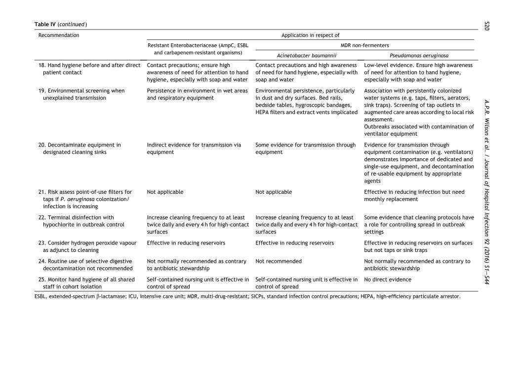
Table IV (continued )
Recommendation Application in respect of
Resistant Enterobacteriaceae (AmpC, ESBL
and carbapenem-resistant organisms)
MDR non-fermenters
Acinetobacter baumannii Pseudomonas aeruginosa
18. Hand hygiene before and after directpatient contact
Contact precautions; ensure highawareness of need for attention to handhygiene, especially with soap and water
Contact precautions and high awarenessof need for hand hygiene, especially withsoap and water
Low-level evidence. Ensure high awarenessof need for attention to hand hygiene,especially with soap and water
19. Environmental screening whenunexplained transmission
Persistence in environment in wet areasand respiratory equipment
Environmental persistence, particularlyin dust and dry surfaces. Bed rails,bedside tables, hygroscopic bandages,HEPA filters and extract vents implicated
Association with persistently colonizedwater systems (e.g. taps, filters, aerators,sink traps). Screening of tap outlets inaugmented care areas according to local riskassessment.Outbreaks associated with contamination ofventilator equipment
20. Decontaminate equipment indesignated cleaning sinks
Indirect evidence for transmission viaequipment
Some evidence for transmission throughequipment
Evidence for transmission throughequipment contamination (e.g. ventilators)demonstrates importance of dedicated andsingle-use equipment, and decontaminationof re-usable equipment by appropriateagents
21. Risk assess point-of-use filters fortaps if P. aeruginosa colonization/infection is increasing
Not applicable Not applicable Effective in reducing infection but needmonthly replacement
22. Terminal disinfection withhypochlorite in outbreak control
Increase cleaning frequency to at leasttwice daily and every 4 h for high-contactsurfaces
Increase cleaning frequency to at leasttwice daily and every 4 h for high-contactsurfaces
Some evidence that cleaning protocols havea role for controlling spread in outbreaksettings
23. Consider hydrogen peroxide vapouras adjunct to cleaning
Effective in reducing reservoirs Effective in reducing reservoirs Effective in reducing reservoirs on surfacesbut not taps or sink traps
24. Routine use of selective digestivedecontamination not recommended
Not normally recommended as contraryto antibiotic stewardship
Not recommended Not normally recommended as contrary toantibiotic stewardship
25. Monitor hand hygiene of all sharedstaff in cohort isolation
Self-contained nursing unit is effective incontrol of spread
Self-contained nursing unit is effective incontrol of spread
No direct evidence
ESBL, extended-spectrum b-lactamase; ICU, intensive care unit; MDR, multi-drug-resistant; SICPs, standard infection control precautions; HEPA, high-efficiency particulate arrestor.
A.P.R
.Wilso
netal.
/Jo
urnalofHospita
lInfectio
n92
(2016)S1eS44
S20
A.P.R. Wilson et al. / Journal of Hospital Infection 92 (2016) S1eS44 S21
All patients with epidemiological links (same hospital unit orcare home) to an index and secondary cases should be screenedto determine the extent of secondary transmission. However,carriage may be prolonged in the community (especially in pa-tients with urinary catheters), and the chronology can bedifficult to determine. As such, isolation is started on re-admission. Admission screening by rectal swab (or axilla/groinswab for Acinetobacter spp.) is required for patients trans-ferred from countries or institutions (including those in the UK)with a prevalence of epidemic or endemic carbapenem-resistant Gram-negative pathogens, as well as those patientswith previous colonization or infection. However, that assumesthe receiving area (infection control practitioner) has beeninformed appropriately of prior hospitalization and carriagedata, and where endemic or epidemic problems are present.
Given reported prolonged gastrointestinal carriage of MDRGram-negative organisms, clearance samples are not recom-mended. Patient isolation should continue for the duration ofthe inpatient stay unless there is extensive spread and largenumbers of colonized or infected patients. Cohorting affectedpatients together may then be necessary. Colonized or infec-ted patients should be isolated if re-admitted as emergencycases, and should then be screened. Previously colonized orinfected patients should be screened as outpatients ifadmission is planned, and isolated on admission if screeningyields the relevant organism. A method of flagging records isneeded.185
9.4.2.3.2. How to screen. As the intestinal flora is the mainsource of MDR Gram-negative bacilli (except Acinetobacterspp.), a rectal swab (or stool) is preferred for ease of collec-tion, handling and processing,162,186 but faecal material mustbe visible on the swab before putting it into transport medium.A stool sample may be used if there is a risk of mucosal trauma.Rectal or perirectal swabs or stool samples have higher yieldthan testing of other body sites.116 In patients with indwellingdevices, specimens from the related site should be screened.Skin swabs, urine and sputum should be checked in those withchronic wounds, indwelling urinary catheters or endotrachealintubation. Acinetobacter spp. are best detected in axillal,groin or wound swabs.187
Screening tests should have a turnaround time of less than48 h. Confirming the specific carbapenemases is important, butrequires molecular methods that often limit availability toreference laboratories. Nevertheless, locally performedphenotypic tests can be extremely helpful as the report isavailable without delay. These tests are easy to implement formost laboratories, provided that the resources are availableand laboratory staff have been trained.162,168 If carbapene-mase confirmation is not possible, isolates should be sent toreference laboratories, although infection control precautionsshould not be delayed. These tests can prove to be even moreuseful if they are interpreted in conjunction with data on thebackground prevalence of carbapenem-resistant organisms in aspecific region. A rapid diagnostic turnaround time and timelycommunication of laboratory results to physicians, nurses andthe infection control team are extremely important for infec-tion prevention and control and clinical therapy.
Commercial media for detection of carbapenemase-producing Enterobacteriaceae are increasingly available. Ina comparison of four chromogenic media used to detectcarbapenemase-producing Enterobacteriaceae, chromIDCarba had the best sensitivity and specificity, although this
may not be adequate for OXA-48.148 Disc or tablet diffusionsynergy tests use meropenem combined with boronic acid toinhibit KPC carbapenemases, or EDTA to inhibit metallo-carbapenemases (IMP, NDM and VIM). Cloxacillin inhibitsAmpC but not KPC, facilitating discrimination between iso-lates with these types of enzyme in an Etest.143 High-leveltemocillin and piperacillin/tazobactam resistance withoutpotentiation of meropenem by EDTA is a marker of OXA-48.Molecular confirmation tests show high sensitivity and speci-ficity, and are used in reference laboratories but are expen-sive and will not detect novel genes. Although severalpseudomonas-selective media are marketed, there are nospecific data on screening methodology, frequency or durationwith respect to P. aeruginosa.161 P. aeruginosa resistant tocarbapenems but susceptible to other b-lactams can beassumed not to have carbapenemases, and do not warrantreference investigation.
The accuracy of carbapenemase detection may be affectedby the species and origin of the pathogen, type of carbapene-mase and other resistance properties, such as porin loss or ESBLproduction. Phenotypic confirmatory tests such as the modifiedHodge test are within the capability of most local laboratories,but depressed AmpC enzymes (and sometimes ESBLs) areassociated with weak false-positive modified Hodge tests,especially with ertapenem. The test can be difficult to inter-pret. Colorimetric and MALDI-ToF methods can be used.188
Some organisms with OXA-48-like carbapenemases onlyexhibit low-level carbapenem resistance without cephalo-sporin resistance, thus escaping the standard identificationmethods.188
High sensitivity and specificity in detecting ESBLs can beachieved using chromogenic selective media, despite mixedflora in catheter urine or faeces. However, competitive bac-terial flora resistant to multiple antibiotics, especially cepha-losporins, can reduce the specificity of selective media. Theuse of CTX-M Chromagar (CHROMagar, Paris, France) is superiorto ESBL chromogenic agars if seeking cases with a CTX-M ESBLin an outbreak, but the medium is less suitable where, forexample, TEM 10 ceftazidimase is present.
Screening for carriage of MDR A. baumannii has beendescribed using a variety of media with samples from variousbody sites including axilla, groin, wounds, rectum orpharynx.104,116,189e192 There is no consensus on site or methodof screening for Acinetobacter spp., and sensitivity is poor.193
Most carbapenem resistance involves OXA-23/40/51/58/143-like carbapenemases, whilst a few isolates have metallo-carbapenemase.194,195
9.4.2.3.3. When to screen. There is insufficient evidence tomandate routine screening of all patients for colonization by allMDR Gram-negative organisms. However, screening of high-riskpatients is used in control efforts for carbapenem-resistantEnterobacteriaceae (e.g. patient transfers from hospitalswhere these organisms are prevalent). The contribution of thispractice to decreasing transmission is unknown. Nevertheless,identifying patients who are at high risk of colonization orinfection with MDR organisms (including carbapenem-resistantorganisms) and performing screening by rectal swab (or skin forAcinetobacter spp.) on admission to healthcare facilities isrecommended, and is now becoming more widespread inhealthcare settings.169 Patients at high risk include patientsadmitted to ICU and from long-term care facilities (e.g. carehomes or endemic areas).

A.P.R. Wilson et al. / Journal of Hospital Infection 92 (2016) S1eS44S22
Screening for carriage of ESBL Enterobacteriaceae canidentify patients in whom empirical treatment with mer-openem may be justified, thus supporting antimicrobial stew-ardship. Discharge screening for ESBL or carbapenem-resistantorganisms is appropriate when admission screening is prac-tised, or if a colonized patient is likely to be re-admitted forfurther procedures or is going to a long-term care facility. Pa-tients with carbapenem-resistant organisms should have notesflagged pending re-admission or if they transfer to long-termcare facilities, where there is a risk of further spread. Long-term care facilities should be informed of positive results ofdischarge screening on their transferees, and may need toconsider if their routine SICPs are sufficiently robust for caringfor these patients.
Good Practice RecommendationsEffective communications between healthcare settings will
help to facilitate efficient patient transfers, and are crucial inreducing spread.
Local screening policies should be developed to define thosepatients at high risk of carriage of, for example, carbapene-mase producers.
9.4.2.4. What can be done in the case of patients unable orunwilling to consent to a rectal swab? On being admitted tohospital, a patient consents to receive those diagnostic andscreening tests that are deemed necessary to the manage-ment of their presenting problem. In situations where a pa-tient is incapacitated, those giving care may proceed withany interventions deemed necessary to provide medicaltreatment for the patient’s well-being. This follows theprinciples of ‘implied consent’ (i.e. it is reasonable to assumethat the person would consent if they were not incapacitatedand unable to do so). Implied consent is already used formeticillin-resistant Staphylococcus aureus (MRSA) screening,even for patients who have the capacity to consent forthemselves.
Screening as part of the ward/hospital policy to guideantimicrobial therapy and/or prevent disease transmissiondoes not require specific written consent, but verbal agree-ment from the patient before sampling is conducted is requiredwhenever possible. Individual, religious and societal concernshave to be respected. Patients should be informed, wheneverpossible, of the need and reason for screening (i.e. that it is fortheir benefit and that of other patients, and what it involves).They should be given the option of who carries it out, includingself-screening, but only after assessment of their ability andwillingness to comply, and safety of the procedure. The optionof a same-sex healthcare practitioner should be provided.Some patients may be unwilling to accept a rectal swab but willprovide a stool sample, although this may result in delay orabsence of a sample. Ideally, patients should be placed pre-emptively in an isolation room while the screening results areawaited, but this is unlikely to be practicable in many highturnover wards. Patients having chemotherapy or with an un-derlying bowel condition (stoma, colon cancer, recent anal orrectal surgery) may be more easily screened using stool.Nevertheless, rectal swabs can be collected safely in haema-tology patients.196
9.4.2.5. How frequently does screening need to be perform-ed? Extensive active screening during outbreaks due to
carbapenem-resistant organisms is recommended168 (e.g.follow-up screening of negative cases at weekly intervals and/or for all inpatient contacts with confirmed cases). Althoughsuch accounts must be interpreted with caution, experiencefrom outbreaks of MDR Gram-negative organisms, includingcarbapenemase- and ESBL-producing Enterobacteriaceae, inacute healthcare settings suggests that the implementation ofscreening for early detection and isolation of colonized pa-tients coupled with contact precautions can help to controltransmission.77,169,197 Screening of those not known to be car-riers of carbapenem-resistant organisms in an endemic situa-tion is advisable at least weekly and on discharge.
9.4.2.6. Is there evidence for effective interventions onpositive patients (i.e. can carriage be cleared)? A number ofstudies have evaluated the duration of colonization with MDRGram-negative bacteria. Studies of hospital inpatients suggestthat they tend to remain colonized for the duration of theirstay.104,198e200 Most studies evaluating the duration of coloni-zation outside of acute settings for a range of MDR Gram-negative bacteria have identified mean durations of coloniza-tion of months rather than days.11,201e204 This duration is likelyto reflect the particular strain, not its resistance.
Several studies have investigated the duration of coloni-zation with carbapenem-resistant Enterobacteriaceaefollowing discharge from acute care facilities. Risk factors forprolonged carriage of MDR Gram-negative bacteria tend to beassociated with healthcare contact, underlying medicalconditions and the presence of invasive devices.11,47,199,205
For example, Lubbert et al. evaluated prolonged coloniza-tion following an outbreak of carbapenem-resistantK. pneumoniae following discharge from an acute hospi-tal.205 Although 26 (31%) of the 84 patients included testednegative for carbapenem-resistant K. pneumoniae at onemonth post discharge, 45% remained colonized at six monthsand one patient remained positive for almost 40 months. Twoother studies found that approximately half of patients colo-nized at the time of hospital discharge were spontaneouslyfree by six months.11,47 However, a number of studies iden-tified patients who retested positive after negative screens,suggesting that gastrointestinal colonization is suppressedrather than eliminated in many cases.47,205 For this reason,the authors of these studies recommend at least threeconsecutive negative screens separated by at least 24 h beforea patient can be considered ‘decolonized’.47,200,205 In prac-tice, colonized inpatients should be considered ‘carriers’during the rest of their hospital admission. The risk periodfrom previous hospitalization exceeds one year.
There is no effective equivalent of the topical suppressionused to reduce shedding of MRSA in the healthcare environ-ment. Attempts at eradication of MDR Gram-negative organ-isms from the gastrointestinal tract have not beensuccessful.206e209 Selective decontamination of the digestivetract (SDD) can produce some temporary reduction in thenumber of organisms in faeces (see Section 9.4.6).
Evidence-based criteria for discontinuing contact precautionsfor carbapenem-resistantGram-negative organisms in acutecaresettings have not been developed. Given the likelihood for pro-longed gastrointestinal carriage by these organisms and risk ofspread, organizations should be cautious in discontinuing contactprecautions (patient isolation). In most cases, contact pre-cautions should continue for the duration of the hospitalization

A.P.R. Wilson et al. / Journal of Hospital Infection 92 (2016) S1eS44 S23
duringwhich the organismwas first found on culture. Patients re-admitted within 12 months of that hospitalization should beconsidered probably colonized, and managed with contact pre-cautions until at least one negative screen is available.
EvidenceEarly recognition of patients infected with MDR Gram-
negative organisms and implementation of rigorous infectioncontrol interventions is usually associated with reduced sec-ondary transmission. 3
Screening (except Acinetobacter spp.) is most sensitivewhen performed on rectal (or perirectal) swabs or stoolspecimens. 3
Patients transferred from, or who have received medicalcare in, a healthcare facility in an endemic area of the UK orabroad are at high risk of carriage of carbapenemase-resistantorganisms. 2þþ
RecommendationsA rectal swab (with visible material) or stool sample (and
urine sample if catheter present) should be used for screeningfor multi-resistant Enterobacteriaceae and P. aeruginosa. ForAcinetobacter spp., sample skin sites or (if a catheter orendotracheal tube is present) urine or respiratory secr-etions. Conditional
Each healthcare organization should have access to robustmicrobiological arrangements for detecting and reporting MDRGram-negative organisms in routine clinical samples, and forscreening using highly-sensitive tests with a rapid diagnosticturnaround time of <48 h. Conditional
All patients transferred from, or with a history in the pre-ceding year of admission to, healthcare facilities with knownendemic carbapenemase-producing Enterobacteriaceae shouldbe screened. Strong
In the event of secondary cases of carbapenem-resistantEnterobacteriaceae, SICPs and contact precautions should bemonitored and re-inforced with clinical staff. Screening ofpatients not identified as carriers should be repeated weeklyand on discharge from affected units until no new cases areidentified for more than seven days. Strong
Where possible, single-room isolation should be provided forpatients with MDR Gram-negative bacterial infection/coloni-zation, and contact precautions should be continued for theduration of their stay. Conditional
Patients with previous samples with carbapenem-resistantor other MDR Gram-negative bacteria should be screened atthe time of admission. Conditional
9.4.3. Isolation and segregationA long-standing principle of infection prevention and control
is to physically segregate those known to be infected or colo-nized with a pathogen of epidemiological importance fromthose who are not infected or colonized. However, criticalappraisal is difficult as segregation is usually assessed as part ofa package of measures. This physical separation can be ach-ieved through placing patients with known/suspected infec-tion/colonization in single rooms; and/or identifying andisolating patients with the same, confirmed pathogen in acohort room/area. An additional cohort measure (nursing staffnumbers permitting) is to identify staff to care for those pa-tients placed in cohorts. The implementation of screeningcultures at the time of admission or during a patient’s stay on a
particular ward is a way to improve the impact of physicalsegregation by identifying those who are colonized with thepathogen of concern.
Placing patients known to be infected or colonized with MDRGram-negative bacteria in single rooms reduces transmission.Several studies have reported the impact of converting a unitfrom multi-occupancy to single rooms on rates of MDR Gram-negative infection/colonization.210e214 A Canadian study re-ported the acquisition rate ratio of various Gram-negativebacteria, comparing an intervention unit that had been con-verted into single rooms with a control unit in a sister hospitalthat had not been converted into single rooms.210 The numberof Acinetobacter spp., Klebsiella spp. and Enterobacter spp.fell significantly, but the number of Pseudomonas spp. andE. coli did not. A 20-year beforeeafter study from a US burnsICU that was converted into single rooms reported a significantreduction in Gram-negative bacteraemia, the time to a firstpositive Gram-negative culture and mortality.212 Significantreductions were also reported in bloodstream infections due toP. aeruginosa, K. pneumoniae, E. cloacae, E. coli and Provi-dencia stuartii. However, there was no control unit, and im-provements in burns care during the study may havecontributed to these reductions. A separate study from thesame renovation reported an overall reduction in infectionrates, mortality and non-enteric Gram-negative species.213
One study from a US ICU reported no significant reduction inoverall infection rates, or in rates of E. coli, Pseudomonas spp.,Acinetobacter spp., Klebsiella spp. or Serratia spp.211 Again,this study did not include a control ward. Hand hygienecompliance was low and did not increase when the unit wasconverted into single rooms, in contrast to other studies.215,216
The impact of enhancing patient isolation aside from otherinterventions has been evaluated. A French study in 2001 foundthat introducing patient isolation to care for ESBL carriersresulted in a sequential reduction in ESBL incidence and hos-pital acquisition.217 However, this preceded the emergence ofCTX-M and E. coli as dominant factors. A study in a VietnameseICU found that improving patient isolation by re-inforcing handhygiene, and limiting exchange of equipment, materials andstaff between patients did not reduce exogenous transmissionof various Gram-negative bacteria (gentamicin-resistantK. pneumoniae, ESBL Enterobacteriaceae, amikacin-resistantAcinetobacter spp. and P. aeruginosa), whereas rates ofMRSA fell significantly. However, this study did not includephysical segregation of patients or the use of gowns oraprons.218
Various studies have evaluated switching from a multi-occupancy ward to a single-occupancy ward. An Israeli studyshowed that there were significantly fewer acquisitions ofresistant organisms when an ICU was converted from a multi-occupancy bay to single rooms.215 The study included a con-trol ward which had not been converted. Patients in thesingle-room ICU had a significantly lower acquisition rate ofresistant organisms when compared with the control multi-occupancy ICU over the same period [3/62 (5%) vs 7/39(18%), respectively, P ¼ 0.043]. SICPs were appliedthroughout, but compliance with hand hygiene was better inthe single-room ICU. Although MDR Gram-negative organismswere included, the significant changes related to Gram-positive pathogens. Another study found that the meannumber of nosocomial infections and length of stay werereduced significantly when a multi-occupancy paediatric ICU
A.P.R. Wilson et al. / Journal of Hospital Infection 92 (2016) S1eS44S24
was converted to single occupancy.219 However, no controlward was used, and no specific data on MDR Gram-negativepathogens were reported.
Although these studies suggest that converting multi-occupancy wards to single rooms reduces the spread of MDRGram-negative pathogens, there are several limitations. First,most studies did not include a control ward (although both ofthe studies that included a control ward found a significantreduction in transmission).210,215 Second, compliance withhand hygiene is higher in a single-room format compared with amulti-occupancy setting.215,216 Thus, it could be that improvedhand hygiene rather than improved physical segregation is thecritical factor for reducing transmission. All of the studies wereperformed in an ICU setting, limiting their applicability tosettings outside of critical care. Converting wards to singlerooms often includes other changes, such as the size of eachbedspace, and the location and number of hand hygiene fa-cilities, which could influence transmission rates.215 Singlerooms are associated with a number of drawbacks, particularlyan increased risk of adverse events due to reduced observationand psychological effects, so the requirement for patientobservation is a key consideration when deciding on theoptimal configuration of wards.220 Finally, patients areincreasingly moved around the hospital for procedures andinvestigations, challenging their segregation.
9.4.3.1. Cohorting staff. No studies have evaluated theimpact of cohorting staff aside from other interventions, butseveral studies have reported cohorting staff as an element of asuccessful multi-faceted strategy.77,104,221,222
9.4.3.2. Disposable aprons and gloves. Data are limited interms of the most appropriate personal protective equipmentto use when caring for patients with MDR Gram-negativebacteria.
Studies have evaluated interventions that have included theuse of gloves and gowns or aprons as an element of contactprecautions.137,217,223,224 Hands and uniforms (or gloves andgowns or aprons if worn) can become contaminated with MDRorganisms.126,127,225e230
The use of gloves is an essential part of prevention of trans-mission of infection, and the evidence has been reviewed else-where.176 While they protect hands from contamination withbiological fluids andmicro-organisms, gloves are not a substitutefor hand hygiene, and unnecessary use can result in increasedcross-contamination. Loveday et al. recommended that glovesshould be removed immediately after the activity has beencompleted, and the hands then decontaminated to preventtransmission as they may be contaminated during glove use orduring removal.176
EvidenceUnits composed of single rooms have less transmission of
MDR Gram-negative bacteria: A. baumannii, ESBL-producing(and, by inference, carbapenemase-producing) Enterobact-eriaceae, P. aeruginosa. 3
RecommendationUse disposable gloves and gowns or aprons to care for pa-
tients with MDR Gram-negative bacteria: A. baumannii,carbapenem-resistant and ESBL Enterobacteriaceae,P. aeruginosa. Strong
9.4.3.3. What is the role of isolation in care home/hospitalsettings? Current practices for the identification and isolationof patients with MDR Gram-negative bacteria (in either singlerooms or cohorts) vary widely among healthcare facil-ities.231,232 A survey of 66 hospitals in 26 US states and 15 othercountries found that 74.2% isolated patients with ESBLs, 93.9%isolated patients with carbapenem-resistant organisms, 81.8%isolated patients with MDR Pseudomonas spp. and 84.9% iso-lated patients with MDR Acinetobacter spp. There wasconsiderable variation in the duration of isolation, and fewfacilities performed screening. Isolation of patients in long-term care facility rooms may not be practicable for psycho-logical reasons.
A number of studies have evaluated the impact of isolatingpatients with ESBL Enterobacteriaceae aside from otherinterventions.217,233e236 A six-year Canadian study evaluatedthe impact of placing patients with ESBL Enterobacteriaceae insingle rooms for the duration of their stay, and applying contactprecautions for symptomatic patients.233 There was an overallincrease in ESBL Enterobacteriaceae colonization/infection,but a relative decrease in cases attributable to the hospital,suggesting that local transmission was reduced. A 2-yearbeforeeafter study in France before the proliferation of CTX-M b-lactamases found that the introduction of admission andweekly surveillance combined with isolation of carriers resul-ted in a significant reduction in the percentage of patientsinfected or colonized with ESBL Enterobacteriaceae.234 Therewas a significant reduction in ESBL-producing K. pneumoniaebut not MDR E. aerogenes. The study has a number of importantconfounders, including an intervention to reduce the use ofimipenem, encouraging prompt discharge and chlorhexidinebathing for isolated patients. A study in a French paediatrichospital evaluated a sequential change in isolation policy fromplacing patients in a single room to placing them in a cohortblock, applying modelling to test the impact on transmission ofESBL producers.235 No significant difference was identifiedbetween the two isolation protocols, but the model suggestedthat single-room or cohort isolation reduced transmission ofESBL producers. Finally, a 12-month cluster randomized studyin 13 European ICUs evaluated chromogenic agar screening forESBL Enterobacteriaceae on admission and isolation of carriersvs no admission screening.236 A hand hygiene improvementprogramme for staff and chlorhexidine body washing for pa-tients preceded the trial. Of the 2129 Enterobacteriaceaeresistant to third- and fourth-generation cephalosporins foundon screening, 29% were resistant to carbapenems. Screeningand source isolation was not associated with any trend (duringthe intervention period) or step change (compared with abaseline period) in rate of ESBL transmission. However, indi-vidual species were not analysed separately.
Two studies have evaluated the impact of isolation on thetransmission of A. baumannii and P. aeruginosa.137,223 A seven-year beforeeafter study in France evaluated the introductionof hospital-wide contact precautions, which included placingthe patient in a single room or cohort with other patients, andthe use of gloves and gowns or aprons, on the transmission ofA. baumannii.223 Isolation precautions were implemented fortwo years, then stopped for three years, then re-implementedfor two years; the incidence of A. baumannii was significantlylower during the two periods when isolation precautions werein use. The implementation of isolation precautions was theonly variable associated with lower incidence of A. baumannii

A.P.R. Wilson et al. / Journal of Hospital Infection 92 (2016) S1eS44 S25
in multi-variate analysis. A follow-up study from the samegroup reported similar findings.237 A study from an ICU in Brazilevaluated the introduction of contact isolation, including theuse of gloves and gowns or aprons, cohorting of medical items(stethoscopes etc.), daily surface cleaning and disinfection onthe rates of MDR A. baumannii and P. aeruginosa.137 Blood-stream infections reduced significantly during the interven-tion, but species were not analysed separately. The studycompared rates of infection at two points during the inter-vention, with no baseline period.
A number of other studies have implemented complex in-terventions that included increased isolation of pa-tients.116,221,222,224,238 One study in a US ICU implemented amulti-faceted intervention centred on improved isolation ofpatients, including admission screening for carbapenem-resistant K. pneumoniae but not for A. baumannii orP. aeruginosa.221 There was a significant reduction in the rateof carbapenem-resistant K. pneumoniae, but no change in therate of A. baumannii or P. aeruginosa. A study of various in-terventions in six US hospitals found that the only consistentpredictor of successful control of K. pneumoniae with KPCcarbapenemases was a shorter length of stay.238 Three studieshave evaluated the impact of a multi-faceted intervention tocontrol the spread of A. baumannii. An intervention at US fieldhospitals in Iraq including improved hand hygiene, contactprecautions, and cohorting patients and staff resulted in asignificant reduction in A. baumannii ventilator-associatedpneumonia.222 A bundle of interventions including contactprecautions, screening (on occasion) and regular staff briefingsreduced the rate of MDR A. baumannii infections significantlyoverall, particularly bloodstream infections due to the organ-isms, in a Spanish hospital.116 Finally, a study in a Thai ICUreported a significant reduction in MDR A. baumannii associ-ated with the introduction of contact precautions, cohortingcolonized patients, screening and environmental disinfectionusing bleach.224 However, acinetobacter infections are morecommon in Thailand than in the UK.
Studies that have evaluated the impact of isolating patientsin either single rooms or cohorts generally lack a concurrentcontrol unit, making it difficult to be certain that changes inrate are attributable to isolation alone. The one study thatincluded a concurrent control ward demonstrated no signifi-cant decrease in transmission of ESBL-producingEnterobacteriaceae.236
EvidenceTransmission of MDR Gram-negative bacteria is reduced by
identifying patients who are infected or colonized, and placingthem in single rooms or cohorts. 3
RecommendationsIdentify and place infected and colonized patients in single
rooms where available in this order of priority: carbapenem-resistant Enterobacteriaceae, carbapenem-resistantA. baumannii, ESBL Klebsiella spp., carbapenemase-producingP. aeruginosa, ESBL E. coli and other Enterobacteriaceae,AmpC Enterobacteriaceae. Strong
If insufficient rooms are available, cohort isolate followinglocal risk assessment. Conditional
Good Practice RecommendationEstablish a flagging system for patient notes. Conditional
9.4.4. Hand hygieneHand hygiene is an essential part of prevention of trans-
mission of infection in health care, and it has been reviewed byLoveday et al.176 Transient flora, including MDR Gram-negativebacteria, is acquired from touching the patient or environ-ment, and is easily transferred to the next patient or surface.In turn, this causes colonization and later (potentially) infec-tion. Use of alcohol hand rub (or liquid soap and water if handsare visibly soiled) has been shown to reduce the carriage ofpotential pathogens on the hands, and therefore is likely toreduce the number and likelihood of healthcare-associatedinfections.174,176 Hand decontamination is considered to havea high impact on outcomes that are important to patients.239
Training of all care workers, combined with audit and feed-back of compliance rates, is central to current guidance onprevention of infection. The WHO Five Moments for Hand Hy-giene has been widely used and supported by NICE, and de-scribes the points at which hand hygiene is required.239 Handhygiene is often missed as the worker enters or leaves thepatient environment, and on contact with potentiallycontaminated surfaces240e242
Despite many activities around the improvement of handhygiene products, the introduction of alcohol-based hand rubs/gel, improving environmental factors, and extensive marketingand education campaigns, evidence of sustained complianceimprovement is lacking. Globally, hand hygiene compliance isvariable, with Creedon243 and Larson et al.244 suggesting a rateof approximately 50%. Therefore, improving compliance withhand hygiene is reliant upon an understanding of the in-teractions within health care between the individuals, thepractices and procedures they perform, the environment thatthey work in, and the culture and safety awareness of the or-ganization. These human factors should be applied to theinfection prevention agenda to ensure that meaningful, sus-tainable interventions are adopted reliably to produce thegreatest impact.245
EvidenceHand hygiene is associated with reduction of carriage of
potential pathogens on the hands. 2þ
RecommendationHand hygiene is required before and after direct patient
contact; after contact with body fluids, mucous membranesand non-intact skin; after contact with the immediate patientenvironment; and immediately after the removal ofgloves. Strong
9.4.5. Environmental hygiene9.4.5.1. When should the environment be sampled? The roleof the environment in the transmission of healthcare-associatedinfection remains controversial and difficult to study. In recentyears, much of the research around this has focused on high-profile Gram-positive pathogens such as MRSA, Clostridiumdifficile and glycopeptide-resistant enterococci, where a sig-nificant body of evidence for the importance of environmentalsources has started to accumulate.246,247 Reservoirs can beidentified when infection control has failed to control anoutbreak.161 The general view has been that Gram-negativebacteria, particularly members of the Enterobacteriaceae, arenot as successful at surviving in the environment for prolongedperiods, and generally are relatively easily removed by

A.P.R. Wilson et al. / Journal of Hospital Infection 92 (2016) S1eS44S26
appropriate conventional cleaning and drying.13 Nevertheless,Enterobacteriaceae, including carbapenem-resistant strains,are able to survive on dry surfaces for extended periods,sometimes measured in weeks and months.248,249 Some strainsof E. coli carrying ESBLs, for example, can survive for a medianof 10 days in the environment.250 In favourable conditions,E. coli, Klebsiella spp. and Pseudomonas spp. can survive foreven longer.161 Acinetobacter spp. have the capacity for long-term survival on dry surfaces.14,251,252,253 Pseudomonas spp.are more traditionally associated with moist environments, andwill be found around handwash basins and respiratory equip-ment.35,254 There is considerable evidence that P. aeruginosacan contaminate waste water systems and spread from them,with these sometimes acting as reservoirs for carbapenemase-producing strains causing prolonged outbreaks.255 If notadequately decontaminated (including the hand piece), endo-scopes have been found to be responsible for outbreaks.256
MDR Gram-negative bacteria, including carbapenem-resistant organisms, Enterobacteriaceae and A. baumannii,can be cultured from sites surrounding infected and/or colo-nized patients.106,257 Several studies have found that environ-mental contamination with resistant K. pneumoniae is morecommon than contamination with resistant E. coli.258e260 Nocontrolled studies have shown that an environmental inter-vention reduces the transmission of MDR Gram-negative rods.However, contamination with MDR Gram-negative bacteria canpersist despite cleaning and disinfection.106,261,262 Epidemio-logical data suggest that admission to an ICU room previouslyoccupied by patients infected or colonized with A. baumanniior P. aeruginosa presents an increased risk for acquiring theseorganisms.252 A significant correlation was noted between thenumber of environmental swabs in monthly screening and thenumber of patients with colonization/infection in the samemonth (P ¼ 0.004).103 However, this association was not seenfor resistant Enterobacteriaceae, suggesting that the environ-ment is less important in their transmission,252,263 albeit withthe caveat that these studies did not stratify by Enter-obacteriaceae spp., which may be important given theincreased capacity for K. pneumoniae, in particular, tocontaminate and survive on hospital surfaces.
The evidence for the benefit of environmental screening islimited, and environmental sampling, in itself, will not limittransmission of MDR Gram-negative bacteria.264 The purpose ofscreening may be to draw attention to failure of clearance of anoutbreak strain by cleaning, or to point to a possible commonsource for a cluster or outbreak. Most of the evidence sur-rounding environmental screening is within reports of manage-ment of outbreaks, and there is often very little detail on sitessampled, sampling technique or method of culture.265 Whenlooking for small numbers of organisms or bacteria living inbiofilms, the yield can be improved by using enriched culture,but this precludes quantitation. An alternative to specificallyseeking the MDR outbreak strain(s) itself is to assess the degreeof microbiological contamination by using surface contactplates and undertaking quantitative bacterial culture.266 Sur-face contact plates using selective media for specific pathogenscan suffer from low sensitivity, but usewith non-selectivemediamay be helpful. Choosing sites for sampling remains problematicas any sampling can only reflect a very small fraction of therelevant environment.267 The general approach should be tochoose sites that are likely to be relevant for cross-transmission,such as work surfaces close to the patient and equipment/
surfaces that are likely to be touched frequently (e.g. computerkeyboards, bed rails and door handles).264,268
Given the difficulties with microbiological sampling of theenvironment, there has been considerable interest in theavailability of technologies that can indirectly assess microbialcontamination. Adenosine triphosphate (ATP) bioluminescencehas been used for many years in the food industry, where visualassessment is considered to be insufficient to assess the risk ofinfection from contaminated surfaces. It has a number of po-tential benefits, including simplicity and immediacy of feed-back; however, it can give only indirect information on thelikelihood of bacterial contamination, and bacteria themselvesare not isolated for investigation and comparison with thosecausing outbreaks. Its use in hospitals can be problematic, asvalues can be variable and there is little consensus on anappropriate benchmark value to indicate inadequate cleanli-ness.166,269,270 Results are prone to interference by differentdisinfectants.271 Where ATP bioluminescence has been usedmost successfully has been on a continuous basis to improvecompliance through feedback and improved cleaning.272,273
Other methods of controlling the process of cleaning, such asthe use of fluorescent gel markers, can be useful in auditing thecleaning process and feedback to cleaning staff.274
EvidenceTransmission of MDR Gram-negative bacteria, particularly
non-fermenters, has been associated with contamination ofthe environment, water systems or equipment. 2�
Sampling of the environment can be useful in identifyingsources of ongoing transmission or a single common source foran outbreak. 2�
RecommendationEnvironmental screening should be considered where there
is any unexplained transmission of MDR Gram-negative organ-isms or a possible common source for an outbreak. Strong
9.4.5.2. What is the evidence that respiratory equipmentcontributes to transmission? The respiratory tract of venti-lated patients in critical care units is a frequent site of car-riage of MDR Gram-negative bacteria. In one study in India,87% of samples from ventilators, humidifiers, nebulizers andother respiratory equipment showed bacterial colonization,and 17/42 Gram-negative isolates were multi-drug resis-tant.275 Endotracheal suctioning is a potential cause of cross-infection, as disconnection of the system may allow airbornespread to the patient’s skin, staff hands and the immediateenvironment. In a crossover study in the Netherlands, therewas no significant difference in acquisition of Gram-negativebacteria during periods of using closed suction vs open suc-tion in ventilated patients.276 However, antibiotic resistancerates were low and similar in each group. Comparing venti-lated with non-ventilated patients in the ICU, non-fermenting and enteric Gram-negative bacilli were reportedmore frequently in ventilator-associated pneumonia, but thisreflected the number of samples cultured per patient. Theoverall proportions of different pathogenic species weresimilar.277
Inappropriate washing of ventilator or endotrachealtubing in handwash sinks risks the spread of Gram-negativepathogens. Environmental sampling in two outbreaks ofMDR P. aeruginosa showed it to have colonized the waste
A.P.R. Wilson et al. / Journal of Hospital Infection 92 (2016) S1eS44 S27
water system, with blockages, splashback and spillage fromshowers being possible modes of spread.35 Direction of waterdirectly into the outlet caused spread of organisms from thesink drain trap. Sinks were fitted with a horizontal drainoutlet at the back of the basin, resolving the problem. Sterilefluids (not tap water) must be used to clear suction equip-ment, as the latter may be contaminated with pseudomonadsor Enterobacteriaceae, and these can be disseminated whenthe equipment is next used, including when catheters arechanged.
EvidenceGram-negative bacteria colonize respiratory equipment and
may be washed into sink traps. 2þ
RecommendationRespiratory and other contaminated equipment should be
decontaminated (or respiratory secretions discarded) awayfrom the immediate bed area in designated cleaning sinks andnot in handwash sinks. Strong
Good Practice RecommendationDo not discard patient wash water, body fluids, secretions or
exudates into handwash basins.
9.4.5.3. What is the evidence that sensor taps contribute totransmission? P. aeruginosa, including MDR strains, in watersources in ICUs has long been recognized as being associatedwith the development of bacteraemia and pneumonia in pa-tients.278 A recent systematic review demonstrated evidenceof transmission of P. aeruginosa from water systems to pa-tients and vice versa.34 Point-of-use filters and increasingchlorine disinfection were effective interventions. Non-touchtaps were identified as probable risk factors for biofilm for-mation and subsequent transmission to patients. Sinks andnurses’ hands have been identified as possible vectors. In aneonatal unit, four neonates were infected and 44 werecolonized by cross-infection with one clone.124 In anotherneonatal unit in Germany, P. aeruginosa was isolated from thenasal prongs of 22 babies and from nine respiratory waterreservoirs.279 The hands of staff were the likely means oftransmission. Other outbreaks have been due to contaminateddetergent-disinfectant solution used in cleaning surfaces.280
Studies have confirmed the effectiveness of point-of-use fil-ters on water outlets in reducing infection in critical careunits, but as they were descriptive cohort studies, they weresubject to temporal variation.281e283 Filters have to bereplaced regularly, are expensive and can themselves be thesource of contamination. P. aeruginosa is found in biofilm inflow straighteners, metal support collars and the adjacentparts of tap bodies. The level of contamination with Pseudo-monas spp. is highest on complex flow straighteners, inte-grated mixers and solenoids.278
Sensor taps have been implicated in some outbreaks ofpseudomonas bacteraemia and other infections in augmentedcare units as the flow is slow and controlled, and the internalmechanism is complex. Decontamination of sensor taps can beperformed284 and, in one major outbreak, all taps, mixervalves, flexible hoses and flow straighteners were replacedwith simpler designs.278
Where there are vulnerable patients (i.e. augmented care),the Department of Health in England recommends a regular
water-testing regimen; clinical surveillance is sufficient else-where. The frequency of testing depends on previous isolationof the organism and proximity to patients at risk (e.g. neo-nates). The purpose is to prevent colonization before thedevelopment of infection. A risk assessment to mitigate risksand a water safety plan are advised, including consideration ofremoval of thermostatic mixer valves and flow straighteners,and the design of the sink.36 Infrequently-used taps should beflushed at full flow for 1 min daily or removed altogether. In theevent of contaminated supplies, sterile water should be usedfor neonates, and single-use wipes should be used for otherpatient hygiene together with additional hand hygiene usingalcohol gel after washing.
EvidenceThe presence of infections with P. aeruginosa in patients is
commonly associated with isolation of these bacteria from unittaps. 2�
The installation of point-of-use filters is associated with areduction in pseudomonal infections. 3
RecommendationFor P. aeruginosa, including MDR strains, at a minimum in
accordance with the organization’s water safety plan, a riskassessment should be made when levels of patient colonizationor infection rise, in order to determine if point-of-use filtersshould be installed or taps changed. Strong
9.4.5.4. Is any cleaning method more effective than others atremoving multi-drug-resistant Gram-negative bacilli from theenvironment? The importance of persistence of transmission ofMDR Gram-negative bacteria in the environment remains uncer-tain.246,285 However, maintaining a clean environment andappropriately decontaminating all relevant equipment is anessential component of any infection prevention and controlprogramme. The cleanliness of healthcare premises is an impor-tant component in the provision of clean safe care.286 In England,the NHS Constitution pledges ‘The NHS commits to ensure thatservicesareprovided inacleanandsafeenvironment that isfit forpurpose, based on national best practice’.287 Whilst there havebeen significant improvements in the cleanliness of Englishhealthcare premises, there is still room for improvement.286
The optimal methods for cleaning have been poorly studiedwith respect to MDR Gram-negative bacteria. Poor cleaningpractice, such as inappropriate dilution of cleaning agent orinactivation of disinfectant by organic matter, is more likely toreduce cleaning efficacy than theoretical cross-resistance be-tween disinfectant and antibiotic.288 In extreme cases, agentsused for cleaning can even become contaminated with Gram-negative bacteria, especially pseudomonads.141,289e291
Conventional decontamination is carried out by a humanoperator, and the reliance on the operator to select the correctproduct, dilution, distribution and surface contact time has thepotential for decontamination failure. Debate around the useof chemical disinfectants vs detergents for routine cleaning isgathering momentum with concerns regarding chemical resis-tance, optimum disinfectant contact times, allergies amongstusers and patients, and costs. In short, practice varies widelyand monitoring is traditionally by visual inspection, which canbe subjective. Efficacy is rarely measured.
Although persistence of Gram-negative organisms on re-usable bedpans is a potential mode of spread, there are no

A.P.R. Wilson et al. / Journal of Hospital Infection 92 (2016) S1eS44S28
recent reviews of performance with respect to Gram-negativebacteria. In automated washer disinfectors, a combination ofalkaline detergent and temperature over 85�C for 1 min issufficient to eliminate C. difficile spores.292 However, visiblefaecal soil can remain on 7e33% of bedpans, so appropriateeducation is important to ensure good practice.293 Earlystudies showed that failure to attain 80
�C was associated with
persistence of E. coli and P. aeruginosa.294 Transmission ofpathogens via contaminated endoscopes is usually due tofailure to comply with appropriate reprocessing practiceguidelines.295 Although some cases of transmission have beenknown, relatively few have been reported in peer-reviewedjournals.
Assessment of the activity of disinfectants against MDRGram-negative bacteria is hampered by the differences be-tween testing against organisms in suspension and on a surface.Acinetobacter spp. and Pseudomonas spp. may survive in theward environment even after bleach disinfection, and increasethe risk of acquisition by patients.106,252 In a prospective cohortstudy, cleaning the environment with sodium hypochloriteinstead of detergent-disinfectant plus chlorhexidine bathing ofpatients reduced the risk of colonization with MDRA. baumannii becoming established beyond that achieved byimproved contact precautions, cohorting, screening and anti-microbial stewardship.296 Termination of acinetobacter out-breaks has been associated with closure and cleaning of units,and removal of a reservoir has been associated with termina-tion of outbreaks for a variety of organisms.161
Most interventions to improve cleaning have been accom-panied by a package of infection control precautions. Educa-tion campaigns and use of audit tools such as marking surfaceswith fluorescent dye reduce contamination, but their effectmay be transient.
Recently, there has been significant interest in the role of‘no-touch’ automated disinfection systems as an additionalmeasure for terminal cleaning of single rooms or hospital areasaffected by clusters or outbreaks.297 Various types of systemsare currently marketed, including aerosolized or vapourizedhydrogen peroxide, and ultraviolet radiation. Each has distinctmicrobiological and practical characteristics. A number ofstudies have shown improved efficacy of killing of variouspathogens compared with cleaning alone, particularly in out-breaks, but there are limited data on whether this reducesacquisition rates of pathogens.298e300 Hydrogen peroxide vapourfrom 30% H2O2 and, in one case, aerosolized hydrogen peroxidefrom 5% H2O2 have been used successfully as part of a bundle ofinterventions to prevent transmission ofMDRAcinetobacter spp.in outbreaks,105,107e109 and hydrogen peroxide vapour has beenused to help control outbreaks of resistant Enterobacteriaceae,including those resistant to carbapenems.262,301,302
Patients admitted to rooms exposed to hydrogen peroxidevapour were significantly less likely to acquire any MDR or-ganisms than patients admitted to rooms that had not beentreated with hydrogen peroxide vapour in a cohort interventionstudy.299 K. pneumoniae, P. aeruginosa and A. baumannii insuspension are all susceptible to low concentrations ofhydrogen peroxide, but efficacy is significantly reduced whenthe organisms are in biofilm; some P. aeruginosa in biofilm hadprolonged survival following hydrogen peroxide exposure in thelaboratory.303 Hydrogen peroxide vapour (from 30% H2O2) ismore effective in eradicating surface A. baumannii thanaerosolized hydrogen peroxide (from 5% H2O2) (>6 log vs 1-4 log
reduction).304 It is essential to ensure that the area has beenthoroughly cleaned first, because efficacy is affected by thepresence of organic soiling.
EvidenceCleaning is important in the control of outbreaks due to MDR
Acinetobacter spp., and failure to clean specific areas or piecesof equipment has been associated with transmission of otherMDR Gram-negative bacteria. However, evidence derived frombeforeeafter studies and outbreak reports is open tobias. 2�
Hydrogen peroxide vapour is effective in reducing environ-mental reservoirs of Acinetobacter spp. and other Gram-negative bacteria on surfaces (but not sink traps) if used inaddition to standard cleaning. 2þ
RecommendationsTerminal disinfection of vacated areas with hypochlorite
should be used in the control of outbreaks of MDR Gram-negative infection. Conditional
Hydrogen peroxide vapour should be considered as anadjunctive measure to follow cleaning of vacated isolationrooms/areas. Conditional
Good Practice RecommendationIncrease cleaning frequency to at least twice daily, and
every 4 h for high-contact surfaces in the presence of resistantEnterobacteriaece and Acinetobacter spp.
9.4.6. Selective decontamination: why is it not used? Isthere a role?
SDD is an intervention that aims to reduce mortality andmorbidity due to healthcare-associated infection in ICUs. Itcomprises application of non-absorbable antibiotics to themouth and stomach together with a course of broad-spectrumintravenous antibiotic(s). A modification uses only the topicalelement of decontamination [selective oropharyngeal decon-tamination (SOD)]. The practice has been investigated exten-sively, largely in the Netherlands, and at least 12 meta-analyses of published papers have been produced.305 Almostone-third of the trials suggest a significant reduction in theincidence of Gram-negative pneumonia, and one large ran-domized study demonstrated a small but significant reductionin mortality in a country with low levels of antibiotic resis-tance.306 Despite issues with blinding, heterogeneity andcompliance, both SDD and SOD appear to be associated with areduction in pneumonia (OR 0.32, 95% CI 0.26e0.38) and mor-tality (OR 0.75, 95% CI 0.65e0.87).307
Nevertheless, only 5% of UK ICUs use SDD, largely becauseuniversal use of prophylactic antibiotics is counter to the te-nets of antimicrobial stewardship.308,309 In some studies withlong-term surveillance, both SDD and SOD were associated withan increase in resistance to ceftazidime in Gram-negative floraof the respiratory tract, although systemic antibiotics werealso given in many cases.310e312 In the short term, however, asystematic review found a significant reduction in resistance ofGram-negative bacilli to third-generation cephalosporins dur-ing the use of selective decontamination.208 The relevance offindings from one country to another is unclear when patternsand prevalence rates of resistant Gram-negative bacteria varyso widely. Preparation requires suitable manufacturing units,and administration can be labour intensive.

A.P.R. Wilson et al. / Journal of Hospital Infection 92 (2016) S1eS44 S29
Recent high-quality studies have provided evidence thatdaily bathing using chlorhexidine gluconate in ICUs helps toreduce bacteraemia, but there is limited evidence of itsspecific effect on MDR Gram-negative bacterialinfection.313e316 This has formed part of successful bundles ofinterventions, but has not been tested as an isolated meas-ure.236,296,317e319 Some studies that have evaluated chlor-hexidine as a single intervention including randomizationhave failed to demonstrate a reduction in Gram-negativebacteraemia.314,316,320 There is no strong evidence that dailybathing with chlorhexidine reduces Gram-negative infectionor colonization, and there is a risk of development of resis-tance.114 The routine use of oropharyngeal chlorhexidine hasbeen associated with an increase in mortality in one system-atic review.321
Selective decontamination can temporarily suppressexcretion of carbapenem-resistant organisms from thegastrointestinal tract and possibly supplement SICPs.207 In aretrospective analysis of a German outbreak, SDD withcolistin and gentamicin as oral solution and gel was used in14 patients with proven carriage of carbapenemase-producing K. pneumoniae KPC-s-KP ST258.311 Loss of car-riage, as defined by three PCR screens 48 h apart, was foundat a mean of 21 days in six treated patients (43%), but also in30% of controls. Resistance to colistin and gentamicin inpost-treatment isolates of K. pneumoniae rose 19% and 45%,respectively, compared with controls. In a randomizedplacebo-controlled trial of a regimen based on colistin andneomycin plus treatment of bacteriuria with nitrofurantoin,the detection of ESBL Enterobacteriaceae in rectal swabswas not affected significantly.322 A randomized trial againstplacebo used oral gentamicin and topical oropharyngealgentamicin and colistin for one week, and was directedagainst carbapenem-resistant K. pneumoniae carriage. Itproduced a significant reduction in carriage at two weeksbut not at six weeks.207 Mortality was not affected signifi-cantly, but throat carriage was reduced from 30% to zero inthe intervention vs 35% to 30% with placebo (P < 0.0001).Isolates did not develop resistance. A lower-qualitycontrolled study reported reductions in faecal and pharyn-geal carriage of Acinetobacter spp. during colistin-/tobra-mycin-selective decontamination.323 Against this, colistin/neomycin/nalidixic acid did not reduce infections or mor-tality due to MDR Enterobacteriaceae compared with noprophylaxis, but carriage was reduced (N ¼ 86, RR 0.28, 95%CI 0.03e2.28).324
EvidenceThe use of SDD or SOD is associated with a reduction in the
incidence of pneumonia due to Gram-negative bact-eria. 1þ
The use of SDD or SOD is associated with a reduction in themortality rate. 1þ
SOD can be used to reduce excretion of MDR Gram-negativebacteria during an outbreak, but no effect on transmission ofinfection has been demonstrated, and application can belogistically difficult. 3
RecommendationThe routine use of SDD or SOD is not recommended for
control of MDR Gram-negative bacteria. Conditional
9.5. What are the minimum standards to stop spreadin public areas, primary care or care homes?
Care homes are recognized as potential reservoirs of MDRpathogens, which can spread among residents, generally ascolonizers, and can be re-introduced into hospitals.325 Pub-licity may alarm residents, their relatives and carers. Clearinformation on the standards of infection prevention andcontrol should be available to promote confidence in thequality of care provided. Despite the relatively poor evidencebase, guidelines are available for managers and carers.239,326
In England, the Code of Practice326 defines what is requiredto ensure compliance with Care Quality Commission regis-tration requirements for cleanliness and infection control.Following general principles, owners are encouraged tocontact the local health protection team in the event ofoutbreaks of infection, increase cleaning and hand hygienecompliance by patients and staff, conduct root cause ana-lyses, and train staff in infection prevention and control. Theprevalence of ESBL Enterobacteriaceae in the care home isusually unknown and sporadic. The need for a urinary cath-eter, if present, should be reviewed regularly and it shouldbe removed as soon as it is not needed. The appropriate useof such devices to prevent healthcare-acquired infection hasbeen reviewed elsewhere.166 Residents with diarrhoea shouldbe isolated in their room with a dedicated commode if no en-suite facilities are available. This isolation may requireadditional psychological support for the resident. Coloniza-tion with MDR organisms should not be construed as a reasonto isolate or screen other residents. SICPs must continue tobe applied.
EvidenceInstitutionalized patients/residents are more likely to carry
MDR organisms and present a risk on re-admission tohospital. 2þ
Good Practice RecommendationsCare homes should adopt national recommendations for
environmental and equipment cleanliness, and infection pre-vention and control.
Clear patient information on MDR Gram-negative infectionmust be provided in accessible formats to encourage good handhygiene by patients and staff.
9.6. Are there organizational structures within ahealthcare facility that play a role in the successfulcontrol of multi-drug-resistant Gram-negative bacilli?
In Israel, with prevalent ST258/KPC K. pneumoniae, a self-contained nursing unit with dedicated staff for patients car-rying carbapenem-producing Enterobacteriaceae was effec-tive as part of a package of measures including mandatoryreporting and a centralized national monitoring system.77 Inthe same way, a centralized national system was effective inthe control of MRSA in the UK. Outbreaks of carbapenemase-producing K. pneumoniae appear to have been controlled bya package of measures, including nursing infected patients inseparate units with different staff until discharge orcohorting.197,262,327e329 The nursing units could be single
A.P.R. Wilson et al. / Journal of Hospital Infection 92 (2016) S1eS44S30
rooms, cohorts or wards, but the nurse did not cross betweenunits during the shift. A sequential intervention study foundthat control was effective when patients were nursed inseparate locations with dedicated nursing personnel. Theshared medical staff had contact precautions vetted by thededicated nurse before entering the area.328 However, theintervention also included education, training, cleaning andscreening.
EvidenceA separate unit with dedicated nursing staff rather than part
of a ward with shared staff is effective in controlpackages. 3
RecommendationMonitor hand hygiene of all staff when patient cohorting is
being applied. Strong
10. Further research
� PCR-based tests for swift detection of MDR Gram-negativepathogens require further testing and development toallow cost-effective and rapid screening and detection ofcarriers.
� Consideration should be given to further investigation ofscreening patients of long-term care facilities as a means oftracking movement and defining reservoirs of organisms.Longitudinal assessment of patients carrying ESBL-producing organisms, particularly E. coli ST131, should bemade on admission and discharge to hospitals and nursinghomes to detect infection and spontaneous clearance ofcarriage.
� The risk factors for development of infection, such as urinarycatheterization, should be separated from the risk factors forcarriage, including hospitalization. Household contacts ofcases of infection with E. coli ST131 or carbapenem-resistantK. pneumoniae should be examined for evidence of spread orsubsequent infection.
� Given the recent changes in the prevalence of, for example,ESBL-producing E. coli ST131 and K. pneumoniae ST258 withKPC carbapenemases, the potential for screening patients forparticular clones as means of controlling transmission in hos-pitals should be assessed. MALDI-ToF, serological and DNA-based detection methods should be developed, togetherwith selective or indicator media for quinolone-resistantEnterobacteriaceae.
� Bacteraemia surveillance including sequence typing forstrains resistant to cephalosporins, fluoroquinolones andcarbapenems should be expanded and open to public andcommissioning groups. This is essential for accurate riskassessment of patients transferred between healthcare fa-cilities. In localities with outbreaks, typing and infectioncontrol should be extended to isolates from other anatom-ical sites.
� Randomized intervention studies of effectiveness measures,including single-room isolation, in the prevention of trans-mission of Gram-negative bacterial infection.
� The potential for endoscopes to disseminate MDR Gram-negative pathogens requires better understanding withreference to complex instruments such as those with wire-containing channels.
Conflict of interest statementSee full details in Section 4.4.
References
1. Scottish Intercollegiate Guidelines Network. SIGN 50: a guidelinedeveloper’s handbook. Revised edition. Edinburgh: HealthcareImprovement Scotland; 2014. Available at: http://www.sign.ac.uk [last accessed December 2014].
2. Higgins JPT, Green S, editors. Cochrane handbook for systematicreviews of interventions version 5.1.0. London: CochraneCollaboration; 2011.
3. Brouwers M, Kho ME, Browman GP, et al.; AGREE Next StepsConsortium. AGREE II: advancing guideline development, reportingand evaluation in healthcare. CMAJ 2010;182:E839eE842.
4. Public Health England. English surveillance programme forantimicrobial utilisation and resistance (ESPAUR). London: PHE;2014. Available at: https://www.gov.uk/government/uploads/system/uploads/attachment_data/file/362374/ESPAUR_Report_2014__3_.pdf [last accessed October 2014].
5. Hawkey P. Treatment of multi-drug resistant (MDR) Gram-negative bacteria e recommendations from a Joint WorkingParty. J Antimicrob Chemother 2015 (submitted).
6. Cochrane Effective Practice and Organisation of Care ReviewGroup. EPOC resources. London: Cochrane Collaboration; 2013.Available at: http://epoc.cochrane.org/epoc-resources [lastaccessed June 2014].
7. Borenstein M, Hedges LV, Higgins JPT, Rothstein HR. Introductionto meta-analysis. Chichester: John Wiley and Sons Ltd; 2009.
8. Review Manager (RevMan) Version 5.1. Copenhagen: NordicCochrane Centre, Cochrane Collaboration; 2011.
9. Rooney PJ, O’Leary MC, Loughrey AC, et al. Nursing homes as areservoir of extended-spectrum beta-lactamase (ESBL)-producingciprofloxacin-resistant Escherichia coli. J Antimicrob Chemother2009;64:635e641.
10. Leaper D, Tanner J, Kiernan M. Surveillance of surgical siteinfection: more accurate definitions and intensive recordingneeded. J Hosp Infect 2013;83:83e86.
11. Zimmerman FS, Assous MV, Bdolah-Abram T, Lachish T,Yinnon AM, Wiener-Well Y. Duration of carriage of carbapenem-resistant Enterobacteriaceae following hospital discharge. Am JInfect Control 2013;41:190e194.
12. Villegas MV, Hartstein AI. Acinetobacter outbreaks, 1977e2000.Infect Control Hosp Epidemiol 2003;24:284e295.
13. Casewell MW, Desai N. Survival of multiply-resistant Klebsiellaaerogenes and other Gram-negative bacilli on finger-tips. J HospInfect 1983;4:350e360.
14. Jawad A, Seifert H, Snelling AM, Heritage J, Hawkey PM. Survival ofAcinetobacter baumannii on dry surfaces: comparison of outbreakand sporadic isolates. J Clin Microbiol 1998;36:1938e1941.
15. Novais A, Rodrigues C, Branquinho R, Antunes P, Grosso F,Boaventura L. Spread of an OmpK36-modified ST15 Klebsiellapneumoniae variant during an outbreak involving multiplecarbapenem-resistant Enterobacteriaceae species and clones.Eur J Clin Microbiol Infect Dis 2012;31:3057e3063.
16. Suh B, Bae IK, Kim J, Jeong SH, Yong D, Lee K. Outbreak ofmeropenem-resistant Serratia marcescens comediated by chro-mosomal AmpC beta-lactamase overproduction and outer mem-brane protein loss. Antimicrob Agents Chemother2010;54:5057e5061.
17. Garcıa-Fernandez A, Miriagou V, Papagiannitsis CC, et al.An ertapenem-resistant extended-spectrum-beta-lactamase-producing Klebsiella pneumoniae clone carries a novel OmpK36porin variant. Antimicrob Agents Chemother2010;54:4178e4184.
18. Swaminathan M, Sharma S, Poliansky Blash S, et al. Prevalenceand risk factors for acquisition of carbapenem-resistant

A.P.R. Wilson et al. / Journal of Hospital Infection 92 (2016) S1eS44 S31
Enterobacteriaceae in the setting of endemicity. Infect ControlHosp Epidemiol 2013;34:809e817.
19. Woodford N, Turton JF, Livermore DM. Multiresistant Gram-negative bacteria: the role of high-risk clones in the dissemina-tion of antibiotic resistance. FEMS Microbiol Rev2011;35:736e755.
20. Banerjee R, Johnston B, Lohse C, Porter SB, Clabots C, Johnson JR.Escherichia coli sequence type 131 is a dominant, antimicrobial-resistant clonal group associated with healthcare and elderlyhosts. Infect Control Hosp Epidemiol 2013;34:361e369.
21. Xu L, Shabir S, Bodah T, et al. Regional survey of CTX-M-typeextended-spectrum b-lactamases among Enterobacteriaceae re-veals marked heterogeneity in the distribution of the ST131clone. J Antimicrob Chemother 2011;66:505e511.
22. Munoz-Price LS, Poirel L, Bonomo RA, et al. Clinical epidemiologyof the global expansion of Klebsiella pneumoniae carbapene-mases. Lancet Infect Dis 2013;13:785e796.
23. Edelstein MV, Skleenova EN, Shevchenko OV, et al. Spread ofextensively resistant VIM-2-positive ST235 Pseudomonas aeru-ginosa in Belarus, Kazakhstan, and Russia: a longitudinalepidemiological and clinical study. Lancet Infect Dis2013;13:867e876.
24. Wright LL, Turton JF, Livermore DM, Hopkins KL, Woodford N.Dominance of international ‘high-risk clones’ among metallo-b-lactamase-producing Pseudomonas aeruginosa in the UK.J Antimicrob Chemother 2015;70:103e110.
25. Coelho JM, Turton JF, Kaufmann ME, et al. Occurrence ofcarbapenem-resistant Acinetobacter baumannii clones at multi-ple hospitals in London and Southeast England. J Clin Microbiol2006;44:3623e3627.
26. Colpan A, Johnston B, Porter S, et al.; VICTORY (Veterans Influ-ence of Clonal Types on Resistance: Year 2011) Investigators.Escherichia coli sequence type 131 (ST131) subclone H30 as anemergent multidrug-resistant pathogen among US veterans. ClinInfect Dis 2013;57:1256e1265.
27. Novais A, Sousa C, de Dios Caballero J, et al. MALDI-TOF mass-spectrometry as a tool for the discrimination of high-riskEscherichia coli clones from phylogenetic groups B2 (ST131) andD (ST69, ST405,ST393). Eur J Clin Microbiol Infect Dis2014;33:2071e2075.
28. Dhanji H, Patel R, Wall R, et al. Variation in the genetic envi-ronments of bla(CTX-M-15) in Escherichia coli from the faeces oftravellers returning to the United Kingdom. J Antimicrob Che-mother 2011;66:1005e1012.
29. Vlek ALM, Cooper BS, Kypraios T, Cox A, Edgeworth JD,Auguet OT. Clustering of antimicrobial resistance outbreaksacross bacterial species in the intensive care unit. Clin Infect Dis2013;57:65e76.
30. Conlan S, Thomas PJ, Deming C, et al. Single-molecule sequencingto track plasmid diversity of hospital-associated carbapenemase-producing Enterobacteriaceae. Sci Transl Med 2014;6. 254ra126.
31. Munoz-Price LS, Fajardo-Aquino Y, Arheart KL, et al. Aero-solization of Acinetobacter baumannii in a trauma ICU. Crit CareMed 2013;41:1915e1918.
32. Gijon D, Curiao T, Baquero F, Coque TM, Canton R. Fecal carriageof carbapenemase-producing Enterobacteriaceae: a hiddenreservoir in hospitalized and nonhospitalized patients. J ClinMicrobiol 2012;50:1558e1563.
33. Crespo MP, Woodford N, Sinclair A, et al. Outbreak ofcarbapenem-resistant Pseudomonas aeruginosa producing VIM-8,a novel metallo-beta-lactamase, in a tertiary care center in Cali,Colombia. J Clin Microbiol 2004;42:5094e5101.
34. Loveday HP, Wilson JA, Kerr K, Pitchers R, Walker JT, Browne J.Association between healthcare water systems and Pseudomonasaeruginosa infections: a rapid systematic review. J Hosp Infect2014;86:7e15.
35. Breathnach AS, Cubbon MD, Karunaharan RN, Pope CF,Planche TD. Multidrug-resistant Pseudomonas aeruginosa
outbreaks in two hospitals: association with contaminated hos-pital waste-water systems. J Hosp Infect 2012;82:19e24.
36. Department of Health. Pseudomonas aeruginosa e advice foraugmented care units. Health Technical Memorandum 04-01:Addendum. London: Department of Health; 2014. Available at:https://www.gov.uk/government/uploads/system/uploads/attachment_data/file/140105/Health_Technical_Memorandum_04-01_Addendum.pdf [last accessed June 2014].
37. Nicolas-Chanoine MH, Jarlier V. Extended-spectrum beta-lactamases in long-term-care facilities. Clin Microbiol Infect2008;14(Suppl. 1):111e116.
38. Brisse S, Diancourt L, Laouenan C, et al.; Coli b Study Group.Phylogenetic distribution of CTX-M- and non-extended-spectrum-b-lactamase-producing Escherichia coli isolates: group B2 iso-lates, except clone ST131, rarely produce CTX-M enzymes. J ClinMicrobiol 2012;50:2974e2981.
39. Centers for Disease Control and Prevention. Guidance for controlof carbapenem-resistant Enterobacteriaceae. Atlanta, GA: CDC;2014. Available at: http://www.cdc.gov/hai/pdfs/cre/cre-guidance-508.pdf [last accessed August 2014].
40. Lievesley N, Crosby G, Bowman C. The changing role of carehomes. London: BUPA; 2011. Available at: http://www.cpa.org.uk/information/reviews/changingroleofcarehomes.pdf [lastaccessed June 2014].
41. Frijters DHM, van der Roest HG, Carpenter IGI, et al. Thecalculation of quality indicators for long term care facilities in 8countries (SHELTER project). BMC Health Serv Res 2013;13:138.
42. Liu WT, Kendig H, editors. Who should care for the elderly? Aneastewest value divide. Singapore: Singapore University Press;2000.
43. World Health Organization. Key policy issues in long-term care.Geneva: WHO; 2003. Available at: http://www.who.int/chp/knowledge/publications/policy_issues_ltc.pdf [last accessedJune 2014].
44. March A, Aschbacher R, Dhanji H, et al. Colonization of residentsand staff of a long-term-care facility and adjacent acute-carehospital geriatric unit by multiresistant bacteria. Clin MicrobiolInfect 2010;16:934e944.
45. Gruber I, Heudorf U, Werner G, et al. Multidrug-resistant bac-teria in geriatric clinics, nursing homes, and ambulant care eprevalence and risk factors. Int J Med Microbiol2013;303:405e409.
46. Viau RA, Hujer AM, Marshall SH, et al. ‘Silent’ dissemination ofKlebsiella pneumoniae isolates bearing K. pneumoniae carbape-nemase in a long-term care facility for children and young adultsin Northeast Ohio. Clin Infect Dis 2012;54:1314e1321.
47. Feldman N, Adler A, Molshatzki N, et al. Gastrointestinal colo-nization by KPC-producing Klebsiella pneumoniae following hos-pital discharge: duration of carriage and risk factors forpersistent carriage. Clin Microbiol Infect 2013;19:E190eE196.
48. Mavroidi A, Miriagou V, Malli E, et al. Emergence of Escherichiacoli sequence type 410 (ST410) with KPC-2 b-lactamase. Int JAntimicrob Agents 2012;39:247e250.
49. Lin MY, Lyles-Banks RD, Lolans K, et al.; Centers for DiseaseControl and Prevention Epicenters Program. The importance oflong-term acute care hospitals in the regional epidemiology ofKlebsiella pneumoniae carbapenemase-producing Enter-obacteriaceae. Clin Infect Dis 2013;57:1246e1252.
50. Prabaker K, Lin MY, McNally M, et al.; Centers for Disease Controland Prevention Epicenters Program. Transfer from high-acuitylong-term care facilities is associated with carriage of Klebsi-ella pneumoniae carbapenemase-producing Enterobacteriaceae:a multihospital study. Infect Control Hosp Epidemiol2012;33:1193e1199.
51. Aschbacher R, Pagani L, Doumith M, et al. Metallo-b-lactamasesamong Enterobacteriaceae from routine samples in an Italiantertiary-care hospital and long-term care facilities during 2008.Clin Microbiol Infect 2011;17:181e189.
A.P.R. Wilson et al. / Journal of Hospital Infection 92 (2016) S1eS44S32
52. Dhanji H, Doumith M, Rooney PJ, et al. Molecular epidemiology offluoroquinolone-resistant ST131 Escherichia coli producing CTX-Mextended-spectrum beta-lactamases in nursing homes in Belfast,UK. J Antimicrob Chemother 2011;66:297e303.
53. Doumith M, Dhanji H, Ellington MJ, Hawkey P, Woodford N.Characterization of plasmids encoding extended-spectrum b-lactamases and their addiction systems circulating amongEscherichia coli clinical isolates in the UK. J Antimicrob Che-mother 2012;67:878e885.
54. Wickramasinghe NH, Xu L, Eustace A, Shabir S, Saluja T,Hawkey PM. High community faecal carriage rates of CTX-M ESBL-producing Escherichia coli in a specific population group in Bir-mingham, UK. J Antimicrob Chemother 2012;67:1108e1113.
55. Pitout JD, Campbell L, Church DL, Gregson DB, Laupland KB.Molecular characteristics of travel-related extended-spectrum-beta-lactamase-producing Escherichia coli isolates from theCalgary Health Region. Antimicrob Agents Chemother2009;53:2539e2543.
56. Gupta N, Limbago BM, Patel JB, Kallen AJ. Carbapenem-resistantEnterobacteriaceae: epidemiology and prevention. Clin InfectDis 2011;53:60e67.
57. Canton R, Akova M, Carmeli Y, et al. Rapid evolution and spreadof carbapenemases among Enterobacteriaceae in Europe. ClinMicrobiol Infect 2012;18:413e431.
58. Higgins PG, Dammhayn C, Hackel M, Seifert H. Global spread ofcarbapenem-resistant Acinetobacter baumannii. J AntimicrobChemother 2010;65:233e238.
59. Day KM, Ali S, Mirza IA, et al. Prevalence and molecularcharacterization of Enterobacteriaceae producing NDM-1 car-bapenemase at a military hospital in Pakistan and evaluation oftwo chromogenic media. Diagn Microbiol Infect Dis2013;75:187e191.
60. Perry JD, Naqvi SH, Mirza IA, et al. Prevalence of faecal carriageof Enterobacteriaceae with NDM-1 carbapenemase at militaryhospitals in Pakistan, and evaluation of two chromogenic media.J Antimicrob Chemother 2011;66:2288e2294.
61. Aaron SD, Vandemheen KL, Ramotar K, et al. Infection withtransmissible strains of Pseudomonas aeruginosa and clinicaloutcomes in adults with cystic fibrosis. JAMA2010;304:2145e2153.
62. Fothergill JL, Walshaw MJ, Winstanley C. Transmissible strains ofPseudomonas aeruginosa in cystic fibrosis lung infections. EurRespir J 2012;40:227e238.
63. Ashish A, Shaw M, Winstanley C, Humphreys L, Walshaw MJ.Halting the spread of epidemic Pseudomonas aeruginosa in anadult cystic fibrosis centre: a prospective cohort study. JRSMShort Rep 2013;4:1.
64. Ashish A, Paterson S, Mowat E, Fothergill JL, Walshaw MJ,Winstanley C. Extensive diversification is a common feature ofPseudomonas aeruginosa populations during respiratory in-fections in cystic fibrosis. J Cyst Fibros 2013;12:790e793.
65. Ewers C, Bethe A, Semmler T, Guenther S, Wieler LH. Extended-spectrum b-lactamase-producing and AmpC-producing Escher-ichia coli from livestock and companion animals, and their pu-tative impact on public health: a global perspective. ClinMicrobiol Infect 2012;18:646e655.
66. Johnson JR, Clabots C, Kuskowski MA. Multiple-host sharing, long-term persistence, and virulence of Escherichia coli clones fromhuman and animal household members. J Clin Microbiol2008;46:4078e4082.
67. Warren RE, Ensor VM, O’Neill PM, et al. Imported chicken meat asa potential source of quinolone-resistant Escherichia coli pro-ducing extended-spectrum beta-lactamases in the UK.J Antimicrob Chemother 2008;61:504e508.
68. Leverstein-van Hall MA, Dierikx CM, Cohen Stuart J, et al.; Na-tional ESBL surveillance group. Dutch patients, retail chickenmeat and poultry share the same ESBL genes, plasmids andstrains. Clin Microbiol Infect 2011;17:873e880.
69. Randall LP, Clouting C, Horton RA, et al. Prevalence of Escher-ichia coli carrying extended-spectrum beta-lactamases (CTX-Mand TEM52) from broiler chickens and turkeys in Great Britainbetween 2006 and 2009. J Antimicrob Chemother2011;66:86e95.
70. Dhanji H, Murphy NM, Doumith M, et al. Cephalosporin resistancemechanisms in Escherichia coli from raw chicken imported intothe UK. J Antimicrob Chemother 2010;65:2534e2537.
71. Wu G, Day MJ, Mafura MT, et al. Comparative analysis of ESBL-positive Escherichia coli isolates from animals and humans fromthe UK, The Netherlands and Germany. PLoS One 2013;8:e75392.
72. Kiernan M. ARHAI E. coli Subgroup final report. London: AdvisoryCommittee on Antimicrobial Resistance and Healthcare Associ-ated Infection; 2014. Available at: https://app.box.com/ARHAI-Minutes-Papers/1/2152374732/18606260536/1 [last accessedOctober 2014].
73. Freeman JT, Anderson DJ, Sexton DJ. Seasonal peaks in Escher-ichia coli infections: possible explanations and implications. ClinMicrobiol Infect 2009;15:951e953.
74. Perencevich EN, McGregor JC, Shardell M, et al. Summer peaks inthe incidences of Gram-negative bacterial infection among hos-pitalized patients. Infect Control Hosp Epidemiol2008;29:1124e1131.
75. Al-Hasan MN, Lahr BD, Eckel-Passow JE, Baddour LM. Seasonalvariation in Escherichia coli bloodstream infection: a population-based study. Clin Microbiol Infect 2009;15:947e950.
76. Wilson J, Elgohari S, Livermore DM, et al. Trends among patho-gens reported as causing bacteraemia in England, 2004e2008.Clin Microbiol Infect 2011;17:451e458.
77. Schwaber MJ, Carmeli Y. An ongoing national intervention tocontain the spread of carbapenem-resistant Enterobacteriaceae.Clin Infect Dis 2014;58:697e703.
78. Lepelletier D, Cady A, Caroff N, et al. Imipenem-resistant Pseu-domonas aeruginosa gastrointestinal carriage among hospitalizedpatients: risk factors and resistance mechanisms. Diagn MicrobiolInfect Dis 2010;66:1e6.
79. Hussein K, Sprecher H, Mashiach T, Oren I, Kassis I, Finkelstein R.Carbapenem resistance among Klebsiella pneumoniae isolates:risk factors, molecular characteristics, and susceptibility pat-terns. Infect Control Hosp Epidemiol 2009;30:666e671.
80. Sheng WH, Liao CH, Lauderdale TL, et al. A multicenter study ofrisk factors and outcome of hospitalized patients with infectionsdue to carbapenem-resistant Acinetobacter baumannii. Int JInfect Dis 2010;14:e764ee769.
81. Wegner C, Hubner NO, Gleich S, Thalmaier U, Kruger CM,Kramer A. One-day point prevalence of emerging bacterialpathogens in a nationwide sample of 62 German hospitals in 2012and comparison with the results of the one-day point prevalenceof 2010. GMS Hyg Infect Control 2013;8:Doc12
82. Moore LS, Freeman R, Gilchrist MJ, et al. Homogeneity of anti-microbial policy, yet heterogeneity of antimicrobial resistance:antimicrobial non-susceptibility among 108 717 clinical isolatesfrom primary, secondary and tertiary care patients in London.J Antimicrob Chemother 2014;69:3409e3422.
83. Zarb P, Coignard B, Griskeviciene J, et al.; National ContactPoints for the ECDC pilot point prevalence survey; HospitalContact Points for the ECDC pilot point prevalence survey. TheEuropean Centre for Disease Prevention and Control (ECDC) pilotpoint prevalence survey of healthcare-associated infections andantimicrobial use. Euro Surveill 2012;17:pii:20316.
84. O’Fallon E, Schreiber R, Kandel R, D’Agata EM. Multidrug-resis-tant Gram-negative bacteria at a long-term care facility:assessment of residents, healthcare workers, and inanimatesurfaces. Infect Control Hosp Epidemiol 2009;30:1172e1179.
85. Endimiani A, Depasquale JM, Forero S, et al. Emergence ofblaKPC-containing Klebsiella pneumoniae in a long-term acutecare hospital: a new challenge to our healthcare system.J Antimicrob Chemother 2009;64:1102e1110.

A.P.R. Wilson et al. / Journal of Hospital Infection 92 (2016) S1eS44 S33
86. Perez F, Endimiani A, Ray AJ, et al. Carbapenem-resistant Aci-netobacter baumannii and Klebsiella pneumoniae across a hos-pital system: impact of post-acute care facilities ondissemination. J Antimicrob Chemother 2010;65:1807e1818.
87. Nicolas-ChanoineMH, Bertrand X, Madec JY. Escherichia coli ST131,an intriguing clonal group. Clin Microbiol Rev 2014;27:543e574.
88. Wang SA, Levine RB, Carson LA, et al. An outbreak of Gram-negative bacteremia in hemodialysis patients traced to hemodi-alysis machine waste drain ports. Infect Control Hosp Epidemiol1999;20:746e751.
89. Bancroft EA, English L, Terashita D, Yasuda L. Outbreak ofEscherichia coli infections associated with a contaminatedtransesophageal echocardiography probe. Infect Control HospEpidemiol 2013;34:1121e1123.
90. Soulier A, Barbut F, Ollivier JM, Petit JC, Lienhart A. Decreasedtransmission of Enterobacteriaceae with extended-spectrumbeta-lactamases in an intensive care unit by nursing reorganiza-tion. J Hosp Infect 1995;31:89e97.
91. van der Zwet WC, van Riessen N, Bergervoet PW, van der Laan JR,Savelkoul PH, Sebens FW. Outbreak of multiresistant Escherichiacoli on a surgical ward: course, measures and consequences forfuture admissions of contaminated patients. Ned Tijdschr Gen-eeskd 2005;149:2281e2286.
92. Warren RE, Harvey G, Carr R, Ward D, Doroshenko A. Control ofinfections due to extended-spectrum beta-lactamase-producingorganisms in hospitals and the community. Clin Microbiol Infect2008;14(Suppl. 1):124e133.
93. Ena J, Arjona F, Martinez PC, Lopez-Perezagua MD, Amador C.Epidemiology of urinary infections caused by extended-spectrumbeta-lactamase-producing Escherichia coli. Urology 2014;68:1169e1174.
94. Wiener J, Quinn JP, Bradford PA, et al. Multiple antibiotic-resistant Klebsiella and Escherichia coli in nursing homes. JAMA1999;281:517e523.
95. Pena C, Gudiol C, Tubau F, et al. Risk-factors for acquisition ofextended-spectrum beta-lactamase-producing Escherichia coliamong hospitalised patients. Clin Microbiol Infect2006;12:279e284.
96. Siedelman L, Kline S, Duval S. Risk factors for community- andhealth facility-acquired extended-spectrum-lactamase-producingbacterial infections in patients at the University of MinnesotaMedical Center, Fairview. Am J Infect Control 2012;40:849e853.
97. Han JH, Kasahara K, Edelstein PH, Bilker WB, Lautenbach E. Riskfactors for infection or colonization with CTX-M extended-spec-trum-lactamase-positive Escherichia coli. Antimicrob Ag Che-mother 2012;56:5575e5580.
98. Jeon MH, Choi SH, Kwak YG, et al. Risk factors for the acquisitionof carbapenem-resistant Escherichia coli among hospitalizedpatients. Diagn Microbiol Infect Dis 2008;62:402e406.
99. Mitchell SL, Shaffer ML, Loeb MB, et al. Infection managementand multidrug-resistant organisms in nursing home residents withadvanced dementia. JAMA Intern Med 2014;174:1660e1667.
100. Fournier PE, Richet H. The epidemiology and control of Acine-tobacter baumannii in health care facilities. Clin Infect Dis2006;42:692e699.
101. Roberts SA, Findlay R, Lang SD. Investigation of an outbreak ofmulti-drug resistant Acinetobacter baumannii in an intensivecare burns unit. J Hosp Infect 2001;48:228e232.
102. Wendt C, Dietze B, Dietz E, Ruden H. Survival of Acinetobacterbaumannii on dry surfaces. J Clin Microbiol 1997;35:1394e1397.
103. Denton M, Wilcox MH, Parnell P, et al. Role of environmentalcleaning in controlling an outbreak of Acinetobacter baumanniion a neurosurgical intensive care unit. J Hosp Infect2004;56:106e110.
104. Enoch DA, Summers C, Brown NM, et al. Investigation and man-agement of an outbreak of multidrug-carbapenem-resistantAcinetobacter baumannii in Cambridge, UK. J Hosp Infect2008;70:109e118.
105. Ray A, Perez F, Beltramini AM, et al. Use of vaporized hydrogenperoxide decontamination during an outbreak of multidrug-resistant Acinetobacter baumannii infection at a long-termacute care hospital. Infect Control Hosp Epidemiol2010;31:1236e1241.
106. Manian FA, Griesenauer S, Senkel D, et al. Isolation of Acineto-bacter baumannii complex and methicillin-resistant Staphylo-coccus aureus from hospital rooms following terminal cleaningand disinfection: can we do better? Infect Control Hosp Epi-demiol 2011;32:667e672.
107. Landelle C, Legrand P, Lesprit P, et al. Protracted outbreak ofmultidrug-resistant Acinetobacter baumannii after interconti-nental transfer of colonised patients. Infect Control Hosp Epi-demiol 2013;34:119e124.
108. Chmielarczyk A, Higgins PG, Wojkowska-Mach J, et al. Control ofan outbreak of Acinetobacter baumannii infections using vapor-ized hydrogen peroxide. J Hosp Infect 2012;81:239e245.
109. Barbut F, Yezli S, MimounM, Pham J, ChaouatM, Otter JA. Reducingthe spread of Acinetobacter baumannii and methicillin-resistantStaphylococcus aureus on a burns unit through the intervention ofan infection control bundle. Burns 2013;39:395e403.
110. Ayats J, Corbella X, Ardanuy C, et al. Epidemiological signifi-cance of cutaneous, pharyngeal, and digestive tract colonizationby multiresistant Acinetobacter baumannii in ICU patients.J Hosp Infect 1997;37:287e295.
111. Valencia R, Arroyo LA, Conde M, et al. Nosocomial outbreak ofinfection with pan-drug-resistant Acinetobacter baumannii in atertiary care university hospital. Infect Control Hosp Epidemiol2009;30:257e263.
112. Corbella X, Montero A, Pujol M, et al. Emergence and rapidspread of carbapenem resistance during a large and sustainedhospital outbreak of multiresistant Acinetobacter baumannii.J Clin Microbiol 2000;38:4086e4095.
113. Urban C, Segal-Maurer S, Rahal JJ. Considerations in controland treatment of nosocomial infections due to multidrug-resistant Acinetobacter baumannii. Clin Infect Dis2003;36:1268e1274.
114. Otter JA, Patel A, Cliff PR, Halligan EP, Tosas O, Edgeworth JD.Selection for qacA carriage in CC22, but not CC30, methicillin-resistant Staphylococcus aureus bloodstream infection isolatesduring a successful institutional infection control programme.J Antimicrob Chemother 2013;68:992e999.
115. Suwantarat N, Carroll KC, Tekle T, et al. High prevalence ofreduced chlorhexidine susceptibility in organisms causing centralline-associated bloodstream infections. Infect Control Hosp Epi-demiol 2014;35:1183e1186.
116. Rodrıguez-Bano J, Garcıa L, Ramırez E, et al. Long-term controlof hospital-wide, endemic multidrug-resistant Acinetobacterbaumannii through a comprehensive ‘bundle’ approach. Am JInfect Control 2009;37:715e722.
117. Kramer A, Schwebke I, Kampf G. How long do nosocomial path-ogens persist on inanimate surfaces? A systematic review. BMCInfect Dis 2006;6:130.
118. Dancer SJ, Coyne M, Robertson C, Thomson A, Guleri A,Alcock S. Antibiotic use is associated with resistance of envi-ronmental organisms in a teaching hospital. J Hosp Infect2006;62:200e206.
119. Widmer AF, Wenzel RP, Trilla A, Bale MJ, Jones RN,Doebbeling BN. Outbreak of Pseudomonas aeruginosa infectionsin a surgical intensive care unit: probable transmission via handsof a health care worker. Clin Infect Dis 1993;16:372.
120. Foca M, Jakob K, Whittier S, et al. Endemic Pseudomonas aeru-ginosa infection in a neonatal intensive care unit. N Engl J Med2000;343:695.
121. Nogueras M, Marinsalta N, Roussell M, Notario R. Importance ofhand germ contamination in health-care workers as possiblecarriers of nosocomial infections. Rev Inst Med Trop Sao Paulo2001;43:149.

A.P.R. Wilson et al. / Journal of Hospital Infection 92 (2016) S1eS44S34
122. Waters V, Larsen E, Wu F, et al. Molecular epidemiology of Gram-negative bacilli from infected neonates and health care workers’hands in neonatal intensive care units. Clin Infect Dis2004;38:1682.
123. Rogues AM, Boulestreau H, Lasheras A, et al. Contribution of tapwater to patient colonisation with Pseudomonas aeruginosa in amedical intensive care unit. J Hosp Infect 2007;67:72.
124. Crivaro V, Di Popolo A, Caprio A, et al. Pseudomonas aeruginosain a neonatal intensive care unit: molecular epidemiology andinfection control measures. BMC Infect Dis 2009;9:70.
125. Moolenaar RL, Crutcher JM, San Joaquin VH, et al. A prolongedoutbreak of P. aeruginosa in a neonatal intensive care unit: didstaff fingernails play a role in disease transmission? Infect ControlHosp Epidemiol 2000;21:80.
126. Morgan DJ, Liang SY, Smith CL, et al. Frequent multidrug-resistant Acinetobacter baumannii contamination of gloves,gowns, and hands of healthcare workers. Infect Control HospEpidemiol 2010;31:716e721.
127. Morgan DJ, Rogawski E, Thom KA, et al. Transfer of multidrug-resistant bacteria to healthcare workers’ gloves and gowns af-ter patient contact increases with environmental contamination.Crit Care Med 2012;40:1045e1051.
128. Saiman L. Infection prevention and control in cystic fibrosis. CurrOpin Infect Dis 2011;24:390.
129. Clifton U, Pecham DG. Defining routes of airborne transmission ofPseudomonas aeruginosa in people with cystic fibrosis. ExpertRev Respir Med 2010;4:519.
130. Tingpej P, Elkins M, Rose B, et al. Clinical profile of adult cysticfibrosis patients with frequent epidemic clones of Pseudomonasaeruginosa. Respirology 2010;15:923.
131. Pena C, Suarez C, Tubau F, et al. Nosocomial spread of Pseu-domonas aeruginosa producing the metallo-beta-lactamase VIM-2 in a Spanish hospital: clinical and epidemiological implications.Clin Microbiol Infect 2007;13:1026.
132. Cortes JA, Cuervo SI, Urdaneta AM, et al. Identifying and con-trolling a multiresistant Pseudomonas aeruginosa outbreak in aLatin-American cancer centre and its associated risk factors.Braz J Infect Dis 2009;13:99.
133. Nagao M, Iinuma Y, Igawa J, et al. Control of an outbreak ofcarbapenem-resistant Pseudomonas aeruginosa in a haemato-oncology unit. J Hosp Infect 2011;79:49.
134. Richard P, Le Floch R, Chamoux C, Pannier M, Espaze E, Richet H.Pseudomonas aeruginosa outbreak in a burn unit: role of anti-microbials in the emergence of multiply resistant strains. J InfectDis 1994;170:377.
135. Bert F, Maubec E, Bruneau B, Berry P, Lambert-Zechovsky N.Multiresistant Pseudomonas aeruginosa outbreak associated withcontaminated tap water in a neurosurgery intensive care unit.J Hosp Infect 1998;39:53.
136. Richet H, Escande MC, Marie JP, Zittoun R, Lagrange PH.Epidemic Pseudomonas aeruginosa serotype 016 bacteraemia inhematology-oncology patients. J Clin Microbiol 1989;27:1992.
137. Martins ST, Moreira M, Furtado GH, et al. Application of controlmeasures for infections caused by multiresistant Gram-negativebacteria in intensive care unit patients. Mem Inst Oswaldo Cruz2004;99:331e334.
138. Kohlenberg A, Weitzel-Kage D, van der Linden P, et al. Outbreakof carbapenem-resistant Pseudomonas aeruginosa infection in asurgical intensive care unit. J Hosp Infect 2010;74:350.
139. Pena C, Dominguez MA, Pujol M, Verdaguer R, Gudiol F, Ariza J.An outbreak of carbapenem-resistant Pseudomonas aeruginosa ina urology ward. Clin Microbiol Infect 2003;9:938e943.
140. Medcraft JW, Hawkins JM, Fletcher BN, Dadswell JV. Potentialhazard from spray cleaning of floors in hospital wards. J HospInfect 1987;9:151e157.
141. Werry C, Lawrence JM, Sanderson PJ. Contamination of deter-gent cleaning solutions during hospital cleaning. J Hosp Infect1988;11:44.
142. Woodford N, Pike R, Meunier D, Loy R, Hill R, Hopkins KL. In vitroactivity of temocillin against multidrug-resistant clinical isolatesof Escherichia coli, Klebsiella spp. and Enterobacter spp., andevaluation of high-level temocillin resistance as a diagnosticmarker for OXA-48 carbapenemase. J Antimicrob Chemother2014;69:564e567.
143. Joint Working Group of DARC and ARHAI, 2012. ESBLs e a threatto human and animal health? London: Advisory Committee onAntimicrobial Resistance and Healthcare Associated Infection,DEFRA Antimicrobial Resistance Coordination; 2012. Available at:https://www.gov.uk/government/uploads/system/uploads/attachment_data/file/215180/dh_132534.pdf [last accessedAugust 2014].
144. European Committee on Antimicrobial Susceptibility Testing.EUCAST guidelines for detection of resistance mechanisms andspecific resistances of clinical and/or epidemiological importance.Version 1.0. Vaxjo: EUCAST; 2013. Available at: http://www.eucast.org/fileadmin/src/media/PDFs/EUCAST_files/Resistance_mechanisms/EUCAST_detection_of_resistance_mechanisms_v1.0_20131211.pdf [last accessed October 2014].
145. Reglier-Poupet H, Naas T, Carrer A, et al. Performance of chro-mID ESBL, a chromogenic medium for detection of Enter-obacteriaceae producing extended-spectrum beta-lactamases.J Med Microbiol 2008;57:310e315.
146. Huang TD, Bogaerts P, Berhin C, Guisset A, Glupczynski Y. Eval-uation of Brilliance ESBL agar, a novel chromogenic medium fordetection of extended-spectrum-beta-lactamase-producingEnterobacteriaceae. J Clin Microbiol 2010;48:2091e2096.
147. Farber J, Moder KA, Layer F, Tammer I, Konig W, Konig B.Extended-spectrum beta-lactamase detection with differentpanels for automated susceptibility testing and with a chromo-genic medium. J Clin Microbiol 2008;46:3721e3727.
148. Wilkinson KM, Winstanley TG, Lanyon C, Cummings SP, Raza MW,Perry JD. Comparison of four chromogenic culture media forcarbapenemase-producing Enterobacteriaceae. J Clin Microbiol2012;50:3102e3104.
149. Livermore DM, Warner M, Mushtaq S. Evaluation of the chromo-genic Cica-beta-test for detecting extended-spectrum, AmpCand metallo-beta-lactamases. J Antimicrob Chemother2007;60:1375e1379.
150. Rubin FA, Smith DH. Characterization of R factor beta-lactamases by the acidimetric method. Antimicrob Agents Che-mother 1973;3:68e73.
151. Dortet L, Brechard L, Cuzon G, Poirel L, Nordmann P. Strategy forrapid detection of carbapenemase-producing Enterobacteriaceae.Antimicrob Agents Chemother 2014;58:2441e2445.
152. Osterblad M, Hakanen AJ, Jalava J. Evaluation of the Carba NPtest for carbapenemase detection. Antimicrob Agents Chemo-ther 2014;58:7553e7556.
153. Burckhardt I, Zimmermann S. Using matrix-assisted laserdesorption ionization-time of flight mass spectrometry to detectcarbapenem resistance within 1 to 2.5 hours. J Clin Microbiol2011;49:3321e3324.
154. Cuzon G, Naas T, Bogaerts P, Glupczynski Y, Nordmann P.Evaluation of a DNA microarray for the rapid detection ofextended-spectrum b-lactamases (TEM, SHV and CTX-M),plasmid-mediated cephalosporinases (CMY-2-like, DHA, FOX,ACC-1, ACT/MIR and CMY-1-like/MOX) and carbapenemases(KPC, OXA-48, VIM, IMP and NDM). J Antimicrob Chemother2012;67:1865e1869.
155. Tenover FC, Canton R, Kop J, et al. Detection of colonization bycarbapenemase-producing Gram-negative bacilli in patients byuse of the Xpert MDRO assay. J Clin Microbiol 2013;51:3780e3787.
156. Lowman W, Marais M, Ahmed K, Marcus L. Routine active sur-veillance for carbapenemase-producing Enterobacteriaceae fromrectal swabs: diagnostic implications of multiplex polymerasechain reaction. J Hosp Infect 2014;88:66e71.
A.P.R. Wilson et al. / Journal of Hospital Infection 92 (2016) S1eS44 S35
157. Livermore DM, Wain J. Revolutionising bacteriology to improvetreatment outcomes and antibiotic stewardship. Infect Chemo-ther 2013;45:1e10.
158. van Belkum A, Tassios PT, Dijkshoorn L, et al.; European So-ciety of Clinical Microbiology and Infectious Diseases (ESCMID)Study Group on Epidemiological Markers (ESGEM). Guidelinesfor the validation and application of typing methods for use inbacterial epidemiology. Clin Microbiol Infect 2007;13(Suppl.3):1e46.
159. Ironmonger D, Edeghere O, Gossain S, Bains A, Hawkey PM.AmWeb: a novel interactive web tool for antimicrobial resistancesurveillance, applicable to both community and hospital pa-tients. J Antimicrob Chemother 2013;68:2406e2413.
160. Magiorakos A, Suetens C, Monnet DL, Carlo Gagliotti C, Heuer OE;EARS-Net Coordination Group and EARS-Net participants. The riseof carbapenem resistance in Europe: just the tip of the iceberg?Antimicrob Resist Infect Control 2013;2:6.
161. Tacconelli E, CataldoMA, Dancer SJ,et al. ESCMIDguidelines for themanagement of the infection control measures to reduce trans-mission of multidrug-resistant Gram-negative bacteria in hospital-ized patients. Clin Microbiol Infect 2014;20(Suppl. 1):1e55.
162. Public Health England. Acute trust toolkit for the early detec-tion, management and control of carbapenemase-producingEnterobacteriaceae. London: PHE; 2013. Available at: http://www.hpa.org.uk/webc/HPAwebFile/HPAweb_C/1317140378646[last accessed August 2014].
163. Bratu S, Landman D, Haag R, et al. Rapid spread of carbapenem-resistant Klebsiella pneumoniae in New York city: a new threatto our antibiotic armamentarium. Arch Intern Med2005;165:1430e1435.
164. Harris AD, Nemoy L, Johnson JA, et al. Co-carriage rates ofvancomycin-resistant enterococcus and extended-spectrum b-lactamase-producing bacteria among a cohort of intensive careunit patients: implications for an active surveillance program.Infect Control Hosp Epidemiol 2004;25:105e108.
165. Schlesinger A, Paul M, Gafter-Gvili A, Rubinovitch B, Leibovici L.Infection-control interventions for cancer patients after chemo-therapy: a systematic review and meta-analysis. Lancet InfectDis 2009;9:97e107.
166. Loveday HP, Wilson JA, Pratt RJ, et al. EPIC3: national evidence-based guidelines for preventing healthcare-associated infectionsin NHS hospitals in England. J Hosp Infect 2014;86:S1eS70.
167. Health Protection Scotland. Guidance for neonatal units (NNUs)(levels 1, 2 & 3), adult and paediatric intensive care units (ICUs)in Scotland to minimise the risk of Pseudomonas aeruginosainfection from water. Glasgow: HPS; 2014. Available at: http://www.documents.hps.scot.nhs.uk/hai/infection-control/guidelines/pseudomonas-2014-07.pdf [last accessed November 2014].
168. European Centre for Disease Control and Prevention. Riskassessment on the spread of carbapenemase-producing Enter-obacteriaceae (CPE) through patient transfer between health-care facilities, with special emphasis on cross-border transfer.Stockholm: ECDC; 2011. Available at: http://ecdc.europa.eu/en/publications/Publications/110913_Risk_assessment_resistant_CPE.pdf [last accessed August 2014].
169. European Centre for Disease Control and Prevention. Summary ofthe results of a survey on carbapenem-resistant bacteria inEurope, 2013. Stockholm: ECDC; 2012.
170. Centers for Disease Control and Prevention. Nursing homes andassisted living (long term care facilities). Atlanta, GA: CDC;2012. Available at: http://www.cdc.gov/longtermcare/ [lastaccessed November 2014].
171. Public Health Agency of Canada. Guidance: infection preventionand control measures for all healthcare workers in all health-care settings e carbapenem-resistant bacteria Gram-negativebacilli. Ottawa: PHAC; 2010. Available at: http://www.phac-aspc.gc.ca/nois-sinp/guide/ipcm-mpci/pdf/guide-eng.pdf [lastaccessed August 2014].
172. Health Protection Scotland. National infection prevention andcontrol manual. Glasgow: HPS; 2014. Available at: http://www.documents.hps.scot.nhs.uk/hai/infection-control/ic-manual/ipcm-p-v2-3.pdf [last accessed August 2014].
173. Siegel JD, Rhinehart E, Jackson M, Chiarello L; Health CareInfection Control Practices Advisory Committee. 2007 Guidelinefor isolation precautions: preventing transmission of infectiousagents in health care settings. Am J Infect Control 2007;35:S65eS164.
174. World Health Organization. WHO guidelines on hand hygiene inhealth care. Geneva: World Health Organization; 2009. Availableat: http://whqlibdoc.who.int/publications/2009/9789241597906_eng.pdf [last accessed August 2014].
175. Advisory Committee Society for Healthcare Epidemiology ofAmerica/Association for Professionals in Infection Control/Infec-tious Diseases Society of America Hand Hygiene Task Force.Guideline for hand hygiene in health-care settings: recommenda-tions of the Healthcare Infection Control Practices Advisory Com-mittee and the HICPAC/SHEA/APIC/IDSA Hand Hygiene Task Force.Infect Control Hosp Epidemiol 2002;23(Suppl. 12):S3eS40.
176. Loveday HP, Lynam S, Singleton J, et al. Clinical glove use:healthcare workers’ actions and perceptions. J Hosp Infect2014;86:110e116.
177. Gudiol C, Tubau F, Calatayud L, et al. Bacteraemia due tomultidrug-resistant Gram-negative bacilli in cancer patients: riskfactors, antibiotic therapy and outcomes. J Antimicrob Chemo-ther 2011;66:657e663.
178. Aoike N, Saga T, Sakata R, et al. Molecular characterization ofextraintestinal Escherichia coli isolates in Japan: relationshipbetween sequence types and mutation patterns of quinoloneresistance-determining regions analyzed by pyrosequencing.J Clin Microbiol 2013;51:1692e1698.
179. Vehreschild MJ, Hamprecht A, Peterson L, et al. A multicentrecohort study on colonization and infection with ESBL-producingEnterobacteriaceae in high-risk patients with haematologicalmalignancies. J Antimicrob Chemother 2014;6:pii:dku305.
180. Kim J, Lee JY, Kim SI, et al. Rates of fecal transmission ofextended-spectrum b-lactamase-producing and carbapenem-resistant Enterobacteriaceae among patients in intensive careunits in Korea. Ann Lab Med 2014;34:20e25.
181. Han JH, Bilker WB, Nachamkin I, et al. The effect of a hospital-wide urine culture screening intervention on the incidence ofextended-spectrum b-lactamase-producing Escherichia coli andKlebsiella species. Infect Control Hosp Epidemiol2013;34:1160e1166.
182. Rodela AR, Perez CD, Sagrado TS, Ruiz-Garbajosa PR,Lopez MJP, Monge V. Emergence and outbreak ofcarbapenemase-producing KPC-3 Klebsiella pneumoniae inSpain. September 2009 to Februrary 2010; control measures.Euro Surveil 2012;17:pii:20086.
183. Larson EL, Cimiotti JP, Haas J, et al. Gram-negative bacilliassociated with catheter-associated and non-catheter-associatedbloodstream infection and hand carriage by healthcare workersin neonatal intensive care units. Pediatr Crit Care Med2005;6:457e461.
184. Jain A, Hopkins KL, Turton J, et al. NDM carbapenemases in theUnited Kingdom: an analysis of the first 250 cases. J AntimicrobChemother 2014;69:1777e1784.
185. Baillie CA, Van Zandbergen C, Tait G, et al. The readmission riskflag: using the electronic health record to automatically identifypatients at risk for 30-day readmission. J Hosp Med2013;8:689e695.
186. Lerner A, Romano J, Chmelnitsky I, Navon-Venezia S, Edgar R,Carmeli Y. Rectal swabs are suitable for quantifying the carriageload of KPC-producing carbapenem-resistant Enterobacteriaceae.Antimicrob Agents Chemother 2013;57:1474e1479.
187. Apisarnthanarak A, Warren DK. Screening for carbapenem-resistant Acinetobacter baumannii colonization sites: an

A.P.R. Wilson et al. / Journal of Hospital Infection 92 (2016) S1eS44S36
implication for combination of horizontal and vertical ap-proaches. Clin Infect Dis 2013;56:1057e1059.
188. Public Health England. UK standards for microbiology in-vestigations. Laboratory detection and reporting of bacteriawith carbapenem-hydrolysing B-lactamases (carbapenemases).London: PHE; 2014.8i1.1.
189. Wybo I, Blommaert L, De Beer T, et al. Outbreak of multidrug-resistant Acinetobacter baumannii in a Belgian university hospi-tal after transfer of patients from Greece. J Hosp Infect2007;67:374e380.
190. Playford EG, Craig JC, Iredell JR. Carbapenem-resistant Acine-tobacter baumannii in intensive care unit patients: risk factorsfor acquisition, infection and their consequences. J Hosp Infect2007;65:204e211.
191. Fierobe L, Lucet JC, Decre D, et al. An outbreak of imipenem-resistant Acinetobacter baumannii in critically ill surgical pa-tients. Infect Control Hosp Epidemiol 2001;22:35e40.
192. Palmore TN, Michelin AV, Bordner M, et al. Use of adher-ence monitors as part of a team approach to control clonalspread of multidrug-resistant Acinetobacter baumannii in aresearch hospital. Infect Control Hosp Epidemiol2011;32:1166e1172.
193. Shivaprasad A, Antony B, Shenoy P. Comparative evaluation offour phenotypic tests for detection of metallo-b-lactamase andcarbapenemase production in Acinetobacter baumannii. J ClinDiagn Res 2014;8:DC05eDC08.
194. Livermore DM, Hill RL, Thomson H, et al.; C-MRAB Study Group.Antimicrobial treatment and clinical outcome for infections withcarbapenem- and multiply-resistant Acinetobacter baumanniiaround London. Int J Antimicrob Agents 2010;35:19e24.
195. Hu Q, Hu Z, Li J, Tian B, Xu H, Li J. Detection of OXA-type car-bapenemases and integrons among carbapenem-resistant Acine-tobactor baumannii in a teaching hospital in China. J BasicMicrobiol 2011;51:467e472.
196. Inverarity D, Kilgour E, Dunn C, et al. Screening haematologypatients for carbapenem-resistant Klebsiella pneumoniae.J Infect Prev 2014;15:50e56.
197. Gregory CJ, Llata E, Stine N, et al. Outbreak of carbapenem-resistant Klebsiella pneumoniae in Puerto Rico with a novelcarbapenemase variant. Infect Control Hosp Epidemiol2010;31:476e484.
198. Weintrob AC, Roediger MP, Barber M, et al. Natural history ofcolonization with Gram-negative multidrug-resistant organismsamong hospitalized patients. Infect Control Hosp Epidemiol2010;31:330e337.
199. Marchaim D, Navon-Venezia S, Schwartz D, et al. Surveillancecultures and duration of carriage of multidrug-resistant Acine-tobacter baumannii. J Clin Microbiol 2007;45:1551e1555.
200. Vonberg RP, Wolter A, Chaberny IF, et al. Epidemiology of multi-drug-resistant Gram-negative bacteria: data from an universityhospital over a 36-month period. Int J Hyg Environ Health2008;211:251e257.
201. Bird J, Browning R, Hobson RP, MacKenzie FM, Brand J, Gould IM.Multiply-resistant Klebsiella pneumoniae: failure of spread incommunity-based elderly care facilities. J Hosp Infect1998;40:243e247.
202. Pacio GA, Visintainer P, Maguire G, Wormser GP, Raffalli J,Montecalvo MA. Natural history of colonization with vancomycin-resistant enterococci, methicillin-resistant Staphylococcusaureus, and resistant Gram-negative bacilli among long-term-carefacility residents. InfectControlHospEpidemiol2003;24:246e250.
203. Zahar JR, Lanternier F, Mechai F, et al. Durationof colonisationbyEnterobacteriaceae producing extended-spectrum beta-lactamaseand risk factors for persistent faecal carriage. J Hosp Infect2010;75:76e78.
204. O’Fallon E, Gautam S, D’Agata EM. Colonization with multidrug-resistant Gram-negative bacteria: prolonged duration andfrequent co-colonization. Clin Infect Dis 2009;48:1375e1381.
205. Lubbert C, Lippmann N, Busch T, et al. Long-term carriage ofKlebsiella pneumoniae carbapenemase-2-producing K.pneumoniae after a large single-center outbreak in Germany. AmJ Infect Control 2014;42:376e380.
206. Oostdijk EA, de Smet AM, Kesecioglu J, Bonten MJ; Dutch SOD-SDD Trialists Group. The role of intestinal colonization withGram-negative bacteria as a source for intensive care unit-acquired bacteraemia. Crit Care Med 2011;39:961e966.
207. Saidel-Odes L, Polachek H, Peled N, et al. A randomized, double-blind, placebo-controlled trial of selective digestive decontami-nation using oral gentamicin and oral polymyxin E for eradicationof carbapenem-resistant Klebsiella pneumoniae carriage. InfectControl Hosp Epidemiol 2012;33:14e19.
208. Daneman N, Sarwar S, Fowler RA, Cuthbertson BH; SuDDICU Ca-nadian Study Group. Effect of selective decontamination onantimicrobial resistance in intensive care units: a systematicreview and meta-analysis. Lancet Infect Dis 2013;13:328e341.
209. Buehlmann M, Bruderer T, Frei R, Widmer AF. Effectiveness of anew decolonisation regimen for eradication of extended-spectrum beta-lactamase-producing Enterobacteriaceae. J HospInfect 2011;77:113e117.
210. Teltsch DY, Hanley J, Loo V, Goldberg P, Gursahaney A,Buckeridge DL. Infection acquisition following intensive care unitroom privatization. Arch Intern Med 2011;171:32e38.
211. Preston GA, Larson EL, Stamm WE. The effect of private isolationrooms on patient care practices, colonization and infection in anintensive care unit. Am J Med 1981;70:641e645.
212. McManus AT, Mason Jr AD, McManus WF, Pruitt Jr BA. A decade ofreduced Gram-negative infections and mortality associated withimproved isolation of burned patients. Arch Surg1994;129:1306e1309.
213. Shirani KZ, McManus AT, Vaughan GM, McManus WF, Pruitt Jr BA,Mason Jr AD. Effects of environment on infection in burn pa-tients. Arch Surg 1986;121:31e36.
214. Mulin B, Rouget C, Clement C, et al. Association of privateisolation rooms with ventilator-associated Acinetobacter bau-manii pneumonia in a surgical intensive-care unit. Infect ControlHosp Epidemiol 1997;18:499e503.
215. Levin PD, Golovanevski M, Moses AE, Sprung CL, Benenson S.Improved ICU design reduces acquisition of antibiotic-resistantbacteria: a quasi-experimental observational study. Crit Care2011;15:R211.
216. Moore G, Ali S, FitzGerald G, et al. Ward assessment of Smart-Ideas Project: bringing source isolation to the patient. J HospInfect 2010;76:103e107.
217. Eveillard M, Eb F, Tramier B, et al. Evaluation of the contributionof isolation precautions in prevention and control of multiresistantbacteria in a teaching hospital. J Hosp Infect 2001;47:116e124.
218. Schultsz C, Bootsma MCJ, Loan HT, et al. Effects of infectioncontrol measures on acquisition of five antimicrobial drug resis-tant microorganisms in a tetanus intensive care unit in Vietnam.Intensive Care Med 2013;39:661.
219. Ben-Abraham R, Keller N, Szold O, et al. Do isolation roomsreduce the rate of nosocomial infections in the pediatric inten-sive care unit? J Crit Care 2002;17:176e180.
220. Stelfox HT, Bates DW, Redelmeier DA. Safety of patients isolatedfor infection control. JAMA 2003;290:1899e1905.
221. Kochar S, Sheard T, Sharma R, et al. Success of an infectioncontrol program to reduce the spread of carbapenem-resistantKlebsiella pneumoniae. Infect Control Hosp Epidemiol2009;30:447e452.
222. Landrum ML, Murray CK. Ventilator associated pneumonia in amilitary deployed setting: the impact of an aggressive infectioncontrol program. J Trauma 2008;64:S123eS127, discussionS127eS128.
223. Gbaguidi-Haore H, Legast S, Thouverez M, Bertrand X, Talon D.Ecological study of the effectiveness of isolation precautions inthe management of hospitalized patients colonised or infected
A.P.R. Wilson et al. / Journal of Hospital Infection 92 (2016) S1eS44 S37
with Acinetobacter baumannii. Infect Control Hosp Epidemiol2008;29:1118e1123.
224. Apisarnthanarak A, Pinitchai U, Thongphubeth K, et al.A multifaceted intervention to reduce pandrug-resistant Acine-tobacter baumannii colonization and infection in 3 intensive careunits in a Thai tertiary care center: a 3-year study. Clin Infect Dis2008;47:760e767.
225. Olsen RJ, Lynch P, Coyle MB, Cummings J, Bokete T, Stamm WE.Examination gloves as barriers to hand contamination in clinicalpractice. JAMA 1993;270:350e353.
226. Wiener-Well Y, Galuty M, Rudensky B, Schlesinger Y,Attias D, Yinnon AM. Nursing and physician attire as possiblesource of nosocomial infections. Am J Infect Control2011;39:555e559.
227. Hess AS, Shardell M, Johnson JK, et al. A randomized controlledtrial of enhanced cleaning to reduce contamination of healthcareworker gowns and gloves with multidrug-resistant bacteria.Infect Control Hosp Epidemiol 2013;34:487e493.
228. Kotsanas D, Scott C, Gillespie EE, Korman TM, Stuart RL. What’shanging around your neck? Pathogenic bacteria on identitybadges and lanyards. Med J Aust 2008;188:5e8.
229. Babb JR, Davies JG, Ayliffe GA. Contamination of protectiveclothing and nurses’ uniforms in an isolation ward. J Hosp Infect1983;4:149e157.
230. Wilson JA, Loveday HP, Hoffman PN, Pratt RJ. Uniform: an evi-dence review of the microbiological significance of uniforms anduniform policy in the prevention and control of healthcare-associated infections. Report to the Department of Health (En-gland). J Hosp Infect 2007;66:301e307.
231. Pogorzelska M, Stone PW, Larson EL. Wide variation in adoptionof screening and infection control interventions for multidrug-resistant organisms: a national study. Am J Infect Control2012;40:696e700.
232. Lowe C, Katz K, McGeer A, Muller MP, Toronto EWG. Disparity ininfection control practices for multidrug-resistant Enter-obacteriaceae. Am J Infect Control 2012;40:836e839.
233. Conterno LO, Shymanski J, Ramotar K, Toye B, Zvonar R, Roth V.Impact and cost of infection control measures to reduce noso-comial transmission of extended-spectrum beta-lactamase-pro-ducing organisms in a non-outbreak setting. J Hosp Infect2007;65:354e360.
234. Souweine B, Traore O, Aublet-Cuvelier B, et al. Role of infectioncontrol measures in limiting morbidity associated with multi-resistant organisms in critically ill patients. J Hosp Infect2000;45:107e116.
235. Domenech de Celles M, Zahar JR, Abadie V, Guillemot D. Limitsof patient isolation measures to control extended-spectrumbeta-lactamase-producing Enterobacteriaceae: model-basedanalysis of clinical data in a pediatric ward. BMC Infect Dis2013;13:187.
236. Derde LP, Cooper BS, Goossens H, et al. Interventions toreduce colonisation and transmission of antimicrobial-resistantbacteria in intensive care units: an interrupted time seriesstudy and cluster randomised trial. Lancet Infect Dis2014;14:31e39.
237. Lefebvre A, Gbaguidi-Haore H, Bertrand X, Thouverez M, Talon D.Impact of barrier precautions and antibiotic consumption on theincidence rate of acquired cases of infection or colonization withAcinetobacter baumannii: a 10-year multi-department study. AmJ Infect Control 2011;39:891e894.
238. Landman D, Babu E, Shah N, et al. Transmission of carbapenem-resistant pathogens in New York City hospitals: progress andfrustration. J Antimicrob Chemother 2012;67:1427e1431.
239. National Institute for Health and Care Excellence. Infection:prevention and control of healthcare-associated infections inprimary and community care. NICE Clinical Guideline 139. Lon-don: NICE; 2012. Available at: http://www.nice.org.uk/guidance/cg139 [last accessed August 2014].
240. Fitzgerald G, Moore G, Wilson APR. Hand hygiene after touching apatient’s surroundings: the opportunities most commonlymissed. J Hosp Infect 2013;84:27e31.
241. Erasmus V, Daha TJ, Brug H, et al. Systematic review of studieson compliance with hand hygiene guidelines in hospital care.Infect Control Hosp Epidemiol 2010;31:283e294.
242. Randle J, Arthur A, Vaughan N. Twenty-four-hour observationalstudy of hospital hand hygiene compliance. J Hosp Infect2010;76:252e255.
243. Creedon SA. Healthcare workers hands decontamination prac-tices: compliance with recommended guidelines. J Adv Nurs2005;51:208e210.
244. Larson EL, Quiros D, Lin SX. Dissemination of the CDC’s handhygiene guideline and impact on infection rates. Am J InfectControl 2007;35:666e675.
245. Storr J, Wigglesworth N, Kilpatrick C. Integrating human factorswith infection prevention & control. London: Health Foundation;2013.
246. Weber DJ, Rutala WA, Miller MB, Huslage K, Sickbert-Bennett E.Role of hospital surfaces in the transmission of emerging healthcare-associated pathogens: norovirus, Clostridium difficile, andAcinetobacter species. Am J Infect Control 2010;38(Suppl.1):S25eS33.
247. Otter JA, Yezli S, Salkeld JA, French GL. Evidence that contam-inated surfaces contribute to the transmission of hospital path-ogens and an overview of strategies to address contaminatedsurfaces in hospital settings. Am J Infect Control2013;41:S6eS11.
248. Otter JA, French GL. Survival of nosocomial bacteria and sporeson surfaces and inactivation by hydrogen peroxide vapor. J ClinMicrobiol 2009;47:205e207.
249. Havill NL, Boyce JM, Otter JA. Extended survival of carbapenem-resistant Enterobacteriaceae on dry surfaces. Infect ControlHosp Epidemiol 2014;35:445e447.
250. Starlander G, Yin H, Edquist P, Melhus A. Survival in the envi-ronment is a possible key factor for the expansion of Escherichiacoli strains producing extended-spectrum b-lactamases. APMIS2014;122:59e67.
251. Wagenvoort JH, Joosten EJ. An outbreak of Acinetobacter bau-mannii that mimics MRSA in its environmental longevity. J HospInfect 2002;52:226e227.
252. Nseir S, Blazejewski C, Lubret R, Wallet F, Courcol R, Durocher A.Risk of acquiring multidrug-resistant Gram-negative bacilli fromprior room occupants in the intensive care unit. Clin MicrobiolInfect 2011;17:1201e1208.
253. Maragakis LL, Perl TM. Acinetobacter baumannii: epidemiology,antimicrobial resistance, and treatment options. Clin Infect Dis2008;46:1254e1263.
254. Kelsey M. Pseudomonas in augmented care: should we worry?J Antimicrob Chemother 2013;68:2697e2700.
255. Witney AA, Gould KA, Pope CF, et al. Genome sequencing andcharacterization of an extensively drug-resistant sequence type111 serotype O12 hospital outbreak strain of Pseudomonas aer-uginosa. Clin Microbiol Infect 2014;20:O609eO618.
256. Koo VS, O’Neill P, Elves A. Multidrug-resistant NDM-1 Klebsiellaoutbreak and infection control in endoscopic urology. BJU Int2012;110:E922eE926.
257. Lerner A, Adler A, Abu-Hanna J, Meitus I, Navon-Venezia S,Carmeli Y. Environmental contamination by carbapenem-resistant Enterobacteriaceae. J Clin Microbiol 2013;51:177e181.
258. Guet-Revillet H, Le Monnier A, Breton N, et al. Environmentalcontamination with extended-spectrum b-lactamases: is thereany difference between Escherichia coli and Klebsiella spp? Am JInfect Control 2012;40:845e848.
259. Gbaguidi-Haore H, Talon D, Hocquet D, Bertrand X. Hospitalenvironmental contamination with Enterobacteriaceae produc-ing extended-spectrum b-lactamase. Am J Infect Control2013;41:664e665.
A.P.R. Wilson et al. / Journal of Hospital Infection 92 (2016) S1eS44S38
260. Freeman JT, Nimmo J, Gregory E, et al. Predictors of hospitalsurface contamination with extended-spectrum b-lactamase-producing Escherichia coli and Klebsiella pneumoniae: patientand organism factors. Antimicrob Resist Infect Control 2014;3:5.
261. Otter JA, Yezli S, Schouten MA, van Zanten AR, Houmes-Zielman G, Nohlmans-Paulssen MK. Hydrogen peroxide vapordecontamination of an intensive care unit to remove environ-mental reservoirs of multidrug-resistant Gram-negative rodsduring an outbreak. Am J Infect Control 2010;38:754e756.
262. Snitkin ES, Zelazny AM, Thomas PJ, et al. Tracking a hospitaloutbreak of carbapenem-resistant Klebsiella pneumoniaewith whole-genome sequencing. Sci Transl Med 2012;4:148ra116.
263. Ajao AO, Johnson JK, Harris AD, et al. Risk of acquiring extended-spectrum beta-lactamase-producing Klebsiella species andEscherichia coli from prior room occupants in the intensive careunit. Infect Control Hosp Epidemiol 2013;34:453e458.
264. Moore G, Muzslay M, Wilson AP. The type, level, and distributionof microorganisms within the ward environment: a zonal analysisof an intensive care unit and a gastrointestinal surgical ward.Infect Control Hosp Epidemiol 2013;34:500e506.
265. Corbella X, Pujol M, Argerich MJ, et al. Environmental sampling ofAcinetobacter baumannii: moistened swabs versus moistenedsterile gauze pads. Infect Control Hosp Epidemiol1999;20:458e460.
266. Dancer SJ, White L, Robertson C. Monitoring environmentalcleanliness on two surgical wards. Int J Environ Health Res2008;18:357e364.
267. Dolan A, Bartlett M, McEntee B, Creamer E, Humphreys H. Eval-uation of different methods to recover meticillin-resistantStaphylococcus aureus from hospital environmental surfaces.J Hosp Infect 2011;79:227e230.
268. Dancer SJ. Hospital cleaning in the 21st century. Eur J ClinMicrobiol Infect Dis 2011;30:1473e1481.
269. Mulvey D, Redding P, Robertson C, et al. Finding a benchmark formonitoring hospital cleanliness. J Hosp Infect 2011;77:25e30.
270. Amodio E, Dino C. Use of ATP bioluminescence for assessing thecleanliness of hospital surfaces: a review of the published liter-ature (1990e2012). J Infect Public Health 2014;7:92e98.
271. Omidbakhsh N, Ahmadpour F, Kenny N. How reliable are ATPbioluminescence meters in assessing decontamination of envi-ronmental surfaces in healthcare settings? PLoS One2014;9:e99951.
272. Smith PW, Beam E, Sayles H, et al. Impact of adenosinetriphosphate detection and feedback on hospital room cleaning.Infect Control Hosp Epidemiol 2014;35:564e569.
273. Branch-Elliman W, Robillard E, McCarthy Jr G, Gupta K. Directfeedback with the ATP luminometer as a process improvementtool for terminal cleaning of patient rooms. Am J Infect Control2014;42:195e197.
274. Trajtman AN, Manickam K, Macrae M, Bruning NS, Alfa MJ.Continuing performance feedback and use of the ultravioletvisible marker to assess cleaning compliance in the healthcareenvironment. J Hosp Infect 2013;84:166e172.
275. Jadhav S, Sahasrabudhe T, Kalley V, Gandham N. The microbialcolonization profile of respiratory devices and the significance ofthe role of disinfection: a blinded study. J Clin Diagn Res2013;7:1021e1026.
276. Jongerden IP, Buiting AG, Leverstein-van Hall MA, et al. Effect ofopen and closed endotracheal suctioning on cross-transmissionwith Gram-negative bacteria: a prospective crossover study.Crit Care Med 2011;39:1313e1321.
277. Esperatti M, Ferrer M, Theessen A, et al. Nosocomial pneumoniain the intensive care unit acquired by mechanically ventilatedversus nonventilated patients. Am J Respir Crit Care Med2010;182:1533e1539.
278. Walker JT, Jhutty A, Parks S, et al. Investigation of healthcareacquired infections associated with Pseudomonas aeruginosabiofilms in taps in neonatal units in Northern Ireland. J HospInfect 2014;86:16e23.
279. Zabel LT, Heeg P, Goelz R. Surveillance of Pseudomonas aeru-ginosa isolates in a neonatal intensive care unit over a one year-period. Int J Hyg Environ Health 2004;207:259e266.
280. Aumeran C, Paillard C, Robin F, et al. Pseudomonas aeruginosaand Pseudomonas putida outbreak associated with contaminatedwater outlets in an oncohaematology paediatric unit. J HospInfect 2007;65:47e53.
281. Vianelli N, Giannini MB, Quarti C, et al. Resolution of a Pseudo-monas aeruginosa outbreak in a hematology unit with the use ofdisposable sterile water filters. Haematologica2006;91:983e985.
282. Trautmann M, Halder S, Hoegel J, Royer H, Haller M. Point-of-usewater filtration reduces endemic Pseudomonas aeruginosa in-fections on a surgical intensive care unit. Am J Infect Control2008;36:421e429.
283. Cervia JS, Farber B, Armellino D, et al. Point-of-use waterfiltration reduces healthcare-associated infections in bonemarrow transplant recipients. Transpl Infect Dis2010;12:238e241.
284. van der Mee-Marquet N, Bloc D, Briand L, Besnier JM, Quentin R.Non-touch fittings in hospitals: a procedure to eradicate Pseu-domonas aeruginosa contamination. J Hosp Infect2005;60:235e239.
285. Hardy KJ, Oppenheim BA, Gossain S, Gao F, Hawkey PM. A studyof the relationship between environmental contamination withmethicillin-resistant Staphylococcus aureus (MRSA) and patients’acquisition of MRSA. Infect Control Hosp Epidemiol2006;27:127e132.
286. National Patient Safety Agency, National Resource and LearningService. Revised healthcare cleaning manual. London: NPSA;2009. Available at: www.nrls.npsa.nhs.uk/EasySiteWeb/getresource.axd?AssetID¼61814 [last accessed September 2014].
287. National Health Service. The NHS constitution: the NHS belongsto us all. London: Department of Health; 2013. Available at:https://www.gov.uk/government/uploads/system/uploads/attachment_data/file/170656/NHS_Constitution.pdf [lastaccessed September 2014].
288. Harbarth S, Tuan Soh S, Horner C, Wilcox MH. Is reduced sus-ceptibility to disinfectants and antiseptics a risk in healthcaresettings? A point/counterpoint review. J Hosp Infect2014;87:194e202.
289. Wishart MM, Riley TV. Infection with Pseudomonas maltophiliahospital outbreak due to contaminated disinfectant. Med J Aust1976;2:710e712.
290. Brooks SE, Walczak MA, Hameed R, Coonan P. Chlorhexidineresistance in antibiotic-resistant bacteria isolated from the sur-faces of dispensers of soap containing chlorhexidine. InfectControl Hosp Epidemiol 2002;23:692e695.
291. Aktas‚ E, Tas‚pınar E, Alay D, Ogedey ED, Kulah C, Comert F.Extrinsic contamination of liquid soap with various Gram-negative bacteria in a hospital in Turkey. Infect Control HospEpidemiol 2010;31:1199e1201.
292. Alfa MJ, Olson N, Buelow-Smith L, Murray BL. Alkalinedetergent combined with a routine ward bedpan washer dis-infector cycle eradicates Clostridium difficile spores from thesurface of plastic bedpans. Am J Infect Control2013;41:381e383.
293. Bryce E, Lamsdale A, Forrester L, et al. Bedpan washer dis-infectors: an in-use evaluation of cleaning and disinfection. Am JInfect Control 2011;39:566e570.
294. Ayliffe GA, Collins BJ, Deverill CE. Tests of disinfection by heat ina bedpan washing machine. J Clin Pathol 1974;27:760e763.
A.P.R. Wilson et al. / Journal of Hospital Infection 92 (2016) S1eS44 S39
295. Dirlam Langlay AM, Ofstead CL, Mueller NJ, Tosh PK, Baron TH,Wetzler HP. Reported gastrointestinal endoscope reprocessinglapses: the tip of the iceberg. Am J Infect Control2013;41:1188e1194.
296. Apisarnthanarak A, Pinitchai U, Warachan B, Warren DK,Khawcharoenporn T, Hayden MK. Effectiveness of infection pre-vention measures featuring advanced source control and envi-ronmental cleaning to limit transmission of extremely-drugresistant Acinetobacter baumannii in a Thai intensive care unit:an analysis before and after extensive flooding. Am J InfectControl 2014;42:116e121.
297. Otter JA, Yezli S, Perl TM, Barbut F, French GL. The role of ‘no-touch’ automated room disinfection systems in infection pre-vention and control. J Hosp Infect 2013;83:1e13.
298. Boyce JM, Havill NL, Otter JA, et al. Impact of hydrogen peroxidevapor room decontamination on Clostridium difficile environ-mental contamination and transmission in a healthcare setting.Infect Control Hosp Epidemiol 2008;29:723e729.
299. Passaretti CL, Otter JA, Reich NG, et al. An evaluation of envi-ronmental decontamination with hydrogen peroxide vapor forreducing the risk of patient acquisition of multidrug-resistantorganisms. Clin Infect Dis 2013;56:27e35.
300. Mahida N, Vaughan N, Boswell T. First UK evaluation of anautomated ultraviolet-C room decontamination device (Tru-D�).J Hosp Infect 2013;84:332e335.
301. Bates CJ, Pearse R. Use of hydrogen peroxide vapour for envi-ronmental control during a Serratia outbreak in a neonatalintensive care unit. J Hosp Infect 2005;61:364e366.
302. Gopinath R, Savard P, Carroll KC, Wilson LE, Landrum BM,Perl TM. Infection prevention considerations related to New Delhimetallo-beta-lactamase Enterobacteriaceae: a case report.Infect Control Hosp Epidemiol 2013;34:99e100.
303. Perumal PK, Wand ME, Sutton JM, Bock LJ. Evaluation of theeffectiveness of hydrogen-peroxide-based disinfectants on bio-films formed by Gram-negative pathogens. J Hosp Infect2014;87:227e233.
304. Fu TY, Gent P, Kumar V. Efficacy, efficiency and safety as-pects of hydrogen peroxide vapour and aerosolized hydrogenoxide room disinfection systems. J Hosp Infect 2012;80:199e205.
305. Francis JJ, Duncan EM, Prior ME, et al. Selective decontamina-tion of the digestive tract in critically ill patients treated inintensive care units: a mixed-methods feasibility study (theSuDDICU study). Health Technol Assess 2014;18:1e170.
306. de Smet AM, Kluytmans JA, Cooper BS, et al. Decontamination ofthe digestive tract and oropharynx in ICU patients. N Engl J Med2009;360:20e31.
307. Liberati A, D’Amico R, Pifferi S, Torri V, Brazzi L, Parmelli E.Antibiotic prophylaxis to reduce respiratory tract infections andmortality in adults receiving intensive care. Cochrane DatabaseSystem Rev 2009;4:CD000022.
308. Canter RR, Harvey SE, Harrison DA, Campbell MK, Rowan KM,Cuthbertson BH; Selective Decontamination of the Digestivetract in critically ill patients treated in Intensive Care Unit(SuDDICU) Investigators. Observational study of current use ofselective decontamination of the digestive tract in UK criticalcare units. Br J Anaesth 2014;113:610e617.
309. Bastin AJ, Ryanna KB. Use of selective decontamination of thedigestive tract in United Kingdom intensive care units. Anaes-thesia 2009;64:46e49.
310. Oostdijk EAN, de Smet AMG, Blok HEM, et al. Ecological effectsof selective decontamination on resistant Gram-negative bac-terial colonization. Am J Respir Crit Care Med 2010;181:452e457.
311. Lubbert C, Faucheux S, Becker-Rux D, et al. Rapid emergence ofsecondary resistance to gentamicin and colistin following
selective digestive decontamination in patients with KPC-2 pro-ducing Klebsiella pneumoniae: a single-centre experience. Int JAntimicrob Agents 2013;42:565e570.
312. Buelow E, Gonzalez TB, Versluis D, et al. Effects of selectivedigestive decontamination (SDD) on the gut resistome.J Antimicrob Chemother 2014;69:2215e2223.
313. Derde LP, Dautzenberg MJ, BontenMJ. Chlorhexidine bodywashingto control antimicrobial-resistant bacteria in intensive care units: asystematic review. Intensive Care Med 2012;38:931e939.
314. Climo MW, Yokoe DS, Warren DK, et al. Effect of daily chlor-hexidine bathing on hospital-acquired infection. N Engl J Med2013;368:533e542.
315. Huang SS, Septimus E, Kleinman K, et al. Targeted versus uni-versal decolonization to prevent ICU infection. N Engl J Med2013;368:2255e2265.
316. Milstone AM, Elward A, Song X, et al. Daily chlorhexidine bathing toreduce bacteraemia in critically ill children: a multicentre,cluster-randomised, crossover trial. Lancet 2013;381:1099e1106.
317. Hayden MK, Lin MY, Lolans K, et al. Prevention of colonizationand infection by Klebsiella pneumoniae carbapenemase-producing Enterobacteriaceae in long term acute care hospi-tals. Clin Infect Dis 2015;60:1153e1161.
318. Munoz-Price LS, Hayden MK, Lolans K, et al. Successful control ofan outbreak of Klebsiella pneumoniae carbapenemase-producingK. pneumoniae at a long-term acute care hospital. Infect ControlHosp Epidemiol 2010;31:341e347.
319. Palmore TN, Henderson DK. Managing transmission ofcarbapenem-resistant Enterobacteriaceae in healthcare settings:a view from the trenches. Clin Infect Dis 2013;57:1593e1599.
320. Camus C, Bellissant E, Sebille V, et al. Prevention of acquiredinfections in intubated patients with the combination of twodecontamination regimens. Crit Care Med 2005;33:307e314.
321. Price R, MacLennan G, Glen J; SuDDICU Collaboration. Selectivedigestive or oropharyngeal decontamination and topicaloropharyngeal chlorhexidine for prevention of death in generalintensive care: systematic review and network meta-analysis.BMJ 2014;348:g2197.
322. Huttner B, Haustein H, Uckay I, et al. Decolonization of intestinalcarriage of extended-spectrum beta-lactamase-producingEnterobacteriaceae with oral colistin and neomycin: a random-ized, double-blind, placebo-controlled trial. J Antimicrob Che-mother 2013;68:2375e2382.
323. Agustı C, Pujol M, Argerich MJ, et al. Short-term effect of theapplication of selective decontamination of the digestive tracton different body site reservoir ICU patients colonised by multi-resistant Acinetobacter baumannii. J Antimicrob Chemother2002;49:205e208.
324. Brun-Buisson C, Legrand P, Rauss A, et al. Intestinal decontam-ination for control of nosocomial multiresistant Gram-negativebacilli. Study of an outbreak in an intensive care unit. AnnIntern Med 1989;110:873e881.
325. Eveillard M, Lafargue S, Guet L, et al. Association betweeninstitutionalization and carriage of multiresistant bacteria in theelderly at the time of admission to a general hospital. Eur J ClinMicrobiol Infect Dis 1999;18:133e136.
326. Health Protection Agency. Prevention and control of infection incare homes e an information resource. London: HPA; 2013.Available at: https://www.gov.uk/government/uploads/system/uploads/attachment_data/file/214929/Care-home-resource-18-February-2013.pdf [last accessed August 2014].
327. Carbonne A, Thiolet JM, Fournier S, et al. Control of a multi-hospital outbreak of KPC-producing Klebsiella pneumoniae type 2in France, September to October 2009. Euro Surveill2010;15:pii:19734.
328. Ciobotaro P, Oved M, Nadir E, Bardenstein R, Zimhony O. Aneffective intervention to limit the spread of an epidemic

Appendix 1. Glossary
Active surveillance: Admission regimen that involves testingof patients to detect the presence of multi-drug-resistantorganisms.
AmpC b-lactamases: Clinically important cephalosporinasesencoded by the chromosomes of many Enterobacteriaceae or(less often) by plasmids. High-level expression confers resis-tance to penicillins (except temocillin), cephalosporins (exceptcefepime), aztreonam and penicillin-b-lactamase inhibitorcombinations.
Antimicrobial: A substance that kills or inhibits the growthof micro-organisms.
Augmented care area: Units where medical/nursing pro-cedures render the patients susceptible to invasive diseasefrom environmental and opportunistic pathogens (e.g. criticalcare, neonatal, burns and haematological wards).
Bacteraemia: The presence of micro-organisms in the bloodstream.
A.P.R. Wilson et al. / Journal of Hospital Infection 92 (2016) S1eS44S40
carbapenem-resistant Klebsiella pneumoniae strain in an acutecare setting: from theory to practice. Am J Infect Control2011;39:671e677.
329. Schwaber MJ, Lev B, Israeli A, et al.; Israel Carbapenem-Resistant Enterobacteriaceae Working Group. Containment of acountry-wide outbreak of carbapenem-resistant Klebsiellapneumoniae in Israeli hospitals via a nationally implementedintervention. Clin Infect Dis 2011;52:848e855.
330. Karah N, Sundsfjord A, Towner K, Samuelsen O. Insights into theglobal molecular epidemiology of carbapenem non-susceptibleclones of Acinetobacter baumannii. Drug Resist Updat2012;15:237e247.
331. Lister PD, Wolter DJ, Hanson ND. Antibacterial-resistant Pseu-domonas aeruginosa: clinical impact and complex regulation ofchromosomally encoded resistance mechanisms. Clin MicrobiolRev 2009;22:582e610.
332. Carmody LA, Spilker T, LiPuma JJ. Reassessment of Steno-trophomonas maltophilia phenotype. J Clin Microbiol2011;49:1101e1103.
333. Brooke JS. Stenotrophomonas maltophilia: an emerging globalopportunistic pathogen. Clin Microbiol Rev 2012;25:2e41.
334. Dijkshoorn L, Nemec A, Seifert H. An increasing threat in hospi-tals: multidrug-resistant Acinetobacter baumannii. Nat RevMicrobiol 2007;5:939e951.
335. Looney WJ, Narita M, Muhlemann K. Stenotrophomonas malto-philia: an emerging opportunist human pathogen. Lancet InfectDis 2009;9:312e323.
336. Medina-Presentado JC, Seija V, Vignoli R, et al. Polyclonal ende-micity of Acinetobacter baumannii in ventilated patients in anintensive care unit in Uruguay. Int J Infect Dis 2013;17:e422e427.
337. Paterson DL. The epidemiological profile of infections withmultidrug-resistant Pseudomonas aeruginosa and Acinetobacterspecies. Clin Infect Dis 2006;43(Suppl. 2):S43eS48.
338. Apisarnthanarak A, Fraser VJ, Dunne WM, et al. Steno-trophomonas maltophilia intestinal colonization in hospitalizedoncology patients with diarrhea. Clin Infect Dis2003;37:1131e1135.
339. Strenger V, Feierl G, Resch B, et al. Fecal carriage and intra-familial spread of extended-spectrum beta-lactamase-producingEnterobacteriaceae following colonization at the neonatal ICU.Pediatr Crit Care Med 2013;14:157e163.
340. Apisarnthanarak A, Bailey TC, Fraser VJ. Duration of stool colo-nization in patients infected with extended-spectrum beta-lactamase-producing Escherichia coli and Klebsiella pneumo-niae. Clin Infect Dis 2008;46:1322e1323.
341. Haverkate M, Dautzenberg M, Ossewaarde T, et al. Within-hostand population transmission of OXA-48. ID Week 2013. Abstract1207.
342. Peleg AY, Hooper DC. Hospital-acquired infections due to Gram-negative bacteria. N Engl J Med 2010;362:1804e1813.
343. Wainwright CE, France MW, O’Rourke P, et al. Cough-generatedaerosols of Pseudomonas aeruginosa and other Gram-negativebacteria from patients with cystic fibrosis. Thorax2009;64:926e931.
344. Magiorakos AP, Srinivasan A, Carey RB, et al. Multidrug-resistant,extensively drug-resistant and pandrug-resistant bacteria: aninternational expert proposal for interim standard definitions foracquired resistance. Clin Microbiol Infect 2011;18:268e281.
345. Driscoll JA, Brody SL, Kollef MH. The epidemiology, pathogenesisand treatment of Pseudomonas aeruginosa infections. Drugs2007;67:351e368.
346. Paterson DL. Resistance in Gram-negative bacteria: Enter-obacteriaceae. Am J Infect Control 2006;34:S20eS28, discussionS64eS73.
347. Bogdanovich T, Adams-Haduch JM, Tian GB, et al. Colistin-resistant, Klebsiella pneumoniae carbapenemase (KPC)-
producing Klebsiella pneumoniae belonging to the internationalepidemic clone ST258. Clin Infect Dis 2011;53:373e376.
348. Diancourt L, Passet V, Nemec A, Dijkshoorn L, Brisse S. Thepopulation structure of Acinetobacter baumannii: expandingmultiresistant clones from an ancestral susceptible genetic pool.PLoS One 2010;5:e10034.
349. Peleg AY, Seifert H, Paterson DL. Acinetobacter baumannii:emergence of a successful pathogen. Clin Microbiol Rev2008;21:538e582.
350. Shorr AF. Review of studies of the impact on Gram-negativebacterial resistance on outcomes in the intensive care unit.Crit Care Med 2009;37:1463e1469.
351. Patel G, Huprikar S, Factor SH, Jenkins SG, Calfee DP. Outcomesof carbapenem-resistant Klebsiella pneumoniae infection andthe impact of antimicrobial and adjunctive therapies. InfectControl Hosp Epidemiol 2008;29:1099e1106.
352. Hirsch EB, Tam VH. Impact of multidrug-resistant Pseudomonasaeruginosa infection on patient outcomes. Expert Rev Pharma-coecon Outcomes Res 2010;10:441e451.
353. Falagas ME, Kastoris AC, Vouloumanou EK, Rafailidis PI,Kapaskelis AM, Dimopoulos G. Attributable mortality of Steno-trophomonas maltophilia infections: a systematic review of theliterature. Future Microbiol 2009;4:1103e1109.
354. Falagas ME, Kopterides P. Risk factors for the isolation of multi-drug-resistant Acinetobacter baumannii and Pseudomonas aeru-ginosa: a systematic review of the literature. J Hosp Infect2006;64:7e15.
355. Ahmed-Bentley J, Chandran AU, Joffe AM, French D, Peirano G,Pitout JD. Gram-negative bacteria that produce carbapenemasescausing death attributed to recent foreign hospitalization.Antimicrob Agents Chemother 2013;57:3085e3091.
356. Eveillard M, Kempf M, Belmonte O, Pailhories H, Joly-Guillou ML.Reservoirs of Acinetobacter baumannii outside the hospital andpotential involvement in emerging human community-acquiredinfections. Int J Infect Dis 2013;17:e802ee805.
357. Safdar A, Rolston KV. Stenotrophomonas maltophilia: changingspectrum of a serious bacterial pathogen in patients with cancer.Clin Infect Dis 2007;45:1602e1609.
358. Moher D, Liberati A, Tetzlaff J, Altman DG; PRISMA Group.Preferred Reporting Items for Systematic Reviews and Meta-Analyses: the PRISMA Statement. PLoS Med 2009;6:e1000097.
b-lactamases: Enzymes produced by some bacteria thatconfer resistance to b-lactam antibiotics, such as penicillins andcephalosporins, by breaking down the central structure of theantibiotic.
Carbapenemases: b-lactamases that inactivate carbape-nems such as meropenem; most also attack and confer resis-tance to penicillins and cephalosporins.
Care area: Any portion of a healthcare facility where pa-tients are intended to be examined or treated.
Cleaning: Methods that physically remove soil, dust and dirtfrom surfaces or equipment.
Cohorting: Imposed groupingwithin an area of a hospital wardof patients or staff potentially exposed to designated diseases.
Colonization: Situation whereby micro-organisms establishthemselves in a particular environment, such as a body surface,without producing disease.
Community-acquired: Infection or colonization that is ac-quired outside of hospitals.
Community-associated: Usually defined as infection orcolonization detected in an outpatient or within 48 h of hos-pital admission.
Contact precautions: Hand hygiene is performed beforetouching the patient and prior to wearing gloves, and wearinggloves when touching the patient and the patient’s environment.A single room is preferred; otherwise, discussion with infectioncontrol personnel to consider cohorting or not moving the pa-tient. Anapron/gown isworn for all patient interactions thatmayinvolve contact with the patient or potentially contaminatedareas in the patient’s environment. Donning personal protectiveequipment on room entry and discarding before exiting the pa-tient room is required to containpathogens, especially those thathave been implicated in transmission through environmentalcontamination.173
Extended-spectrum b-lactamases (ESBLs): b-lactamases thatattack cephalosporins with an oxyimino side chain (e.g. cefo-taxime, ceftriaxone and ceftazidime), as well as the oxyimino-monobactam aztreonam. Unlike AmpC b-lactamases (q.v.),they are inhibited by clavulanic acid and tazobactam, and unlikecarbapenemases (q.v.), they do not attack carbapenems
Healthcare-associated (acquired): Infection or colonizationdetected in an inpatient more than 48 h after hospitaladmission.
High-risk: Used to describe those patients or facilities wherethe risk of acquiring infection is in the upper 10% of the totalpatient population.
Infection: Invasion by and multiplication of pathogenicmicro-organisms in the body, producing tissue injury and dis-ease, requiring treatment.
Long-term care facility (care home/nursing home): Providesaccommodation and meets the needs of patients with chronicillness or disability who cannot care for themselves.
Multi-drug-resistant (MDR) Gram-negative bacteria: Bacte-ria resistant to at least three different antibiotics.
Outbreak: At least two similar (i.e. not distinct) casesrelated in time and place.
Passive surveillance: Review of routine clinical samples ofall patients by microbiologists on reporting results from thelaboratory.
Personal protective equipment: The equipment a personwears to protect themselves from risks to their health or
safety, including exposure to infections (e.g. disposable glovesand disposable aprons).
Porins: Proteins that span the outer membrane of Gram-negative bacteria and mycobacteria forming pores that allowtheentry of smallwater-solublemolecules, includingantibiotics.
Screening of patients: Sampling potential colonization sitesfor multi-drug-resistant Gram-negative pathogens.
Selective decontamination: The prophylactic use of topicaland systemic antibiotics to remove pathogenic organisms fromthe gastrointestinal tract to reduce the incidence of respira-tory tract infections.
Standard infection control precautions (SICPs): Basic infec-tion prevention and control measures necessary to reduce therisk of transmission of infectious agents from both recognizedand unrecognized sources of infection. Sources of (potential)infection include blood and other body fluid secretions or ex-cretions (excluding sweat), non-intact skin or mucous mem-branes, and any equipment or items in the care environmentthat could have become contaminated.
Appendix 2. Guideline development
The subject was identified by the Scientific DevelopmentCommittee of HIS in February 2011 and approved by HIS in May2011. The BSAC Council agreed a similar proposal at the sametime. The BIA Council agreed to join in September 2011. Themembers were chosen to reflect the range of stakeholders, andwere not limited to members of the three societies. Thequestions were decided at the first meeting of the WorkingParty Group in November 2011 from issues presented to themembers and patient representatives by staff and patients inthe preceding months. Each was debated by the Working PartyGroup before adoption. Enhance Reviews Ltd was paid for thesearch and data extraction. Working Party members were notpaid, except for travel expenses.
Conflict of interest
Conflicts of interestwereregisteredat theoutsetand renewedduring the process. They are stated in the report. In the event of apotential conflict being identified, theWorking Party agreed thatthe member should not contribute to the section affected.
Infection control: systematic review process
Patient Intervention Comparison Outcome processPatients: All patient groups were included. The guideline is
careful not to make recommendations that may prejudiceclinical care based on sex, age, ethnicity or socio-economicstatus.
Interventions: Interventions were identified in the literatureto generate intervention-specific recommendations.
Comparisons: Comparisons between intervention and stan-dard management were used.
Outcomes: Objective referring to length of hospital stay,mortality, rate of acquisition or infection.
Systematic review questions
1. What is the definition of MDR Gram-negative bacilli?
A.P.R. Wilson et al. / Journal of Hospital Infection 92 (2016) S1eS44 S41

2. What Gram-negative bacilli cause infection controlproblems?
3. What are the relative contributions of community andhospital acquisition?
4. What is the evidence for reservoir and spread of MDRGram-negative bacteria in care homes and secondarycare?
5. What is the role of agricultural use of sewage and anti-biotic treatment in veterinary practice in spreadingESBLs?
6. What insights have national E. coli bacteraemia surveil-lance provided?
7. What is the role for screening in patients and staff?8. What organisms should screening include?9. Who, how and when to screen patients for MDR Gram-
negative bacilli?10. What can be done concerning patients unable to consent
to a rectal swab?11. How frequently does screening need to be performed?12. Is there evidence for effective interventions on positive
patients (i.e. can carriage be cleared)?13. Selective decontamination: why is it not used? Is there a
role?14. When should the environment be sampled?15. What is the evidence that respiratory equipment con-
tributes to transmission?16. What national surveillance is performed and how should
it be developed?17. What is the evidence that sensor taps contribute to
transmission?18. Is any cleaning method more effective than others
at removing MDR Gram-negative bacilli from theenvironment?
19. What is the evidence that infection control precautionsprevent transmission?
20. Are SICPs sufficient to stop transmission?21. What are the minimum standards to stop spread in public
areas, primary care or care homes?22. Is there evidence for high-/low-risk areas within a
healthcare facility?23. Are there any organizational structures within a health-
care facility that play a role in the successful control ofMDR Gram-negative bacilli?
24. How should we undertake local screening, why is itimportant and how should it be interpreted?
25. At what point should passive surveillance switch to activesurveillance (i.e. screening)?
26. What is the role of isolation in care home/hospitalsettings?
27. Is there evidence of differences between organisms inrespect of transmission, morbidity and mortality?
Antimicrobial chemotherapy: systematic reviewprocess
Systematic review questions
1. What is the clinical importance of carbapenemases vsAmp C and CTX-M strains?
2. What impact have returning travellers had on UKepidemiology?
3. What is the global epidemiology of MDR Gram-negativebacteria?
4. How do MDR Enterobacteriaceae differ from the non-fermenters in terms of their prevalence and associatedresistance genes?
5. What is the efficacy of carbapenems, mecillinam, temo-cillin, fosfomycin and colistin against specific pathogens?
6. What are the recommended antibiotics for community/secondary/tertiary care?
7. What is the threshold level of resistance for changingchoice of empirical treatment for urinary infection?
Databases and search terms used 23rd May 2014358
DatabasesThe Cochrane Library; MEDLINE; EMBASE; CINAHLMeSH terms Appendix F, see online supplementary materiialSearch dates: Medline 1946e2014; Embase 1980e2012;
CINAHL (1984e2012)Search results (Figure 1)Total number of articles located after duplicates
removed ¼ 2523
Sift 1 criteriaAbstract screening: systematic review, primary research,
infection relates to MDR Gram-negative infection, informs oneor more review questions
Articles retrievedTotal number of studies selected ¼ 597
Sift 2 criteriaFull text confirms that the article is primary research (ran-
domized controlled trial, non-randomized controlled trial,controlled beforeeafter study, interrupted time series,caseecontrol study, case series, prospective cohort, system-atic review), informs one or more review questions
Articles selected for appraisal (10 full-text publicationscould not be retrieved)
Total number of studies selected ¼ 49
Critical appraisalArticles presenting primary research or a systematic review
and meeting the sift criteria were critically appraised by tworeviewers using SIGN and EPOC criteria. Consensus was ach-ieved through discussion
Accepted and rejected evidenceNo meta-analyses were availableAccepted after critical appraisal ¼ 49Rejected after critical appraisal ¼ 0
Appendix 3. Consultation stakeholders
Antimicrobial Resistance and Hospital Acquired InfectionAdvisory Committee
British Medical AssociationBritish Society of Antimicrobial ChemotherapyBritish Infection AssociationC. Diff SupportEuropean Society of Clinical Microbiology and Infectious
Diseases
A.P.R. Wilson et al. / Journal of Hospital Infection 92 (2016) S1eS44S42

Records identified throughdatabase search
N = 2398
Scre
enin
gIn
clud
ed
Elig
ibili
ty
Iden
tific
atio
n
Additional records identifiedthrough other sources
N = 1
Records after duplicates removedN = 2385
Records screenedN = 2385
Records excludedN = 1902
Full-text articles assessedfor eligibility
N = 2523
Full-text articles excluded,with reasons
N = 440
Full-text articles notretrieved
N = 6Studies included in
qualitative synthesisN = 49
Studies included inquantitative synthesis
(meta-analysis)N = 0
Full-text articles assessedfor eligibility in September
2014N = 114
Studies included inqualitative synthesis in
September 2014N = 16
Figure 1. Flow chart of systematic review.
Faculty of Intensive Care MedicineFoundation Trust NetworkHand Hygiene AllianceHealthcare Infection SocietyInfection Prevention SocietyLee Spark FoundationMRSA Action UKNHS ConfederationNHS EnglandNHS Trust Development AuthorityPatient’s AssociationPublic Health England/Wales/Scotland/Northern IrelandRoyal College of PathologistsRoyal College of General PractitionersRoyal College of NursingRoyal College of PhysiciansRoyal College of SurgeonsService User Research Forum Healthcare Acquired
InfectionsUK Clinical Pharmacists AssociationUnison
Appendix 4. Continuing ProfessionalDevelopment material
1. In deciding which patients to screen for carbapenem-resistant organisms, which two groups should takepriority:
a) Admitted to intensive careb) Admitted to surgical wardc) Admitted from long-term care facilityd) Admitted to medical warde) Discharge to community
Answer: A C
2. For the purposes of national surveillance, which twoantimicrobial susceptibilities for Gram-negative patho-gens are most important to test?
a. Meropenemb. Amoxicillinc. Trimethoprimd. Gentamicin
A.P.R. Wilson et al. / Journal of Hospital Infection 92 (2016) S1eS44 S43

e. CefpodoximeAnswer: A E
3. To prevent spread of multi-drug-resistant Gram-negativepathogens:
a. Assess all patients for risk of infection on arrivalb. Maintain standard infection control precautions in only those
patients at high riskc. Do not discard body fluids into hand basins
d. Minimize use of invasive medical devicese. Audit and feedback staff compliance
Answer: A C D E
Appendices AeG. Supplementary data
Supplementary data related to this article can be foundonline at http://dx.doi.org/10.1016/j.jhin.2015.08.007.
A.P.R. Wilson et al. / Journal of Hospital Infection 92 (2016) S1eS44S44